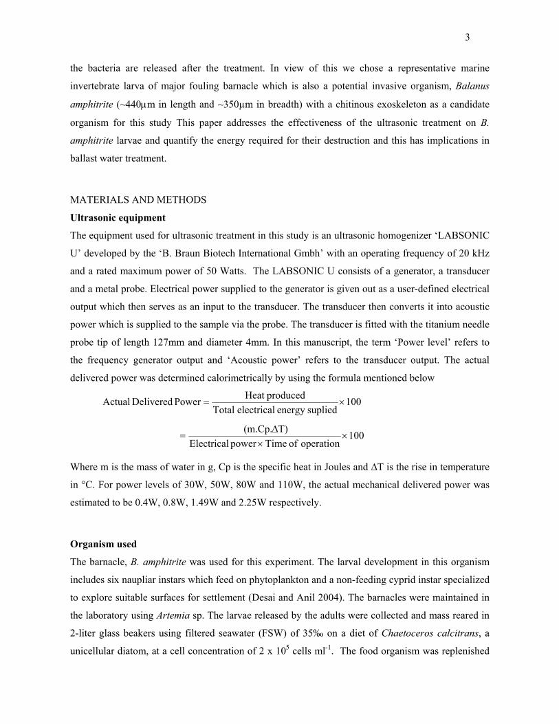
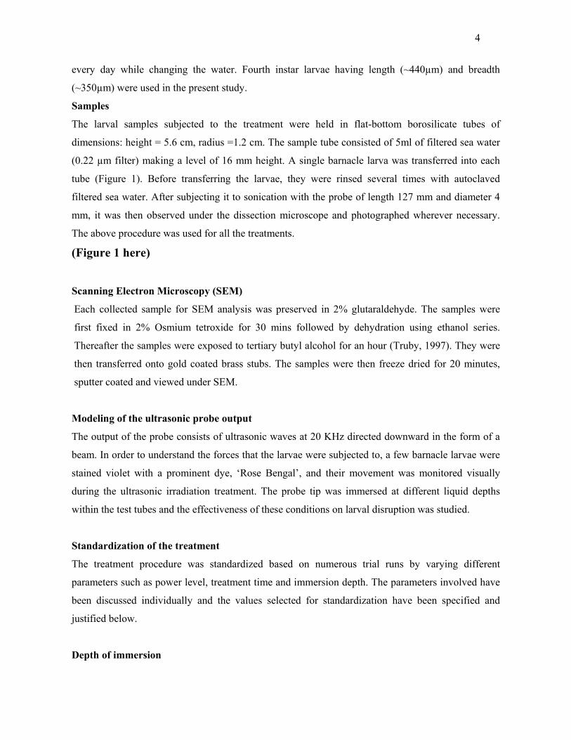
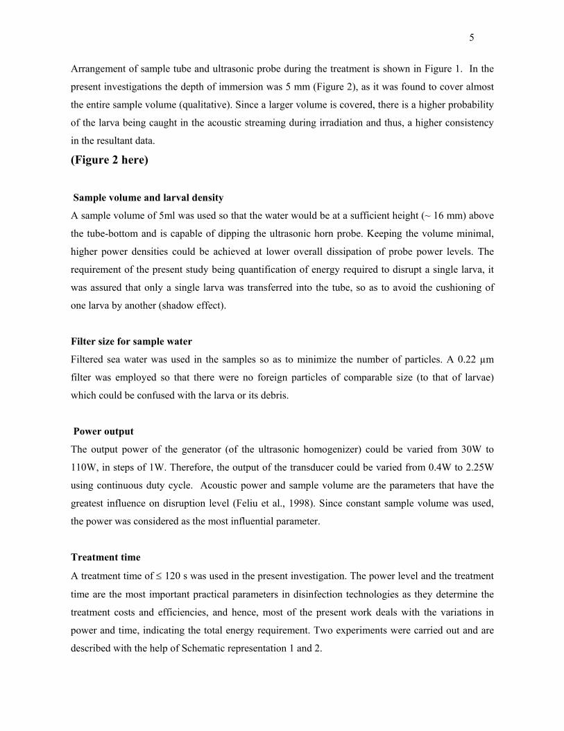
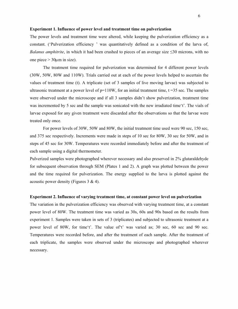
Embed Size (px)
Citation preview
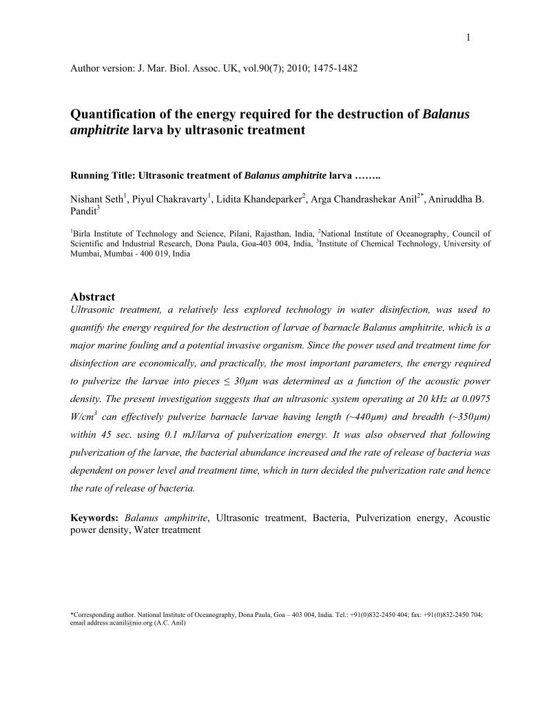
1
Author version: J. Mar. Biol. Assoc. UK, vol.90(7); 2010; 1475-1482 Quantification of the energy required for the destruction of Balanus amphitrite larva by ultrasonic treatment
Running Title: Ultrasonic treatment of Balanus amphitrite larva …….. Nishant Seth1, Piyul Chakravarty1, Lidita Khandeparker2, Arga Chandrashekar Anil2*, Aniruddha B. Pandit3 1Birla Institute of Technology and Science, Pilani, Rajasthan, India, 2National Institute of Oceanography, Council of Scientific and Industrial Research, Dona Paula, Goa-403 004, India, 3Institute of Chemical Technology, University of Mumbai, Mumbai - 400 019, India
Abstract Ultrasonic treatment, a relatively less explored technology in water disinfection, was used to
quantify the energy required for the destruction of larvae of barnacle Balanus amphitrite, which is a
major marine fouling and a potential invasive organism. Since the power used and treatment time for
disinfection are economically, and practically, the most important parameters, the energy required
to pulverize the larvae into pieces ≤ 30µm was determined as a function of the acoustic power
density. The present investigation suggests that an ultrasonic system operating at 20 kHz at 0.0975
W/cm3 can effectively pulverize barnacle larvae having length (~440µm) and breadth (~350µm)
within 45 sec. using 0.1 mJ/larva of pulverization energy. It was also observed that following
pulverization of the larvae, the bacterial abundance increased and the rate of release of bacteria was
dependent on power level and treatment time, which in turn decided the pulverization rate and hence
the rate of release of bacteria.
Keywords: Balanus amphitrite, Ultrasonic treatment, Bacteria, Pulverization energy, Acoustic power density, Water treatment
*Corresponding author. National Institute of Oceanography, Dona Paula, Goa – 403 004, India. Tel.: +91(0)832-2450 404; fax: +91(0)832-2450 704; email address:[email protected] (A.C. Anil)
2
INTRODUCTION
Acoustic cavitation is a relatively less explored technique in the treatment of water and
investigations are needed to ascertain its practical applicability especially in brackish/ saline
environment or with specific marine microbes. Apart from making water effective for drinking
purpose, it can find utility in a ship to treat ship’s ballast water, treatment of industrial effluent etc.
There are various existing, as well as, newly emerging technologies available for the disinfection of
water, ranging from physical methods such as heating and UV treatment, to chemical treatment with
biocides; however they have many inherent problems. Firstly, they are energy wise intensive and
hence expensive, they can cause contamination of the treated water (due to addition of chemicals),
and therefore, of the environment into which the water is subsequently released. Their mechanism of
action is often complicated and poorly understood, and their effect varies with different organisms.
Also there is a possibility of generation of treatment byproducts.
Although there are number of illustrations regarding use of cavitation for waste water
treatment, the theoretical analysis to develop the fundamentals and experimental studies to
implement the same is a necessity. We have addressed the implication of ultrasonic treatment in
disrupting and/ or pulverizing the organisms that could be transported via ballast water. Ballast water
is the sea-water taken up by the ships in order to weigh down and/or balance the latter while
unloading their cargo. Conversely, when loading cargo, they discharge the ballast water. In this
process, ships transfer millions of tons of ballast water from one place to another worldwide,
inadvertently discharging non-indigenous aquatic organisms into receiving waters (Carlton & Geller,
1993). Translocation of organisms through ships (bio-invasion) is considered to be one of the central
issues that have plagued the naturally evolved biodiversity, the consequences of which are being
realized increasingly in recent years (Gollasch et al., 2000; Anil et al., 2002). Marine invertebrate
larvae are of major concern in ballast water treatment/management programmes and in addition they
also harbour many bacteria within them. The release of bacteria while these large planktonic
organisms are killed or destroyed by rupturing is an additional concern in the ballast water treatment.
It has been found that most of the pathogens introduced to Chesapeake Bay came from bacteria
associated with plankton rather than that in water (Ruiz et al., 2000). In this context it is important to
get the maximum extent of microorganisms released out of the planktonic organisms during the
latter’s destruction so that additional treatment measures can be effectively applied to reduce or
eliminate them. Very little is known about the strength of their exoskeleton and the mechanical or
thermal energy required for breaking or pulverizing them into small pieces (≤30 µm) so that most of
3
the bacteria are released after the treatment. In view of this we chose a representative marine
invertebrate larva of major fouling barnacle which is also a potential invasive organism, Balanus
amphitrite (~440μm in length and ~350µm in breadth) with a chitinous exoskeleton as a candidate
organism for this study This paper addresses the effectiveness of the ultrasonic treatment on B.
amphitrite larvae and quantify the energy required for their destruction and this has implications in
ballast water treatment.
MATERIALS AND METHODS
Ultrasonic equipment
The equipment used for ultrasonic treatment in this study is an ultrasonic homogenizer ‘LABSONIC
U’ developed by the ‘B. Braun Biotech International Gmbh’ with an operating frequency of 20 kHz
and a rated maximum power of 50 Watts. The LABSONIC U consists of a generator, a transducer
and a metal probe. Electrical power supplied to the generator is given out as a user-defined electrical
output which then serves as an input to the transducer. The transducer then converts it into acoustic
power which is supplied to the sample via the probe. The transducer is fitted with the titanium needle
probe tip of length 127mm and diameter 4mm. In this manuscript, the term ‘Power level’ refers to
the frequency generator output and ‘Acoustic power’ refers to the transducer output. The actual
delivered power was determined calorimetrically by using the formula mentioned below
Where m is the mass of water in g, Cp is the specific heat in Joules and ∆T is the rise in temperature
in °C. For power levels of 30W, 50W, 80W and 110W, the actual mechanical delivered power was
estimated to be 0.4W, 0.8W, 1.49W and 2.25W respectively.
Organism used
The barnacle, B. amphitrite was used for this experiment. The larval development in this organism
includes six naupliar instars which feed on phytoplankton and a non-feeding cyprid instar specialized
to explore suitable surfaces for settlement (Desai and Anil 2004). The barnacles were maintained in
the laboratory using Artemia sp. The larvae released by the adults were collected and mass reared in
2-liter glass beakers using filtered seawater (FSW) of 35‰ on a diet of Chaetoceros calcitrans, a
unicellular diatom, at a cell concentration of 2 x 10⁵ cells ml-1. The food organism was replenished
100operation of Timepower Electrical
T)(m.Cp. ×
×Δ
=
100supliedenergyelectricalTotal
producedHeat Power Delivered Actual ×=
4
every day while changing the water. Fourth instar larvae having length (~440µm) and breadth
(~350µm) were used in the present study.
Samples
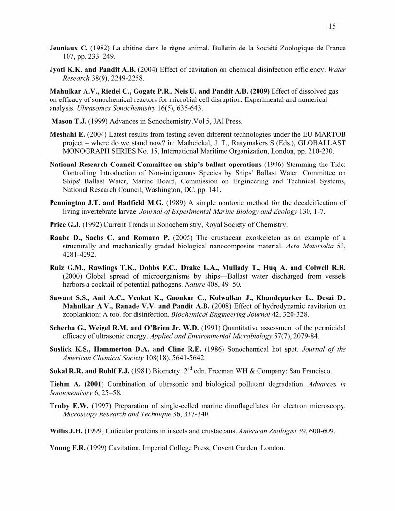
The larval samples subjected to the treatment were held in flat-bottom borosilicate tubes of
dimensions: height = 5.6 cm, radius =1.2 cm. The sample tube consisted of 5ml of filtered sea water
(0.22 µm filter) making a level of 16 mm height. A single barnacle larva was transferred into each
tube (Figure 1). Before transferring the larvae, they were rinsed several times with autoclaved
filtered sea water. After subjecting it to sonication with the probe of length 127 mm and diameter 4
mm, it was then observed under the dissection microscope and photographed wherever necessary.
The above procedure was used for all the treatments.
(Figure 1 here)
Scanning Electron Microscopy (SEM)
Each collected sample for SEM analysis was preserved in 2% glutaraldehyde. The samples were
first fixed in 2% Osmium tetroxide for 30 mins followed by dehydration using ethanol series.
Thereafter the samples were exposed to tertiary butyl alcohol for an hour (Truby, 1997). They were
then transferred onto gold coated brass stubs. The samples were then freeze dried for 20 minutes,
sputter coated and viewed under SEM.
Modeling of the ultrasonic probe output
The output of the probe consists of ultrasonic waves at 20 KHz directed downward in the form of a
beam. In order to understand the forces that the larvae were subjected to, a few barnacle larvae were
stained violet with a prominent dye, ‘Rose Bengal’, and their movement was monitored visually
during the ultrasonic irradiation treatment. The probe tip was immersed at different liquid depths
within the test tubes and the effectiveness of these conditions on larval disruption was studied.
Standardization of the treatment
The treatment procedure was standardized based on numerous trial runs by varying different
parameters such as power level, treatment time and immersion depth. The parameters involved have
been discussed individually and the values selected for standardization have been specified and
justified below.
Depth of immersion
5
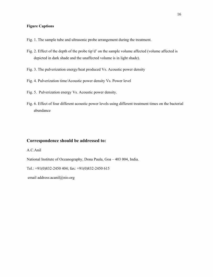
Arrangement of sample tube and ultrasonic probe during the treatment is shown in Figure 1. In the
present investigations the depth of immersion was 5 mm (Figure 2), as it was found to cover almost
the entire sample volume (qualitative). Since a larger volume is covered, there is a higher probability
of the larva being caught in the acoustic streaming during irradiation and thus, a higher consistency
in the resultant data.
(Figure 2 here)
Sample volume and larval density
A sample volume of 5ml was used so that the water would be at a sufficient height (~ 16 mm) above
the tube-bottom and is capable of dipping the ultrasonic horn probe. Keeping the volume minimal,
higher power densities could be achieved at lower overall dissipation of probe power levels. The
requirement of the present study being quantification of energy required to disrupt a single larva, it
was assured that only a single larva was transferred into the tube, so as to avoid the cushioning of
one larva by another (shadow effect).
Filter size for sample water
Filtered sea water was used in the samples so as to minimize the number of particles. A 0.22 µm
filter was employed so that there were no foreign particles of comparable size (to that of larvae)
which could be confused with the larva or its debris.
Power output
The output power of the generator (of the ultrasonic homogenizer) could be varied from 30W to
110W, in steps of 1W. Therefore, the output of the transducer could be varied from 0.4W to 2.25W
using continuous duty cycle. Acoustic power and sample volume are the parameters that have the
greatest influence on disruption level (Feliu et al., 1998). Since constant sample volume was used,
the power was considered as the most influential parameter.
Treatment time
A treatment time of ≤ 120 s was used in the present investigation. The power level and the treatment
time are the most important practical parameters in disinfection technologies as they determine the
treatment costs and efficiencies, and hence, most of the present work deals with the variations in
power and time, indicating the total energy requirement. Two experiments were carried out and are
described with the help of Schematic representation 1 and 2.
6
Experiment 1. Influence of power level and treatment time on pulverization
The power levels and treatment time were altered, while keeping the pulverization efficiency as a
constant. (‘Pulverization efficiency ’ was quantitatively defined as a condition of the larva of,
Balanus amphitrite, in which it had been crushed to pieces of an average size ≤30 microns, with no
one piece > 30μm in size).
The treatment time required for pulverization was determined for 4 different power levels
(30W, 50W, 80W and 110W). Trials carried out at each of the power levels helped to ascertain the
values of treatment time (t). A triplicate (set of 3 samples of live moving larvae) was subjected to
ultrasonic treatment at a power level of p=110W, for an initial treatment time, t =35 sec. The samples
were observed under the microscope and if all 3 samples didn’t show pulverization, treatment time
was incremented by 5 sec and the sample was sonicated with the new irradiated time‘t’. The vials of
larvae exposed for any given treatment were discarded after the observations so that the larvae were
treated only once.
For power levels of 30W, 50W and 80W, the initial treatment time used were 90 sec, 150 sec,
and 375 sec respectively. Increments were made in steps of 10 sec for 80W, 30 sec for 50W, and in
steps of 45 sec for 30W. Temperatures were recorded immediately before and after the treatment of
each sample using a digital thermometer.
Pulverized samples were photographed wherever necessary and also preserved in 2% glutaraldehyde
for subsequent observation through SEM (Plates 1 and 2). A graph was plotted between the power
and the time required for pulverization. The energy supplied to the larva is plotted against the
acoustic power density (Figures 3 & 4).
Experiment 2. Influence of varying treatment time, at constant power level on pulverization
The variation in the pulverization efficiency was observed with varying treatment time, at a constant
power level of 80W. The treatment time was varied as 30s, 60s and 90s based on the results from
experiment 1. Samples were taken in sets of 3 (triplicates) and subjected to ultrasonic treatment at a
power level of 80W, for time‘t’. The value of‘t’ was varied as; 30 sec, 60 sec and 90 sec.
Temperatures were recorded before, and after the treatment of each sample. After the treatment of
each triplicate, the samples were observed under the microscope and photographed wherever
necessary.
7
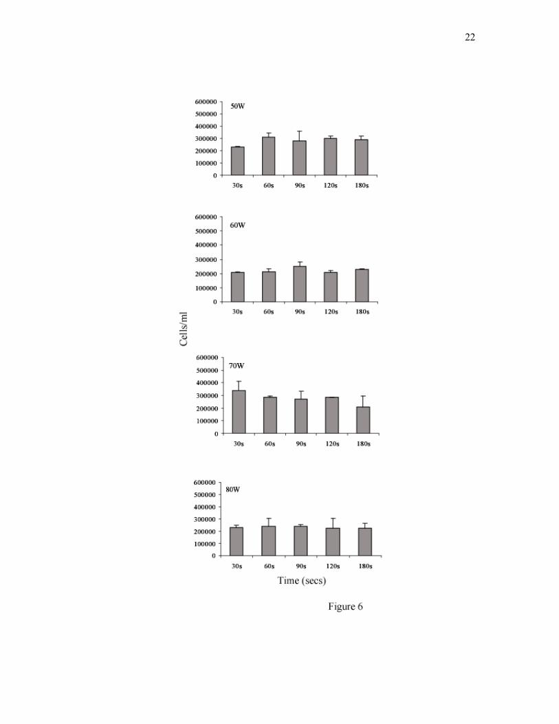
Quantification of bacteria
In another set of experiment the larvae were sonicated using a range of mid power levels used in the
above experiment (i.e. 50, 60, 70 and 80W) upto180 seconds. The water from the sample tube was
then fixed with formaldehyde (final concentration 1 to 2%; v/v) to quantify the bacterial numbers.
The quantification of bacteria was done by using acridine orange and epifluorescence microscopy
(Daley & Hobbie, 1975).
Statistical analysis
The data on bacterial abundance was log transformed to ensure normality and homogeneity of
variances before subjecting to statistical analysis. The pattern seen in the bacterial abundance with
increasing input power and exposure time was subjected to one-way ANOVA followed by a post-
ANOVA Scheffe’s test (Sokal & Rohlf, 1981).
RESULTS After standardizing the depth of probe immersion for effective disruption to 5 mm, numerous trials
with the rose Bengal preserved and live larvae were carried out. Since the stain used had preserved
the larvae, the movement shown by them was purely due to the acoustic streaming generated by the
passage of ultrasound. Trials with, live, larvae also revealed, that larvae close to, or anywhere below
the probe tip were immediately sucked into the liquid jet created by the oscillation of the horn, while
those that were well above the probe tip, were relatively unaffected, and hence, much more
independent in their movement.
In conclusion, almost all the ultrasonic energy is concentrated in the volume of water
adjacent to, and/or below the probe tip. This can be called as the ‘processed volume’. Since the exact
ultrasonic power distribution in the processed volume was unknown, the power density that the larva
is subjected to is approximated, as the average power density over the processed volume i.e.
Acoustic power density = (transducer power output, watts/ processed volume, cm3).
The ‘acoustic power density’ is a more universal treatment parameter than the ‘power level’
as it is independent of the methods, equipment, sample volume etc. However, it suffers from
inaccuracy due to the approximation of the transducer conversion efficiency. Hence, throughout this
report both parameters have been used.
So, given a treatment at a particular power level ‘p’, watt (W) for a treatment time of ‘t’, sec,
with the depth of the probe as ‘d’, mm , the height of water above the tube-bottom as ‘h’, mm and
the radius of the tube as ‘r’, mm (Figure 1), the acoustic power density ‘P’ and the acoustic energy
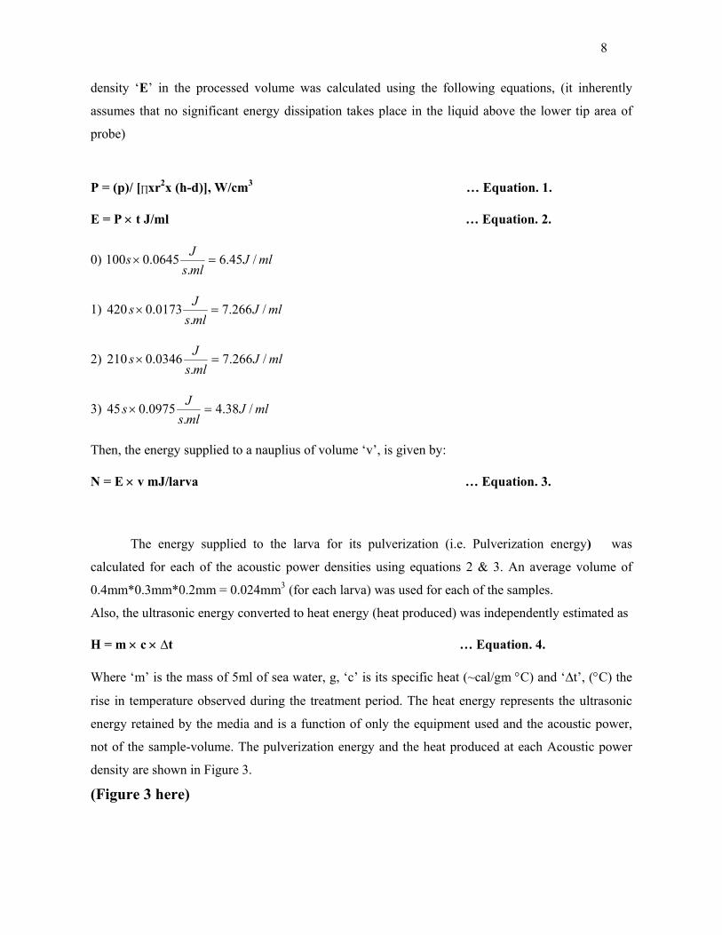
8
density ‘E’ in the processed volume was calculated using the following equations, (it inherently
assumes that no significant energy dissipation takes place in the liquid above the lower tip area of
probe)
P = (p)/ [∏xr2x (h-d)], W/cm3 … Equation. 1.
E = P × t J/ml … Equation. 2.
0) mlJmlsJs /45.6.
0645.0100 =×
1) mlJmlsJs /266.7.
0173.0420 =×
2) mlJmlsJs /266.7.
0346.0210 =×
3) mlJmlsJs /38.4.
0975.045 =×
Then, the energy supplied to a nauplius of volume ‘v’, is given by:
N = E × v mJ/larva … Equation. 3.
The energy supplied to the larva for its pulverization (i.e. Pulverization energy) was
calculated for each of the acoustic power densities using equations 2 & 3. An average volume of
0.4mm*0.3mm*0.2mm = 0.024mm3 (for each larva) was used for each of the samples.
Also, the ultrasonic energy converted to heat energy (heat produced) was independently estimated as
H = m × c × ∆t … Equation. 4.
Where ‘m’ is the mass of 5ml of sea water, g, ‘c’ is its specific heat (~cal/gm °C) and ‘∆t’, (°C) the
rise in temperature observed during the treatment period. The heat energy represents the ultrasonic
energy retained by the media and is a function of only the equipment used and the acoustic power,
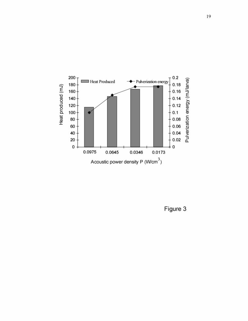
not of the sample-volume. The pulverization energy and the heat produced at each Acoustic power
density are shown in Figure 3.
(Figure 3 here)
9
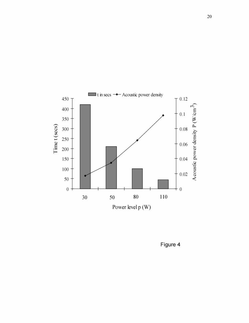
The results of Experiment 1 are shown in Fig. 4, plotted between the time required for pulverization
(i.e. pulverization time) of the sample, and the power used. The ‘pulverization time’, expressed as
(sec), and ‘Acoustic power density’ values, calculated using equations 2 & 3 expressed as W/cm3
are plotted on the Y-axis and the corresponding power levels, expressed as watts (W), are plotted
along the X-axis. The results indicated that from a pulverization time of 420s at 0.0173 W/cm3 the
curve drops to half its value; 210s at 0.0346 W/cm3, again to 100s at 0.0645 W/cm3, and then again
to 45s at 0.0975 W/cm3. The rise in temperature at higher power density is low i.e. 5.5°C at 0.0975
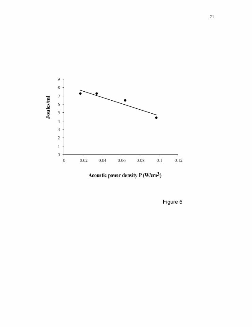
W/cm3 when compared at 0.0173 W/cm3 (8.5°C). The results of Experiment 2 are shown in Figure 5.
The pulverization energies expressed as J/mL are plotted against the acoustic power densities. The
heat produced during a pulverization treatment is seen to decrease, approximately linearly, with
increasing acoustic power. This clearly indicates that at higher power density, pulverization is more
energy efficient i.e. requires less total energy. (Figures 4 & 5 here) The bacterial abundance after the pulverization of larvae using different power levels and
time is shown in Figure 6. When the larvae were sonicated at lower power levels, an increase in the
bacterial numbers was evident up to 90 secs after which there was a slow decline (17.79%). When a
comparison was made across the power treatments, a significant difference in bacterial abundance
was observed at 30 and 90 secs (p≤0.001, One-way ANOVA; p≤0.05, Scheffe’s test). At higher
power levels, less time was required to obtain the highest bacterial numbers followed by a faster
decline in their numbers subsequent to further sonication till 180 secs.
(Figure 6 here)
DISCUSSION Ultrasonic liquid treatment uses high frequency wave energy to cause vibrations in liquids resulting
in a phenomenon called as acoustic cavitation, which is the formation, growth, and implosive
collapse of microscopic gas and/or vapor bubbles that are generated when ultrasonic waves are
propagated through a liquid medium. Thus, acoustic cavitation results from the mechanical
interaction between sound waves and bubbles in liquids (Price, 1992; Crum et al., 1999; Young,
1999; Mason, 1999; Ashokkumar et al., 2007). When these cavitation bubbles collapse, a series of
mechanical and physical effects such as shear forces and shock waves are generated. The collapse of
cavitation bubbles also results in the generation of heat within the bubbles for which these bubbles
are also referred as micro-reactors or hot spots (Suslick et al., 1986; Ashokkumar et al., 2007). The
chemical changes that take place due to the cavitation induced by the passage of sound waves are
commonly known as sonochemistry (Gogate, 2002). The present study revealed that when the larvae
were sonicated using ultrasound energy, they were caught in violent agitations that forced them into
10
moving along random, looped pathways. This observation agrees with the mechanism proposed
earlier (Doulah, 1977), that an ultrasound results in the formation of eddies (acoustic streaming).
Also, it was observed that all the ultrasonic energy is concentrated in the volume of water adjacent
to, and/or below the probe tip referred as ‘processed volume’. Given that the larva is within this
processed volume just before the treatment, it is necessarily subjected to movement within it,
assuming that the larva had equal probability of being at any position within the processed volume.
But, the total number of cavitation events per unit time is independent of the volume of the
suspension irradiated and is a function only of the power input to the transducer (Davies, 1959). A
1970s study on the treatment of shipboard waste-water demonstrated that effective cavitation could
not be attained for the volumes of water being treated (NRC, 1996). Consequently, sufficient
sterilization of the wastewater was not achieved. Traditional methods of ultrasonic treatments were
reviewed in Stemming the Tide (NRC, 1996), which states that effectiveness is a variable when
treatment is applied to higher organisms such as zebra mussel veligers and fish. NRC concluded
however, that such ultrasound systems could destroy fungi, yeasts, and pathogenic bacteria. The
energy that exists within the cavity, at the time of collapse, causes both physical and chemical effects
that are responsible for the rupture of organisms during cavitation. The conditions immediately
preceding collapse of a cavitation bubble are similar in magnitude to ultra-high energy combustion
conditions. Within the cavitation bubble and the immediate surround area, temperatures range from
2000 to 5000 °C, and pressure reaches 1800 atmospheres (Buchholz et al., 1998). Higher
frequencies, warmer temperatures and lower concentrations of dissolved matter have been found to
increase the effect of ultrasound pulses (Mesbahi, 2004). Evaluating the effect of ultrasound at a
frequency similar to that employed in present investigation i.e. 20 kHz it was opined that stand-
alone ultrasonic treatment systems for ballast water may be effective for planktonic organisms >100
µm in size but smaller planktonic forms such as phytoplankton and bacteria will require an additional
or alternative treatment system (Holm et al., 2008). It was also observed that zooplankton tested all
suffered 90% mortality after exposure times <10 s and at low energy densities <20 J/ml. In the
present investigation 0.1 mJ/larva of pulverization energy at power density of 0.0975 W/cm3 was
efficient in pulverization of barnacle larva with slow rise in temperature. Thus, temperature can be
considered to have little effect on the rate of killing in such organisms and therefore, it can be
concluded that mechanical disruption was the main mechanism of pulverization. Previous reports
also suggest that physical processes could be more important at the frequencies employed in the
present investigation (Tiehm, 2001). Thus, under such conditions, the primary means for biological
eradication are the mechanical effects rather than chemical reactions that result from cavitation. Cell
11
membranes and organisms are literally ruptured or blown apart from the intense energy delivered in
the form of shock waves. These (mechanical) effects include: (1) Complete destruction of larger
biota, and (2) Disturbance or rupture of biological cell membranes, leading to subsequent death of an
organism.
The cell breakage by ultrasound is a single hit type of phenomenon, such as the occurrence of a
cavitation (implosion) in close proximity to a cell (Anand et al., 2007). The shock waves generated
by transient cavitation are the main physical force responsible for mechanical disruption of the
microbe. The strength of the exoskeleton of the organisms subjected to cavitation also plays a
significant role in its effective disruption. It has been shown earlier that micro streaming resulting
from stable cavitation produce stresses that are sufficient to disrupt cell membranes (Scherba et al.,
1991). The damage caused by fluid shear stress is thought to depend on the erosion of the outer cell
wall polymers, particularly at weakened places such as division or budding scars (Anand et al.,
2007). Most of the zooplankton develops either external or internal skeletons (Hardy, 1956;
Pennington & Hadfield, 1989) for support or protection from predators in the aquatic environments.
The exoskeletons are either chitinous as in the crustacea or calcareous as in the larval mollusks or
brachiopods (Hardy, 1956). The major component of the cuticles and exoskeletons of worms,
mollusks and arthropods consists of chitin, which is one of the most abundant polysaccharides in
nature (Jeuniaux, 1982). Crustaceans and millipedes have calcium as a major component of cuticle
and play a significant role in cuticle hardness (Willis, 1999). The gradient in the stiffness and
hardness through the cuticle thickness is related to honeycomb mechanism of the twisted plywood
structure which is formed by the helicoidal stacking sequence of the fibrous chitin-protein layers
(Raabe et al., 2005). Once the cell membrane is sheared (a physical consequence of cavitation),
chemical oxidants can then enter the cell attacking internal structures (Anand et al., 2007). Chemical
oxidants produced as a result of ultrasonic irradiation include free radicals like the hydroxyl radicals.
One of the recent studies by Gavand et al. (2007) reported that a combination of sonication and
advanced chemical oxidants could be more promising method to eradicate aquatic algae and macro
invertebrates in ballast water. The biochemical composition of the exoskeleton and its strength
differs for different invertebrate larvae, thus the energy required to pulverize or sonicate different
sizes of larval forms would be different (Holm et al., 2008). Thus the present study aimed at
elucidating the energy required for the pulverization of barnacle larva, as well as, the heat produced
in the process using four different acoustic power densities. In the present study the dimension of the
larvae subjected to pulverization and the level of disruption as quantified by the size of pulverized
fragments were kept constant. Some preliminary experiments carried out on different larval stages of
barnacles and copepods of different sizes indicated variations in the energies required for disruption,
12
copepods requiring higher energies than barnacle larvae (personal observation). In real-life situation
there will be a gamut of organisms belonging to different taxa and thus energy required to
treat/disrupt these organisms would be different along with the shadow effect (shock wave impact
attenuated by physical obstruction) and this needs further investigation.
For the range of acoustic power densities used, the following conclusions can be drawn about
the requirements of time and energy, trends shown by the heat produced and the extent of
pulverization. From Figure 4 it is evident that pulverization time showed an exponential decrease
with an increase in the acoustic power density, such that, at very low power densities, the time
required tends toward infinity. The temperature rise during a pulverization treatment is seen to
marginally decrease, approximately linearly, with increasing acoustic power.
Treatment time is of critical importance, especially, in high flow systems that are used in
large-scale treatments. So, higher acoustic power densities can be used to achieve exponentially
lower treatment times, so as to make ultrasonic treatment feasible for use in such systems.
Pulverization energy is the energy that must be supplied to the larva to pulverize it, and not
the total input energy by the transducer. From Figure 5 it is observed that the exponential decrease in
treatment time has resulted in an almost linear decrease in required pulverization energy with
increasing acoustic power densities. The four points in the graph in Figure 5, representing
combinations of energy and acoustic power densities, can be utilized to design disinfection systems
for pulverizing larvae of Balanus amphitrite or similar barnacle species. However, the data provided
has limited application in that it covers only a narrow range of acoustic power densities, viz. ‘0.0173’
to ‘0.0975’ W/cm3.
It was observed that when the organism is pulverized, various microbes embedded in its
exoskeleton, as well as, those within its gut, are dislodged and thus, become vulnerable to subsequent
treatment. Most disinfection treatments use a combination of treatment technologies in stages.
Experimental results have also shown that hydrodynamic cavitation and or turbulent shear
dominantly originating from cavitation are effective tools and could kill 80% zooplankton present in
sea water (Sawant et al., 2008). Jyoti & Pandit (2004) reported that hybrid technique which
combines hydrodynamic cavitation, acoustic cavitation and hydrogen peroxide appear to be an
attractive alternative to any one technique on its own for the reduction in the heterotrophic plate
count bacteria as well as indicator microorganisms like the total coliforms, fecal coliforms and fecal
streptococci. Thus, while treating macro-organisms, it becomes important, not only to destroy them,
but also kill or at least, make vulnerable, the microbes harbored within them.
Our observations indicated that larvae were split into pieces measuring ≤ 30µm. The surfaces of the
naupliar debris showed numerous perforations and indentations (0.2 to 3 µm), however there was no
13
peculiar pattern in the way they were destroyed. When low power levels were used, the numbers of
bacteria released in the water column following pulverization increased and was evident up to 90
secs followed by a slow decline, whereas at higher power density (0.0975 W/cm3) this could be
achieved in 45 s of exposure time and pulverization energy of 0.1 mJ/larva. The destruction of
bacteria (size approximately 5μm) is brought about only after they are released after the disruption of
the bacteria bearing organism. Earlier work (Mahulkar et al., 2009), clearly shows that continued
irradiation with ultrasound is capable of destroying the bacteria. The bacterial population plotted v/s
time goes through maxima, which more or less coincides with the complete disruption of the main
(target), bacteria bearing larva. The ultrasonic irradiation, in the initial stages disrupts the larva and
also the bacteria, but the rate of release of the bacteria in the initial stages is significantly higher than
its destruction and hence it goes through maxima. After this, the bacterial population shows a steady
decrease with continued irradiation using ultrasound. The effects related to the microbial/larval
concentration does show an optimum and it has been observed to be in the range of 1% wt/volume
(Anand et al., 2007). The fragmentation size analysis is necessary as it will be useful for subsequent
solid-liquid separation requirements. Once, the time of exposure v/s fragmentation rate is known, a
flow through system can be designed, following two possible strategies are suggested (a) Multiple
circulation of the suspension through the cavitating zone, where the cumulative exposure time
matches the ultrasound exposure time obtained from the batch studies Or (b) Continuous flow
through system, having circulation rate, much higher than the addition and withdrawal rate, again
matching the required exposure time.
ACKNOWLEDGEMENTS
We are grateful to Dr. Satish R Shetye, the Director National Institute of Oceanography, Goa for his
support and encouragement. We thank Dr. Dattesh Desai, Mr. K. Venkat, and other colleagues of
MCMRD for their help. This is a NIO contribution ####
14
REFERENCES
Anand H., Balasundaram B., Pandit A.B. and Harrison S.T.L. (2007) The effect of chemical pretreatment combined with mechanical disruption on the extent of disruption and release of intracellular protein from E. coli. Biochemical Engineering Journal 35(2), 166-173.
Anil A.C., Venkat K., Sawant S.S., Dileepkumar M., Dhargalkar V.K., Ramaiah N., Harkantra S.N. and Ansari Z.A. (2002) Marine bio-invasion: Concern for ecology and shipping. Current Science 83(3), 214-218.
Ashokkumar M., Lee J., Kentish S. and Grieser F. (2007) Bubbles in an acoustic field: An overview. Ultrasonics Sonochemistry 14(4), 470-475.
Buchholz K., Deborah T., Scott M. and Emmanuele F. (1998) Ballast Water Secondary Treatment Technology Review: Battelle Duxbury Operations.
Carlton J.J. and Geller J.B. (1993) Ecological Roulette: Biological invasions and the global transport of non-indigenous marine organisms. Science 261, 78-82.
Crum L.A., Mason T.J., Reisse J.-L. and Suslick K.S. (1999) Sonochemistry and Sonoluminescence. In NATO ASI Series C vol. 524, Kluwer Publishers, London.
Daley R.J. and Hobbie J.E. (1975) Direct counts of aquatic bacteria by a modified epifluorescence technique. Limnology and Oceanography 20, 875-882.
Desai D.V. and Anil A.C. (2004) The impact of food type, temperature and starvation on larval development of Balanus amphitrite Darwin (Cirripedia: Thoracica). Journal of Experimental Marine Biology and Ecology 306, 113–137.
Davies R. (1959) Effects on the use of ultrasound waves for the disruption of microorganisms. Biochimica et Biophysica Acta 33, 481-493.
Doulah M.S. (1977) Mechanism of disintegration of biological cells in ultrasonic cavitation. Biotechnology and Bioengineering 19(5), 649-660.
Feliu J.X., Cubarsi R. and Villaverde A. (1998) Optimized release of recombinant protein by ultrasonication of E. coli cells. Biotechnology and Bioengineering 58(5), 536-540.
Gavand M.R., McClintock J.B., Amsler C.D., Peters R.W. and Angus R.A. (2007) Effects of sonication and advanced chemical oxidants on the unicellular alga Dunaliella tertiolecta and cysts, larvae and adults of the brine shrimp Artemia salina: a prospective treatment to eradicate invasive organisms from ballast water. Marine Pollution Bulletin 54(11), 1777-1788.
Gogate P.R. (2002) Cavitation: an auxiliary technique in wastewater treatment schemes. Advances in Environmental Research 6, 335-358.
Gollasch S., Lenz J., Dammer M. and Andres H.G. (2000) Survival of tropical ballast water organisms during a cruise from the Indian Ocean to the North Sea. Journal of Plankton Research 22, 923-937.
Hardy A. (1956) The open sea, Houghton Mifflin, Boston, pp. 335.
Holm E.R., Stamper D.M., Brizzolara R.A., Barnes L., Deamer N. and Burkholder J.M. (2008) Sonication of bacteria, phytoplankton and zooplankton: Application to treatment of ballast water. Marine Pollution Bulletin 56, 1201-1208.
15
Jeuniaux C. (1982) La chitine dans le règne animal. Bulletin de la Société Zoologique de France 107, pp. 233–249.
Jyoti K.K. and Pandit A.B. (2004) Effect of cavitation on chemical disinfection efficiency. Water Research 38(9), 2249-2258.
Mahulkar A.V., Riedel C., Gogate P.R., Neis U. and Pandit A.B. (2009) Effect of dissolved gas on efficacy of sonochemical reactors for microbial cell disruption: Experimental and numerical analysis. Ultrasonics Sonochemistry 16(5), 635-643.
Mason T.J. (1999) Advances in Sonochemistry.Vol 5, JAI Press.
Meshahi E. (2004) Latest results from testing seven different technologies under the EU MARTOB project – where do we stand now? in: Matheickal, J. T., Raaymakers S (Eds.), GLOBALLAST MONOGRAPH SERIES No. 15, International Maritime Organization, London, pp. 210-230.
National Research Council Committee on ship’s ballast operations (1996) Stemming the Tide: Controlling Introduction of Non-indigenous Species by Ships' Ballast Water. Committee on Ships' Ballast Water, Marine Board, Commission on Engineering and Technical Systems, National Research Council, Washington, DC, pp. 141.
Pennington J.T. and Hadfield M.G. (1989) A simple nontoxic method for the decalcification of living invertebrate larvae. Journal of Experimental Marine Biology and Ecology 130, 1-7.
Price G.J. (1992) Current Trends in Sonochemistry, Royal Society of Chemistry.
Raabe D., Sachs C. and Romano P. (2005) The crustacean exoskeleton as an example of a structurally and mechanically graded biological nanocomposite material. Acta Materialia 53, 4281-4292.
Ruiz G.M., Rawlings T.K., Dobbs F.C., Drake L.A., Mullady T., Huq A. and Colwell R.R. (2000) Global spread of microorganisms by ships––Ballast water discharged from vessels harbors a cocktail of potential pathogens. Nature 408, 49–50.
Sawant S.S., Anil A.C., Venkat K., Gaonkar C., Kolwalkar J., Khandeparker L., Desai D., Mahulkar A.V., Ranade V.V. and Pandit A.B. (2008) Effect of hydrodynamic cavitation on zooplankton: A tool for disinfection. Biochemical Engineering Journal 42, 320-328.
Scherba G., Weigel R.M. and O’Brien Jr. W.D. (1991) Quantitative assessment of the germicidal efficacy of ultrasonic energy. Applied and Environmental Microbiology 57(7), 2079-84.
Suslick K.S., Hammerton D.A. and Cline R.E. (1986) Sonochemical hot spot. Journal of the American Chemical Society 108(18), 5641-5642.
Sokal R.R. and Rohlf F.J. (1981) Biometry. 2nd edn. Freeman WH & Company: San Francisco.
Tiehm A. (2001) Combination of ultrasonic and biological pollutant degradation. Advances in Sonochemistry 6, 25–58.
Truby E.W. (1997) Preparation of single-celled marine dinoflagellates for electron microscopy. Microscopy Research and Technique 36, 337-340.
Willis J.H. (1999) Cuticular proteins in insects and crustaceans. American Zoologist 39, 600-609.
Young F.R. (1999) Cavitation, Imperial College Press, Covent Garden, London.
16
Figure Captions
Fig. 1. The sample tube and ultrasonic probe arrangement during the treatment.
Fig. 2. Effect of the depth of the probe tip‘d’ on the sample volume affected (volume affected is
depicted in dark shade and the unaffected volume is in light shade).
Fig. 3. The pulverization energy/heat produced Vs. Acoustic power density
Fig. 4. Pulverization time/Acoustic power density Vs. Power level
Fig. 5. Pulverization energy Vs. Acoustic power density.
Fig. 6. Effect of four different acoustic power levels using different treatment times on the bacterial
abundance
Correspondence should be addressed to:
A.C.Anil
National Institute of Oceanography, Dona Paula, Goa – 403 004, India.
Tel.: +91(0)832-2450 404; fax: +91(0)832-2450 615
email address:[email protected]
17
18
19
20
21
22