Embed Size (px)
Citation preview
RinA controls phage-mediated packagingand transfer of virulence genes inGram-positive bacteriaMarıa Desamparados Ferrer1,2, Nuria Quiles-Puchalt2, Michael D. Harwich3,
Marıa Angeles Tormo-Mas1,2, Susana Campoy4, Jordi Barbe4, Inigo Lasa5,
Richard P. Novick6, Gail E. Christie3 and Jose R. Penades1,7,*
1Departamento de Quımica, Bioquımica y Biologıa Molecular, Universidad CEU Cardenal Herrera, 46113Moncada, Valencia, 2Centro de Investigacion y Tecnologıa Animal, Instituto Valenciano de InvestigacionesAgrarias (CITA-IVIA), Apdo. 187, 12.400 Segorbe, Castellon, Spain, 3Department of Microbiology andImmunology, Virginia Commonwealth University School of Medicine, Richmond, VA 23298-0678, USA,4Departament de Genetica i Microbiologia, Universitat Autonoma de Barcelona, 08193 Barcelona, 5Instituto deAgrobiotecnologıa, CSIC-Universidad Publica de Navarra, 31006 Pamplona, Navarra, Spain, 6Skirball InstituteProgram in Molecular Pathogenesis and Departments of Microbiology and Medicine, New York UniversityMedical Center, 540 First Avenue, New York, NY 10016, USA and 7Instituto de Investigacion en Ganaderıade Montana (IGM-CSIC), 24346, Grulleros, Leon, Spain
Received October 25, 2010; Revised February 18, 2011; Accepted March 4, 2011
ABSTRACT
Phage-mediated transfer of microbial genetic elem-ents plays a crucial role in bacterial life style andevolution. In this study, we identify the RinA familyof phage-encoded proteins as activators requiredfor transcription of the late operon in a large groupof temperate staphylococcal phages. RinA binds toa tightly regulated promoter region, situatedupstream of the terS gene, that controls expressionof the morphogenetic and lysis modules of thephage, activating their transcription. As expected,rinA deletion eliminated formation of functionalphage particles and significantly decreased thetransfer of phage and pathogenicity island encodedvirulence factors. A genetic analysis of the latepromoter region showed that a fragment of 272 bpcontains both the promoter and the region neces-sary for activation by RinA. In addition, we demon-strated that RinA is the only phage-encoded proteinrequired for the activation of this promoter region.This region was shown to be divergent among dif-ferent phages. Consequently, phages with divergentpromoter regions carried allelic variants of theRinA protein, which specifically recognize its ownpromoter sequence. Finally, most Gram-
postive bacteria carry bacteriophages encodingRinA homologue proteins. Characterization ofseveral of these proteins demonstrated thatcontrol by RinA of the phage-mediated packagingand transfer of virulence factor is a conservedmechanism regulating horizontal gene transfer.
INTRODUCTION
Horizontal gene transfer (HGT) between microorganismshas a great impact on the evolution of bacterial pathogens.Phages, plasmids and pathogenicity islands have all beenrecognized as mobile carriers of virulence-associated geneclusters. In HGT between bacteria, phages play importantroles by carrying accessory virulence factors and by actingas gene transfer vehicles. Both functions are well docu-mented in Staphylococcus aureus phages. Many clinicallyrelevant toxins from staphylococci, including Panton–Valentine leukocidin (PVL), staphylokinase, enterotoxinA and exfoliative toxin A, are phage-encoded [recentlyreviewed in (1)]. In addition, a number of staphylococcalsuperantigens are encoded on a family of pathogenicityislands (SaPIs) that exploit certain helper bacteriophagesfor high frequency horizontal transfer. In the absence of ahelper phage, SaPIs reside stably in the chromosomes oftheir host bacteria under the control of the master repres-sor Stl (2). Following helper phage infection, SaPIs are
*To whom correspondence should be addressed. Tel: +34 987 317 064; Fax: +34 987 317 161; Email: [email protected]; [email protected]
The authors wish it to be known that, in their opinion, the first three authors should be regarded as joint First Authors.
5866–5878 Nucleic Acids Research, 2011, Vol. 39, No. 14 Published online 30 March 2011doi:10.1093/nar/gkr158
� The Author(s) 2011. Published by Oxford University Press.This is an Open Access article distributed under the terms of the Creative Commons Attribution Non-Commercial License (http://creativecommons.org/licenses/by-nc/2.5), which permits unrestricted non-commercial use, distribution, and reproduction in any medium, provided the original work is properly cited.
Dow
nloaded from https://academ
ic.oup.com/nar/article/39/14/5866/1370560 by guest on 07 D
ecember 2021
specifically de-repressed by phage-encoded antirepressors(3) (M.D. Harwich et al., in preparation), leading totheir excision from the chromosome and their autono-mous replication (4). The SaPI-encoded small terminasesubunit then directs encapsidation of the replicated SaPIDNA (5) in phage-like particles composed of phage virionproteins (6,7) but with smaller capsids that accommodatethe SaPI genome (generally 15–18 kb) while excluding thelarger genome of the helper phage (8,9).
Relatively little is known about the biology of staphylo-coccal phages, despite their relevance in the pathogenicprocess of S. aureus and the availability of more than ahundred S. aureus phage and prophage genome sequencesin public databases. Most known staphylococcal phagesare temperate, tailed bacterial viruses belonging to thefamily Siphoviridae. The genomes of these phages areorganized in discrete functional modules, including thosefor lysogeny, DNA replication, phage assembly, DNApackaging and host cell lysis. Like other bacteriophages,these staphylococcal phages are predicted to exert tem-poral control of gene expression during infection of a sen-sitive host, or during induction from a lysogenic strain.This temporal transcription of phage genes is expectedto be divided into at least three stages; early (expressionof the lysogeny and control modules), middle (replication)and late (packaging, assembly and lysis modules).
In this work, we have characterized genes controllingexpression of the phage late genes. Previous results fromour group, using lysogenic, SaPI-positive strains, demon-strated that in the absence of phage induction, no phage orSaPI particles were produced and there was no horizontaltransfer of the SaPI-encoded virulence factors. Since SaPIsare encapsidated in phage-encoded particles, we expectedthat control of expression of the morphogenetic and lysismodules of the staphylococcal phages is an essential stepin phage-mediated transfer of virulence factors encodedon these mobile genetic elements (MGE) as well as forthe transfer of toxins carried on the phage genomes them-selves. In this study we have examined transcriptionalcontrol of the highly conserved morphogenetic gene clusterpresent in phages f11 and 80a, which serve as helpers forsome of the well-studied SaPIs, as well as in the clinicallyrelevant phage fSLT, which encodes the PVL toxin.Phage f11 mobilizes SaPIbov1 while 80a induces severalSaPIs, including SaPI1, SaPI2, SaPIn1, SaPIbov1 andSaPIbov2 (10). We demonstrate that the phage rinAgene, previously reported to be a regulator of int transcrip-tion (11), is actually an activator of phage late gene tran-scription and not of int. Phages lacking rinA did notgenerate either phage or SaPI functional particles, andwere defective in horizontal transfer of MGE-encodedvirulence factors. Furthermore, since RinA homologs arealso present in phages from other clinically relevantGram-positive bacteria, including Streptococcus suis,Clostridium botulinum, Listeria monocytogenes andEnterococcus faecalis, we extended our results bydemonstrating that these RinA homologues are function-ally identical to those characterized from staphylococci,which indicates that control of phage morphogeneticgenes by RinA is a widespread mechanism used byphages infecting Gram-positive bacteria.
MATERIAL AND METHODS
Bacterial strains and growth conditions
Bacterial strains used in these studies are listed inSupplementary Table S1. Bacteria were grown at 37�Covernight on Trypticase Soy (TSA) agar medium, supple-mented with antibiotics as appropriate. Broth cultureswere grown at 37�C in Trypticase Soy Broth (TSB)broth with shaking (240 rpm).For prophage induction, bacteria were grown in TSB to
OD540=0.4 and induced by adding mitomycin C (2 ug/ml). Cultures were grown at 32�C with slow shaking(80 rpm). Lysis usually occurred within 3 h. Sampleswere removed at various time points after phage induc-tion, and standard SDS minilysates were prepared andseparated on 0.7% agarose gels, as described previously(12,13). Standard procedures were used for preparationand analysis of phage lysates, lysogens and transductionin S. aureus, as described previously (12).
DNA methods
General DNA manipulations were performed by standardprocedures (14,15). Oligonucleotides used in this study arelisted in Supplementary Table S3. Labeling of the probesand DNA hybridization were performed according to theprotocol supplied with the PCR-DIG DNA-labeling andchemiluminescent detection kit (Roche). To produce thestrains carrying mutant prophages, allelic exchange wasperformed using derivatives of plasmid pMAD (16)carrying the desired mutations, as described previously (8).Plasmid constructs (Supplementary Table S2) were pre-pared by cloning PCR products obtained from oligo-nucleotide primers as listed in Supplementary Table S3.All constructs were sequenced by the Institute coresequencing laboratory.
Enzyme assays
b-Lactamase assays, using nitrocefin as substrate, wereperformed as described (17), using a Thermomax(Molecular Devices) microtiter plate reader. Cells wereobtained in exponential phase. b-Lactamase units aredefined as (Vmax)/OD650.
Real-time quantitative PCR
Total S. aureus RNA was prepared using the Fast RNA-Blue kit (Bio101) according to the manufacturer’s instruc-tions. Two micrograms of each RNA were subjected, induplicate, to DNase I (Invitrogen) treatment for 30min at37�C. The enzyme was inactivated at 65�C in the presenceof EDTA. To verify the absence of genomic DNA in everysample, the RNA duplicates were reverse transcribed in thepresence and absence of M-MLV Reverse Transcriptase(Invitrogen). All preparations were purified usingQIAquick PCR purification kit (Qiagen). Twenty-fivenanograms of each reaction product was used for areal-time quantitative PCR using the iCycler machine(Bio-Rad) and the LC-DNA Master SYBR Green I mix(Biorad). The different genes were amplified using oligo-nucleotides listed in Supplementary Table S2. The gyrBtranscripts that are constitutively expressed were amplified
Nucleic Acids Research, 2011, Vol. 39, No. 14 5867
Dow
nloaded from https://academ
ic.oup.com/nar/article/39/14/5866/1370560 by guest on 07 D
ecember 2021
as an endogenous control. The level of expression of thedifferent genes were normalized with respect to gyrB ex-pression. Only samples with no amplification of gyrB inthe minus reverse transcriptase aliquot were included inthe study. To monitor the specificity, the final PCRproducts were analyzed by melting curves and electro-phoresis. In each experiment, all the reactions were per-formed in triplicate. The relative transcriptional levelswithin distinct experiments were determined by using the2���CT method (18). The results show the average±SEM of at least four independent experiments.
Purification of RinA protein
The rinA gene was amplified by PCR using primersorf28phi11-5cX and orf28phi11-7mB (SupplementaryTable S3), digested with XbaI and BamHI, and clonedinto plasmid pGEX-4T-1 (GE Healthcare). Expressionand purification of the GST-tagged fusion RinA proteinwas performed following the instructions of the manufac-turer. The purity of the purified GST-tagged RinA fusionprotein was confirmed by sodium dodecyl sulphate (SDS)gels stained with Commassie Brilliant Blue R-250. Thepurified protein was found to be more than 98% pure inan SDS-12% polyacrylamide gel. The concentration of thepurified proteins was determined by the Bradford proteinassay (Bio-Rad, Hercules, CA), using bovine serumalbumin as the standard.
Mobility shift assays
Electrophoretic mobility shift assays (EMSAs) were per-formed as described before (3,5) using purified RinAprotein and a DIG-labeled DNA fragment obtained byPCR using the oligonucleotides listed in SupplementaryTable S3.
Southern blot analysis of phage late promoters
RNA samples isolated during a time course of 80a infec-tion were pooled to yield 100mg of total RNA which wasconcentrated to 12 ml using an RNeasy MinElute column(Qiagen). This RNA was then used as a substrate forvaccinia virus capping enzyme, using the ScriptCap m7GCapping System (Epicenter Biotechnologies). The RNAwas heated at 65�C for 10min, placed immediately onice, and then incubated at 37�C for 1 h in a reaction con-taining 1� reaction buffer (provided by the manufactur-er), 1mM GTP (including 1mCi of a-32P GTP), 0.1mMSAM, 50U of RNase inhibitor and 25U of cappingenzyme. The labeled RNA was purified using a RNeasyMinelute column, and an aliquot was checked on a gel toensure that most of the labeled RNA was small enough(>200 bp) to hybridize only to fragments containing theinitial transcribed region. The RNA probe was incubatedat 70�C for 10min, and added to 10ml of UltraHyb(Ambion) for hybridization to Southern blots. A positive-ly charged nylon membrane (ICN) containing restrictionfragments of 80a DNA that had been resolved by agarosegel electrophoresis was prepared by capillary transferusing standard methods (15) and pre-hybridized inUltraHyb for 1 h at 42�C prior to addition of the probe.After hybridization overnight at 42�C, membranes were
washed twice at 42�C for 10min in 2� SSC, 0.1% SDSand twice at 42�C for 10min in 0.1� SSC, 0.1% SDS priorto exposure to a phosphor screen (Molecular Dynamics).
Rapid amplification of cDNA 50-ends
Amplification of the terS cDNA 50-end of f11 was per-formed using the 50/30 RACE kit (Roche), according to themanufacturer’s protocol. First-strand cDNA synthesiswas performed using the oligo ORF29-phi11-sp1c(Supplementary Table S3). The cDNA mixtures wereamplified by PCR using the oligo(dT) anchor primer andthe gene-specific primers ORF29-phi11-sp2c and ORF29-phi11-sp3c (Supplementary Table S3). The PCR productswere purified from 1.2% agarose gels and subjected toDNA sequence analysis. The 50-end of 80a terS wasamplified using the FirstChoice RLM-RACE kit(Ambion) according to the manufacturer’s instructions,after treatment of RNA with Terminator enzyme(Epicentre Biotechnologies) to degrade fragments contain-ing 50 monophosphate termini. First strand cDNA synthe-sis was performed using random decamers, after ligationof the 50 rapid amplification of cDNA 50/30 ends (RACE)adapter. Following cDNA synthesis, nested PCR was per-formed using Pfu Turbo (Stratagene) with terS specificprimers MH22 and MH23 (Supplementary Table S3)and the 50 outer and inner adapter specific primers pro-vided with the kit. Amplicons from the MH22/inneradapter primer reaction were ligated into NruI-digestedpBR322 and individual clones were sequenced.
RESULTS
Deletion of rinA eliminates production of both phage andSaPI particles
The virion proteins of f11 and 80a, which are involved inthe production of both phage and SaPI particles (6,7,19),are encoded by a cluster of genes that are all oriented inthe same direction on the phage genome (Figure 1). Weexpected phage-encoded factor(s) to be required for ex-pression of these morphogenetic genes. Two attractivecandidates for late gene regulation were RinA and RinB,homologous of which are found in the vast majority of S.aureus Siphoviridae and which are encoded by genes im-mediately upstream of the morphogenetic gene cluster.These two genes had been reported previously to beinvolved in activation of expression of the cloned phagef11 int gene (11), consistent with a regulatory function.To analyze their possible role as regulators of the phagemorphogenetic genes, we separately inactivated eitherrinA or rinB in a f11 prophage by constructing in-framedeletions. We also introduced SaPIbov1 containing atetracycline marker inserted in the tst locus (SaPIbov1tst::tetM) into the strains carrying the prophage mutants,in order to examine effects on production of SaPIbov1transducing particles as well as infectious phage. The dif-ferent phage mutants (with or without SaPIs) wereanalyzed for definable stages of the phage and SaPI lifecycles following induction with mitomycin C.
Deletion of rinA eliminated or significantly reduced theproduction of functional phage or SaPI particles,
5868 Nucleic Acids Research, 2011, Vol. 39, No. 14
Dow
nloaded from https://academ
ic.oup.com/nar/article/39/14/5866/1370560 by guest on 07 D
ecember 2021
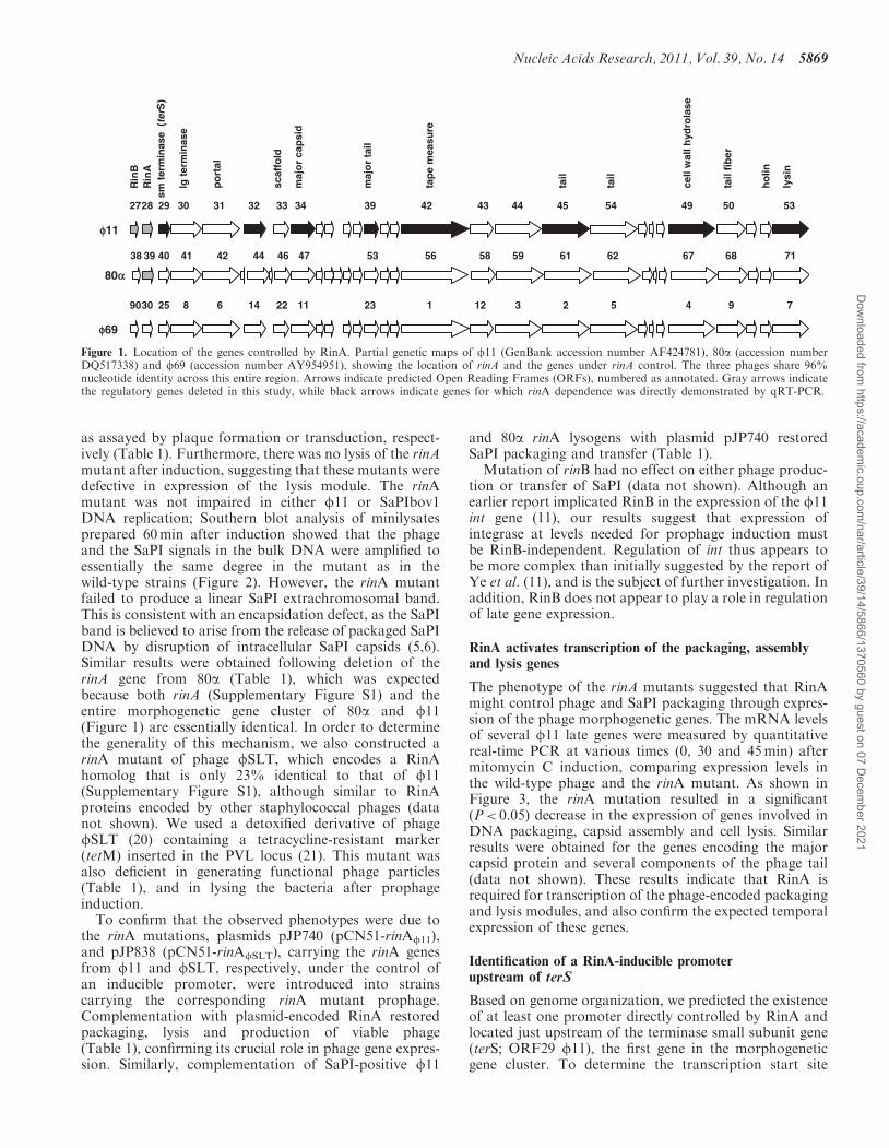
as assayed by plaque formation or transduction, respect-ively (Table 1). Furthermore, there was no lysis of the rinAmutant after induction, suggesting that these mutants weredefective in expression of the lysis module. The rinAmutant was not impaired in either f11 or SaPIbov1DNA replication; Southern blot analysis of minilysatesprepared 60min after induction showed that the phageand the SaPI signals in the bulk DNA were amplified toessentially the same degree in the mutant as in thewild-type strains (Figure 2). However, the rinA mutantfailed to produce a linear SaPI extrachromosomal band.This is consistent with an encapsidation defect, as the SaPIband is believed to arise from the release of packaged SaPIDNA by disruption of intracellular SaPI capsids (5,6).Similar results were obtained following deletion of therinA gene from 80a (Table 1), which was expectedbecause both rinA (Supplementary Figure S1) and theentire morphogenetic gene cluster of 80a and f11(Figure 1) are essentially identical. In order to determinethe generality of this mechanism, we also constructed arinA mutant of phage fSLT, which encodes a RinAhomolog that is only 23% identical to that of f11(Supplementary Figure S1), although similar to RinAproteins encoded by other staphylococcal phages (datanot shown). We used a detoxified derivative of phagefSLT (20) containing a tetracycline-resistant marker(tetM) inserted in the PVL locus (21). This mutant wasalso deficient in generating functional phage particles(Table 1), and in lysing the bacteria after prophageinduction.
To confirm that the observed phenotypes were due tothe rinA mutations, plasmids pJP740 (pCN51-rinAf11),and pJP838 (pCN51-rinAfSLT), carrying the rinA genesfrom f11 and fSLT, respectively, under the control ofan inducible promoter, were introduced into strainscarrying the corresponding rinA mutant prophage.Complementation with plasmid-encoded RinA restoredpackaging, lysis and production of viable phage(Table 1), confirming its crucial role in phage gene expres-sion. Similarly, complementation of SaPI-positive f11
and 80a rinA lysogens with plasmid pJP740 restoredSaPI packaging and transfer (Table 1).Mutation of rinB had no effect on either phage produc-
tion or transfer of SaPI (data not shown). Although anearlier report implicated RinB in the expression of the f11int gene (11), our results suggest that expression ofintegrase at levels needed for prophage induction mustbe RinB-independent. Regulation of int thus appears tobe more complex than initially suggested by the report ofYe et al. (11), and is the subject of further investigation. Inaddition, RinB does not appear to play a role in regulationof late gene expression.
RinA activates transcription of the packaging, assemblyand lysis genes
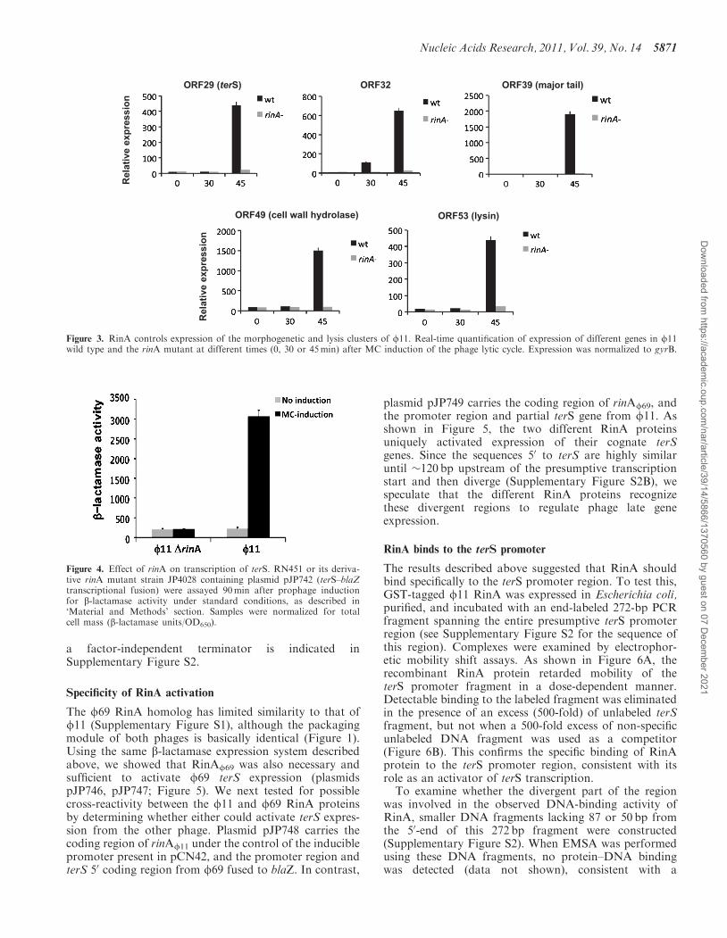
The phenotype of the rinA mutants suggested that RinAmight control phage and SaPI packaging through expres-sion of the phage morphogenetic genes. The mRNA levelsof several f11 late genes were measured by quantitativereal-time PCR at various times (0, 30 and 45min) aftermitomycin C induction, comparing expression levels inthe wild-type phage and the rinA mutant. As shown inFigure 3, the rinA mutation resulted in a significant(P< 0.05) decrease in the expression of genes involved inDNA packaging, capsid assembly and cell lysis. Similarresults were obtained for the genes encoding the majorcapsid protein and several components of the phage tail(data not shown). These results indicate that RinA isrequired for transcription of the phage-encoded packagingand lysis modules, and also confirm the expected temporalexpression of these genes.
Identification of a RinA-inducible promoterupstream of terS
Based on genome organization, we predicted the existenceof at least one promoter directly controlled by RinA andlocated just upstream of the terminase small subunit gene(terS; ORF29 f11), the first gene in the morphogeneticgene cluster. To determine the transcription start site
f11
f69
80a
3505945444342493433323130392 54
1786761695856535746444241404 62
sm t
erm
inas
e (
terS
)
lg t
erm
inas
e
tap
e m
easu
re
cell
wal
l hyd
rola
se
tail
fib
er
ho
lin
lysi
n
maj
or
cap
sid
maj
or
tail
po
rtal
scaf
fold
tail
tail
2827
Rin
AR
inB
3938
79423211321122416852 53090
Figure 1. Location of the genes controlled by RinA. Partial genetic maps of f11 (GenBank accession number AF424781), 80a (accession numberDQ517338) and f69 (accession number AY954951), showing the location of rinA and the genes under rinA control. The three phages share 96%nucleotide identity across this entire region. Arrows indicate predicted Open Reading Frames (ORFs), numbered as annotated. Gray arrows indicatethe regulatory genes deleted in this study, while black arrows indicate genes for which rinA dependence was directly demonstrated by qRT-PCR.
Nucleic Acids Research, 2011, Vol. 39, No. 14 5869
Dow
nloaded from https://academ
ic.oup.com/nar/article/39/14/5866/1370560 by guest on 07 D
ecember 2021
and locate this putative promoter, 50 RACE analysis wasperformed with total RNA isolated after induction of thef11 prophage in strain RN451. A rinA-dependent 50-endwas identified in RNA isolated 60min after MC inductionof the wild-type (wt) strain RN451, which mapped to a Cresidue located 23 bp upstream of the terS ATG initiationcodon (Supplementary Figure S2). In the case of 80a latemRNA, the 50-end was mapped to the adjacent G residue24 bp upstream of the terS gene.To determine whether the observed 50-end corresponded
to a promoter that was indeed regulated by RinA, a 425 bpfragment spanning the rinA-terS junction was fused to theb-lactamase reporter gene present in plasmid pCN41,generating plasmid pJP742. This plasmid was introducedinto strains RN451 (f11 lysogen) and JP4028 (RN451�rinA), and, after prophage induction, the transcription
of the b-lactamase reporter was analyzed. As shown inFigure 4, b-lactamase expression increased significantlyafter induction of the wt strain RN451 but not in its de-rivative rinA mutant, clearly confirming the existence of apromoter regulated by RinA in this region of the f11genome.
RinA is sufficient to activate ter expression
We next examined whether expression of RinA wasenough to induce expression of the terS gene in theabsence of other phage-encoded proteins. To test this wemade use of plasmid pCN42, which contains a Pcad indu-cible promoter and a b-lactamase reporter (Figure 5). Weintroduced into this plasmid a PCR fragment containingthe rinA coding sequence (expression of which depends onthe Pcad promoter present in the plasmid), the terSpromoter region, and the 50 coding region of the terSgene, which was fused to the b-lactamase reporter. Theresulting plasmid, pJP743, is illustrated in Figure 5. As acontrol, we amplified this region using as a template theDNA from the f11 rinA mutant, generating plasmidpJP744, which does not express RinA. These plasmidswere introduced into strain RN4220, and the expressionof the b-lactamase reporter was measured. As shown inFigure 5, only the plasmid expressing RinA also expressedthe b-lactamase reporter, confirming the previous resultsand indicating that RinA is the only phage-encodedprotein required for the expression of terS. Furthermore,the lack of b-lactamase expression from the rinA mutantplasmid indicates the presence of a transcriptional ter-minator between the rinA and terS genes. One po-tential sequence in the region with the characteristics of
Table 1. Effect of phage mutations on phage titre and SaPI transfera
Donor strain f Plasmid SaPI Phage titreb Transduction titrec
RN451 f11 wt – – 1.3� 109 –JP1794 f11 wt – SaPIbov1 1.4� 107 1.6� 108
JP4028 f11 �rinA – – <10 –JP4221 f11 �rinA pCN51-rinAf11 – 2.4� 108 –JP4128 f11 �rinA – SaPIbov1 <10 8.0� 103
JP5961 f11 �rinA pCN51-rinAf11 SaPIbov1 2.2� 106 2.4� 107
RN10359 80a wt – – 2.9� 1010 –JP3603 80a wt – SaPIbov1 6.9� 108 7.8� 107
JP3602 80a wt – SaPI1 4.5� 108 8.2� 108
JP4717 80a �rinA – – <10 –JP5418 80a �rinA pCN51-rinAf11 – 6.1� 108 –JP5293 80a �rinA – SaPIbov1 <10 770JP5294 80a �rinA – SaPI1 <10 22JP5419 80a �rinA pCN51-rinAf11 SaPIbov1 9.0� 107 4.0� 107
JP5420 80a �rinA pCN51-rinAf11 SaPI1 2.8� 107 6.9� 107
JP5011 fSLT wt – – 1.7� 105 8.7� 104
JP6895 fSLT �rinA – – <10 <10JP6391 fSLT �rinA pCN51-rinAfSLT – 9.0� 104 7.7� 103
JP7188 f11-f69 chimera – – <10 –JP7242 f11-f69 chimera – SaPIbov1 <10 1.2� 103
JP7218 f11-f69 chimera pCN51-rinAf69 – ND –JP7243 f11-f69 chimera pCN51-rinAf69 SaPIbov1 ND 4.2� 106
aThe means of results from three independent experiments are presented. Variation was within ±5% in all cases.bPfu/ml of induced culture, using RN4220 (or pCN51-complemented RN4200) as recipient.cTransductants/ml of induced culture, using RN4220 as recipient.
φ11 probe
0 60 0 60 0 60 0 60 min
φ11 φ11 ΔrinA φ11 φ11 ΔrinA
SaPIbov1probe
Bulk DNASaPI monomer
Bulk DNA
SaPIbov1
Figure 2. Characterization of the f11 rinA mutant. Southern blot off11wt and rinA mutant lysates from strains with or without SaPIbov1tst::tetM. Samples were taken before or 60min after MC induction,minilysates were prepared, the DNA was separated on agarose andblotted with a phage- or SaPIbov1-specific probe, as indicated. Theupper band is ‘bulk’ DNA, including chromosomal, phage andreplicating SaPI DNA; the lower band is SaPI linear monomersreleased from phage heads.
5870 Nucleic Acids Research, 2011, Vol. 39, No. 14
Dow
nloaded from https://academ
ic.oup.com/nar/article/39/14/5866/1370560 by guest on 07 D
ecember 2021
a factor-independent terminator is indicated inSupplementary Figure S2.
Specificity of RinA activation
The f69 RinA homolog has limited similarity to that off11 (Supplementary Figure S1), although the packagingmodule of both phages is basically identical (Figure 1).Using the same b-lactamase expression system describedabove, we showed that RinAf69 was also necessary andsufficient to activate f69 terS expression (plasmidspJP746, pJP747; Figure 5). We next tested for possiblecross-reactivity between the f11 and f69 RinA proteinsby determining whether either could activate terS expres-sion from the other phage. Plasmid pJP748 carries thecoding region of rinAf11 under the control of the induciblepromoter present in pCN42, and the promoter region andterS 50 coding region from f69 fused to blaZ. In contrast,
plasmid pJP749 carries the coding region of rinAf69, andthe promoter region and partial terS gene from f11. Asshown in Figure 5, the two different RinA proteinsuniquely activated expression of their cognate terSgenes. Since the sequences 50 to terS are highly similaruntil �120 bp upstream of the presumptive transcriptionstart and then diverge (Supplementary Figure S2B), wespeculate that the different RinA proteins recognizethese divergent regions to regulate phage late geneexpression.
RinA binds to the terS promoter
The results described above suggested that RinA shouldbind specifically to the terS promoter region. To test this,GST-tagged f11 RinA was expressed in Escherichia coli,purified, and incubated with an end-labeled 272-bp PCRfragment spanning the entire presumptive terS promoterregion (see Supplementary Figure S2 for the sequence ofthis region). Complexes were examined by electrophor-etic mobility shift assays. As shown in Figure 6A, therecombinant RinA protein retarded mobility of theterS promoter fragment in a dose-dependent manner.Detectable binding to the labeled fragment was eliminatedin the presence of an excess (500-fold) of unlabeled terSfragment, but not when a 500-fold excess of non-specificunlabeled DNA fragment was used as a competitor(Figure 6B). This confirms the specific binding of RinAprotein to the terS promoter region, consistent with itsrole as an activator of terS transcription.To examine whether the divergent part of the region
was involved in the observed DNA-binding activity ofRinA, smaller DNA fragments lacking 87 or 50 bp fromthe 50-end of this 272 bp fragment were constructed(Supplementary Figure S2). When EMSA was performedusing these DNA fragments, no protein–DNA bindingwas detected (data not shown), consistent with a
ORF29 (terS) ORF32 ORF39 (major tail)
Rel
ativ
e ex
pres
sion
ORF49 (cell wall hydrolase) ORF53 (lysin)R
elat
ive
expr
essi
on
Figure 3. RinA controls expression of the morphogenetic and lysis clusters of f11. Real-time quantiEcation of expression of different genes in f11wild type and the rinA mutant at different times (0, 30 or 45min) after MC induction of the phage lytic cycle. Expression was normalized to gyrB.
Figure 4. Effect of rinA on transcription of terS. RN451 or its deriva-tive rinA mutant strain JP4028 containing plasmid pJP742 (terS–blaZtranscriptional fusion) were assayed 90min after prophage inductionfor b-lactamase activity under standard conditions, as described in‘Material and Methods’ section. Samples were normalized for totalcell mass (b-lactamase units/OD650).
Nucleic Acids Research, 2011, Vol. 39, No. 14 5871
Dow
nloaded from https://academ
ic.oup.com/nar/article/39/14/5866/1370560 by guest on 07 D
ecember 2021
requirement for virtually all of this upstream divergentregion in RinA binding.
Characterization of the rinA-dependent transcription unit
The terS gene lies just downstream of rinA, and at thebeginning of the phage morphogenetic gene cluster. All
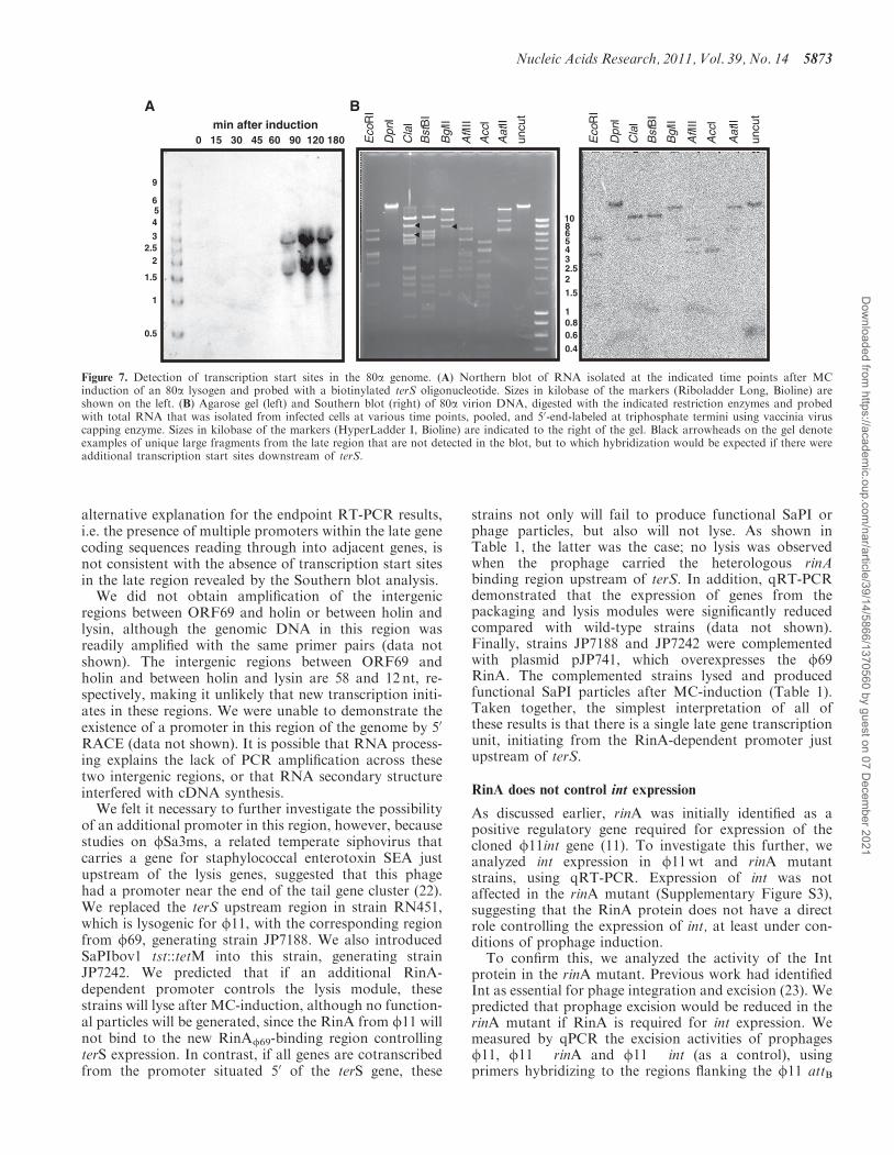
of the morphogenetic genes are encoded on the samestrand, with little intergenic space between them, suggest-ing that they might comprise a single large transcriptionunit. In an attempt to characterize the phage transcriptionunits, independent Northern blot analyses were performedon RNA isolated during both 80a and f11 infections. Inboth cases, although specific hybridization was obtainedat appropriate time points, only small RNAs in the 2–4 kbsize range could be detected, using probes from severaldifferent genes across this region. A representativeexample is shown in Figure 7A. This suggests that eitherthere are multiple transcription start sites throughout thelate gene cluster or that the RNA is rapidly processed.Multiple transcription start sites should yield multipleRNAs containing 50 triphosphate ends. To estimate thenumber of promoters present in the phage genome,Southern blot analysis of 80a DNA was performed withpooled phage RNA collected at various times after infec-tion and labeled specifically at the 50 triphosphate terminiarising from initiation of transcription. This analysis doesnot allow unambiguous identification of uniquepromoter-containing fragments, but did indicate that thetotal number of promoters in the phage genome is rela-tively small (Figure 7B). Furthermore, several uniquelarge restriction fragments that included only late regionDNA downstream of terS were not detected in thisanalysis, consistent with the hypothesis that there is asingle promoter for the late genes.
To investigate further the transcription of the morpho-genetic genes, we isolated RNA late in infection with 80a,converted it to cDNA and used that cDNA in endpointRT-PCR primer pairs flanking intergenic regions through-out the late gene cluster, from terminase through the lysisgenes. A product was obtained with all primer pairs fromterL to ORF69 (data not shown). The presence of RNAspanning each of the late gene intergenic regions alsosuggests that these genes are all cotranscribed. The
A BpCN42
φ11φ69
pJP743
pJP744
rinA ter
pJP746
pJP747
pJP748
pJP749
Pcad-cadC blaZTT b-lactamase activity
6000
5000
4000
3000
2000
10000
Figure 5. Specificity of RinA proteins. (A) Schematic representation of the different blaZ transcriptional fusions. (B) Derivatives of strain RN4220containing each of the indicated plasmids were assayed at mid-exponential phase for b-lactamase activity under standard conditions. Samples werenormalized for total cell mass (b-lactamase units/OD650).
RinA protein (100 nM)- + + +
- - + -
- - - +
Specific DNA
Non-specific DNA
-0 40 80 100 RinA protein (nM)
A
B
Complex
Complex
DNA probe
DNA probe
Figure 6. Binding of RinA to the terS promoter. (A) Electrophoreticmobility of a DIG-labeled promoter fragment was measured in thepresence or absence of increasing amounts of puriEed f11 RinAprotein, as indicated on the top line. (B) In the competition assays,500-fold excesses of speciEc- or non-speciEc unlabeled DNA fragmentswere added, as indicated. In all cases, the concentration of theDig-labeled probe was 21 nM.
5872 Nucleic Acids Research, 2011, Vol. 39, No. 14
Dow
nloaded from https://academ
ic.oup.com/nar/article/39/14/5866/1370560 by guest on 07 D
ecember 2021
alternative explanation for the endpoint RT-PCR results,i.e. the presence of multiple promoters within the late genecoding sequences reading through into adjacent genes, isnot consistent with the absence of transcription start sitesin the late region revealed by the Southern blot analysis.
We did not obtain amplification of the intergenicregions between ORF69 and holin or between holin andlysin, although the genomic DNA in this region wasreadily amplified with the same primer pairs (data notshown). The intergenic regions between ORF69 andholin and between holin and lysin are 58 and 12 nt, re-spectively, making it unlikely that new transcription initi-ates in these regions. We were unable to demonstrate theexistence of a promoter in this region of the genome by 50
RACE (data not shown). It is possible that RNA process-ing explains the lack of PCR amplification across thesetwo intergenic regions, or that RNA secondary structureinterfered with cDNA synthesis.
We felt it necessary to further investigate the possibilityof an additional promoter in this region, however, becausestudies on fSa3ms, a related temperate siphovirus thatcarries a gene for staphylococcal enterotoxin SEA justupstream of the lysis genes, suggested that this phagehad a promoter near the end of the tail gene cluster (22).We replaced the terS upstream region in strain RN451,which is lysogenic for f11, with the corresponding regionfrom f69, generating strain JP7188. We also introducedSaPIbov1 tst::tetM into this strain, generating strainJP7242. We predicted that if an additional RinA-dependent promoter controls the lysis module, thesestrains will lyse after MC-induction, although no function-al particles will be generated, since the RinA from f11 willnot bind to the new RinAf69-binding region controllingterS expression. In contrast, if all genes are cotranscribedfrom the promoter situated 50 of the terS gene, these
strains not only will fail to produce functional SaPI orphage particles, but also will not lyse. As shown inTable 1, the latter was the case; no lysis was observedwhen the prophage carried the heterologous rinAbinding region upstream of terS. In addition, qRT-PCRdemonstrated that the expression of genes from thepackaging and lysis modules were significantly reducedcompared with wild-type strains (data not shown).Finally, strains JP7188 and JP7242 were complementedwith plasmid pJP741, which overexpresses the f69RinA. The complemented strains lysed and producedfunctional SaPI particles after MC-induction (Table 1).Taken together, the simplest interpretation of all ofthese results is that there is a single late gene transcriptionunit, initiating from the RinA-dependent promoter justupstream of terS.
RinA does not control int expression
As discussed earlier, rinA was initially identified as apositive regulatory gene required for expression of thecloned f11int gene (11). To investigate this further, weanalyzed int expression in f11wt and rinA mutantstrains, using qRT-PCR. Expression of int was notaffected in the rinA mutant (Supplementary Figure S3),suggesting that the RinA protein does not have a directrole controlling the expression of int, at least under con-ditions of prophage induction.To confirm this, we analyzed the activity of the Int
protein in the rinA mutant. Previous work had identifiedInt as essential for phage integration and excision (23). Wepredicted that prophage excision would be reduced in therinA mutant if RinA is required for int expression. Wemeasured by qPCR the excision activities of prophagesf11, f11 �rinA and f11 �int (as a control), usingprimers hybridizing to the regions flanking the f11 attB
2.5
1.5
0.80.6
0.4
10
6543
2
1
8
0.5
1
1.5
2.5
456
9
0 15 30 45 60 90 120 180
2
3
min after induction
A B
Eco
RI
Dpn
I
Cla
I
Bst
BI
Bgl
II
AflI
II
Acc
I
Aat
II
uncu
t
Eco
RI
Dpn
I
Cla
I
Bst
BI
Bgl
II
AflI
II
Acc
I
Aat
II
uncu
t
Figure 7. Detection of transcription start sites in the 80a genome. (A) Northern blot of RNA isolated at the indicated time points after MCinduction of an 80a lysogen and probed with a biotinylated terS oligonucleotide. Sizes in kilobase of the markers (Riboladder Long, Bioline) areshown on the left. (B) Agarose gel (left) and Southern blot (right) of 80a virion DNA, digested with the indicated restriction enzymes and probedwith total RNA that was isolated from infected cells at various time points, pooled, and 50-end-labeled at triphosphate termini using vaccinia viruscapping enzyme. Sizes in kilobase of the markers (HyperLadder I, Bioline) are indicated to the right of the gel. Black arrowheads on the gel denoteexamples of unique large fragments from the late region that are not detected in the blot, but to which hybridization would be expected if there wereadditional transcription start sites downstream of terS.
Nucleic Acids Research, 2011, Vol. 39, No. 14 5873
Dow
nloaded from https://academ
ic.oup.com/nar/article/39/14/5866/1370560 by guest on 07 D
ecember 2021
site. As expected, excision of the f11 rinA mutant wassimilar to wt, while no excision was observed in the f11�int mutant (data not shown). This confirms the previ-ously reported requirement for Int in prophage excision aswell as the lack of involvement of RinA in regulation of intexpression during this process.
RinA does not control SaPI-encoded operon I expression
We have previously reported that SaPI packaging dependson expression of the SaPI-encoded homologue of thephage terminase small subunit (terS). In addition, SaPIsremodel assembly of the phage capsid proteins to generatecapsids one-third the size of the helper phage capsids,which accommodate their smaller genomes whileexcluding complete helper phage genomes. This requirestwo SaPI genes, cp1 and cp2, which are adjacent in asix-gene LexA-regulated operon, operon I, that alsoencodes the SaPI-specific terS (5). In order to determineif RinA controls expression of SaPIbov1 operon I, weanalyzed SaPIbov1 terS expression in f11wt and rinAmutant strains, using qRT-PCR. Expression ofSaPIbov1 terS was not impaired in the rinA mutant,even under conditions where LexA-dependent SaPIoperon I expression was blocked by mutation (data notshown), suggesting that the RinA protein does not have adirect role controlling the expression of SaPI-encodedoperon I.
RinA homologues control packaging in phages from otherGram-positive bacteria
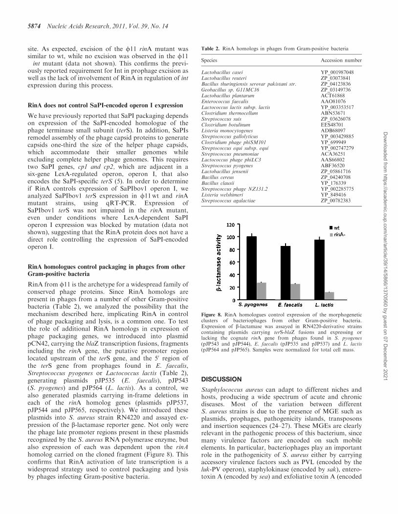
RinA from f11 is the archetype for a widespread family ofconserved phage proteins. Since RinA homologs arepresent in phages from a number of other Gram-positivebacteria (Table 2), we analyzed the possibility that themechanism described here, implicating RinA in controlof phage packaging and lysis, is a common one. To testthe role of additional RinA homologs in expression ofphage packaging genes, we introduced into plasmidpCN42, carrying the blaZ transcription fusions, fragmentsincluding the rinA gene, the putative promoter regionlocated upstream of the terS gene, and the 50 region ofthe terS gene from prophages found in E. faecalis,Streptococcus pyogenes or Lactococcus lactis (Table 2),generating plasmids pJP535 (E. faecalis), pJP543(S. pyogenes) and pJP564 (L. lactis). As a control, wealso generated plasmids carrying in-frame deletions ineach of the rinA homolog genes (plasmids pJP537,pJP544 and pJP565, respectively). We introduced theseplasmids into S. aureus strain RN4220 and assayed ex-pression of the b-lactamase reporter gene. Not only werethe phage late promoter regions present in these plasmidsrecognized by the S. aureus RNA polymerase enzyme, butalso expression of each was dependent upon the rinAhomolog carried on the cloned fragment (Figure 8). Thisconfirms that RinA activation of late transcription is awidespread strategy used to control packaging and lysisby phages infecting Gram-positive bacteria.
DISCUSSION
Staphylococcus aureus can adapt to different niches andhosts, producing a wide spectrum of acute and chronicdiseases. Most of the variation between differentS. aureus strains is due to the presence of MGE such asplasmids, prophages, pathogenicity islands, transposonsand insertion sequences (24–27). These MGEs are clearlyrelevant in the pathogenic process of this bacterium, sincemany virulence factors are encoded on such mobileelements. In particular, bacteriophages play an importantrole in the pathogenicity of S. aureus either by carryingaccessory virulence factors such as PVL (encoded by theluk-PV operon), staphylokinase (encoded by sak), entero-toxin A (encoded by sea) and exfoliative toxin A (encoded
Table 2. RinA homologs in phages from Gram-positive bacteria
Species Accession number
Lactobacillus casei YP_001987048Lactobacillus reuteri ZP_03073841Bacillus thuringiensis serovar pakistani str. ZP_04123836Geobacillus sp. G11MC16 ZP_03149736Lactobacillus plantarum ACT61868Enterococcus faecalis AAO81076Lactococcus lactis subsp. lactis YP_003353517Clostridium thermocellum ABN53671Streptococcus suis ZP_03626078Clostridium botulinum EES48701Listeria monocytogenes ADB68097Streptococcus gallolyticus YP_003429885Clostridium phage phiSM101 YP_699949Streptococcus equi subsp. equi YP_002747279Streptococcus pneumoniae ACA36251Lactococcus phage phiLC3 AAS66802Streptococcus pyogenes ABF36520Lactobacillus jensenii ZP_05861716Bacillus cereus ZP_04240708Bacillus clausii YP_176339Streptococcus phage NZ131.2 YP_002285775Listeria welshimeri YP_849416Streptococcus agalactiae ZP_00782383
Figure 8. RinA homologues control expression of the morphogeneticclusters of bacteriophages from other Gram-positive bacteria.Expression of b-lactamase was assayed in RN4220-derivative strainscontaining plasmids carrying terS-blaZ fusions and expressing orlacking the cognate rinA gene from phages found in S. pyogenes(pJP543 and pJP544), E. faecalis (pJP535 and pJP537) and L. lactis(pJP564 and pJP565). Samples were normalized for total cell mass.
5874 Nucleic Acids Research, 2011, Vol. 39, No. 14
Dow
nloaded from https://academ
ic.oup.com/nar/article/39/14/5866/1370560 by guest on 07 D
ecember 2021
by eta) or by interrupting chromosomal virulence genessuch as those for b-hemolysin (hlb) and lipase (geh)upon insertion [reviewed in (1)]. Phages are also the pri-mary vehicle of lateral gene transfer between S. aureusstrains, providing the species with the potential forbroad genetic variation. It has been reported recentlythat phages increase the genome plasticity of S. aureusduring infection, facilitating the adaptation of the patho-gen to various host conditions (28). Additionally, certainstaphylococcal phages carry out a highly specializedexample of phage-mediated HGT, the mobilization ofSaPIs. SaPI replication and high frequency transductionis closely linked to the phage lytic cycle, which inducesSaPI replication and provides the proteins required forSaPI packaging and transfer [for review see (10)].Despite the clear importance of staphylococcal phages toS. aureus pathogenesis, and the large number of sequencedstaphylococcal phage genomes now available in publicgenome databases, the number of studies characterizingthe biology of these relevant elements is limited.
To ensure tight control of genes required in the laterstages of infection, bacteriophages have evolved avariety of mechanisms involving synthesis of phage-encoded control factors during the early stages of infec-tion. In this work, we have identified the activator ofphage late transcription and localized the promoter re-sponsible for the expression of the morphogenetic genesin several representatives from a large family of relatedS. aureus phages. This promoter is active only duringthe late phase of the lytic cycle and requires the phage-encoded RinA protein for activity. The late promoter islocated upstream of the morphogenetic cluster andcontrols the synthesis of a single large operon encodingthe proteins responsible for assembly of phage particles,DNA packaging and host cell lysis. Expression of thisgene cluster is essential not only for propagation of thephage, but also for transfer of phage-borne toxin genes,generalized transduction and the horizontal transmissionof other MGEs that exploit the phage for their owntransfer, such as SaPIs. As would be expected for an ac-tivator of late transcription, preliminary analysis off11-encoded genes after SOS induction, using tilingarrays, indicate that rinA is expressed earlier than terS,in a transcription unit containing genes involved inphage replication (data not shown).
Activation of late transcription by positive regulatoryproteins is a strategy employed by a variety of phages withdsDNA genomes. Enterobacteriophage P2 and relatedphages encode a small zinc-binding protein, Ogr, thatbinds to conserved inverted repeats centered at about�55 relative to the transcription initiation sites of P2late promoters (29–32). Protein–protein interactionbetween Ogr and the a subunit of the host RNA polymer-ase is required to stimulate P2 late gene transcription (33).Activation of phage T4 late promoters requires twophage-encoded proteins that function as core RNA poly-merase subunits. T4 gp55 functions as a sigma factor thatallows recognition of the 8 bp late promoter consensusmotif, while gp33 is a coactivator for high level late tran-scription (34,35). T4 late transcription also requires aunique transcriptional activator, gp45, which is a sliding
clamp that interacts with gp55, gp33 and T4 DNA poly-merase to couple transcription to phage DNA replication(36). Phage P1 late transcription requires the phage-encoded Lpa protein, which binds to a conserved sequencecentered at �22, as well as host-encoded coactivator, SspA(37). The phage Mu late activator protein, C, binds to aregion of Mu late promoters from �30 to �55 and stimu-lates both binding of RNA polymerase and subsequentpromoter release (38–40). Much less is known about themechanism of transcription activation in the dsDNAphages of Gram-positive bacteria. The classic example isBacillus subtilis phage SP01, which employs successivephage-encoded sigma factors to direct the host RNA poly-merase to phage middle and late promoters (41) and alsorequires a T4 gp45-like protein for late transcription (42).The regulatory protein p4 from B. subtilis phage f29 bindsbetween �58 and �104 upstream of the late promotertranscription start site and activates through contactwith the a subunit of RNA polymerase (43,44). An acti-vator of late gene transcription (Alt) has also beenidentified in the L. lactis phage TP901-1 (45). Alt bindsto a promoter region located upstream of the terS gene,recognizing a series of repeats �76 to �32 relative to thetranscription start site (46). A common feature of all ofthese mechanisms is the recognition of sequences within orjust upstream of the RNA polymerase binding site by aprotein involved in activation of late transcription. RinA,in contrast, appears to require, for binding, sequenceslocated more than 155 bp upstream of the terS transcrip-tion start site. Since essentially the same+1 was independ-ently mapped by RACE for both f11 and 80a, and the80a RNA was treated to eliminate molecules with 50
monophosphate termini, we have high confidence thatthis site corresponds to the actual start of transcriptionand not an end generated by processing. The largedistance between the transcription start and sequencesneeded for RinA binding therefore suggests a mechanismfor transcription activation that is more complex than adirect interaction between RinA and an adjacent RNApolymerase.RinA was initially reported as an activator of the f11
int gene (11). In earlier work, Ye et al. (23) identified theputative promoter region for the f11 int gene andobserved that the DNA sequence of the region betweenthe translation start sites of the int and xis genes, whichare transcribed divergently, was conserved between phagesL54a and phage f11, suggesting that this conserved regioncould contain the potential regulatory sites for expressionof the int and xis genes. Using a reporter plasmid contain-ing the gene xylE under control of this interveningsequence, including the f11 int promoter, differentcloned regions of the f11 genome were screened for acti-vation of expression. It was found that regulatory proteinsRinA and RinB were both required to activate expressionof the f11int gene, while RinA alone was capable ofactivating L54a int gene transcription (11). Surprisingly,our results with the effects of rinA and rinB mutants donot confirm this initial characterization, suggesting thatcontrol of int expression is more complicated than pre-dicted. Furthermore, RinA from phage L54a is evenmore divergent from f11 than the RinA of f69, sharing
Nucleic Acids Research, 2011, Vol. 39, No. 14 5875
Dow
nloaded from https://academ
ic.oup.com/nar/article/39/14/5866/1370560 by guest on 07 D
ecember 2021
only 25% amino acid identity. Since we have demon-strated that the RinA proteins from f11 and f69 recog-nize and bind to different specific DNA regulatorysequences, it is difficult to explain how the f11 andL54a proteins could bind to a conserved DNA sequencebetween int and xis to activate f11 int expression.RinA homologues can be found in bacteriophages in-
fecting a variety of Gram-positive bacteria. We testedseveral of these and found that all were transcriptionalactivators that controlled expression of terS, the firstgene of the morphogenetic cluster, in their respectivephages. Thus, we propose that this family of proteins rep-resents a conserved strategy for regulating the packagingand transfer of many bacteriophages in Gram-positivebacteria. Interestingly, an NCBI Conserved DomainDatabase search indicates that the RinA family of phageproteins also shows weak similarity to another large familyof transcriptional regulators, ArpU. ArpU was describedas a regulator of cellular muramidase-2 of Enterococcushirae (47). Although initially reported as a chromosomallyencoded gene, ArpU appears to have been cloned from aprophage. ArpU homologues are also present in a varietyof different Gram-positive bacteriophages, and, like RinA,are localized upstream of the first gene of the morpho-genetic cluster of the phage. This suggests that theArpU-related proteins have a similar role in activatingbacteriophage late gene expression, although this has notbeen tested directly. The C-terminal half of RinA alsoshows limited similarity to a portion of several sigmafactors corresponding to conserved region 4. The signifi-cance of this is unclear. This region of sigma is involved inrecognition of the �35 element of promoters (48). While itseems quite unlikely that RinA is functioning as a sigmafactor, this may indicate that the C-terminal part of theprotein contains the DNA recognition determinants. Onthe other hand, sigma region 4 is also a target for anumber of transcription factors (49) so this could be aprotein–protein interaction domain. Further investigationof the structure and function of RinA is clearly needed todetermine its mechanism of action in activation oftranscription.Horizontal transfer of DNA can occur in bacteria by
transformation, conjugation and transduction. InS. aureus, there is little evidence that transformationoccurs. In addition, conjugative plasmids are not wide-spread and conjugative transposons are even lesscommon. In contrast, many of the known S. aureus bac-teriophages are generalized transducing phages that canpackage S. aureus chromosomal DNA as well as MGEs,and transfer them to other S. aureus strains (12).Transduction is likely the predominant mechanism ofHGT in S. aureus. Since transfer of the different MGEsrequires exploitation of the phage mechanisms involved invirion assembly and DNA packaging, characterization ofthe phage proteins controlling these processes are import-ant in understanding how MGEs are transferred. In thiswork we have identified and characterized a key regulatorof genes involved in phage assembly and packaging.This activator, RinA, is not only present in most of the
S. aureus phages, but also in phages infecting a widevariety of other Gram-positive bacteria. Deletion of thisactivator resulted not only in elimination of thephage titre, but also significantly reduced transfer ofSaPI-encoded virulence factors, and is predicted to showa similar reduction in transmission of plasmid-encoded virulence factors. Thus, we have identified an im-portant mechanism involved in phage-mediated HGT,which plays a key role in bacterial evolution andpathogenesis.
SUPPLEMENTARY DATA
Supplementary Data are available at NAR Online.
FUNDING
Consolider-Ingenio CSD2009-00006, BIO2005-08399-C02-02, BIO2008-05284-C02-02 and BIO2008-00642-E/Cfrom the Ministerio de Ciencia e Innovacion (MICINN),and grants from the Cardenal Herrera-CEU University(PRCEU-UCH39/10 and Copernicus-Banco Santanderprogram), from the Conselleria de Agricultura, Pesca iAlimentacio (CAPiA) and from the GeneralitatValenciana (ACOMP07/258) to J.R.P., grants[BFU2008-01078] from the MICINN and[2009SGR1106] from the Generalitat de Catalunya toJ.B., NIH grants [R21 AI067654 and R56 AI081837]and a grant-in-aid from the A.D. Williams Trust and theBaruch Foundation Trust to G.E.C., a VCU GraduateSchool Thesis and Dissertation Award to M.D.H., andNIH grant [R01AI022159-23A2] to R.P.N. Funding foropen access charge: Consolider-Ingenio CSD2009-00006,BIO2005-08399-C02-02, BIO2008-05284-C02-02 andBIO2008-00642-E/C from the Ministerio de Ciencia eInnovacion (MICINN), Spain.
Conflict of interest statement. None declared.
REFERENCES
1. L�os,M., Kuzio,J., McConnell,M.R., Kropinski,A.M., Wegrzyn,G.and Christie,G.E. (2010) Lysogenic conversion in bacteria ofimportance to the food industry. In Sabour,M.P. and Griffiths,M.(eds), Bacteriophage in the Detection and Control of FoodbornePathogens. ASM Press, Washington, DC, pp. 157–198.
2. Ubeda,C., Maiques,E., Barry,P., Matthews,A., Tormo,M.A.,Lasa,I., Novick,R.P. and Penades,J.R. (2008) SaPI mutationsaffecting replication and transfer and enabling autonomousreplication in the absence of helper phage. Mol. Microbiol., 67,493–503.
3. Tormo-Mas,M.A., Mir,I., Shrestha,A., Tallent,S.M., Campoy,S.,Lasa,I., Barbe,J., Novick,R.P., Christie,G.E. and Penades,J.R.(2010) Moonlighting phage proteins de-repress staphylococcalpathogenicity islands. Nature, 465, 779–782.
4. Ubeda,C., Barry,P., Penades,J.R. and Novick,R.P. (2007) Apathogenicity island replicon in Staphylococcus aureus replicatesas an unstable plasmid. Proc. Natl Acad. Sci. USA, 104,14182–14188.
5. Ubeda,C., Maiques,E., Tormo,M.A., Campoy,S., Lasa,I.,Barbe,J., Novick,R.P. and Penades,J.R. (2007) SaPI operon I isrequired for SaPI packaging and is controlled by LexA.Mol. Microbiol., 65, 41–50.
5876 Nucleic Acids Research, 2011, Vol. 39, No. 14
Dow
nloaded from https://academ
ic.oup.com/nar/article/39/14/5866/1370560 by guest on 07 D
ecember 2021
6. Tormo,M.A., Ferrer,M.D., Maiques,E., Ubeda,C., Selva,L.,Lasa,I., Calvete,J.J., Novick,R.P. and Penades,J.R. (2008)Staphylococcus aureus pathogenicity island DNA is packaged inparticles composed of phage proteins. J. Bacteriol., 190,2434–2440.
7. Tallent,S.M., Langston,T.B., Moran,R.G. and Christie,G.E.(2007) Transducing particles of Staphylococcus aureuspathogenicity island SaPI1 are comprised of helper phage-encodedproteins. J. Bacteriol., 189, 7520–7524.
8. Ubeda,C., Maiques,E., Knecht,E., Lasa,I., Novick,R.P. andPenades,J.R. (2005) Antibiotic-induced SOS responsepromotes horizontal dissemination of pathogenicity island-encoded virulence factors in staphylococci. Mol. Microbiol., 56,836–844.
9. Ruzin,A., Lindsay,J. and Novick,R.P. (2001) Molecular geneticsof SaPI1 - a mobile pathogenicity island in Staphylococcus aureus.Mol. Microbiol., 41, 365–377.
10. Novick,R.P., Christie,G.E. and Penades,J.R. (2010) Thephage-related chromosomal islands of Gram-positive bacteria.Nat. Rev. Microbiol., 8, 541–551.
11. Ye,Z.H. and Lee,C.Y. (1993) Cloning, sequencing, and geneticcharacterization of regulatory genes, rinA and rinB, required forthe activation of staphylococcal phage phi 11 int expression.J. Bacteriol., 175, 1095–1102.
12. Novick,R.P. (1991) Genetic systems in staphylococci.Methods Enzymol., 204, 587–636.
13. Lindsay,J.A., Ruzin,A., Ross,H.F., Kurepina,N. and Novick,R.P.(1998) The gene for toxic shock toxin is carried by a family ofmobile pathogenicity islands in Staphylococcus aureus.Mol. Microbiol., 29, 527–543.
14. Ausubel,F.M., Brent,R., Kingston,R.E., Moore,D.D.,Seidman,J.G., Smith,J.A. and Struhl,K. (1990) Current Protocolsin Molecular Biology. John Wiley & Sons, New York, NY.
15. Sambrook,J., Fritsch,E.F. and Maniatis,T. (1989) MolecularCloning: a Laboratory Manual. Cold Spring Harbor LaboratoryPress, NY.
16. Arnaud,M., Chastanet,A. and Debarbouille,M. (2004) New vectorfor efficient allelic replacement in naturally nontransformable,low-GC-content, gram-positive bacteria. Appl. Environ. Microbiol.,70, 6887–6891.
17. Ji,G., Beavis,R. and Novick,R.P. (1997) Bacterial interferencecaused by autoinducing peptide variants. Science, 276, 2027–2030.
18. Livak,K.J. and Schmittgen,T.D. (2001) Analysis of relative geneexpression data using real-time quantitative PCR and the 2(-DeltaDelta C(T)) Method. Methods, 25, 402–408.
19. Poliakov,A., Chang,J.R., Spilman,M.S., Damle,P.K.,Christie,G.E., Mobley,J.A. and Dokland,T. (2008) Capsid sizedetermination by Staphylococcus aureus pathogenicity islandSaPI1 involves specific incorporation of SaPI1 proteins intoprocapsids. J. Mol. Biol., 380, 465–475.
20. Narita,S., Kaneko,J., Chiba,J., Piemont,Y., Jarraud,S., Etienne,J.and Kamio,Y. (2001) Phage conversion of Panton-Valentineleukocidin in Staphylococcus aureus: molecular analysis of aPVL-converting phage, phiSLT. Gene, 268, 195–206.
21. Labandeira-Rey,M., Couzon,F., Boisset,S., Brown,E.L., Bes,M.,Benito,Y., Barbu,E.M., Vazquez,V., Hook,M., Etienne,J. et al.(2007) Staphylococcus aureus Panton-Valentine leukocidin causesnecrotizing pneumonia. Science, 315, 1130–1133.
22. Sumby,P. and Waldor,M.K. (2003) Transcription of the toxingenes present within the Staphylococcal phage phiSa3ms isintimately linked with the phage’s life cycle. J. Bacteriol., 185,6841–6851.
23. Ye,Z.H., Buranen,S.L. and Lee,C.Y. (1990) Sequence analysisand comparison of int and xis genes from staphylococcalbacteriophages L54a and phi 11. J. Bacteriol., 172, 2568–2575.
24. Herron-Olson,L., Fitzgerald,J.R., Musser,J.M. and Kapur,V.(2007) Molecular correlates of host specialization inStaphylococcus aureus. PLoS One, 2, e1120.
25. Lowder,B.V., Guinane,C.M., Ben Zakour,N.L., Weinert,L.A.,Conway-Morris,A., Cartwright,R.A., Simpson,A.J., Rambaut,A.,Nubel,U. and Fitzgerald,J.R. (2009) Recent human-to-poultryhost jump, adaptation, and pandemic spread of Staphylococcusaureus. Proc. Natl Acad. Sci. USA, 106, 19545–19550.
26. Guinane,C.M., Ben Zakour,N.L., Tormo-Mas,M.A.,Weinert,L.A., Lowder,B.V., Cartwright,R.A., Smyth,D.S.,Smyth,C.J., Lindsay,J.A., Gould,K.A. et al. (2010) Evolutionarygenomics of Staphylococcus aureus reveals insights into the originand molecular basis of ruminant host adaptation. Genome Biol.Evol., 2, 454–466.
27. Viana,D., Blanco,J., Tormo-Mas,M.A., Selva,L., Guinane,C.M.,Baselga,R., Corpa,J.M., Lasa,I., Novick,R.P., Fitzgerald,R. et al.(2010) Adaptation of Staphylococcus aureus to ruminant andequine hosts involves SaPI-carried variants of von Willebrandfactor-binding protein. Mol. Microbiol., 77, 1583–1594.
28. Goerke,C., Wirtz,C., Fluckiger,U. and Wolz,C. (2006) Extensivephage dynamics in Staphylococcus aureus contributes toadaptation to the human host during infection. Mol. Microbiol.,61, 1673–1685.
29. Christie,G.E. and Calendar,R. (1985) Bacteriophage P2 latepromoters. II. Comparison of the four late promoter sequences.J. Mol. Biol., 181, 373–382.
30. Grambow,N.J., Birkeland,N.K., Anders,D.L. and Christie,G.E.(1990) Deletion analysis of a bacteriophage P2 late promoter.Gene, 95, 9–15.
31. Birkeland,N.K., Lindqvist,B.H. and Christie,G.E. (1991) Controlof bacteriophage P2 gene expression: analysis of transcription ofthe ogr gene. J. Bacteriol., 173, 6927–6934.
32. Van Bokkelen,G.B., Dale,E.C., Halling,C. and Calendar,R. (1991)Mutational analysis of a bacteriophage P4 late promoter.J. Bacteriol., 173, 37–45.
33. Wood,L.F., Tszine,N.Y. and Christie,G.E. (1997) Activation ofP2 late transcription by P2 Ogr protein requires a discrete contactsite on the C terminus of the alpha subunit of Escherichia coliRNA polymerase. J. Mol. Biol., 274, 1–7.
34. Kassavetis,G.A. and Geiduschek,E.P. (1984) Defining abacteriophage T4 late promoter: bacteriophage T4 gene 55protein suffices for directing late promoter recognition.Proc. Natl Acad. Sci. USA, 81, 5101–5105.
35. Herendeen,D.R., Williams,K.P., Kassavetis,G.A. andGeiduschek,E.P. (1990) An RNA polymerase-binding protein thatis required for communication between an enhancer and apromoter. Science, 248, 573–578.
36. Nechaev,S. and Geiduschek,E.P. (2008) Dissection of thebacteriophage T4 late promoter complex. J. Mol. Biol., 379,402–413.
37. Hansen,A.M., Lehnherr,H., Wang,X., Mobley,V. and Jin,D.J.(2003) Escherichia coli SspA is a transcription activator forbacteriophage P1 late genes. Mol. Microbiol., 48, 1621–1631.
38. Margolin,W. and Howe,M.M. (1986) Localization and DNAsequence analysis of the C gene of bacteriophage Mu, the positiveregulator of Mu late transcription. Nucleic Acids Res., 14,4881–4897.
39. Sun,W., Hattman,S. and Kool,E. (1997) Interaction of thebacteriophage Mu transcriptional activator protein, C,with its target site in the mom promoter. J. Mol. Biol., 273,765–774.
40. Chakraborty,A. and Nagaraja,V. (2006) Dual role fortransactivator protein C in activation of mom promoter ofbacteriophage Mu. J. Biol. Chem., 281, 8511–8517.
41. Talkington,C. and Pero,J. (1978) Promoter recognition by phageSP01-modified RNA polymerase. Proc. Natl Acad. Sci. USA, 75,1185–1189.
42. Greene,J.R., Chelm,B.K. and Geiduschek,E.P. (1982) SP01gene 27 is required for viral late transcription. J. Virol., 41,715–720.
43. Barthelemy,I., Lazaro,J.M., Mendez,E., Mellado,R.P. andSalas,M. (1987) Purification in an active form of the phage phi29 protein p4 that controls the viral late transcription.Nucleic Acids Res., 15, 7781–7793.
44. Mencia,M., Monsalve,M., Rojo,F. and Salas,M. (1996)Transcription activation by phage phi29 protein p4 is mediatedby interaction with the alpha subunit of Bacillus subtilis RNApolymerase. Proc. Natl Acad. Sci. USA, 93, 6616–6620.
45. Brondsted,L., Pedersen,M. and Hammer,K. (2001) An activatorof transcription regulates phage TP901-1 late gene expression.Appl. Environ. Microbiol., 67, 5626–5633.
Nucleic Acids Research, 2011, Vol. 39, No. 14 5877
Dow
nloaded from https://academ
ic.oup.com/nar/article/39/14/5866/1370560 by guest on 07 D
ecember 2021
46. Pedersen,M., Kilstrup,M. and Hammer,K. (2006) Identification ofDNA-binding sites for the activator involved in late transcriptionof the temperate lactococcal phage TP901-1. Virology, 345,446–456.
47. Lleo,M.M., Fontana,R. and Solioz,M. (1995) Identification of agene (arpU) controlling muramidase-2 export in Enterococcushirae. J. Bacteriol., 177, 5912–5917.
48. Gross,C., Lonetto,M. and Losick,R. (1992) Bacterial SigmaFactors. In McKnight,S.Y.K. (ed.), Transcriptional Regulation.
Cold Spring Harbor Laboratory, Cold Spring Harbor, NY,pp. 129–176.
49. Lonetto,M.A., Rhodius,V., Lamberg,K., Kiley,P., Busby,S. andGross,C. (1998) Identification of a contact site for differenttranscription activators in region 4 of the Escherichia coli RNApolymerase sigma70 subunit. J. Mol. Biol., 284, 1353–1365.
5878 Nucleic Acids Research, 2011, Vol. 39, No. 14
Dow
nloaded from https://academ
ic.oup.com/nar/article/39/14/5866/1370560 by guest on 07 D
ecember 2021

![BCL9PromotesTumorProgressionbyConferringEnhanced ...Res2009;69(19):7577–86] Introduction The Wnt pathway consists of a highly conserved and tightly regulated receptor-mediated signal](https://img.pdfslide.fr/doc/110x75/612571378eb4a3086b4f647d/bcl9promotestumorprogressionbyconferringenhanced-res200969197577a86-introduction.jpg)