Embed Size (px)
Citation preview

1
Université de Montréal
Structure d'une tagatose-1,6-bisphosphate aldolase de classe I :
étude d'une apparente perte de stéréospécificité
par Clotilde LowKam
Département de Biochimie
Faculté de Médecine
Mémoire présenté à la Faculté des études supérieures
en vue de l'obtention du grade de
Maitrise en Biochimie
Novembre 2009
Clotilde LowKam 2009

2
Université de Montréal
Faculté des études supérieures
Ce mémoire intitulé:
Structure d'une tagatose-1,6-bisphosphate aldolase de classe I :
étude d'une apparente perte de stéréospécificité
présenté par :
Clotilde LowKam
a été évalué par un jury composé des personnes suivantes :
Jim Omichinski, président rapporteur
Jurgen Sygusch, directeur de recherche
Mirek Cygler, membre du jury

3
Résumé
La tagatose-1,6-biphosphate aldolase de Streptococcus pyogenes est une aldolase de
classe I qui fait montre d'un remarquable manque de spécificité vis à vis de ses
substrats. En effet, elle catalyse le clivage réversible du tagatose-1,6-biphosphate
(TBP), mais également du fructose-1,6-biphosphate (FBP), du sorbose-1,6-biphosphate
et du psicose-1,6-biphosphate, quatre stéréoisomères, en dihydroxyacétone phosphate
(DHAP) et en glycéraldéhyde-3-phosphate (G3P). Afin de mettre à jour les
caractéristiques du mécanisme enzymatique, une étude structurale de la TBP aldolase de
S. pyogenes, un pathogène humain extrêmement versatile, a été entreprise. Elle a permis
la résolution de la structure native et en complexe avec le DHAP, a respectivement 1.87
et 1.92 Å de résolution. Ces mêmes structures ont permis de se représenter plus
clairement le site actif de l'enzyme en général, et les résidus catalytiques en particulier.
Le trempage des cristaux de TBP aldolase dans une solution saturante de DHAP a en
outre permis de piéger un authentique intermédiaire iminium, ainsi que sa géométrie
particulière en atteste. Des expériences d'échange de proton, entreprises afin d'évaluer le
stéréoisomérisme du transfert de proton catalytique, ont également permis de faire une
intéressante découverte : la TBP aldolase ne peut déprotoner le coté pro-R du C3 du
DHAP, mais peut le protonner. Ce résultat, ainsi que la comparaison de la structure du
complexe TBP aldolase-DHAP avec la structure du complexe FBP aldolase de muscle
de lapin- DHAP, pointe vers un isomérisme cis-trans autour du lien C2-C3 de la base
de Schiff formée avec le DHAP.
4
De plus, la résolution de ces deux structures a permis de mettre en évidence trois
régions très mobiles de la protéine, ce qui pourrait être relié au rôle postulé de son
isozyme chez S. pyogenes dans la régulation de l’expression génétique et de la virulence
de la bactérie.
La cristallographie par rayons X et la cinétique enzymatique ont ainsi permis d'avancer
dans l'élucidation du mécanisme et des propriétés structurales de cette enzyme aux
caractéristiques particulières.
Mots-clés : Streptococcus pyogenes, tagatose-1,6-biphosphate aldolase, transfert de
proton, base de Schiff, stéréospécificité, énantiomèrie, isomérisme, structure,
cristallographie par rayons X.

5
Abstract:
Tagatose-1,6-biphosphate aldolase from Streptococcus pyogenes is a class I aldolase
that shows a lack of stereospecificity that is rare in enzymes in general, and in aldolases
in particular. This aldolase catalyzes the reversible cleavage of tagatose-1,6-biphosphate
(TBP), fructose-1,6-biphosphate (FBP), sorbose-1,6-biphosphate and psicose-1,6-
biphosphate, four stereoisomers, in dihydroxyacetone phosphate and glyceraldehyde-3-
phosphate (DHAP). In order to understand its mechanism, a structural study of TBP
aldolase from S. pyogenes, one of the most versatile and virulent human pathogen, was
initiated and high resolution crystallographic structures of native and DHAP-liganded
TBP aldolase were solved. These structures allowed us to gain informations regarding
active site residues implicated in catalysis and that give rise to the apparent lack of
specificity. Soaking of TBP aldolase crystals in saturating DHAP solution specifically
trapped the iminium intermediate, as demonstrated by its geometry. Furthermore, proton
transfer studies uncovered an interesting phenomenon: TBP aldolase from S. pyogenes
is unable to detritiate pro-R labelled hydrogen position at C3 of DHAP, yet it is able to
tritiate both the pro-R and the pro-S position. These results, taken together with the
superposition of the DHAP-TBP aldolase with the DHAP-FBP aldolase from rabbit
muscle, suggest a cis-trans isomerism about the Schiff base C2-C3 bond.
The resolution of both the native and the liganded structure also proved useful in
identifying three very mobile regions in the protein. This trend could be linked to the
6
putative metabolic sensor and genetic expression regulator role of LacD.1 in S.
pyogenes.
X-rays crystallography and traditional enzymatic kinetics allowed us to gain insights
into the catalytic mechanism and others structural properties of this important metabolic
enzyme.
Keywords: Streptococcus pyogenes, tagatose-1,6-biphosphate aldolase, proton transfer,
Schiff base, stereospecificity, enantiomerism, isomerism, structure, X-rays
crystallography
7
Table des matières:
Résumé
Abstract
Table des matières
Liste des tableaux
Liste des figures
Liste des abréviations
CHAPITRE 1: Introduction
1.1 Streptococcus pyogenes
1.1.1 Histoire et classification des streptocoques
1.1.2 Bactériologie de Streptococcus pyogenes
1.1.3Constituants cellulaires, produits extracellulaires et virulence
associée
1.1.4 Pathogénèse
1.1.5 Métabolisme
1.2 Les aldolases
1.2.1 Généralités et classification
1.2.2 Structure tridimensionnelle
1.2.3 Site actif et mécanisme
1.2.4 Les aldolases des streptocoques : la tagatose-1,6-bisphosphate
aldolase de S. pyogenes
1.2.5 LacD.1 : un régulateur de l’expression génétique ?

8
1.3 Objectif du projet de recherche
1.4 Méthodes
1.4.1 Purification et cristallisation de la TBP aldolase de S. pyogenes
1.4.2 Diffraction des rayons X
1.4.3 Hexacyanoferrate III
1.4.4 Marquage isotopique
1.4.5 Chromatographie sur papier
CHAPITRE 2 : Article
Abstract
Introduction
Experimental Procedures
Results
Discussion
Figures and tables
Footnotes
References
CHAPITRE 3 : Discussion
CHAPITRE 4 : Perspectives
Références

9
Liste des tableaux
Tableau I : Taxonomie de Streptococcus pyogenes
Tableau II : Infections et pathogénies de S. pyogenes
Tableau III : Aldolases pyruvate-dépendantes (non exhaustif)
Tableau IV : Aldolases DHAP-dépendantes (non exhaustif)
Table 1 : Data collection and refinement statistics
Table 2 : Proton exchange at C3 of DHAP
Table 3 : Control experiment of the proton exchange at C3 of DHAP
10
Liste des figures :
Figure 1 : A. Bêta-hémolyse sur agar contenant du sang
B. S. pyogenes en division sous le microscope, grossissement x 900
Figure 2 : Schéma des constituants cellulaires de S. pyogenes
Figure 3 : Métabolisme du glucose de Streptococcus pyogenes
Figure 4 : Voie du tagatose-6-phosphate
Figure 5 : A. Homotétramère de la FBP aldolase de muscle de lapin, vu
parallèlement à l’axe du tonneau beta.
B. Sous-unité de la FBP aldolase de muscle de lapin vue parallèlement à
l’axe du tonneau beta.
Figure 6 : Mécanisme simplifié et site actif de la FBP aldolase de muscle de lapin
Figure 7 : Structures des quatre hexoses biphosphorylés clivés par la TBP aldolase de S.
pyogenes
Figure 8 : Diagramme de phase selon la concentration en protéine et en agent
précipitant.
Figure 9 : Cristaux de TBP aldolase de S. pyogenes
Figure 10 : Patron de diffraction de la TBP aldolase de S. pyogenes
Figure 11 : Dispositif de chromatographie descendante sur couche mince
11
Figure 1: Structure of a TBP aldolase dimer and subunit.
Figure 2: Electron density showing the carbanion intermediate in the active site of
tagatose-1,6-biphosphate aldolase from Streptococcus pyogenes.
Figure 3: Enzymatic intermediate formed with DHAP in TBP aldolase active site.
Figure 4: Superposition of the trapped DHAP-TBP aldolase structure with the
unliganded enzyme TBP aldolase.
Figure 5: Superposition of the trapped DHAP-TBP aldolase structure with the
DHAP-rabbit muscle aldolase.
Figure 6: Possible conformation of the carbanion enzymatic intermediate formed
with DHAP in the TBP aldolase active site.
Figure 7: Superposition of the TBP aldolase structure from S. pyogenes with three
putative TBP aldolases from other Streptococcus.

12
Liste des abréviations utilisées:
ADN Acide désoxyribonucléique
ADP Adénosine di-phosphate
ATP Adénosine triphosphate
DHAP Dihydroxyacétone phosphate
FBP Fructose-1,6-bisphosphate
F1P Fructose-1-phosphate
G3P Glycéraldéhyde-3-phosphate
GAPDH Glycéraldéhyde-3-phosphate déshydrogénase
kDa kilo Dalton
KM Constante de Michaelis
MAD Multiple Anomalous Dispersion
mM Millimolaire
μM Micromolaire
NAD Nicotinamide adénine dinucléotide
PDB Protein Data Bank
RMA Rabbit Muscle Aldolase
S. pyogenes Streptococcus pyogenes
TBP Tagatose-1,6-bisphosphate
TIM Triose-phosphate isomérase
13
CHAPITRE 1: Introduction
1.1 Streptococcus pyogenes
1.1.1 Histoire et classification des streptocoques
La première description de la croissance en chaine de micro-organismes globulaires a
été faite par le chirurgien Viennois Theodor Billroth en 1874 (Peuckert, 1985), à partir
d'exsudats de blessures ouvertes infectées (Billroth, 2003). Ces mêmes organismes,
finalement nommés streptococci (du grec streptos, tordable comme une chaine, et
coccus, micro-organisme sphérique), étaient presque au même moment isolés de la
gorge de patients affectés de scarlatine, et du sang d’individus présentant la fièvre
puerpérale (Whiffin, 1903). Bien qu’au prime abord, il ait semblé que chaque type de
maladie streptocoquale soit causé par une variété différente de streptocoques, ce que
semblait corroborer le nombre sans cesse grandissant de types différents isolés chez
l’homme et chez l’animal, il devint bientôt clair que dans les faits une seule variété de
streptocoques pouvait provoquer une multitude de maladies (Sherman et Albus, 1918;
Evans, 1936). C’est Schottmuller puis Brown qui, en 1903 et 1919, proposèrent que les
streptocoques soient classifiés selon leur capacité à hémolyser les érythrocytes
(Schottmuller, 1903; Brown, 1919; Budelmann, 1969). Le terme alpha, bêta, et gamma
hémolyse fut introduit quelques années plus tard. Il faut noter que la classification des
streptocoques basée sur l’hémolyse est loin de faire l’unanimité (Horstmeier, 1973), et
14
ce pour plusieurs raisons : certaines souches de certains streptocoques, considérées
comme non hémolytiques après une incubation de 24 heures, peuvent présenter une
alpha-hémolyse après 24 à 48 heures d’incubation supplémentaires (Jassim, 1989). De
même, certaines variétés, considérées comme étant bêta-hémolytiques, sont en fait non
hémolytiques dans certaines conditions (Orden, 1991). Malgré ces défauts, c’est ce
système qui est le plus largement employé, encore de nos jours, pour classifier les
streptocoques, l'un des plus large et hétérogène groupe du règne bactérien.
Dans les années 1930, Rebecca Lancefield s’attacha à avancer un peu plus la
classification des streptocoques en différenciant quinze groupes immunologiques (A à
O) parmi les streptocoques bêta-hémolytiques (Lancefield, 1933). C’est la nature d’un
sucre sur la surface de la bactérie qui est immunogénique et discriminant. Le groupe A,
au sein duquel sont retrouvés la majorité des streptocoques infectant l’homme, fut
ensuite subdivisé en différents types antigéniques selon la nature d’une protéine de
surface (Davis, 1973), et contient S. pyogenes.

15
1.1.2. Bactériologie de S. pyogenes
Au sein du règne bactérien, de la classe Bacilli et de l'ordre Lactobacillales, la famille
Streptococcaceae est divisée en trois genres : streptococcus, lactococcus et
enterococcus (Bottone, 2006).
Tableau I : Taxonomie de Streptococcus pyogenes
Règne Bacteria
Division Firmicutes
Classe Bacilli
Ordre Lactobacillales
Famille Streptococcaceae
Genre Streptococcus
Espèce Pyogenes
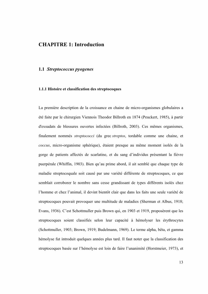
Comme ses congénères du genre Streptococcus, l’espèce S. pyogenes se présente sous la
forme de chaînes de cellules (Figure 1). In vivo néanmoins, elle est souvent trouvée sous
forme de diplococci. Il semble en effet que la longueur de la chaîne soit inversement
proportionnelle à la disponibilité en nutriments dans le milieu (Stollerman et Ekstedt,
1957). Après division d’un coccus individuel, si la paire résultante ou diplococcus ne se
16
sépare pas de ses voisines, il y a allongement subséquent de la chaîne. La préservation
de la paroi intercoquale et donc la non-séparation du diplococcus du reste de la chaîne
est promue par des conditions défavorables à la croissance (antibiotiques, manque de
nutriments, froid par exemple), de même que par la présence d’anticorps contre les
antigènes de la paroi de la bactérie (Iacono, 1985; Stollerman et Ekstedt, 1957;
Lominski, 1968).
Les streptocoques sont des bactéries Gram-positive, du fait de la couleur mauve qu'elles
adoptent après avoir été colorées à la coloration de Gram, coloration mettant en
évidence des caractéristiques de la paroi cellulaire. D'après le critère d'hémolyse,
l'espèce Streptococcus pyogenes est donc une bactérie bêta-hémolytique (Figure 1), c'est
à dire qu'elle provoque la complète destruction des érythrocytes lorsque cultivée sur de
l'agar contenant du sang (Brooks, 2007) (Figure 1). De plus, selon la classification de
Lancefield, S. pyogenes fait partie du groupe A des streptocoques (Evans, 1936). Enfin,
c'est une bactérie non motile, c'est-à-dire qu’elle ne possède pas de flagelles moteurs, et
elle ne forme pas de spores.

17
A B
Figure 1 : A. Bêta-hémolyse sur agar contenant du sang
B. S. pyogenes en division sous le microscope, grossissement x 900 : Les
chaînes de cellules sont caractéristiques du genre Streptococcus et lui ont donné son
nom.
Images tirées de la Public Health Image Library, sous l'égide du Center for Disease Control and
Prevention du United States Department of Health and Human Services (domaine public).

18
1.1.3 Constituants cellulaires, produits extra-cellulaires et virulence associée
Les constituants cellulaires spécifiques des streptocoques de groupe A en général, et de
S. pyogenes en particulier, sont présents à l’extérieur de la membrane cytoplasmique de
la bactérie et comprennent :
Antigène C spécifique du groupe : La classification des streptocoques bêta-
hémolytiques en quinze groupes immunologiques A à O se fait selon la présence de
sucres (carbohydrates C) dans leur paroi cellulaire. Dans le cas du groupe A, le
rhamnose-N-acetylgalactosamine peut constituer jusqu’à 10 % de la masse nette du
micro-organisme (Fung, 1982). La spécificité immunologique du sucre réside
principalement dans la nature du saccharide terminal sur la chaîne rhamnose, le N-
acetylgalactosamine chez le groupe A (Coligan, 1975; Pritchard, 1981).
Antigène M spécifique du type : Chez les streptocoques bêta-hémolytiques, le
groupe A peut être subdivisé en plus d’une centaine de types sérologiques selon
l’antigène M protéique de leur paroi cellulaire (Bottone, 2006; Kahn, 2008). C’est l’un
des plus notables facteurs de virulence de S. pyogenes, en effet responsable de la
résistance de la bactérie à la phagocytose (Fischetti, 1989). L'absence de cette protéine
chez un sérotype de S. pyogenes se traduit par un manque de virulence (Brooks, 2007).
La capsule d’acide hyaluronique : Un autre élément participant grandement à la
virulence de la bactérie est sa capsule d’acide hyaluronique (Okamoto, 2004; Gryllos,
2008). Moins étudiée et peut-être un peu négligée du fait de son caractère évanescent
(elle disparait chez les cellules en culture artificielle, peut-être en raison de
l’accumulation dans le milieu de l’hyaluronidase exprimée par la bactérie elle-même), et

19
non immunogénique (car indifférentiable chimiquement de l’acide hyaluronique des
tissus conjonctifs de l’hôte), elle est néanmoins de première importance (Stollerman,
2008). De concert avec la protéine M, elle permet en effet à la bactérie de se jouer de la
phagocytose.
La protéine F : Cette protéine permet d’augmenter la capacité de S. pyogenes à
adhérer aux tissus hôtes, en se liant à la fibronectine des tissus conjonctifs (Hanski,
1992)
Figure 2 : Schéma des constituants cellulaires de S. pyogenes

20
Viennent ensuite les toxines et enzymes exprimées par S. pyogenes. Il semble certain
aujourd’hui (Nakamura, 2004) que la grande variété de maladies causées par les
streptocoques de groupe A soit corrélée à l’extraordinaire nombre de produits extra-
cellulaires exprimés par ces bactéries, dont voici quelques exemples.
Les toxines érythro- ou pyrogéniques : S. pyogenes exprime une variété
d’exotoxines, dont les exotoxines A, B, C, G, H, J, K, et L (Bisno, 2003). Les
exotoxines A et C par exemple, SpeA et SpeC (pour Streptococcal Pyrogenic Exotoxin
A et C), historiquement connues comme les toxines de la scarlatine, se lient au
complexe d'histocompatibilité de classe II dans la région Vβ du récepteur des cellules T.
Elles agissent ainsi comme des super-antigènes et provoquent la destruction des tissus
de l'hôte en stimulant la production incontrôlable de certaines cytokines, ce qui active
différentes cascades fibrinolytiques et amène hypotension et défaillance polyviscérale
(Oelschlaeger, 2000). Ces toxines sont de nature protéique (Dong, 2008; Fraser, 2008).
La toxine B, ou SpeB, est une protéase à cystéine, qui est en fait la protéine la plus
abondamment secrétée par S. pyogenes. Elle a une activité endopeptidase et est capable
de s’attaquer à la matrice extra-cellulaire de l’hôte, à ses immunoglobulines et aux
composants de son complément immunitaire, en plus d’être à même d’indirectement
stimuler la production de peptides au rôle biologique important, comme l’interleukine-1
ou encore l’histamine (Chiang-Ni, 2008; Bisno, 2003).
Les streptolysines S et O : Ces deux hémolysines sont responsables de la β-
hémolyse caractéristique des streptocoques du groupe A. La streptolysine O doit son
nom au fait qu’elle soit inactivée par la présence d’oxygène, alors que ce facteur n’a
aucun effet sur la streptolysine S. On peut également ajouter que la streptolysine S, bien
21
que présentée ici comme une exotoxine, reste en fait largement attachée à la paroi
cellulaire de S. pyogenes (Fontaine, 2003; Davis, 2006). La streptolysine O est, elle,
strictement extra-cellulaire. Toutes deux peuvent, outre celle des érythrocytes, abîmer
les parois d’autres cellules hôtes, comme les leucocytes et les macrophages (Goldman,
2009).
La streptokinase : Il en existe deux isozymes (A et B) au sein du groupe A des
streptocoques. Il semble que cette enzyme digestive soit capable de s’attaquer aux
barrières de fibrine érigées par l’hôte autour des lésions streptocoquales (Nelson, 2004),
permettant la propagation extrêmement rapide de la bactérie (Chhatwal, 2005). Il est
intéressant de noter que cette enzyme est l’un des facteurs les plus importants de la
spécificité des infections streptocoquales pour l’homme : l’interaction de la
streptokinase de la bactérie avec le plasminogène humain est en effet cruciale dans le
cadre d’une pathogénèse streptocoquale (Sun, 2004).
La hyaluronidase : Cette autre enzyme digestive exprimée par S. pyogenes a un
effet lytique sur les tissus conjonctifs de l’hôte, et lui permet donc également une
propagation rapide (Hynes, 2000). Il est curieux de remarquer, comme cela a été déjà
mentionné, que cette enzyme peut dans certains cas s’attaquer à la capsule d’acide
hyaluronique de la bactérie elle-même, mais que chez certaines souches de la bactérie,
l’enzyme exprimée ne soit pas assez active pour lui attribuer ce rôle. L’hypothèse de
l’utilisation de l’acide hyaluronique (de l’hôte ou de la bactérie), comme source
d’énergie alternative pour la bactérie a d’ailleurs récemment été formulée (Starr 2006).

22
S. pyogenes exprime et excrète de nombreuses autres protéines, comme des
protéines de choc thermique, une streptodornase, une ADN dépolymérase, une ADNase,
ou encore une α-amylase (Nakamura, 2003)
Enfin, la simple sensibilité de la bactérie vis-à-vis de son environnement et de la
disponibilité en nutriments constitue un mécanisme grâce auquel S. pyogenes peut
progresser de la simple colonisation à la pathogénie caractérisée (Fung, 1992; Rosch,
2007; Shelburn, 2008; Loughman et Caparon, 2006).

23
1.1.3 Pathogénèse
Si S. pyogenes fait l'objet d'une attention toute particulière, c'est qu'elle est une bactérie
extraordinairement versatile responsable d'une myriade de maladies, des plus bénignes
aux plus dangereuses (Quinn, 1950; Cunningham, 2000). Bien que la bactérie soit
présente sans conséquence chez jusqu’à 15 % de la population, ou porteurs sains, (Shet
2004), elle ne provoque des dommages sévères que si elle est capable de disséminer de
son point d’infection initial et souvent superficiel jusqu’à des tissus plus profonds, via la
circulation sanguine (Medina, 2003). Parmi les maladies purulentes, elle cause
directement pharyngites et pneumonies par infection du conduit respiratoire, de même
que l'impétigo par infection de la peau (Cvjetkovic, 2008; Shelburne, 2008; Papadas,
2008; Hedrick, 2003). Plus drastique et extrêmement rapide, le syndrome du choc
toxique cause insuffisance respiratoire et défaillance polyviscérale, et entraine la mort
chez 30 % des patients (Luca-Harari, 2009; Thomas, 2008). C'est la production des
exotoxines A ou C qui est associée à ce type d'infection létale. Ces protéines, excrétées
par S. pyogenes se lient au récepteur des cellules T et agissent comme des super-
antigènes (Fraser, 2008). Enfin, et c'est évidemment la pathologie qui frappe le plus les
esprits, S. pyogenes conjointement avec les autres membres du groupe A des
streptocoques, est également responsable de la fascitiis necroticans. Cette gangrène
streptococcocale est une nécrose extrêmement rapide des tissus sous-cutanés, et a
récemment valu à S. pyogenes l'appellation de « bactérie mangeuse de chair » (Weidle,
2009; Bingol-Kologlu, 2007; Gonzalez Castro, 2008). La pathogénèse de ces maladies
suppuratives par S. pyogenes dépend en grande partie de sa faculté de propagation, et

24
donc repose sur sa hyaluronidase et sa streptokinase, facteurs qui la promeuvent. De
même sa capacité à éviter la phagocytose grâce à sa capsule d’acide hyaluronique et à sa
protéine M est cruciale (Olsen, 2009; Stollerman, 2008)
Enfin, il faut mentionner les maladies non purulentes post-streptocoquales, qui se
manifestent donc après infection par S. pyogenes et en sont les séquelles. Ainsi, la fièvre
rhumatismale endommage le muscle et les valves cardiaques chez les individus ayant
préalablement développé une infection streptocoquale (Cunningham, 2008). Dans le cas
de la glomérulonéphrite (Skattum, 2006), la réaction auto-immune observée pourrait
être due aux exotoxines A et C, ou à la protéine M (Burova, 2003)
25
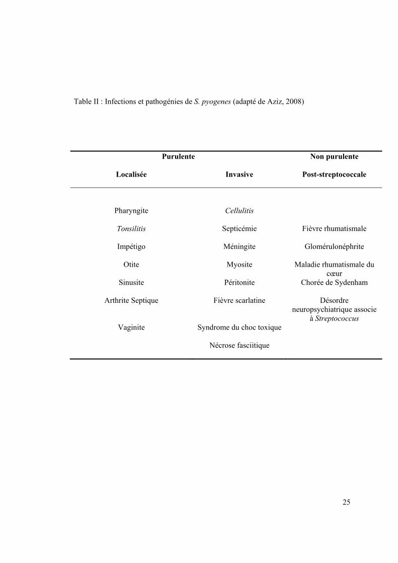
Table II : Infections et pathogénies de S. pyogenes (adapté de Aziz, 2008)
Purulente Non purulente
Localisée Invasive Post-streptococcale
Pharyngite Cellulitis
Tonsilitis Septicémie Fièvre rhumatismale
Impétigo Méningite Glomérulonéphrite
Otite Myosite Maladie rhumatismale du cœur
Sinusite Péritonite Chorée de Sydenham
Arthrite Septique Fièvre scarlatine Désordre neuropsychiatrique associe
à Streptococcus Vaginite Syndrome du choc toxique
Nécrose fasciitique
26
1.1.4 Métabolisme de Streptococcus pyogenes
Bien que S. pyogenes produise la superoxide dismutase (SOD), essentielle à la survie
des streptocoques en milieu aérobique, elle est catalase-négative et cytochrome
oxydase-négative, c'est à dire qu'elle n'exprime pas les autres enzymes habituellement
liées à un métabolisme oxydatif (Dworkin, 2006). Malgré le fait qu'elle présente des
enzymes alternatives qui lui permettent de résister au stress oxydatif (Gibson, 2000),
son métabolisme est homofermentatif et non-respiratoire. La fermentation est définie
comme le métabolisme au cours duquel des composés organiques servent à la fois de
donneurs et d’accepteurs d’électrons (Davis, 1973). La fermentation lactique, celle des
bactéries de l’ordre des Lactobacillales, est la plus simple des fermentations. Le
pyruvate y est réduit en acide lactique, et chaque hexose produit deux molécules d’ATP.
Bien que les streptocoques soient organotrophes et aérotolérants anaérobiques, ou
anaérobiques facultatifs et que dans certaines conditions (anaérobique ou alcaline en
particulier), la totalité du pyruvate résultat du catabolisme du glucose ne soit pas
transformé en acide lactique (Pearce, 1957) et qu’une portion non négligeable se trouve
oxydée en acide acétique selon la réaction :
CH3COCOOH + ½ O2 CH3COOH + CO2
l'énergie est essentiellement tirée du métabolisme du glucose avec l'acide lactique
comme produit final de la voie (Brooks, 2007) (Figure 3).

27
Figure 3 : Métabolisme du glucose de Streptococcus pyogenes

28
L’accumulation d’acide lactique dans le milieu peut limiter la croissance de la bactérie,
jusqu’à ce que le pH soit ajusté en conséquence.
Des hexoses autres que le glucose, comme le mannose, le galactose et le fructose sont
également utilisés par S. pyogenes (Salminen, 2004). Ces sucres s’insèrent dans la
glycolyse au niveau du glucose-6-phosphate ou du fructose-6-phosphate après
isomérisation ou phosphorylation. Le lactose est hydrolysé en ses moitiés glucose, ce
dernier étant dirigé vers la glycolyse, et galactose. Le galactose néanmoins peut
connaitre deux sorts différents : la voie de Leloir, qui voit le galactose transformé en
galactose-1-phosphate, en glucose-1-phosphate puis en glucose-6-phosphate qui peut
ensuite rejoindre la glycolyse (Maxwell, 1962). Il peut également être métabolisé grâce
à la voie du tagatose-6-phosphate après avoir été phosphorylé en galactose-6-phosphate
(Steele, 1954) (Figure 4).
Le groupe de gènes des enzymes de la voie du tagatose-6-phosphate, lacABCD, codant
pour la galactose-6-phosphate isomérase, la tagatose-6-phosphate kinase, et la tagatose-
1,6-bisphosphate aldoldase, sont en fait les premiers gènes de l’opéron lactose lacR-
lacABCDFEGX (van Rooijen, 1991) de cette bactérie à acide lactique.
C'est justement au sein de cette voie que se retrouve l'enzyme au cœur de ce projet, la
tagatose-1,6-bisphosphate aldolase, qui catalyse le clivage réversible du tagatose-1,6-
bisphosphate en DHAP et G3P.

29
Figure 4 : Voie du tagatose-6-phosphate
30
1.2 Les aldolases
1.2.1. Généralités et classification :
C’est dans les années 1930 que l’activité enzymatique de clivage de certains hexoses
biphosphorylés fut identifiée pour la première fois (Meyerhorf, 1943; Meyerhorf, 1947;
Kresge, 2005), et l’enzyme responsable isolée et nommée. La réaction aldolasique, soit
l’addition d’une cétone-énolate sur un accepteur aldéhyque, effectivement crée un lien
carbone-carbone. Cette réaction est réversible et résulte en une multitude de différents
produits, en général selon la nature de l’accepteur aldéhyque. Il existe donc de
nombreuses aldolases, baptisées selon l’identité du produit de la condensation.
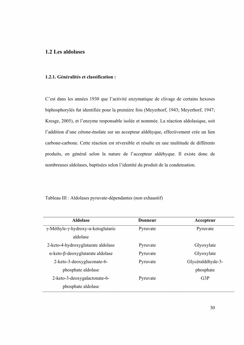
Tableau III : Aldolases pyruvate-dépendantes (non exhaustif)
Aldolase Donneur Accepteur
γ-Méthyle-γ-hydroxy-α-ketoglutaric
aldolase
Pyruvate Pyruvate
2-keto-4-hydroxyglutarate aldolase Pyruvate Glyoxylate
α-keto-β-deoxyglutarate aldolase Pyruvate Glyoxylate
2-keto-3-deoxygluconate-6-
phosphate aldolase
Pyruvate Glycéraldéhyde-3-
phosphate
2-keto-3-deoxygalactonate-6-
phosphate aldolase
Pyruvate G3P

31
Tableau IV : Aldolases DHAP-dépendantes (non exhaustif)
Aldolase Donneur Accepteur
Fructose-1,6-biphosphate
aldolase
Dihydroxyacétone
phosphate
G3P
Tagatose-1,6-biphosphate
aldolase
DHAP G3P
Fuculose-1-phosphate
aldolase
DHAP Lactaldéhyde
Rhamnulose-1-phosphate
aldolase
DHAP Lactaldéhyde
Ce n’est pourtant pas la nature du produit de la catalyse qui permet de classifier les
aldolases. Leur séparation en deux classes se fait en effet selon leur mécanisme
catalytique (Rutter, 1964). Les aldolases de classe I forment un intermédiaire
réactionnel covalent ou base de Schiff avec le donneur de la réaction. Cet intermédiaire
se crée entre le groupe carboxyle de la fonction cétone du donneur et l’atome d’azote de
la lysine catalytique après attaque nucléophile du dernier sur le premier (Blonski, 1997;
Thomson, 1998). Chez les aldolases de classe II, il y a polarisation du donneur grâce à
un cation divalent, Ca2+ ou Zn2+, qui aide également à stabiliser l’intermédiaire
réactionnel (Hall, 2002). Bien qu’initialement, il ait semblé que les aldolases de classe I
soient retrouvées essentiellement chez les organismes dits supérieurs, les eucaryotes, et
les aldolases de classe II chez les procaryotes, l’étanchéité de cette distribution est
32
fortement remise en question aujourd’hui (Kroth, 2005; Lorentzen, 2005), notamment
avec l’avènement d’une sous-classe d’aldolases qui consiste en une troisième famille de
séquence primaire, la classe I des Archae (Lorentzen, 2004). Il est intéressant de noter
que les deux classes d’enzymes, en plus de catalyser la même réaction, partagent en
outre le même type de repliement, un tonneau (α⁄β)8 de type triose-phosphate isomérase.
Cette caractéristique conforte l’hypothèse d’une évolution divergente des deux classes
(Ramsaywak, 2004).

33
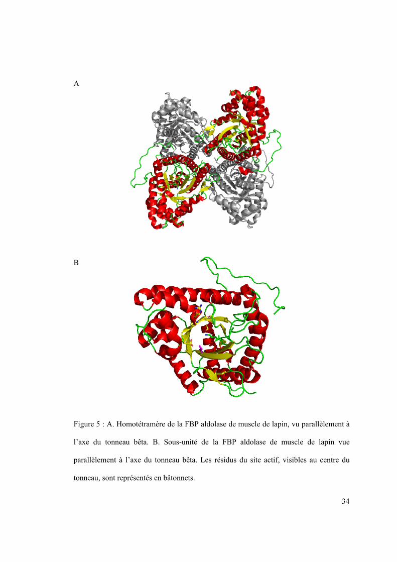
1.2.2 Structure tridimensionnelle :
Les aldolases, environ 40 kilodaltons par sous-unité, se présentent généralement sous
forme d’homotétramères d’environ 160 kDa (Lorentzen, 2005) pour les enzymes de
classe I (Figure 5 A.), et d’homodimères d’environ 80 kDa pour celles de classe II
(Galkin, 2009). Les deux classes d’aldolases présentent un repliement de type TIM. Le
tonneau bêta, au centre de l’enzyme, est formé par les huit brins bêta parallèles séparés
par les hélices alpha en enjambement à droite. C’est au cœur de ce tonneau que l’on
trouve le site actif de l’enzyme (Figure 5 B.) (Figure et suivantes réalisées grâce au
programme PyMOL (DeLano, 2002).
34
A
B
Figure 5 : A. Homotétramère de la FBP aldolase de muscle de lapin, vu parallèlement à
l’axe du tonneau bêta. B. Sous-unité de la FBP aldolase de muscle de lapin vue
parallèlement à l’axe du tonneau bêta. Les résidus du site actif, visibles au centre du
tonneau, sont représentés en bâtonnets.

35
1.2.3 Site actif et mécanisme L'aldolase de muscle de lapin, FBP aldolase de classe I, est depuis toujours le modèle
de prédilection pour l'étude de cette classe d'enzyme. De multiples structures
tridimensionnelles ont été déposées dans la PDB (codes 1ADO, 1ZAH, 1EWD, ou
6ALD), et son mécanisme catalytique est depuis peu complètement élucidé (St-Jean,
2005; St-Jean, 2007).
Dans le sens de la condensation du DHAP et G3P en FBP, la catalyse débute par la
liaison covalente du DHAP au site actif via la Lys229. Lys229 attaque nucléophiliquement
le C2 du DHAP, et Glu187
catalyse la formation de l'intermédiaire iminium, ce qui
entraîne un rétrécissement conformationnel du site actif. La chaîne latérale de Arg303 par
exemple, est considérablement déplacée afin de lier électrostatiquement l'oxyanion du
phosphate P1. Après avoir été activé par un transfert de proton séquentiel via une
molécule d'eau, et stabilisé par Lys146
, Tyr363
peut ensuite abstraire
stéréospécifiquement le proton pro-S du C3 de l'intermédiaire substrat-enzyme,
produisant l'énamine. Glu187
est ensuite protonné par un transfert de proton séquentiel,
et peut à son tour activer le G3P arrivant en le protonnant en C4. Il y a ensuite attaque
nucléophile du C3 de l'intermédiaire DHAP-enzyme sur le carbanion en C4 du G3P, ce
qui génère un nouveau lien C-C. L'étape finale de ce mécanisme catalytique est
évidemment la relâche du FBP (Figure 6) (St-Jean, 2007).

Figure 6 :
avec son s
Mécanism
site actif en
me simplifié
encadré (a
de la cataly
adapté de St
yse par la F
-Jean, 2007
FBP aldolas
7)
se du musc
36
le de lapin,
6
,

37
Le phosphate P1 du DHAP est impliqué dans de nombreuses interactions stabilisatrices
avec des résidus du site actif : outre Arg303, Ser271, Gly272 et Gly302 lient directement
l'oxyanion phosphate. Enfin, l'hydroxyle C3 du DHAP et de FBP interagit avec Asp33
et Lys146 (St-Jean, 2007)
La liaison des substrats au site actif de la FBP aldolase de muscle provoque de plus
d'importants changements conformationnels (St-Jean, 2005). Outre l'Arg303 qui, comme
vu précédemment, interagit directement avec l'oxyanion du phosphate P1 du DHAP, et
qui, de la structure native à celle ligandée (DHAP ou FBP), présente un r.m.s.d.
d'environ 0.7 Å, les régions comprises entre les résidus 33 à 65, et 302 à 329 montrent
des r.m.s.d. pouvant aller jusqu'à 1.02 et 1.25 Å, respectivement.
38
1.2.4. Les aldolases chez les streptocoques : la tagatose-1,6-bisphosphate aldolase
de S. pyogenes
S. pyogenes et ses congénères streptocoques expriment plusieurs aldolases : la 4-
hydroxy-2-oxoglutarate aldolase (EC 4.1.2.14), la 2-déhydro-3-déoxyphosphogluconate
aldolase (EC 4.1.3.16), la déoxyribose-phosphate aldolase (EC 4.1.2.4), la
dihydronéopterine aldolase (EC 4.1.2.25), la fructose-1,6-bisphosphate aldolase (EC
4.1.2.13), la phospho-2-déhydro-3-déoxyheptonate aldolase (EC 2.5.1.54), la tagatose
1,6-bisphosphate aldolase 1 (EC 4.1.2.40), et la tagatose 1,6-bisphosphate aldolase 2
(EC 4.1.2.40). Certaines autres lyases, telles la 3-déhydroquinate déhydratase (EC
4.2.1.10) ou encore la nicotinate-nucléotide pyrophosphorylase (EC 2.4.2.19), du fait de
la nature de la réaction qu'elles catalysent, peuvent ou non également rentrer dans cette
large catégorie d'enzyme. S. pyogenes étant une bactérie à acide lactique, c'est
néanmoins la fructose-1,6-bisphosphate aldolase et la tagatose 1,6-bisphosphate
aldolase qui ont le plus d'importance dans le cadre d'un métabolisme fermentatif.
L'aldolase au cœur de ce projet est la tagatose 1,6-bisphosphate aldolase 2, produit du
gène lacD.2. C'est une protéine de 37 kDa qui se présente sous la forme d'un
homodimère de 74 kDa. Comme mentionné dans la section 1.1.5, la TBP aldolase de S.
pyogenes se trouve à la jonction de la voie du tagatose et de la glycolyse où elle catalyse
le clivage réversible du tagatose-1,6-bisphosphate provenant du galactose en DHAP et
G3P. Néanmoins, sa particularité réside dans son singulier manque de stéréospécificité.
En effet, la TBP aldolase de S. pyogenes, au-delà du tagatose-1,6-bisphosphate, est
capable de cliver réversiblement le fructose-1,6-bisphosphate, le sorbose-1,6-
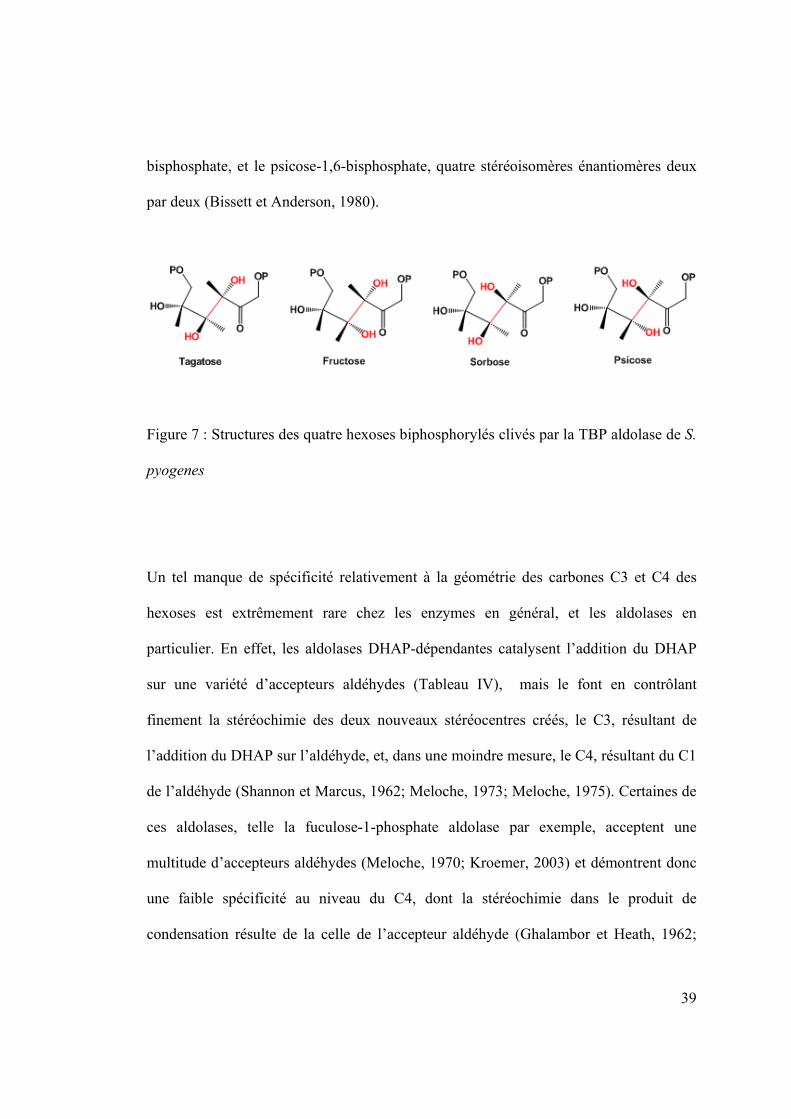
39
bisphosphate, et le psicose-1,6-bisphosphate, quatre stéréoisomères énantiomères deux
par deux (Bissett et Anderson, 1980).
Figure 7 : Structures des quatre hexoses biphosphorylés clivés par la TBP aldolase de S.
pyogenes
Un tel manque de spécificité relativement à la géométrie des carbones C3 et C4 des
hexoses est extrêmement rare chez les enzymes en général, et les aldolases en
particulier. En effet, les aldolases DHAP-dépendantes catalysent l’addition du DHAP
sur une variété d’accepteurs aldéhydes (Tableau IV), mais le font en contrôlant
finement la stéréochimie des deux nouveaux stéréocentres créés, le C3, résultant de
l’addition du DHAP sur l’aldéhyde, et, dans une moindre mesure, le C4, résultant du C1
de l’aldéhyde (Shannon et Marcus, 1962; Meloche, 1973; Meloche, 1975). Certaines de
ces aldolases, telle la fuculose-1-phosphate aldolase par exemple, acceptent une
multitude d’accepteurs aldéhydes (Meloche, 1970; Kroemer, 2003) et démontrent donc
une faible spécificité au niveau du C4, dont la stéréochimie dans le produit de
condensation résulte de la celle de l’accepteur aldéhyde (Ghalambor et Heath, 1962;

40
Espelt, 2005). Toutefois, la stéréospécificité relativement au C3 est, elle, strictement
conservée (Takayama, 1997; Fessner, 1998). Aucune structure tridimensionnelle de
TBP aldolase de classe I n’ayant été déposée à ce jour, les informations structurales
concernant cette enzyme et pouvant aider à comprendre son manque extraordinaire de
stéréospécificité sont donc manquantes.

41
1.2.5. LacD.1 : un régulateur de l’expression génétique ?
Pour terminer, au-delà de son activité purement métabolique, la TBP aldolase de S.
pyogenes pourrait également démontrer un rôle de senseur et par extension, de
régulateur de virulence chez la bactérie (Loughman et Caparon, 2006). Il est déjà connu
que des enzymes glycolytiques, comme l’hexokinase, la lactate déshydrogénase ou
encore la glycéraldéhyde-3-phosphate déshydrogénase, ont été adaptées afin de jouer un
rôle de régulation de l’expression génétique chez certains organismes (Kim et Dang,
2005). L’autre isozyme de la TBP aldolase chez S. pyogenes, la TBP aldolase 1, ou
LacD.1, avec laquelle elle partage 72.6 % d’identité de séquence, serait pour sa part
capable de lier directement RopB, un facteur impliqué dans la régulation du
métabolisme du streptocoque, et dans l’activation de l’expression de certains gènes
impliqués dans sa virulence, dont celui codant pour SpeB (Loughman et Caparon, 2006;
Loughman et Caparon, 2007). Le gène codant pour cette protéase à cystéine est l’un des
plus hautement exprimé in vivo lors d’une infection par S. pyogenes, et répond à
différents signaux environnementaux. Or, le mutant LacD.1- n’est plus sensible à ces
signaux, et présente une expression aberrante de SpeB, phénotype complètement corrigé
par l’introduction d’un plasmide codant pour LacD.1. Dans le modèle proposé, la
liaison de LacD.1 à ses substrats, DHAP et G3P, en conditions d’excès d’énergie via la
glycolyse, y induirait un changement conformationnel qui lui permettrait de séquestrer
RopB, inhibant ainsi l’activation des gènes cibles de RopB. Ce rôle de senseur
métabolique et de régulateur de virulence serait donc subordonné aux différents
changements conformationnels adoptés par cette aldolase après liaison du substrat. La

42
résolution de la structure native et ligandée de cette TBP aldolase de classe I pourrait
donc permettre d’identifier les régions les plus mobile et les plus susceptibles d’être
liées à cette facette de la TBP aldolase de S. pyogenes.

43
1.3 Objectif du projet de recherche
La tagatose-1,6-bisphosphate aldolase, du fait de son manque singulier de
stéréospécificité, est une exception dans le monde des enzymes. De plus, son rôle
putatif de régulateur de l’expression génétique de S. pyogenes pourrait avoir une part
dans la virulence de cette bactérie. Son appartenance à la classe I des aldolases signifie
qu'elle catalyse le clivage réversible de quatre hexoses biphosphorylés via la formation
d'une base de Schiff. Bien qu’une structure en trois dimensions d'une tagatose-1,6-
bisphosphate aldolase ait déjà été déposée dans la PDB, il s'agissait d'une classe II dont
le mécanisme est donc par essence différent de celui de la TBP aldolase de S. pyogenes.
Les informations structurales concernant les résidus du site actif impliqués dans la
catalyse ou les régions mobiles peut-être liées au rôle de senseur métabolique étant très
fragmentaires, nous avons décidé de tenter de remédier à cette situation en initiant une
étude structurale de l'enzyme. L'objectif principal de ladite étude était donc de déposer
la première structure d'une tagatose-1,6-bisphosphate aldolase de classe I, native tout
d'abord, et en complexe avec l'un de ses substrats ensuite. La résolution de ces deux
structures permettant de se représenter un peu mieux la topologie du site actif et
l'identité des résidus catalytique, des études de cinétique enzymologique ont ensuite été
entreprises afin de mieux cerner les modalités de la catalyse. Une cinétique suivant
l'oxydation de l'intermédiaire enzyme-substrat suivies par réduction de l'indicateur
hexacyanoferrate III a ainsi été réalisée. Enfin, le stéréoisomérisme du transfert de

44
proton a été étudié grâce à une cinétique de marquage isotopique et l'utilisation d'eau
tritiée et de DHAP marqué.
1.4 Méthodes
1.4.1 Purification et cristallisation de la TBP aldolase de S. pyogenes
La TBP aldolase recombinante a été exprimée dans Escherichia coli. La souche JM-109
a été transformée avec un plasmide pKK222 codant pour la protéine d'intérêt. La
surexpression est induite grâce à l'adjonction d'IPTG dans le milieu de culture. La TBP
aldolase est ensuite purifiée grâce à un système en trois étapes (Liotard, 2003). Après la
lyse des bactéries sur-exprimant la protéine, le culot est passé sur un échangeur
anionique de type DEAE. Les fractions sont testées pour l'activité aldolasique grâce à
un test couplé (Racker, 1947), et les fractions actives sont rassemblées et passées sur
une colonne hydrophobe Phényl-sépharose. Après une précipitation la nuit durant dans
une solution saturante de sulfate d'ammonium, les fractions actives sont ensuite
soumises à une filtration sur gel. Le résultat est environ 30 milligrammes par litre de
culture bactérienne de TBP aldolase parfaitement pure.
La cristallisation de la protéine est réalisée grâce à la méthode de la goutte pendante. La
solution de 5 milligramme par millilitre de protéine est mélangée à proportion égale
avec la solution précipitante. La goutte ainsi obtenue est suspendue dans le puit

45
hermétique d'une plaque de cristallisation dont le fond contient environ 1 millilitre de la
solution précipitante. Après équilibration du système via l'évaporation de l'eau contenue
dans la goutte, la supersaturation est atteinte. En effet, l’évaporation progressive de
l’eau de la goutte permet d’y augmenter de manière tout aussi progressive la
concentration en protéine et en agents précipitants (Figure 8). Il y a alors nucléation de
la protéine, ce qui a pour effet d’en faire diminuer la concentration en solution dans la
goutte. Les cristaux de TBP aldolase commencent de croitre.
Figure 8: Diagramme de phase selon la concentration en protéine et en agent
précipitant. Le chemin emprunté par le système lors d’une expérience de goutte
pendante, par évaporation de l'eau contenue dans la goutte et équilibration avec le
réservoir contenant la solution précipitante, est représenté par une flèche.

46
Les cristaux de TBP aldolase adaptés à la diffraction des rayons X apparaissent selon ce
protocole en moins d’une heure sous forme de plaques minces.
Figure 9 : Cristaux de TBP aldolase après 48 heures de croissance

47
1.4.2 Diffraction des rayons X
Après trois jours environ, les cristaux ainsi obtenus sont suffisamment gros et surtout
épais pour l'expérience de diffraction des rayons X. Les cristaux choisis pour la
diffraction sont trempés dans une solution à hauteur de 20 % de cryoprotectant, du
glycérol en l’occurrence, et dans une solution saturante de ligand le cas échéant. Dans le
cas de la structure de la TBP aldolase complexée au DHAP, une solution de 5 mM de
DHAP a ainsi été utilisée. Le cristal est ensuite congelé par immersion immédiate dans
de l'azote liquide, et soumis au flux de rayons X. La congélation du cristal permet de
minimiser les dommages causés par le faisceau de photons de haute énergie, et la
cryoprotection permet elle d'éviter la formation de glace dommageable à l'analyse des
résultats. Le patron de diffraction ainsi obtenu (Figure 10) est ensuite analysé grâce à
des programmes informatiques (Otwinowski, 1997), réduit et utilisé pour résoudre la
structure en trois dimensions de la protéine. La structure native de la TBP aldolase de S.
pyogenes a été résolue grâce à un modèle préalablement construit par Mutliple-
wavelenght Anomalous Dispersion phasing. Ce modèle initial a servi de départ à un
remplacement moléculaire dont le résultat, après de multiples étapes d'affinement, est la
structure native de la TBP aldolase (code pdb). Le trempage de cristaux de la TBP
aldolase dans une solution saturante de DHAP a de même permis de piéger un
intermédiaire réactionnel enzyme-substrat, soit la structure de la TBP aldolase liée au
DHAP (code pdb).

48
Figure 10 : Patron de diffraction de la TBP aldolase de S. pyogenes, collecté au faisceau
X29 du Brookhaven National Laboratory, NY.

49
1.4.3 Hexacyanoferrate III
Afin de vérifier la présence d'un intermédiaire covalent de nature carbanionique lorsque
la TBP aldolase est liée au DHAP, la réduction de l'indicateur redox hexacyanoferrate
III a été suivie spectrophotomètriquement à 420 nm (Healy et Christen, 1973). La
réaction, gardée à 25 °C et initiée par l'adjonction de TBP aldolase, nécessite la
présence à la fois de l'enzyme et du DHAP, et a permis d'évaluer le KM et Vmax de la
liaison du DHAP à la TBP aldolase. Il faut noter que la réduction de l'hexacyanoferrate
a également été observée lorsque le FBP remplace le DHAP.
1.4.4 Marquage isotopique
Afin de caractériser l'échange 3H/1H au C3 du DHAP catalyse par TBP aldolase, et d'en
mesurer la vitesse, des expériences de marquages ont été entreprises. Le protocole
(Rose et Rieder, 1958; Rose, 1958) permet de suivre le marquage du DHAP sous forme
de (S)-[3-3H1]DHAP or (R)-[3-3H1]DHAP par la TBP aldolase a partir d'eau tritiée et de
DHAP. La position du H3, pro-(R) ou pro-(S), est déterminée grâce a deux enzymes
capables de déprotoner de manière stéréospécifique le C3 du DHAP : en pro-R pour la
TIM et en pro-S pour la RMA. De même, la formation d'eau tritiée à partir de (S)-[3-
3H1]DHAP and (R)-[3-3H1]DHAP a été suivie afin de mesurer le démarquage du DHAP
catalysé par la TBP aldolase.
50
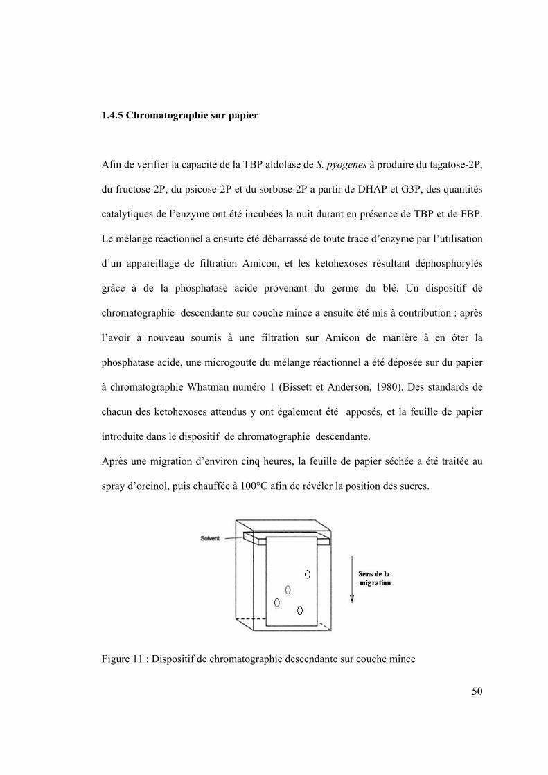
1.4.5 Chromatographie sur papier
Afin de vérifier la capacité de la TBP aldolase de S. pyogenes à produire du tagatose-2P,
du fructose-2P, du psicose-2P et du sorbose-2P a partir de DHAP et G3P, des quantités
catalytiques de l’enzyme ont été incubées la nuit durant en présence de TBP et de FBP.
Le mélange réactionnel a ensuite été débarrassé de toute trace d’enzyme par l’utilisation
d’un appareillage de filtration Amicon, et les ketohexoses résultant déphosphorylés
grâce à de la phosphatase acide provenant du germe du blé. Un dispositif de
chromatographie descendante sur couche mince a ensuite été mis à contribution : après
l’avoir à nouveau soumis à une filtration sur Amicon de manière à en ôter la
phosphatase acide, une microgoutte du mélange réactionnel a été déposée sur du papier
à chromatographie Whatman numéro 1 (Bissett et Anderson, 1980). Des standards de
chacun des ketohexoses attendus y ont également été apposés, et la feuille de papier
introduite dans le dispositif de chromatographie descendante.
Après une migration d’environ cinq heures, la feuille de papier séchée a été traitée au
spray d’orcinol, puis chauffée à 100°C afin de révéler la position des sucres.
Figure 11 : Dispositif de chromatographie descendante sur couche mince

51
CHAPITRE 2 : Article
STRUCTURE OF A CLASS I
TAGATOSE-1,6-BIPHOSPHATE ALDOLASE :
STUDY INTO AN APPARENT LOSS OF
STEREOSPECIFICITY
Clotilde LowKam, Brigitte Liotard, and Jurgen Sygusch1
From the Department of Biochemistry, Université de Montréal,
Montréal H3C 3J7, Québec, Canada
1Correspondance to: Jurgen Sygusch, CP 6128, Station Centre Ville, Montréal
H3C 3J7, Quebec, Canada. Tel.: 514-343-2389; Fax: 514-343-6463

52
Abstract
Tagatose-1,6-biphosphate (TBP) aldolase from Streptococcus pyogenes is a class
I aldolase that exhibits a remarkable lack of chiral discrimination with respect to
the configuration of hydroxyl groups at both C3 and C4 positions. The enzyme
catalyzes the reversible cleavage of four diastereoisomers: fructose-1,6-
bisphosphate (FBP), psicose-1,6-bis-phosphate, sorbose-1,6-bisphosphate and
tagatose-1,6-bisphosphate to dihydroxyacetone-P and D-glyceraldehyde 3-P with
high catalytic efficiency. To investigate its enzymatic mechanism, high resolution
crystal structures were determined of both native enzyme and native enzyme in
complex with dihydroxyacetone-P. The electron density map revealed a (α/β)8 fold
in each dimeric subunit. Flash cooled crystals of native enzyme soaked with
dihydroxyacetone-P trapped a covalent intermediate with carbanionic character at
Lys205, different from the enamine mesomer bound in stereospecific class I FBP
aldolase. Structural analysis indicates extensive active site conservation with
respect to class I FBP aldolases including conserved conformational responses to
DHAP binding and conserved stereospecific proton transfer at the DHAP C3
carbon mediated by a proximal water molecule. Exchange reactions with tritiated
water and tritium labelled DHAP at C3 hydrogen were carried out in both solution
and crystalline state to assess stereochemical control at C3. The kinetic studies
show labelling at both pro-R and pro-S C3 positions of DHAP yet detriation only
at the C3 pro-S labelled position. Detritiation of the C3 pro-R label was not
detected and is consistent with preferential cis-trans isomerism about the C2-C3

53
bond in the carbanion as the mechanism responsible for C3 epimerization in TBP
aldolase.
Introduction
Aldolases are crucial enzymes in living organisms because of their role in essential
metabolic pathways such as gluconeogenesis and glycolysis. Their ability to control the
stereochemistry of one of the most important biochemical transformation, the carbon-
carbon bond formation, makes them models for de novo preparation of carbohydrates
(1), and ideal alternatives to traditional methods in synthetic organic chemistry (2, 3, 4).
Tagatose-1,6-bisphosphate (TBP) aldolase is an inducible enzyme which, although
demonstrating greatest affinity for D-tagatose-1,6-bisphosphate, can also use as
substrate the bisphosphorylated D-hexose stereoisomers: sorbose-P2, psicose-P2 and
fructose-P2 (5). The four sugars are diastereoisomers and differ in stereochemistry at
carbon 3 and at carbon 4 with respect to the configuration of their hydroxyl groups. The
cleavage of the four sugars produces glyceraldehyde 3-phosphate (G3P) and
dihydroxyacetone phosphate (DHAP), while the condensation of G3P and DHAP
produces a mixture of the four D-hexoses in Staphylococcus aureus (5). Fructose-1,6-
bisphosphate (FBP) aldolases, on the other hand, are constitutively expressed and
catalyze generally a highly stereospecific reaction, although exceptions have been
reported (6).
Aldolases are broadly categorized with respect to their catalytic mechanism into two
classes. Class I aldolases are characterized by formation of a covalent Schiff base

54
intermediates (7, 8), while class II aldolases are metallo-dependant enzymes and use a
divalent transition metal ion to polarize the substrate ketose (9, 10). Of all aldolases,
class I FBP aldolase from rabbit muscle has been the most extensively studied (11) and
its catalytic mechanism implicates a protonated Schiff base or iminium formed between
the lysine residue in the active site of the enzyme and a ketose substrate (12). To form
the FBP C3-C4 bond in aldol condensation, the enzyme stereospecifically abstracts the
pro-S C3 proton of the iminium intermediate formed with DHAP (13, 14, 15), thereby
generating the carbanionic character at C3 of DHAP for the aldol reaction. The nascent
carbon-carbon bond has the same orientation as the pro-S α-hydrogen initially
abstracted from the DHAP imine intermediate; the carbanion si face being subjected to
a nucleophilic attack by the si face of the incoming G3P aldehyde. The overall retention
of configuration requires that proton abstraction to yield the enamine and condensation
with the aldehyde must take place from the same direction on the enzyme (16). The
iminium intermediate formed is then hydrolyzed and FBP is released. Similarly, in L-
rhamnulose-1-phosphate aldolase, the attack by the re face of L-lactaldehyde on the re
face of DHAP yields rhamulose-1-phosphate (17,18) and also retains configuration.
Racemic aldol condensation products with respect to the configuration at C4 have
been noted in a number of class I or class II aldolases. In 2-keto-4-hydroxyglutarate
aldolase, a class I aldolase (19) and α-keto-β-deoxyhexarate aldolase, a class II enzyme
(20), racemisation occurs at C4 of their condensation product, yielding (4-RS)-2-keto-4-
hydroxyglutarate from pyruvate and glyoxalate (21). The epimerisation at the C4
hydroxyl is a result of the random orientation of the aldehyde during stereofacial attack
on the enzyme-bound pyruvate by either the si or re face of the incoming aldehyde.

55
Similarly, analysis of condensation products in a stereoselectivity study of L-fuculose-
1-phosphate aldolase, a DHAP dependent class II aldolase, showed stereofacial attack
on the bound DHAP with stereochemistry at the C4 position in products controlled by
the chemical nature of the attacking aldehyde (22). In contrast, configuration at both C3
and at C4 is not retained in the catalytic mechanism of the class I TBP aldolase from S.
aureus, given that aldol condensation yields a mixture of sorbose-P2, psicose-P2,
fructose-P2 and tagatose-P2 (5)i. Attempts have been made to switch specificity of class
II TBP aldolases; however these have been met with limited success (23, 24).
Three-dimensional structures of class I FBP aldolases have been determined from
different organisms and correspond to a subunit (α/β)8 barrel fold for aldolase tetramer
(25, 26, 27). Reaction intermediates have been characterized particularly in FBP
aldolases (28) and have provided a detailed description of the catalytic mechanism at
the molecular level, including a structural explanation for stereospecific proton
exchange at C3 (11). For TBP aldolase, only a single structure has been reported: a class
II tetrameric enzyme from Escherichia coli also displaying a (α/β)8 barrel fold in
complex with a putative enediolate transition state intermediate (29). This enzyme has
reduced substrate specificity at C4 and can cleave both TBP and FBP substrates, which
was attributed to an active site that is more open at the G3P binding side, hence
affording more conformational freedom to different aldehyde binding modes. Insight
into how class I TBP aldolase catalyze racemic aldol condensation products at the
molecular level has been hampered by an absence of structures of reaction
i This observation was verified for S. pyogenes TBP aldolase and is described in Experimental Procedures.

56
intermediates. We thus initiated high resolution crystallographic studies of class I TBP
aldolase from Streptococcus pyogenes in conjunction with isotopic labelling studies to
investigate its reaction mechanism and the structural basis for the apparent loss of
stereospecificity.
Here, we present the three-dimensional structure of a native class I TBP aldolase, as
well as its complex with DHAP using TBP aldolase crystals incubated in the presence
of DHAP. Flash cooling of a native TBP aldolase crystal soaked in a saturating DHAP
solution trapped a covalent reaction intermediate. The complex structure revealed
conformational changes upon binding whereby the active site undergoes asymmetrical
narrowing that grasps DHAP. The location of an invariant water molecule within close
contact of the DHAP C3 carbon in the trapped intermediate underscored a stereospecific
proton transfer mechanism conserved in Schiff base forming aldolases.
The extent of stereochemical control in class I TBP aldolases with respect to C3
epimerization was also examined in both solution and crystalline state. Catalytic proton
transfer was assessed by the lability of the pro-R and pro-S C3 hydrogens of DHAP to
exchange in tritiated water, and by stereospecific loss of label at either C3 hydrogen of
DHAP. The proton exchange studies in conjunction with the structural data afforded
identification of the carbanion as the dominant steady state reaction intermediate and
indicated cis-trans isomerism about the carbanion C2-C3 bond in explaining the loss of
stereospecific proton exchange at the DHAP C3 carbon in TBP aldolase. These findings
stand in contrast to the highly stereospecific proton exchange mechanism found in class
I FBP aldolases and point to rotational isomerisation in specific reaction intermediates
as an evolutionary simple solution capable of generating enantiomeric promiscuity.
57
Experimental Procedures
Purification & Crystallization: Plasmid pKK-223-3 with T7 IPTG-inducible
promoter and coding for the lacD2 gene product from Streptococcus pyogenes,
tagatose-1-6-biphosphate aldolase 2, was transformed and overexpressed using the
JM109 strain (Promega) in Escherichia coli. Recombinant TBP aldolase was purified to
homogeneity using a three steps chromatographic protocol as reported previously (30).
Briefly, cells were lysed at 4°C and the lysate was applied onto an anion-exchange
column (DEAE Sepharose Fast Flow) and eluted with a NaCl gradient at pH 7.5.
Enzymatic activity was determined by following NADH oxidation at 340 nm using a
coupled assay (31). The active fractions were dialyzed overnight against a solution of
1.5 M (NH4)2 SO4, 50 mM NaH2PO4, pH 7.0 and then applied onto a hydrophobic
affinity column (Phenyl Sepharose, Amersham Biosciences). TBP aldolase was eluted
using a continuous gradient of NaH2PO4. The active fractions were then dialyzed
overnight against a precipitating solution of 90 % saturating (NH4)2 SO4, pH 7.0, and
then applied after resolubilization onto a size exclusion column (SuperdexTM 200,
Amersham Biosciences). The purified protein was precipitated overnight using the same
90 % saturated (NH4)2 SO4, pH 7.0 solution as before, and stored at 4°C. Yields
corresponded to ~ 50 mg of purified enzyme per liter of E. coli culture.
TBP aldolase crystals were grown using the hanging drop method, from a 1:1
mixture of protein solution (initial concentration 5 mg/ml in 10 mM Tris-HCl, pH 7.0)
and precipitant buffer (9-11% polyethylene glycol 4000 in 0.2 M Calcium Acetate and
0.1 M Tris-acetic acid, pH 7.5) that was equilibrated against a reservoir of precipitant.

58
Cleavage Assay: The substrate cleavage rate was determined by measuring the
decrease in A340/min in a coupled assay. Aldolase was diluted in 50 mM Tris-HCl, pH
7.5, and added to a cuvette containing 50 mM Tris-HCI, pH 7.5, 10 mM EDTA, 0.16
mM NADH, and 10 µg/mL glycerol-3-phosphate dehydrogenase / triose-phosphate
isomerase. Assays (1 mL) were performed in triplicate at 23°C following addition of
substrate. The cleavage rate for FBP and TBP substrates was measured over a substrate
concentration range of 1 - 5000 μM, using 1 μg of wild type enzyme. TBP aldolase
concentration was determined by BCA protein assay reagent (Pierce), with bovine
serum albumin serving as a standard.
Data Collection and Processing: TBP aldolase crystals were soaked in DHAP
buffer (mother liquor plus 5 mM DHAP) for 5 minutes. Crystals were then mounted in
nylon cryoloops (Hampton Research) after brief immersion in cryoprotectant (mother
liquor plus 5 mM DHAP plus 20% glycerol) and then immediately flash cooled in a
nitrogen-gas stream at 100 K. Diffraction data were collected at beamlines X25 of the
National Synchrotron Light Source (Brookhaven National Laboratory, Upton, NY). A
native dataset was also collected at beamline X29. Data indexing and integration was
performed using HKL2000 (32).
MAD phasing and Refinement: The three-dimensional structure of TBP aldolase
from Streptococcus pyogenes was solved using MAD phasing on previously collected
data of a SeMet crystal (30). The three different wavelengths (inflexion, peak and
remote) MAD datasets allowed the program SOLVE (33) to determine the positions of
5 selenium atoms out of 6 possible in each protomer, and to estimate initial protein
phases. Solvent flattening with the program RESOLVE (34) yielded an interpretable

59
electron density map calculated at 2.6 Å resolution. Iterative cycles of chain tracing
using O (35) and model refinement using the Crystallography and NMR System (CNS)
(36) defined a trace of the polypeptide backbone that refined to Rcryst (Rfree) values of
0.246 (0.290).
Molecular replacement and Refinement: Native free TBP aldolase crystallized in the
P212121 space group with four protomers in the asymmetric unit and diffracted to 1.87 Å
resolution; the structure was obtained by molecular replacement using the initial MAD
phased model as a search probe. The structures were subjected to iterative rounds of
refinement (simulated annealing and minimization) with CNS and Phenix (37), and
model building using O and Coot (38). Water molecules
were initially added
automatically by CNS and Phenix and by manual addition in subsequent rounds. The
final model has 322 residues per subunit, 2031 water molecules and 7 atoms of calcium.
The liganded crystal structure was isomorphous with the crystal structure of the
native
aldolase and diffracted to 1.92 Å resolution. Initial phases used
for model
building of the liganded structure were obtained by molecular replacement using the
MAD phased model for the TBP aldolase as a search probe. The polypeptide trace of
the liganded structure was determined using the same model building and refinement
protocol as described for the native enzyme. DHAP was refined as a covalent
intermediate bound to Lys205 bound in all four protomers.
Structure refinement used all reflections having an I/σ(I) > 1, however electron
density maps were calculated to the resolution indicated in Table 1 to ensure at least
~80% completeness in the highest resolution shell with an I/σ(I) > 2. The PRODRG
server was used to generate ligand topology and parameter files (39). The presence of

60
ligands in the final models was confirmed by inspection of simulated annealing Fo-Fc
omit maps. Final model statistics, calculated with CNS and MolProbity are shown in
Table 1. The atomic coordinates and structure factors for native and DHAP bound TBP
aldolases have been deposited with the Protein Data Bank (PDB # xxxx, PDB # yyyy,
respectively). The final structure models have Rcryst (Rfree) values of 0.175 (0.218) and
0.161 (0.199), respectively. The corresponding positional errors in atomic coordinates
using Maximum Likelihood plots were estimated to be 0.21 Å and 0.17 Å, respectively.
Errors in hydrogen bond distances and positional differences are reported as standard
deviations and were estimated based on their value in each aldolase protomer unless
specified otherwise. Inter-subunit variability among the four protomers in the
asymmetric unit cell was analyzed by the program Polypose (40) and yielded rms
differences, based on Cα atom coordinates, that were less than the error in the atomic
coordinates for each structure. All figures were prepared using the program PyMOL
(41).
Chemical identity of the enzymatic intermediate trapped in the DHAP-TBP aldolase
structure was determined with the program O (35), using a simulated annealing omit
map. The real space statistic Rfact was calculated in O (RS_FIT function) to evaluate the
fit of Lys205 Nζ and Cε atoms, and DHAP C1, C2, C3 and O3 atoms modelled as
enamine and iminium forms to the election density. The discrimination between those
two chemical species was made using Student’s t test comparing pairwise Rfact statistics
in each subunit with bound DHAP refined as either iminium/carbanion or enamine form
(11).

61
Superpositions: Superpositions of the native and liganded structures of the TBP
aldolase from S. pyogenes were performed with the program PyMOL overlaying Cα
atom coordinates of TBP aldolase residues 100-230, which were invariant to binding
events based on difference matrix plot analysis. Difference distance matrix plots were
produced using the DDMP program from the Center for Structural Biology at Yale
University (New Haven, Connecticut, USA). R.m.s.d. of coordinates was calculated
using Cα atoms for the appropriate residues.
Superposition of the TBP aldolase structures onto the FBP rabbit muscle aldolase
structure was performed using PyMOL and Swiss-PdbViewer (42). For the active site
comparison, Cα atoms of conserved residues in TBP and FBP aldolase active sites in
proximity of the Schiff-base forming lysine residue were overlaid using PyMOL, while
the Fragment Alternate Fit function of the Swiss-PdbViewer program was used for
sequence alignment comparison of the overall fold.
A Blast search (NCBI BLAST2) was performed against the UniprotKB database
with the S. pyogenes aldolase sequence (accession number P63706) to identify
orthologues. Orthologue sequences were submitted to SWISS-MODEL workspace for
three dimensional model building (43).
Detection of ketohexoses: To verify the ability of S. pyogenes TBP aldolase to form
the hexose 1,6-bisphosphates: tagatose-1,6-bisphophate, fructose-1,6-bisphophate,
sorbose-1,6-bisphophate and psicose-1,6-bisphophate, from DHAP and G3P, catalytic
amounts of the enzyme were incubated in presence of TBP or FBP overnight. Wheat
germ acid phosphatase was used to dephosphorylate the phosphated ketohexoses, and
the resulting mixture was then spotted onto Whatman # 1 chromatography paper (5).

62
Spots corresponding to migration of sugar molecules were identified using an orcinol
spray. Four spots were detected and their migration pattern corresponded to the
ketohexoses: fructose, tagatose, sorbose and psicose, used as reference standards for
migration.
Hexacyanoferrate (III) oxidation of trapped DHAP: The rate of hexacyanoferrate
(III) reduction was monitored by the decrease in absorbance at 420 nm (44). The initial
rate of reaction was concentration dependant and followed saturation kinetics. The
reaction mixture contained 1.0 mM hexacyanoferrate (III) in 0.1 M Tris-hydrochloric
acid at pH 8.0, and either 0.05 - 5 mM TBP or 0.05 - 5mM FBP or 0.05 - 5 mM DHAP
and was started at time zero by addition of TBP aldolase (final concentration 50 μg/mL)
to the cuvette. The concentration of reduced indicator formed was calculated from
absorbance readings at λmax= 420 nm, with molar absorption ε λmax = 1000 M-1cm-1. The
rate of decrease in absorbance was measured within the first 30 seconds at 23°C.
Proton Exchange: Tritium was used as tracer to measure label incorporation at the
DHAP C3 hydrogen catalyzed by TBP aldolase in solution and in crystals.
Incorporation of the label at (S)-C3 and (R)-C3 of DHAP was determined using an ion
exchange protocol (Dowex Cl- resin) described previously (16). Stereospecific
deprotonation at DHAP C3 by rabbit muscle aldolase and triose isomerase was used to
determine extent of stereospecific labelling as (S)-[3-3H1]DHAP and (R)-[3-3H1]DHAP,
respectively (16). Label released as tritiated water (detritiation) was determined as
previously described and quantitated to assess epimerisation at C3 by TBP aldolase.
DHAP labelled as (S)-[3-3H1]DHAP or (R)-[3-3H1]DHAP from tritiated water
(tritiation) was prepared using the ability of FBP aldolase and triosephosphate

63
isomerase to stereospecifically label pro-S and pro-R C3 positions of DHAP,
respectively. Detritiation catalyzed by TBP aldolase in solution and in crystals was
followed by appearance of label as tritiated water from (S)-[3-3H1]DHAP or (R)-[3-
3H1]DHAP using the same ion exchange protocol. Rates of label exchange were
determined under conditions corresponding to initial rate kinetics.
Aliquots of soluble TBP aldolase ranging from 1-5µg made up in 10 mM Tris-HCl,
100 mM NaCl, pH 7.0, were used for the exchange studies. For the titriation
experiments, 20 mM DHAP and 5 mCi T2O (average specific activity of 70884
cpm/micromole) were added to the mixture and incubated for 1 to 10 minutes at room
temperature. For the detritiation experiments, 4 mM DHAP and 1 mM of DHAP
labelled at either pro-R or pro-S position of carbon C3 (average specific activity of 2053
cpm/micromole), were added and incubated for 1 to 10 minutes at room temperature.
For tritiation and detritiation by TBP aldolase in the crystalline state crystals (~ 25 x 75
x 20 microns average dimensions) were washed three times in fresh mother precipitant
solution, then incubated in the same tritiation and detritiation mixtures as was the
soluble protein. Quantities of crystalline TBP aldolase used, ranged from 38 - 51 µg,
and incubation times, from 10 minutes to 4 hours. The amount of crystalline enzyme
used and incubation times was greater compared to soluble TBP aldolase to ensure
sufficient count rates that were significant above background. Net count rates were
calculated by subtracting background count rate measured in the absence of enzyme.
Background represented < 0.1 % of the lowest exchange rate measured in the presence
of soluble enzyme and between 25-50% of the count rate in presence of crystalline

64
enzyme. The final essay volume was 1 mL, and 300 μL aliquots were used for analysis.
Exchange rates were measured in triplicate with both soluble and crystalline enzyme.
Kinetic solvent isotope effect: To evaluate D2O kinetic solvent isotope effects,
enzyme activity measurements were performed in D2O using 50mM Tris-Acetic acid
buffer pH 7.5 and effect of D2O on pH was corrected for. Substrate and Tris-Acetic acid
buffer was made up in D2O and coupling enzymes were diluted in D2O prior to usage.
Together this amounted to less than a 5% reduction in D2O concentration in the activity
assay. TBP aldolase was dialyzed against three successive changes of D2O for at least 1
hour each until measured activity became stable. Enzyme dialyzed against similar three
successive changes of H2O was used for control.
Results
Native TBP aldolase: TBP aldolase from S. pyogenes exhibits a α/β8 -TIM barrel
fold similar to the fold observed for rabbit muscle aldolase (25), with which it shares
less than 12% sequence identity. The secondary structure elements assigned using
PyMOL consist of eleven α-helices and eight β-strands. The native enzyme has four
protomers in the asymmetric unit cell and arrangement of the protomers is best
described in terms of two identical dimers. Each dimer interface consists primarily of
hydrogen bonds formed between subunit α helices, α5 and α6 of one subunit interacting
with the same helices α5 and α6 of the other. The surface buried at the subunit dimer
interface represents 1908 Å2 while the dimer-dimer interface, constituted by one dimer
subunit interacting with the subunit dimer interface of the second dimer buries 1319 Å2.
All calcium ions were located at the dimer-dimer interfaces and participated in bridging

65
interactions. Size exclusion chromatography corroborated the dimeric quaternary
structure of TBP aldolase, which elutes at relative molecular weight of 70k Daltons. A
cartoon representation of the TBP aldolase dimer with overall dimensions of 34 x 36 x
44 Å is given in Figure 1 whose subunits are related by a non-crystallographic twofold
axis of rotation such that subunit active sites face in the same direction.
Carbanion intermediate: A TBP aldolase crystal soaked in a DHAP solution and
flash cooled trapped a covalent enzymatic intermediate in each of the four subunits in
the asymmetric unit that permitted unambiguous identification of the active site. The
active site of TBP aldolase is situated deep in the center of the α/β8 -TIM barrel fold, as
was observed for rabbit muscle aldolase. Continuous electron density extending from
Lys205 Nζ to DHAP C2 shows formation of a stable covalent intermediate (Fig. 2). The
iminium and/or carbanion intermediate can be distinguished from the enamine
intermediate because of sp2 hybridization at Lys205 Nζ, as shown by the planar shape of
the electron density observed about the Lys205 Nζ. Furthermore, DHAP O3 points
slightly out of the plane defined by Lys205 Nξ and DHAP carbon atoms C1, C2, and C3,
which is not possible in the enamine intermediate and is consistent with the trapping of
an iminium and/or carbanion intermediate in each TBP aldolase subunit (Fig. 3).
Average B-factors of the bound DHAP (18.0 ± 5.5 Å2) and of interacting side chains
(16.3 ± 4 Å2) indicate full active site occupancy by DHAP.
To further corroborate the identity of the bound intermediate, real space Rfact
statistic was calculated using the O program in order to gauge the model fit to the
electron density. The derived Student’s t-test comapred Rfact values for DHAP modeled
either as an enamine intermediate with planar geometry for DHAP atoms C1, C2, C3

66
and O3 and sp3 hybridization at Lys205 Nζ or as an iminium/carbanion intermediate with
sp2 hydridization at Lys205 Nζ and coplanarity for DHAP atoms C1, C2, and C3 only.
The statistic was discriminatory with p = 0.072, the iminium and / or carbanion showing
significantly lower Rfact than the enamine in all four subunits.
The DHAP intermediate is engaged in several stabilizing bonds with active site
residues. DHAP P1 hydrogen bonds with Arg278, Gly277 and Ser249: Arg278 curls around
and interacts electrostatically with the P1 oxyanion, creating a binding pocket while
Gly277 and Ser249 provide three additional hydrogen bonds with the oxyanion. The
intermediate is further stabilized by Gln28 hydrogen bonding DHAP O1 and oxyanion.
DHAP C3 via its hydroxyl forms two hydrogen bonds with Asp27 and Lys125. A water
molecule W1, hydrogen bonded to Glu163, is positioned nearly perpendicular to the
plane of the iminium/carbanion and makes si face close contact with DHAP C3
hydroxyl (3.45 ± 0.16 Å). Comparison of B-factors of W1 with those of proximal water
molecules suggests variable occupancy by W1 in the four protomers. Water molecule,
W2, hydrogen bonds to the phosphate oxyanion and makes close contact with W1, and
contrary to W1, shows full occupancy in all protomers. Superposition of the four
subunits of the native and DHAP-bound aldolase showed the positions of these two
water molecules to be invariant to active site binding.
Sequence similarity: BLAST2 using S. pyogenes TBP aldolase as search probe
yielded high sequence similarity with the TBP aldolases from S. uberis, S. mutans, S.
equi, S. gordonii, and S. sanguinis, an uncharacterized protein from S. infantarius, S.
aureus TBP aldolase (5) and a paralogue of S. pyogenes LacD.2 gene, LacD.1.
Sequence homologies for these proteins with the S. pyogenes lacD.2 gene product were

67
80 %, 78%, 75 %, 74 %, 74 %, 80 %, 73.6 %, and 72.6 %, respectively and were
corroborated using the SIM program on the Expasy server (45). Sequence homology
between class I FBP aldolase from rabbit muscle and class I TBP aldolase from S.
pyogenes revealed 11.3 % for sequence identity and 16.1% in terms of sequence
homology.
Three dimensional models were built for the uncharacterized protein from S.
infantarius, LacD.1 from S. pyogenes, and TBP aldolases from S. mutans, S. gordonii
and S. aureus, and were successfully threaded onto the native structure determined for
S. pyogenes LacD.2 TBP aldolase. Comparison of invariant residues 100-230 of S.
pyogenes TBP aldolase with equivalent residues in the modelled proteins corresponded
to a r.m.s.d. < 0.40 Å. Residues Asp27, Gln28, Arg29, Glu92, Lys125, Glu163, Lys205,
Leu275, Gly277 and Arg278 proximal to the bound DHAP intermediate in S.pyogenes TBP
LacD.2 aldolase were conserved and threaded into identical positions for all structures.
Residues within ~ 15 Å radius around the Schiff base forming lysine were also
conserved in both identity and position; residue differences were observed primarily at
the protein surface.
A structure similarity search was also submitted to the Dali server 46 using the native
S. pyogenes TBP aldolase as search probe. The structures ranked according to highest
scoring were : native putative class I Yiht aldolase from Salmonella typhimurium (PDB
ID: 1TO3) (Z score : 31.5), rabbit muscle FBP aldolase in complex with DHAP (PDB
ID: 2QUT) (Z score : 26.4), a dimer variant of native rabbit muscle FBP aldolase (PDB
ID: 3BV4) (Z score : 26.0), and mutant rabbit muscle FBP aldolase in complex with
68
FBP (PDB ID: 6ALD) (Z score : 25.7), respectively. The next thirty best hits were all
variants of the rabbit muscle FBP aldolase structure (Z scores from 25.6 to 25.4).
Conformational changes and active site binding: Comparison of the four protomers
in the asymmetric unit cell of the native TBP aldolase with those of the DHAP bound
enzyme yielded r.m.s.d. values of 0.98 Å using all residues and 0.36 Å comparing only
residues 100 to 230 delineating the same central domain in each subunit that is invariant
to binding events. Superposition of the native and the DHAP-bound structures of TBP
aldolase defined three regions that underwent systematic conformational changes due to
active site binding (Fig. 4). Region 1 (residues 25 - 50), region 2 (residues 245 - 250)
and region 3 (residues 275 - 295) contributed residues Asp27 and Gln28, residue Ser249,
and residues Gly277 and Arg278, respectively, which interacted with the covalent
intermediate thereby narrowing the active site with respect to the free enzyme. The
calculated r.m.s.d. values between the native and liganded structures corresponding to
the rigid body movements of region 1, 2, and 3 were 0.65 ± 0.24 Å, 0.80 ± 0.02 Å and
1.17 ± 0.24 Å, respectively.
Active site comparison of class I TBP and FBP aldolases: To investigate structural
similarity of the active site of TBP aldolase with that of FBP aldolase, superposition of
the DHAP-bound structures of TBP aldolase and of FBP rabbit muscle aldolase (PDB
ID: 2QUT) (11) were performed. The best alignment corresponded to a match of 148
residues that yielded a calculated r.m.s.d. of 1.64 Å (based on Cα atoms) and revealed
extensive homology among active site residues. Finer alignment based on conserved
active site residues and DHAP atoms yielded r.m.s.d. values between the two aldolase
active sites of 1.06 Å based on Cα atom coordinates and 0.28 Å for DHAP atoms; the

69
latter r.m.s.d. value could be further decreased to 0.12 Å when the DHAP O3 atom was
excluded. Active site residues interacting and those in close contact with the trapped
covalent intermediate, shown in Fig. 5, were largely conserved between the two
different aldolases. Spatial disposition of active site residues Lys205, Glu163, Lys125,
Asp27, Gly277, Arg278, Ser249 and Leu248, conserved in class I TBP aldolases, was
isomorphous with active site residues Lys229, Glu187, Lys146, Asp33, Gly302, Arg303, Ser271
and Leu270, also conserved in class I FBP aldolases. The positions of water molecules
W1 and W2 were also spatially similarly disposed as observed in FBP aldolases. The
only striking difference was the replacement of rabbit muscle aldolase Ser300 by Leu275
in the TBP aldolase structure. Ser300 hydroxyl in the DHAP-RMA structure forms a
hydrogen bond with Lys229 Nζ, thus stabilizing the enamine intermediate (11), which is
not possible by Leu275 in TBP aldolase.
Kinetic characterization: Michaelis-Menten kinetics was used to analyze initial rate
velocities measured using FBP and TBP as substrates and kinetic parameters kcat and Km
are shown in Table 2. While kcat was higher for the TBP substrate than for FBP (13 s-1 as
opposed to 4 s-1), Km was lower for TBP than for FBP (543 μM and 931 μM,
respectively); substrate specificity, defined by kcat / Km, clearly shows preference by the
enzyme for its natural substrate, TBP.
To verify the presence of carbanion in the TBP aldolase-DHAP intermediate,
carbanion oxidation reaction was analyzed using Michaelis-Menten kinetics and yielded
kcat and Km values shown in Table 2. Hexacyanoferrate (III) was also reduced when in
presence of FBP and TBP, although with less efficiency. The turnovers and Km for TBP
aldolase substrates in the oxidation reaction were significantly lower than for the

70
cleavage reaction and comparable to carbanion oxidation in presence of DHAP (Table
2). Comparable turnover of TBP, FBP and DHAP by hexacyanoferrate (III) oxidation
indicates a common reaction step subsequent to substrate cleavage and specific to
oxidation of the carbanion that is rate determining and different from DHAP release.
The reduction of Km values of substrates, FBP and TBP, in carbanion oxidation,
significant compared to cleavage, is compatible with rapid equilibration of rate
determining enzymatic intermediates that occur subsequent to substrate cleavage in the
carbanion oxidative shunt.
To evaluate D2O kinetic solvent isotope effects, enzyme activity measurements were
performed in D2O and resultant turnover compared with turnover in H2O and is shown
in table 2. A significant isotope effect was observed with the largest kcatH
2O / kcat
D2
O ratio
of 3.3 measured for FBP as substrate compared to 2.3 for TBP.
Proton exchange: To determine the rate of label exchange at DHAP C3 hydrogen,
initial rate velocities were measured in tritiated water to assess label uptake as (S)-[3-
3H1]DHAP or (R)-[3-3H1]DHAP or by appearance of label in tritiated water from (S)-[3-
3H1]DHAP or (R)-[3-3H1]DHAP. The rates of proton exchange at C3 of DHAP
determined at saturation concentrations in solution and crystalline state are shown in
Table 3. TBP aldolase labelled both pro-R and pro-S C3 hydrogen positions of DHAP in
solution with an approximate two-fold kinetic preference for pro-S labelling as
compared to pro-R. Detritiation results were surprising as the enzyme was unable to
detritiate label at the pro-R C3 hydrogen position of DHAP; label release was
indistinguishable from that measured in absence of enzyme. A control experiment was
performed in order to confirm this result: DHAP was first labelled using TBP aldolase,

71
the labelled DHAP was then detritiated again using TBP aldolase. The amount of
residual label present as (S)-[3-3H1]DHAP or (R)-[3-3H1]DHAP was then measured and
rates shown in Table 3. The results show that the enzyme was not capable of
significantly detritiating pro-R labelled DHAP even though it had effectively labelled
the pro-R C3 position.
Exchange rates for the crystalline state shown in Table 3 were considerably smaller.
Label uptake in TBP aldolase crystals had strong kinetic preference for labelling the
pro-S C3 hydrogen position of DHAP. Rate of label uptake as (R)-[3-3H1]DHAP is not
statistically different from background and indicated a limit on label partitioning of >
3:1 in favour of pro-S labelling in the crystalline state. Similar to the soluble enzyme,
detritiation of label at the pro-R C3 hydrogen position of DHAP was not detected in the
crystalline enzyme as label release was statistically indistinguishable from background.
Both the rate for labelling and detritiation at the pro-S DHAP C3 hydrogen in the
crystalline state indicate that soaking of TBP aldolase crystals for a period of 5 minutes
with DHAP corresponds to multiple turnovers in both directions and is consistent with
exchange occurring under steady state conditions.
The 7 fold greater rate of detritiation in solution compared to tritium label
incorporation in Table 3 corresponds to an equilibrium distribution of covalent
intermediates on the enzyme where the combined enamine and carbanion population
exceeds that of the Schiff base intermediate by ~ 85:15. In TBP aldolase crystals,
although the uncertainties are greater, the exchange rates indicate a similar preference
(6.6 fold) for enamine and carbanion populations over the Schiff base. The dominant
covalent intermediate trapped on the enzyme in the crystalline state is thus not an imine
72
and is consistent with cryotrapping of a predominant carbanion species in the active
site.
Discussion
The structure determination of this class I TBP aldolase has afforded insight into
this enzyme’s mechanism and evolution. The folding of the polypeptide chain of TBP
aldolase, shown in Figure 1, corresponds to a (α/β)8 or TIM barrel fold that is typical of
class I and II aldolase classes (47). The extensive conservation of the active site residues
and their identical spatial disposition between class I FBP and TBP aldolases is
consistent with the hypothesis of divergent evolution from a common ancestor (48). The
largest difference between the two aldolases was a loss in TBP aldolase of 23 residues
that comprise the flexible C-terminal region in FBP aldolase which interacts with the
active site (11); the remaining differences were associated with conformations of
connecting loops between secondary structure elements and a positional shift in helices
5 and 6 to accommodate differential subunit interfaces between the two aldolases. In
TBP aldolase, the C-terminus position terminates at the end of helix α11 and is ~ 20 Å
distant from its active site and therefore cannot interact with it. Loss of the C-terminal
region in FBP aldolase reduces cleavage activity by ~ 20 fold (49) and Schiff-base-
carbanion interconversion by 2 orders of magnitude (50). Thus, although the two
aldolases have comparable catalytic activity, the truncation of the C-terminal region in
TBP aldolase implies a divergent evolutionary solution to the mechanism of α-proton
exchange by the two aldolases.

73
DHAP carbanion intermediate: The trapping of the DHAP enzymatic intermediate
clearly demarked the active site locus in TBP aldolase and delineated amino acid
residues that are responsible for stabilization of the covalent intermediate. Both
structural characterisation and proton exchange data at C3 corroborate the identification
of the dominant steady state intermediate as the carbanion (Figure 3). The carbanion is
extensively stabilized by numerous electrostatic interactions and hydrogen bonds with
active site residues which are nearly identical as those observed in the enamine complex
that was trapped in class I FBP aldolase (Figure 5). In addition to covalent intermediate
formation involving an equivalent lysine residue on each enzyme, the mode of DHAP
binding in the active sites of both enzymes is isomorphous and makes use of similar
arginine, serine and glycine residues to grasp the P1 oxyanion while an equivalent
aspartate and lysine residues are used to bind the DHAP O3. A distinctive feature in the
FBP aldolase was the preferential enamine stabilization, promoted by Ser300 hydroxyl
hydrogen bonding Lys229 Nζ that allowed the enzyme to differentiate between enamine
and carbanion mesomers (11). The equivalent serine residue is absent in the TBP
aldolase active site and is replaced by Leu275. The mesomeric discrimination against the
enamine in TBP aldolase is consistent with Leu275 destabilizing the enamine
intermediate because the residue cannot engage in hydrogen bond formation with
Lys205.
C3 epimerisation: Class I TBP aldolases, contrary to FBP aldolases, lack a flexible
C-terminal region capable of mediating si face proton transfer at C3 of the carbanion.
The active site of TBP aldolase however contains two water molecules, W1 and W2,
that are invariant to binding events (Figure 2) and are similarly positioned with respect

74
to bound DHAP as two water molecules (similarly designated W1 and W2) found in the
structure of FBP aldolase (Figure 5). Water molecule W1 in TBP aldolase is well
positioned si face to the DHAP C3 by hydrogen bonding with Glu163 and is relatively
mobile, while water molecule W2 is tightly bound to the phosphate oxyanion. W2 is
also in close contact with W1, making them well situated to mediate a sequential proton
transfer as was described for FBP aldolase (11). Such a proton exchange role is not
inconsistent with the observed kinetic solvent isotope effect and is supported by the
virtual identity of active site residues between FBP and TBP aldolases. In FBP aldolase,
the P1 phosphate dianion acts as a conjugate base to catalyze proton transfer at
physiological pH that is relayed through its invariant water molecule W2 and activates
W1. This activated water molecule with a slight lateral shift (< 1 Å) towards the DHAP
C3 carbon in FBP aldolase would hydrogen bond with Lys146 and positioning it to
catalyze stereospecific pro-S α-proton transfer in FBP aldolase. The ability to label C3
pro-S hydrogen of DHAP by TBP aldolase would support such a mechanism as water
molecule, W1, is positioned within close contact and si face with respect to C3. A lateral
shift by W1 of ~ 1 Å enables Lys125 to hydrogen bond W1 and positioning it for pro-S α-
proton transfer.
Labelling of the C3 pro-R hydrogen from the re face of DHAP cannot be
reconciled with a proton transfer mechanism such as described for si face proton
exchange at C3. The crystal structure shows no water molecule positioned roughly
perpendicularly and re face with respect to the plane defined by DHAP C1, C2, and C3
that could mediate pro-R labelling. A water molecule positioned as the mirror image of
W1 with respect to C3 cannot be accommodated, since this would result in steric clash

75
with Ala25. Asp27 could act as a conjugate base to catalyze re face proton transfer,
however, from Figure 2, the residue is not well positioned with respect to C3 to mediate
proton transfer. Rotations by the Asp27 side chain do not result in an orientation
favourable for re face proton transfer by Asp27. Furthermore the trajectory of
conformational change by helical region 1 (residues 25-50) which brings Asp27 within
hydrogen bonding of DHAP C3 hydroxyl also does not result in a geometry capable of
pro-R labelling by Asp27 and makes this residue an unlikely candidate for re face proton
transfer.
Cis-trans isomerisation: Rotation of the hydroxymethyl group of the bound DHAP
has been postulated as a mechanism for the apparent C3 epimerisation and requires that
enolization as well as C-C bond cleavage or synthesis proceed at rates faster than
release of the products from the enzyme (51). Isotopic studies on deoxyribose 5-P
aldolase have indicated that, during the cleavage of deoxyribose 5-P, rotation of the
methyl group of the enzyme-bound acetaldehyde and enolization occurred prior to
product release, resulting in an apparent lack of stereospecificity with respect to proton
uptake at C2 (equivalent to C3 of a pyruvate lyase substrate) (51). Indeed proton
exchanges in TBP aldolase, shown in table 2, appear to proceed at rates faster than C-C
bond cleavage. Rotation about the equivalent C2-C3 bond in the DHAP intermediate of
TBP and FBP aldolases is however restricted due to its double bond character and limits
the rotamers to the two discrete isomers; E where the DHAP O3 hydroxyl is oriented cis
to the DHAP C2- Lys205 Nζ bond and Z where the orientation is trans. Their relative
populations depend on the active site stabilization of each isomer and their rate of
interconversion is determined by the height of the rotational barrier which depends on

76
the extent of double bond character at C2-C3. The rotational barrier would be greater in
the enamine than in the carbanion which has reduced C2-C3 double character.
Figure 2 shows the DHAP intermediate bound as the E isomer while Figure 6 shows
a possible conformation for the Z isomer as modelled from the E isomer. Stabilization
of the DHAP C3 hydroxyl in the E isomer implicates three hydrogen bonds with active
residues Asp27 and Lys125. The DHAP C3 hydroxyl by adopting a trans configuration in
Figure 6 forms a hydrogen bond with DHAP O1 (2.4-2.6 Å) that would be further
stabilized by hydrogen bonding with Gln28. The observed 2:1 ratio in the rates of pro-S
and pro-R labelling at C3 of DHAP in TBP aldolase is consistent with a mechanism of
stereofacial si face proton transfer at C3 in both E and Z isomers with more extensive
hydrogen bonding stablizing the E isomer. Preferential stabilization of carbanion
mesomer by the enzyme would favour the requisite cis-trans isomerisation about the
C2-C3 bond due to its reduced double bond character in the carbanion. In FBP aldolase,
the presence of Glu34, equivalent to Gln28 in TBP aldolase (Fig. 5), would not favour
hydrogen bonding with DHAP O3 hydroxyl in the Z isomer compared to TBP aldolase,
because such an interaction with Glu34 would simultaneously make a repulsive
interaction with DHAP O1. Furthermore, the greater double bond character of C2-C3
bond in the enamine heightens the barrier to cis-trans isomerisation. Together, these
considerations explain the inability to detect pro-R labelling of the DHAP C3 hydrogen
in FBP aldolase (16). The loss of stereospecific labelling at C3 in TBP aldolase is thus a
likely consequence of cis-trans isomerization of the carbanion promoted by Gln28 and
Leu275. From an evolutionary perspective, C3 epimerisation in class I DHAP dependent

77
aldolases is based on a conserved stereospecific proton transfer mechanism exploiting
cis-trans isomerization in the carbanion intermediate.
The preferential labelling in the crystalline state of the pro-S C3 hydrogen with
respect to the pro-R DHAP C3 hydrogen of > 3:1 implies a population for the Z isomer
that would have occupancy of less than 25% in the crystal structure and which would
make it technically challenging to observe above the noise level in the electron density
map. The small exchange rates in the crystalline state as well as labelling pattern would
argue that crystal lattice packing decreases exchange rates and differentially stabilize Z
and E isomer populations with respect to solution. Indeed, in all subunits,
conformational mobile regions 1 and 2 that respond to DHAP binding participate in
crystal packing contacts and thus could redirect Z and E isomer stability and the barrier
height to cis-trans isomerisation. Fluctuations along the conformational trajectory made
by region 1 upon DHAP binding which could disrupt Asp27 hydrogen bond formation
with DHAP O3 and facilitate cis-trans isomerisation also would be hindered by the
crystal lattice packing.
The Z isomer configuration shown in Figure 6 is however only possible if the
carbanion intermediate has sp2 hybridization at C3 as sp3 hybridization at C3 would
result in an unacceptable close contact between DHAP C3 hydroxyl and DHAP O1 in
this isomer. A planar carbanion center is favoured as the nucleophile is adjacent to the
DHAP C2- Lys205 Nζ double bond that affords conjugative stabilization. The
destabilisation of the Z isomer in the Schiff base, due to its sp3 hybridization at C3, is
consistent with a very low rate of pro-R detritiation at C3, which was not detected, in
comparison to a very high rate of pro-S detritiation at C3 as shown in Table 3. The
78
inhibition of Schiff base to carbanion interconversion and a kinetic preference for pro-S
labelling indicates a transaldolase rather than aldolase activity towards sorbose-P2 and
psicose-P2 as substrates.
Induced conformational changes: In Figure 4, three regions in the native enzyme are
shown to undergo small yet significant conformational changes upon active site binding
by DHAP. The conformational changes consist of rigid body movements induced by
DHAP that serve to stabilize the DHAP-enzyme intermediate and are very akin to those
observed in the structures of rabbit muscle aldolase bound to DHAP and FBP (11, 28).
Both FBP and TBP aldolases respond to active site binding by identical asymmetrical
active site narrowing implicating equivalent α helices and regions of β turn flanking the
active site, as shown in Figure 7. Conformational displacements by these secondary
structure features serve in both cases to bind the DHAP phosphate oxyanion and C3
hydroxyl. The low sequence homology between those two class I aldolases that are
functionally very similar suggests not only that the alpha/beta barrel platform is
functionally robust and able to tolerate considerable sequence divergence, but also that
the conformational changes requisite to binding are strongly conserved, being invariant
to the same degree of sequence divergence.
The conformational changes observed in TBP aldolase may thus represent the
molecular basis enabling LacD.1, a paralogue of TBP aldolase in S. pyogenes and to
which TBP aldolase is highly homologous (73% identical and 86% similar), to act as a
putative metabolic sensor that regulates global transcription of virulence genes in S.
pyogenes (52, 53). Sequence differences between the two paralogues are largely at the
level of surface-exposed residues while active site residues are conserved and implicitly

79
support the hypothesis of homologous conformational changes in LacD.1 triggered by
active site binding. All residues in S. pyogenes LacD.2 found proximal to the bound
DHAP intermediate, namely: Asp27, Gln28, Arg29, Glu92, Lys125, Glu163, Lys205, Leu275,
Gly277 and Arg278, are sequence and spatially conserved in the computed LacD.1 model.
Furthermore, residues vicinal to the active site were also conserved both in identity and
position. Experimental data indicates that the role of LacD.1 depends on substrate
recognition, especially on DHAP, but not on enzymatic activity, and is most sensitive to
changes in DHAP levels (52). Under conditions of high glycolysis, product binding by
DHAP could thus induce a conformational change in LacD.1. Indirect evidence
suggests that LacD.1 sequesters a regulatory partner and by inhibiting it from activating
its target genes, triggers virulence gene expression (52). Interference with or
exploitation of these conformational changes in S. pyogenes would represent a novel
target to inhibit virulence of this versatile human pathogen, which is the causative agent
of numerous human diseases, ranging from pharyngitis and impetigo to the often fatal
necrotizing fasciitis and septicaemia (54).
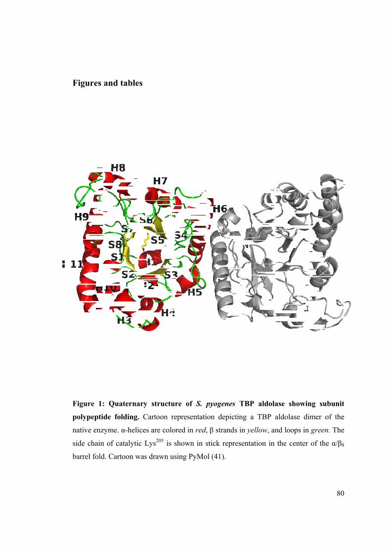
Figures
Figure 1:
polypepti
native enz
side chain
barrel fold
and table
: Quaterna
de folding
zyme. α-heli
n of catalyti
d. Cartoon w
es
ary structu
. Cartoon r
ices are col
ic Lys205 is
was drawn u
ure of S. p
representati
ored in red,
shown in s
using PyMo
pyogenes T
ion depictin
, β strands i
stick repres
ol (41).
TBP aldola
ng a TBP
in yellow, an
sentation in
ase showin
aldolase di
nd loops in
the center
80
ng subunit
mer of the
green. The
of the α/β8
0
t
e
e
8

Figure 2:
S. pyogen
and DHA
makes clo
W2 intera
Arg278. Di
Fo-Fc om
Covalent e
es TBP ald
AP C3 via it
ose contact
acts with the
ifference el
mit map enco
enzymatic
dolase. DHA
ts hydroxyl
with DHA
e DHAP ox
lectron dens
ompassing L
intermedia
AP P1 make
l interacts w
P C3 hydro
xyanion and
sity was ca
Lys205 and D
ate formed
es interactio
with Asp27
oxyl and hy
d makes clo
alculated fro
DHAP and c
with DHA
ons with Ar
and Lys125
ydrogen bo
ose contacts
om a 1.92 Å
contoured a
AP in the ac
rg278, Gly277
5. Water mo
nds with G
s with W1,
Å simulated
at 4.0 σ.
81
ctive site of
, and Ser249
olecule W1
Glu163 while
Glu163 and
d annealing
f 9
e
d
g

Figure 3:
active sit
consistent
the active
intermedia
perpendicu
DHAP O3
carbanion
simulated
4.0 σ. Gre
: Electron
te of TBP
t with geom
site of all s
ate has sp2
ular to the p
3 points ou
and/or imi
annealing F
een dashes r
density sh
aldolase.
metry corresp
subunits of t
hybridizati
plane contai
ut of this p
inium. Diff
Fo-Fc omit
represent hy
howing trap
Electron d
ponding to a
the trapped
on at Lys20
ining DHAP
lane corrob
ference elec
t map encom
ydrogen bon
pping of a
density of t
a carbanion
DHAP-TB05 Nξ and h
P carbon at
borating the
ctron densit
mpassing Ly
nding and re
carbanion
the enzyma
n and/or imi
P aldolase c
hence plana
toms C1, C2
e intermedi
ty was calc
ys205 and D
ed spheres w
n intermed
atic interm
inium specie
complex. Th
ar shape wh
2, and C3; a
ate identifi
culated from
HAP and c
water molec
82
iate in the
mediate was
es bound in
he covalent
hen viewed
additionally
cation as a
m a 1.92 Å
ontoured at
cules.
2
e
s
n
t
d
y
a
Å
t

Figure 4:
native en
undergo
representa
binding, in
Gly277 an
conformat
to the free
: Superpos
nzyme (yel
conformatio
ation. For r
n region 2,
nd Arg278
tional displa
e enzyme.
sition of lig
low). Thr
onal displa
region 1, r
, residue Se
are shown
acements na
ganded TB
ree regions
acement up
residues A
er249 interac
n interactin
arrow asym
BP aldolase
of continu
pon DHAP
sp27 and G
cts with DH
ng with th
mmetrically
e structure
uous seque
P binding
Gln28 intera
HAP, while
he carbanio
the active s
e (cyan) wi
ence whose
are shown
act with D
e in region
on interme
site cavity w
83
ith that of
e backbone
n in stick
HAP upon
3, residues
ediate. The
with respect
f
e
k
n
s
e
t

Figure 5:
the DHA
interacting
structures.
Leu248 in t
Lys146, A
complex, r
were also
FBP aldol
Superpos
AP bound
g or in clo
. The resid
the DHAP-
sp33, Gly302
respectively
conserved.
lase by Gln2
sition of th
FBP ald
se contact
dues are Ly
TBP aldola2, Arg303, S
y. Loci of in
Two notab28 and Leu27
he DHAP b
dolase (ma
with DHA
ys205, Glu16
ase complex
Ser271 and
nvariant wa
ble differenc75 in the TB
bound TBP
agenta). S
AP are cons3, Lys125, A
x that superp
Leu270 in t
ater molecul
ces are the r
BP aldolase s
P aldolase s
Superpositio
served betw
Asp27, Gly2
pose with r
the trapped
les, W1 and
replacement
structure, re
structure (
on showin
ween the tw277, Arg278,
residues Lys
DHAP-FB
d W2, in eac
t of Glu34 an
espectively.
84
cyan) with
g residues
wo aldolase
Ser249 and
s229, Glu187,
BP aldolase
ch structure
nd Ser300 in
4
h
s
e
d
,
e
e
n
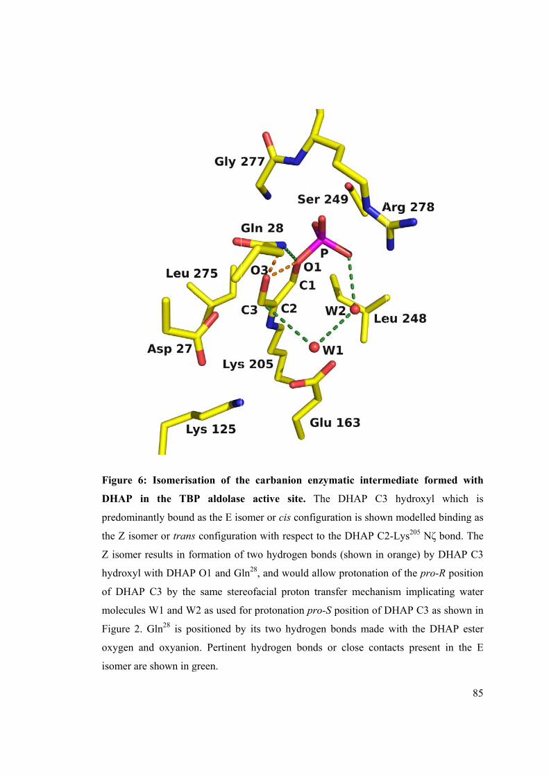
Figure 6:
DHAP in
predomina
the Z isom
Z isomer
hydroxyl w
of DHAP
molecules
Figure 2.
oxygen an
isomer are
: Isomerisa
n the TB
antly bound
mer or trans
results in fo
with DHAP
C3 by the
s W1 and W
Gln28 is po
nd oxyanio
e shown in g
ation of th
P aldolase
d as the E is
s configurat
formation of
P O1 and G
e same ster
W2 as used f
ositioned by
on. Pertinen
green.
he carbani
e active s
somer or cis
tion with re
f two hydro
Gln28, and w
reofacial pr
for protonat
y its two h
nt hydrogen
ion enzym
site. The
s configurat
espect to the
ogen bonds
ould allow
roton transf
tion pro-S p
hydrogen bo
n bonds or
atic interm
DHAP C3
tion is show
e DHAP C2
(shown in
protonation
fer mechani
position of D
onds made
close cont
mediate for
3 hydroxyl
wn modelled
2-Lys205 Nζ
orange) by
n of the pro
ism implica
DHAP C3 a
with the D
tacts presen
85
rmed with
which is
d binding as
ζ bond. The
y DHAP C3
o-R position
ating water
as shown in
DHAP ester
nt in the E
5
h
s
s
e
n
r
n
r
E

Figure 7:
bound (c
(magenta
conformat
of the DH
Superposi
yan) TBP
) FBP al
tional displa
AP bound T
ition of the
aldolase w
dolase. Th
acement up
TBP and FB
e crystal st
with the n
he regions
pon ligand b
BP aldolases
ructures o
native (pale
1, 2 and
binding are
s are shown
f native (p
e magenta
d 3 which
shown as s
n in cartoon
ale cyan) a
a) and DH
h undergo
sticks. The o
representat
86
and DHAP
AP bound
conserved
overall fold
tion.
6
P
d
d
d

87
Table 1: Data collection and refinement statistics
native DHAP-bound Data collection Resolution 50-1.87
(1.97-1.87)a 50-1.92
(2.02-1.92) Wavelength 0.9795 0.9795 Unique reflections 144212 (15756) 142361 (12153) Completeness (%) 82.5 (74.8) 85.9 (74.1) Average I/σ(I) 7.1 (1.4) 7.8 (2.9) Rsymb 0.092 (0.513) 0.101 (0.326) Space group P212121 P212121Unit cell parameters a(Å), b(Å), c(Å) 63.7, 106.2, 238.5 64.1, 108.2, 238.7 Refinement Number of atoms Protein 10248 10192 Water 2336 2597 Hetero 7 Ca 7 Ca, 36 DHAP atoms Sigma cutoff; I/σ(I) > 1 1 Rcryst (%)c 17.45 16.11 Rfree (%)d 21.81 19.90 Root mean square deviation Bond length (Å) 0.005 0.004 Bond angle (˚) 0.929 0.869 Average B-factor (Å ) 32.75 22.22 Ramachandran analysis e (%) Favoured 96.3 97.1 Allowed 3.7 2.9
a Values in parentheses are given for the highest resolution shell. b Rsym = Σhkl Σi|Ii(hkl) - Īi(hkl)|/ Σhkl ΣiIi(hkl) with i running over the number of independent
observations of reflection hkl. c Rcryst = Σhkl||Io(hkl)| - |Ic(hkl)||/Σhkl|Io(hkl)|. d Rfree = Σhkl∈T|| Io(hkl) - |Ic(hkl)||/Σhkl∈T|Io(hkl)|, where T is a test data set randomly selected from
the observed reflections prior to refinement. Test data set was not used throughout refinement and contains 7.5% of the total unique reflections for native and native aldolase bound with DHAP.
e Analyzed by MolProbity [34]

88
Table 2: Kinetic parameters of FBP and TBP cleavage reaction for TBP aldolase
and carbanion oxidation rates in presence of DHAP, FBP and TBP.
a kinetic solvent isotope effect represented by ratio of kcatH
2O / kcat
D2O
Cleavage reaction Carbanion oxidation
Substrate kcat (s-1) Km (μM) D2
Okcata kcat (s-1) Km (μM)
FBP 4.06±0.07 931±48 3.3 1.92±0.08 292±36
TBP 13.0±0.27 543±32 2.3 2.25±0.08 191±20
DHAP - - 2.64±0.11 144±20
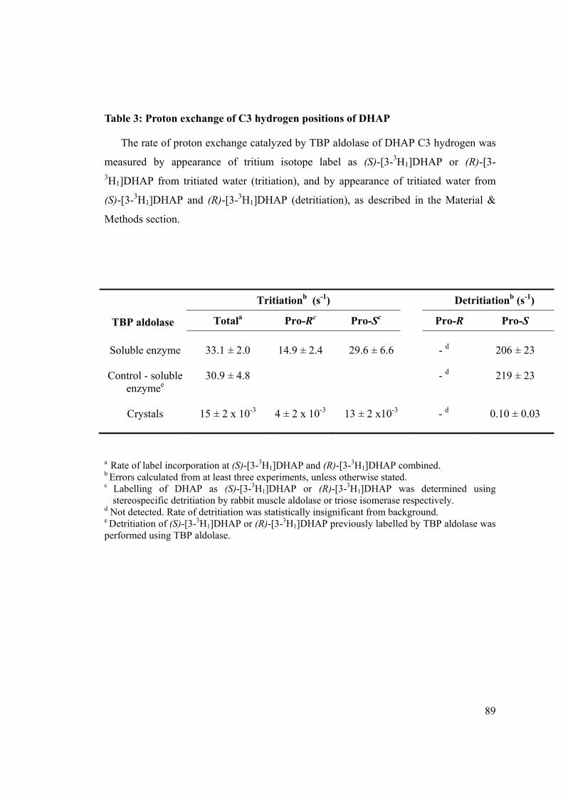
89
Table 3: Proton exchange of C3 hydrogen positions of DHAP
The rate of proton exchange catalyzed by TBP aldolase of DHAP C3 hydrogen was
measured by appearance of tritium isotope label as (S)-[3-3H1]DHAP or (R)-[3-3H1]DHAP from tritiated water (tritiation), and by appearance of tritiated water from
(S)-[3-3H1]DHAP and (R)-[3-3H1]DHAP (detritiation), as described in the Material &
Methods section.
TBP aldolase
Tritiationb (s-1) Detritiationb (s-1)
Totala Pro-Rc Pro-Sc Pro-R Pro-S
Soluble enzyme
33.1 ± 2.0
14.9 ± 2.4
29.6 ± 6.6
- d
206 ± 23
Control - soluble enzymee
30.9 ± 4.8 - d
219 ± 23
Crystals
15 ± 2 x 10⁻³
4 ± 2 x 10⁻³
13 ± 2 x10⁻³
- d
0.10 ± 0.03
a Rate of label incorporation at (S)-[3-3H1]DHAP and (R)-[3-3H1]DHAP combined. b Errors calculated from at least three experiments, unless otherwise stated. c Labelling of DHAP as (S)-[3-3H1]DHAP or (R)-[3-3H1]DHAP was determined using
stereospecific detritiation by rabbit muscle aldolase or triose isomerase respectively. d Not detected. Rate of detritiation was statistically insignificant from background. e Detritiation of (S)-[3-3H1]DHAP or (R)-[3-3H1]DHAP previously labelled by TBP aldolase was performed using TBP aldolase.

90
References 1 Suri, J.T., Mitsumori, S., Albertshofer, K.,Tanaka, F., Barbas, C.F. 3rd. Dihydroxyacetone
variants in the organocatalytic construction of carbohydrates: mimicking tagatose and
fuculose aldolase. (2006) J. Org. Chem. 71, 3822-3828.
2 Takayama, S., McGarvey, G.J., Wong, C.H. Microbial aldolases and transketolases: new
biocatalytic approaches to simple and complex sugars.(1997) Annu. Rev. Microbiol. 51, 285-
310.
3 Fessner, W.D. Enzyme mediated C-C bond formation.(1998) Curr. Opin. Chem. Biol. 2(1),
85-97.
4 Wong, C.H., Whitesides, G. M. (1994), Enzymes in Synthetic Organic Chemistry 1st ed.,
Elsevier Science & Technology Books.
5 Bissett, D.L. and Anderson, R.L. Lactose and D-galactose metabolism in Staphylococcus
aureus. (1980) J. Biol. Chem. 255, 8750-8755.
6 Lee, J.H., Bae, J., Kim, D., Choi, Y., Im, Y.J., Koh, S., Kim, J.S., Kim, M.K., Kang, G.B.,
Hong, S.I., Lee, D.S., Eom, S.H. Stereoselectivity of fructose-1,6-bisphosphate aldolase in
Thermus caldophilus. (2006) Biochem. Biophys. Res. Commun. 347, 616-625.
7 Grazi, E., Rowley, P.T., Cheng, T., Tchola, O., Horecker, B.L. The mechanism of action of
aldolases. III. Schiff base formation with lysine. (1962) Biochem. Biophys. Res. Commun. 9,
38-43.
8 Kobashi, K., Lai, C.Y., Horecker, B.L. Organic phosphate groups in native and
borohydride-reduced aldolase. (1966) Arch. Biochem. Biophys. 117, 437-444.
9 Kobes, R.D., Simpson, R.T., Vallee, R.L., Rutter, W.J. A functional role of metal ions in a
class II aldolase. (1969) Biochemistry. 8, 585-588.
10 Harris, C.E., Kobes, R.D., Teller, D.C., Rutter, W.J. The molecular characteristics of yeast
aldolase. (1969) Biochemistry. 8, 2442-2454.
11 St-Jean, M. and Sygusch, J. Stereospecific proton transfer by a mobile catalyst in
mammalian fructose-1,6-bisphosphate aldolase. (2007) J. Biol. Chem. 282, 31028-31037.
12 Rose, I.A. (1969) Glycolysis : Regulation and mechanisms of the enzymes; Comprehensive
Biochemistry, Vol. 17, ed. Florkin, M. & Stoltz, E.H., Elsevier, Amsterdam, London and
New York
13 Jencks, W.P. (1987), Catalysis in Chemistry and Enzymology, Courier Dover Publications.
14 Grazi, E., Cheng, T., Horecker, B.L. The formation of a stable aldolase-dihydroxyacetone

91
phosphate complex (1962) Biochem. Biophys. Res. Commun. 7, 250-253.
15 Rose, I.A. and Rieder, S.V. Studies on the mechanism on the aldolase reaction; isotope
exchange reactions of muscle and yeast aldolase. (1958) J. Biol. Chem. 231, 315-329.
16 Rose, I.A. The absolute configuration of dihydroxyacetone phosphate tritiated by aldolase
reaction. (1958) J. Am. Chem. Soc. 80, 5835-5836.
17 Chiu, T.H. and Feingold, D.S. Substrate specificity of L-rhamnulose 1-phosphate aldolase
(1965) Biochem. Biophys. Res. Commun. 19, 511-516.
18 Chiu, T.H. and Feingold, D.S. L-rhamnulose 1-phosphate aldolase from Escherichia coli.
Crystallization and properties. (1969), Biochemistry. 8, 98-108.
19 Kobes, R.D., and Dekker, K.E. 2-Keto-4-hydroxyglutarate aldolase of bovine liver.
Purification, criteria of purity, and general properties, (1969) J. Biol. Chem. 244, 1919-1925
20 Fish, D.C., and Blumenthal, H.J., 2-keto-3-deoxy-d-glucarate aldolase (1966) Methods
Enzymol. 9, 529-534
21 Meloche, H.P. and Mehler, L. The stereochemistry at carbon 3 of pyruvate lyase
condensation products. Aldolases forming condensation products racemic at carbon 4.
(1973) J. Biol. Chem. 248, 6333-6338.
22 Espelt, L., Bujons, J., Parella, T., Calveras, J., Joglar, J., Delgado, A., Clapés, P. Aldol
additions of dihydroxyacetone phosphate to N-Cbz-amino aldehydes catalyzed by L-
fuculose-1-phosphate aldolase in emulsion systems: inversion of stereoselectivity as a
function of the acceptor aldehyde (2005) Chemistry. 11, 1392-1401.
23 Zgiby, S.M., Thomson, G.J., Qamar, S., Berry, A. Exploring substrate binding and
discrimination in fructose-1,6-bisphosphate and tagatose-1,6-bisphosphate aldolases (2000)
Eur.J.Biochem. 267, 1858-1868.
24 Williams, G.J., Domann, S., Nelson, A., Berry, A. Modifying the stereochemistry of an
enzyme-catalyzed reaction by directed evolution. (2002) Proc. Natl. Acad. Sci. U S A. 100,
3143-3148.
25 Sygusch, J., Beaudry, D.F., Allaire, M. Molecular architecture of rabbit skeletal muscle
aldolase at 2.7-A resolution. (1987) Proc. Natl. Acad. Sci. U S A. 84, 7846-7850.
26 Chudzik, D.M., Michels, P.A., de Walque, S., Hol, W.G. Structures of type 2 peroxisomal
targeting signals in two trypanosomatid aldolases. (2000) J. Mol. Biol. 300, 697-707.
27 Dalby, A.R., Tolan, D.R., Littlechild, J.A. The structure of human liver fructose-1,6-
bisphosphate aldolase. (2001) Acta Crystallogr D Biol Crystallogr. 57, 1526-33.

92
28 St-Jean, M., Lafrance-Vanasse, J., Liotard, B., Sygusch, J. High resolution reaction
intermediates of rabbit muscle fructose-1,6-biphosphate aldolase. (2005) J.Biol.Chem. 280,
27262-27270.
29 Hall, D.R., Bond, C.S., Leonard, G.A., Watt, C.I., Berry, A., Hunter, W.N. Structure of
tagatose-1,6-bisphosphate aldolase. Insight into chiral discrimination, mechanism, and
specificity of class II aldolases. (2002) J. Biol. Chem. 277, 22018-22024
30 Liotard, B. and Sygusch, J. Purification, crystallization and preliminary X-ray analysis of
native and selenomethionine class I tagatose-1,6-bisphosphate aldolase from Streptococcus
pyogenes. (2003) Acta.Cryst.Sect.D. 60, 528-530.
31 Racker, E. Spectrophotometric measurement of hexokinase and phosphohexokinase
activity.(1947) J.Biol.Chem. 47, 843-854.
32 Otwinowski, Z., Minor, W. Processing of X-ray diffraction data collected in oscillation
mode. (1997) Methods Enzymol. 276, 307-326.
33 Terwilliger, T.C. and Berendzen J. Automated MAD and MIR structure solution. (1999)
Acta Cryst. D55, 849-861.
34 Terwilliger, TC. Maximum likelihood density modification. (2000) Acta Cryst. D56, 965-
972.
35 Jones, T.A., Zou, J.Y., Cowan, S.W., Kjeldgaard, M. Improved methods for building protein
models in electron density maps and the location of errors in these models. (1991) Acta
Crystallogr.Sect.A. 47, 110-111.
36 Brünger, A.T., Adams, P.D., Clore, G.M., DeLano, W.L., Gros, P., Grosse-Kunstleve, R.W.,
Jiang, J.S., Kuszewski, J., Nilges, M., Pannu, N.S., Read, R.J., Rice, L.M., Simonson, T.,
Warren, G.L. Crystallography & NMR system: A new software suite for macromolecular
structure determination. (1998) Acta Crystallogr.Sect.D. 54, 905-921.
37 Adams, P.D., Grosse-Kunstleve, R.W., Hung, L.W., Ioerger, T.R., McCoy, A.J., Moriarty,
N.W., Read, R.J., Sacchettini, J.C., Sauter, N.K., Terwilliger, T.C. PHENIX: building new
software for automated crystallographic structure determination. (2002) Acta Crystallogr.
D58, 1948-1954.
38 Emsley, P. and Cowtan, K. Coot: model-building tools for molecular graphics. (2004) Acta
Crystallogr.Sect.D. 60, 2126-2132.
39 Schuttelkopf, A. W., and van Aalten, D. M. PRODRG: a tool for high-throughput
crystallography of protein-ligand complexes. (2004) Acta Crystallogr., Sect. D: Biol.

93
Crystallogr. 60, 1355-1363.
40 Diamond, R. On the multiple simultaneous superposition of molecular structures by rigid
body transformations. (1992) Protein Sci. 1, 1279-1287.
41 DeLano, W. L. (2002) The PyMOL Molecular Graphics System on World Wide Web
http://www.pymol.org.
42 Guex, N. and Peitsch, M.C. SWISS-MODEL and the Swiss-PdbViewer: An environment
for comparative protein modeling. (1997), Electrophoresis 18, 2714-2723.
43 Arnold, K., Bordoli, L., Kopp, J., Schwede, T. The SWISS-MODEL workspace: a web-
based environment for protein structure homology modelling. (2006) Bioinformatics. 22,
195-201.
44 Healy, M.J., and Christen, P. Mechanistic probes for enzymatic reactions. Oxidation-
reduction indicators as oxidants of intermediary carbanions (studies with aldolase, aspartate
aminotransferase, pyruvate decarboxylase, and 6-phosphogluconate dehydrogenase) (1973)
Biochemistry. 12, 35-41
45 Miller, W., Huang, X. A Time-Efficient, Linear-Space Local Similarity Algorithm (1991)
Advances in Applied Mathematics. 12, 337-357.
46 Holm L, Kaariainen S, Rosenstrom P, Schenkel A. Searching protein structure databases
with DaliLite v.3. (2008) Bioinformatics 24, 2780-2781.
47 Lorentzen, E., Siebers, B., Hensel, R., Pohl, E. Structure, function and evolution of the
Archaeal class I fructose-1,6-bisphosphate aldolase. (2004) Biochem Soc Trans. 32, 259-63.
48 Wierenga RK. The TIM-barrel fold: a versatile framework for efficient enzymes. (2001)
FEBS Lett. Mar 16;492(3):193-8.
49 Spolter, P. D., Adelman, R. C., Weinhouse, S. Distinctive properties of native and
carboxypeptidase-treated aldolases of rabbit muscle and liver. (1965) J. Biol. Chem. 244,
126–134.
50 Rutter, W. J., Richards, O. C., Woodfin, B. M. Comparative studies of liver and muscle
aldolase. I. Effect of carboxypeptidase on catalytic activity. (1961) J. Biol. Chem. 236,
3193–3197
51 Corina, D. L., and Wilton, D. C. An apparent lack of stereospecificity in the reaction
catalysed by deoxyribose 5-phosphate aldolase due to methyl-group rotation and enolization
before product release. (1976) Biochem. J. 157, 573-576
52 Loughman, J.A. and Caparon, M.G. A novel adaptation of aldolase regulates virulence in

94
Streptococcus pyogenes. (2006) EMBO J. 25, 5414-5422.
53 Loughman, J.A. and Caparon, M.G. Comparative functional analysis of the lac operon in
Streptococcus pyogenes. (2007) Molecular Microbiology. 64, 269-280.
54 Cunningham, M.W. Pathogenesis of group A streptococcal infections. (2000) Clin Microbiol
Rev. 13, 470–511

Footnotes:
The atomic coordinates and structure factors (code … and … ) have been deposited
in the Protein Data Bank, Research Collaboratory for Structural Bioinformatics,
Rutgers University, New Brunswick, NJ (http://www.rcsb.org/).
Research was supported by funding to JS from Natural Science and Engineering
Research Council (Canada) and Canadian Institutes for Health Research.
Work was carried out in part at beamlines X12B, X25 and X29 of the National
Synchrotron Light Source. Financial support comes principally from the Offices of
Biological and Environmental Research and of Basic Energy Sciences of the US
Department of Energy, and from the National Center for Research Resources of the
National Institutes of Health. Assistance by beamline personnel, Drs L. Flaks, D.K.
Schneider, A. Soares, A. Héroux and H. Robinson, is gratefully acknowledged. The
generous help of Laurent Cappadoccia in data collection was also particularly
appreciated. Critical reading of the manuscript by Dr. Casimir Blonski was appreciated.
Tagatose-bisphosphate was generous gift of Dr. W. Fessner.
The abbreviations used are: TBP, tagatose-1,6-bis(phosphate); FBP, fructose-1,6-
bis(phosphate); G3P, D-glyceraldehyde 3-phosphate; DHAP, dihydroxyacetone
phosphate; TIM, triose phosphate isomerase; NADH, nicotinamide adenine dinucleotide

CHAPITRE 3: Discussion
La résolution des structures de la tagatose-1,6-bisphosphate aldolase de Streptococcus
pyogenes native et en complexe avec son substrat DHAP a permis de déposer une des
premières structures tridimensionnelles d’une TBP aldolase de classe I. Son repliement,
un tonneau (α/β)8 caractéristique des aldolases de classe I ou II, voit le site actif enfoui
au cœur du tonneau, et gardé par trois régions très mobiles et de faible densité
électronique dans la structure native. Une recherche dans la base de données UniProt
grâce au serveur Blast (NCBI BLAST2) a permis de mettre en évidence de nombreuses
similitudes de séquences avec des TBP aldolases de divers streptocoques. L’alignement
des structures modelées via le programme Swiss-model (Arnold, 2006) des meilleurs
résultats du serveur Blast sur la structure native de la TBP aldolase de S. pyogenes a été
réalisé grâce au programme PyMOL. Les modèles, ceux d’une protéine non identifiée
de Streptococcus infantarius, d’une TBP aldolase de Streptococcus mutans, d’une TBP
aldolase de Streptococcus gordonii, et d’une TBP aldolase de Streptococcus sanguinis,
et la structure de la TBP aldolase de Streptococcus pyogenes, partagent tous les mêmes
résidus au sein de leur site actif : Arg278, Gly277, Ser249, Gly277, Ser249, Asp27, Gln28,
Lys125 et Leu275 (numérotation de la TBP aldolase de S. pyogenes). De même,
l’alignement de la structure de la TBP aldolase de S. pyogenes sur la structure modelée
de la TBP aldolase de Straphylococcus aureus, avec laquelle elle partage 73,6 %
d’identité, a permis de souligner les mêmes résidus du site actif.

Le trempage pendant cinq minutes de cristaux de la TBP aldolase de S. pyogenes dans
une solution saturante de DHAP a permis de piéger un intermédiaire réactionnel
enzyme-substrat. Cet intermédiaire présente un caractère iminium certain, en contraste
avec la FBP aldolase de muscle qui, elle, montre une énamine dans ces conditions (St-
Jean, 2007). L’intermédiaire iminium chez la TBP aldolase de S. pyogenes est stabilisé
par de nombreux ponts hydrogènes et interactions électrostatiques avec des résidus du
site actif, très similaires à celles présentes dans la structure de la FBP aldolase de
muscle en complexe avec le DHAP. L'alignement de la structure de la TBP aldolase de
S. pyogenes sur celle de la FBP aldolase de muscle a ainsi permis de vérifier que tous
les résidus du site actif considérés comme importants pour la catalyse étaient conservés
chez les deux enzymes, mis à part la Leu275 de la TBP aldolase. Cette Leucine est, dans
la FBP aldolase, remplacée par Ser300, résidu que l'on pense à présent (St-Jean, 2007)
capable de former un pont hydrogène stabilisateur avec l'atome Nζ de la Lys229 ce qui
stabilise la forme énamine de l'intermédiaire. La formation de ce pont hydrogène n'est
possible que dans la forme énamine de l'intermédiaire réactionnel, et non dans la forme
iminium. Il est donc intéressant de noter que cette Serine est remplacée par une Leucine
dans la structure de la TBP aldolase de S. pyogenes, et que c'est l'iminium qui y est
stabilisé. La figure 6 de l'article montre une possible conformation de l'intermédiaire
réactionnel iminium : l'hydroxyle du C3 du DHAP pourrait subir une isomérisation et
adopter une conformation cis autour du lien C2-C3 du DHAP. Cette isomérisation cis-
trans pourrait permettre la formation d'un pont hydrogène de 2.39 Å entre l'hydroxyle
C3 du DHAP et son O1. Une telle géométrie n'est néanmoins acceptable que s'il y a

hybridation sp2 au niveau du C3 de l'intermédiaire réactionnel. En effet, une
hybridation sp3 induirait une distance trop courte et un encombrement stérique entre
l'hydroxyle du C3 et son O1. Enfin, une telle géométrie permettrait d'expliquer les
résultats de transfert de proton obtenus lors de la tritiation/détritiation du DHAP
catalysée par la TBP aldolase. En effet, l’enzyme, bien que capable marquer au tritium
les deux cotés pro-S et pro-R du carbone C3 de l'intermédiaire réactionnel, ne semble
capable de ne démarquer que le cote pro-S. Or, la formation du sorbose-bisphosphate et
du psicose-bisphosphate à partir du DHAP et G3P catalysée par la TBP aldolase
implique
Dans le cadre de la condensation catalysée par la FBP aldolase de muscle, la face si du
DHAP est attaquée nucléophiliquement par la face si du G3P, ce qui provoque la
rétention de la configuration au niveau du carbone C3, et donc résulte en la formation
du fructose-1,6-bisphosphate (St-Jean, 2005; St-Jean, 2007). De même, la L-
rahmnulose-1-phosphate aldolase est capable de produire du L-rahmnulose-1-phosphate
en catalysant l'attaque de la face re du L-lactaldéhyde sur la face re du DHAP
(Kroemer, 2003). A l'opposé, il existe des aldolases qui génèrent des produits
racémiques au niveau de l'orientation de leurs hydroxyles. Ainsi, la α-keto-β-
deoxyhexarate aldolase et la 2-keto-4-hydroxyglutarate aldolase, deux enzymes
pyruvate-dépendantes (Tableau III), démontrent une racémisation au carbone C4 de leur
produit de condensation (Fish, 1966; Kobes, 1969). Il a déjà été démontré (Meloche,
1973), que ce manque de stéréospécificité et de discrimination chirale est le résultat de

l'orientation peu contrôlée de l'aldéhyde au sein du site actif de ces aldolases. Ce sont
donc les deux faces si et re de l'accepteur aldéhyde qui sont ainsi capables d'être
attaquées nucléophiliquement par une seule face du donneur pyruvate, provoquant ainsi
une rétention de configuration au niveau du carbone C3 mais non du C4. La
racémisation des produits de condensation n'est donc pas le résultat de l'attaque de
l'aldéhyde sur n'importe laquelle des faces de l'accepteur pyruvate, ce qui amènerait une
racémisation au niveau du carbone C3 et serait un phénomène atypique des aldolases.
Pour ce qui est des aldolases DHAP-dépendantes, la L-fuculose-1-phosphate aldolase
génère également une isomérisation des produits (Ghalambor, 1962). Dans ce dernier
cas, il a été démontré que la stéréochimie du carbone C4 dépend de la structure de
l'accepteur aldéhyde (Espelt, 2005).
L'incapacité de la TBP aldolase de S. pyogenes à détritier la position pro-R du C3 du
DHAP, alors qu'elle semble à même de tritier les deux positions pro-R et pro-S, s'inscrit
dans ce contexte. En effet, et ce malgré les apparences, cette enzyme semble bien
démontrer une certaine spécificité au niveau de la déprotonation du DHAP lié au site
actif. Afin de vérifier ce point qui, bien qu'en accord avec le mécanisme aldolasique
standard, est tout de même étonnant au regard des quatre produits de condensation de la
TBP aldolase de S. pyogenes, nous avons concocté une expérience de contrôle
supplémentaire. Ainsi que la section Matériel et Méthodes de l'article l'explique en
détails, la TBP aldolase a été incubée avec du DHAP dans un milieu contenant du 3H.
Le DHAP marqué résultant a ensuite été mis en présence de la même TBP aldolase, afin
que cette dernière puisse détritier le DHAP qu'elle avait elle-même tritié précédemment.
L'incubation à la triose phosphate isomérase ou à la FBP aldolase de muscle a
finalement permis d'évaluer la préférence de la TBP aldolase pour le proton en position
pro-R ou pro-S.
La superposition de la structure de la TBP aldolase liée au DHAP sur celle de l'enzyme
libre a permis de distinguer trois régions particulièrement mobiles. Incidemment, ces
trois régions étaient celles qui étaient les moins bien définies et dessinées dans la carte
de densité électronique de la structure native. Le déplacement de ces régions de la
structure native à celle liée au DHAP indique que la liaison du DHAP à l'atome Nζ de la
Lys205 induit un mouvement de fermeture du site actif, ce qui a pour effet de stabiliser
l'intermédiaire enzyme-substrat. Des mouvements conformationnels de ce type ont déjà
été observés dans la FBP aldolase lorsque liée à ses substrats DHAP et FBP (St-Jean,
2007). La région qui fait preuve du plus large déplacement, baptisée région 3, est une
hélice alpha qui renferme l'Arg278 impliquée dans de nombreuses interactions
stabilisatrices avec l'oxyanion du phosphate P1. Le r.m.s.d. moyen de la région 3 atteint
les 1.17 ± 0.24 Å.
De même, deux molécules d'eau W1 et W2, conservées de la structure de la TBP
aldolase native à celle ligandée ont également été mises en évidence. Ces deux
molécules, baptisées W1 et W2 comme dans la structure de la FBP aldolase de muscle
(St-Jean, 2007), y ont des positions comparables : W1 est perpendiculaire au plan de
l'iminium intermédiaire, et proche de son carbone C3 (3.45 ± 0.16 Å). W2 est, elle,
engagée dans un pont hydrogène avec l'un des oxygènes de l'oxyanion phosphate P1
dans la structure complexée au DHAP (2.85 ± 0.06 Å), et avec W1 dans les structures

native et ligandée (3.50 ± 0.27 Å et 3.37 ± 0.53 Å, respectivement). La possibilité d'un
transfert de proton séquentiel via ces deux molécules d'eau, si proches du carbone C3 de
l'intermédiaire réactionnel, est donc à envisager.

CHAPITRE 4: Perspectives
La résolution de la structure de la tagatose-1,6-biphosphate aldolase de Streptococcus
pyogenes native et en complexe avec un de ses substrats naturels le DHAP a donc
permis un premier pas vers la compréhension de son mécanisme catalytique et en
particulier de son étonnant manque de stéréospécificité. La suite évidente à ce projet est
donc naturellement la résolution de structures trois-dimensionnelles de la TBP aldolase
complexées à ses cinq autres substrats le fructose-1,6-bisphosphate aldolase, le
tagatose-1,6-phosphate aldolase, le sorbose-1,6-bisphosphate, le psicose-1,6-
bisphophate aldolase, ainsi que le glycéraldéhyde-3-phosphate. De même, les structures
de la TBP aldolase en complexe avec divers inhibiteurs, compétitifs ou non, pourraient
permettre d'éclairer un peu plus son mécanisme, et les caractéristiques qui lui permettent
d'être si peu spécifique au niveau du carbone 3 et 4 de ses substrats. Elles pourraient
également conforter l'hypothèse d'un isomérisme cis-trans autour du lien C2-C3 du
DHAP dans le sens de la condensation. Dans le cadre de la suite à amener a ce projet,
certaines de ses structures ont déjà été entreprises, conjointement avec les études
enzymologiques adéquates, et des résultats prometteurs obtenus. Bien que la structure
de la TBP aldolase en complexe avec son substrat préféré, le tagatose-1,6-phosphate, de
même que celle de la TBP aldolase complexée au fructose-1,6-bisphosphate, aient été
difficile a obtenir du fait de l'équilibre favorable au clivage de la catalyse, cela a été
réalisé récemment. En effet, la mise au point d'une technique de trempage des cristaux
de TBP aldolase a des pH décroissants successifs a permis de piéger à un pH acide

moins favorable à la catalyse les deux substrats dans le site actif de l'enzyme.
L'affinement et l'analyse structurale des ces deux structures sont en court.
De plus, la mutation des résidus du site actif mis en exergue par la résolution de sa
structure trois-dimensionnelle devrait également permettre d'identifier leur rôle
spécifique dans la catalyse. Les mutations ponctuelles de Arg278 et Gln28 en résidus
isomorphes mais non hydrophobes ou non chargés par exemple pourrait se traduire par
une perte d'affinité de la TBP aldolase pour ses substrats. De même, les mutations de
Lys125, Glu163, Asp27 et Leu275 en résidus isomorphes de charge opposées pourrait
amener une perte d'activité de l'enzyme. Certaines de ces mutations ont déjà été
réalisées, et ici aussi, les résultats, sont prometteurs. Le mutant Leu275Ser par exemple,
semble exhiber une perte totale d'activité de clivage du FBP, ce qui conforte l'hypothèse
d'un rôle important de ce résidu dans la catalyse.
L'identification de trois régions mobiles dans la structure de la TBP aldolase, régions
dont le mouvement est dépendant de la liaison de l'enzyme à son substrat, offre une
piste de recherche dans le rôle putatif de l'homologue de LacD.2 dans S. pyogenes,
LacD.1. En effet, la liaison de LacD.1 à RopB semble être subordonnée à sa capacité à
lier ses substrats naturels, et pourrait imputer un rôle aux différents mouvements
conformationnels adoptés par la protéine. Diverses mutations au sein de ces régions
dans LacD.2, qui auraient pour effet de minimiser ou maximiser ces changements
conformationnels après liaison au substrat devraient donc permettre de mesurer leur

importance dans le rôle de senseur métabolique attribué à LacD.1. Pour cela, des
expériences in vivo, faites conjointement avec Caparon et al. (Loughman et Capron,
2006; Loughman et Caparon, 2007), et qui permettraient d'observer les effets de ces
mutations affectant les changements conformationnels sur la virulence du streptocoque
lui même, sont envisagées. Enfin, la résolution de la structure trois-dimensionnelle de
LacD1, native ou mutée et complexée a ses substrats et peut être également à RopB, est
prévue.

Références :
1. Peuckert, W., Microbiology of streptococcal infections, Klin. Padiatr., 1985,
197(1), 5-8.
2. Billroth, T., Pathology and therapeutics, in fifty lectures 1871, Clin. Orthop.
Relat. Res., 2003, 408, 4-11.
3. Whiffin, R.A., Puerperal Septicemia, Cal. State J. Med., 1903, 1(13), 395-7.
4. Sherman, J.M. and Albus, W.R., Some Characters which Differentiate the
Lactic-acid Streptococcus from Streptococci of the Pyogenes Type Occurring in
Milk, J. Bacteriol., 1918, 3(2), 153-74
5. Evans, A.C., Studies on Hemolytic Streptococci: II. Streptococcus pyogenes, J.
Bacteriol., 1936, 31(6), 611-24.
6. Brown, J.H., The use of blood agar for the study of streptococci, Rockefeller
Institute Medical Research Monograph, 1919, 9
7. Schottmuller, H., Die Arunterscheidung des fur den Menschen pathogenen
Streptokokken durch Blutagar, Munch. med. Wochschr., 1903, 50, 849-853, 909-
12.
8. Budelmann, G., Hugo Schottmuller, 1867-1936. The problem of sepsis, Internist
(Berl), 1969. 10(3), 92-101.
9. Horstmeier, C. and Washington, J.A. 2nd, Microbiological study of
streptococcal bacteremia, Appl. Microbiol., 1973. 26(4), 589-91.
10. Jassim, S., Salt, W.G., Stretton, R.J., In vitro studies of haemolysis by some
staphylococci grown in chemically defined media, J. Appl. Bacteriol., 1989,
67(5), 511-20.
11. Orden, B., Streptococcus pyogenes not producing beta hemolysis during
aerobiosis, Enferm. Infecc. Microbiol. Clin., 1991, 9(8) 516-7
12. Lancefield, R.C., A sereological differentiation of human and other groups of
hemolytic streptococci. J. Exptl. Med., 1933, 57, 571-95.
13. Davis, B.D., Dulbecco, R., Eisen, H.N., Ginsberg, H.S., Wood, W.B.,
Microbiology, 1973, 2nd Edition, Harper & Row Publishers.

14. Bottone, E.J., An Atlas of the Clinical Microbiology of Infectious Diseases,
2006, Illustrated Edition, Informa Health Care.
15. Iacono, V.J., Lysozyme-mediated de-chaining of Streptococcus mutans and its
antibacterial significance in an acidic environment, J. Dent. Res., 1985. 64(1),
48-53.
16. Stollerman, G.H. and Ekstedt, R., Long chain formation by strains of group A
streptococci in the presence of homologous antiserum: a type-specific reaction. J
Exp Med, 1957. 106(3): p. 345-56.
17. Lominski, I. and Shaikh, M.R., Long-chain mutants of Streptococcus faecalis
induced by ultraviolet irradiation, J. Med. Microbiol., 1968, 1(2), 219-20.
18. Brooks, G.F, Carroll, K.C., Butel, J.S., Morse, S.A., Jawetz, Melnick &
Adelberg's Medical Microbiology, 2007, 24th Edition, illustrated, revised,
McGraw-Hill Professional.
19. Fung, J.C., Wicher, K., McCarty, M., Immunochemical analysis of streptococcal
group A, B, and C carbohydrates, with emphasis on group A, Infect. Immun.,
1982. 37(1), 209-15.
20. Coligan, J.E., Schnute, W.C. Jr., Kindt, T.J., Immunochemical and chemical
studies on streptococcal group-specific carbohydrates, J. Immunol., 1975,
114(6), 1654-8.
21. Pritchard, D.G., Coligan, J.E., Speed, S.E., Gray, B.M., Carbohydrate
fingerprints of streptococcal cells, J. Clin. Microbiol., 1981, 13(1), 89-92.
22. Kahn, F., Mörgelin, M., Shannon, O., Norrby-Teglund, A., Herwald, H., Olin,
A.I., Björck, L., Antibodies against a surface protein of Streptococcus pyogenes
promote a pathological inflammatory response, PLoS Pathog., 2008, 4(9),
e1000149.
23. Fischetti, V.A., Streptococcal M protein: molecular design and biological
behavior, Clin. Microbiol. Rev., 1989. 2(3), 285-314.
24. Okamoto, S., Kawabata, S., Terao, Y., Fujitaka, H., Okuno, Y., Hamada, S., The
Streptococcus pyogenes capsule is required for adhesion of bacteria to virus-

infected alveolar epithelial cells and lethal bacterial-viral superinfection, Infect.
Immun., 2004. 72(10), 6068-75.
25. Gryllos, I., Tran-Winkler, H.J., Cheng, M.F., Chung, H., Bolcome, R. 3rd, Lu,
W., Lehrer, R.I., Wessels, M.R., Induction of group A Streptococcus virulence
by a human antimicrobial peptide, Proc Natl Acad Sci U S A, 2008, 105(43),
16755-60.
26. Stollerman G. H. and Dale J. B., The importance of group A strepcococcus
capsule in the pathogenesis of human infections: a historical perspective,
Clinical Infectious Diseases, 2008, 46, 1038-45
27. Hanski, E., Horwitz, P.A., Caparon, M.G., Expression of protein F, the
fibronectin-binding protein of Streptococcus pyogenes JRS4, in heterologous
streptococcal and enterococcal strains promotes their adherence to respiratory
epithelial cells, Infect. Immun., 1992, 60(12), 5119-25.
28. Nakamura, T., Hasegawa, T., Torii, K., Hasegawa, Y., Shimokata, K., Ohta, M.,
Two-dimensional gel electrophoresis analysis of the abundance of virulent
exoproteins of group A streptococcus caused by environmental changes, Arch.
Microbiol., 2004, 181(1), 74-81.
29. Bisno, A.L., Brito, M.O., Collins, C.M., Molecular basis of group A
streptococcal virulence, Lancet Infect. Dis., 2003, 3(4), 191-200.
30. Oelschlaeger, T.A., Hacker, J., Bacterial Invasion Into Eukaryotic Cells, 2000,
Illustrated Edition, Springer.
31. Dong, H., Li, S., Song, Q., Liu, S., Lin, H., Chai, Y., Zhou, A., Fang, T., Zhang,
H., Jin, C., Lu, W., Cao, G., Beta-haemolytic group A streptococci emm75
carrying altered pyrogenic exotoxin A linked to scarlet fever in adults, J. Infect.,
2008, 56(4), 261-7.
32. Fraser, J.D. and Proft, T., The bacterial superantigen and superantigen-like
proteins, Immunol. Rev., 2008, 225, 226-43.
33. Chiang-Ni C., Wu J.J., Effects of streptococcal pyrogenic exotoxin B on
pathogenesis of Streptococcus pyogenes, J. Formos. Med. Assoc., 2008, 107(9),
677-85

34. Fontaine, M.C., Lee, J.J., Kehoe, M.A, Combined contributions of streptolysin
O and streptolysin S to virulence of serotype M5 Streptococcus pyogenes strain
Manfredo, Infect. Immun., 2003, 71(7), 3857-65.
35. Goldmann, O., Sastalla, I., Wos-Oxley, M., Rohde, M., Medina, E.,
Streptococcus pyogenes induces oncosis in macrophages through the activation
of an inflammatory programmed cell death pathway, Cell Microbiol., 2009,
11(1), 138-55.
36. Nelson, L., Potential drug target for flesh-eating bug, Drug Discov. Today, 2004,
9(20), 862.
37. Chhatwal, G.S. and McMillan, D.J., Uncovering the mysteries of invasive
streptococcal diseases, Trends Mol. Med., 2005, 11(4), 152-5.
38. Sun, H., Ringdahl, U., Homeister, J.W., Fay, W.P., Engleberg, N.C., Yang,
A.Y., Rozek, L.S., Wang, X., Sjöbring, U., Ginsburg, D., Plasminogen is a
critical host pathogenicity factor for group A streptococcal infection, 2004,
305(5688), 1283-6.
39. Hynes, W.L., Dixon, A.R., Walton, S.L., Aridgides, L.J., The extracellular
hyaluronidase gene (hylA) of Streptococcus pyogenes, FEMS Microbiol. Lett.,
2000, 184(1), 109-12.
40. Starr, C.R. and Engleberg, N.C., Role of hyaluronidase in subcutaneous spread
and growth of group A streptococcus, Infect. Immun., 2006, 74(1), 40-8.
41. Rosch J. W. and Tuomanen E., Adapting a diet from sugar to meat: double-
dealing genes of Streptococcus pyogenes, Molecular Microbiology, 2007, 64,
257-9
42. Quinn, R.W. and Liao, S.J., A comparative study of antihyaluronidase,
antistreptolysin "O," antistreptokinase, and streptococcal agglutination titers in
patients with rheumatic fever, acute hemolytic streptococcal infections,
rheumatoid arthritis and non-rheumatoid forms of arthritis, J. Clin. Invest., 1950,
29(9), 1156-66.
43. Cunningham, M.W., Pathogenesis of group A streptococcal infections, Clin.
Microbiol. Rev., 2000, 13, 470–511

44. Shet, A., Kaplan, E., Addressing the burden of group A streptococcal disease in
India, Indian J. Pediatr., 2004, 71(1), 41-8.
45. Medina, E., Goldmann, O., Toppel, A.W., Chhatwal, G.S., Survival of
Streptococcus pyogenes within host phagocytic cells: a pathogenic mechanism
for persistence and systemic invasion, J. Infect. Dis., 2003, 187(4), 597-603.
46. Cvjetkovic, D., Jovanović, J., Hrnjaković-Cvjetković, I., Aleksić-Dordević, M.,
Stefan-Mikić, S., Significance of group A streptococcal infections in human
pathology, Med. Pregl., 2008, 61(9-10), 529-32.
47. Shelburne, S.A. 3rd, Keith, D.B., Davenport, M.T., Horstmann, N., Brennan,
R.G., Musser, J.M., Molecular characterization of group A Streptococcus
maltodextrin catabolism and its role in pharyngitis, Mol. Microbiol., 2008,
69(2), 436-52.
48. Papadas, T.A., Karkoulias, K.P., Mastronikolis, N.S., Sampsonas, F.L.,
Spiropoulos, K.B., Concurrent tonsillitis and pneumonia due to S. pyogenes,
Clin. Ter., 2008, 159(1), 33-4.
49. Hedrick, J., Acute bacterial skin infections in pediatric medicine: current issues
in presentation and treatment, Paediatr. Drugs, 2003, 5 Suppl 1, 35-46.
50. Luca-Harari, B., Darenberg, J., Neal, S., Siljander, T., Strakova, L., Tanna, A.,
Creti, R., Ekelund, K., Koliou, M., Tassios, P.T., van der Linden, M., Straut, M.,
Vuopio-Varkila, J., Bouvet, A., Efstratiou, A., Schalén, C., Henriques-Normark,
B., Strep-EURO Study Group, Jasir, A., Clinical and Microbiological
Characteristics of Severe Streptococcus pyogenes Disease in Europe, J. Clin.
Microbiol., 2009, 47, 1155-65.
51. Thomas, D., Perpoint, T., Dauwalder, O., Lina, G., Floccard, B., Richard, J.C.,
Bouvet, A., Peyramond, D., Allaouchiche, B., Chidiac, C., Vandenesch, F.,
Etienne, J., Ferry, T., In vivo and in vitro detection of a superantigenic toxin
Vbeta signature in two forms of streptococcal toxic shock syndrome, Eur. J.
Clin. Microbiol. Infect. Dis., 2008, 28(6), 671-6.

52. Weidle, P.A., Brankamp, J., Dedy, N., Haenisch, C., Windolf, J., Jonas, M.,
Complication of a closed Colles-fracture: necrotising fasciitis with lethal
outcome. A case report, Arch. Orthop. Trauma Surg., 2009, 129(1), 75-8.
53. Bingöl-Koloğlu, M., Yildiz, R.V., Alper, B., Yağmurlu, A., Ciftçi, E., Gökçora,
I.H., Ince, E., Emiroğlu, M., Dindar, H., Necrotizing fasciitis in children:
diagnostic and therapeutic aspects, J Pediatr Surg., 2007, 42(11), 1892-7.
54. González Castro A, Rodriguez-Borregan JC, Obeso T, Castellanos A, Perez-
Ceballos A, Sesmero JR., Necrotizing fasciitis after cesarean section., Arch.
Gynecol. Obstet.., 2008, 277(6), 579-81. Erratum in: Arch. Gynecol. Obstet.,
2008, 277(6), 583.
55. Olsen, R.J., Shelburne, S.A., Musser, J.M., Molecular mechanisms underlying
group A streptococcal pathogenesis, Cell Microbiol., 2009, 11(1), 1-12
56. Cunningham, M.W., Pathogenesis of group A streptococcal infections and their
sequelae, Adv. Exp. Med. Biol., 2008, 609, 29-42.
57. Skattum, L., Akesson, P., Truedsson, L., Sjöholm, A.G., Antibodies against four
proteins from a Streptococcus pyogenes serotype M1 strain and levels of
circulating mannan-binding lectin in acute poststreptococcal glomerulonephritis,
Int. Arch. Allergy Immunol., 2006, 140(1), 9-19.
58. Burova, L., Thern, A., Pigarevsky, P., Gladilina, M., Seliverstova, V.,
Gavrilova, E., Nagornev, V., Schalén, C., Totolian, A., Role of group A
streptococcal IgG-binding proteins in triggering experimental
glomerulonephritis in the rabbit, APMIS, 2003, 111(10), 955-62.
59. Aziz, R.K. and Kotb, M., Rise and persistence of global M1 T1 clone of
Streptococcus pyogenes, Emerg. Infect. Di., 2008, 14(10), 1511-7
60. Dworkin, M., Falkow, S., The Prokaryotes: a handbook on the biology of
bacteria, 2006, 3rd Edition, Springer.
61. Gibson, C.M., Mallett, T.C., Claiborne, A., Caparon, M.G., Contribution of
NADH oxidase to aerobic metabolism of Streptococcus pyogenes, J. Bacteriol.,
2000, 182, 448-455.

62. Pearce, W. A. Jr, Glucose and galactose metabolism in streptococcus pyogenes,
J. Bacteriol., 1957, 74(2), 186-93.
63. Salminen, S., von Wright, A., Ouwehand, A., Lactic Acid Bacteria:
Microbiological and Functional Aspects, 2004, 3rd Edition, illustrated, revised,
CRC Press.
64. Maxwell, E. S., K. Kurahashi, H. M. Kalckar, Enzymes of the Leloir pathway,
Methods in enzymology, 1962, vol. 5. Academic Press Inc., New York, 174-
189.
65. Steele, R.H., White, A.G., and Pierce, W.A., Jr., The fermentation of galactose
by Streptococcus pyogenes, J. Bacteriol., 1954, 67(1), 86-9.
66. van Rooijen, R.J., van Schalkwijk, S., de Vos, W.M., Molecular cloning,
characterization, and nucleotide sequence of the tagatose 6-phosphate pathway
gene cluster of the lactose operon of Lactococcus lactis, J. Biol. Chem, 1991,
266, 7176-7181.
67. Meyerhof O., Junowicz-Kocholaty, R., The equilibria of isomerase and aldolase,
and the problem of the phosphorylation of glyceraldehyde phosphate, J. Bio.
Chem., 1943, 149, 71-92.
68. Meyerhof O., Oesper, P., The mechanism of the oxydative reaction in
fermentation, J. Biol. Chem., 1947, 170, 1-22.
69. Kresge, N., Simoni, R.D., and Hill, R.L., Otto Fritz Meyerhof and the
elucidation of the glycolytic pathway, J. Biol. Chem., 2005, 280(4), e3.
70. Rutter, W.J., Evolution of Aldolase, Fed. Proc., 1964. 23, 1248-57.
71. Blonski, C., De Moissac, D., Périé, J., Sygusch, J., Inhibition of rabbit muscle
aldolase by phosphorylated aromatic compounds, Biochem. J., 1997, 323 ( Pt 1),
71-7.
72. Thomson, G.J., Howlett, G.J., Ashcroft, A.E., Berry, A., The dhnA gene of
Escherichia coli encodes a class I fructose bisphosphate aldolase, Biochem. J.,
1998, 331(2), 437-45.

73. Hall, D.R., Bond, C.S., Leonard, G.A., Watt, C.I., Berry, A., Hunter,W.N.,
Structure of tagatose-1,6-bisphosphate aldolase, J. Biol. Chem., 2002, 277,
22018-24.
74. Kroth, P.G., Schroers, Y., Kilian, O., The peculiar distribution of class I and
class II aldolases in diatoms and in red algae, Curr. Genet., 2005, 48(6), 389-
400.
75. Lorentzen, E., Siebers, B., Hensel, R., Pohl, E., Mechanism of the Schiff base
forming fructose-1,6-bisphosphate aldolase: structural analysis of reaction
intermediates, Biochemistry, 2005, 44(11), 4222-9.
76. Lorentzen, E., Siebers, B., Hensel, R., Pohl, E., Structure, function and evolution
of the Archaeal class I fructose-1,6-bisphosphate aldolase, Biochem. Soc. Trans.,
2004, 32(Pt 2), 259-63.
77. Ramsaywak, P.C., Labbé, G., Siemann, S., Dmitrienko, G.I., Guillemette, J.G.,
Molecular cloning, expression, purification, and characterization of fructose 1,6-
bisphosphate aldolase from Mycobacterium tuberculosis--a novel Class II A
tetramer, Protein Expr. Purif., 2004, 37(1), 220-8.
78. Galkin, A., Li, Z., Li, L., Kulakova, L., Pal. L.R., Dunaway-Mariano, D.,
Herzberg, O., Structural insights into the substrate binding and stereoselectivity
of giardia fructose-1,6-bisphosphate aldolase, Biochemistry, 2009, 48, 3186-96
79. DeLano, W. L. (2002) The PyMOL Molecular Graphics System on World Wide
Web, http://www.pymol.org.
80. St-Jean, M., Lafrance.-Vanasse, J., Liotard, B., Sygusch, J., High resolution
reaction intermediates of rabbit muscle fructose-1,6-biphosphate aldolase,
J.Biol.Chem., 2005, 280, 27262-70.
81. St-Jean, M. and Sygusch, J., Stereospecific proton transfer by a mobile catalyst
in mammalian fructose-1,6-bisphosphate aldolase, J.Biol.Chem, 2007, 282,
31028-37.
82. Bissett, D.L. and Anderson, R.L., Lactose and D-galactose metabolism in
Staphylococcus aureus, J.Biol.Chem., 1980. 255, 8750-55.

83. Shannon, L.M. and Marcus, A., Gamma-Methyl-gamma-hydroxy-alpha-
ketoglutaric aldolase. I. Purification and properties, J. Biol. Chem, 1962, 237,
3342-7.
84. Shannon, L.M. and Marcus, A., Gamma-Methyl-gamma-hydroxy-alpha-
ketoglutaric aldolase. II. Studies with pyruvate-C14, J. Biol. Chem, 1962, 237,
3348-53.
85. Meloche, H.P., The 2-keto-3-deoxy-6-phosphogluconic aldolase catalyzed
pyruvate-proton exchange reaction: enzymic synthesis of crystalline sodium 3H-
pyruvate, Anal. Biochem., 1970, 38(2), 389-92.
86. Meloche, H.P. and Mehler, L., The stereochemistry at carbon 3 of pyruvate
lyase condensation products: Aldolases forming condensation products racemic
at carbon 4, J. Biol. Chem., 1973, 248(18), 6333-8.
87. Meloche, H.P., Mehler, L., Wurster, J.M., The stereochemistry at carbon 3 of
pyruvate lyase condensation products: 2-Keto-3-deoxygluconate-6-phosphate
aldolase, J. Biol. Chem., 1975, 250(17), 6870-4.
88. Kroemer, M., Merkel, I., Schulz, G.E., Structure and catalytic mechanism of L-
rhamnulose-1-phosphate aldolase, Biochemistry, 2003, 42, 10560-8.
89. Heath, E.C. and Ghalambor, M.A., The metabolism of L-fucose. I. The
purification and properties of L-fuculose kinase, J. Biol. Chem, 1962, 237, 2423-
6.
90. Ghalambor and Heath, The metabolism of L-fucose. II. The enzymatic cleavage
of L-fuculose 1-phosphate, J. Biol. Chem, 1962, 237, 2427-33.
91. Espelt, L., Bujons, J., Parella, T., Calveras, J., Joglar, J., Delgado, A., Clapés, P.,
Aldol additions of dihydroxyacetone phosphate to N-Cbz-amino aldehydes
catalyzed by L-fuculose-1-phosphate aldolase in emulsion systems: inversion of
stereoselectivity as a function of the acceptor aldehyde, Chemistry, 2005, 11(5),
1392-401.
92. Takayama, S., McGarvey, G.J., Wong, C.H., Microbial aldolases and
transketolases: new biocatalytic approaches to simple and complex sugars,
Annu. Rev. Microbiol., 1997, 51, 285-310.

93. Fessner, W.D., Enzyme mediated C-C bond formation, Curr. Opin. Chem. Biol.,
1998, 2(1), 85-97.
94. Loughman, J.A. and Caparon, M.G., A novel adaptation of aldolase regulates
virulence in Streptococcus pyogenes. EMBO, 2006, 25, 5414-22.
95. Loughman, J.A. and Caparon, M.G., Comparative functional analysis of the lac
operon in Streptococcus pyogenes, Molecular Microbiology, 2007, 64, 269-80.
96. Kim, J.W. and Dang, C.V., Multifaceted roles of glycolytic enzymes, Trends
Biochem. Sci., 2005, 30(3), 142-50.
97. Liotard, B. and Sygusch, J., Purification, crystallization and preliminary X-ray
analysis of native and selenomethionine class I tagatose-1,6-bisphosphate
aldolase from Streptococcus pyogenes, Acta.Cryst.Sect.D, 2003, 60, 528-30.
98. Racker, E., Spectrophotometric measurement of hexokinase and
phosphohexokinase activity, J.Biol.Chem, 1947, 47, 843-54.
99. Otwinowski, Z.a.M.W., Processing of X-ray diffraction data collected in
oscillation mode, Methods Enzymol., 1997. 276, 307-26.
100. Healy, M.J. and Christen, P., Mechanistic probes for enzymatic reactions.
Oxidation-reduction indicators as oxidants of intermediary carbanions (studies
with aldolase, aspartate aminotransferase, pyruvate decarboxylase, and 6-
phosphogluconate dehydrogenase, Biochemistry, 1973, 12, 35-41
101. Rose, I.A. and Rieder, S.V., Studies on the mechanism on the aldolase
reaction; isotope exchange reactions of muscle and yeast aldolase, J. Biol.
Chem., 1958, 231, 315-29.
102. Rose, I.A., The absolute configuration of dihydroxyacetone phosphate
tritiated by aldolase reaction, J. Am. Chem. Soc., 1958. 80, 5835-36.
103. Arnold, K., Bordoli,L., Kopp, J., Schwede, T., The SWISS-MODEL
workspace: a web-based environment for protein structure homology modelling,
Bioinformatics, 2006, 22(2), 195-201.