Embed Size (px)
Citation preview
Volume 4 Issue 9 September 2021
Surgical Treatment of Late Complication Resulting from Zygomatic Implant
Matheus Lourenço dos Santos1, Jenifer Lourenço dos Santos Bitencourt1, Taciano Bezerra dos Santos1, Daniel Nuciatelli Pinto de Mello2, Renato Martins Vaz de Almeida3, Caleb Shitsuka4 and Irineu Gregnanin Pedron5*1Undergraduate Student, Universidade Brasil, São Paulo, Brazil2DDS, Birmingham, UK3Professor, Chedid Odontológica, São Paulo, Brazil4Professor, Department of Cariology and Pediatric Dentistry, Universidade Brasil, São Paulo, Brazil5Professor, Department of Periodontology, Implantology, Stomatology and Therapeutics, Universidade Brasil, São Paulo, Brazil*Corresponding Author: Irineu Gregnanin Pedron, Professor, Department of Periodontology, Implantology, Stomatology and Therapeutics, Universidade Brasil, São Paulo, Brazil.
Case Report
Received: August 11, 2021; Published: September 01, 2021
SCIENTIFIC ARCHIVES OF DENTAL SCIENCES (ISSN: 2642-1623)
Abstract
Keywords: Zygomatic Implants; Dental Implants; Complications; Implant Apicoectomy; Oral Surgery
Introduction
The zygomatic implants were developed to meet the indications for atrophic edentulous maxillae, or when it is not possible to install osseointegrated implants or to perform bone grafts. The technique requires some care and particularly the expertise of the dental surgeon. Several complications can occur with the zygomatic implant technique, among them mucositis; peri-implantitis; gingival hyperplasia; oroantral fistula formation; sinusitis and sinusopathies; hypoesthesia; epistaxis; periorbital and facial hematomas; subcutaneous malar emphysema; and penetration of the orbital cavity. The purpose of this article is to present a case of subcutaneous granulation tissue formation in the periapical region of a zygomatic implant, installed 3 years ago, which was treated by removal and curettage.
Discovered by Per Ingvar Branemark, osseointegration is cha-racterized by the condition of direct anatomical and functional bio-logical union between the bone tissue and the titanium implant, obtaining the replacement of the root of dental organ in partially or totally edentulous patients. After exodontia, the process of bone resorption of the alveolar bone starts, reaching, in more advanced cases, the basal bone of the jaws [1-5].
Osseointegration depends on several factors, among them the quantity and quality of bone-implant contact; cellular phenomena (healing, repair and bone remodeling); the mechanical and bio-compatible characteristics of the implant surface; the amount of bone available, in height, width, and thickness; bone quality; the skill and knowledge of the dental surgeon; the surgical technique used; the load and its conditions [1,3].
Anatomical anomalies of the maxillary bones can also cause sur-gical and implant prosthetic rehabilitation restrictions or difficul-ties. In the maxilla, low bone quality and increased pneumatization of the maxillary sinus, associated with excessive bone resorption can make it difficult to perform onlay bone grafts and sinus floor elevation (maxillary sinus lift). Subsequently, these clinical condi-tions may hinder the installation of conventional osseointegrated implants [5,6].
The zygomatic implant technique has been developed since 1998 to meet the indications for atrophic, edentulous maxillae when it is not possible to install osseointegrated implants or per-form bone grafts [2,3,5-8]. Initially, zygomatic implants were also indicated for patients with cleft palate, oncologic patients who un-derwent maxilectomy, or large maxillary defects caused by tumor resections or trauma [1,3]. The technique has been used in patients
Citation: Irineu Gregnanin Pedron., et al. “Surgical Treatment of Late Complication Resulting from Zygomatic Implant". Scientific Archives Of Dental Sciences 4.9 (2021): 30-36.
31
Surgical Treatment of Late Complication Resulting from Zygomatic Implant
with total edentulous atrophic maxillas or syndromic conditions such as maxillary hypoplasia due to ectodermal dysplasia or clei-docranial dysostosis, allowing rehabilitation of masticatory func-tion and improving facial aesthetics and appearance [1-3].
The length of zygomatic implants ranges from 35 to 55 mm and are installed at 45° to the occlusal plane, anchored to the zygoma-tic bone by its apex [2-4,7]. Activation by immediate loading raises the survival rate compared to delayed loading [8].
The installation of zygomatic implants presents several risks due to its technical complexity of execution. The limited intraope-rative visibility and the adjacent anatomical structures involved in the surgical procedure require high expertise of the dental surgeon [3,5,6]. Since it is a very complex and invasive procedure, it is re-commended that zygomatic implants be performed under general anesthesia in a hospital environment [1].
Zygomatic implants present several complications and morbidi-ty resulting from the surgical procedure. Lip laceration is an imme-diate complication resulting from the surgical procedure, in which great effort is required to open the mouth of the patient. Late com-plications related to the installation of zygomatic implants include mucositis; periimplantitis; gingival hyperplasia; oroantral fistula formation; sinusitis and sinusopathies; temporary sensory nerve deficits; temporary epistaxis; periorbital and facial hematomas; subcutaneous malar emphysema; and penetration of the orbital cavity. Granulation tissue formation after exposure of the implant apex in the zygomatic region has been reported [1-10]. The purpo-se of this article is to present a case of subcutaneous granulation tissue formation in the periapical region of a zygomatic implant, installed 3 years ago, which was treated by removal and curettage.
Case ReportA Caucasian female patient, 49-years-old, came to the clinic
with a complaint of swelling in the right malar region. She was re-ported to have bilateral zygomatic implants installed 3 years ago, and other conventional osseointegrated implants 6 years ago.
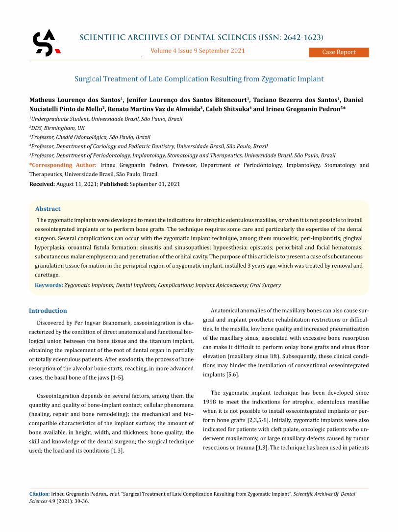
Clinically, a slight symptomatic edema was observed in the ma-lar region on the right side (Figure 1). During palpation on clinical examination, the presence of more consistent granulation tissue was noted in the malar region, with painful symptoms.
Figure 1: Slight symptomatic edema observed in the malar region on the right side. Frontal view (A). Right side view (B).
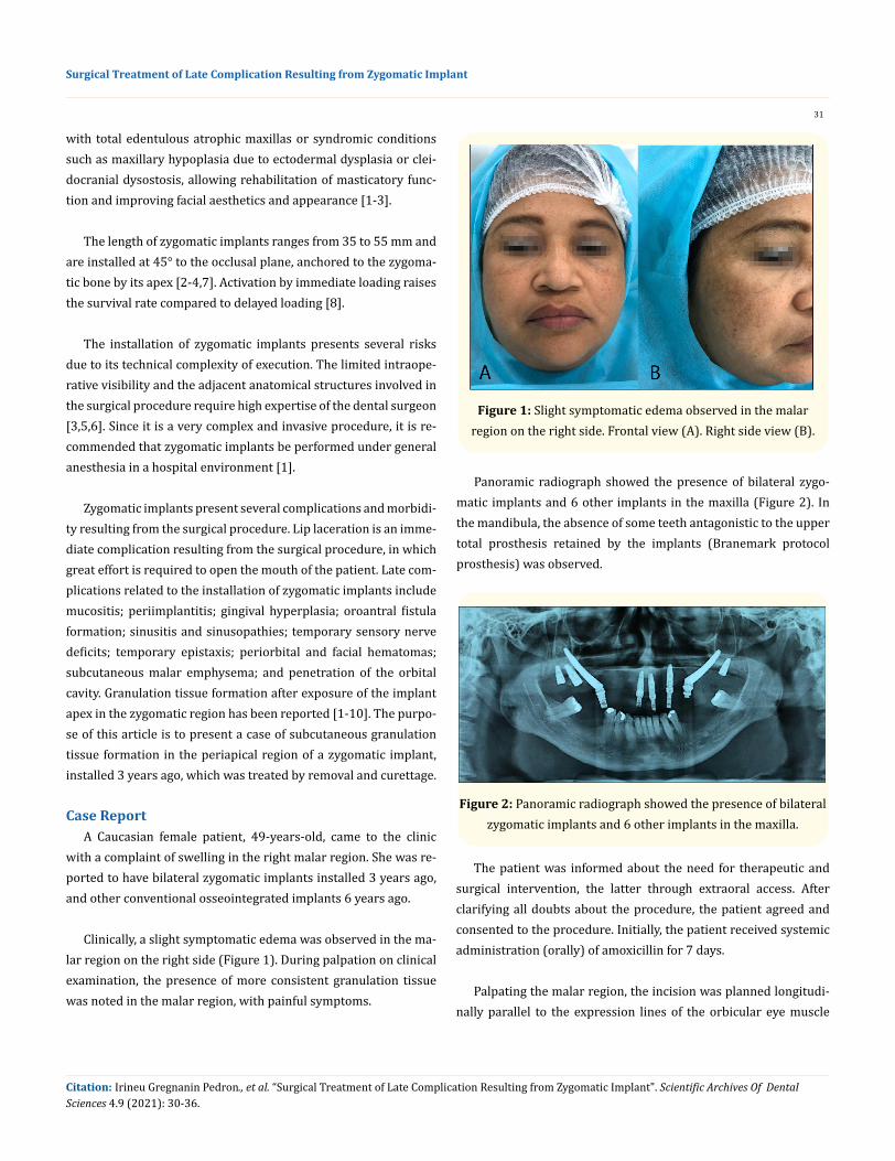
Panoramic radiograph showed the presence of bilateral zygo-matic implants and 6 other implants in the maxilla (Figure 2). In the mandibula, the absence of some teeth antagonistic to the upper total prosthesis retained by the implants (Branemark protocol prosthesis) was observed.
Figure 2: Panoramic radiograph showed the presence of bilateral zygomatic implants and 6 other implants in the maxilla.
The patient was informed about the need for therapeutic and surgical intervention, the latter through extraoral access. After clarifying all doubts about the procedure, the patient agreed and consented to the procedure. Initially, the patient received systemic administration (orally) of amoxicillin for 7 days.
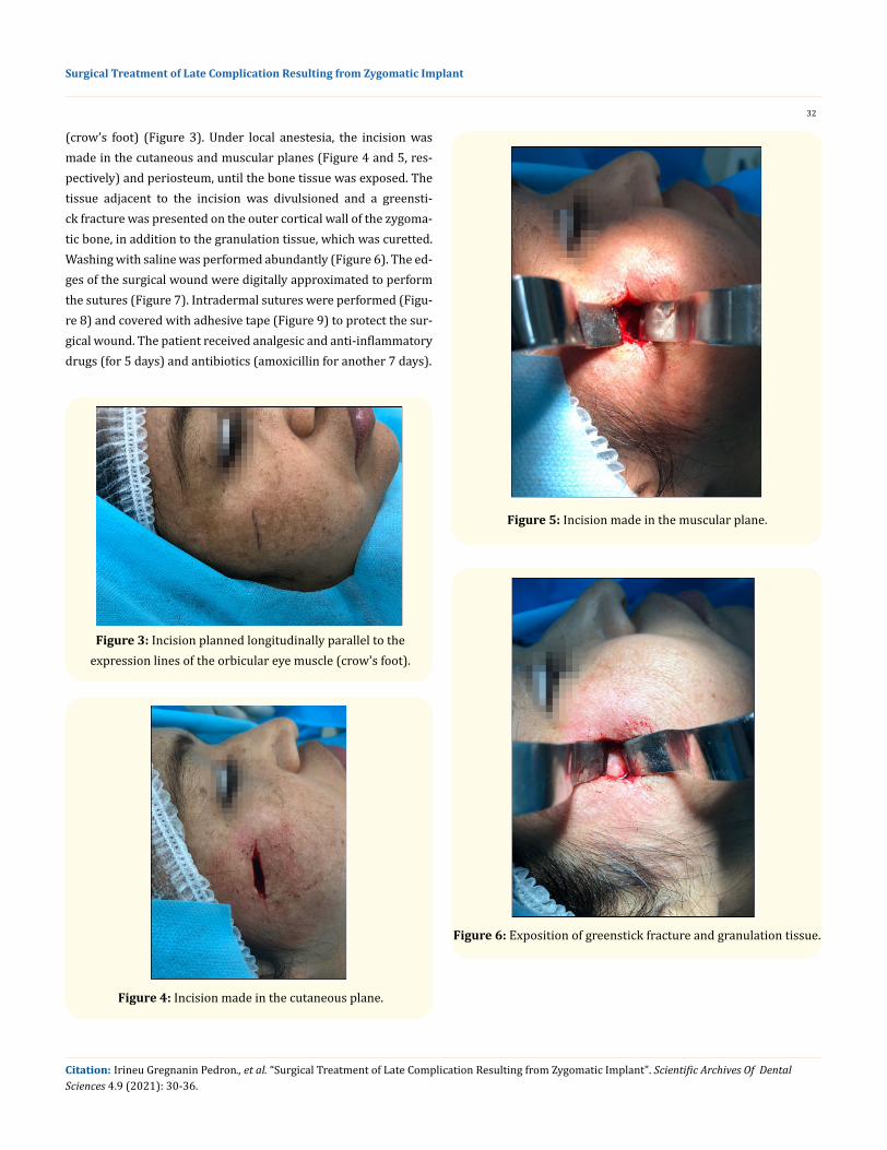
Palpating the malar region, the incision was planned longitudi-nally parallel to the expression lines of the orbicular eye muscle
Citation: Irineu Gregnanin Pedron., et al. “Surgical Treatment of Late Complication Resulting from Zygomatic Implant". Scientific Archives Of Dental Sciences 4.9 (2021): 30-36.
32
Surgical Treatment of Late Complication Resulting from Zygomatic Implant
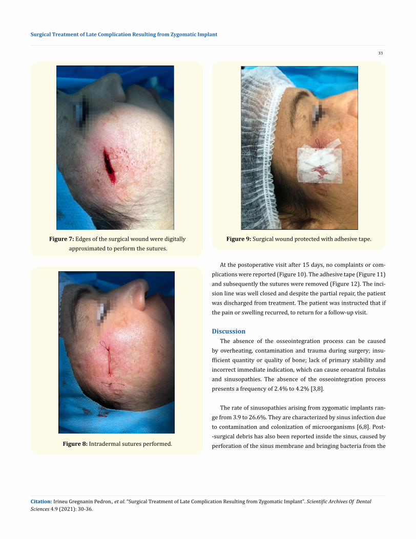
(crow’s foot) (Figure 3). Under local anestesia, the incision was made in the cutaneous and muscular planes (Figure 4 and 5, res-pectively) and periosteum, until the bone tissue was exposed. The tissue adjacent to the incision was divulsioned and a greensti-ck fracture was presented on the outer cortical wall of the zygoma-tic bone, in addition to the granulation tissue, which was curetted. Washing with saline was performed abundantly (Figure 6). The ed-ges of the surgical wound were digitally approximated to perform the sutures (Figure 7). Intradermal sutures were performed (Figu-re 8) and covered with adhesive tape (Figure 9) to protect the sur-gical wound. The patient received analgesic and anti-inflammatory drugs (for 5 days) and antibiotics (amoxicillin for another 7 days).
Figure 3: Incision planned longitudinally parallel to the expression lines of the orbicular eye muscle (crow's foot).
Figure 4: Incision made in the cutaneous plane.
Figure 5: Incision made in the muscular plane.
Figure 6: Exposition of greenstick fracture and granulation tissue.
Citation: Irineu Gregnanin Pedron., et al. “Surgical Treatment of Late Complication Resulting from Zygomatic Implant". Scientific Archives Of Dental Sciences 4.9 (2021): 30-36.
33
Surgical Treatment of Late Complication Resulting from Zygomatic Implant
Figure 7: Edges of the surgical wound were digitally approximated to perform the sutures.
Figure 8: Intradermal sutures performed.
Figure 9: Surgical wound protected with adhesive tape.
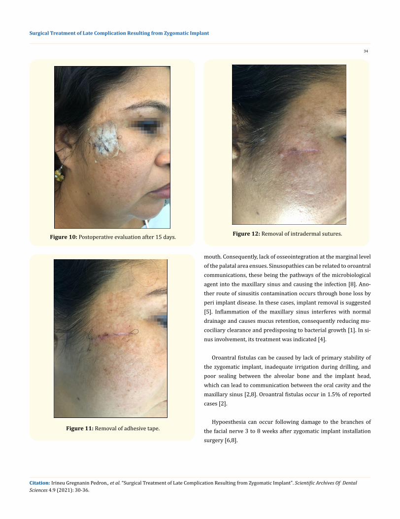
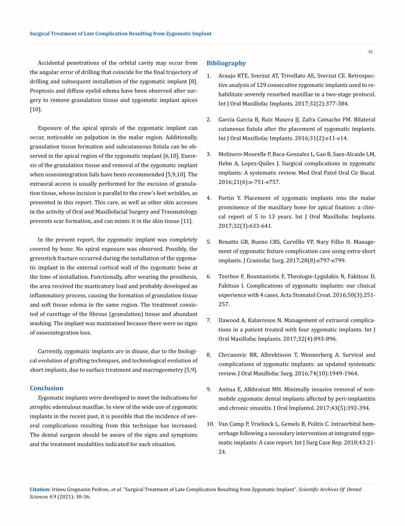
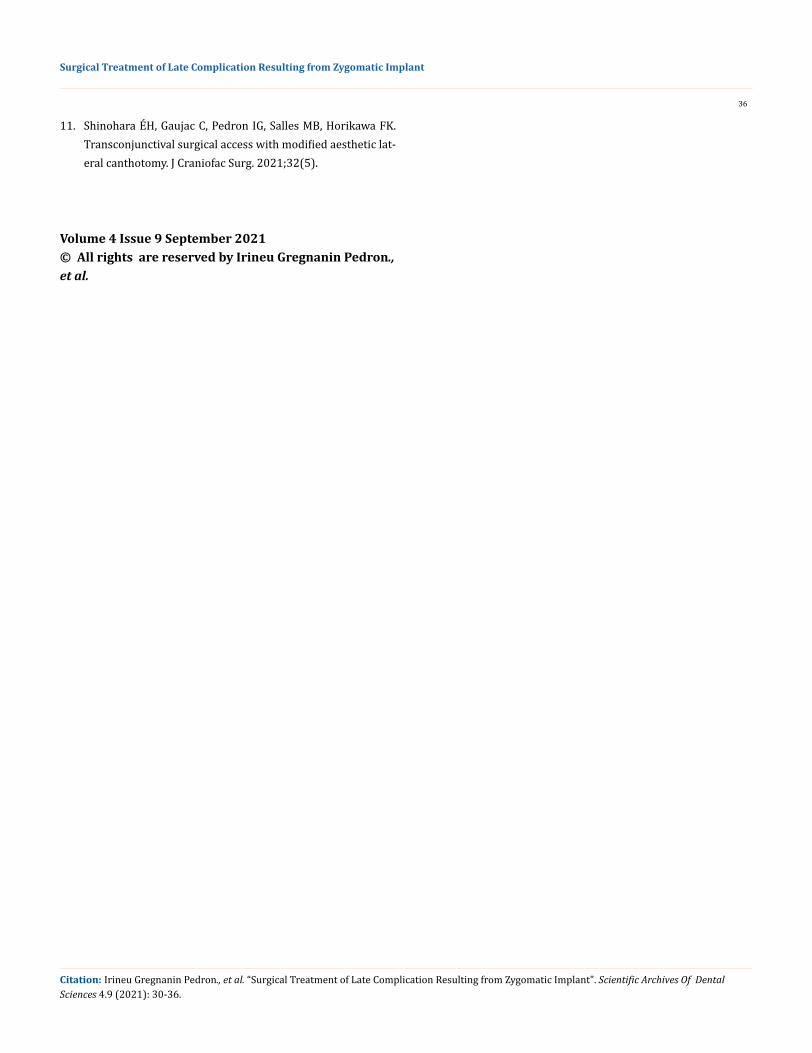
At the postoperative visit after 15 days, no complaints or com-plications were reported (Figure 10). The adhesive tape (Figure 11) and subsequently the sutures were removed (Figure 12). The inci-sion line was well closed and despite the partial repair, the patient was discharged from treatment. The patient was instructed that if the pain or swelling recurred, to return for a follow-up visit.
DiscussionThe absence of the osseointegration process can be caused
by overheating, contamination and trauma during surgery; insu-fficient quantity or quality of bone; lack of primary stability and incorrect immediate indication, which can cause oroantral fistulas and sinusopathies. The absence of the osseointegration process presents a frequency of 2.4% to 4.2% [3,8].
The rate of sinusopathies arising from zygomatic implants ran-ge from 3.9 to 26.6%. They are characterized by sinus infection due to contamination and colonization of microorganisms [6,8]. Post--surgical debris has also been reported inside the sinus, caused by perforation of the sinus membrane and bringing bacteria from the
Citation: Irineu Gregnanin Pedron., et al. “Surgical Treatment of Late Complication Resulting from Zygomatic Implant". Scientific Archives Of Dental Sciences 4.9 (2021): 30-36.
34
Surgical Treatment of Late Complication Resulting from Zygomatic Implant
Figure 10: Postoperative evaluation after 15 days.
Figure 11: Removal of adhesive tape.
Figure 12: Removal of intradermal sutures.
mouth. Consequently, lack of osseointegration at the marginal level of the palatal area ensues. Sinusopathies can be related to oroantral communications, these being the pathways of the microbiological agent into the maxillary sinus and causing the infection [8]. Ano-ther route of sinusitis contamination occurs through bone loss by peri implant disease. In these cases, implant removal is suggested [5]. Inflammation of the maxillary sinus interferes with normal drainage and causes mucus retention, consequently reducing mu-cociliary clearance and predisposing to bacterial growth [1]. In si-nus involvement, its treatment was indicated [4].
Oroantral fistulas can be caused by lack of primary stability of the zygomatic implant, inadequate irrigation during drilling, and poor sealing between the alveolar bone and the implant head, which can lead to communication between the oral cavity and the maxillary sinus [2,8]. Oroantral fistulas occur in 1.5% of reported cases [2].
Hypoesthesia can occur following damage to the branches of the facial nerve 3 to 8 weeks after zygomatic implant installation surgery [6,8].
Citation: Irineu Gregnanin Pedron., et al. “Surgical Treatment of Late Complication Resulting from Zygomatic Implant". Scientific Archives Of Dental Sciences 4.9 (2021): 30-36.
35
Surgical Treatment of Late Complication Resulting from Zygomatic Implant
Accidental penetrations of the orbital cavity may occur from the angular error of drilling that coincide for the final trajectory of drilling and subsequent installation of the zygomatic implant [8]. Proptosis and diffuse eyelid edema have been observed after sur-gery to remove granulation tissue and zygomatic implant apices [10].
Exposure of the apical spirals of the zygomatic implant can occur, noticeable on palpation in the malar region. Additionally, granulation tissue formation and subcutaneous fistula can be ob-served in the apical region of the zygomatic implant [6,10]. Exere-sis of the granulation tissue and removal of the zygomatic implant when osseointegration fails have been recommended [5,9,10]. The extraoral access is usually performed for the excision of granula-tion tissue, whose incision is parallel to the crow’s feet wrinkles, as presented in this report. This care, as well as other skin accesses in the activity of Oral and Maxillofacial Surgery and Traumatology, prevents scar formation, and can mimic it in the skin tissue [11].
In the present report, the zygomatic implant was completely covered by bone. No spiral exposure was observed. Possibly, the greenstick fracture occurred during the installation of the zygoma-tic implant in the external cortical wall of the zygomatic bone at the time of installation. Functionally, after wearing the prosthesis, the area received the masticatory load and probably developed an inflammatory process, causing the formation of granulation tissue and soft tissue edema in the same region. The treatment consis-ted of curettage of the fibrous (granulation) tissue and abundant washing. The implant was maintained because there were no signs of osseointegration loss.
Currently, zygomatic implants are in disuse, due to the biologi-cal evolution of grafting techniques, and technological evolution of short implants, due to surface treatment and macrogeometry [5,9].
ConclusionZygomatic implants were developed to meet the indications for
atrophic edentulous maxillae. In view of the wide use of zygomatic implants in the recent past, it is possible that the incidence of sev-eral complications resulting from this technique has increased. The dental surgeon should be aware of the signs and symptoms and the treatment modalities indicated for each situation.
Bibliography
1. Araujo RTE, Sverzut AT, Trivellato AE, Sverzut CE. Retrospec-tive analysis of 129 consecutive zygomatic implants used to re-habilitate severely resorbed maxillae in a two-stage protocol. Int J Oral Maxillofac Implants. 2017;32(2):377-384.
2. Garcia Garcia B, Ruiz Masera JJ, Zafra Camacho FM. Bilateral cutaneous fistula after the placement of zygomatic implants. Int J Oral Maxillofac Implants. 2016;31(2):e11-e14.
3. Molinero-Mourelle P, Baca-Gonzalez L, Gao B, Saez-Alcaide LM, Helm A, Lopez-Quiles J. Surgical complications in zygomatic implants: A systematic review. Med Oral Patol Oral Cir Bucal. 2016;21(6):e-751-e757.
4. Fortin Y. Placement of zygomatic implants into the malar prominence of the maxillary bone for apical fixation: a clini-cal report of 5 to 13 years. Int J Oral Maxillofac Implants. 2017;32(3):633-641.
5. Benatto GB, Bueno CRS, Curvêllo VP, Nary Filho H. Manage-ment of zygomatic fixture complication case using extra-short implants. J Craniofac Surg. 2017;28(8):e797-e799.
6. Tzerbos F, Bountaniotis F, Theologie-Lygidakis N, Fakitsas D, Fakitsas I. Complications of zygomatic implants: our clinical experience with 4 cases. Acta Stomatol Croat. 2016;50(3):251-257.
7. Dawood A, Kalavresos N. Management of extraoral complica-tions in a patient treated with four zygomatic implants. Int J Oral Maxillofac Implants. 2017;32(4):893-896.
8. Chrcanovic BR, Albrektsson T, Wennerberg A. Survival and complications of zygomatic implants: an updated systematic review. J Oral Maxillofac Surg. 2016;74(10):1949-1964.
9. Anitua E, Alkhraisat MH. Minimally invasive removal of non-mobile zygomatic dental implants affected by peri-implantitis and chronic sinusitis. J Oral Implantol. 2017;43(5):392-394.
10. Van Camp P, Vrielinck L, Gemels B, Politis C. Intraorbital hem-orrhage following a secondary intervention at integrated zygo-matic implants: A case report. Int J Surg Case Rep. 2018;43:21-24.
Citation: Irineu Gregnanin Pedron., et al. “Surgical Treatment of Late Complication Resulting from Zygomatic Implant". Scientific Archives Of Dental Sciences 4.9 (2021): 30-36.
36
Surgical Treatment of Late Complication Resulting from Zygomatic Implant
11. Shinohara ÉH, Gaujac C, Pedron IG, Salles MB, Horikawa FK. Transconjunctival surgical access with modified aesthetic lat-eral canthotomy. J Craniofac Surg. 2021;32(5).
Volume 4 Issue 9 September 2021© All rights are reserved by Irineu Gregnanin Pedron., et al.
Citation: Irineu Gregnanin Pedron., et al. “Surgical Treatment of Late Complication Resulting from Zygomatic Implant". Scientific Archives Of Dental Sciences 4.9 (2021): 30-36.