Embed Size (px)
Citation preview
IRANIAN SOCIETY OFCARDIAC SURGEONS
The Iranian Journal of Cardiac Surgery
President of ISCS
Vice President of ISCS
General Secretary
Editorial Board
Editor-in-chief
Managing Editor
Technical Editors
Address
M. A. Yousefnia M.D
S. Davoodi M.D.
R. Baghaei M.D.
H. Radmehr M.D.S. H. Hassantash M.D.E. Asdaghpoor M.D.M. H. Mandegar M.D.D. Javidi M.D.K. Abbassi M.D.K. Babazadeh M.D.M. Delavarkhan M.D.A. Tabib M.D.F. Roshanali M.D.A. Noori M.D.A.A. Ghavidel M.D.
M. A. Yousefnia M.D.
R. Baghaei M.D.
M. Marzban M.D.M. Mehrparvar M.D.
ContributersM. Abbassi M.D.N.M. Noori M.D.K. Mozaffari M.D.M. Mahjoubifaed M.D.A.Monirpoor M.DS.A.Mirdehghani M.D.Sh. Roodpeyma M.DH. Hoseinkhah M.D.
2nd Floor; #4; Nikrai Alley; Kazeroon St.; Mirdamad Blvd.; Tehran. IRAN • P. Code: 1919913534Tel: 26401341 - 22279743 • Fax: 26401340Email: [email protected] • www.iscs.org.ir
OfficeM. Arab
Art & Printing SupervisorM. Ghandi
English EditorNick Austin
IRANIAN SOCIETY OFCARDIAC SURGEONS
The Iranian Journal of Cardiac Surgery Vol.3 No.4 & Vol.4 No.1 May 2012
Contents
Information for Authors: 1
Predicted Operative Factors for Early Mortality after OFFPUMP Coronary Artery Bypass Grafting Surgery (CABG)t
3
Evaluation of Brain Natriuretic Peptides in Early Diag-nosis of Cardiac Involvement Comparing to Echocar-diographic Findings in Major Thalassemia Patients
7
The Effect of Starch VS Crystalloid Administration of Cardiopulmonary Bypass Prime Solution on Tissue and Organ Perfusion and Coagulation Status
13
Clinical Course of Ventricular Septal Defect in Children Referred to Aliasghar Center of Zahedan during 2001-2011
16
All That Glitters Is Not Gold and All Myxoid Tumors Are Not Myxomas
22
What’s New in Cardiac Surgery? 24
Aorta to Left Atrial Fistula Following Transcatheter Closure of Atrial Septal Defect
34
Aortic valve endocarditis. A rare presentation of brucellosis
38
Emergency CABG and Surgical Retrieval of Entrapped Coronary Stent Balloon: A Case Report
42
Congenital Left Main Coronary Artery Fistula to Right Atrium : A Case Report
44
May 20121
The Iranian Journal of Cardiac Surgery is the official quarterly publication of the Iranian Society of Cardiac Surgeons (ISCS). The editorial board encourages submission of original papers concerned on any aspects of cardiovascular medicine including cardiac surgery, adult and pediatric cardiology, cardiac anesthesia and cardiac intensive care in the form of both basic and clinical research. Submitted articles should neither have been published previously nor be considered for publication elsewhere.Each article will be carefully reviewed by the editorial board. Additional review may be requested from the specialists in the related field. Then the corresponding author will be informed regarding acceptance or rejection of the article.
Papers in the following categories are accepted for publication in the journal;
• Guest editorial • Original article (Basic or Clinical science)• Review article • Case report• How to do it? (Presentation of a new technique of surgery
or interventis, Conclusion. Abstract of review articles and case reports should be nonstructured and no more than 200 and 60 words respectively. A maximum of six key words may be added at the end of the abstract.
5- Prepare your manuscript as precise, descriptive, and conclusive as possible. For this purpose, the Introduction should be brief and set out the aim for which the study has been performed. The Materials and Methods should be sufficiently detailed so that readers can understand precisely what has been done. The Results should be presented clearly with definition of relevant positive and negative findings. The Discussion should relate directly to the study and interpret the results and their relevance as well as indicate the limitations of the study.
6- Reviews of recent developments are welcome. Materials in the Review Article should be informative, presenting the most recent advances and challenges about the subject.
Information for Authors:
May 20122
7- Presentation of interesting cases which add new or important information about specific diseases and description of innovative technique of surgery or intervention will be accepted for publication as Case Report and How to do it?, respectively. Articles in these sections should have no more than three authors, 1200 words, three figures or tables and a maximum of eight references.
8- References should be numbered consecutively (in superscript) as they appear in the text. Style and punctuation of references should conform to the Index Medicus format.
9- Tables should be typed double- spaced, each with a number and title above the table and explanatory footnotes. Figures must be submitted in three sets, indicating their numbers, and be suitable for high quality reproduction. Legends to illustrations must be typed double-spaced separately. Figure numbers should correspond to the order in which they appear in the text.
10- Send all printed manuscripts, accompanied by their electronic files on compact disc (CD) to the following address;Iranian Society of Cardiac Surgeons2nd floor, No. 4, Ni
IntroductionCABG is still the main treatment for pa-tients with three-vessel coronary disease. The indications are well documented and the results are relatively satisfying in terms of low mortality and morbidity (1). The incidence of risk factors and pre-operative comorbidities is increasing (2) and many patients candidated for surgery, are at an advanced age, with severe left ventricular dysfunction, chronic renal dis-ease, peripheral vascular disease, chronic bronchopulmonary disease, etc. To im-prove the management of these patients, surgeons needed to adapt their operating
techniques. CPB can trigger numerous complications or worsen pre-existing or-gan damage (cardiac, pulmonary, renal), which in turn may increase operative morbidity and mortality. OPCAB strategy is to be able to carry out revascularization as complete as possible, under technical conditions offering maximum safety for the patient, by avoiding the triggering of possible complications induced by CPB and by avoiding myocardial ischemia (3, 4). It seems that OPCAB offers results at least equal compared to CPB under car-dioplegic arrest in terms of low or mod-erate risk patients (5). Patients condition,
Predicted Operative Factors for Early Mortality after OFFPUMP Coronary Artery Bypass Grafting Surgery (CABG)Mohammad Abbasi Tashnize MD, Hamid Hoseinikhah Manshady MD*, Nahid Zirak MD, Mahmood Hoseinzade Maleki MD, Yavar Shams Hojaty MD
AbstractObjective: OFFPump CABG surgery (opcab) is performing worldwide and the rate of coronary revascularization without CPB (cardiopulmonary Bypass) is growing sig-nificantly. The aim of this study is to evaluate the risk factors for early mortality after OPCABG.Methods: From April 2009 to April 2011, data were collected from a total of 920 patients who underwent OFFPump CABG. Variables that were recorded were age, gender, EF, preoperative Cr, any comorbidity diseases like DM, HTN, Hyperlipidemia, Valvular pathology especially MR and TR. In postoperative period, need for Reex-ploration and any neurologic complications.Result: Older age was a risk factor for early mortality (30 days) after OFFPump CABG, but female gender was not a risk factor. Although in dead patients the average EF was lower but low EF was not an important risk factor for early mortality. Mild Cr elevation was not a risk factor for early mortality as well. In CAD patients, valvular pathology is a known risk factor for mortality. In our study Sever TR was a risk factor for early mortality but MR was not a risk factor for early mortality. Need for early reexploration was a factor for early mortality.Conclusion: risk factors of CABG- Ischemic heart disease- cardiopulmonary bypass- coronary heart disease- OPCAB post operative mortality
Key words: Beta (β)-thalassemia Major; Systolic and diastolic dysfunction; Echocar-diography.
* Corresponding Author: Hamid Hoseinikhah ManshadyEmail: [email protected] Tel:09153046163- 0511-8525307Department of Cardiac Surgery, Imam Reza Hospital, Mashhad, IRAN
May 20124
Iranian Society of Cardiac Surgeons
who candidated for myocardial revascularization surgery has changed in recent years: patients are older, with myo-cardial infarction and Left ventricular dysfunction, with greater comorbidities, which means the predicted opera-tive risk is greater than for those operated in the past. How-ever, despite the worse characteristics of the patients and increased predicted risk, Mortality of CABG was decreased recently. Until the mid-1990s, coronary bypass surgery was performed with cardiopulmonary bypass (CPB) and cardio-plegia in almost all cases, producing an arrested and flaccid heart and providing ideal conditions for the construction of anastomoses in arteries 1 to 2 mm in diameter. However, by placing the blood in contact with nonbiological sur-faces, CPB causes systemic inflammatory reactions and a series of deleterious effects in various organs (6). With the aim of further reducing mortality and preventing morbid-ity resulting from the use of CPB, at the end of the 1990s various teams began performing beating heart or off-pump coronary artery bypass (OPCAB) surgery, originally for revascularization of the left anterior descending artery and later for arteries located in the inferior and lateral walls ( 7). There is little consensus in the literature on the advan-tages of not using CPB. Some studies have indicated that mortality is lower in OFFPump coronary surgery compared to patients operated with CPB. Others have shown reduced morbidity, particularly a lower incidence of transfusion and fewer renal and neurologic complications, lower levels of biochemical markers of myocardial ischemia, and shorter ICU and hospital stay (8-10). The objective of this study was evaluation of risk factors for early mortality (30 days) after OFFPUMP Coronary artery bypass surgery.
Methods:Between April 2009 and April 2011, 920 patients undergo-ing isolated myocardial revascularization surgery, in Car-diovascular surgery department in Imam Reza hospital of Mashhad medical science university were evaluated in a cross sectional study. Exclusion criteria was recent MI, unstable hemodynamic states, recurrent and intractable ventricular arrhythmia especially VT/VF. Only patients undergoing Primary isolated OPCAB through median ster-notomy were included. Re-operative OFFPump CABG and minimal invasive direct coronary artery bypass (MIDCAB) procedures were also excluded. Preoperative, intraopera-
tive and postoperative data were recorded. The variables that were recorded consisted of preoperative EF, history of HTN and DM, hyperlipidemia, smoking and addiction. Coronary Angiography showed that 760 patients (68%) had 3VD, and others had 1VD and 2VD. Renal function tests also were recorded with preoperative Cr. Echocardiogra-phy data about valvular heart pathology were recorded with special attention to MR and TR. Patients were operated with median sternotomy approach and OFFPump facili-ties. During surgery, if there was any necessity to emergent conversion OFFPump to conventional CABG, patient was excluded from the study. The number of constructed grafts and any significant problems were recorded, and in the end of procedure, intubated patient was transferred to ICU, un-der monitoring and with infusion of low dose dopamine (5 microgr/Kg/Min). In ICU, any mortality and morbidity of patients were recorded and they closely followed up, dur-ing hospital stay, and after releasing from hospital, patients re-evaluated and visited in a regular program for 30 days. Any event that was necessitated to Re-explore the Sternum was recorded too.
Result:In this study 920 patients with Coronary Artery Disease who were candidated for OFFPump CABG was evaluated. 557 patients (60.5%) were male and 363 (39.5%) were female. Mean age of patients was 58.47 with range 27-80 years. Due to Echocardiography data, mean EF was 45.12% and in range of 15%-66%. In this study, Prevalence of HTN was 47.7% (439 patients), for DM was 27.8% (256patients), and for Hyperlipidemia was 37.9% (349 patients). Smoking and other addiction history was seen in 195 patients (21.2%). Preoperative Cr greater than 2mg/dl was seen in 93 patients (10.1%). Preoperative Echocardiography showed that MR was seen in 189 patients (20.5%) with different degrees. 115 patients(12.5%) had mild MR, 55 patients (5%) had moderate MR and 19 patient (2%) had sever MR, and also 88 patients (9.6%) had some degrees of TR that 86 (9.3%) had mild TR and only Two patients (.2%) had Sever TR (Table 1). In our study 40 patients (4.3%) were necessitated to Sternum Re-exploration, due to excessive hemorrhage and Drainage, Tamponade, Cardiac Arrest, Graft failure and other reasons during ICU or Hospital stay and until 30 days. In our study the mortality rate was 1.2% (11 patients).
May 20125
The Iranian Journal of Cardiac Surgery
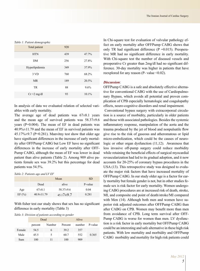
Table 1: Patient demographicTotal patient 920
HTN 439 47.7%
DM 256 27.8%
Hyperlipidemia 349 37.9%
3 VD 760 68.2%
MR 189 20.5%
TR 88 9.6%
Cr >2 mg/dl 93 10.1%
In analysis of data we evaluated relation of selected vari-ables with early mortality.The average age of dead patients was 67±8.1 years and the mean age of survived patients was 58.37±9.6 years (P=0.004). The mean of EF in dead patients was 40.9%±11.79 and the mean of EF in survived patients was 45.17%±9.7 (P=0.281). Manvitny test show that older age have significant differences in the increase of early mortal-ity after OFFPump CABG but Low EF have no significant differences in the increase of early mortality after OFF-Pump CABG, although the mean of EF was lower in dead patient than alive patients (Table 2). Among 909 alive pa-tients female sex was 39.2% but this percentage for dead patients was 54.5%.
Table 2: Patients age and LV EF
Mean SD
Dead alive P-value
Age 67±8.1 58.37±9.6 0.04EF (%) 40.9±11.79 45.17±9.7 0.281
With fisher test our study shows that sex has no significant difference in early mortality (Table 3)
Table 3: Division of patients according to genderDead Alive
percent Number Percent number P-value
Female 54.5 6 39.2 3570.365Male 45.5 5 60.7 552
Sum 100 11 100 909
In Chi-square test for evaluation of valvular pathology ef-fect on early mortality after OFFPump CABG shows that only TR had significant difference (P =0.015). Preopera-tive MR had no significant difference in early mortality. With Chi-square test the number of diseased vessels and preoperative Cr greater than 2mg/dl had no significant dif-ference. 30-day mortality was higher in patients that have reexplored for any reason (P- value <0.02).
Discussion:OFFPump CABG is a safe and absolutely effective alterna-tive for conventional CABG with the use of Cardiopulmo-nary Bypass, which avoids all potential and proven com-plication of CPB especially hematologic and coagulopathy effects, neuro-cognitive disorders and renal impairment. Conventional bypass surgery with extracorporeal circula-tion is a source of morbidity, particularly in older patients and those with associated pathologies. Besides the systemic inflammatory response, manipulation of the aorta and the trauma produced by the jet of blood and nonpulsatile flow give rise to the risk of gaseous and atheromatous or lipid micro-embolization, which could be the causes of neuro-logic or other organ dysfunction (11,12). Awareness that less invasive off-pump surgery could reduce morbidity while retaining the beneficial effects of surgical myocardial revascularization had led to its gradual adoption, and it now accounts for 20-25% of coronary bypass procedures in the USA (13). This retrospective study was designed to evalu-ate the major risk factors that have increased mortality of OFFPump CABG. In our study older age is a factor for ear-ly mortality but female gender is not, but in other studies fe-male sex is risk factor for early mortality. Women undergo-ing CABG procedures are at increased risk of death, stroke, MI, and composite end point of death/stroke/MI compared with Men (14). Although both men and women have su-perior risk adjusted outcomes after OFFPump CABG than after CABG on CPB. Women may benefit more than men from avoidance of CPB. Long term survival after OFF-Pump CABG is worse for women than men. LV dysfunc-tion is a risk factor in early mortality but OFFPump CABG could be an interesting and safe alternative in these high risk patients. With low mortality and morbidity and OFFPump CABG morbidity and mortality for high risk patients could
May 20126
Iranian Society of Cardiac Surgeons
be improved. In contrast, we found that preoperative EF 30% was an independent risk factor for postoperative com-plications and in-hospital mortality. A large study on 55,515 patients undergoing CABG concluded that those with low EF were sicker at baseline and had more than 4-times high-er mortality than patients with normal EF. We found signifi-cantly higher rates of postoperative cardiac complications, neurologic events, and infections, intra-aortic balloon pump and inotropic support in patients with LV dysfunction (15). CABG without CPB has a renal protection in compare to conventional coronary revascularization surgery and there-fore in patients with borderline renal function OFFPump CAB is recommended. Mild Cr elevation (Cr = 2-3 mg/dl) may not worsen the prognosis for OFFPump CABG, but in CRF patients with Dialysis dependent, it is a strong risk factor for early mortality after surgery (16). DM is a risk factor for early mortality and morbidity. These patients almost have small and diffuse coronary artery and multiple prior MI. Mortality in diabetic patients is greater than non-diabetic patients and also they have greater morbidity due to sternal and wound complications and renal impairment. Development of different neurologic complications from TIA to Stroke is a strong factor for early mortality and ma-jor morbidity in OFFPump CABG (17).
Conclusion:OFFPump CABG is a safe and effective method for coro-nary artery revascularization with fewer mortality and mor-bidity compared with conventional CABG with Cardiopul-monary Bypass.OFFPump CABG can be done for nearly all of coronary artery disease patients and risk factor for early mortality is similar to conventional CABG.
Refferences: 1. Eagle KA, Guyton RA, Davidoff R, Edwards FH. Coronary artery by-
pass graft surgery: summary article: a report of the Ameican College of cardiology/American heart Assosiation Task Force of practice Guidline. Crculation 2204; 110:1168-1176.
2. Lioyd Gones DM, Wilson PW, Larson MG, Beiser A. Framingham risk score and prediction of lifetime risk for Coronary artery disease. AM J Cardiol 2004; 94:20-24.
3. El-Hamasy L,Cartier R, Demers P, Bouchard D.Long term result after sys-tematic Off-pump Coronary artery bypass graft surgery in 1000 consecu-tive patients. Circulation 2006; 114:1486-91.
4. Brown JM, Poston RS, Gammie JS, Cardarelli MG, Schwartz K. Offpump versus Onpump coronary artery bypass grafting in consecutive patients: decision –making algorithm and outcomes. Ann Thorac Surg 2006; 81:555-561.
5. Nathoe HM, Van Dijk D, Jansen EW, Sukker WJ. A comparison of On-pump and Off-pump coronary bypass surgery in low risk patients. N Engl J Med 2003; 348:394-402.
6. Menasche P. The systemic factor: the comparative roles of cardiopulmo-nary bypass and Offpump surgery in the genesis of patient injury during and following cardiac surgery. Ann Thorac Surg 2001; 72:s2260-5.
7. UVA Mi, Matias F, Pereira F, Rodrigues V, Pedro A. Less invasive coro-nary artery bypass surgery: Early result in 1083 patients operated Off-pump. Rev port Cardiol 2005; 24:925-941.
8. Al Ruzzeh S, Nakmura K, Athanasiou T. Off-pump coronary artery bypass surgery reduce risk-stratified morbidity and mortality.Circulation 2003; 108:1-8.
9. Calafiore AM, Di Mauro M, Canosa C, Di Gianmmarco G. Myocardial revascularization with and without cardiopulmonary bypass: advantages and disadvantages and similaries. Eur J Cardiothorac Surg 2003; 24:953-60.
10. Mack MJ, Pfister A, Bchard D. Comparsion of coronary bypass surgery with and without cardiopulmonary bypass in patients with multivessel disease. J Thorac Cardiovas Surg 2003;127:167-73.
11. Abu-omar Y, Balacumaraswami L, Pigott DW, Matthews PM, Taggart D. Solid and gaseous cereberal microorganism during Offpump, on-pump and open cardiac surgery procedures. J Thorac cardiovas surg 2004; 127:1759-65.
12. Paparella D, Yau TM, Young E.Cardiopulmonary bypass induced inflam-mation: Pathophysiology AND Treatment. Eur J Thorac Cardiovasc Surg 2002; 21:232-44.
13. Mack MJ, Pfister A, Bachand D et al. Comparison of coronary bypass surgery with and without cardiopulmonary bypass in patients with multi-vessel disease. J Thorac cardiovasc Surg 2003; 127:167-73.
14. Eifet S, Kilian E, Fernandez A, Juchem G. Early and mid term mortality after coronary artery bypass grafting in women depends on th surgical protocol: Retrospective analysis of 3441 On- and Off-Pump Coronary ar-tery bypass grafting procedures. Journal of Cardiothoracic surgery; 2010, 90.
15. Calafiore A, Di Mauro M, Teodori G.Impact of aortic manipulatiob on incidence of cereberovascular accidents afte surgical myocardial revascu-larization. Ann Thorac Surg . 2002; 73:1387-1393.
16. Barandon L, Richebe P, Munos E, Caldernon J.OffPump Coronary artery bypass surgery in very high risk patients: adjustment and preliminary re-sults. Interact Cardio Vasc Thorac Surg 2008; 7:789-793.
17. Puskas J, Kilgo P, Lattouf O,Thourani V.Off-Pump coronary Bypass pro-vides Reduced Mortality and Morbidity and equivalent 10 Year survival. Ann Thorac Surg 208; 86:1139-46.
Evaluation of Brain Natriuretic Peptides in Early Diagnosis of Cardiac Involvement Comparing to Echocardiographic Findings in Major Thalassemia PatientsNoor Mohammad Noori MD 1, Mehdi Mohammadi MD2, Hossein Ali Khazaei MD3, Maziar Mahjoubifard MD4*
1-Professor of Pediatric cardiology, Research Center of Children and Adolescent Health, ALI-EBN E-ABITALEB hospital, Zahedan University of medical sciences, Zahedan, Iran 2-Holder of Doctorate Degree in Health and Statistic – Assistant Professor of Zahedan University of medical sciences, Zahedan, Iran 3-Immunology, PHD, Zahedan University of medical sciences, Zahedan, Iran4-*corresponding author: Assistant professor of anesthesiology, fellowship of cardiac anesthesia, Research Center for Children and Adolescent Health, ALI-EBN E-ABITALEB hospital, Khash road, Zahedan university of medical sciences, Zahedan, Iran, E- mail: [email protected] center for children and adolescents health of Zahedan medical Sciences University and, Zahedan University of Medical Sciences, Zahedan-Iran
Abstract:Background: Heart disease is one of the leading causes of disability in major thalas-semia patients. Timely diagnosis and effective treatment in these patients are essential. The aim of this study is to evaluate the diagnostic value of BNP in the diagnosis of heart involvement compared with echocardiographic findings in patients with major thalassemia.Methods: This case-control study was carried out in patients with major thalassemia aged 9-25 years old admitted to Aliasghar hospital from October 2010 to November 2011. Patients with no obvious cardiac abnormalities were included. 80 major thalas-semia patients with 80 healthy children matched by age and sex were entered and for both, echocardiography was performed by a pediatric cardiologist. The serum level of Brain Natriuretic Peptides (BNP) was also evaluated. The data were analyzed by SPSS17.Results: The groups studied were matched well regarding age and gender (P =0.1346, 0.429). Regarding the echocardiographic results, some of the parameters of the left heart in case group were significantly higher than control group as well as some of the parameters of right heart. The mean value of BNP in case group was higher signifi-cantly. There was a significant correlation between BNP and right heart MPI (r = 0.229, P = 0.041) and age (r = 0.237, P = 0.035).Conclusion: Based on the results, systolic and diastolic function in patients with major beta thalassemia were impaired. Therefore, measurement of BNP level in addition to serial echocardiography is recommended to early diagnose heart involvement in pa-tients with major beta thalassemia without clinical symptoms.
Key words: major thalassemia, echocardiography, BNP and children
IntroductionPrecursor NT PRO BNP, BNP, is a prehor-mone with 134 amino acids that formed in moist and changed to BNP with 108 amino acid. This pre hormone is released by stress. Natriuretic peptides are cleared by kidneys. Hypovolemia, decreased blood pressure and renal failure lead to increase secretion
of BNP, especially “NT PRO BNP in the patients (1). NT Pro BNP and BNP bio-markers are commercially available and these biomarkers are widely used in the di-agnosis of heart failure. BNP measurement appears to be beneficial in order to diag-nose and classify the patients with chronic heart failure. This provides a better predic-
May 20128
Iranian Society of Cardiac Surgeons
tion of death than plasma norepinephrine or endothelinone (1).Increase in plasma BNP has been observed in the first days of life and its rapid decline occurs during the first week. The reason for increasing level of BNP immedi-ately after birth is unknown. Increased preoperative BNP was not due to chronic heart disease but is due to congeni-tal heart disease that is assumed that stretching the muscle cell walls is the primary stimulus for BNP secretion (2). Voskaridou and colleagues demonstrated that the occurrence of pulmonary hypertension in patients with β/S thalassemia was similar to patients with sickle cell disease (SCD) and the serum amount of NT Pro BNP was a strong indicator for patients with β/S thalassemia with pulmonary hyperten-sion and in addition to the findings of echocardiography, it may be used for diagnosis of pulmonary hypertension (3). Kremastinous and colleagues showed that in patients with ma-jor thalassemia plasma level of BNP and NT pro BNP signifi-cantly increased when diastolic left ventricular dysfunction occurred. NT Pro BNP marker seemed to have better predic-tive value than BNP in detecting abnormalities of left ventric-ular diastolic function in patients with major thalassemia (4). Garadah and colleagues indicated that patients with ma-jor thalassemia had high serum NT Pro BNP in relation with levels of E / Em in Doppler tissue imaging (DTI) (5). Marwick and colleagues have indicated that activ-ity can increase BNP biomarker in patients with dia-stolic dysfunction confirmed evidently by Dop-pler tissue imaging (DTI) echocardiography (6). Berger has concluded that multidisciplinary care and BNP mea-surement, improved clinical outcomes in patients with heart failure after hospitalization (7). Since studies of the diagnostic value of BNP in determination of heart dysfunction in patients with beta major thalassemia are limited and early diagnosis of cardiac involvement in these patients reduces the incidence of mortality, this study was conducted with the purpose of early diagnosis of cardiac involvement in patients with major thalassemia compared to the findings of echocardiography.
Methods and materialsThis study was carried out in all asymptomatic major thalas-semia patients who referred to the center for patients with special diseases of Aliasghar hospital from 2010-2011. These patients were examined for chest radiography and electro-cardiogram. Patients with no important clinical symptoms of disease entered the study and those with hypertension,
endocrine disease, heart failure and valve involvement were excluded from the study. Patients underwent echocardiogra-phy 48-72 hours after receiving blood transfusions. Physi-cal examination and echocardiography were performed by one pediatric cardiologist in the same place using challenge 7000(made in Italy). There were eighty individuals aged 9-25 years old in each group matched by sex and age. Fol-lowing parameters were measured: MPI: myocardial per-formance index, LVMI: Left ventricular mass index, PEP: pre-ejection period, ET: ejection time, PEP/ET: pre-ejection period/ejection time ratio, IVSD: interventricular septal di-mension in diastole, LVPWD: left ventricular posterior wall dimension in diastole , IVSS: interventricular septal dimension in systole, LVPWS: left ventricular posterior wall dimension in systole, LVEDD :left ventricular end – diastolic dimension, LVESD: left ventricular end- systolic dimension, LVEDV: left ventricular end-diastolic volume, EF: ejection fraction, FS: fractional shortening, AO: Aorta diameter, LA: left atrium diameter, LA/AO: left atrium, aorta ratio, ICT: isovolumic contraction time, IRT: Isovolu-mic relaxation time, AT: acceleration time, DT :decelera-tion time, E/A ratio peak E/Peak A velocity in both groups. Three cardiac cycles were measured by Doppler echo M-Mode 2D and the mean value of each parameter was con-sidered. This method was performed in the supine position (supine) while breathing. M-Mode in the tip mitral valve in position of Para sternal view was obtained. The thickness of the wall between ventricular in systole and diastole, the wall thickness of posterior left ventricle in diastole and systole, the end diastolic and systolic left ventricular ejection fraction (EF), fraction shortening were obtained using M-Mode. The pulsed Doppler method was used to determine the veloc-ity of blood in the heart valves through E-velocity, A-veloc-ity, ejection time, pre ejection time, E/A, PEP/ET. After 3 or 4 days of transfusion, 5 cc blood was taken and the level of BNP was measured using ELISA kit BNP. Patients remained at rest for 30 minutes before blood sampling. All samples were centrifuged in around 3000 rps for 10 minutes at 4 cen-tigrade degree and plasma was kept in minus 80⁰c. The specificity and sensitivity of BNP laboratory testing compared to the results of echo were calculated. To analyze data, T-test, the Kappa coefficient of agreement, Mc-Namar Test and correlation coefficients were used.Consent form was obtained from patients and they were en-rolled if they were satisfied.
May 20129
The Iranian Journal of Cardiac Surgery
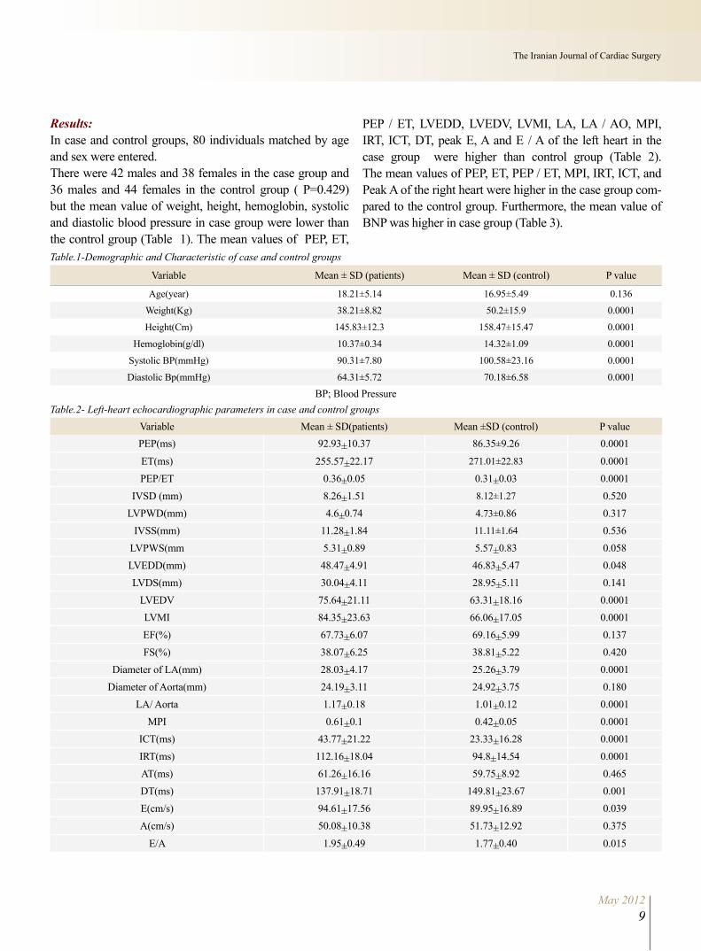
Results:In case and control groups, 80 individuals matched by age and sex were entered. There were 42 males and 38 females in the case group and 36 males and 44 females in the control group ( P=0.429) but the mean value of weight, height, hemoglobin, systolic and diastolic blood pressure in case group were lower than the control group (Table 1). The mean values of PEP, ET,
PEP / ET, LVEDD, LVEDV, LVMI, LA, LA / AO, MPI, IRT, ICT, DT, peak E, A and E / A of the left heart in the case group were higher than control group (Table 2). The mean values of PEP, ET, PEP / ET, MPI, IRT, ICT, and Peak A of the right heart were higher in the case group com-pared to the control group. Furthermore, the mean value of BNP was higher in case group (Table 3).
Table.1-Demographic and Characteristic of case and control groups
Variable Mean ± SD (patients) Mean ± SD (control) P value
Age(year) 18.21±5.14 16.95±5.49 0.136
Weight(Kg) 38.21±8.82 50.2±15.9 0.0001
Height(Cm) 145.83±12.3 158.47±15.47 0.0001
Hemoglobin(g/dl) 10.37±0.34 14.32±1.09 0.0001
Systolic BP(mmHg) 90.31±7.80 100.58±23.16 0.0001
Diastolic Bp(mmHg) 64.31±5.72 70.18±6.58 0.0001
BP; Blood PressureTable.2- Left-heart echocardiographic parameters in case and control groups
Variable Mean ± SD(patients) Mean ±SD (control) P valuePEP(ms) 92.93±10.37 86.35±9.26 0.0001
ET(ms) 255.57±22.17 271.01±22.83 0.0001PEP/ET 0.36±0.05 0.31±0.03 0.0001
IVSD (mm) 8.26±1.51 8.12±1.27 0.520LVPWD(mm) 4.6±0.74 4.73±0.86 0.317
IVSS(mm) 11.28±1.84 11.11±1.64 0.536LVPWS(mm 5.31±0.89 5.57±0.83 0.058LVEDD(mm) 48.47±4.91 46.83±5.47 0.048LVDS(mm) 30.04±4.11 28.95±5.11 0.141
LVEDV 75.64±21.11 63.31±18.16 0.0001LVMI 84.35±23.63 66.06±17.05 0.0001EF(%) 67.73±6.07 69.16±5.99 0.137FS(%) 38.07±6.25 38.81±5.22 0.420
Diameter of LA(mm) 28.03±4.17 25.26±3.79 0.0001Diameter of Aorta(mm) 24.19±3.11 24.92±3.75 0.180
LA/ Aorta 1.17±0.18 1.01±0.12 0.0001MPI 0.61±0.1 0.42±0.05 0.0001
ICT(ms) 43.77±21.22 23.33±16.28 0.0001IRT(ms) 112.16±18.04 94.8±14.54 0.0001AT(ms) 61.26±16.16 59.75±8.92 0.465DT(ms) 137.91±18.71 149.81±23.67 0.001E(cm/s) 94.61±17.56 89.95±16.89 0.039A(cm/s) 50.08±10.38 51.73±12.92 0.375
E/A 1.95±0.49 1.77±0.40 0.015
May 201210
Iranian Society of Cardiac Surgeons
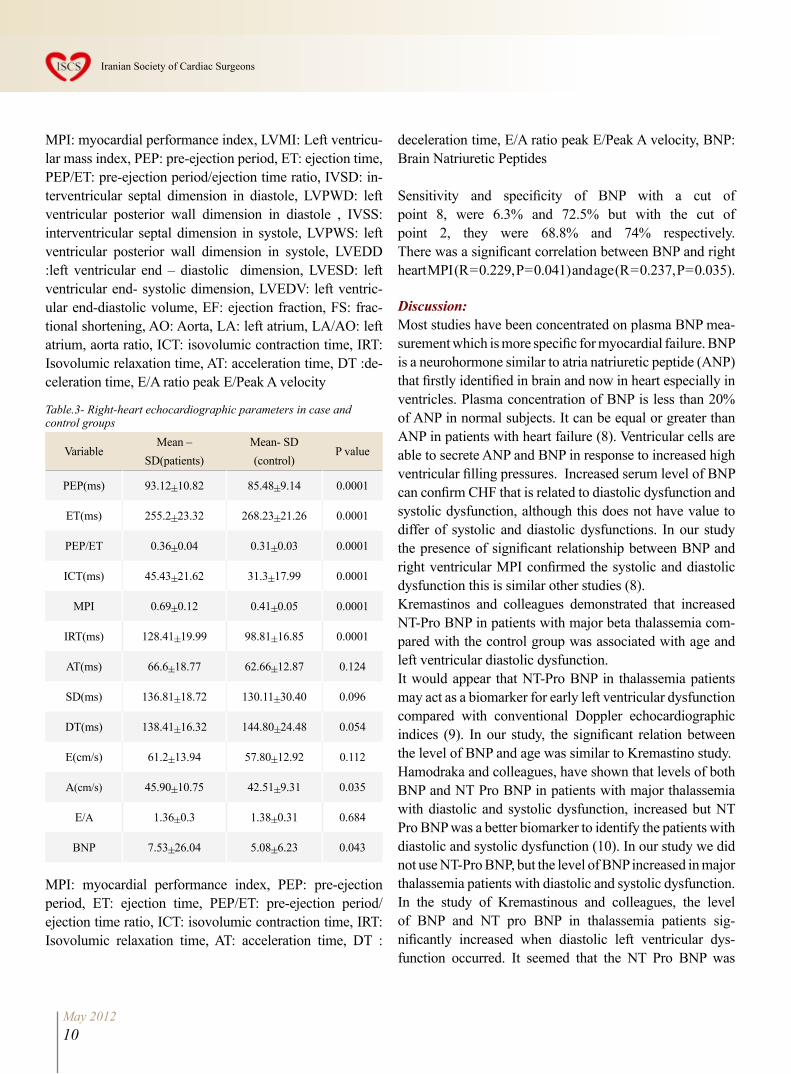
MPI: myocardial performance index, LVMI: Left ventricu-lar mass index, PEP: pre-ejection period, ET: ejection time, PEP/ET: pre-ejection period/ejection time ratio, IVSD: in-terventricular septal dimension in diastole, LVPWD: left ventricular posterior wall dimension in diastole , IVSS: interventricular septal dimension in systole, LVPWS: left ventricular posterior wall dimension in systole, LVEDD :left ventricular end – diastolic dimension, LVESD: left ventricular end- systolic dimension, LVEDV: left ventric-ular end-diastolic volume, EF: ejection fraction, FS: frac-tional shortening, AO: Aorta, LA: left atrium, LA/AO: left atrium, aorta ratio, ICT: isovolumic contraction time, IRT: Isovolumic relaxation time, AT: acceleration time, DT :de-celeration time, E/A ratio peak E/Peak A velocity
Table.3- Right-heart echocardiographic parameters in case andcontrol groups
VariableMean –
SD(patients)Mean- SD (control)
P value
PEP(ms) 93.12±10.82 85.48±9.14 0.0001
ET(ms) 255.2±23.32 268.23±21.26 0.0001
PEP/ET 0.36±0.04 0.31±0.03 0.0001
ICT(ms) 45.43±21.62 31.3±17.99 0.0001
MPI 0.69±0.12 0.41±0.05 0.0001
IRT(ms) 128.41±19.99 98.81±16.85 0.0001
AT(ms) 66.6±18.77 62.66±12.87 0.124
SD(ms) 136.81±18.72 130.11±30.40 0.096
DT(ms) 138.41±16.32 144.80±24.48 0.054
E(cm/s) 61.2±13.94 57.80±12.92 0.112
A(cm/s) 45.90±10.75 42.51±9.31 0.035
E/A 1.36±0.3 1.38±0.31 0.684
BNP 7.53±26.04 5.08±6.23 0.043
MPI: myocardial performance index, PEP: pre-ejection period, ET: ejection time, PEP/ET: pre-ejection period/ejection time ratio, ICT: isovolumic contraction time, IRT: Isovolumic relaxation time, AT: acceleration time, DT :
deceleration time, E/A ratio peak E/Peak A velocity, BNP: Brain Natriuretic Peptides
Sensitivity and specificity of BNP with a cut of point 8, were 6.3% and 72.5% but with the cut of point 2, they were 68.8% and 74% respectively. There was a significant correlation between BNP and right heart MPI (R = 0.229, P = 0.041) and age (R = 0.237, P = 0.035).
Discussion:Most studies have been concentrated on plasma BNP mea-surement which is more specific for myocardial failure. BNP is a neurohormone similar to atria natriuretic peptide (ANP) that firstly identified in brain and now in heart especially in ventricles. Plasma concentration of BNP is less than 20% of ANP in normal subjects. It can be equal or greater than ANP in patients with heart failure (8). Ventricular cells are able to secrete ANP and BNP in response to increased high ventricular filling pressures. Increased serum level of BNP can confirm CHF that is related to diastolic dysfunction and systolic dysfunction, although this does not have value to differ of systolic and diastolic dysfunctions. In our study the presence of significant relationship between BNP and right ventricular MPI confirmed the systolic and diastolic dysfunction this is similar other studies (8).Kremastinos and colleagues demonstrated that increased NT-Pro BNP in patients with major beta thalassemia com-pared with the control group was associated with age and left ventricular diastolic dysfunction.It would appear that NT-Pro BNP in thalassemia patients may act as a biomarker for early left ventricular dysfunction compared with conventional Doppler echocardiographic indices (9). In our study, the significant relation between the level of BNP and age was similar to Kremastino study.Hamodraka and colleagues, have shown that levels of both BNP and NT Pro BNP in patients with major thalassemia with diastolic and systolic dysfunction, increased but NT Pro BNP was a better biomarker to identify the patients with diastolic and systolic dysfunction (10). In our study we did not use NT-Pro BNP, but the level of BNP increased in major thalassemia patients with diastolic and systolic dysfunction. In the study of Kremastinous and colleagues, the level of BNP and NT pro BNP in thalassemia patients sig-nificantly increased when diastolic left ventricular dys-function occurred. It seemed that the NT Pro BNP was
May 201211
The Iranian Journal of Cardiac Surgery
a better predictive marker than BNP in order to detect left ventricle diastolic dysfunction in thalassemia pa-tients who have no significant signs of the diseases (4). In our study, there was no correlation between BNP and left ventricular markers, since we studied patients with ma-jor thalassemia without clinical symptoms of diseases with younger age. In the study of Aessopos and colleagues, cardiac compli-cations in major thalassemia were the causes of mortality in these patients and the main characteristic of disease was left ventricular dysfunction that was due to consume iron consumption. Levels of BNP in patients with impaired regional or global function and left ventricular diastolic dysfunction will arise and appear after increased wall stretch of left ventricle that is associated with the severity of symptoms and the prognosis of disease therefore the measurement of serum level of BNP in this cases can be important (11). In our study, patients who had symptoms of heart failure were excluded from the study and as a result, this study does not conform to our research. Krematinos and colleagues indicated that NT Pro BNP in patients with major thalassemia is related to aging and left ventricular diastolic dysfunction. NT Pro BNP biomarker appeared to be a marker of early left ventricular diastolic dysfunction compared with conventional Doppler echocar-diography index (9). In this study BNP was only available for research and because our patients were asymptomatic without clinical signs of dysfunction.Meloni and colleagues indicated in their study that in pa-tients with major thalassemia, NT Pro BNP was associated significantly with diastolic right ventricular dysfunction and the presence of myocardial fibrosis. (12). The present study demonstrated that there was a direct relationship be-tween increasing right ventricular MPI and BNP. Aessopos and colleagues showed that BNP was use-ful in predicting the risk of developing heart disease.BNP only increased in patients with obvious and signifi-cant dysfunction in heart disease and BNP levels did not reflect the severity of heart failure in these patients. In our study that was conducted on patients without sig-nificant symptom of the disease, BNP had direct corre-lation with increased right ventricular MPI since right ventricular involvement is earlier than left ventricle (13). Wahl and his colleague indicated that NT Pro BNP lev-
els had correlation with increased severity of pulmo-nary artery pressure and right ventricular dysfunction in patients with primary pulmonary artery pressure (14). This study has been performed in symptomatic patients whereas in our study patients were clinically asymp-tomatic. Thus the findings of two studies are different. Lim (15) and Galasko (16) studied the development of screening programs for the treatment of left ventricular systemic dysfunction. This study showed that ECG, Echo, BNP and NT Pro BNP are all cost effective for screening. All of the above studies were used for screening of left ven-tricular dysfunction, therefore this study like our study were conducted to prevent the occurrence of heart failure.Bursi and colleagues indicated that more than 1/ 2 of pa-tients with heart failure diastolic dysfunction occurred in 40% of isolated patients. EF and diastolic dysfunction was independently related to the level of BNP (17). Maisel had shown that mid regional Pro BNP as BNP were useful in diagnosis of heart failure with dyspnea (18). This research is consistent with our study since our patients were thalas-semia without clinical symptoms.
Conclusions:Based on the findings of this study, the patients with beta thalassemia, systolic and diastolic function of left and right heart were damaged. Therefore, it is recommended that in patients with major beta thalassemia without clear symp-toms and signs of heart involvement, the measurement of plasma level of BNP is necessary in addition to serial echo-cardiography in order to diagnose the early involvement of heart.
References:1. Braunwald E. Biomarkers in Heart Failure. N Engl J Med 2008;358:2148-
59.2. Law YM, Keller BB, Feingod BM, et al. Usefulness of Plasma B_type
Natriuretic Peptide to Identify Ventricular Dysfunction in Pediat-ric and Adult Patients With Congenital Heart Disease. Am J Cardiol 2005;95:474-478.
3. Voskaridou E, Tsetsos G, Tsoutsias A, Spyropoulou E, Christoulas D, Terpos E, Pulmonary hypertension in patients with sickle cell/b thalas-semia: incidence and correlation with serum N-terminal pro-brain natri-uretic peptide concentrations. Haematologica 2007; 92:738-743.
4. Kremastinos DT ,Hamodraka E, Parissis J, Tsiapras D, Dima K. Pre-dictive value of B-type natriuretic peptides in detecting latent left ven-tricular diastolic dysfunction in β-thalassemia major. American Heart Journal 2010; 159(1): 68-74.
5. Garadah TS , Mahdi N, Kassab S, Al Shoroqi I, Abu-Taleb A, Jamsheer A. The pro-BNP Serum Level and Echocardiographic Tissue Doppler
May 201212
Iranian Society of Cardiac Surgeons
Abnormalities in Patients with Beta Thalassemia Major, Clinical Medi-cine Insights: Cardiology2010; 4: 135–141.
6. Marwick TH, Schwaiger M. The Future of Cardiovascular Imaging in the Diagnosis and Management of Heart Failure, Part 2 Clinical Ap-plications, Circ Cardiovasc Imaging 2008;1:162-170.
7. Berger R, Moertl D, Peter S, Ahmadi R, Huelsmann M, Yamuti S , Wag-ner B, Pacher R : N-Terminal Pro–B-Type Natriuretic Peptide–Guided, Intensive Patient Management in Addition to Multidisciplinary Care in Chronic Heart Failure: A 3-Arm, Prospective, Randomized Pilot Study, Journal of the American College of Cardiology2010; 55( 7): 645-653.
8. Bhatia V, Nayyar P, Dhindsa S. Brain natriuretic peptide in diagnosis and treatment of heart failure. J Postgrad Med 2003; 49:182-5.
9. Kremastinos DT, Tsiapras DP, Kostopoulou AG, Hamodraka ES, Chaid-aroglou AS, Kapsali ED. NT-proBNP levels and diastolic dysfunction in βThalassaemia major patients, European Journal of Heart Failure 2007; 9 :531–536.
10. Hamodraka E, Paraskevaidis I, Parissis J, Tsiapras D, Kostopoulou A, Vrettou E,.Kapsali E, Kremastinos D. NT-pro BNP is a better biomarker than BNP for the identification of left ventricular dysfunction in beta-thalassemia major patients Eur J Heart Fail Suppl 2008;7(Suppl 1): 192-193.
11. Aessopos A, Farmakis D, Polonifi A, Tsironi M, Fragodimitri C, Hatzil-iami A, Karagiorga M, Diamanti-Kandarakis E. Plasma B-type natri-uretic peptide concentration in β-thalassaemia patients, European Jour-nal of Heart Failure 2007; 9: 537–541.
12. Meloni A, Pepe A, Zyw L, Positano V, Chiara Dell’Amico M, Passino C, et al. N-terminal fragment of proBNP is a marker of risk for right ventricular dysfunction and cardiac complications in thalassemia major,
Journal of Cardiovascular Magnetic Resonance 2010, 12 (Suppl 1): 284.
13. Aessopos A , Berdoukas V, Tsironi M . TheBrain natriuretic peptide in diagnosis and treatment of heart failure. e heart in transfusion dependent homozygous thalassaemia today – prediction, prevention and manage-ment, European Journal of Haematology2008; 80 (2) : 93–106.
14. Wahl S, Vichinsky E. Pulmonary hypertension in hemolytic anemias, f1000 Medicine Reports 2010, 2:10.
15. Lim TK, Dwivedi G, Hayat S, Collinson PO, Senior R. Cost Effective-ness of the B Type Natriuretic Peptide, Electrocardiography, and Por-table Echocardiography for the Assessment of Patients from the Com-munity with Suspected Heart Failure, Echocardiography 2007; 24(3) : 228–236.
16. Galasko GIW, Barnes SC, Collinson P, Lahiri A, Senior R. What is the most cost-effective strategy to screen for left ventricular systolic dysfunction: Natriuretic peptides, the electrocardiogram, hand-held echocardiography, traditional echocardiography, or their combination? European Heart Journal 2006; 27:193–200.
17. Bursi F, Weston SA, Redfield MM, Jacobsen SJ, Pakhomov S, Nkomo VT, Meverden RA, Roger VL. Systolic and Diastolic Heart Failure in the Community, JAMA. 2006; 296:2209-2216.
18. Maisel A, Mueller C, Nowak R , Peacock WF , Landsberg JW, Poni-kowski P , et al: Mid-Region Pro-Hormone Markers for Diagnosis and Prognosis in Acute Dyspnea: Results From the BACH (Biomarkers in Acute Heart Failure) Trial,Journal of the American College of Cardiol-ogy2010; 55( 19: 2062-2076.
The Effect of Starch VS Crystalloid Admin-istration of Cardiopulmonary Bypass Prime Solution on Tissue and Organ Perfusion and Coagulation StatusMohammad Abbasi MD, Hamid Hoseinikhah Manshady * MD, Aliasghar Moinipoor MD, Nahid Zirak MD, Mahmoud Hosseinzade Maleky MD, Ahmad Amozeshy MD
*Correspondence to: Hamid Hoseinikhah ManshadyEmail: [email protected] Tell: 09153046163- 05118525307Department of Cardiac surgery, Imam Reza Hospital of Mashhad University, Mashhad, Iran.
Background: We evaluated the effects of tissue and organ perfusion and Coagulation status and hemorrhage in open heart surgery with the use of Cardiopulmonary Bypass with Starch (colloid) or crystalloid (Lactated ringer’s) as prime solution.Methods: In this prospective randomized-controlled trial study, 40 patients undergoing on-pump open heart surgery were randomly assigned to receive either colloid (Starch) or crystalloid (Lactated ringer’s) as prime solution, for initiation of cardiopulmonary bypass machine procedure. Tissue and organ perfusion markers and Coagulation test including lactate, and renal function tests and PTT , INR were measured sequentiallyResults: Although the differences in PTT/INR/ Cr/ Plt Count between two groups was not significant but Bleeding and drainage in 6 and 24 interval after operation was high-er in Starch group and also Urine Out Pout during operation in Ringer Lactate group was higher than Starch group.Conclusion: In Tissue and Organ perfusion variable there was no Statistical differ-ences but Priming with Starch have tendency to excessive Bleeding and Coagulopathy.
Key words: Prime, Colloid, Crystalloid, Cardiopulmonary Bypass
Background:Cardiopulmonary bypass (CPB) provides the extracorporeal maintenance of respi-ration and circulation at hypothermic and normothermic temperatures, despite its association with a number of profound physiological perturbations. The central nervous system, kidneys, gut, and heart are especially vulnerable to ischemic events associated with extracorporeal cir-culation.(1) The heart-lung machine ( Car-diopulmonary Bypass Circuit) wad made first with Gibbon and then most of cardiac Operation have done with it. The heart-lung machine and the joined lines must be prepared before starting the cardiopulmo-nary bypass. Prime solutions are solutions which are used to prepare the extracorpo-real perfusion line in cardiopulmonary by-pass applications. Crystalloid (Ringer so-
lution) as the base of prime solution is the classic method.(2) but other options ex-ist for Prime Solutions like whole Blood, FFP, Albumin ,Gelatin , Starch, Voluven. Adult extracorporeal perfusion circuits require 1.5 to 2 lit of balanced electrolyte solution like Lactated Ringer. Before con-nection is made to the patient, the patient is recirculated through a micropure Filter to remove Particular matter and air. The Priming volume represents approximate-ly 30 % to 35% of patient blood volume and reduces Hematocrit about two-third of the preoperative value. The addition of Cardioplegic cause further dilution.(3) The redistribution of circulation produces hypovolemia for which volume loading is necessary and which also takes advan-tage of the vasodilators by maintaining constant filling pressures. Colloid as well
May 201214
Iranian Society of Cardiac Surgeons
as crystalloid solutions is used for this purpose. As CPB is occasionally followed by capillary leaking, the qualities of the most preferable infusion solution are still being de-bated. (4) The use of colloid (Albumin, Gelatin, Dextran and Hetastarch) in priming volume is controversial. Col-loid reduce the fall in colloid osmotic pressure and may re-duce the amount of fluid entering the extracellular space. Postoperative clinical studies failed to document any sig-nificant clinical benefits with Albumin which is expensive and may have adverse effects. Hetastarch may contribute to postoperative bleeding. Volume replacement is essential in the management of cardiac surgery patients. Different intravascular volume replacement regimens have been pro-posed for providing hemodynamic stability in this situation, including blood and its components (e.g., human albumin), synthetic colloidal (dextrans, gelatins, hydroxyethyl starch [HES]), or crystalloids (e.g.,Lactated Ringer’s solution). Various modifications of approved HES have different mo-lecular weights (MWs) (450 kd, 200–260 kd, and 70 kd) and degrees of substitution (DSs) (0.7, 0.62, and 0.5). HES with an intermediate MW (130 kd) and a very low DS (0.4) has been developed that has already been approved in sev-eral countries for treating hypovolemia. (5) In this study we have examined Lactated Ringer’s solution and Hetastarch for the prime of Cardiopulmonary bypass (CPB) circuit and evaluate the differences in tissue and organ perfusion fac-tor and also bleeding and coagulopathy in two groups of patients. Method and Material:This was a randomized prospective investigation and in-cluded 40 patients candidated for Open heart surgery using CPB and those with renal or hepatic insufficiency (Cr> 2 mg/dl and ASTandALT>2.5×normal), low ejection fraction (EF<25%) and reoperation were excluded. The anesthetic drug doses were calculated according to body weight, and anesthesia was induced with midazolam 0.03-0.05 mg/kg, sufentanil 1.5-2.0 mcg/kg, and sodium thiopental 1-2mg/ kg. Pancruniom bromide 0.15 mg/kg was administered to facilitate tracheal intubation. Anesthesia was maintained by continuous infusion of propofol 50-150 mcg/kg/min and remifentanil 0.1-1.0 mcg/ kg/min. Operation with standard median sternotomy was done in all of patients. Heparin (3mg /kg) was given for anticoagulationbefore the initiation of CPB. CPB was introduced by arte-
rial cannulation from the ascending aorta and by two stage venous cannulation or Bicaval cannulation from the auricle of the right atrium. Cardioplegia cannula was positioned into the root of the aorta and anterograde crystalloid car-dioplegia was given to all the patients. For Priming of CPB in 20 patients we use 1500 ml Ringer solution plus 200 ml mannitol 10% and 60 ml sodium bicarbonate 5%, contain-ing 150 IU/kg heparin was used as the prime solution. On the other hand, to begin the CPB on the second group of 20 subjects , 1500 ml HES 130-0,4 , 200 ml mannitol 10% , 60 ml sodium bicarbonate 5% and 150 IU/kg heparin was used as the prime solution. At moderate hypothermia (32° to 34°C rectal temperature), pump flows on CPB were ad-justed to maintain a mean arterial pressure of more than 50 mm Hg and a flow rate of 2.2 L/min/m2 body surface area. Intravascular volume replacement was managed with equivalent amount of crystalloid and colloid solutions to maintaina central venous pressure of 8-16 mmHg according to base-line values. The variable that we have recorded consist of Urine out pout ( u/o) in operation Time and 6 hours after operation, bleeding and Drainage in 6 hours and 24 hours after operation and differences in Preoperative and Postop-erative Value of Cr, PLT Count, PTT, INR.
Results:In 20 patients of Ringer group 12 were female (80%) and in Starch group 10 patients were female (50%). The Mean age of patients in Ringer group was 50.5 and in Starch group it was 52.2. The Mean Drainage in 6 hours in Ringer Lac-tate group was 296 Ml and Mean Drainage in 6 hours in Starch group was 504 Ml with significant differences (P value = 0.005). The Mean Drainage in 24 hours in Ringer Lactate group was 668 Ml and Mean Drainage in 24 hours in Starch group was 1102 Ml with significant differences (P value = 0.004). The Mean of PTT difference (preoperative and postoperative value) in Ringer Lactate group was 28 s and in Starch group was 18 s. The Mean of INR difference in Ringer Lactate group was 0.5 s and in Starch group was 0.6 s. The Mean of Plt count difference in Ringer Lactate group was 105000 and in Starch group was 113000. The Mean of Cr differences in Ringer Lactate group was 0.19 mg/dl and in Starch group was 0.28 mg/dl. In statistical analysis, PTT and INR and Plt Count and Cr between two groups have no significant differences. The
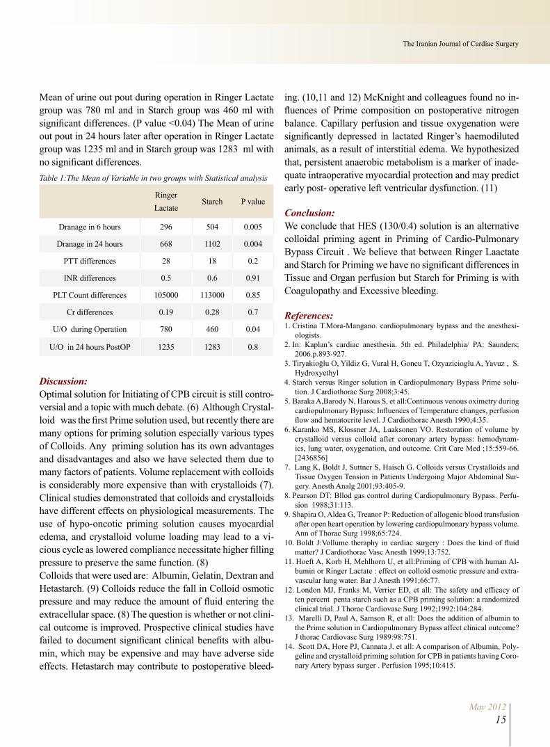
May 201215
The Iranian Journal of Cardiac Surgery
Mean of urine out pout during operation in Ringer Lactate group was 780 ml and in Starch group was 460 ml with significant differences. (P value <0.04) The Mean of urine out pout in 24 hours later after operation in Ringer Lactate group was 1235 ml and in Starch group was 1283 ml with no significant differences.Table 1:The Mean of Variable in two groups with Statistical analysis
Ringer Lactate
Starch P value
Dranage in 6 hours 296 504 0.005
Dranage in 24 hours 668 1102 0.004
PTT differences 28 18 0.2
INR differences 0.5 0.6 0.91
PLT Count differences 105000 113000 0.85
Cr differences 0.19 0.28 0.7
U/O during Operation 780 460 0.04
U/O in 24 hours PostOP 1235 1283 0.8
Discussion:Optimal solution for Initiating of CPB circuit is still contro-versial and a topic with much debate. (6) Although Crystal-loid was the first Prime solution used, but recently there are many options for priming solution especially various types of Colloids. Any priming solution has its own advantages and disadvantages and also we have selected them due to many factors of patients. Volume replacement with colloids is considerably more expensive than with crystalloids (7). Clinical studies demonstrated that colloids and crystalloids have different effects on physiological measurements. The use of hypo-oncotic priming solution causes myocardial edema, and crystalloid volume loading may lead to a vi-cious cycle as lowered compliance necessitate higher filling pressure to preserve the same function. (8)Colloids that were used are: Albumin, Gelatin, Dextran and Hetastarch. (9) Colloids reduce the fall in Colloid osmotic pressure and may reduce the amount of fluid entering the extracellular space. (8) The question is whether or not clini-cal outcome is improved. Prospective clinical studies have failed to document significant clinical benefits with albu-min, which may be expensive and may have adverse side effects. Hetastarch may contribute to postoperative bleed-
ing. (10,11 and 12) McKnight and colleagues found no in-fluences of Prime composition on postoperative nitrogen balance. Capillary perfusion and tissue oxygenation were significantly depressed in lactated Ringer’s haemodiluted animals, as a result of interstitial edema. We hypothesized that, persistent anaerobic metabolism is a marker of inade-quate intraoperative myocardial protection and may predict early post- operative left ventricular dysfunction. (11)
Conclusion:We conclude that HES (130/0.4) solution is an alternative colloidal priming agent in Priming of Cardio-Pulmonary Bypass Circuit . We believe that between Ringer Laactate and Starch for Priming we have no significant differences in Tissue and Organ perfusion but Starch for Priming is with Coagulopathy and Excessive bleeding.
References:1. Cristina T.Mora-Mangano. cardiopulmonary bypass and the anesthesi-
ologists.2. In: Kaplan’s cardiac anesthesia. 5th ed. Philadelphia/ PA: Saunders;
2006.p.893-927.3. Tiryakioğlu O, Yildiz G, Vural H, Goncu T, Ozyazicioglu A, Yavuz , S.
Hydroxyethyl 4. Starch versus Ringer solution in Cardiopulmonary Bypass Prime solu-
tion. J Cardiothorac Surg 2008;3:45.5. Baraka A,Barody N, Harous S, et all:Continuous venous oximetry during
cardiopulmonary Bypass: Influences of Temperature changes, perfusion flow and hematocrite level. J Cardiothorac Anesth 1990;4:35.
6. Karanko MS, Klossner JA, Laaksonen VO. Restoration of volume by crystalloid versus colloid after coronary artery bypass: hemodynam-ics, lung water, oxygenation, and outcome. Crit Care Med ;15:559-66. [2436856]
7. Lang K, Boldt J, Suttner S, Haisch G. Colloids versus Crystalloids and Tissue Oxygen Tension in Patients Undergoing Major Abdominal Sur-gery. Anesth Analg 2001;93:405-9.
8. Pearson DT: Bllod gas control during Cardiopulmonary Bypass. Perfu-sion 1988;31:113.
9. Shapira O, Aldea G, Treanor P: Reduction of allogenic blood transfusion after open heart operation by lowering cardiopulmonary bypass volume. Ann of Thorac Surg 1998;65:724.
10. Boldt J:Vollume theraphy in cardiac surgery : Does the kind of fluid matter? J Cardiothorac Vasc Anesth 1999;13:752.
11. Hoeft A, Korb H, Mehlhorn U, et all:Priming of CPB with human Al-bumin or Ringer Lactate : effect on colloid osmotic pressure and extra-vascular lung water. Bar J Anesth 1991;66:77.
12. London MJ, Franks M, Verrier ED, et all: The safety and efficacy of ten percent penta starch such as a CPB priming solution: a randomized clinical trial. J Thorac Cardiovasc Surg 1992;1992:104:284.
13. Marelli D, Paul A, Samson R, et all: Does the addition of albumin to the Prime solution in Cardiopulmonary Bypass affect clinical outcome? J thorac Cardiovasc Surg 1989:98:751.
14. Scott DA, Hore PJ, Cannata J. et all: A comparison of Albumin, Poly-geline and crystalloid priming solution for CPB in patients having Coro-nary Artery bypass surger . Perfusion 1995;10:415.

Clinical Course of Ventricular Septal Defect in Children Referred to Aliasghar Center of Zahedan during 2001-2011
1- Professor of Pediatric cardiology, Research Center of Children and Adolescent Health, ALI-EBN E-ABITALEB hospital, Zahedan University of medical sciences, Zahedan, Iran 2- Holder of Doctorate Degree in Health and Statistic – Assistant Professor of Zahedan University of medical sciences, Zahedan, Iran 3- *Corresponding author: Assistant professor of anesthesiology, fellowship of cardiac anesthesia, Research Center for Children and Adolescent Health, ALI-EBN E-ABITALEB hospital, Khash road, Zahedan university of medical sciences, Zahedan, Iran4- General Physician, Zahedan University of medical sciences, Zahedan, Iran 5- General Physician, Heart valve disease research center, Rajaie Cardiovascular Medical and Research center, Tehran University of Medical Sciences, Tehran, Iran6- Fellowship of cardiac anesthesia, Rajaie Cardiovascular Medical & Research Center, Tehran University of Medical Sciences, Tehran, Iran7- Fellowship of cardiac anesthesia, Rajaie Cardiovascular Medical & Research Center, Tehran University of Medical Sciences, Tehran, Iran
Noor Mohammad Noori MD1, Mahnaz Shahraki MD2, Maziar Mahjoubifard MD3*, Bahareh Bagherzadeh MD4, Yalda Mirmesdagh MD5, Korosh Ghorbannejad MD6, Alireza Jahangiri Fard MD7
Abstract:Objective: The aim of this study is a review of clinical progress of ventricular septal defect in children referred to Aliasghar Center of Zahedan during 2001 to 2011.Method: In this research we have studied all files existing in the archive of the patients referred to Aliasghar Diseases Center of Zahedan, from 2001 to September 2011. The cases with ventricular septal defect diagnosed by echocardiography were selected and required data including location and size of VSD, patient’s age along with the manner of VSD closure (spontaneously, surgery, intervention) were collected. Results: Among 1750 patients with congenital heart diseases, 621 cases (35.5%) were suffering from VSD. In 32.3% of cases, the defect was closed spontaneously, 12.7% underwent closed heart operation, and 53% received medical therapy. Based on the defect location, there were 11.9% muscular, 72.8% perimembranous, 8.1% outlet, and 7.2% inlet type. Also the size of the defect in patients with isolated VSD was as fol-lows: 45.1% small type, 25.9% moderate type, 29% large type. In general, serious complications have been occurred in 4.34% of the patients in long term follow up.Conclusions: In this study, it has been indicated that the incidence of spontaneously closure of small defects has been increased with time and a large number of moderate defects convert to minor defects by time and few number of large defects spontane-ously closed by time. The incidence of serious complications in long term was 4.34% and mostly the results were similar to the results of other studies in this regard.
Key words: Children, Congenital Heart Disease, Ventricular Septal Defect
Introduction:Congenital heart diseases have been re-ported in 4-50 thousands of live births. Congenital defects have wide spectrum from severity points of view in children. About 2-3 infants of every 1000 births have symptoms of heart diseases in the first year of their lives. By progress in
treatment methods, number of children suffering from congenital heart diseases who survived until adulthood period, have been increased. Despite the latest progress, congenital heart diseases are the first reason of death in patients with con-genital anomalies.Ventricular Septal Defect (VSD) alone
May 201217
The Iranian Journal of Cardiac Surgery
forms 30-35 percent of the whole heart congenital diseases. VSD also is seen along with other heart diseases including: Tetralogy of Fallot (TOF), complete atrioventricular septal defects, transposition of great arteries (2). Clinical symp-toms and natural development of VSD in children are dif-ferent depending on the defects. The clinical presentation of VSD with small size is mostly a holosystolic murmur and is often without symptoms and in most cases spontaneously closure occur (3).In newborns suffer from VSD with moderate size; the clini-cal symptoms are mostly in form of heart failure and growth anomalies. The sign of VSD with moderate size is in form of holosystolic murmur with or without trill in left sternal border and prolonged S2. Also diastolic murmur in heart apex can be osculated in these children. Usually VSDs with moderate sizes have regressed in size by time and even a number of them may be closed spontaneously. Newborns suffering from VSD with large size usually have symptoms during the first month after birth (2) and in two studies on premature and term newborns the incidence of VSD has been reported as 5 % (3).In the study of Miyake and colleagues study, in order to de-tect the spontaneously closure of VSD, physical examina-tion, heart catheterization, echocardiography with colored pulse and Doppler were performed. In this study physical examination for long follow up and colored Doppler for short period were performed. In long term reports, the fol-low up period (10-year), the spontaneously closure of small VSD, was reported as about 75%, however it is reported in other studies as 60%. In short time follow ups (12-month) done by colored Doppler and Doppler pulse, the incidence of spontaneously closure in type of Perimembranous was nearly 45%, and in muscular type it was71.8% (4).Allen and colleagues reported spontaneously closure dur-ing the first 2 years of life between 75-80% in small size and in case of VSDs with moderate sizes, they believed that 15-20% of them may need surgical operation and in case of large VSDs spontaneously closure was occurred in 8 % ( 5).Most studies have been conducted in patients with VSD along with good results after operation as well as children with normal growth and activities. Long term follow up implicates occurrence of unusual pulmonary hypertension (4%), sinus dysfunction (4%) and aortic valve insufficiency (16%) in VSD with large sizes (6). In another study it was
proposed that patients with clinical course and with small to moderate VSD must be followed up in longer period of medical therapy, unless they are affected by heart failure or pulmonary hypertension (7).Regarding the outbreak and importance of VSD, and since the studies regarding natural course of VSD are limited in our country, we decided to study the 10-year prognosis of VSD in children suffering from this congenital disease in Aliasghar Center of Special Diseases of Zahedan.
Methods:The study was a case descriptive one and was conducted on all children with VSD referred to Aliasghar Center of Spe-cial Diseases of Zahedan from 2001 to 2011. Patients with other anomalies except for VSD were excluded from the study. Regarding the number of referring patients during ten years, the number of subject patients in the study was determined as 621 cases. The sampling method is easy and accessible. The research was conducted by enumeration of the subject society, meaning patients with VSD, in order to reach the desired sample group. The approval of the study protocol was granted and the files of all patients existing in the archive of Aliasghar Center of Special Diseases of Za-hedan, from 2001 to September 2011 were studied. Among them, the cases with VSD were divided based on echocar-diography and the information existing in the file of every patient, including location and size of the VSD, patient’s age along with the manner of closure (spontaneously, sur-gery, intervention) and the side effects were extracted and entered in the prepared questionnaire form. The patients with bicuspid aorta, mitral valve prolapse and those with patent duct us arteriosus in premature newborns were ex-cluded from the study. Meanwhile the incomplete files were left aside. The data were analyzed using SPSS, descriptive statistical methods of central indexes, dispersing, drawing the table of distribution of plentitude, and drawing chart.
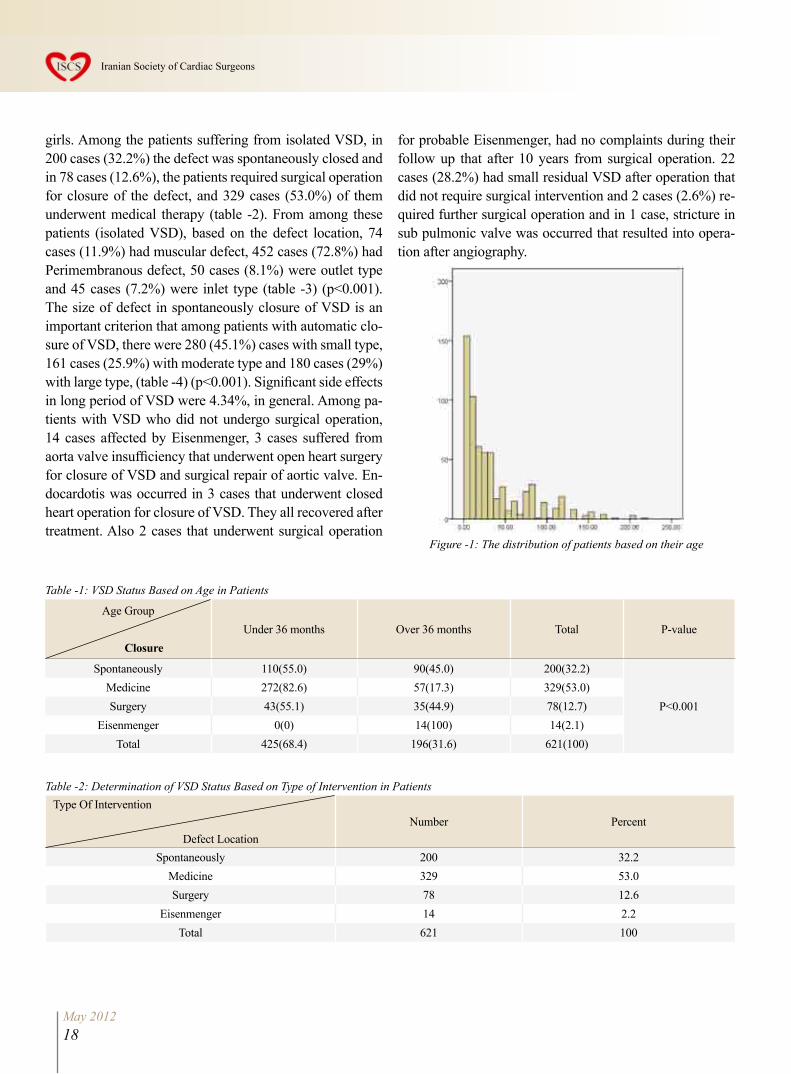
Results:In this study 621 children with the mean age of 36.2±40.3 (figure -1) and VSD who referred to Aliasghar Center of Special Diseases of Zahedan for diagnosis and treatment during 2001 to 2011 were studied. In this study, 425 children (48.4%) were under 36 months and 196 (31.6%) were over 36 months (table-1) (p<0.001). In this study, 339 children of subject patients were boys (54.4%) and 284 (45.6%) were
May 201218
Iranian Society of Cardiac Surgeons
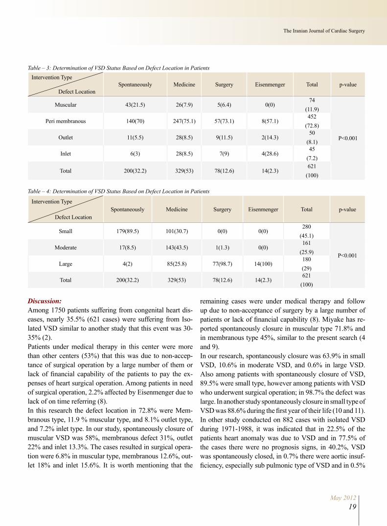
girls. Among the patients suffering from isolated VSD, in 200 cases (32.2%) the defect was spontaneously closed and in 78 cases (12.6%), the patients required surgical operation for closure of the defect, and 329 cases (53.0%) of them underwent medical therapy (table -2). From among these patients (isolated VSD), based on the defect location, 74 cases (11.9%) had muscular defect, 452 cases (72.8%) had Perimembranous defect, 50 cases (8.1%) were outlet type and 45 cases (7.2%) were inlet type (table -3) (p<0.001). The size of defect in spontaneously closure of VSD is an important criterion that among patients with automatic clo-sure of VSD, there were 280 (45.1%) cases with small type, 161 cases (25.9%) with moderate type and 180 cases (29%) with large type, (table -4) (p<0.001). Significant side effects in long period of VSD were 4.34%, in general. Among pa-tients with VSD who did not undergo surgical operation, 14 cases affected by Eisenmenger, 3 cases suffered from aorta valve insufficiency that underwent open heart surgery for closure of VSD and surgical repair of aortic valve. En-docardotis was occurred in 3 cases that underwent closed heart operation for closure of VSD. They all recovered after treatment. Also 2 cases that underwent surgical operation
for probable Eisenmenger, had no complaints during their follow up that after 10 years from surgical operation. 22 cases (28.2%) had small residual VSD after operation that did not require surgical intervention and 2 cases (2.6%) re-quired further surgical operation and in 1 case, stricture in sub pulmonic valve was occurred that resulted into opera-tion after angiography.
Figure -1: The distribution of patients based on their age
Table -1: VSD Status Based on Age in Patients
Age Group
Closure Under 36 months Over 36 months Total P-value
Spontaneously 110(55.0) 90(45.0) 200(32.2)
P<0.001Medicine 272(82.6) 57(17.3) 329(53.0)Surgery 43(55.1) 35(44.9) 78(12.7)
Eisenmenger 0(0) 14(100) 14(2.1)Total 425(68.4) 196(31.6) 621(100)
Table -2: Determination of VSD Status Based on Type of Intervention in Patients Type Of Intervention
Defect LocationNumber Percent
Spontaneously 200 32.2Medicine 329 53.0Surgery 78 12.6
Eisenmenger 14 2.2Total 621 100
May 201219
The Iranian Journal of Cardiac Surgery
Discussion: Among 1750 patients suffering from congenital heart dis-eases, nearly 35.5% (621 cases) were suffering from Iso-lated VSD similar to another study that this event was 30-35% (2). Patients under medical therapy in this center were more than other centers (53%) that this was due to non-accep-tance of surgical operation by a large number of them or lack of financial capability of the patients to pay the ex-penses of heart surgical operation. Among patients in need of surgical operation, 2.2% affected by Eisenmenger due to lack of on time referring (8). In this research the defect location in 72.8% were Mem-branous type, 11.9 % muscular type, and 8.1% outlet type, and 7.2% inlet type. In our study, spontaneously closure of muscular VSD was 58%, membranous defect 31%, outlet 22% and inlet 13.3%. The cases resulted in surgical opera-tion were 6.8% in muscular type, membranous 12.6%, out-let 18% and inlet 15.6%. It is worth mentioning that the
remaining cases were under medical therapy and follow up due to non-acceptance of surgery by a large number of patients or lack of financial capability (8). Miyake has re-ported spontaneously closure in muscular type 71.8% and in membranous type 45%, similar to the present search (4 and 9).In our research, spontaneously closure was 63.9% in small VSD, 10.6% in moderate VSD, and 0.6% in large VSD. Also among patients with spontaneously closure of VSD, 89.5% were small type, however among patients with VSD who underwent surgical operation; in 98.7% the defect was large. In another study spontaneously closure in small type of VSD was 88.6% during the first year of their life (10 and 11).In other study conducted on 882 cases with isolated VSD during 1971-1988, it was indicated that in 22.5% of the patients heart anomaly was due to VSD and in 77.5% of the cases there were no prognosis signs, in 40.2%, VSD was spontaneously closed, in 0.7% there were aortic insuf-ficiency, especially sub pulmonic type of VSD and in 0.5%
Table – 3: Determination of VSD Status Based on Defect Location in Patients Intervention Type
Defect LocationSpontaneously Medicine Surgery Eisenmenger Total p-value
Muscular 43(21.5) 26(7.9) 5(6.4) 0(0)74
(11.9)
P<0.001
Peri membranous 140(70) 247(75.1) 57(73.1) 8(57.1)452
(72.8)
Outlet 11(5.5) 28(8.5) 9(11.5) 2(14.3)50
(8.1)
Inlet 6(3) 28(8.5) 7(9) 4(28.6)45
(7.2)
Total 200(32.2) 329(53) 78(12.6) 14(2.3)621
(100)
Table – 4: Determination of VSD Status Based on Defect Location in Patients
Intervention Type
Defect LocationSpontaneously Medicine Surgery Eisenmenger Total p-value
Small 179(89.5) 101(30.7) 0(0) 0(0)280
(45.1)
P<0.001Moderate 17(8.5) 143(43.5) 1(1.3) 0(0)
161(25.9)
Large 4(2) 85(25.8) 77(98.7) 14(100)180(29)
Total 200(32.2) 329(53) 78(12.6) 14(2.3)621
(100)
May 201220
Iranian Society of Cardiac Surgeons
bacterial endocarditis have occurred. But nearly 15.5% of VSD cases required open heart operation. The findings of this study were, to large extend, similar to our research (4, 12). In another study Onat and colleagues concluded that in children with VSD, before puberty period, this defect is also affected. These researchers have also reported that the incidence of spontaneously closure of the defect in their patients who had no increase in pulmonary hypertension about 32%. This research is also coordinated with our re-search in this regard (13).In other research, Tuner has studied the clinical course of VSD based on the size of the defect, its location and pa-tient’s age and indicated that from 68 subject patients with isolated VSD, in 49 cases (72%) the size of VSD was small, in 14 cases (20.5%) the size of VSD was moderate, and in 5 cases (7.3%) the size of VSD was large. And 35 cases had spontaneous closure and 13 case required surgical opera-tion. Also the researchers of this study showed that from 35 cases of spontaneous closure of VSD (77%), 27 cases were muscular types that this point is also similar to the present research (9,8 and 14).In the study of Miyake and colleagues study is also shown that the spontaneous closure in long-term follow up of small type VSD was 75%. They also reported the incidence of spontaneous closure in muscular type as 71.8% and in perimembranous 45%. Also these researchers have re-ported the incidence of abnormal pulmonary hypertension as (4%), aortic valve insufficiency (16%) along with large VSD. These findings are similar with our study (4, 9). In another study, 1075 newborns in Russia were studied after birth by echocardiography and the incidence of spon-taneous closure of VSD was 88.6% in the first year, in pre-term newborns this criteria was 100% versus 78.8% in term newborns. Theses researchers proved that spontaneous clo-sure of VSD in newborns occurred in a large scale (10, 15). In our research, the patients were studied after the period of being newborns. In Roos and colleagues’ study, it is indicated that the pa-tients with VSD who underwent surgical operation, had good prognosis in long term follow up. Nearly 92% of their subject patients were in class 1 regarding NYHA class. In other study, Wu and colleagues indicated that those patients who were affected by diseases like aortic valve insuffi-
ciency, left ventricular stretch and pulmonary hypertension were in need of closed surgical operation for closure of VSD. Also in other study Gu and colleagues showed that it was possible to close the remaining VSD successfully af-ter operation by intervention method and also these authors emphasized that there were no need for further surgery af-ter closure of VSD. In the present research surgical opera-tion was done in order to repair VSD for patients with large VSD, patients with aortic valve insufficiency after VSD, as well as patients with VSD and pulmonary hypertension. Although, these days VSD can be repaired by intervention method (6, 16 and 17).In other research by Heuvel and colleagues, it is indicated that clinical course of patients with VSD was good in adult-hood and in 6% of the patients the VSD had been closed spontaneously. Also no death has been occurred to any of the patients. 1.8% patient with endocarditis and 4% also due to hemodynamic problems underwent surgical opera-tion. Also they indicated that VSDs with small defects have good pre-information in the future and are not in need of surgical operation and have no remarkable side effects. In our study, long term follow up of side effects was similar to other studies but bacterial endocarditis was less in this research (18).In long term follow up, totally 4.34% of patients were af-fected by serious complications. In 28.2 % of the patients who underwent surgical operation the VSD remained in form of a small defect that did not require surgical inter-vention. Except for these cases: in 14 patients Eisenmenger syndrome was occurred due to delay in surgical operation time, In 2 cases, the further defect was repaired by surgi-cal operation due to non-closure of VSD, and one case af-fected by stricture of pulmonary valve after surgical repair of VSD for whom, first echocardiography was performed, then angiography was performed in order to determine the severity of stricture and finally the cases were introduced to the surgeon for removal of the stricture. Among total, 3 patients with small VSD, were affected by aortic valve insufficiency that required VSD closure and valve repair. Also 3 cases were affected by endocarditis that was treated by antibacterial medical therapy and 2 patients with Eisen-menger underwent surgical operation with no complaints after 10 year follow up with normal pulmonary artery pres-sure in the recent catheterization. Also this study indicated
May 201221
The Iranian Journal of Cardiac Surgery
that in 54% of patients the defect was closed spontaneously before 36 months and in 10.2% of the cases surgery was closure of VSD, 43% of VSD cases the defect was closed over 36 months and in 17.9% the closure of VSD required surgical operation.In recent years, the intervention method for closure of VSD (perimembranous and muscular types) has been employed in wide range as an alternative method. The study of Zheng and colleagues indicated that closure of VSD by percutane-ous method under echocardiography had been useful and was associated with fewer complications. Because of short-ed duration of hospitalization and less remaining scars, this method is more acceptable than open heart surgery. Also this method may decrease the incidence of heart block and aortic valve insufficiency with less morbidity (19 and 20).Spontaneous closure of large VSD in present research was 0.6%, and in 98.7% this defect required surgical operation. The rest of the cases with large VSD could not undergo sur-gical operation due to the occurrence of the Eisenmenger syndrome. It is worth mentioning that other cases of VSD (53%) were under medical therapy and follow up, that this situation had occurred more than other centers, since a large number of patients refused to undergo surgical operation due to lack of financial capability .The long term compli-cations in this study were similar to other studies (434%) except that small defects remained after surgical operation (28.32%)
Conclusions:In this study, it is indicated that spontaneous closure of small VSDs will increase by time and a large number of moderate defects convert to small defects by time and treat-ment. Also a few numbers of large defects convert to small defects by time. The long term complications in this study were similar to other studies.
References:1. Hoffman JIE, Kaplan S. The incidence of congenital heart disease, J Am
Coll Cardiol 2002; 39(12): 1890-1900.2. Kliegman RM, Stanton BF, Schor NF, Geme III JWSt, Behrman RE.
Nelson Textbook of Pediatrics.19th edition, Philadelphia, Saun-ders,2011:1549-1551.
3. Pierpont ME, Basson TC, Bensow DW, et al. Genetic Basis for con-genital Heart Detects: Current Knowledge : A scientific statement form the American Heart Association congenital Heart defects commi He , council on cardiovascular Disease m the Young . Circulation 2007 ; 15 : 3015 – 3038.
4. Miyake T, Shinohara T, Nakamura Y, Fukuda T, Tasato H, Toyohara K, et al. Spontaneous closure of ventricular septal defects followed up from <3 months of age. Pediatr Int 2004; 46:135-140.
5. Allen HD, Driscoll DJ, Shaddy RE, Feltes TF. Moss and Adams’ Heart Disease in Infants, Children, and Adolescents: Including the Fetus and Young Adult 7th ed,Philadelphia, Lippincott Williams & Wilkins 2008: 667-682
6. Roos-Hesselink JW, Meijboom FJ, Spitaels SE, Van Domburg R, Van Rijen EH, Utens EM, Bogers AJ, Simoons ML. Outcome of patients after surgical closure of ventricular septal defect at young age: longitu-dinal follow-up of 22-34 years, Eur Heart J 2004;25(12):1057-62.
7. Turner SW, Hornung T, Hunter S. Closure of ventricular septal defects: a study of factors influencing spontaneous and surgical closure, Cardiol-ogy in the Young 2002;12:357-363.
8. Chaudhry TA, Younas M, Baig A. Ventricular Septal defect and associ-ated complications, J Pak Med Assoc2011; 61(10):1001-1004.
9. Miyake T. Shinohara T. Inoue T. Marutani S and Takemura T. Spontane-ous closure of muscular trabecular ventricular septal defect: comparison of defect positions. Acta Paediatrica 2011; 100(10): e158–e162.
10. Shirali GS, Smith EO, Geva T. Quantitation of echocardiographic pre-dictors of outcome in infants with isolated ventricular septal defect. Am Heart J. 1995;130(6):1228–1235.
11. Sadoh WE. Natural history of ventricular septal defects in Nigerian children,South African Journal of Child Health 2010;4(1):16-19.
12. Frontera-Izquierdo P, Cabezuelo-Huerta G. Natural and modified his-tory of isolated ventricular septal defect: a 17-year study. Pediatr Car-diol. 1992; 13(4):193-7.
13. Onat T, Ahunbay G, Batmaz G, Celebi A. The natural course of iso-lated ventricular septal defect during adolescence. Pediatr Cardiol. 1998;19(3):230-4.
14. Turner SW, Hunter S, Wyllie JP. The natural history of ventricular sep-tal defects. Arch Dis Child. 1999;81(5):413-6.
15. Ekici F, Tutar E, Atalay S, Arsan S, Özçelik N. The incidence and fol-low-up of isolated ventricular septal defect in newborns by echocardio-graphic screening, The Turkish Journal of Pediatrics 2008; 50: 223-227.
16. Ming-Biao Gu, Yuan Bai, Xian-Xian Zhao, Xing Zheng,Wei-Ping Li, Yong-Wen Qin. Transcatheter Closure of Postoperative Resid-ual Perimembranous Ventricular Septal Defects, Ann Thorac Surg 2009;88:1551-1555.
17. Wu JC, Child JS. Common Congenital Heart Disorders in Adults, Cur-rent problems in Cardiology 2004; 29:641-700.
18. Van den Heuvel F, Timmers T, Hess J. Morphological, haemodynamic, and clinical variables as predictors for management of isolated ventricu-lar septal defect. Br. Heart J. 1995; 73: 49−52.
19. Zheng Q, Zhao Z, Zuo J, Yang J, Wang H, Yu S, Yi D. A Comparative Study: Early Results and Complications of Percutaneous and Surgical Closure of Ventricular Septal Defect, Cardiology 2009;114:238-243.
20. Oses P, Hugues N, Dahdah N, Vobecky SJ, Miro J, Pellerin M, Poirier NC. Treatment of Isolated Ventricular Septal Defects in Children: Am-platzer Versus Surgical Closure, Ann Thorac Surg 2010; 90:1593-1598.
All That Glitters Is Not Gold and All Myxoid Tumors Are Not Myxomas Kambiz Mozaffari: Department of PathologyCorrespondence to: Kambiz Mozaffari, MD, Surgical Pathology Laboratory, Shaheed Rajaie Cardiovascular, Medical and Research Center, Tehran University of Medical Sciences, Mellat Park, Vali Asr Ave., Tehran , Iran. Tel: 23921
AbstractPrimary tumors of the heart are rare, as opposed to metastatic lesions in this organ. Among the primary tumors, cardiac myxoma is the most prevalent neoplasia.Here, we discuss the varied nature of myxoid lesions, which may be mistaken for myxomas, in two categories: benign and malignant.Not only should the pathologist, but also the cardiac surgeon, be familiar with the diag-nostic pitfalls and the differential diagnoses of myxoid lesions of the heart.Echocardiographic findings, the rapidity of symptom development, and the location of the tumor, all serve as clues to a better approach in such patients.
Key words: Cardiac tumors, Myxoma, Myxofibrosarcoma
Cardiac myxomas are primary neoplas-tic lesions, arising in the endocardium. Primary tumors are rare in the heart; nevertheless, myxomas constitute about 80% of these primary tumors.A wide range of tumors or pseudo-tumors, albeit rare, may be found in the heart. Therefore, both the pa-thologist and the surgeon should be familiar with the diagnostic pit-falls and the differential diagnoses.The myxoid lesions, which may be mis-taken for myxomas, may be either be-nign or malignant. In the former group, only to name a few, we can mention papillary fibroelastomas, which occur on leaflets and are sometimes large in size. They have a characteristic gross appearance that is likened to “sea anemone” and in contrast to myxomas, which are vascular lesions, they are avascular structures and do not pos-sess the stellate cells seen in myxomas.Another benign mass mimicking myx-oma is cardiac hemangioma, in light of the fact that it is a vascular tumor like
myxoma and may also adopt a myxoid stroma. What is used as the diagnostic aid here is the lack of myxoma cell on one hand, and the abundant presence of he-mosiderin in myxoma on the other hand.Last but not least, in the first group, we can add organized thrombus as a lesion very likely to be frequently confused with myxoma. Thrombi lack myxoma cells, but possess granulation tissue and areas of scar and hyalinization.As was mentioned before, the second category of lesions is malignant, and in this group we can name myxosar-comas. They are fibroblastic or fibro-histiocytic tumors that demonstrate extensive myxoid stromal component; however, they should not be considered as benign myxomas evolving into sar-comas. In fact, there is little evidence, if any, that myxomas undergo such progressive malignant transformation.Some 80% of these sarcomas occur in the left atrium, hence the significance of an accurate differential diagnosis from myxomas. A high proportion of myxoid
May 201223
The Iranian Journal of Cardiac Surgery
sarcomas of the heart are initially misdiagnosed as myxo-mas, but the clues that help differentiate them from myxo-mas include myxoma or stellate cells, hemosiderin-laden macrophages, and factor-XIII-positive dendritic cells. All these features are lacking in the malignant sarcomas.In the malignant category of diagnostically challenging tumors, we should add leiomyosarcomas, which are of smooth muscle origins. A myxoid stroma is noted in about 25 % of these tumors, but the dense fascicles of smooth mus-cle easily distinguish this tumor from cardiac myxoma (1).During the operation, the myxoid appearance of the tumor may further support this assumption that we are dealing with a myxoma, but histopathological examination unex-pectedly shows that the mass is not an atrial myxoma but rather a myxoid variant of a primary leiomyosarcoma. For such cases, immunohistochemistry and electron micros-copy studies may be utilized to confirm the diagnosis (2).A great number of malignant tumors in the heart are sar-comatous in origin, and it is not rare to see a patient who presents with symptoms of mitral valve stenosis because of a malignant tumor of the left atrium. We came across a case in the literature where echocardiography first re-vealed a left atrial mass supposedly interpreted as an atrial myxoma. Upon operation, the tumor appeared lobulated with a smooth surface. It arose from the posterior wall of the left atrium and extended into the mitral annulus. Wide surgical excision was possible with atrial reconstruction and mitral valve replacement. Histopathological and im-munohistochemical studies were indicative of a malignant undifferentiated sarcoma (3).The clinical and echocardiographic findings are extreme-ly crucial to discriminate malignant tumors from benign myxomas, although the diagnosis may not be reached un-til the histological sections are available. To name a few, a rapidly progressive clinical course, or multiple tumoral growths, and non-septal attachment of the mass all sug-gest a malignant process. Therefore, careful and precise preoperative evaluation is said to be essential to identify such malignancies (4). Other authors too stress the impor-tance of a comprehensive preoperative workup (5).Here, we focus again on the intra-operative observation by the surgeon of the left atrial masses attached to the lateral atrial wall. On inspection during surgery, the malignant tu-mors are prone to infiltrate the mitral annulus and leaflet (6).
ConclusionIn view of the frequency of reports indicating the impor-tance of preoperative workup and intra-operative observa-tion as well as the varied nature of the lesions that may be mistakenly designated as myxoma, one should always remember that all myxoid tumors are not actually myxo-mas, as all that glitters is not gold.AcknowledgmentsWe wish to thank Mr. Farshad Amouzadeh for his techni-cal assistance.
References1. Silver MD, Gotlieb AI, Schoen FJ. Cardiovascular Pathology. 3rd edi-
tion, New York, Churchill Livingstone Company, 2001; pp. 583-5992. Morin JE, Rahal DP, Hüttner I Myxoid leiomyosarcoma of the left
atrium: a rare malignancy of the heart and its comparison with atrial myxoma. Can J Cardiol. 2001 Mar;17(3):331-6.
3. 3-Ariki H, Mizuno S, Tsunekawa T, Doi T. Left atrial sarcoma with the initial diagnosis of myxoma. Kyobu Geka. 2005 Jun;58(6):462-5: Available @ http://www.ncbi.nlm.nih.gov/pubmed/15957419
4. Hasegawa T, Nakagawa S, Chino M, Kunihiro T, Ui S, Kimura M. Pri-mary cardiac sarcoma mimicking benign myxoma: a case report. J Car-diol. 2002 Jun;39(6):321-5.
5. Kim JT, Baek WK, Kim KH, Yoon YH, Kim DH, Lim HK. A primary cardiac sarcoma preoperatively presented as a benign left atrial myxo-ma. Yonsei Med J. 2003 Jun 30;44(3):530-3.
6. Lazaros GA, Matsakas EP, Madas JS, Toli DI, Nikas DJ, Kershaw MA, Alpert MA. Primary myxofibrosarcoma of the left atrium: case report and review of the literature. Angiology. 2008 Oct-Nov;59(5):632-5. Epub 2008 Apr 2.
May 201224
Iranian Society of Cardiac Surgeons
Abstracts selected and summarized by: R. Baghaei M.D ,Shahid Rajaee Cardiovascular, Medical and Research Center, Tehran University of Medical Sciences, Tehran, Iran
Emergency Surgery for Native Mitral Valve Endocarditis: The Impact of Septic and Cardiogenic Shock
Ann Thorac Surg 2012;93:1469-1476.
Background: Autograft dilatation after the Ross procedure is the most common cause of late autograft failure. We looked at results after reoperation for autograft dysfunction using au-tograft sparing and composite root replacement techniques.Methods: Data were abstracted from our prospectively col-lected Ross registry for 160 consecutive patients who un-derwent a Ross procedure by a single surgeon between 1994 and 2008. Follow-up records were obtained, and the last echocardiographic report after reoperation was analyzed.Results: Autograft reoperation was necessary in 17 patients, at a median interval of 6.9 years after the original proce-dure. Indications for reoperation were insufficiency with autograft dilatation in 16 patients, and without dilatation in 1 patient. Surgical procedures used at reoperation included autograft reimplantation in 6 patients (35.3%), autograft remodeling procedure in 1 patient (5.9%), composite root
replacement with mechanical valved conduit in 5 patients (29.4%), composite root replacement with biologic valved conduit in 3 patients (17.6%), and mechanical aortic valve replacement in 2 patients (11.8%). At a median follow-up of 5.0 years after reoperation, freedom from greater than 2+ aortic insufficiency was 100% (17 of 17 patients) in both re-implantation and replacement groups. There was 1 death af-ter reoperation (at >14 years) related to complications from systemic lupus erythematosus. There have been no strokes after autograft reimplantation.Conclusions: Autograft valve reimplantation and compos-ite aortic root replacement are effective treatments for aortic root dilatation and aortic insufficiency after the Ross proce-dure. Echocardiographic follow-up demonstrates reasonable short-term function after autograft preservation procedures.
Redo Autograft Operations After the Ross Procedure
Ann Thorac Surg 2012;93:1477-1482.
Background: Limited information exists about the real im-pact of the etiology of shock on early and late outcome after emergency surgery in acute native mitral valve endocarditis (ANMVE). This multicenter study analyzed the impact of the etiology of shock on early and late outcome in patients with ANMVE.Methods: Data were collected in eight institutions. Three hundred-seventy-nine ANMVE patients undergoing sur-gery on an emergency basis between May 1991 and Decem-ber 2009 were eligible for the study. According to current criteria used for the differential diagnosis of shock, patients were retrospectively assigned to one of three groups: group 1, no shock (n = 154), group 2, cardiogenic shock (CS [n = 118]), and group 3, septic shock (SS [n = 107]). Median follow-up was 69.8 months.Results: Early mortality was significantly higher in patients with SS (p < 0.001). At multivariable logistic regression
analysis, compared with patients with CS, patients with SS had more than 3.8 times higher risk of death. That rose to more than 4 times versus patients without shock. In addi-tion, patients with SS had 4.2 times and 4.3 times higher risk of complications compared with patients with CS and without shock, respectively. Sepsis was also an independent predictor of prolonged artificial ventilation (p = 0.04) and stroke (p = 0.003) whereas CS was associated with a higher postoperative occurrence of low output syndrome and myo-cardial infarction (p < 0.001). No difference was detected between groups in 18-year survival, freedom from endocar-ditis, and freedom from reoperation.Conclusions: Our study suggests that emergency surgery for ANMVE in patients with CS achieved satisfactory early and late results. In contrast, the presence of SS was linked to dismal early prognosis. Our findings need to be confirmed by further larger studies.
May 201225
The Iranian Journal of Cardiac Surgery
Objective: This study was undertaken to examine the pos-sible adverse effect of the mitral valve prosthesis on the he-modynamic performance of the aortic valve prosthesis in patients who have undergone double valve replacement.Methods: Patients who underwent double valve replace-ment were matched for age, body surface area, left ventric-ular function, and size and type of aortic valve prosthesis with patients who underwent isolated aortic valve replace-ment. Two types of prosthetic valves were examined: the St Jude Medical mechanical valve (St Jude Medical, St Paul, Minn) and the Hancock II bioprosthesis (Medtronic Inc, Minneapolis, Minn). Five patients for each size and type of aortic valve prosthesis in the double valve replacement group were matched at 1:2 with patients in the isolated aor-
tic valve replacement group. Only valve sizes 21 to 27 were matched. Hemodynamic assessment of the aortic valve prosthesis was performed by transthoracic echocardiogram before hospital discharge.Results: Matched patients had similar clinical profiles. There were no differences in the systolic gradients, effec-tive aortic valve areas, or flow velocity across the aortic valve prostheses after isolated aortic valve replacement or double valve replacement.Conclusions: Early after surgery, the hemodynamic perfor-mance of aortic valve prostheses was not affected by the presence of mitral valve prostheses in patients who under-went combined aortic and mitral valve replacement.
Background: All neurologic events in the PARTNER ran-domized trial comparing transcatheter aortic valve replace-ment (TAVR) with surgical aortic valve replacement (AVR) were analyzed.Methods: High-risk patients with aortic stenosis were strat-ified into transfemoral (TF, n = 461) or transapical (TA, n = 196) strata based on their arterial anatomy and ran-domized: 657 received treatment assigned (“as treated”), 313 underwent AVR, and 344 TAVR. Neurologic events were prospectively adjudicated by an independent Clinical Events Committee. Multivariable, multiphase hazard anal-ysis elucidated factors associated with increased likelihood of neurologic events.Results: Forty-nine neurologic events (15 transient isch-emic attacks, 34 strokes) occurred in 47 patients (TAVR, n = 31; AVR, n = 16). An early peaking high hazard phase
occurred within the first week, which declined to a constant late hazard phase out to 2 years. The risk in the early phase was higher after TAVR than AVR, and in the TAVR arm in patients with a smaller aortic valve area index. In the late risk phase, the likelihood of neurologic event was linked to patient-related factors in both arms (“non-TF candidate,” history of recent stroke or transient ischemic attack, and ad-vanced functional disability), but not by treatment (TAVR vs AVR) or any intraprocedural variables. The likelihood of sustaining a neurologic event was lowest in the AVR sub-group in the TF stratum during all available follow-up.Conclusions: After either treatment, there were 2 distinct hazard phases for neurologic events that were driven by dif-ferent risk factors. Neurologic complications occurred more frequently after TAVR than AVR early, but thereafter the risk was influenced by patient- and disease-related factors.
Does the mitral valve prosthesis adversely affect the hemodynamic performance of the aortic valve prosthesis in patients with double valve replacement?
Transcatheter (TAVR) versus surgical (AVR) aortic valve replacement: Occurrence, hazard, risk factors, and consequences of neurologic events in the PARTNER trial
J Thorac Cardiovasc Surg 2012;143:S74-S77
J Thorac Cardiovasc Surg 2012;143:832-843.e13
May 201226
Iranian Society of Cardiac Surgeons
Objective: We developed a validated 50-point recipient risk index predicting short-term mortality after orthotopic heart transplant (OHT). This study examined the relationship be-tween institutional volume and recipient risk on post-OHT mortality.Methods: We used United Network for Organ Sharing (UNOS) data to identify primary OHT recipients between January 2000 and April 2010. Centers were stratified by mean annual volume. Preoperative Index for Mortality Pre-diction After Cardiac Transplantation risk scores were cal-culated for each patient with our validated 50-point system. Primary outcomes were 30-day and 1-year survivals. Mul-tivariable logistic regression analysis included interaction terms to examine effect modification of risk and volume on mortality.Results: In all, 18,226 patients underwent transplant at 141 centers: 1173 (6.4%) recipients at low-volume centers (<7 procedures/y), 5353 (29.4%) at medium-volume centers (7-
15 procedures/y), and 11,700 (64.2%) at high-volume cen-ters (>15 procedures/y). Low center volume was associated with worse 1-year mortality (odds ratio, 1.58; 95% confi-dence interval, 1.30-1.92; P < .001). For 1-year survival, there was significant positive interaction between center volume and recipient risk score (odds ratio, 1.04; 95% con-fidence interval, 1.01-1.07; P = .02), indicating effect of risk on mortality at low-volume centers greater than from either variable analyzed individually. Among high-risk recipients (score 10), 1-year survival was improved at high-volume centers (high, 79%; medium, 75%; low, 64%).Conclusions: In analysis of UNOS data with our validated recipient risk index, institutional volume acted as an effect modifier on association between risk and mortality. High-risk patients had higher mortality at low-volume centers; differences dissipated among lower-risk recipients. These data support a mandate for high-risk transplants at higher-volume centers.
Background: This case-control study was conducted to identify factors predictive of 10-year survival after ortho-topic heart transplantation (OHT).Methods: Prospectively collected data from the United Network for Organ Sharing registry were reviewed to iden-tify adult patients undergoing OHT between 1987 and 1999 (N = 22,385) who had survived 10 years. Controls were those who had died within 10 years of OHT. Factors associ-ated with 10-year survival were identified with multivariate logistic regression analysis. Lowess smoothing plots were used to identify linear breakpoints in continuous variables, and splines were incorporated when appropriate.Results: There were 9,404 ten-year survivors (42%; mean follow-up, 14.0 ± 3.0 years) and 10,373 controls (46%) with a mean survival of 3.7 ± 3.3 years post-OHT. Predictors of 10-year survival in the optimal multivariate model were age
younger than 55 (odds ratio [OR], 1.24; 95% confidence interval [CI], 1.10 to 1.38; p < 0.001), white race (OR, 1.35; 95% CI, 1.17 to 1.56; p < 0.001), shorter ischemic time (OR, 1.11; 95% CI, 1.05 to 1.18; p < 0.001), younger donor age (OR, 1.01; 95% CI, 1.01 to 1.02; p < 0.001), an-nual center volume of 9 or more (OR, 1.31; 95% CI, 1.17 to 1.47; p < 0.001), mechanical ventilation (OR, 0.53; 95% CI, 0.36 to 0.78; p = 0.001), and diabetes (OR, 0.67; 95% CI, 0.57 to 0.78; p < 0.001).Conclusions: Age younger than 55 years, annual center volume of 9 or more, white race, shorter ischemic time, and younger donor age improved the likelihood of 10-year survival after OHT. Mechanical ventilation and diabetes re-duced this likelihood. These data should serve as a useful guide to long-term prognostication in adult OHT.
Institutional volume and the effect of recipient risk on short-term mortality after ortho-topic heart transplant
What Predicts Long-Term Survival After Heart Transplantation? An Analysis of 9,400 Ten-Year Survivors
J Thorac Cardiovasc Surg 2012;143:157-167
Ann Thorac Surg 2012;93:699-704
May 201227
The Iranian Journal of Cardiac Surgery
Objective: An in vivo study of piglets on cardiopulmonary bypass was performed to determine whether postcondition-ing has a cardioprotective effect after cardioplegic arrest in large animals. Methods: Eighteen piglets were subjected to 90 minutes of cardioplegic arrest followed by 30 minutes of reperfusion. In 6 animals (control), there was no intervention at reperfu-sion. In 6 other animals, 6 cycles of unclamping and rec-lamping for 10 seconds each were done before reperfusion (postconditioning 10), whereas 3 cycles of unclamping and reclamping for 30 seconds each were performed in another 6 piglets (postconditioning 30). Results: Recovery of left ventricular contractility and dia-stolic function (percent of preischemic value) was signifi-cantly better in both postconditioning groups (contractility: 89.2% and 118.2; diastolic function: 142.3% and 120.4; in
the postconditioning 10 and 30 groups, respectively) com-pared with the control (contractility: 46.1%; diastolic func-tion: 218.5%). Recovery of global cardiac function (ven-tricular function curve analysis) was improved only in the postconditioning 30 group. Troponin-T release during re-perfusion was significantly reduced in the postconditioning 10 group compared with all groups (plasma troponin-T was 0.58 ng/mL in postconditioning 10, 1.85 in postcondition-ing 30, and 2.54 in control). The myocardial lipid peroxide was significantly higher in the control group than in both postconditioning groups after reperfusion (199% vs 112% and 131%). Conclusions: Both postconditioning algorisms promoted functional recovery after cardioplegic arrest in a large ani-mal model along with the limitation of lipid peroxidation with or without the reduction of troponin-T release.
Objective: Acute kidney injury after cardiac surgery with cardiopulmonary bypass is closely related to systemic in-flammatory reactions and oxidative stresses. Remote isch-emic preconditioning is a systemic protective strategy whereby brief limb ischemia confers systemic protection against prolonged ischemia and inflammatory reactions in distant organs. This study investigated whether remote ischemic preconditioning provides systemic protective ef-fect on kidneys that are not directly exposed to ischemia–reperfusion injury during complex valvular heart surgery. Methods: Seventy-six adult patients undergoing complex valvular heart surgery were randomly assigned to either remote ischemic preconditioning group (n = 38) or control group (n = 38). Remote ischemic preconditioning consisted of 3 10-minute cycles of lower limb ischemia and reper-fusion with an automated cuff inflator. Primary end points were comparisons of biomarkers of renal injury including
serum creatinine, cystatin C and neutrophil gelatinase–as-sociated lipocalin, and incidence of acute kidney injury. Secondary end points were comparisons of myocardial en-zyme release and pulmonary parameters. Results: There were no significant differences in serum lev-els of biomarkers of renal injury between groups through-out the study period. The incidence of acute kidney injury did not differ between groups. Creatine kinase isoenzyme MB at 24 hours after surgery was lower, and intensive care unit stay was shorter in the remote ischemic precondition-ing group than in the control group. Conclusions: In patients undergoing complex valvular heart surgery, remote ischemic preconditioning did not re-duce degree of renal injury or incidence of acute kidney in-jury whereas it did reduce myocardial injury and intensive care unit stay.
Effect of remote ischemic preconditioning on renal dysfunction after complex valvular heart surgery: A randomized controlled trial
J Thorac Cardiovasc Surg 2011;142:926-932
J Thorac Cardiovasc Surg 2011;142:148-154
Ischemic postconditioning promotes left ventricular functional recovery after cardiople-gic arrest in an in vivo piglet model of global ischemia reperfusion injury on cardiopul-monary bypass
May 201228
Iranian Society of Cardiac Surgeons
Background: Cerebral protection during aortic arch sur-gery can be performed using various surgical strategies. We retrospectively analyzed our results of different brain pro-tection modalities during aortic arch surgery.Methods: Between January 2003 and November 2009, 636 consecutive patients underwent aortic arch replacement sur-gery using unilateral antegrade cerebral perfusion (UACP [n = 123]), bilateral antegrade cerebral perfusion (BACP [n = 242]), retrograde cerebral perfusion (RCP [n = 51]), or deep hypothermia and circulatory arrest (DHCA [n = 220]). Mean age of patients was 62±14 years, 64% were male, 15% were reoperations, and 37% were performed for acute type A dissections. Mean follow-up was 4.9 ± 0.1 years and was 97% complete.Results: Circulatory arrest time was 22 ± 17 minutes UACP, 23 ± 21 minutes BACP, 18 ± 12 minutes RCP, and 15 ± 13
minutes DHCA; p < 0.001). Early mortality was 11% (n = 72) and was not different between the surgical groups. Stroke rate was 9% for ACP patients (n = 33) versus 15% (n = 39) for patients who did not receive ACP (p = 0.035). Indepen-dent predictors of stroke were type A aortic dissection (odds ratio [OR], 1.9; 95% confidence interval [CI], 1.3 to 3.2; p < 0.001), age (OR, 1.04; 95% CI, 1.01 to 1.06; p = 0.001), duration of circulatory arrest (OR, 1.01, 95% CI, 1.002 to 1.03; p = 0.02), and total aortic arch replacement (OR, 2.7; 95% CI, 1.3 to 5.7; p = 0.005). Five year survival was 68% ± 4% and was not significantly different between groups.Conclusions: Antegrade cerebral perfusion is associated with significantly less neurologic complications than RCP and DHCA, despite longer circulatory arrest times. Medium-term survival is worse for patients with postoperative permanent neurologic deficit and preoperative type A aortic dissection.
What Is the Best Strategy for Brain Protection in Patients Undergoing Aortic Arch Sur-gery? A Single Center Experience of 636 Patients
Ann Thorac Surg 2012;93:1502-1508.
Objectives: An array of neuroprotective strategies has evolved to limit spinal cord injury during descending tho-racic aneurysm and thoracoabdominal aortic aneurysm re-pair. This study prospectively assessed the neuroprotective impact of intrathecal papaverine added to other techniques in aortic aneurysm repairs.Methods: From January 2002 to January 2010, 398 con-secutive patients underwent descending thoracic aneurysm and thoracoabdominal aortic aneurysm repairs at Cleveland Clinic, 68 under hypothermic circulatory arrest. We focused on the remaining 330, in whom a combination of neuro-protective adjuncts was used intraoperatively to mitigate spinal cord ischemia. These included distal aortic perfusion with moderate hypothermia, cerebrospinal fluid drainage, and intrathecal papaverine. Two patient groups were dis-criminated according to whether intrathecal papaverine was (n = 250) or was not (n = 80) administered. Postoperative
outcomes were analyzed from a prospectively maintained clinical database.Results: Preoperative patient characteristics and comorbid-ities were similar between groups. Extent of aortic disease was also similar: descending thoracic aneurysm (34% with papaverine vs 28%) and Crawford types I (25% vs 34%), II (27% vs 24%), III (13% vs 13%), and IV (2% vs 2.5%). Groups had similar in-hospital mortality (6.4% vs 11%; P = .11) and permanent stroke (4.4% vs 7.5%; P=.3). Per-manent paraplegia (3.6% vs 7.5%; P = .01) and paraparesis (1.6% vs 6.3%; P = .01) were significantly lower in the in-trathecal papaverine group.Conclusions: Adding intrathecal papaverine to the neuropro-tective protocol for descending thoracic aneurysm and tho-racoabdominal aortic aneurysm repairs may enhance spinal cord perfusion and provide additional spinal cord protection.
J Thorac Cardiovasc Surg 2012;143:945-952.e1
Spinal cord protective strategies during descending and thoracoabdominal aortic aneu-rysm repair in the modern era: The role of intrathecal papaverine
May 201229
The Iranian Journal of Cardiac Surgery
Objectives: This study assessed comparative effectiveness of minimally invasive versus traditional sternotomy mitral valve surgery in elderly patients.Methods: From January 1, 2000, to December 31, 2008, 1005 patients underwent isolated mitral valve surgery at our institution. Patients 75-years-old were included in analy-sis (sternotomy, n = 105; minimally invasive, n = 70). Clini-cal outcomes included bypass and crossclamp time, length of hospitalization, morbidity, and mortality. To assess re-source use, total hospital costs and discharge location were analyzed. Three standardized inpatient functional status outcomes were also assessed.Results: The minimally invasive approach was associated with a 9.2-minute longer crossclamp time (P = .037) and a 25.2-minute longer bypass time (P < .001). Minimally in-vasive surgery was associated with a 3.1-day shorter hospi-
talization (P = .033). There were no significant differences in rate of major postoperative complications (P = .085) or long-term survival (P = .60). Minimally invasive approach was associated with a $6721 lower median cost of hospi-talization (P = .007) and more common discharge to home, routinely or with a health aide, rather than to rehabilitation (P=.021). Minimally invasive patients achieved faster rates of independent ambulation (P = .039) and independent sit-to-stand activity (P = .003), although there were no differ-ences in time to independent stair climbing (P = .31).Conclusions: Among elderly patients, minimally invasive mitral valve surgery is associated with slightly longer cross-clamp and bypass times but with equivalent morbidity and mortality and shorter hospitalization, decreased resource use, and improved postoperative functional status.
Comparative effectiveness of minimally invasive versus traditional sternotomy mitral valve surgery in elderly patientsJ Thorac Cardiovasc Surg 2012;143:S86-S90
Objective: The study objective was to determine the predic-tors of postoperative atrial fibrillation (POAF) in patients randomized to conventional coronary artery bypass graft (on-pump coronary artery bypass [ONCAB]) versus beat-ing heart coronary surgery (off-pump coronary artery by-pass [OPCAB]).Methods: The subgroup of 2103 patients (of 2203 enroll-ees) in the Randomized On Versus Off Bypass trial with no POAF was studied (1056 patients in the ONCAB group and 1047 patients in the OPCAB group). Univariate and multivariate analyses were used to identify the predictors of POAF and the impact of POAF on outcomes.Results: Use of ONCAB versus OPCAB was not associ-ated with increased rates of POAF. Older age (P < .0001), white race (P < .001), and hypertension (P < .002) were predictors of POAF on multivariate analysis. In general, POAF led to a higher rates of reintubation (ONCAB: 6.3% vs 0.8% no POAF, P < .001; OPCAB: 7.4% vs 1.8% no POAF, P < .0001) and prolonged ventilatory support (ON-CAB: 7.1% vs 2.3% no POAF, P = .001; OPCAB: 9.2% vs
3.4% no POAF, P = .0003). The rate of any early adverse outcome was higher in patients with POAF (all patients: 10% POAF vs 4.7% no POAF, P < .0001; ONCAB: 9% POAF vs 4.3% no POAF, P = .008; OPCAB: 11% POAF vs 5.1% no POAF, P = .001). The 1-year all cause mortal-ity was higher with POAF for both groups (ONCAB: 5.4% POAF vs 2% no POAF, P = .009; OPCAB: 5.1% POAF vs 2.6% no POAF, P = .07). POAF was independently as-sociated with early composite end point (odds ratio [OR], 2.23; confidence interval [CI], 1.55–3.22; P < .0001), need for new mechanical support (OR, 3.25; CI, 1.39–7.61; P = .007), prolonged ventilatory support (OR, 2.93; CI, 1.89–4.55; P < .0001), renal failure (OR, 5.42; CI, 1.94–15.15; P = .001), and mortality at 12 months (OR, 1.94; CI, 1.14–3.28; P = .01).Conclusions: In the Randomized On Versus Off Bypass trial, the strategy of revascularization did not affect the rate of POAF. Age, race, and hypertension were predictors of POAF. POAF was independently associated with a higher short-term morbidity and higher 1-year mortality rates.
Predictors and impact of postoperative atrial fibrillation on patients’ outcomes: A report from the Randomized On Versus Off Bypass trialJ Thorac Cardiovasc Surg 2012;143:93-102
May 201230
Iranian Society of Cardiac Surgeons
Objective: Bilateral internal thoracic artery (BITA) grafting has been shown to improve long-term survival after coro-nary artery bypass grafting. However, there has been reluc-tance to use this technique in higher-risk patients. Patients with reduced ejection fraction (EF) have been shown to present a higher operative risk and reduced long-term sur-vival. We studied the perioperative and long-term results of BITA versus single internal thoracic artery grafting (SITA) in a large population of patients with reduced EF in whom BITA grafting was broadly applied.Methods: Between February 1972 and May 1994, 4537 consecutive patients in whom EF was recorded underwent SITA (2340) or BITA (2197) grafting. Prospectively col-lected clinical data recorded EF categorically as less than 0.30 (group I; n = 233), 0.30 to 0.50 (group II; n = 1256), or greater than 0.50 (group III; n = 3048). Multivariable anal-yses were performed to determine correlates of operative and late mortality. Optimal matching using propensity scor-ing was used to create matched SITA and BITA cohorts: group I, SITA and BITA, n = 87 each; group II, SITA and BITA, n = 448 each; group III, SITA and BITA, n = 1137 each. Equality of survival distribution was tested by the log-rank algorithm.
Results: There was no difference in operative mortality be-tween matched SITA and BITA groups (group I: SITA vs BITA, 10.3% vs 6.9%, P = .418; group II: 4.7% vs 4.5%, P = .873; group III: 3.2% vs 2.0%, P = .086). SITA versus BITA was not a predictor of operative mortality on logis-tic regression analysis. There was no difference in freedom from any postoperative complication, including sternal wound infection, between matched SITA and BITA groups. Late survival was significantly enhanced with the use of BITA grafting in groups II and III (10- and 20-year survival, SITA vs BITA, in group II: 57.7% ± 0.3% and 19% ± 2.5% vs 62.0% ± 2.3% and 33.1% ± 3.4%, respectively, P = .016; and in group III: 67.1% ± 1.4% and 35.8% ± 1.7% vs 74.6% ± 1.3% and 38.1% ± 2.1%, respectively, P = .012). Like-wise, choice of SITA versus BITA was a significant predic-tor of late mortality on Cox regression in both groups II (P < .007) and III (P < .001).Conclusions: Broadly applied BITA compared with SITA grafting in propensity-matched patients provides enhanced long-term survival with no increase in operative mortality or morbidity for patients with normal and reduced EF. The ex-panded use of BITA grafting should be seriously considered.
Bilateral internal thoracic artery grafting improves long-term survival in patients with reduced ejection fraction: A propensity-matched study with 30-year follow-upJ Thorac Cardiovasc Surg 2012;143:844-853.e4
Objective: The purpose of this study is to assess perceived health status and quality of life in adults with congenitally corrected transposition of the great arteries who have not undergone anatomic repair.Methods: Quality of life as measured by the satisfaction with life scale and linear analog scales and perceived health status as measured by the Short Form 36 Health Survey (ver-sion 1) were evaluated in 25 adults with congenitally cor-rected transposition and compared with a control group of 25 adults with mild, hemodynamically insignificant defects.Results: Instruments were returned by 83% of patients (25/30; 11 male; mean age, 44.6 ± 16 years). Health status by the linear analog scale was significantly lower (P = .03)
in subjects (median, 80; range, 15–100) than in controls (median, 85; range, 65–100). Quality of life by the satisfac-tion with life scale was also lower (P = .009) in subjects (mean, 24 ± 8) compared with controls. Age was negatively correlated with the Short Form 36 Health Survey physi-cal functioning (r = –0.41, P = .04), bodily pain (r = –0.5, P = .01), and physical component (r = –0.56, P = .004) summary scores in adults with congenitally corrected trans-position but not in controls.Conclusions: Adults with congenitally corrected transposi-tion have lower reported health status and satisfaction with life than a control population, with perceived health status declining with advancing age.
Quality of life and perceived health status in adults with congenitally corrected transpo-sition of the great arteriesJ Thorac Cardiovasc Surg 2012;143:885-890
May 201231
The Iranian Journal of Cardiac Surgery
Background: We sought to determine the impact of the completeness of surgical revascularization among octoge-narians with triple-vessel disease.Methods: Between 1992 and 2008, 476 consecutive pa-tients aged 80 years or more who underwent primary iso-lated coronary artery bypass grafting (CABG) procedures were identified. Early and late survival were compared among patients who underwent complete revascularization (CR, n = 391) and incomplete revascularization (IR, n = 85). IR was present when 1 or more of the 3 main coronary arteries with 50% or greater stenosis that were identified preoperatively as a surgical target by the operating surgeon were not grafted. The mean follow-up was 5.4 ± 3.0 years (maximum 15.3 years).Results: Baseline risk was similar between the 2 groups of patients. IR was more frequent in off-pump compared with
on-pump CABG (34.9% versus 16.2%, respectively; p = 0.002). The most common reason for IR was small or se-verely diseased arteries (87%). The incidence of postopera-tive myocardial infarction (MI) was similar in both groups (CR, 18.4% versus IR, 17.3%; p = 0.81). In-hospital mor-tality was 7.2% among patients with CR and 4.7% among patients with IR (p = 0.60). Three, 5-, and 8-year freedom from all-cause mortality among patients who underwent CR were 89.2%, 74.1%, and 54.3%, respectively, and were not significantly different from those patients who underwent IR (86.6%, 74.5%, and 49.4%, respectively) (p = 0.40).Conclusions: In octogenarians with triple-vessel disease, a strategy of incomplete revascularization during CABG does not negatively impact early or long-term survival.
Completeness of Revascularization and Survival Among Octogenarians With Triple-Vessel Disease
Ann Thorac Surg 2012;93:1432-1437.
Objectives: We reviewed the use of pediatric mechanical circulatory support before and after transplantation to ex-aminine current results and future strategies.Methods: All patients listed for transplantation from Janu-ary 2000 to December 2010 who required either extracor-poreal membrane oxygenation (ECMO) or ventricular as-sist device (VAD) support before (“intention to transplant”) or after transplantation were included. Indications for me-chanical assistance, age, weight, duration of support, com-plications while on support, causes of death, and overall actuarial survival were recorded.Results: Thirty-seven patients were received VADs; 32 (86.5%) survived to transplantation. Postoperative hemor-rhagic or thrombotic complications affected all of those un-der 15 kg. One patient in the survivor cohort demonstrated focal neurologic findings. Three (8.1%) had panel reactive antibody levels of 10% or more while on device support; all received transplants. ECMO as an intention to bridge
to transplantation was used in 28 patients; 7 died, 7 were weaned, and 14 were bridged to transplantation. Nineteen patients required ECMO after transplantation; 3 additional patients had percutaneous VAD support for late rejection. There was a significant (P = .02) difference in survival af-ter listing for transplantation among those supported with ECMO, with VAD, and those not supported with a device. No difference in posttransplant survival was demonstrated between those patients supported with either ECMO or VAD before transplant compared with all others not bridged to transplantation.Conclusions: Both VAD and ECMO support are highly ef-fective means of bridging patients to transplantation and supporting patients after transplanatation. Ideally, the avail-ability of smaller devices for children will have a favorable impact on the morbidity related to anticoagulation in the smallest patients.
J Thorac Cardiovasc Surg 2012;143:344-351
A decade of pediatric mechanical circulatory support before and after cardiac trans-plantation
May 201232
Iranian Society of Cardiac Surgeons
Background: Extracardiac conduit and lateral atrial tunnel total cavopulmonary connection are both widely used in the management of functionally univentricular hearts. The effect of the type of connection on early outcomes after Fontan op-eration remains unclear. We evaluated the effect of Fontan type on early outcome in a large clinical database.Methods: Patients in the Society of Thoracic Surgeons Con-genital Heart Surgery Database undergoing the Fontan op-eration (2000 to 2009) were included. We used multivariable analysis to evaluate the effect of Fontan type (extracardiac conduit vs lateral tunnel) on in-hospital death, Fontan take-down/revision, Fontan failure (in-hospital death or Fontan takedown/revision), postoperative length of stay, and compli-cations, adjusting for patient, procedural, and center factors.Results: The study included 2,747 patients (61% male) from
68 centers. A right-dominant ventricle was present in 45%. Extracardiac conduit Fontan (vs lateral atrial tunnel) was per-formed in 63%; in all, 65% were fenestrated. In multivariable analysis with adjustment for patient, procedural (including fenestration), and center factors (including Fontan volume), the extracardiac conduit Fontan was associated with signifi-cantly higher Fontan takedown/revision (odds ratio, 2.73; 95% confidence interval, 1.09 to 6.87) and Fontan failure (odds ratio, 2.28; 95% confidence interval, 1.13 to 4.59), and longer postoperative hospital stay (adjusted estimated differ-ence in postoperative hospital stay: +1.4 days).Conclusions: These multicenter data suggest that of the two prevalent forms of Fontan connection in current use, the lateral atrial tunnel Fontan may be associated with superior early outcomes.
Contemporary Fontan Operation: Association Between Early Outcome and Type of Ca-vopulmonary ConnectionAnn Thorac Surg 2012;93:1254-1261.
Background: Low birth weight and prematurity and are known risks for mortality in congenital heart lesions. It is not
Objective: Evaluate the impact of near-term delivery on neu-rodevelopmental (ND) outcomes in children with congenital heart disease (CHD).Methods: Secondary analysis of data from a study of genetic polymorphisms and ND outcomes after cardiac surgery in infants. The effect of gestational age (GA) as a continuous variable on ND outcomes was evaluated using general linear regression models. GA was also evaluated as a categorical variable to seek a threshold for better outcomes. ND do-mains tested at 4 years of age included cognition, language skills, attention, impulsivity, memory, executive function, social competence, visual-motor, and fine-motor skills.Results: ND outcomes and GA were available for 378 in-fants. Median GA was 39 weeks (range, 28-42 weeks) with 351 born at 36 weeks or more (near-term/term). In univariate
analysis of the near-term/term subgroup, older GA predicted better performance for cognition, visual-motor, and fine-mo-tor skills. After covariate adjustment, older GA predicted bet-ter performance for fine-motor skills (P = .018). Performance for cognition, language, executive function, social skills, vi-sual-motor, and fine-motor skills was better for those born at 39 to 40 weeks of GA or more versus those born at less than 39 weeks (all P < .05).Conclusions: These findings are consistent with the hypoth-esis that delivery before 39 to 40 weeks of GA is associated with worse outcomes in patients with CHD. Early delivery of a child with CHD is often indicated because of maternal or fetal health issues. In the absence of these concerns, these data suggest that elective (or spontaneous) delivery at 39 to 40 weeks of GA is associated with better ND outcomes.
Younger gestational age is associated with worse neurodevelopmental outcomes after cardiac surgery in infancy
J Thorac Cardiovasc Surg 2012;143:126-136.
J Thorac Cardiovasc Surg 2012;143:535-542
Very low-birth-weight infants with congenital cardiac lesions: Is there merit in delaying intervention to permit growth and maturation?
May 201233
The Iranian Journal of Cardiac Surgery
known whether risks of delayed intervention are offset by benefits of growth and maturation. We explored this question.Methods: All 1618 infants admitted to our institution within 30 days after birth for a congenital heart defect since 2000 were analyzed. Birth details and admission progress notes were detailed on all. For infants requiring cardiac interven-tions, clinical conference records and progress notes enabled their management to be classified as either USUAL (normal timing and mode of intervention) or DELAYED (intentional delay for growth/maturation). The survival implications of birth weight and prematurity were examined via parametric multiphase methodology with bootstrap resampling. Subse-quently, the impact of DELAYED management was sought in propensity-adjusted and multivariable time-related models.Results: Low birth weight is a strong, robust and indepen-dent predictor of death within the first year of life (P < .0001; 99.6% bootstrap resamples). The relationship is nonlinear with an inflection point at approximately 2.0 kg, below which decrements in survival are increasingly pronounced. Prema-turity is also associated with poor outcome but less reliably so (P < .0001; 53% resamples); its variance appears partially mitigated by colinearity with multiple factors including diag-
nosis and chromosomal aneuploidy. Of the 149 infants with birth weight less than 2.0 kg (highest risk and most likely to receive delayed care in this cohort), care was USUAL in 34 and DELAYED in 46. The remaining children received comfort care only (27), were not considered for intervention owing to severe noncardiac problems (12) or were routinely observed for nonurgent lesions (30). Survival between the children weighing less than 2.0 kg and receiving USUAL or DELAYED care was identical (78% ± 2% at 1 year; P = .88), even when adjusted via propensity score (P = 0.65) or multi-variable analysis (P = 0.55). Major determinants of death in this very low-birth-weight population were antenatal diagno-sis (P = .01), presence of congenital gastrointestinal defects (P = .07), or lesion type (all higher risk: anomalous pulmo-nary venous drainage, P = .03; pulmonary atresia and intact septum, P = .05; and truncus, P = .01).Conclusions: For very low-birth-weight neonates (<2.0 kg) with congenital heart defects, imposed delays in interven-tion neither compromise nor improve survival. Other factors instead appear to account for survival differences, including lesion type, associated noncardiac congenital defects, and an-tenatal diagnosis.
Background: This prospective double-blinded randomized study tested the hypothesis that preoperative treatment with dexamethasone would attenuate inflammatory priming of the myocardium, reduce the systemic inflammatory reaction upon cardiac operation, and provide organ protection in neonates.Methods: Twenty neonates (age, 8 to 21 days) with trans-position of the great arteries scheduled for arterial switch operation were included. Nine received dexamethasone (1 mg/kg body weight) 4 hours before cardiopulmonary by-pass, and 11 received natrium chloride. We studied intra-myocardial messenger RNA expression of interleukin (IL)-6, IL-8, IL-1β, and tumor necrosis factor-α (TNF-α), as well as IL-10 and expression of TNF-α on protein level in right atrial tissue taken before institution of CPB. We measured plasma levels of IL-6, IL-10, lipopolysaccharide binding protein, and cardiac troponin T. Cytokine expression was related to postoperative outcome.
Results: Pretreatment with dexamethasone led to a signifi-cant decrease in myocardial expression of IL-6, IL-8, IL-1β, and TNF-α messenger RNA and to a decrease in protein synthesis of TNF-α. Plasma concentrations of IL-6 were significantly lower and those of IL-10 significantly higher in pretreated patients. This was associated with lower car-diac troponin T values and lower dobutamine requirement. Levels of lipopolysaccharide binding protein were signifi-cantly higher postoperatively in pretreated neonates.Conclusions: Dexamethasone administration before arterial switch operation leads to a shift in the myocardial and sys-temic cytokine expression profile in neonates with transposi-tion of the great arteries, with downregulation of proinflam-matory and upregulation of antiinflammatory cytokines. Lower myocardial cell damage and lower catecholamine re-quirement suggest myocardial protection in treated patients.
Dexamethasone Pretreatment Provides Antiinflammatory and Myocardial Protection in Neonatal Arterial Switch Operation
Ann Thorac Surg 2012;93:869-876.
Aorta to Left Atrial Fistula Following Transcatheter Closure of Atrial Septal Defect
Introduction:Surgical repair is the gold standard for treatment of a secundum type of atrial septal defect (ASD). Morbidity and mor-tality are extremely low, and long-term follow-up has demonstrated excellent survival and functional capacity 1. Re-cently major advances have been made in device closure of ASDs. Percutane-ous transcatheter ASD closure provides superior cosmetics, is less invasive and allows for shorter hospital stays. Even so, transcatheter ASD closure is not com-pletely risk free and the seriousness of the device-related complications has not been adequately described 2-22. The most fre-quently cited complications are: emboli-sation (i.e., complete dislodgement of the ASD device into any part of the cardio-vascular system), thrombosis (formation of thrombus on the device), thromboem-bolism (i.e., embolisation of thrombotic material originating on the ASD device),
transient cerebral ischemia or stroke, in-complete ASD closure with significant residual shunt, atrial and/or aortic injury or erosion, device impingement on caval veins on the right upper pulmonary vein, and on the mitral and tricuspid valves, complete atrioventricular block, haemo-pericardium with tamponade, aortic or mitral valve injury, endocarditis and sud-den death 2,23,24. Correction of many of these complications warrants open sur-gery. We have tried to report a case with late fistula formation between aorta and left atrium following transcatheter closure of secundum type ASD.
Case Presentation:A16-year-old boy, a known case of ASD since he was 3 years old, was planned for interventional closure of ASD. His ASD diameter was 2.4 cm with a left to right shunt. His left ventricular ejection frac-tion was 65%. Pulmonary artery was di-
Seyyed Abolghasem Mirdehghani1, Gholamreza Behdadmehr2, 3. Mohsen Mir-Mohammad Sadeghi 3, Amir Mir-Mohammad Sadeghi 4, Seyyed Ahmad Mirdamadi 5,
Abstract:Surgical repair is the gold standard for treatment of a secundum type of atrial septal defect (ASD). Recently major advances have been made in device closure of ASDs. Although percutaneous transcatheter ASD closure provides superior cosmetics it is not completely risk free. In this article we have reported a late fistula formation be-tween aorta and left atrium following transcatheter closure of secundum type ASD. Review of similar complications shows that lifelong follow-up of patients whose ASD is closed by devices seems mandatory to detect potentially serious late device-related complications.
Key words:atrial septal defect, amplatzer septal occluder, catheterization, complications
1. Assistant Professor of Cardiovascular Surgery, Chamran Heart Center, Isfahan University of Medical Sciences (IUMS), Isfahan, Iran2. Assistant Professor of Cardiovascular Surgery, Chamran Heart Center, Isfahan University of Medical Sciences (IUMS), Isfahan, Iran3. Assistant Professor of Cardiovascular Surgery, Chamran Heart Center, Isfahan University of Medical Sciences (IUMS), Isfahan, Iran4. Resident of Cardiovascular Surgery, Chamran Heart Center, Isfahan University of Medical Sciences (IUMS), Isfahan, Iran5. Assistant Professor of Cardiology, Islamic Azad University, Najaf Abad Branch, Isfahan, Iran
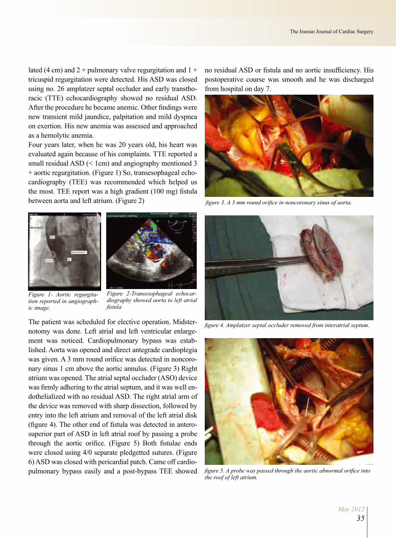
May 201235
The Iranian Journal of Cardiac Surgery
lated (4 cm) and 2 + pulmonary valve regurgitation and 1 + tricuspid regurgitation were detected. His ASD was closed using no. 26 amplatzer septal occluder and early transtho-racic (TTE) echocardiography showed no residual ASD. After the procedure he became anemic. Other findings were new transient mild jaundice, palpitation and mild dyspnea on exertion. His new anemia was assessed and approached as a hemolytic anemia. Four years later, when he was 20 years old, his heart was evaluated again because of his complaints. TTE reported a small residual ASD (< 1cm) and angiography mentioned 3 + aortic regurgitation. (Figure 1) So, transesophageal echo-cardiography (TEE) was recommended which helped us the most. TEE report was a high gradient (100 mg) fistula between aorta and left atrium. (Figure 2)
Figure 1- Aortic regurgita-tion reported in angiograph-ic image.
Figure 2-Transesophageal echocar-diography showed aorta to left atrial fistula
The patient was scheduled for elective operation. Midster-notomy was done. Left atrial and left ventricular enlarge-ment was noticed. Cardiopulmonary bypass was estab-lished. Aorta was opened and direct antegrade cardioplegia was given. A 3 mm round orifice was detected in noncoro-nary sinus 1 cm above the aortic annulus. (Figure 3) Right atrium was opened. The atrial septal occluder (ASO) device was firmly adhering to the atrial septum, and it was well en-dothelialized with no residual ASD. The right atrial arm of the device was removed with sharp dissection, followed by entry into the left atrium and removal of the left atrial disk (figure 4). The other end of fistula was detected in antero-superior part of ASD in left atrial roof by passing a probe through the aortic orifice. (Figure 5) Both fistulae ends were closed using 4/0 separate pledgetted sutures. (Figure 6) ASD was closed with pericardial patch. Came off cardio-pulmonary bypass easily and a post-bypass TEE showed
no residual ASD or fistula and no aortic insufficiency. His postoperative course was smooth and he was discharged from hospital on day 7.
figure 5. A probe was passed through the aortic abnormal orifice into the roof of left atrium.
figure 3. A 3 mm round orifice in noncoronary sinus of aorta.
figure 4. Amplatzer septal occluder removed from interatrial septum.
May 201236
Iranian Society of Cardiac Surgeons
Discussion:Surgical repair of all types of atrial septal defects (ASDs) has been practiced since the dawn of cardiac surgery in the 1950s with a large accumulated experience document-ing nearly 100% efficacy, near zero mortality, minimal and only short-term morbidity, and, with the introduction of less invasive surgical techniques, improved cosmetic results 25, 26 In 1976 King and Mills 27 reported the feasibility of per-cutaneous closure of ASD. Latson et al 28 in 1991 reported successful closure of ASDs in 500 patients with Bard clam-shell device. Percutaneous closure of ASD is gaining popu-larity because of the short learning curve, cosmetic benefits, reduced pain and reduced hospital stay. It also obviates an open cardiac procedure and many reports document high success rates and low morbidity 29-31. However, there are increasingly more frequent reports of serious complications of device ASD closures, including fatalities or major events necessitating surgical intervention 6-22. According to the report of Chessa et al, the overall incidence of complica-tions after the interventional occlusion of an ASD was 8.6% 24. European Association of Cardio-thoracic Surgery Congeni-tal Database has been reviewed for 10 years and 56 patients were reported to need early or late surgical repair after in-terventional closure of ASD23. The median time interval between device implantation and late surgery has been 3 years (12 days to 8 years) 23. Complications leading to sur-gery included embolisation (n = 29), thrombosis/thrombo-mbolism/cerebral ischemia or stroke (n = 12), significant residual shunt (n = 12), aortic or atrial perforation or ero-sion (n = 9), haemopericardium with tamponade (n = 5), aortic or mitral valve injury (n = 2) and endocarditis (n = 1) 23. Of these complications, the most worrisome were car-diac erosion or perforation, both of which may culminate in circulatory collapse. Fistula formation has different causes which one of the usual causes is deficient retroaortic rim. Although it is presumed that an oversized amplatzer may induce atrial erosion or a fistulous connection between the aorta and the atrium14, 32, serious complications may hap-pen regardless of the size or type of current devices 23.
In this case the aortic to left atrial fistula was detected 4 years after percutaneous implantation of atrial septal oc-
cluder while his new hemolytic anemia was under phar-macologic treatment. Although transthoracic echocardiog-raphy and angiography were not so accurate, the precise report of TEE helped us a lot in detecting the exact pathol-ogy. Operation was done successfully in this case but pa-tients needing surgical repair of complicated percutaneous closure of ASD are reported to have considerable mortality (5.4%) 23. There are two points which we may get out of this case: First, tranesophageal echocardiography could be considered as a helpful method of detecting complications of percutaneous closure of ASD; Second, and more impor-tant, comparing with patients who have undergone surgical closure of ASD and are considered cured, lifelong follow-up of patients whose ASD is closed by devices seems man-datory to detect potentially serious late device-related com-plications.
References:1. Murphy JG, Gersh BJ, McGoon MD, et al. Long-term outcome after sur-
gical repair of isolated atrial septal defect. Follow-up at 27 to 32 years. N Engl J Med 1990;323:1645–50.
2. Berdat PA, Chatterjee T, Pfammatter JP, et al. Surgical management of complications after transcatheter closure of an atrial septal defect or pat-ent foramen ovale. J Thorac Cardiovasc Surg 2000;120:1034 –9.
3. Preventza O, Sampath-Kumar S, Wasnick J, Gold JP. Late cardiac perfora-tion following transcatheter atrial septal defect closure. Ann Thorac Surg 2004;77:1435–7.
4. Vojacek J, Mates M, Popelova J, Pavel P. Perforation of the right atrium and the ascending aorta following percutaneous transcatheter atrial septal defect closure. Interact CardioVasc Thorac Surg 2005;4:157–9.
5. Sadeghpour Tabaee A, Rostami AR, Ghafari R, Sadeghpour Tabaee AH. Surgical removal of an embolized amplatzer device from left ventricle. Iranian Cardiovasc Res J 2007;1 (1):53-58.
6. Contrafouris CA, Chatzis AC, Giannopoulos NM, et al. Emergency surgi-cal intervention for runaway atrial septal defect closure devices: a word of caution. J Thorac Cardiovasc Surg 2006;132:1234—5.
7. Voja´cek J, Mates M, Popelova´ J, Pave P. Perforation of the right atrium and the ascending aorta following percutaneous transcatheter atrial septal defect closure. Interact Cardio Vasc Thorac Surg 2005;4:157—9.
8. Sauer HH, Ntalakoura K, Haun C, et al. Early cardiac perforation after atrial septal defect closure with the amplatzer septal occluder. Ann Thorac Surg 2006;81:2312—3.
9. Misra M, Sadiq A, Namboodiri N, Karunakaran J. The ‘aortic rim’ recount: embolization of interatrial septal occluder into the main pulmonary artery bifurcation after atrial septal defect closure. Interact Cardio Vasc Thorac Surg 2007;6:384—6.
10. Konstantinov IE, Saxena P, Friederich L, Newman MAJ. Emergency surgery after failed device closure of the atrial septal defect. J Thorac Cardiovasc Surg 2007;133:1370—1.
11. Bonatti J, Bonaros N, Mu¨ller S, Bartel T. Completely endoscopic remov-al of a dislocated Amplatzer atrial septal defect closure device. Interact Cardio Vasc Thorac Surg 2008;7:130—2.
12. Knott-Craig CJ, Goldberg SP. Emergent surgical retrieval of embolized atrial septal defect closure device. Ann Thorac Surg 2008;85:319—21.
13. Raghuram AR, Krishnan R, Kumar S, Balamurugan K. Complications
May 201237
The Iranian Journal of Cardiac Surgery
in atrial septal defect device closure. Interact Cardio Vasc Thorac Surg 2008;7:167—9.
14. Mello DM, Fahey J, Kopf GS. Repair of aortic-left fistula following the transcatheter closure of an atrial septal defect. Ann Thorac Surg 2005;80:1495—8.
15. Bo¨rgermann J, Hakim K, Friedrich I, Diez C. Recurrent thromboem-bolic events after percutaneous device closure of patent foramen ovale. Interact Cardiovasc Thorac Surg 2003;2:125—7.
16. 16. Tsilimingas NB, Reiter B, Kodolitsch YV, et al. Surgical revision of an uncommonly dislocated self-expanding Amplatzer septal occlude de-vice. Ann Thorac Surg 2004;78:686—7.
17. Siong Soo AW, Healy DG, Walsh K, Wood F. Inferior vena cava and coronary sinus obstruction after percutaneous atrial septal defect device closure requiring surgical revision. J Thorac Cardiovasc Surg 2006;131: 1405—6.
18. Walther T, Binner C, Rastan A, et al. Surgical atrial septal defect closure after interventional occlude placement: incidence and outcome. J Thorac Cardiovasc Surg 2007;134:731—7.
19. Nehgme RA, Huddleston AR, Cheatham JP. Progression to late complete atrioventricular block following Amplatzer device closure of atrial septal defect in a child. Pediatr Cardiol 2009;30(3):367—70.
20. Sto¨llberger C, Finsterer J, Krexner E, Schneider B. Stroke and peripheral embolism from an Amplatzer septal occlude 5 years after implantation. J Neurol 2008;255(8):1270—1.
21. Modine T, Larrue B, Leroy O, Fayad G. Surgical atrial septal defect closure after interventional occlude placement. Eur J Cardiothorac Surg 2008;33:736.
22. Rezaian GR, Amirghofran AA, Afifi S, et al. Nitinol wire mesh fracture and traumatic left atrial thrombus in a patient with atrial septal defect amplatzer occluder. J Card Surg 2011; 26(1):41-3.
23. Sarris GE, Kirvassilis G, Zavaropoulos P, et al. Surgery for complications of trans-catheter closure of atrial septal defects: a multi-institutional study from the European Congenital Heart Surgeons Association. Eur J Cardio-thorac Surg 2010;37:1285-90.
24. Chessa M, Carminati M, Butera G, et al. Early and late complications associated with transcatheter occlusion of secundum atrial septal defect. J Am Coll Cardiol 2002;39:1061–5.
25. Schreiber C, Bleiziffer S, Kostolny M, et al. Minimally invasive midaxil-lary muscle sparing thoracotomy for atrial septal defect closure in prepu-bescent patients. Ann Thorac Surg 2005;80:673—6.
26. Vida VL, Padalino MA, Boccuzzo G, et al. Minimally invasive opera-tion for congenital heart disease: a sex differentiated approach. J Thorac Cardiovasc Surg 2009;138:933—6.
27. King TD, Mills NL. Secundum atrial septal defects: nonoperative closure during cardiac catheterization. J Am Med Assoc 1976;235:2506–9.
28. Latson LA, Benson LN, Hellenbrand WE, et al. Transcatheter closure of ASD – early results of multicenter trial of the Bard clamshell septal oc-cluder. Circulation 1991;84(2):44.
29. Nugent AW, Britt A, Gauvreau K, et al. Device closure rates of simple atrial septal defects optimized by the starflex device. J Am Coll Cardiol 2006;48:538—44.
30. 30. Romagnoli BGE, Carminati M, Chessa M, et al. Treatment of isolated secundum atrial septal defects: impact of age and defect morphology in 1013 consecutive patients. Am Heart J 2008;156(4):706—12.
31. Berger F, Vogel M, Meskishvili VA, Lange PE. Comparison of results and complications of surgical and amplatzer device closure of atrial septal defects. J Thorac Cardiovasc Surg 1999;118:674—8.
32. Jang GY, Lee JY, Kim SJ, et al. Aorta to Right Atrial Fistula Follow-ing Transcatheter Closure of an Atrial Septal Defect. Am J Cardiol 2005;96:1605–1606.
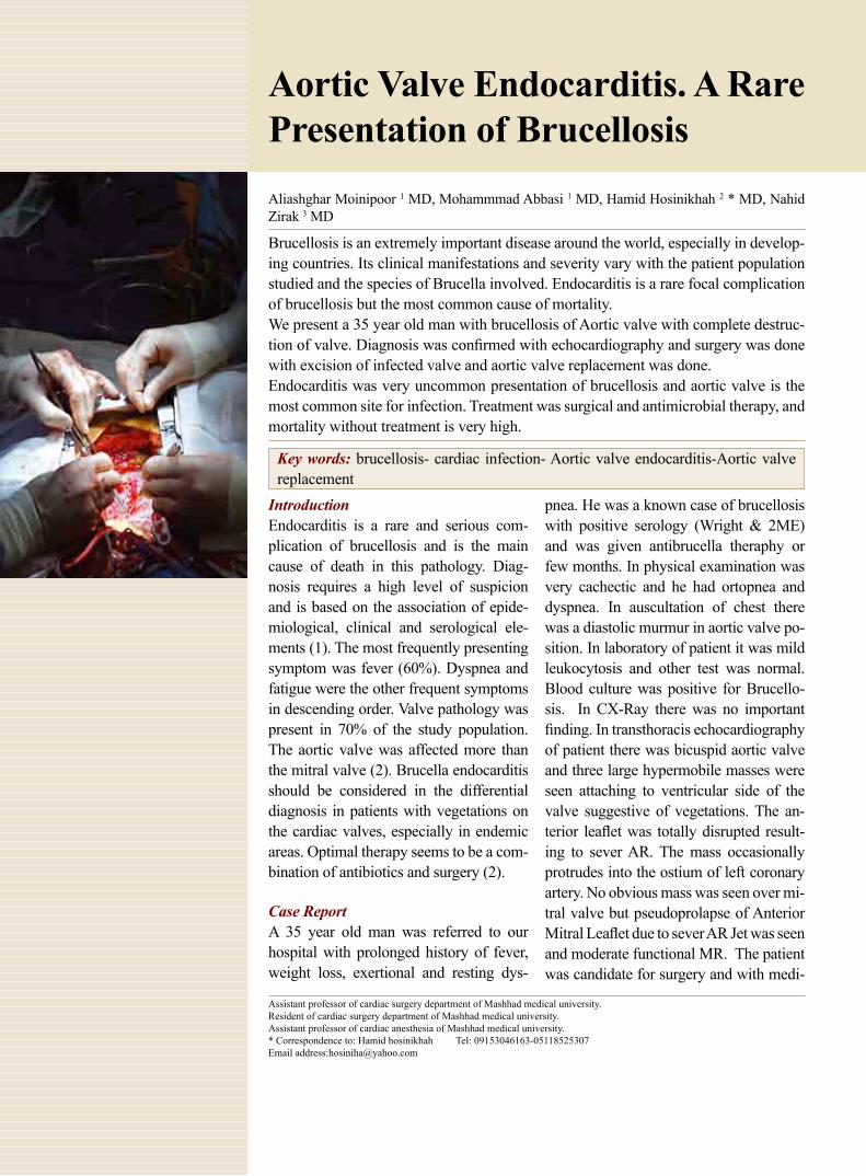
Aortic Valve Endocarditis. A Rare Presentation of BrucellosisAliashghar Moinipoor 1 MD, Mohammmad Abbasi 1 MD, Hamid Hosinikhah 2 * MD, Nahid Zirak 3 MD
Assistant professor of cardiac surgery department of Mashhad medical university.Resident of cardiac surgery department of Mashhad medical university.Assistant professor of cardiac anesthesia of Mashhad medical university.* Correspondence to: Hamid hosinikhah Tel: 09153046163-05118525307Email address:[email protected]
Brucellosis is an extremely important disease around the world, especially in develop-ing countries. Its clinical manifestations and severity vary with the patient population studied and the species of Brucella involved. Endocarditis is a rare focal complication of brucellosis but the most common cause of mortality.We present a 35 year old man with brucellosis of Aortic valve with complete destruc-tion of valve. Diagnosis was confirmed with echocardiography and surgery was done with excision of infected valve and aortic valve replacement was done.Endocarditis was very uncommon presentation of brucellosis and aortic valve is the most common site for infection. Treatment was surgical and antimicrobial therapy, and mortality without treatment is very high.
Key words: brucellosis- cardiac infection- Aortic valve endocarditis-Aortic valve replacement
IntroductionEndocarditis is a rare and serious com-plication of brucellosis and is the main cause of death in this pathology. Diag-nosis requires a high level of suspicion and is based on the association of epide-miological, clinical and serological ele-ments (1). The most frequently presenting symptom was fever (60%). Dyspnea and fatigue were the other frequent symptoms in descending order. Valve pathology was present in 70% of the study population. The aortic valve was affected more than the mitral valve (2). Brucella endocarditis should be considered in the differential diagnosis in patients with vegetations on the cardiac valves, especially in endemic areas. Optimal therapy seems to be a com-bination of antibiotics and surgery (2).
Case ReportA 35 year old man was referred to our hospital with prolonged history of fever, weight loss, exertional and resting dys-
pnea. He was a known case of brucellosis with positive serology (Wright & 2ME) and was given antibrucella theraphy or few months. In physical examination was very cachectic and he had ortopnea and dyspnea. In auscultation of chest there was a diastolic murmur in aortic valve po-sition. In laboratory of patient it was mild leukocytosis and other test was normal. Blood culture was positive for Brucello-sis. In CX-Ray there was no important finding. In transthoracis echocardiography of patient there was bicuspid aortic valve and three large hypermobile masses were seen attaching to ventricular side of the valve suggestive of vegetations. The an-terior leaflet was totally disrupted result-ing to sever AR. The mass occasionally protrudes into the ostium of left coronary artery. No obvious mass was seen over mi-tral valve but pseudoprolapse of Anterior Mitral Leaflet due to sever AR Jet was seen and moderate functional MR. The patient was candidate for surgery and with medi-
May 201239
The Iranian Journal of Cardiac Surgery
an sternotomy approach, CPB was done in standard fashion and cardiac arrest was done with cardioplegic infusion via anterograde and direct in coronary ostium after opening the Aorta. Aortic leaflet was completely destructed and leaflet was flaccid and a lot of vegetation especially in noncoronary and Right coronary leaflet of aortic valve was seen. After ex-cision of aortic leaflet and cleaning of annulus and debride-ment, irrigation was done, Specimen was sent for pathologic examination, size of annulus was measured and aortic valve replacement was done with mechanical Aortic valve (n 21). Postoperative and recovery state was uneventful. Pathologic examination of aortic valve confirms endocarditis. Patient was discharge of hospital in 10 day after operation with antibiotic and in 2 months follow up was in good status.
DiscussionBrucellosis is a zoonosis transmitted to humans through the consumption of products derived from unpasteurized milk and through direct contact with infected animal remains. This means the disease is most frequently found among in-dividuals who have regular contact with livestock and their products, such as shepherds, slaughterhouse workers and veterinary staff, as well as people working in microbiol-ogy laboratories, who become infected through inhalation of aerosolized particles (1). A zoonosis, Brucella, causes a special form of infectious disease named brucellosis. Any organ system may be affected, although the musculoskel-etal, especially osteoarticular part, is the most commonly affected. Involvement of the cardiovascular system is rela-tively rare (2). Human brucellosis is a systemic disease that involves multiple organs and tissues concomitantly (4). The most common form of presentation being fever of unknown origin, together with constitutional symptoms such as asthe-nia, sweating and joint pain (1). As is usual in brucellosis, young men were infected more frequently than females. In addition they all had a history of ingestion of contaminated milk or milk products, more commonly non-pasteurized cheese. The predominant symptoms have been fever and dyspnea (NYHA class 3 or 4) (2, 3). Wright agglutination test in a titer of 1/160 or higher is very sensitive and spe-cific for the diagnosis of brucellosis (2). Brucella endocar-ditis was diagnosed by physical examination, laboratory findings, serological tests, blood culture, transthoracic and trans-esophageal echocardiography and elevated ESR and CRP (3, 7). Etiological diagnosis is not easy; it requires a
high level of suspicion and is based on the association of epidemiological data with serological test results or isolation of Brucella in previously collected blood or tissue samples. The sensitivity of blood cultures is extremely variable (15 to 70%), depending on the time elapsed between symptom onset and diagnosis, previous recent antibiotic therapy and culture conditions (culture medium and incubation time) Se-rological methods are more sensitive but have lower speci-ficity, and are limited by the difficulty of interpreting results in endemic regions and the significant percentage of cases with late or no seroconversion (1). Transthoracic echocar-diography plays a crucial role in diagnosing cardiac compli-cations, and a transesophageal approach is required in some cases. However, in the presence of predisposing cardiac factors, the detection of Brucella bacteremia is equivalent to a diagnosis of endocarditis. The aortic valve is most fre-quently affected, with mitral valve involvement more com-mon in cases of previous changes in its structure, as in rheu-matic valve disease. The most common echocardiographic findings are vegetations, normally large, and ulcerations or abscesses, but any structural element of the affected valve may be involved. The most frequent complication is con-gestive heart failure, with an incidence of 75-90%, and the rate of embolic events is similar to that seen in endocarditis caused by other microbiological agents. Cardiovascular in-volvement is uncommon, and may present in the form of myocarditis, pericarditis, aortic root abscess or endocarditis, the latter being the principal cause of death (1). Endocardi-tis is a rare complication of brucellosis with an incidence of 1 to 2%; it is more common in patients with previous valve disease. It is a serious, potentially fatal condition, and pathological alterations typical of endocarditis are found in 80% of autopsy studies performed on patients who have died from brucellosis (1). The 70% of patients had underly-ing valve pathology for endocarditis. Brucella endocarditis affected the aortic valves more than the mitral valves as ex-pected (70%). The 43% of the infected aortic valves had no predisposing pathology. All the affected mitral valves had rheumatic pathology (2). Brucella endocarditis is a rare type of endocarditis. In endemic regions, its prevalence is higher than in nonendemic parts of the world. It has a much higher mortality rate than endocarditis caused by other pathogens due to its rapid and wide tissue destruction. In addition, the high mortality rate might be due to late diagnosis of the in-
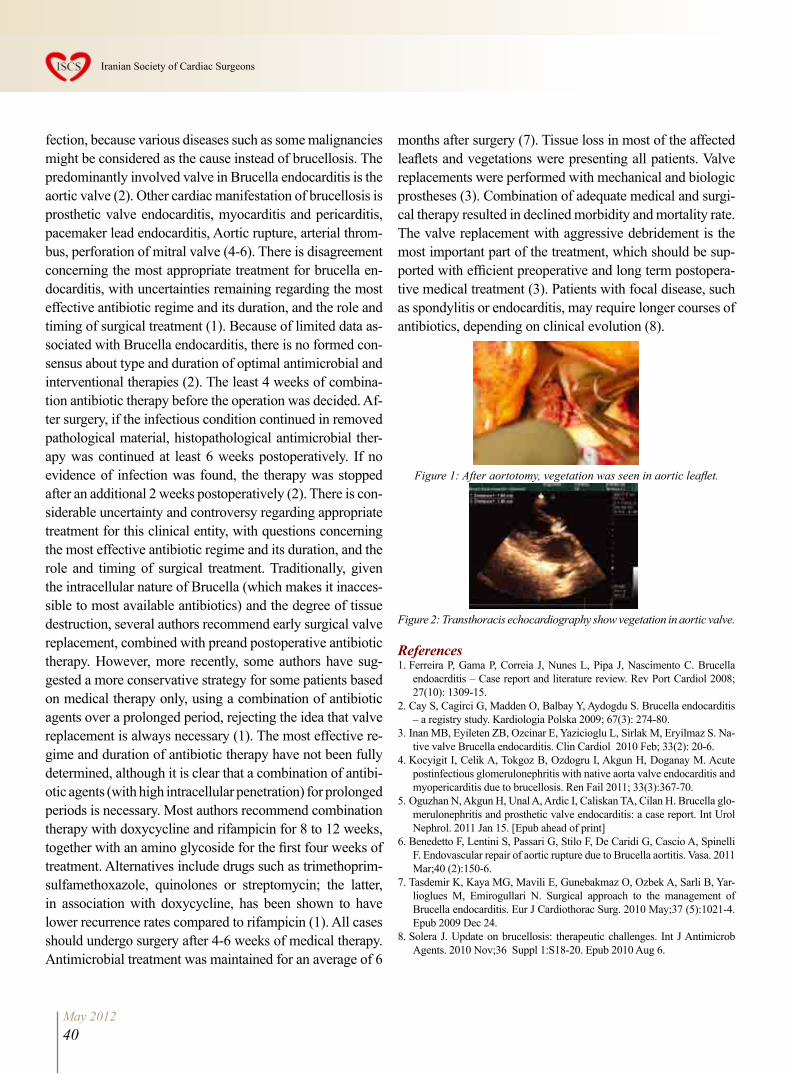
May 201240
Iranian Society of Cardiac Surgeons
fection, because various diseases such as some malignancies might be considered as the cause instead of brucellosis. The predominantly involved valve in Brucella endocarditis is the aortic valve (2). Other cardiac manifestation of brucellosis is prosthetic valve endocarditis, myocarditis and pericarditis, pacemaker lead endocarditis, Aortic rupture, arterial throm-bus, perforation of mitral valve (4-6). There is disagreement concerning the most appropriate treatment for brucella en-docarditis, with uncertainties remaining regarding the most effective antibiotic regime and its duration, and the role and timing of surgical treatment (1). Because of limited data as-sociated with Brucella endocarditis, there is no formed con-sensus about type and duration of optimal antimicrobial and interventional therapies (2). The least 4 weeks of combina-tion antibiotic therapy before the operation was decided. Af-ter surgery, if the infectious condition continued in removed pathological material, histopathological antimicrobial ther-apy was continued at least 6 weeks postoperatively. If no evidence of infection was found, the therapy was stopped after an additional 2 weeks postoperatively (2). There is con-siderable uncertainty and controversy regarding appropriate treatment for this clinical entity, with questions concerning the most effective antibiotic regime and its duration, and the role and timing of surgical treatment. Traditionally, given the intracellular nature of Brucella (which makes it inacces-sible to most available antibiotics) and the degree of tissue destruction, several authors recommend early surgical valve replacement, combined with preand postoperative antibiotic therapy. However, more recently, some authors have sug-gested a more conservative strategy for some patients based on medical therapy only, using a combination of antibiotic agents over a prolonged period, rejecting the idea that valve replacement is always necessary (1). The most effective re-gime and duration of antibiotic therapy have not been fully determined, although it is clear that a combination of antibi-otic agents (with high intracellular penetration) for prolonged periods is necessary. Most authors recommend combination therapy with doxycycline and rifampicin for 8 to 12 weeks, together with an amino glycoside for the first four weeks of treatment. Alternatives include drugs such as trimethoprim-sulfamethoxazole, quinolones or streptomycin; the latter, in association with doxycycline, has been shown to have lower recurrence rates compared to rifampicin (1). All cases should undergo surgery after 4-6 weeks of medical therapy. Antimicrobial treatment was maintained for an average of 6
months after surgery (7). Tissue loss in most of the affected leaflets and vegetations were presenting all patients. Valve replacements were performed with mechanical and biologic prostheses (3). Combination of adequate medical and surgi-cal therapy resulted in declined morbidity and mortality rate. The valve replacement with aggressive debridement is the most important part of the treatment, which should be sup-ported with efficient preoperative and long term postopera-tive medical treatment (3). Patients with focal disease, such as spondylitis or endocarditis, may require longer courses of antibiotics, depending on clinical evolution (8).
Figure 1: After aortotomy, vegetation was seen in aortic leaflet.
Figure 2: Transthoracis echocardiography show vegetation in aortic valve.
References1. Ferreira P, Gama P, Correia J, Nunes L, Pipa J, Nascimento C. Brucella
endoacrditis – Case report and literature review. Rev Port Cardiol 2008; 27(10): 1309-15.
2. Cay S, Cagirci G, Madden O, Balbay Y, Aydogdu S. Brucella endocarditis – a registry study. Kardiologia Polska 2009; 67(3): 274-80.
3. Inan MB, Eyileten ZB, Ozcinar E, Yazicioglu L, Sirlak M, Eryilmaz S. Na-tive valve Brucella endocarditis. Clin Cardiol 2010 Feb; 33(2): 20-6.
4. Kocyigit I, Celik A, Tokgoz B, Ozdogru I, Akgun H, Doganay M. Acute postinfectious glomerulonephritis with native aorta valve endocarditis and myopericarditis due to brucellosis. Ren Fail 2011; 33(3):367-70.
5. Oguzhan N, Akgun H, Unal A, Ardic I, Caliskan TA, Cilan H. Brucella glo-merulonephritis and prosthetic valve endocarditis: a case report. Int Urol Nephrol. 2011 Jan 15. [Epub ahead of print]
6. Benedetto F, Lentini S, Passari G, Stilo F, De Caridi G, Cascio A, Spinelli F. Endovascular repair of aortic rupture due to Brucella aortitis. Vasa. 2011 Mar;40 (2):150-6.
7. Tasdemir K, Kaya MG, Mavili E, Gunebakmaz O, Ozbek A, Sarli B, Yar-lioglues M, Emirogullari N. Surgical approach to the management of Brucella endocarditis. Eur J Cardiothorac Surg. 2010 May;37 (5):1021-4. Epub 2009 Dec 24.
8. Solera J. Update on brucellosis: therapeutic challenges. Int J Antimicrob Agents. 2010 Nov;36 Suppl 1:S18-20. Epub 2010 Aug 6.
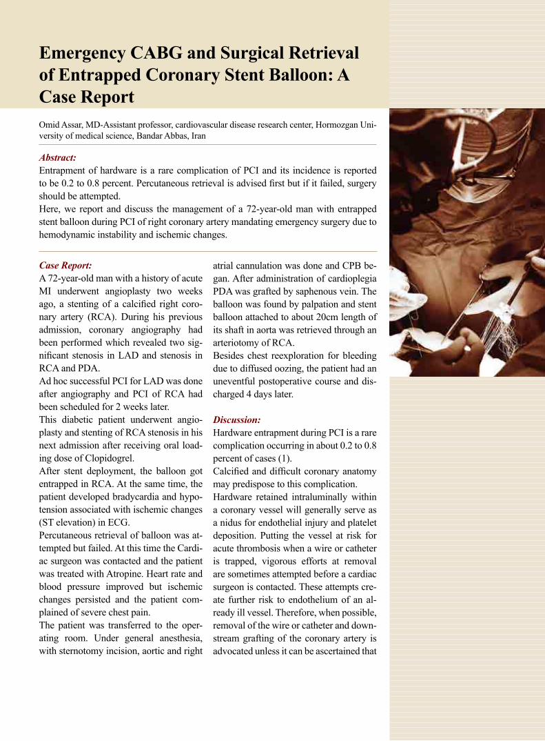
Emergency CABG and Surgical Retrieval of Entrapped Coronary Stent Balloon: A Case Report Omid Assar, MD-Assistant professor, cardiovascular disease research center, Hormozgan Uni-versity of medical science, Bandar Abbas, Iran
Abstract:Entrapment of hardware is a rare complication of PCI and its incidence is reported to be 0.2 to 0.8 percent. Percutaneous retrieval is advised first but if it failed, surgery should be attempted.Here, we report and discuss the management of a 72-year-old man with entrapped stent balloon during PCI of right coronary artery mandating emergency surgery due to hemodynamic instability and ischemic changes.
Case Report:A 72-year-old man with a history of acute MI underwent angioplasty two weeks ago, a stenting of a calcified right coro-nary artery (RCA). During his previous admission, coronary angiography had been performed which revealed two sig-nificant stenosis in LAD and stenosis in RCA and PDA.Ad hoc successful PCI for LAD was done after angiography and PCI of RCA had been scheduled for 2 weeks later.This diabetic patient underwent angio-plasty and stenting of RCA stenosis in his next admission after receiving oral load-ing dose of Clopidogrel. After stent deployment, the balloon got entrapped in RCA. At the same time, the patient developed bradycardia and hypo-tension associated with ischemic changes (ST elevation) in ECG.Percutaneous retrieval of balloon was at-tempted but failed. At this time the Cardi-ac surgeon was contacted and the patient was treated with Atropine. Heart rate and blood pressure improved but ischemic changes persisted and the patient com-plained of severe chest pain.The patient was transferred to the oper-ating room. Under general anesthesia, with sternotomy incision, aortic and right
atrial cannulation was done and CPB be-gan. After administration of cardioplegia PDA was grafted by saphenous vein. The balloon was found by palpation and stent balloon attached to about 20cm length of its shaft in aorta was retrieved through an arteriotomy of RCA.Besides chest reexploration for bleeding due to diffused oozing, the patient had an uneventful postoperative course and dis-charged 4 days later.
Discussion:Hardware entrapment during PCI is a rare complication occurring in about 0.2 to 0.8 percent of cases (1).Calcified and difficult coronary anatomy may predispose to this complication.Hardware retained intraluminally within a coronary vessel will generally serve as a nidus for endothelial injury and platelet deposition. Putting the vessel at risk for acute thrombosis when a wire or catheter is trapped, vigorous efforts at removal are sometimes attempted before a cardiac surgeon is contacted. These attempts cre-ate further risk to endothelium of an al-ready ill vessel. Therefore, when possible, removal of the wire or catheter and down-stream grafting of the coronary artery is advocated unless it can be ascertained that
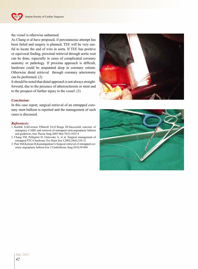
May 201242
Iranian Society of Cardiac Surgeons
the vessel is otherwise unharmed.As Chang et al have proposed, if percutaneous attempt has been failed and surgery is planned, TEE will be very use-ful to locate the end of wire in aorta. If TEE has positive or equivocal finding, proximal retrieval through aortic root can be done, especially in cases of complicated coronary anatomy or pathology. If proxima approach is difficult, hardware could be amputated deep in coronary ostium. Otherwise distal retrieval through coronary arteriotomy can be performed. (2)It should be noted that distal approach is not always straight-forward, due to the presence of atherosclerosis or stent and to the prospect of further injury to the vessel. (3)
Conclusion:In this case report, surgical retrieval of an entrapped coro-nary stent balloon is reported and the management of such cases is discussed.
References:1. Karthik S,Silverston P,Blaxill JA,O`Reaga DJ.Successful outcome of
emergency CABG and retrieval of entrapped stent,angioplasty balloon and guidewire.Ann Thorac Surg.2005 Mar;79(3):1032-4
2. Chang TM, Pellegrini D, Ostrovsky A, et al. Surgical management of entrapped PTCA hardware.Tex Heart Inst J,2002;29(4):329-32
3. Peer SM,Kalyani R,Kamalapurkar G.Surgical retrieval of entrapped cor-onary angioplasty balloon.Eur J Cardiothorac Surg.2010;38:804
Congenital Left Main Coronary ArteryFistula to Right Atrium : A Case Report
Pediatric Cardiology ward, Shahid Modarres Hospital, Shahid Beheshti University of Medical Sciences, Tehran, IranAddress of Corresponding authorDr Shahla Roodpeyma, Pediatric cardiology ward, Shahid Modarres hospital, Saadat Abad, Tehran 1998734383, IranTel : 0098 021 22074087-98 Fax : 0098 021 22074101 Email : [email protected]
Fatemeh Vaziri , MD, Shahla Roodpeyma , MD, Manuchehr Hekmat , MD
AbstractCoronary artery fistulas are rare abnormalities with an estimated frequency of 0.002% in the general population. The majority of these fistulas arise from the right coronary artery. The left coronary artery is rarely involved. This study presents a 5-year old girl with a left coronary artery fistula to right atrium who underwent a successful surgical operation.
IntroductionCoronary artery fistulas (CAF) are rare congenital anomalies; they connect a ma-jor coronary artery directly to a cardiac chamber, coronary sinus, superior vena cava or pulmonary artery. They consti-tute the most common form of hemody-namically significant coronary malforma-tion with an incidence of 0.002% in the general population and 0.2% in patients undergoing coronary angiography. The most plausible explanation for a congeni-tal CAF is the persistence of embryonic vascular sinusoids in the myocardium (1). The right coronary artery or its branches are the site of fistula in about 55% of cases; the left coronary artery in about 35%; and both coronary arteries in 5%. Over 90% of the fistulas drain into the venous circulation. These include right – sided chambers, pulmonary artery, su-perior vena cava and coronary sinus. Fistulas drainage occurs into the RV in (41%), RA in (26%), pulmonary artery in (17%), LV in (3%), and SVC in (1%). Most fistulas are single communications, but multiple fistulas have been reported (2). The myocardial blood flow is usually not compromised and shunt through the fistula usually is of small magnitude. A left – to – right shunt exists in more than 90% of cases. The majority of adult pa-tients are usually asymptomatic. Unlike
adults a smaller percentage of pediatric patients are asymptomatic (2). Sometimes the blood in CAF bypasses the myocar-dial capillary network (steal phenomena) and symptoms range from mild shortness of breath to myocardial ischemia, an-gina, congestive heart failure, cardiac ar-rhythmia, rupture or dissection of fistula with or without cardiac tamponade and endocarditis (3,4). The acquired causes of coronary fistulas are rare and include atherosclerosis, takayasu arteritis, and trauma (2). Although selective coronary angiography is capable of identifying the origin of aneurysmal CAF, it is difficult to clarify the relation of the distal site of the CAF to other cardiac chambers because of dilution of a contrast medium or over-lapping of adjacent structure. A multi – slice CT angiography might well become the modality of choice for the diagnosis of these rare congenital anomalies (5). It is well accepted that all symptomatic pa-tients should be treated with surgical op-eration because the surgical risk in most cases appears to be considerably less than the potential development of serious and fatal complication (6).The aim of this study is to present an as-ymptomatic pediatric patient with a rare type of CAF who underwent a successful surgical repair.
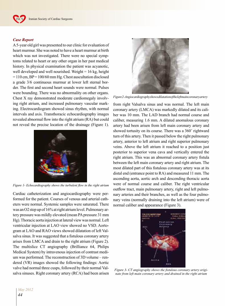
May 201244
Iranian Society of Cardiac Surgeons
Case ReportA 5-year old girl was presented to our clinic for evaluation of heart murmur. She was noted to have a heart murmur at birth which was not investigated. There were no special symp-toms related to heart or any other organ in her past medical history. In physical examination the patient was acyanotic, well developed and well nourished. Weight = 16 kg, height = 110 cm, BP = 100/60 mm Hg. Chest auscultation disclosed a grade 3/6 continuous murmur at lower left sternal bor-der. The first and second heart sounds were normal. Pulses were bounding. There was no abnormality on other organs. Chest X ray demonstrated moderate cardiomegaly involv-ing right atrium, and increased pulmonary vascular mark-ing. Electrocardiogram showed sinus rhythm, with normal intervals and axis. Transthoracic echocardiography images revealed abnormal flow into the right atrium (RA) but could not reveal the precise location of the drainage (Figure 1).
Figure 1- Echocardiography shows the turbulent flow in the right atrium
Cardiac catheterization and angiocardiography were per-formed for the patient. Courses of venous and arterial cath-eters were normal. Systemic samples were saturated. There was an O2 step up of 16% at right atrium level. Pulmonary ar-tery pressure was mildly elevated (mean PA pressure 31 mm Hg). Thoracic aorta injection at lateral view was normal. Left ventricular injection at LAO view showed no VSD. Aorto-gram at LAO and RAO views showed dilatation of left Val-salva sinus. It was suggested that a fistulous coronary artery arises from LMCA and drain to the right atrium (Figure 2). The multislice CT angiography (Brilliance 64, Philips Medical System) by intravenous injection of contrast medi-um was performed. The reconstruction of 3D volume – ren-dered (VR) images showed the following findings: Aortic valve had normal three cusps, followed by their normal Val-salva sinuses. Right coronary artery (RCA) had been arisen
from right Valsalva sinus and was normal. The left main coronary artery (LMCA) was markedly dilated and its cali-ber was 10 mm. The LAD branch had normal course and caliber, measuring 1.6 mm. A dilated anomalous coronary artery had been arisen from left main coronary artery and showed tortusity on its course. There was a 360˚ rightward turn of this artery. Then it passed below the right pulmonary artery, anterior to left atrium and right superior pulmonary veins. Above the left atrium it reached to a position just posterior to superior vena cava and vertically entered the right atrium. This was an abnormal coronary artery fistula between the left main coronary artery and right atrium. The most dilated part of this fistulous coronary artery was at its distal end (entrance point to RA) and measured 11 mm. The ascending aorta, aortic arch and descending thoracic aorta were of normal course and caliber. The right ventricular outflow tract, main pulmonary artery, right and left pulmo-nary arteries and their branches, as well as the four pulmo-nary veins (normally draining into the left atrium) were of normal caliber and appearance (Figure 3).
Figure 3- CT angiography shows the fistulous coronary artery origi-nate from left main coronary artery and drained in the right atrium
Figure 2- Angiocardiography shows dilatation of the left main coronary artery
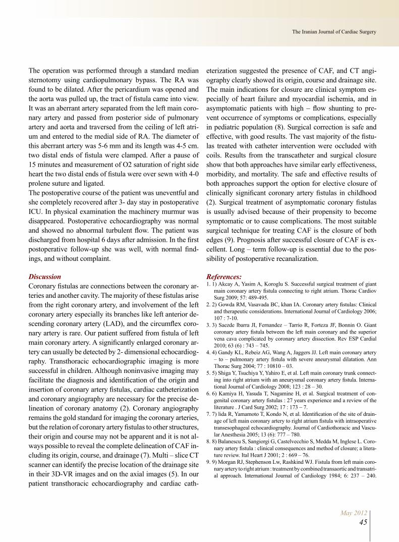
May 201245
The Iranian Journal of Cardiac Surgery
The operation was performed through a standard median sternotomy using cardiopulmonary bypass. The RA was found to be dilated. After the pericardium was opened and the aorta was pulled up, the tract of fistula came into view. It was an aberrant artery separated from the left main coro-nary artery and passed from posterior side of pulmonary artery and aorta and traversed from the ceiling of left atri-um and entered to the medial side of RA. The diameter of this aberrant artery was 5-6 mm and its length was 4-5 cm. two distal ends of fistula were clamped. After a pause of 15 minutes and measurement of O2 saturation of right side heart the two distal ends of fistula were over sewn with 4-0 prolene suture and ligated.The postoperative course of the patient was uneventful and she completely recovered after 3- day stay in postoperative ICU. In physical examination the machinery murmur was disappeared. Postoperative echocardiography was normal and showed no abnormal turbulent flow. The patient was discharged from hospital 6 days after admission. In the first postoperative follow-up she was well, with normal find-ings, and without complaint.
DiscussionCoronary fistulas are connections between the coronary ar-teries and another cavity. The majority of these fistulas arise from the right coronary artery, and involvement of the left coronary artery especially its branches like left anterior de-scending coronary artery (LAD), and the circumflex coro-nary artery is rare. Our patient suffered from fistula of left main coronary artery. A significantly enlarged coronary ar-tery can usually be detected by 2- dimensional echocardiog-raphy. Transthoracic echocardiographic imaging is more successful in children. Although noninvasive imaging may facilitate the diagnosis and identification of the origin and insertion of coronary artery fistulas, cardiac catheterization and coronary angiography are necessary for the precise de-lineation of coronary anatomy (2). Coronary angiography remains the gold standard for imaging the coronary arteries, but the relation of coronary artery fistulas to other structures, their origin and course may not be apparent and it is not al-ways possible to reveal the complete delineation of CAF in-cluding its origin, course, and drainage (7). Multi – slice CT scanner can identify the precise location of the drainage site in their 3D-VR images and on the axial images (5). In our patient transthoracic echocardiography and cardiac cath-
eterization suggested the presence of CAF, and CT angi-ography clearly showed its origin, course and drainage site.The main indications for closure are clinical symptom es-pecially of heart failure and myocardial ischemia, and in asymptomatic patients with high – flow shunting to pre-vent occurrence of symptoms or complications, especially in pediatric population (8). Surgical correction is safe and effective, with good results. The vast majority of the fistu-las treated with catheter intervention were occluded with coils. Results from the transcatheter and surgical closure show that both approaches have similar early effectiveness, morbidity, and mortality. The safe and effective results of both approaches support the option for elective closure of clinically significant coronary artery fistulas in childhood (2). Surgical treatment of asymptomatic coronary fistulas is usually advised because of their propensity to become symptomatic or to cause complications. The most suitable surgical technique for treating CAF is the closure of both edges (9). Prognosis after successful closure of CAF is ex-cellent. Long – term follow-up is essential due to the pos-sibility of postoperative recanalization.
References: 1. 1) Akcay A, Yasim A, Koroglu S. Successful surgical treatment of giant
main coronary artery fistula connecting to right atrium. Thorac Cardiov Surg 2009; 57: 489-495.
2. 2) Gowda RM, Vasavada BC, khan IA. Coronary artery fistulas: Clinical and therapeutic considerations. International Journal of Cardiology 2006; 107 : 7-10.
3. 3) Saezde lbarra JI, Fernandez – Tarrio R, Forteza JF, Bonnin O. Giant coronary artery fistula between the left main coronary and the superior vena cava complicated by coronary artery dissection. Rev ESP Cardial 2010; 63 (6) : 743 – 745.
4. 4) Gandy KL, Rebeiz AG, Wang A, Jaggers JJ. Left main coronary artery – to – pulmonary artery fistula with severe aneurysmal dilatation. Ann Thorac Surg 2004; 77 : 10810 – 03.
5. 5) Shiga Y, Tsuchiya Y, Yahiro E, et al. Left main coronary trunk connect-ing into right atrium with an aneurysmal coronary artery fistula. Interna-tional Journal of Cardiology 2008; 123 : 28 – 30.
6. 6) Kamiya H, Yasuda T, Nagamine H, et al. Surgical treatment of con-genital coronary artery fistulas : 27 years experience and a review of the literature . J Card Surg 2002; 17 : 173 – 7.
7. 7) lida R, Yamamoto T, Kondo N, et al. Identification of the site of drain-age of left main coronary artery to right atrium fistula with intraoperative transesophageal echocardiography. Journal of Cardiothoracic and Vascu-lar Anesthesia 2005; 13 (6): 777 – 780.
8. 8) Balanescu S, Sangiorgi G, Castelvecchio S, Medda M, Inglese L. Coro-nary artery fistula : clinical consequences and method of closure; a litera-ture review. Ital Heart J 2001; 2 : 669 – 76.
9. 9) Morgan RJ, Stephenson Lw, Rashkind WJ. Fistula from left main coro-nary artery to right atrium : treatment by combined transaortic and transatri-al approach. International Journal of Cardiology 1984; 6: 237 – 240.
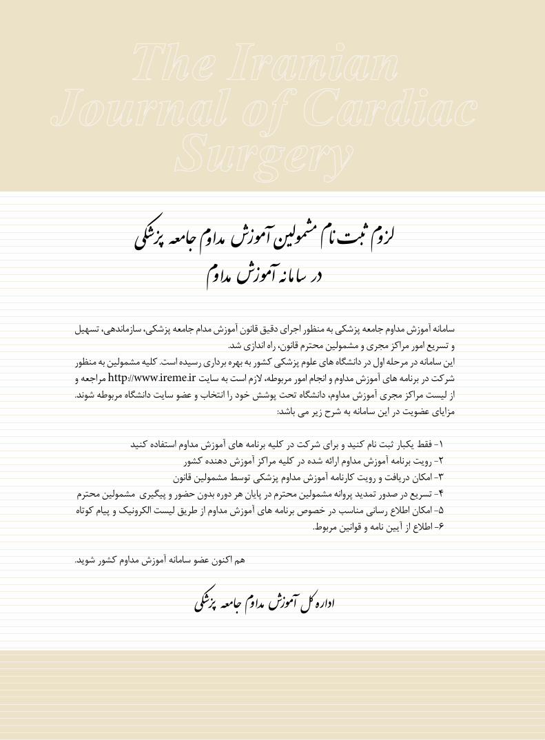
سامانه آموزش مداوم جامعه پزشکی به منظور اجرای دقیق قانون آموزش مدام جامعه پزشکی، سازماندهی، تسهیل و تسریع امور مراکز مجری و مشمولین محترم قانون، راه اندازی شد.
این سامانه در مرحله اول در دانشگاه های علوم پزشکی کشور به بهره برداری رسیده است. کلیه مشمولین به منظور شرکت در برنامه های آموزش مداوم و انجام امور مربوطه، الزم است به سایت http://www.ireme.ir مراجعه و از لیست مراکز مجری آموزش مداوم، دانشگاه تحت پوشش خود را انتخاب و عضو سایت دانشگاه مربوطه شوند.
مزایای عضویت در این سامانه به شرح زیر می باشد:
1- فقط یکبار ثبت نام کنید و برای شرکت در کلیه برنامه های آموزش مداوم استفاده کنید2- رویت برنامه آموزش مداوم ارائه شده در کلیه مراکز آموزش دهنده کشور
3- امکان دریافت و رویت کارنامه آموزش مداوم پزشکی توسط مشمولین قانون 4- تسریع در صدور تمدید پروانه مشمولین محترم در پایان هر دوره بدون حضور و پیگیری مشمولین محترم 5- امکان اطالع رسانی مناسب در خصوص برنامه های آموزش مداوم از طریق لیست الکرونیک و پیام کوتاه
6- اطالع از آیین نامه و قوانین مربوط.
هم اکنون عضو سامانه آموزش مداوم کشور شوید.


![Cardiac Care - Homemycardiaccare.ca › ~mycardia › images › pdfs › forms › 2017 › ... · CIO HCM a C] ARVD PROFESSIONAL SERVICES Consultation Clinical Consultation [2 OTN](https://img.pdfslide.fr/doc/110x75/5f28737725376d19c36d3ab5/cardiac-care-a-mycardia-a-images-a-pdfs-a-forms-a-2017-a-cio.jpg)