Embed Size (px)
Citation preview

1
The isolated human umbilical vein as a bioassay for kinin-generating proteases: an in
vitro model for therapeutic angioedema agents
Melissa Jeana, Arvind Raghavanb, Matthew L. Charlesc, Mark S. Robbinsb,d, Eric
Wagnera, Georges-Étienne Rivarde, Xavier Charest-Morina, François Marceaua*
a Axe Microbiologie-Infectiologie et Immunologie, CHU de Québec, and Faculté de
médecine, Université Laval, Québec QC, Canada G1V 4G2 ;
bTansna Therapeutics, St. Louis, MO, USA, 63108;
cMinnetonka, MN, USA;
dKodiak Strategic Consultants, LLC, Minneapolis, MN, USA.
e Division of Hematology / Oncology, CHU Sainte-Justine, Montréal, QC, Canada
Presented in part at the 9th C1-inhibitor Deficiency Workshop, Budapest, Hungary, 29
May 2015.
Correspondence:
Dr F Marceau, T1-49, CHU de Québec, 2705 Laurier Blvd., Québec (Québec), Canada
G1V 4G2. Tel. 1-418-525-4444 x46155. E-mail: [email protected]

2
Abstract
Aims: The isolated human umbilical vein is a robust contractile bioassay for ligands of
the bradykinin (BK) B2 receptor (B2R), also extendable to B1 receptor (B1R)
pharmacology. We hypothesized that, as a freshly isolated vessel, it also contains traces
of plasma proteins that may confer responses to exogenous proteases via the formation of
kinins.
Main methods: Rings of human umbilical veins were mounted in organ baths containing
Krebs buffer maintained at 37°C and purified proteases were introduced in the bathing
fluid along with additional drugs/proteins that permit mechanistic analysis of effects.
Key findings: The previously described contractile response to human recombinant tissue
kallikrein (KLK-1, 1-10 nM) is not influenced by metabolic inhibitors, suggesting its
dependence on a preexisting reservoir of low molecular weight-kininogen (LK). Active
plasma kallikrein (apK, inactive in fresh tissues, unless high molecular
weight-kininogen (HK, 39-197 nM) replenishment was applied. The effects of KLK-1
and HK+apK are abolished by pretreating tissues with icatibant, but not with tranexamic
acid. C1-esterase inhibitor inhibited only HK+apK. Purified plasmin and neutrophil
proteinase-3 produced small contractions in the presence of HK only, and tissue
plasminogen activator, none. B1R stimulation was pharmacologically evidenced in
response to KLK-1 if LK was supplied.

3
Significance: The pharmacology of KLK-1 and HK+apK in the human isolated umbilical
vein is essentially based on the activity of locally generated kinins and this assay models
the inhibitory action of some therapeutic agents active in angioedema states. Proteases
that indirectly generate kinins have little activity in the system.
Keywords: tissue kallikrein, plasma kallikrein, plasmin, bradykinin B2 receptor.

4
1. Introduction
The human isolated umbilical vein is a robust contractile bioassay for agonist and
antagonist ligands of the bradykinin (BK) B2 receptor (B2R), extendable to the inducible
B1 receptor (B1R) [1-4]. In this system with low intrinsic sensitivity to endothelium-
dependent vasorelaxation and possessing , BK or Lys-BK
essentially induce B2R-mediated contractions. The kinin B1R is preferentially stimulated
by kinin metabolites generated by ubiquitous arginine-carboxypeptidases. The umbilical
vein preparation, consistent with the pharmacologic profile of the human B1R [5], is
~100-fold more sensitive to Lys-des-Arg9-BK than to des-Arg9-BK [6].
We recently analyzed the effect of a pharmaceutically refined form of human
recombinant tissue kallikrein (KLK-1) on umbilical vein rings maintained in Krebs buffer
[7]. KLK-1 induced contractions that were highly tachyphylactic, dependent on the B2R
(as shown by the effect of a non-peptide B2R antagonist) and on the catalytic effect of the
protease (inhibited by aprotinin). The tachyphylaxis was reversed if tissues were
replenished with low-molecular weight kininogen (LK), the preferential substrate of
KLK-1. Thus, the freshly isolated vein contains traces of plasma proteins that may confer
an effect to exogenous proteases via the formation of kinins.
The present therapeutic showcase of the kallikrein-kinin system is hereditary angioedema
(HAE); in this autosomal dominant disease, most patients exhibit a mutated SERPING1
gene that codes for a defective of non-expressed C1-esterase inhibitor (C1-inh) protein
5
[8]. Other patients have a constitutively active form of Factor XII. All these molecular
alterations point to a hyperactive contact system, with active plasma kallikrein (apK)
generating kinins during attacks. In addition to the replenishment of C1-inh, a B2R
antagonist, icatibant, as well the pharmacological inhibition of plasma kallikrein are
effective to abort attacks of HAE angioedema [8]. The acquired angioedema occasionally
associated with the pharmacological blockade of angiotensin converting enzyme (a major
kinin-destroying peptidase in the extracellular compartment) is also responsive to
icatibant [9]. Various other forms of angioedema, many idiopatic, are clinically observed
and may be associated with the use of other drugs, malignancies or autoimmune disease
[10]. The place of plasmin(ogen) inhibitors in the therapy of angioedema is debated:
tranexamic acid is reportedly effective to prevent attacks in a fraction of HAE patients
and in certain atypical angioedema cases [8]. Kinin generation may explain acquired
angioedema associated with tissue plasminogen activator (tPA) treatment in patients with
arterial thrombosis [11, 12]. Other proteases reported to release vasoactive kinins from
HK include neutrophil proteinase-3 (PR3), the lectin pathway complement component
MSAP-1, and pancreatic trypsin [13-16].
We have exploited the umbilical vein assay to investigate the effect of additional
proteases, human purified apK, plasmin and tPA, that may also generate kinins. Further,
therapeutic agents used in the treatment of HAE were tested against the effect of the
active proteases in vitro effect of currently
used drugs vs. the putative pathways of vasoactive kinin generation. The assay

6
theoretically allows the detection of possible non-conventional effects of proteases, such
as the direct activation of the B2R (as previously proposed [17]).

7
2. Methods
2.1. Drugs
Human recombinant tissue kallikrein (KLK-1; DM199) was provided as a catalytically
active and pharmaceutically refined form (average molecular weight of 38.5 kDa) by
DiaMedica, Inc. (Minneapolis, MN) [18]. Human active plasma kallikrein (apK) purified
) was from EMD Millipore. Human plasmin
was obtained from Sigma-Aldrich (St. Louis,
MO) under a lyophilized powder form. Recombinant human tPA (ateplase, Cathflow,
Roche) was reconstituted as recommended by the manufacturer. Proteinase 3 (PR3),
purified from human neutrophils (>95%), was from Athens Research & Technology
(Athens, GA).
Purified single chain high molecular weight kininogen (>95%, 120 kDa) was purchased
from Enzyme Research Laboratories (South Bend, IN), icatibant (Hoe 140; D-Arg[Hyp3,
Thi5, D-Tic7, Oic8]BK), from Phoenix Pharmaceuticals (Burlingame, CA), and
tranexamic acid and pefabloc SC (4-(2-aminoethyl)benzenesulfonyl fluoride
hydrochloride), from Sigma-Aldrich. C1-esterase inhibitor (C1-inh) was under the form
of Berinert (CSL Behring Canada, Ottawa, ON), reconstituted as recommended by the
manufacturer. Compound 11 (2-{(2R)-1-[(3,4-dichlorophenyl)sulfonyl]-3-oxo-1,2,3,4-
tetrahydroquinoxalin-2-yl}-N-{2-[4-(4,5-dihydro-1H-imidazol-2-
yl)phenyl]ethyl}acetamide) is a powerful antagonist at the human and rabbit B1 receptor
[19] (gift from Dr. D. J. Pettibone, Merck Research Laboratories, West Point, PA).
8
Bradykinin and histamine were from Sigma-Aldrich and Sar-[D-Phe8]des-Arg9-
bradykinin, a selective B1R agonist resistant to peptidases [3], was purchased from
Phoenix Pharmaceuticals (Burlingame, CA).
2.2. Contractility assay involving the human umbilical vein
The institutional research ethics board (CHU de Québec) approved the anonymous use of
human umbilical cord segments obtained after elective cesarean section deliveries.
Informed consent was obtained from mothers. The experimental procedures have been
recently reported [7]. Briefly, most experiments were based on a 3 hr-equilibration period
dealing with B1Rs had an extended equilibration period and a cytokine mixture was
present (interleukin- -
time- and stimulus-dependent induction of this pharmacological entity in vascular smooth
muscle cells [2, 3, 20].
2.3. Radioligand binding competition assays
The construction of a myc-tagged human B2R is reported elsewhere, as well as the
techniques applied in a competition assay for the binding of [3H]BK to the recombinant
receptor transiently expressed in HEK 293a cells [7]. This assay is performed at 0°C to
identify unlabeled ligands of the B2R by their displacement of the specific binding of 3
nM [3H]BK. Similarly designed experiments were the basis of a binding competition

9
assay involving the displacement of 1 nM [3H]Lys-des-Arg9-BK from recombinant
FLAG-tagged human B1Rs transiently expressed in HEK 293a cells [21].
2.4. Data analysis
Numerical results are presented as mean ± S.E.M. Considering the non-normal
distributions observed in several experimental groups, sets of numerical data were
compared by the non-parametric ANOVA, the Kruskal Wallis test, and
comparison test was applied to compare pairs of values. Pairs of values were likewise
compared with the non-parametric Mann-Whitney test. All computations were performed
using the InStat3.05 computer program, GraphPad Software (SanDiego, CA). Data from
the radioligand competition assays were fitted by nonlinear regression to a one-site
competition equation (Prism 4.0, GraphPad Software Inc.).
10
3. Results
3.1. Contractility studies of the human umbilical vein preparation
The direct application of apK (up to 5 nM) to rings of human umbilical artery did not
contract the human umbilical vein preparation maintained in Krebs buffer (Fig. 1A, B).
However, HK replenishment 30 min prior to testing (39 or 197 nM; physiological plasma
concentration ~600 nM) revealed that apK can slowly contract the preparation in a
manner dependent on the HK concentration (Fig. 1C, D). The tissues remained
responsive to BK (10 nM) under all circumstances.
Therapeutic agents active in the therapy of HAE were tested against responses induced
by the combination of HK + apK (99 nM and 5 nM, respectively), BK (10 nM) or an
1 receptor agonist in this
preparation) [22] (Fig. 2A, sample control tracing). The peptide B2R antagonist icatibant
(100 nM) effectively prevented the effect of apK and BK, but not that of histamine (Fig.
2B). C1-inh and tranexamic acid, both used a clinically relevant plasma concentrations,
were tested against the 3 contractile agents. Only C1-inh showed an inhibitory effect
against HK + apK.
Minor or ineffective contractile proteases were identified in the contractility assay (Fig.
3). Neutrophil PR3 (up to 31 nM) had a negligible direct effect, but protracted contractile
responses were observed if the HK replenishment scheme was applied (Fig. 3A-C). The

11
effect of PR3 was not dependent on the concentration of HK. The latencies are reported
in the Fig. 3 legend and this phenomenon is specific for PR3, because all other contractile
proteases produce immediate responses upon application. For instance, immediate
contractions were induced by apK (5 nM) in the presence of HK (Fig. 3A). Similarly,
plasmin (10 nM) had no direct contractile effect on the umbilical vein preparation, unless
a HK replenishment protocol was applied (Fig. 3D, E). The effects did not seem
concentration-related vs. HK, and the amplitude of plasmin effects was small and
inconsistent from one preparation to the other. Recombinant tPA (ateplase) had no
contractile effect on the venous preparation, whether or not HK replenishment was
applied (Fig. 3F, G).
The lack of direct effect of apK on the umbilical vein preparation contrasted with the
reproducible, but tachyphylactic effect of KLK-1 [7]. A possible explanation for this is
that their respective preferential substrates, HK and LK respectively, have a differential
expression in the isolated tissue. To test whether LK presence is due to its post-isolation
de novo formation (as reported in cultured endothelial cells derived from this vein) [23],
we have treated venous rings continuously with metabolic inhibitors from the time of
organ bath mounting (Fig. 4A). Blockade of protein synthesis with cycloheximide, of
RNA synthesis with actinomycin D and of the endoplasmic reticulum-Golgi transition
with brefeldin A failed to modify the initial contractile effect of KLK-1 or the one later
recorded in response to BK. These results suggest that a certain LK reservoir derived
from fetal blood plasma persists in the freshly isolated vein. In this set of experiments,
the serine protease inhibitor pefabloc SC ) inhibited by 79% the effect of KLK-1

12
vs. responses recorded in paired control tissues (Fig. 4B). However, it had a cumulative
toxicity on tissues, depressing the late effect of BK by 42%, showing that both responses
are reactive to this particular metabolic inhibitor, if not always for the expected reason.
The previously described tachyphylactic effect of KLK-1 on the venous preparation was
re-examined in relationship with drugs active against HEA (Fig. 5, control tracing).
Icatibant abated the effects of both KLK-1 and BK; this was previously observed with the
alternate B2R antagonist anatibant [7]. In contrast with the susceptibility of apK, KLK-1-
induced contractions were not abated in the presence of C1-inh. Tranexamic acid also
failed to inhibit KLK-1-induced responses (Fig. 5).
Since KLK-1 reportedly releases Lys-BK from LK, it is possible that widely distributed
arginine-carboxypeptidases generate in situ the high affinity of the human B1R, Lys-des-
Arg9-BK, from it. We applied special conditions to upregulate the expression of the B1R
in the venous assay and we controlled its presence in experiments reported in Fig. 6.
Thus, umbilical vein rings were stimulated for the first 3 hrs of incubation with
inflammatory cytokines; the contractile response to the selective B1R agonist Sar-[D-
Phe8]des-Arg9-BK recorded 1 hr later proved the time- and protein synthesis-dependent
expression of this entity [2, 3]. Then, B2Rs were blocked with a low concentration of
icatibant (20 nM) and the KLK-1 stimulation (10 nM) took place. Very small or no
contractions were recorded in response to the protease (Fig. 6A, B). However, if LK
replenishment (15.2 nM) was applied before KLK-1 stimulation, a contractile response

13
was recorded in icatibant-treated tissues; it was attributed to the B1Rs because addition of
the non-peptide B1R antagonist, compound 11, significantly reduced the response to
KLK-1 in separate tissues (Fig. 6B).
The same reasoning was applied in tissues stimulated with HK+aPK (Fig. 6C). In tissues
sensitized to the B1R agonist and in the presence of icatibant, HK+aPK had no reliable
effect, consistent with the low affinity of des-Arg9-BK in the system.
3.2. Ancillary experiments
The binding of some agents to human recombinant B2R was examined using a [3H]BK
binding competition assay. While BK, Lys-BK and their antagonist icatibant were
approximately equipotent competitors of this binding with nanomolar potencies (Fig.
7A), KLK-1 failed to displace [3H]BK in the same experimental system [7], failing to
support a direct effect of KLK-1 on the receptor. We have verified that tranexamic acid
has no affinity for either human recombinant B2R or B1R using radioligand binding
competition assays (no displacement of the cognate tritiated agonists from the receptors,
Fig. 7A, B).

14
4. Discussion
The human umbilical vein preparation is suitable to examine the pharmacology of B2R
ligands, as well as that of B1R ligands if special experimental conditions are applied. The
present study extends the finding that KLK-1 contracts the preparation in a
tachyphylactic manner by enzymatically releasing a kinin from a substrate, probably LK,
present in limited quantity in the blood free system [7]. Consistent with the probable
consumption of the contact system in the veins collected post-partum, apK contracts the
preparation only if HK replenishment is applied. The umbilical vein system also suggests
that multistep enzymatic pathways leading to kinin formation are not favorable stimuli.
Thus, as one introduces proteases with more distal actions relative to apK, i.e. plasmin
and ateplase (tPA; Fig. 8), the contractile effects progressively decreases. Both tPA and
plasmin generate BK when added to human blood, parallel to HK consumption [11].
However, this may be indirect, rather mediated by an effect of plasmin on Factor XII
[25], and the present experimental tissues seem to contain a trace of the latter component
of the contact system, accounting for the small effect of plasmin. Neutrophil PR3, that
reportedly releases Met-Lys-BK-Ser-Ser from HK [13], generates a delayed contractile
effect on the venous system in the presence of HK (Fig. 3A-C). We have recently shown
that Met-Lys-BK-Ser-Ser has virtually no affinity for the B2R, but that it is paradoxically
activated by angiotensin converting enzyme (ACE) present in vascular tissues, including
the human umbilical vein [22] (Fig. 8). Thus, this obligatory additional reaction may
make the neutrophil PR3 enzyme a less effective contractile agent than aPK, the latter
releasing BK from HK.

15
There is no animal model of the HAE attack, as mice with a complete knockout of the
gene (SERPING1) corresponding to the C1-inh protein have no phenotype, except for an
asymptomatic increase in microvascular permeability mediated by B2Rs [26]. Mental and
physical stressors, including infection, surgery, trauma and chronic inflammation, are
statistically associated with the frequency of HEA and ACE inhibitor-induced attacks
[27, 28]. A certain fraction of these factors may be parallel to the expression of the
cytokine-controlled B1R. Also, local circulatory condition such as fibrinolysis, KLK-1
trigger the attack or amplify it. Therefore, a comparison of the proposed BK-generating
proteases (Fig. 8) may help to unravel their role in angioedema states, as well as the
spectrum of effect of drugs currently used or proposed in the therapy of such states. As
far as effects of kinins on B2R are concerned, icatibant may be the universal inhibitor of
the vascular effects of these proteases. C1-inh, as expected, inhibited the contractile
effect of apK+HK, but not that of KLK-1, itself previously shown to be sensitive to
aprotinin [7] and to pefabloc SC in present experiments (Fig. 4B). This spectrum of
susceptibility to inhibitors is well known for KLK-1 [24], however the cell impermeant
peptide was not toxic to tissues, as opposed to pefabloc SC. Tranexamic acid at the used
concentration inhibits both tPA and plasmin [29], proteases that possess kringle
domains and that are upstream of the contact system vs. the generation of BK (Fig. 8).
The lack of effect of this drug on the contractile effects of either type of kallikrein is
consistent with this idea. We verified that tranexamic acid has no unexpected effect on

16
kinin receptors using radioligand competition assays; this cyclic amine is not dissimilar to
some non-peptide antagonists of either B1 or B2Rs [30].
The only subnanomolar affinity agonist of the human B1R is Lys-des-Arg9-BK [5, 6].
The formation of this peptide may be limited to the KLK-1/LK pathway, and a moderate
LK supplementation has indeed led to
the pharmacological stimulation of the B1Rs in the venous preparation (Fig. 6A, B).
Speculations about a role of the inducible B1R in HAE attacks [31] may be criticized on
the account that the apK/HK pathway theoretically only produces some des-Arg9-BK, a
low affinity B1R agonist. Accordingly and within the limitations of the present
experimental system, the HK+apK combination failed to stimulate B1Rs (Fig. 6C).
However, it is not excluded that KLK-1 activity is secreted and/or upregulated during
hereditary or other forms of angioedema attacks, and participates in a C1-inh-resistant
manner to symptoms via B1 and B2Rs.
Conclusion
The pharmacology of KLK-1 and HK+apK in the human isolated umbilical vein is
essentially based on the activity of locally generated kinins and this assay models the
inhibitory action of some therapeutic agents active in angioedema states. Proteases that
initiate the formation of pharmacologically active kinin via multiple enzymatic steps
exhibit little or no activity in the system.
17
Conflict of interest
None declared.
Acknowledgements
This work was supported by the grant MOP-93773 from the Canadian Institutes of Health
Research, the Fonds de recherche Santé du Québec (Studentship award to XCM) and by
an Investigator-Initiated Research Grant from Shire Canada, Inc. We thank Ms. Johanne
Bouthillier for technical help, and DiaMedica, Inc. (Minneapolis, MN, USA) for
supplying human recombinant KLK-1.

18
References
[1] F. Marceau, L. Levesque, G. Drapeau, F. Rioux, J.M. Salvino, H.R. Wolfe, P.R.
Seoane, D.G. Sawutz. Effects of peptide and nonpeptide antagonists of bradykinin B2
receptors on the venoconstrictor action of bradykinin. J. Pharmacol. Exp. Ther. 269
(1994) 1136-1143.
[2] S.P. Sardi, F.M. Daray, A.E. Errasti, F.G. Pelorosso, V.A. Pujol-Lereis, V. Rey-Ares,
M.P. Rogines-Velo, R.P. Rothlin. Further pharmacological characterization of bradykinin
B1 receptor up-regulation in human umbilical vein. J. Pharmacol. Exp. Ther. 290 (1999)
1019-1025.
[3] S. Houle, M. Landry, R. Audet, J. Bouthillier, D.R. Bachvarov, F. Marceau. Effect of
allelic polymorphism of the B1 and B2 receptor genes on the contractile responses of the
human umbilical vein to kinins. J. Pharmacol. Exp. Ther. 294 (2000) 45-51.
[4] F. Marceau, D. deBlois, E. Petitclerc, L. Levesque, G. Drapeau, R. Audet, D. Godin,
J.F. Larrivée, S. Houle, T. Sabourin, J.P. Fortin, G. Morissette, L. Gera, M.T. Bawolak,
G.A. Koumbadinga, J. Bouthillier. Vascular smooth muscle contractility assays for
inflammatory and immunological mediators. Int. Immunopharmacol. 10 (2010) 1344-
1353.

19
[5] L.M. Leeb-Lundberg, F. Marceau, W. Müller-Esterl, D.J. Pettibone, B.L. Zuraw.
International Union of Pharmacology. XLV. Classification of the kinin receptor family:
from molecular mechanisms to pathophysiological consequences. Pharmacol. Rev. 57
(2005) 27-77.
[6] F. Gobeil, L.H. Pheng, I. Badini, X.K. Nguyen-Le, A. Pizard, A. Rizzi, D. Blouin, D.
Regoli. Receptors for kinins in the human isolated umbilical vein. Br. J. Pharmacol. 118
(1996) 289 294.
[7] X. Charest-Morin, A. Raghavan, M.L. Charles, T. Kolodka, J. Bouthillier, M. Jean,
M.S. Robbins, F. Marceau. Pharmacological effects of recombinant human tissue
kallikrein on bradykinin B2 receptors. Pharmacol. Res. Perspect. 3 (2015) e00119.
[8] M. Cicardi, W. Aberer, A. Banerji, M. Bas, J.A. Bernstein, K. Bork, T. Caballero, H.
Farkas, A. Grumach, A.P. Kaplan, M.A. Riedl, M. Triggiani, A. Zanichelli, B. Zuraw,
HAWK under the patronage of EAACI (European Academy of Allergy and Clinical
Immunology). Classification, diagnosis, and approach to treatment for angioedema:
consensus report from the Hereditary Angioedema International Working Group. Allergy
69 (2014) 602-616.

20
U. Strassen, N. Rotter, J. Veit, B. Schossow,
A. Hapfelmeier, V. Kehl, G. Kojda, T.K. Hoffmann. A randomized trial of icatibant in
ACE-inhibitor-induced angioedema. N. Engl. J. Med. 372 (2015) 418-425.
[10] J. Levy, G.E. Rivard, E. Wagner, D. Beezhold, N. Berlin, L. Fan, Z. Zhang, G.L.
Sussman. Examination of genetic variants involved in generation and biodisposition of
kinins in patients with angioedema. Allergy Asthma Clin. Immunol. 10 (2014) 60.
[11] G. Molinaro, N. Gervais, A. Adam. Biochemical basis of angioedema associated
with recombinant tissue plasminogen activator treatment: an in vitro experimental
approach. Stroke 33 (2002) 1712 1716.
[12] M.E. Moreau, N. Garbacki, G. Molinaro, N.J. Brown, F. Marceau, A. Adam. The
kallikrein-kinin system: current and future pharmacological targets. J. Pharmacol. Sci. 99
(2005) 6-38.
[13] R. Kahn, T. Hellmark, L.M. Leeb-Lundberg, N. Akbari, M. Todiras, T. Olofsson, J.
Wieslander, A. Christensson, K. Westman, M. Bader, W. Müller-Esterl, D. Karpman.
Neutrophil-derived proteinase 3 induces kallikrein-independent release of a novel
vasoactive kinin. J. Immunol. 182 (2009) 7906-7915.
21
[14] L. Gera, C. Roy, M.T. Bawolak, J. Bouthillier, A. Adam, F. Marceau. Met-Lys-
bradykinin-Ser-Ser, a peptide produced by the neutrophil from kininogen, is
metabolically activated by angiotensin converting enzyme in vascular tissue. Pharmacol.
Res. 64 (2011) 528-534.
[15] J. Dobó, B. Major, K.A. Kékesi, I. Szabó, M. Megyeri, K. Hajela, G. Juhász, P.
Závodszky, P Gál. Cleavage of kininogen and subsequent bradykinin release by the
complement component: mannose-binding lectin-associated serine protease (MASP)-1.
PLoS One 6 (2011) e20036.
[16] G.R. Drummond, S. Selemidis, T.M. Cocks. B2 kinin receptor activation is the
predominant mechanism by which trypsin mediates endothelium-dependent relaxation in
bovine coronary arteries. Naunyn Schmiedebergs Arch. Pharmacol. 378 (2008) 33-41.
[17] C. Hecquet, F. Tan, D.M. Marcic, E.G. Erdös. Human bradykinin B2 receptor is
activated by kallikrein and other serine proteases. Mol. Pharmacol. 58 (2000) 828-836.
[18] T. Kolodka, M.L. Charles, A. Raghavan, I.A. Radichev, C. Amatya, J. Ellefson,
A.Y. Savinov, A. Nag, M.S. Williams, M.S. Robbins. Preclinical characterization of
recombinant human tissue kallikrein-1 as a novel treatment for type 2 diabetes mellitus.
PLOS One 9 (2014) e103981.

22
[19] G. Morissette, J.P. Fortin, S. Otis, J. Bouthillier, F. Marceau. A novel nonpeptide
antagonist of the kinin B1 receptor: effects at the rabbit receptor. J. Pharmacol. Exp. Ther.
311 (2004) 1121-1130.
[20] G.A. Koumbadinga, A. Désormeaux, A. Adam, F. Marceau F. Effect of interferon-
on inflammatory cytokine-induced bradykinin B1 receptor expression in human vascular
cells. Eur. J. Pharmacol. 647 (2010) 117-125.
[21] G. Morissette, J.P. Couture, A. Désormeaux, A. Adam, F. Marceau. Lack of direct
interaction between enalaprilat and the kinin B1 receptors. Peptides 29 (2008) 606-612.
[22] L. Gera, C. Roy, X. Charest-Morin, F. Marceau. Vasopeptidase-activated latent
ligands of the histamine receptor-1. Int. Immunopharmacol. 17 (2013) 677-683.
[23] K. Yayama, N. Kunimatsu, Y. Teranishi, M. Takano, H. Okamoto. Tissue kallikrein
is synthesized and secreted by human vascular endothelial cells. Biochim. Biophys. Acta
1593 (2003) 231-238.
23
[24] P. Goetig, V. Magdolen, H. Brandstetter. Natural and synthetic inhibitors of
kallikrein-related peptidases (KLKs). Biochimie 92 (2010) 1546-1567.
[25] S. de Maat, P.G. de Groot, C Maas. Contact system activation on endothelial cells.
Semin. Thromb. Hemost. 40 (2014) 887 894.
[26] E.D. Han, R.C. MacFarlane, A.N. Mulligan, J. Scafidi, A.E. Davis AE. Increased
vascular permeability in C1 inhibitor-deficient mice mediated by the bradykinin type 2
receptor. J. Clin. Invest. 109 (2002) 1057-1063.
[27] T. Hoover, M. Lippmann, E. Grouzmann, F. Marceau, P. Herscu. Angiotensin
converting enzyme inhibitor induced angio-oedema: a review of the pathophysiology and
risk factors. Clin. Exp. Allergy 40 (2010) 50-61.
[28] Z. Zotter, D. Csuka, E. Szabó, I. Czaller, Z. Nébenführer, G. Temesszentandrási, G.
Fust, L. Varga, H. Farkas. The influence of trigger factors on hereditary angioedema due
to C1-inhibitor deficiency. Orphanet J. Rare Dis. 9 (2014) 44.
[29] R.A. Al-Horani, U.R. Desai. Recent advances on plasmin inhibitors for the treatment
of fibrinolysis-related disorders. Med. Res. Rev. 34 (2014) 1168-1216.
24
[30] G. Morissette, J. Bouthillier, F. Marceau. Dual antagonists of the bradykinin B1 and
B2 receptors based on a postulated common pharmacophore from existing non-peptide
antagonists. Biol. Chem. 387 (2006) 189-194.
[31] Z.L.M. Hofman, A. Relan, C.E. Hack. C-reactive protein levels in hereditary
angioedema. Clin. Exp. Immunol. 177 (2014) 280-286.

25
Figure legends
Figure 1. Effect of purified active plasma kallikrein (apK) on the human isolated
umbilical vein. A. Representative tracings of the effect of apK (5 nM) and bradykinin
(BK, 10 nM). apK, applied twice at 1-hr interval, had no effect. B. Maximal effects
recorded in replicated experiments. C, D. Effect of HK replenishment, applied 30 min
before stimulation, on the contractile effect of apK (5 nM; C. representative tracing; D.
effect of two concentration levels of HK).
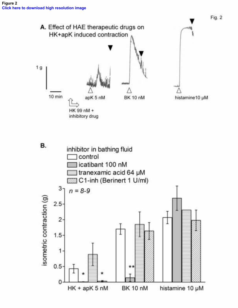
Figure 2. Mechanism of the contraction induced by the HK + apK combination in the
human umbilical vein. Tissues were randomly assigned to one of the inhibitory drug
treatment, as indicated, from the equilibration time point 2.5 hr; this treatment was
maintained for all subsequent recordings. Inhibitory drugs were introduced 30 min before
apK stimulation (5 nM), at the same time as HK replenishment (99 nM), and maintained
thereafter in the bathing fluid. Kruskall-Wallis test showed that the response to HK+apK
and BK stimulation significantly differed across treatments with inhibitory drugs
(HK+apK: P = 0.0004; BK: P = 0.0011). The effect of each drug vs. control responses
* P<0.05; ** P<0.01.
Figure 3. Effect of minor or ineffective contractile proteases. A. Representative tracing of
the protracted effect of purified neutrophil proteinase 3 (PR3, 31 nM) in the presence of
HK. This was followed by stimulations with BK (10 nM) and apK (5 nM) in the presence

26
of HK to put the effect of PR3 in perspective. B. Purified neutrophil proteinase 3 (3.1 or
31 nM), applied twice at 1-hr interval, had negligible effect on the umbilical vein. C.
When HK replenishment was applied 30 min before stimulation, protracted contractile
effects were recorded (latency at the 39 nM HK concentration: 9.8 ± 1.8 min; at the 197
HK concentration: 7.7 ± 1.2 min). D. Purified plasmin (10 nM), applied twice at 1-hr
interval, had no effect. E. When HK replenishment was applied 30 min before
stimulation, small and inconsistent contractile effects were recorded. F, G. Recombinant
tissue plasminogen activator (169 nM) has no effect, whether or not HK replenishment
was applied.
Figure 4. Effect of metabolic inhibitors applied in a continuous manner on the
contractions induced by human recombinant tissue kallikrein (KLK-1) or BK (10 nM of
each). A. Inhibitors of intracellular processes. ANOVA indicated that the responses to
either KLK-1 or BK were homogeneous between groups (P>0.05). B. Inhibitor of serine
proteases, pefabloc SC. * P<0.05, ** P<0.01, vs. control response to each
stimulus.
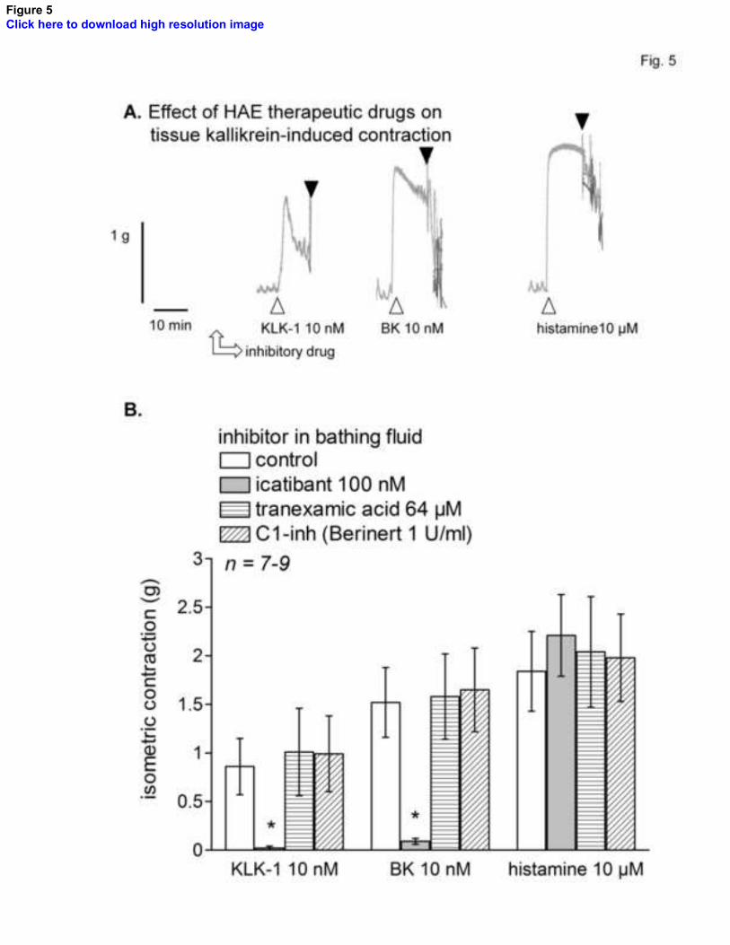
Figure 5. Mechanism of KLK-1-induced contraction in the human umbilical vein. A.
Representative tracing of a control tissue: effect of continuous drug treatments on the
contractile response to KLK-1 (10 nM, recorded at the 3-hr equilibration time point), BK
B. Tissues were randomly assigned to one
of the inhibitory drug treatment, as indicated, from the equilibration time point 2.5 hr;
27
this treatment was maintained for all subsequent recordings. Kruskall-Wallis test showed
that the response to KLK-1 and BK stimulation significantly differed across treatments
with inhibitory drugs (KLK-1: P = 0.0024; BK: P = 0.0012). The effect of each drug vs.
control responses was further t multiple comparison test. * P<0.01.
Figure 6. Investigation of the generation of a B1R stimulant by kallikreins. A. Isolated
umbilical vein preparations were treated to optimize the expression of B1Rs, with
successive cytokine pretreatment during a long in vitro incubation, testing of the B1R
presence using the selective agonist Sar-[D-Phe8]des-Arg9-BK, B2R blockade with
icatibant (20 nM) and optional kininogen replenishment, as outlined in the sample
tracings (LK and KLK-1 tested). B. Effect of KLK-1 (10 nM) during B2R blockade with
or without LK supplementation. The effect of Sar-[D-Phe8]des-Arg9-BK was recorded in
each tissue prior to B2R blockade. In the presence of LK and icatibant, adding compound
11 had a significant effect only for KLK-1 (* P<0.05, Mann-Whitney test). C.
Inconsistent effect of apK (5 nM) during B2R blockade with HK supplementation in
tissues expressing the B1Rs. Responses recorded in the presence or absence of compound
11 did not significantly differ (Mann-Whitney test).
Figure 7. A. Competition of [3H]BK binding to human recombinant B2Rs stably
expressed in HEK 293a cells by a panel of unlabeled peptides/drugs. BK, Lys-BK and
icatibant are approximately equipotent competitors of nanomolar potency. KLK-1 (10-10-
10-6 M) or tranexamic acid do not compete for [3H]BK in this assay (Charest-Morin et al.,
2015;present results). B. Tranexamic acid fails to displace [3H]Lys-des-Arg9-BK from
28
either human recombinant B1Rs. The unlabeled form of the radioligand is used as a
positive control.
Figure 8. Schematic representation of the kinin-mediated responses to proteases in the
isolated human umbilical vein preparation. Reactions difficult to evidence in the system
are represented in progressively shaded areas. A reservoir of LK and preformed BK B2Rs
are present in freshly isolated veins, explaining the tachyphylactic response to KLK-1;
however the system is depleted of HK. The contractile effect of apK is revealed by HK
replenishment. Fibrinolysis is postulated to potentiate kinin generation upstream of the
contact system. The inducible B1Rs may be shown to mediate KLK-1 effects under
specific experimental conditions. The effects of some inhibitors are indicated, that of
tranexamic acid postulated. Some of the peptidases that metabolize kinin are abbreviated
as ap, aminopeptidase; cp, carboxypeptidase; ace, angiotensin converting enzyme.
1
The isolated human umbilical vein as a bioassay for kinin-generating proteases: an in
vitro model for therapeutic angioedema agents
Melissa Jeana, Arvind Raghavanb, Matthew L. Charlesc, Mark S. Robbinsb,d, Eric
Wagnera, Georges-Étienne Rivarde, Xavier Charest-Morina, François Marceaua*
a Axe Microbiologie-Infectiologie et Immunologie, CHU de Québec, and Faculté de
médecine, Université Laval, Québec QC, Canada G1V 4G2 ;
bTansna Therapeutics, St. Louis, MO, USA, 63108;
cMinnetonka, MN, USA;
dKodiak Strategic Consultants, LLC, Minneapolis, MN, USA.
e Division of Hematology / Oncology, CHU Sainte-Justine, Montréal, QC, Canada
Presented in part at the 9th C1-inhibitor Deficiency Workshop, Budapest, Hungary, 29
May 2015.
Correspondence:
Dr F Marceau, T1-49, CHU de Québec, 2705 Laurier Blvd., Québec (Québec), Canada
G1V 4G2. Tel. 1-418-525-4444 x46155. E-mail: [email protected]
2
Abstract
Aims: The isolated human umbilical vein is a robust contractile bioassay for ligands of
the bradykinin (BK) B2 receptor (B2R), also extendable to B1 receptor (B1R)
pharmacology. We hypothesized that, as a freshly isolated vessel, it also contains traces
of plasma proteins that may confer responses to exogenous proteases via the formation of
kinins.
Main methods: Rings of human umbilical veins were mounted in organ baths containing
Krebs buffer maintained at 37°C and purified proteases were introduced in the bathing
fluid along with additional drugs/proteins that permit mechanistic analysis of effects.
Key findings: The previously described contractile response to human recombinant tissue
kallikrein (KLK-1, 1-10 nM) is not influenced by metabolic inhibitors, suggesting its
dependence on a preexisting reservoir of low molecular weight-kininogen (LK). Active
plasma kallikrein (apK, inactive in fresh tissues, unless high molecular
weight-kininogen (HK, 39-197 nM) replenishment was applied. The effects of KLK-1
and HK+apK are abolished by pretreating tissues with icatibant, but not with tranexamic
acid. C1-esterase inhibitor inhibited only HK+apK. Purified plasmin and neutrophil
proteinase-3 produced small contractions in the presence of HK only, and tissue
plasminogen activator, none. B1R stimulation was pharmacologically evidenced in
response to KLK-1 if LK was supplied.

3
Significance: The pharmacology of KLK-1 and HK+apK in the human isolated umbilical
vein is essentially based on the activity of locally generated kinins and this assay models
the inhibitory action of some therapeutic agents active in angioedema states. Proteases
that indirectly generate kinins have little activity in the system.
Keywords: tissue kallikrein, plasma kallikrein, plasmin, bradykinin B2 receptor.

4
1. Introduction
The human isolated umbilical vein is a robust contractile bioassay for agonist and
antagonist ligands of the bradykinin (BK) B2 receptor (B2R), extendable to the inducible
B1 receptor (B1R) [1-4]. In this system with low intrinsic sensitivity to endothelium-
dependent vasorelaxation and possessing , BK or Lys-BK
essentially induce B2R-mediated contractions. The kinin B1R is preferentially stimulated
by kinin metabolites generated by ubiquitous arginine-carboxypeptidases. The umbilical
vein preparation, consistent with the pharmacologic profile of the human B1R [5], is
~100-fold more sensitive to Lys-des-Arg9-BK than to des-Arg9-BK [6].
We recently analyzed the effect of a pharmaceutically refined form of human
recombinant tissue kallikrein (KLK-1) on umbilical vein rings maintained in Krebs buffer
[7]. KLK-1 induced contractions that were highly tachyphylactic, dependent on the B2R
(as shown by the effect of a non-peptide B2R antagonist) and on the catalytic effect of the
protease (inhibited by aprotinin). The tachyphylaxis was reversed if tissues were
replenished with low-molecular weight kininogen (LK), the preferential substrate of
KLK-1. Thus, the freshly isolated vein contains traces of plasma proteins that may confer
an effect to exogenous proteases via the formation of kinins.
The present therapeutic showcase of the kallikrein-kinin system is hereditary angioedema
(HAE); in this autosomal dominant disease, most patients exhibit a mutated SERPING1
gene that codes for a defective of non-expressed C1-esterase inhibitor (C1-inh) protein

5
[8]. Other patients have a constitutively active form of Factor XII. All these molecular
alterations point to a hyperactive contact system, with active plasma kallikrein (apK)
generating kinins during attacks. In addition to the replenishment of C1-inh, a B2R
antagonist, icatibant, as well the pharmacological inhibition of plasma kallikrein are
effective to abort attacks of HAE angioedema [8]. The acquired angioedema occasionally
associated with the pharmacological blockade of angiotensin converting enzyme (a major
kinin-destroying peptidase in the extracellular compartment) is also responsive to
icatibant [9]. Various other forms of angioedema, many idiopatic, are clinically observed
and may be associated with the use of other drugs, malignancies or autoimmune disease
[10]. The place of plasmin(ogen) inhibitors in the therapy of angioedema is debated:
tranexamic acid is reportedly effective to prevent attacks in a fraction of HAE patients
and in certain atypical angioedema cases [8]. Kinin generation may explain acquired
angioedema associated with tissue plasminogen activator (tPA) treatment in patients with
arterial thrombosis [11, 12]. Other proteases reported to release vasoactive kinins from
HK include neutrophil proteinase-3 (PR3), the lectin pathway complement component
MSAP-1, and pancreatic trypsin [13-16].
We have exploited the umbilical vein assay to investigate the effect of additional
proteases, human purified apK, plasmin and tPA, that may also generate kinins. Further,
therapeutic agents used in the treatment of HAE were tested against the effect of the
active proteases, in in vitro effect of currently
used drugs vs. the putative pathways of vasoactive kinin generation. The assay

6
theoretically allows the detection of possible non-conventional effects of proteases, such
as the direct activation of the B2R (as previously proposed [17]).

7
2. Methods
2.1. Drugs
Human recombinant tissue kallikrein (KLK-1; DM199) was provided as a catalytically
active and pharmaceutically refined form (average molecular weight of 38.5 kDa) by
DiaMedica, Inc. (Minneapolis, MN) [18]. Human active plasma kallikrein (apK) purified
from plasma ( ; ) was from EMD Millipore. Human plasmin
purified from plasma ( 2 units/mg protein) was obtained from Sigma-Aldrich (St. Louis,
MO) under a lyophilized powder form. Recombinant human tPA (ateplase, Cathflow,
Roche) was reconstituted as recommended by the manufacturer. Proteinase 3 (PR3),
purified from human neutrophils (>95%), was from Athens Research & Technology
(Athens, GA).
Purified single chain high molecular weight kininogen (>95%, 120 kDa) was purchased
from Enzyme Research Laboratories (South Bend, IN), icatibant (Hoe 140; D-Arg[Hyp3,
Thi5, D-Tic7, Oic8]BK), from Phoenix Pharmaceuticals (Burlingame, CA), and
tranexamic acid and pefabloc SC (4-(2-aminoethyl)benzenesulfonyl fluoride
hydrochloride), from Sigma-Aldrich. C1-esterase inhibitor (C1-inh) was under the form
of Berinert (CSL Behring Canada, Ottawa, ON), reconstituted as recommended by the
manufacturer. Compound 11 (2-{(2R)-1-[(3,4-dichlorophenyl)sulfonyl]-3-oxo-1,2,3,4-
tetrahydroquinoxalin-2-yl}-N-{2-[4-(4,5-dihydro-1H-imidazol-2-
yl)phenyl]ethyl}acetamide) is a powerful antagonist at the human and rabbit B1 receptor
[19] (gift from Dr. D. J. Pettibone, Merck Research Laboratories, West Point, PA).

8
Bradykinin and histamine were from Sigma-Aldrich and Sar-[D-Phe8]des-Arg9-
bradykinin, a selective B1R agonist resistant to peptidases [3], was purchased from
Phoenix Pharmaceuticals (Burlingame, CA).
2.2. Contractility assay involving the human umbilical vein
The institutional research ethics board (CHU de Québec) approved the anonymous use of
human umbilical cord segments obtained after elective cesarean section deliveries.
Informed consent was obtained from mothers. The experimental procedures have been
recently reported [7]. Briefly, most experiments were based on a 3 hr-equilibration period
protease and other agents. Experiments
dealing with B1Rs had an extended equilibration period and a cytokine mixture was
present (interleukin- -
time- and stimulus-dependent induction of this pharmacological entity in vascular smooth
muscle cells [2, 3, 20].
2.3. Radioligand binding competition assays
The construction of a myc-tagged human B2R is reported elsewhere, as well as the
techniques applied in a competition assay for the binding of [3H]BK to the recombinant
receptor transiently expressed in HEK 293a cells [7]. This assay is performed at 0°C to
identify unlabeled ligands of the B2R by their displacement of the specific binding of 3
nM [3H]BK. Similarly designed experiments were the basis of a binding competition

9
assay involving the displacement of 1 nM [3H]Lys-des-Arg9-BK from recombinant
FLAG-tagged human B1Rs transiently expressed in HEK 293a cells [21].
2.4. Data analysis
Numerical results are presented as mean ± S.E.M. Considering the non-normal
distributions observed in several experimental groups, sets of numerical data were
compared by the non-parametric ANOVA, the Kruskal Wallis test, and
comparison test was applied to compare pairs of values. Pairs of values were likewise
compared with the non-parametric Mann-Whitney test. All computations were performed
using the InStat3.05 computer program, GraphPad Software (SanDiego, CA). Data from
the radioligand competition assays were fitted by nonlinear regression to a one-site
competition equation (Prism 4.0, GraphPad Software Inc.).
10
3. Results
3.1. Contractility studies of the human umbilical vein preparation
The direct application of apK (up to 5 nM) to rings of human umbilical artery did not
contract the human umbilical vein preparation maintained in Krebs buffer (Fig. 1A, B).
However, HK replenishment 30 min prior to testing (39 or 197 nM; physiological plasma
concentration ~600 nM) revealed that apK can slowly contract the preparation in a
manner dependent on the HK concentration (Fig. 1C, D). The tissues remained
responsive to BK (10 nM) under all circumstances.
Therapeutic agents active in the therapy of HAE were tested against responses induced
by the combination of HK + apK (99 nM and 5 nM, respectively), BK (10 nM) or an
1 receptor agonist in this
preparation) [22] (Fig. 2A, sample control tracing). The peptide B2R antagonist icatibant
(100 nM) effectively prevented the effect of apK and BK, but not that of histamine (Fig.
2B). C1-inh and tranexamic acid, both used a clinically relevant plasma concentrations,
were tested against the 3 contractile agents. Only C1-inh showed an inhibitory effect
against HK + apK.
Minor or ineffective contractile proteases were identified in the contractility assay (Fig.
3). Neutrophil PR3 (up to 31 nM) had a negligible direct effect, but protracted contractile
responses were observed if the HK replenishment scheme was applied (Fig. 3A-C). The

11
effect of PR3 was not dependent on the concentration of HK. The latencies are reported
in the Fig. 3 legend and this phenomenon is specific for PR3, because all other contractile
proteases produce immediate responses upon application. For instance, immediate
contractions were induced by apK (5 nM) in the presence of HK (Fig. 3A). Similarly,
plasmin (10 nM) had no direct contractile effect on the umbilical vein preparation, unless
a HK replenishment protocol was applied (Fig. 3D, E). The effects did not seem
concentration-related vs. HK, and the amplitude of plasmin effects was small and
inconsistent from one preparation to the other. Recombinant tPA (ateplase) had no
contractile effect on the venous preparation, whether or not HK replenishment was
applied (Fig. 3F, G).
The lack of direct effect of apK on the umbilical vein preparation contrasted with the
reproducible, but tachyphylactic effect of KLK-1 [7]. A possible explanation for this is
that their respective preferential substrates, HK and LK respectively, have a differential
expression in the isolated tissue. To test whether LK presence is due to its post-isolation
de novo formation (as reported in cultured endothelial cells derived from this vein) [23],
we have treated venous rings continuously with metabolic inhibitors from the time of
organ bath mounting (Fig. 4A). Blockade of protein synthesis with cycloheximide, of
RNA synthesis with actinomycin D and of the endoplasmic reticulum-Golgi transition
with brefeldin A failed to modify the initial contractile effect of KLK-1 or the one later
recorded in response to BK. These results suggest that a certain LK reservoir derived
from fetal blood plasma persists in the freshly isolated vein. In this set of experiments,
the serine protease inhibitor pefabloc SC -1

12
vs. responses recorded in paired control tissues (Fig. 4B). However, it had a cumulative
toxicity on tissues, depressing the late effect of BK by 42%, showing that both responses
are reactive to this particular metabolic inhibitor, if not always for the expected reason.
The previously described tachyphylactic effect of KLK-1 on the venous preparation was
re-examined in relationship with drugs active against HEA (Fig. 5, control tracing).
Icatibant abated the effects of both KLK-1 and BK; this was previously observed with the
alternate B2R antagonist anatibant [7]. In contrast with the susceptibility of apK, KLK-1-
induced contractions were not abated in the presence of C1-inh. Tranexamic acid also
failed to inhibit KLK-1-induced responses (Fig. 5).
Since KLK-1 reportedly releases Lys-BK from LK, it is possible that widely distributed
arginine-carboxypeptidases generate in situ the high affinity of the human B1R, Lys-des-
Arg9-BK, from it. We applied special conditions to upregulate the expression of the B1R
in the venous assay and we controlled its presence in experiments reported in Fig. 6.
Thus, umbilical vein rings were stimulated for the first 3 hrs of incubation with
inflammatory cytokines; the contractile response to the selective B1R agonist Sar-[D-
Phe8]des-Arg9-BK recorded 1 hr later proved the time- and protein synthesis-dependent
expression of this entity [2, 3]. Then, B2Rs were blocked with a low concentration of
icatibant (20 nM) and the KLK-1 stimulation (10 nM) took place. Very small or no
contractions were recorded in response to the protease (Fig. 6A, B). However, if LK
replenishment (15.2 nM) was applied before KLK-1 stimulation, a contractile response

13
was recorded in icatibant-treated tissues; it was attributed to the B1Rs because addition of
the non-peptide B1R antagonist, compound 11, significantly reduced the response to
KLK-1 in separate tissues (Fig. 6B).
The same reasoning was applied in tissues stimulated with HK+aPK (Fig. 6C). In tissues
sensitized to the B1R agonist and in the presence of icatibant, HK+aPK had no reliable
effect, consistent with the low affinity of des-Arg9-BK in the system.
3.2. Ancillary experiments
The binding of some agents to human recombinant B2R was examined using a [3H]BK
binding competition assay. While BK, Lys-BK and their antagonist icatibant were
approximately equipotent competitors of this binding with nanomolar potencies (Fig.
7A), KLK-1 failed to displace [3H]BK in the same experimental system [7], failing to
support a direct effect of KLK-1 on the receptor. We have verified that tranexamic acid
has no affinity for either human recombinant B2R or B1R using radioligand binding
competition assays (no displacement of the cognate tritiated agonists from the receptors,
Fig. 7A, B).

14
4. Discussion
The human umbilical vein preparation is suitable to examine the pharmacology of B2R
ligands, as well as that of B1R ligands if special experimental conditions are applied. The
present study extends the finding that KLK-1 contracts the preparation in a
tachyphylactic manner by enzymatically releasing a kinin from a substrate, probably LK,
present in limited quantity in the blood free system [7]. Consistent with the probable
consumption of the contact system in the veins collected post-partum, apK contracts the
preparation only if HK replenishment is applied. The umbilical vein system also suggests
that multistep enzymatic pathways leading to kinin formation are not favorable stimuli.
Thus, as one introduces proteases with more distal actions relative to apK, i.e. plasmin
and ateplase (tPA; Fig. 8), the contractile effects progressively decreases. Both tPA and
plasmin generate BK when added to human blood, parallel to HK consumption [11].
However, this may be indirect, rather mediated by an effect of plasmin on Factor XII
[25], and the present experimental tissues seem to contain a trace of the latter component
of the contact system, accounting for the small effect of plasmin. Neutrophil PR3, that
reportedly releases Met-Lys-BK-Ser-Ser from HK [13], generates a delayed contractile
effect on the venous system in the presence of HK (Fig. 3A-C). We have recently shown
that Met-Lys-BK-Ser-Ser has virtually no affinity for the B2R, but that it is paradoxically
activated by angiotensin converting enzyme (ACE) present in vascular tissues, including
the human umbilical vein [22] (Fig. 8). Thus, this obligatory additional reaction may
make the neutrophil PR3 enzyme a less effective contractile agent than aPK, the latter
releasing BK from HK.

15
There is no animal model of the HAE attack, as mice with a complete knockout of the
gene (SERPING1) corresponding to the C1-inh protein have no phenotype, except for an
asymptomatic increase in microvascular permeability mediated by B2Rs [26]. Mental and
physical stressors, including infection, surgery, trauma and chronic inflammation, are
statistically associated with the frequency of HEA and ACE inhibitor-induced attacks
[27, 28]. A certain fraction of these factors may be parallel to the expression of the
cytokine-controlled B1R. Also, local circulatory condition such as fibrinolysis, KLK-1
generation, neutrophil leukocyte
trigger the attack or amplify it. Therefore, a comparison of the proposed BK-generating
proteases (Fig. 8) may help to unravel their role in angioedema states, as well as the
spectrum of effect of drugs currently used or proposed in the therapy of such states. As
far as effects of kinins on B2R are concerned, icatibant may be the universal inhibitor of
the vascular effects of these proteases. C1-inh, as expected, inhibited the contractile
effect of apK+HK, but not that of KLK-1, itself previously shown to be sensitive to
aprotinin [7] and to pefabloc SC in present experiments (Fig. 4B). This spectrum of
susceptibility to inhibitors is well known for KLK-1 [24], however the cell impermeant
peptide was not toxic to tissues, as opposed to pefabloc SC. Tranexamic acid at the used
concentration inhibits both tPA and plasmin [29], proteases that possess kringle
domains and that are upstream of the contact system vs. the generation of BK (Fig. 8).
The lack of effect of this drug on the contractile effects of either type of kallikrein is
consistent with this idea. We verified that tranexamic acid has no unexpected effect on
16
kinin receptors using radioligand competition assays; this cyclic amine is not dissimilar to
some non-peptide antagonists of either B1 or B2Rs [30].
The only subnanomolar affinity agonist of the human B1R is Lys-des-Arg9-BK [5, 6].
The formation of this peptide may be limited to the KLK-1/LK pathway, and a moderate
LK supplementation has indeed led to
the pharmacological stimulation of the B1Rs in the venous preparation (Fig. 6A, B).
Speculations about a role of the inducible B1R in HAE attacks [31] may be criticized on
the account that the apK/HK pathway theoretically only produces some des-Arg9-BK, a
low affinity B1R agonist. Accordingly and within the limitations of the present
experimental system, the HK+apK combination failed to stimulate B1Rs (Fig. 6C).
However, it is not excluded that KLK-1 activity is secreted and/or upregulated during
hereditary or other forms of angioedema attacks, and participates in a C1-inh-resistant
manner to symptoms via B1 and B2Rs.
Conclusion
The pharmacology of KLK-1 and HK+apK in the human isolated umbilical vein is
essentially based on the activity of locally generated kinins and this assay models the
inhibitory action of some therapeutic agents active in angioedema states. Proteases that
initiate the formation of pharmacologically active kinin via multiple enzymatic steps
exhibit little or no activity in the system.
17
Conflict of interest
None declared.
Acknowledgements
This work was supported by the grant MOP-93773 from the Canadian Institutes of Health
Research, the Fonds de recherche Santé du Québec (Studentship award to XCM) and by
an Investigator-Initiated Research Grant from Shire Canada, Inc. We thank Ms. Johanne
Bouthillier for technical help, and DiaMedica, Inc. (Minneapolis, MN, USA) for
supplying human recombinant KLK-1.
18
References
[1] F. Marceau, L. Levesque, G. Drapeau, F. Rioux, J.M. Salvino, H.R. Wolfe, P.R.
Seoane, D.G. Sawutz. Effects of peptide and nonpeptide antagonists of bradykinin B2
receptors on the venoconstrictor action of bradykinin. J. Pharmacol. Exp. Ther. 269
(1994) 1136-1143.
[2] S.P. Sardi, F.M. Daray, A.E. Errasti, F.G. Pelorosso, V.A. Pujol-Lereis, V. Rey-Ares,
M.P. Rogines-Velo, R.P. Rothlin. Further pharmacological characterization of bradykinin
B1 receptor up-regulation in human umbilical vein. J. Pharmacol. Exp. Ther. 290 (1999)
1019-1025.
[3] S. Houle, M. Landry, R. Audet, J. Bouthillier, D.R. Bachvarov, F. Marceau. Effect of
allelic polymorphism of the B1 and B2 receptor genes on the contractile responses of the
human umbilical vein to kinins. J. Pharmacol. Exp. Ther. 294 (2000) 45-51.
[4] F. Marceau, D. deBlois, E. Petitclerc, L. Levesque, G. Drapeau, R. Audet, D. Godin,
J.F. Larrivée, S. Houle, T. Sabourin, J.P. Fortin, G. Morissette, L. Gera, M.T. Bawolak,
G.A. Koumbadinga, J. Bouthillier. Vascular smooth muscle contractility assays for
inflammatory and immunological mediators. Int. Immunopharmacol. 10 (2010) 1344-
1353.

19
[5] L.M. Leeb-Lundberg, F. Marceau, W. Müller-Esterl, D.J. Pettibone, B.L. Zuraw.
International Union of Pharmacology. XLV. Classification of the kinin receptor family:
from molecular mechanisms to pathophysiological consequences. Pharmacol. Rev. 57
(2005) 27-77.
[6] F. Gobeil, L.H. Pheng, I. Badini, X.K. Nguyen-Le, A. Pizard, A. Rizzi, D. Blouin, D.
Regoli. Receptors for kinins in the human isolated umbilical vein. Br. J. Pharmacol. 118
(1996) 289 294.
[7] X. Charest-Morin, A. Raghavan, M.L. Charles, T. Kolodka, J. Bouthillier, M. Jean,
M.S. Robbins, F. Marceau. Pharmacological effects of recombinant human tissue
kallikrein on bradykinin B2 receptors. Pharmacol. Res. Perspect. 3 (2015) e00119.
[8] M. Cicardi, W. Aberer, A. Banerji, M. Bas, J.A. Bernstein, K. Bork, T. Caballero, H.
Farkas, A. Grumach, A.P. Kaplan, M.A. Riedl, M. Triggiani, A. Zanichelli, B. Zuraw,
HAWK under the patronage of EAACI (European Academy of Allergy and Clinical
Immunology). Classification, diagnosis, and approach to treatment for angioedema:
consensus report from the Hereditary Angioedema International Working Group. Allergy
69 (2014) 602-616.

20
[9] M. J. Greve, K. Stelter, M. Havel, U. Strassen, N. Rotter, J. Veit, B. Schossow,
A. Hapfelmeier, V. Kehl, G. Kojda, T.K. Hoffmann. A randomized trial of icatibant in
ACE-inhibitor-induced angioedema. N. Engl. J. Med. 372 (2015) 418-425.
[10] J. Levy, G.E. Rivard, E. Wagner, D. Beezhold, N. Berlin, L. Fan, Z. Zhang, G.L.
Sussman. Examination of genetic variants involved in generation and biodisposition of
kinins in patients with angioedema. Allergy Asthma Clin. Immunol. 10 (2014) 60.
[11] G. Molinaro, N. Gervais, A. Adam. Biochemical basis of angioedema associated
with recombinant tissue plasminogen activator treatment: an in vitro experimental
approach. Stroke 33 (2002) 1712 1716.
[12] M.E. Moreau, N. Garbacki, G. Molinaro, N.J. Brown, F. Marceau, A. Adam. The
kallikrein-kinin system: current and future pharmacological targets. J. Pharmacol. Sci. 99
(2005) 6-38.
[13] R. Kahn, T. Hellmark, L.M. Leeb-Lundberg, N. Akbari, M. Todiras, T. Olofsson, J.
Wieslander, A. Christensson, K. Westman, M. Bader, W. Müller-Esterl, D. Karpman.
Neutrophil-derived proteinase 3 induces kallikrein-independent release of a novel
vasoactive kinin. J. Immunol. 182 (2009) 7906-7915.

21
[14] L. Gera, C. Roy, M.T. Bawolak, J. Bouthillier, A. Adam, F. Marceau. Met-Lys-
bradykinin-Ser-Ser, a peptide produced by the neutrophil from kininogen, is
metabolically activated by angiotensin converting enzyme in vascular tissue. Pharmacol.
Res. 64 (2011) 528-534.
[15] J. Dobó, B. Major, K.A. Kékesi, I. Szabó, M. Megyeri, K. Hajela, G. Juhász, P.
Závodszky, P Gál. Cleavage of kininogen and subsequent bradykinin release by the
complement component: mannose-binding lectin-associated serine protease (MASP)-1.
PLoS One 6 (2011) e20036.
[16] G.R. Drummond, S. Selemidis, T.M. Cocks. B2 kinin receptor activation is the
predominant mechanism by which trypsin mediates endothelium-dependent relaxation in
bovine coronary arteries. Naunyn Schmiedebergs Arch. Pharmacol. 378 (2008) 33-41.
[17] C. Hecquet, F. Tan, D.M. Marcic, E.G. Erdös. Human bradykinin B2 receptor is
activated by kallikrein and other serine proteases. Mol. Pharmacol. 58 (2000) 828-836.
[18] T. Kolodka, M.L. Charles, A. Raghavan, I.A. Radichev, C. Amatya, J. Ellefson,
A.Y. Savinov, A. Nag, M.S. Williams, M.S. Robbins. Preclinical characterization of
recombinant human tissue kallikrein-1 as a novel treatment for type 2 diabetes mellitus.
PLOS One 9 (2014) e103981.

22
[19] G. Morissette, J.P. Fortin, S. Otis, J. Bouthillier, F. Marceau. A novel nonpeptide
antagonist of the kinin B1 receptor: effects at the rabbit receptor. J. Pharmacol. Exp. Ther.
311 (2004) 1121-1130.
[20] G.A. Koumbadinga, A. Désormeaux, A. Adam, F. Marceau F. Effect of interferon-
on inflammatory cytokine-induced bradykinin B1 receptor expression in human vascular
cells. Eur. J. Pharmacol. 647 (2010) 117-125.
[21] G. Morissette, J.P. Couture, A. Désormeaux, A. Adam, F. Marceau. Lack of direct
interaction between enalaprilat and the kinin B1 receptors. Peptides 29 (2008) 606-612.
[22] L. Gera, C. Roy, X. Charest-Morin, F. Marceau. Vasopeptidase-activated latent
ligands of the histamine receptor-1. Int. Immunopharmacol. 17 (2013) 677-683.
[23] K. Yayama, N. Kunimatsu, Y. Teranishi, M. Takano, H. Okamoto. Tissue kallikrein
is synthesized and secreted by human vascular endothelial cells. Biochim. Biophys. Acta
1593 (2003) 231-238.

23
[24] P. Goetig, V. Magdolen, H. Brandstetter. Natural and synthetic inhibitors of
kallikrein-related peptidases (KLKs). Biochimie 92 (2010) 1546-1567.
[25] S. de Maat, P.G. de Groot, C Maas. Contact system activation on endothelial cells.
Semin. Thromb. Hemost. 40 (2014) 887 894.
[26] E.D. Han, R.C. MacFarlane, A.N. Mulligan, J. Scafidi, A.E. Davis AE. Increased
vascular permeability in C1 inhibitor-deficient mice mediated by the bradykinin type 2
receptor. J. Clin. Invest. 109 (2002) 1057-1063.
[27] T. Hoover, M. Lippmann, E. Grouzmann, F. Marceau, P. Herscu. Angiotensin
converting enzyme inhibitor induced angio-oedema: a review of the pathophysiology and
risk factors. Clin. Exp. Allergy 40 (2010) 50-61.
[28] Z. Zotter, D. Csuka, E. Szabó, I. Czaller, Z. Nébenführer, G. Temesszentandrási, G.
Fust, L. Varga, H. Farkas. The influence of trigger factors on hereditary angioedema due
to C1-inhibitor deficiency. Orphanet J. Rare Dis. 9 (2014) 44.
[29] R.A. Al-Horani, U.R. Desai. Recent advances on plasmin inhibitors for the treatment
of fibrinolysis-related disorders. Med. Res. Rev. 34 (2014) 1168-1216.

24
[30] G. Morissette, J. Bouthillier, F. Marceau. Dual antagonists of the bradykinin B1 and
B2 receptors based on a postulated common pharmacophore from existing non-peptide
antagonists. Biol. Chem. 387 (2006) 189-194.
[31] Z.L.M. Hofman, A. Relan, C.E. Hack. C-reactive protein levels in hereditary
angioedema. Clin. Exp. Immunol. 177 (2014) 280-286.
25
Figure legends
Figure 1. Effect of purified active plasma kallikrein (apK) on the human isolated
umbilical vein. A. Representative tracings of the effect of apK (5 nM) and bradykinin
(BK, 10 nM). apK, applied twice at 1-hr interval, had no effect. B. Maximal effects
recorded in replicated experiments. C, D. Effect of HK replenishment, applied 30 min
before stimulation, on the contractile effect of apK (5 nM; C. representative tracing; D.
effect of two concentration levels of HK).
Figure 2. Mechanism of the contraction induced by the HK + apK combination in the
human umbilical vein. Tissues were randomly assigned to one of the inhibitory drug
treatment, as indicated, from the equilibration time point 2.5 hr; this treatment was
maintained for all subsequent recordings. Inhibitory drugs were introduced 30 min before
apK stimulation (5 nM), at the same time as HK replenishment (99 nM), and maintained
thereafter in the bathing fluid. Kruskall-Wallis test showed that the response to HK+apK
and BK stimulation significantly differed across treatments with inhibitory drugs
(HK+apK: P = 0.0004; BK: P = 0.0011). The effect of each drug vs. control responses
* P<0.05; ** P<0.01.
Figure 3. Effect of minor or ineffective contractile proteases. A. Representative tracing of
the protracted effect of purified neutrophil proteinase 3 (PR3, 31 nM) in the presence of
HK. This was followed by stimulations with BK (10 nM) and apK (5 nM) in the presence

26
of HK to put the effect of PR3 in perspective. B. Purified neutrophil proteinase 3 (3.1 or
31 nM), applied twice at 1-hr interval, had negligible effect on the umbilical vein. C.
When HK replenishment was applied 30 min before stimulation, protracted contractile
effects were recorded (latency at the 39 nM HK concentration: 9.8 ± 1.8 min; at the 197
HK concentration: 7.7 ± 1.2 min). D. Purified plasmin (10 nM), applied twice at 1-hr
interval, had no effect. E. When HK replenishment was applied 30 min before
stimulation, small and inconsistent contractile effects were recorded. F, G. Recombinant
tissue plasminogen activator (169 nM) has no effect, whether or not HK replenishment
was applied.
Figure 4. Effect of metabolic inhibitors applied in a continuous manner on the
contractions induced by human recombinant tissue kallikrein (KLK-1) or BK (10 nM of
each). A. Inhibitors of intracellular processes. ANOVA indicated that the responses to
either KLK-1 or BK were homogeneous between groups (P>0.05). B. Inhibitor of serine
proteases, pefabloc SC. * P<0.05, ** P<0.01, t test vs. control response to each
stimulus.
Figure 5. Mechanism of KLK-1-induced contraction in the human umbilical vein. A.
Representative tracing of a control tissue: effect of continuous drug treatments on the
contractile response to KLK-1 (10 nM, recorded at the 3-hr equilibration time point), BK
B. Tissues were randomly assigned to one
of the inhibitory drug treatment, as indicated, from the equilibration time point 2.5 hr;
27
this treatment was maintained for all subsequent recordings. Kruskall-Wallis test showed
that the response to KLK-1 and BK stimulation significantly differed across treatments
with inhibitory drugs (KLK-1: P = 0.0024; BK: P = 0.0012). The effect of each drug vs.
control responses multiple comparison test. * P<0.01.
Figure 6. Investigation of the generation of a B1R stimulant by kallikreins. A. Isolated
umbilical vein preparations were treated to optimize the expression of B1Rs, with
successive cytokine pretreatment during a long in vitro incubation, testing of the B1R
presence using the selective agonist Sar-[D-Phe8]des-Arg9-BK, B2R blockade with
icatibant (20 nM) and optional kininogen replenishment, as outlined in the sample
tracings (LK and KLK-1 tested). B. Effect of KLK-1 (10 nM) during B2R blockade with
or without LK supplementation. The effect of Sar-[D-Phe8]des-Arg9-BK was recorded in
each tissue prior to B2R blockade. In the presence of LK and icatibant, adding compound
11 had a significant effect only for KLK-1 (* P<0.05, Mann-Whitney test). C.
Inconsistent effect of apK (5 nM) during B2R blockade with HK supplementation in
tissues expressing the B1Rs. Responses recorded in the presence or absence of compound
11 did not significantly differ (Mann-Whitney test).
Figure 7. A. Competition of [3H]BK binding to human recombinant B2Rs stably
expressed in HEK 293a cells by a panel of unlabeled peptides/drugs. BK, Lys-BK and
icatibant are approximately equipotent competitors of nanomolar potency. KLK-1 (10-10-
10-6 M) or tranexamic acid do not compete for [3H]BK in this assay (Charest-Morin et al.,
2015;present results). B. Tranexamic acid fails to displace [3H]Lys-des-Arg9-BK from

28
either human recombinant B1Rs. The unlabeled form of the radioligand is used as a
positive control.
Figure 8. Schematic representation of the kinin-mediated responses to proteases in the
isolated human umbilical vein preparation. Reactions difficult to evidence in the system
are represented in progressively shaded areas. A reservoir of LK and preformed BK B2Rs
are present in freshly isolated veins, explaining the tachyphylactic response to KLK-1;
however the system is depleted of HK. The contractile effect of apK is revealed by HK
replenishment. Fibrinolysis is postulated to potentiate kinin generation upstream of the
contact system. The inducible B1Rs may be shown to mediate KLK-1 effects under
specific experimental conditions. The effects of some inhibitors are indicated, that of
tranexamic acid postulated. Some of the peptidases that metabolize kinin are abbreviated
as ap, aminopeptidase; cp, carboxypeptidase; ace, angiotensin converting enzyme.


![Use of molecular biology tools for rapid identification ... · vaccine (VSVRI, Egypt) was isolated from cattle during HS outbreak in Egypt since 1962 and identified as PM Type B [4,5,23,24]](https://img.pdfslide.fr/doc/110x75/5e8b0927bd8172489267fc18/use-of-molecular-biology-tools-for-rapid-identification-vaccine-vsvri-egypt.jpg)


![Mutation of the Rice Narrow leaf1 Gene, Which …...Mutation of the RiceNarrow leaf1 Gene, Which Encodes a Novel Protein, Affects Vein Patterning and Polar Auxin Transport1[OA] Jing](https://img.pdfslide.fr/doc/110x75/5e67715455d8a57ede40e3d0/mutation-of-the-rice-narrow-leaf1-gene-which-mutation-of-the-ricenarrow-leaf1.jpg)


![Optical mesoscopy without the scatter: broadband multispectral … · single GFP-labeled neurons within dendritic trees in isolated hippocampi [3]. SPIM has also been able to offer](https://img.pdfslide.fr/doc/110x75/60b4cc937ba1593eee0be699/optical-mesoscopy-without-the-scatter-broadband-multispectral-single-gfp-labeled.jpg)