Embed Size (px)
Citation preview
UNIVERSITE DE GENÈVE FACULTÉ DES SCIENCES
Département de chimie minérale, Professeur J. Buffle
analytique et appliquée Dr. K. J. Wilkinson
Bio-physicochemical Parameters Influencing Cadmium Uptake by the
Unicellular Green Alga Chlamydomonas reinhardtii
THÈSE
Présentée à la Faculté des sciences à l’Université de Genève
pour obtenir le grade de Docteur ès sciences, mention chimie
par
Heliana KOLA de
Tirana (Albanie)
Thèse No 3601
GENÈVE
Atelier de reproduction de la Section de Physique
2005
i
REMERCIEMENTS
Ma reconnaissance va en premier lieu à M. le Docteur Kevin Wilkinson, co-directeur de
thèse, pour ses précieux conseils, sa disponibilité constante et la bienveillance qu’il n’a cessé
de me témoigner durant l’élaboration de ce travail.
Je tiens aussi à remercier très chaleureusement M. le Docteur Augusto Porta pour ses
encouragements, sa confiance envers moi et l’institut BATTELLE pour son assistance
financière; leur soutien m’a été indispensable.
Mes remerciements s’adressent également à MM. les Professeurs Herman van Leeuwen et
Jacques Buffle, qui, par l’intérêt qu’ils ont porté à ce travail, m’ont permis de le mener à
terme! Les discussions avec eux étaient précieuses.
J’ai en outre bénéficié de l’assistance technique de MM. les Docteurs Michel Martin, Luis M.
Laglera, Michel Pelletier et Mme les Docteurs Nalini Parthasarathy et Cécile Gehin-Deval.
Que tous soient ici vivement remerciés!
Ma gratitude s’adresse aussi au Docteur M. Michel Goldschmidt-Clermont et à M. le
Professeur M. Bill Zerges et Mmes les Professeures Sabine Waffenschmidt et Elizabeth
Harris pour les discussions fructueuses que j’ai pu avoir avec eux et qui m’ont aidé à mieux
comprendre le comportement des algues.
J’aimerais exprimer ma sincère reconnaissance au Dr. Klaus Müller pour son aide initiale
dans la compilation de la bibliographie et M. Jacques Auger pour son aide occasionnelle au
laboratoire.
Je tiens à remercier également les Dr. Christel Hassler, Dr. Vera Slaveykova, Dana Simon,
Isabelle Worms, Dr. Alain Reinhardt et Cristina Lamellas qui ont partagé avec moi leurs
connaissances pratiques et théoriques.
Ma reconnaissance va enfin à ma famille et mes proches ainsi qu’à toutes les personnes qui
m’ont soutenue – et supportée – ! – durant ce long périple.
ii
DETAILED LIST OF CONTENTS
ACKNOWLEDGEMENTS i GLOSSARY iv FRENCH SUMMARY vii 1-INTRODUCTION 1
1.1 - Chemical speciation of metals in natural waters 2 1.2 – Physicochemical interaction of metals with aquatic organism 3 1.3 - Models used in the kinetics of metal adsorption and internalization 7 1.4 - Factors influencing metal uptake 11 1.5 - The case of cadmium and C. reinhardtii 12
1.5.1 - Levels of Cd in the environment 12 1.5.2 - Metal adsorption 14 1.5.3 - Metal internalization 19 1.5.4 - Effects of metals on growth and assimilatory pathways 24 1.5.5 - Morphologic changes in the presence of metals 29 1.5.6 - Genetic mutation in response to heavy metals 29
1.6 - Implications of using algae in bioavailability studies 31 2- OBJECTIVES 36 3-MATERIALS AND METHODS 38
3.1 - Cell culture maintenance, growth and isolation 38 3.2 - Uptake experiments 40 3.2.1 - Experimental conditions 40 3.2.2 – Adsorbed Cd 41 3.2.3 - Cellular and carrier bound Cd 41 3.2.4 - Competition experiments 42 3.2.5 - Cd uptake fluxes in the presence of a fulvic acid 43
3.2.6 - Cd efflux 44 3.3 - Lability calculations 44 3.4 - Cd speciation measurements 45
3.4.1 - PLM measurements 45 3.4.2 - ASV measurements 45
3.5 - Quantification and characterization of algal exudates 46 3.5.1 - Bradford assay 46 3.5.2 - SDS-PAGE 47
3.5.3 - Total organic carbon and nitrogen analysis 47 3.6 - Algal membrane lipidic permeability and metabolism 48
4 - METHOD OPTIMIZATION AND PRELIMINARY EXPERIMENTS 49
4.1 - Optimization of the EDTA wash to distinguish cellular and adsorbed Cd 49 4.2 - Comparison of bioaccumulation methods 55 4.3 - Effect of centrifugation (G-force) on exudate release 58
iii
4.4 - Bioaccumulation experiments 59 4.4.1 - Cd internalization fluxes in the presence of hardness cations 60
4.4.2 - Competition effects on biouptake: Cu, Zn 62 4.4.3 - Ligand effects on Cd internalization fluxes 65 5 - CADMIUM INTERACTION WITH CHLAMYDOMONAS REINHARDTII AND ITS CELL WALL PROTEINS 70
5.1 - Results and Discussion 71
5.1.1 - Cd adsorption by C. reinhardtii WT and CW-2 strains 71 5.1.2 - Exudate release 75 5.1.3 - Cd (II) speciation measurements: PLM and ASV 77 5.1.4 - Implications for bioaccumulation studies 78
5.2 - Conclusion 80 6 - CADMIUM UPTAKE A GREEN ALGA CAN BE PREDICTED BY
EQUILIBRIUM MODELLING 81
6.1 - Results and Discussion 83
6.1.1 - Cd internalization 83 6.1.2 - Cd efflux 86 6.1.3 - Carrier bound Cd (WT strain) 87 6.1.4 - Competition studies 88 6.1.5 - Influence of pH 90 6.1.6 - Role of a fulvic acid in Cd uptake fluxes 92
7 - SUMMARY AND CONCLUSIONS 94 8 - REFERENCES 96 9 - ANNEXES 112 112 9.1 - Cadmium 9.1.1 - Cadmium in environment 112 9.1.1.1 - Cadmium emissions 112 9.1.1.2 - Cadmium in air 113 9.1.1.2 - Cadmium in soils 113
9.1.2 - Uses of Cd in industry 114 9.1.3 - Implications for the future 116 9.1.4 - Cd exposure and human health 117
9.1.4.1 - Factors determining levels of human exposure 118 9.1.4.2 - Human intake of Cd 118 9.1.4.3 - Human health effects of Cd 120 9.2 - Biological interaction of trace metals with Chlamydomonas 121
9.2.1 - Unicellular green algae 121 9.2.2 - Different strategies to control the availability of essential metals 123 9.2.3 - Protein induction 124 9.3 – Biosensors 129 9.3.1 - Microorganisms as a sensing component 131 9.4 – References (annexes) 134
iv
GLOSSARY
AAP Algal Assay Procedure medium
AAS flame atomic absorption spectrometry
APS ammonium persulfate
ASV anodic stripping voltammetry
ATSDR Agency for Toxic Substances and Disease Registry
BLM biotic ligand model
CW-2 cell wall-less strain of C. reinhardtii
CaM calmodulin
Dave average diffusion coefficient of metal species: function of DML, DM, KS
DM diffusion coefficient of metal
DML diffusion coefficient of metal species
DETA diethylen triamine
DGA diglycolic acid
EDTA ethylenediamine-tetraacetic acid
EPM electrophoretic mobility measurements
FIAM free ion activity model
∆G ° change in the Gibbs free energy of the system (under a set conditions)
∆Gads change in the Gibbs free energy for the adsorption reaction
GS glutamine synthetase
GSH glutathione
HEPES N-2-hydroxyethylpiperazine-N’-2-ethanesulfonic acid
HEPPS N-2-hydroxyethylpiperazine-N’-3-propanesulfonic acid
HEPPSO N-[2-hydroxyethyl]piperazine-N’-2-hydroxypropanesulfonic acid
8-HQ 8-hydroxyquinoline-5-sulfonic acid
HMW high molecular weight
HRGP hydroxyproline-rich glycoproteins
∆H° change in the enthalpy of the system
I ionic strength
IDA iminodiacetic acid
ICP-MS inductively coupled plasma mass spectrometry
ISE ion selective electrode
IS-FET ion-sensitive field effect transistors
v
Jdiff diffusion flux
Jint metal internalization flux (uptake rate)
Jmax maximum metal internalization flux
Kads equilibrium constant for metal adsorption to binding sites
ka rate constant for the formation of the transport site complex
kd rate constant for the dissociation of the transport site complex
Kcond conditional stability constant
kint first order rate constant for the transfer of the bound metal into the cell
KD equilibrium dissociation constant (KD = kd / ka)
KH Henry constant
KM Michaelis-Menten half-saturation constant
KM-Rcell affinity constants of the metal for the transporter
KS stability constant of the M-L(z-n) complex
L lability of the complexes
Ln- free ligands
LMW low molecular weight
M metal
Mz+ free metal ion
ML or M-L(z-n) metal species (complexes)
M0 metal at the vicinity of the microorganism
Mcell internalized metal
[M]diss dissolved metal
M-Rcell transport sites bound by M
µ reaction layer thickness
MES 2-[N-morpholino] ethanesulfonic acid
MOPS morpholino propane sulphonic acid
mt metric tonnes
NTA nitrilotriacetic acid
NIST National Institute of Standards and Technology
NiR nitrite reductase
OECD Organization for Economic Co-operation and Development
PC phytochelatin
pKa affinity constant of the proton of the binding sites
PLM permeation liquid membrane
vi
POPSO piperazine-N, N’-bis[hydroxypropanesulfonic acid]
POR protochlorophyllide reductase
PTWI provisional tolerable weekly intake
PVC polyvinylchloride
R gas constant
R0 radius of the microorganism
Rcell transport binding sites
Rcell-tot total transport sites
SDS-PAGE sodium dodecyl sulphate – polyacrylamide gel electrophoresis
∆S° change in the entropy of the system
SRFA Suwannee River fulvic acid
δ thickness of the diffusion layer
T temperature
TAP tris-acetate-phosphate
TETA triethylen tetramine
TEMED N, N, N', N'-tetramethyl-1-,2-diaminomethane
TN nitrogen content
TOC total organic carbon
TRIS tris-(hydroxymethyl)aminomethane
TRX thioredoxins (small ubiquitous proteins)
ΓMeq
metal amount adsorbed to the binding sites at equilibrium at [Mz+].
Γmax maximum amount of metal adsorbed to the binding sites
τ time to diffuse through the diffusion layer and the cell wall
Xcell non-specific binding sites
WT wild type
W1 to W7 layers of the cell wall of C. reinhardtii green algae
WHO World Health Organization
vii
RESUME DE LA THESE EN FRANCAIS 1 - INTRODUCTION
La pollution par les métaux toxiques dans l'environnement aquatique est un des problèmes
auxquels doit faire face la société actuelle. Ces métaux entrent dans les systèmes aquatiques
par des sources ponctuelles et tendent à s'accumuler dans les sédiments et à se concentrer dans
les chaînes alimentaires aquatiques. La biodisponibilité et la toxicité des métaux traces
présents en milieu aquatique dépend de leur type et de leurs formes chimiques (spéciation des
métaux). Dans les eaux naturelles, les métaux peuvent exister sous différentes formes: cations
libres, métaux complexés à la matière organique dissoute, à des ions inorganiques et à des
particules solides (i.e. colloïdes, microorganismes).
Pour des métaux bivalents étudiés au laboratoire en milieux artificiels, il existe beaucoup
d’exemples que la réponse biologique provoquée par le métal dissous dépend de la
concentration de l'ion métallique libre, Mz+. Pour un organisme aquatique donné, les effets
biologiques d'un métal dissous vont également dépendre de divers facteurs environnementaux
(i.e., pH, dureté, [Ca], concentration en matière organique dissoute). Ces facteurs physico-
chimiques peuvent en principe agir de deux manières: (i) directement sur l'organisme, en
influençant sur la physiologie et donc la sensibilité au métal; (ii) indirectement, en influençant
notamment la spéciation du métal dans le milieu. Dans ce contexte, le sujet de thèse est
orienté vers la compréhension des mécanismes de transferts de métaux depuis la solution
aqueuse vers les microorganismes. Etant donné que l'effet de ces différents facteurs varie
selon le métal étudié ainsi que d'une espèce à l'autre, une étude plus spécifique est nécessaire.
Le but de ce travail est d’établir un lien entre la biodisponibilité du cadmium et les effets
biologiques chez l’algue verte unicellulaire Chlamydomonas reinhardtii. Le "Modèle de l'Ion
Libre" (MIL, ou "Free Ion Activity Model", FIAM), compte tenu de sa capacité à expliquer la
grande majorité des données toxicologiques obtenues en laboratoire dans des milieux
artificiels, a été choisi comme point de départ pour prédire la biodisponibilité du cadmium. Le
MIL suppose un processus d’internalisation suffisamment lent qui implique l’existence d’un
équilibre entre la surface de l’organisme et la solution. Dans cette étude sont testées les
limites du MIL dans des conditions relativement réalistes, notamment en ce qui concerne le
pH, [Ca2+], et la présence de ligands organiques naturels (par exemple : acides fulviques).
Cette validation du modèle a été rigoureusement vérifiée par des expériences de prise en
viii
charge ("uptake") où on suit de près la cinétique des réactions impliquées (adsorption du
métal sur la parois membranaire et aux sites de transport, son transport à travers la membrane
plasmique). De plus, dans cette étude, la prise en compte de la matière organique relâchée par
l’algue est indispensable pour comprendre la spéciation et la biodisponibilité potentielle du
Cd dans les milieux naturels.
Etant donné que la mesure de la spéciation des métaux traces dans les eaux naturelles
demande des techniques de manipulation élaborées et très propres, il est hautement
souhaitable de mettre au point et d'utiliser des techniques possédant une sélectivité pour les
espèces chimiques étudiées. Pour cela cette étude permettra d’améliorer les évaluations de la
biodisponibilité ou toxicité des métaux chez les organismes aquatiques.
2 - Résultats et discussion
2.1 - Croissance des algues
Deux souches de C. reinhardtii ont été utilisées durant de cette étude: une souche sauvage
(WT) et une souche qui ne possède pas de parois cellulaires (CW-2). Dans un premier temps,
les algues sont mises en croissance dans un milieu de culture standardisé (Tris-Acétate-
Phosphate, TAP). Afin d’éviter l’agrégation entre les cellules, les concentrations des micro- et
macro-nutriments du milieu de TAP ont été diluées 4 fois. Le milieu modifié (TAP) paraît
optimal pour la croissance de C. reinhardtii du point de vue de la concentration algale en
phase exponentielle (2 x 10⁻⁶ cellules mL-1) et du taux de croissance.
2.2 - La prise en charge (« uptake ») du Cd
Afin de tester le MIL, les algues ont été resuspendues dans une solution contenant seulement
le tampon biologique (MOPS, pH = 7.0), les sels inorganiques (i.e., NaNO3) afin de pouvoir
ajuster la force ionique à 10-2 M, ainsi que le cadmium et les ligands (tampons métalliques).
L’accumulation de Cd y est mesuré sur un intervalle de temps court (25 min) afin de limiter la
diminution du Cd en solution dû à la forte adsorption ainsi que les effets biologiques dûs à la
ix
présence du métal (par exemple, mortalité des cellules) et l’absence de nutriments (par
exemple, relâchement des exudats). Les algues ont été récupérées de la solution de croissance
par centrifugation et utilisées pour l’expérience de bioaccumulation. Les concentrations de Cd
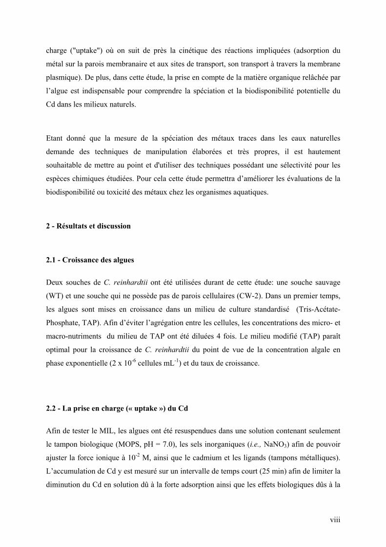
adsorbées (Fig. vf.1a), internalisées (Fig. vf.1b) et dissoutes (Fig. vf.1c) sont mesurées en
fonction du temps pour [Cd2+] en solution variant de 10-11 M à 10-3 M. Le flux
d’internalisation, Jint, est calculé à partir de la pente de l’augmentation linéaire du Cd
cellulaire en fonction de temps ; l’intercept donne la concentration du Cd lié aux
transporteurs, Cd-Rcell (Fig. vf.1b). Le Cd adsorbé sur les parois cellulaires de l’algue est
donné par la quantité de Cd désorbée par le lavage à l’EDTA (10 min).
[Cd]
ads (
x 10
11 m
ol c
m-2
)
0
1
2
3
Temps (min)
0 5 10 15 20 25
[Cd]diss (x 10
7 M)1
2
3
4(b)
Temps (min)
0 5 10 15 20 25 30
[Cd]
int (
x 10
11m
ol c
m-2
)
0
2
4
6
(a)
(b) (c)pente = Jint
intercept = Cd-Rcell
Figure vf.1: Cd adsorbé (a), internalisé (b) et dissout (c) pour les souches WT () et CW-2 ().
Un lavage à l’EDTA de 10 minutes s’est avéré suffisant pour désorber le Cd de la surface de
l’algue. Afin de distinguer les deux processus possibles (désorption et efflux) qui pourraient
avoir lieu pendant le lavage à l ‘EDTA, les expériences de désorption ont été conduites avec
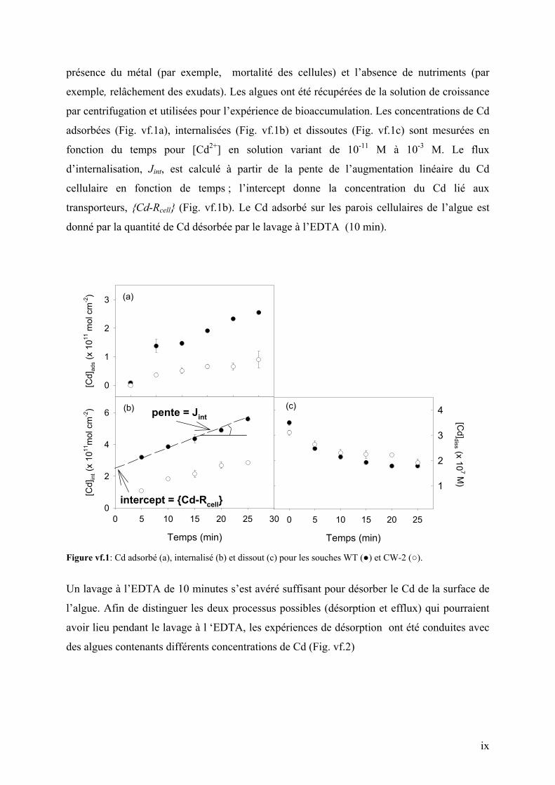
des algues contenants différents concentrations de Cd (Fig. vf.2)
x
Temps (min)
0 10 20 30 40
[Cd]
int (
x 10
10 m
ol c
m-2
)
0.0
0.2
0.4
0.6
0.8
1.0
1.2
Figure vf.2: Cd cellulaire après 10 (), 30 () et 40 () minutes d’exposition au 4 x 10⁻⁷ M Cd en fonction du temps de contact avec EDTA. Les lignes pleines, interrompues et pointillées représentent une diminution en Cd cellulaire de premier ordre avec kd = 4 x10⁻² min-1.
En effet, la diminution du Cd cellulaire, [Cd]int, pour différents concentrations initiales de
[Cd]int en présence de EDTA, n’a pas pu être décrite par un simple processus de premier ordre
(voir en Fig. vf.2 que les points expérimentales ne suivent pas l’équation de diminution
exponentielle du premier ordre), témoignant que probablement deux processus sont présents,
au moins initialement. L’efflux de Cd pendant le lavage à l ‘EDTA (10 min.) a été considéré
négligeable par rapport au flux d’internalisation et les calculs ont démontrés que l’erreur dûe à
ce processus était < 5%.
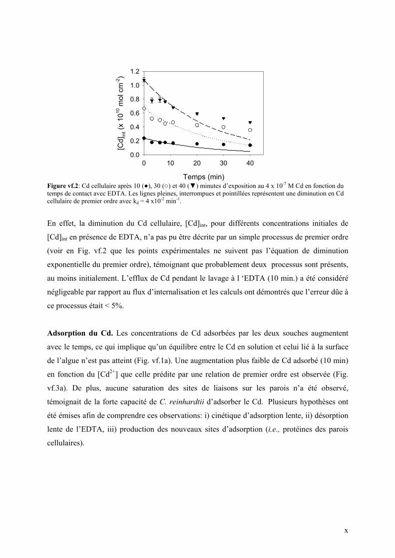
Adsorption du Cd. Les concentrations de Cd adsorbées par les deux souches augmentent
avec le temps, ce qui implique qu’un équilibre entre le Cd en solution et celui lié à la surface
de l’algue n’est pas atteint (Fig. vf.1a). Une augmentation plus faible de Cd adsorbé (10 min)
en fonction du [Cd2+] que celle prédite par une relation de premier ordre est observée (Fig.
vf.3a). De plus, aucune saturation des sites de liaisons sur les parois n’a été observé,
témoignait de la forte capacité de C. reinhardtii d’adsorber le Cd. Plusieurs hypothèses ont
été émises afin de comprendre ces observations: i) cinétique d’adsorption lente, ii) désorption
lente de l’EDTA, iii) production des nouveaux sites d’adsorption (i.e., protéines des parois
cellulaires).
xi
log [Cd2+]
-9 -8 -7 -6 -5
(a)
log [Cd2+]
-10 -9 -8 -7 -6 -5 -4 -3
log
[Cd]
ads (
mol
cm
-2)
-13
-12
-11
-10
-9 (b)(a)
Figure vf.3: a) Log Cd adsorbé (10 min) par le souches WT (cercle plein) et CW-2 (cercle vide) de C. reinhardtii en fonction du Log [Cd2+] (pente = 0.5, R2 = 0.98). b) Log Cd adsorbé (WT) en fonction du (log) [Cd2+] pour la souche WT exposé pendant 5 (), 20 () et 60 (∆) minutes au Cd.
Dans le cas d’un taux d’adsorption limitant, la pente de Cd adsorbé en fonction du [Cd2+]
devrait diminuer progressivement avec l’augmentation des temps d’exposition au Cd, ce qui
n’est pas le cas (pente ~0.6 in Fig. vf.3b). De plus, les calculs basés sur les valeurs de ka,
[site]tot, KH, DMCd et R0 ont démontrés que la désorption est un processus rapide (kd =10-2 –
10-4 s-1) et que l’équilibre entre Cd en solution et celui lié à la surface de l’algue devrait être
atteint en moins de 2 minutes pour [Cd2+] = 2 x 10⁻⁷ M, ce qui n’est pas le cas (voir Fig.
vf.1a).
L’hypothèse de la production de nouveaux sites non-spécifiques semble être la meilleure
explication de l’augmentation du Cd adsorbé en fonction du temps, ce qui implique que
l’adsorption du Cd à la surface de Chlamydomonas est un processus dynamique dans les
conditions étudiées. Néanmoins, ce résultat n’a aucun effet sur l’application du MIL puisque
le Cd lié aux sites non-spécifiques des parois n’est pas responsable de son internalisation.
C’est plutôt l’adsorption spécifique (i.e. Cd lié aux transporteurs) qui est responsable pour le
transport de Cd à l’intérieur de la cellule.
Internalisation du Cd. Une relation linéaire comme prédit par MIL (i.e., pente de 1 en
échelle Log-Log) entre les flux d’internalisations de Cd et sa concentration en solution est
observée pour n’importe quel [Cd2+] et tampon métallique utilisé pour les deux mutants de C.
reinhardtii (Fig. vf.4a). De plus, le flux d’internalisation de Cd était un ordre de grandeur plus
petit que le flux de diffusion de Cd2+. A cause de la nature limitante du flux d’internalisation,
xii
les différences du flux de diffusion de Cd2+ (<10 fois) dues à la parois cellulaire n’auront
aucun effet sur la bioaccumulation du Cd.
log
J int (
mol
cm
-2 m
in-1
)
-15
-14
-13
-12
-11
log [Cd2+] (M)
-10 -9 -8 -7 -6 -5 -4
log
Cd-
Rce
ll (m
ol c
m-2
)
-14
-13
-12
-11
-10
(a)
(b)
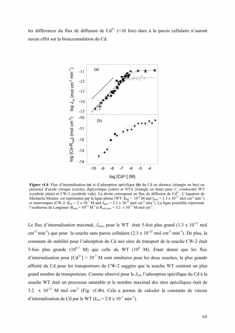
Figure vf.4: Flux d’internalisation (a) et d’adsorption spécifique (b) du Cd en absence (triangle en bas) ou présence d’acide citrique (cercle), diglycolique (carré) et NTA (triangle en haut) pour C. reinhardtii WT (symbole plein) et CW-2 (symbole vide). La droite correspond au flux de diffusion de Cd2+. L’équation de Michaelis-Menten est représentée par la ligne pleine (WT: KM = 10-6 M and Jmax = 1.3 x 10⁻¹¹ mol cm-2 min-1) et interrompue (CW-2: KM = 2 x 10⁻⁷ M and Jmax = 2.3 x 10⁻¹² mol cm-2 min-1). La ligne pointillée représente l’isotherme de Langmuir (Kads = 106.2 M-1 et Rcell-max = 3.2 x 10⁻¹¹ M mol cm-2.
Le flux d’internalisation maximal, Jmax, pour le WT était 5-fois plus grand (1.3 x 10⁻¹¹ mol
cm-2 min-1) que pour la souche sans parois cellulaire (2.3 x 10⁻¹² mol cm-2 min-1). De plus, la
constante de stabilité pour l’adsorption du Cd aux sites de transport de la souche CW-2 était
5-fois plus grande (106.7 M) que celle du WT (106 M). Etant donné que les flux
d’internalisation pour [Cd2+] < 10-7 M sont similaires pour les deux souches, la plus grande
affinité du Cd pour les transporteurs du CW-2 suggère que la souche WT contient un plus
grand nombre de transporteurs. Comme observé pour le Jint, l’adsorption spécifique du Cd à la
souche WT était un processus saturable et le nombre maximal des sites spécifiques était de
3.2 x 10⁻¹¹ M mol cm-2 (Fig. vf.4b). Cela a permis de calculer la constante de vitesse
d’internalisation de Cd par le WT (kint = 2.8 x 10⁻¹ min-1).
xiii
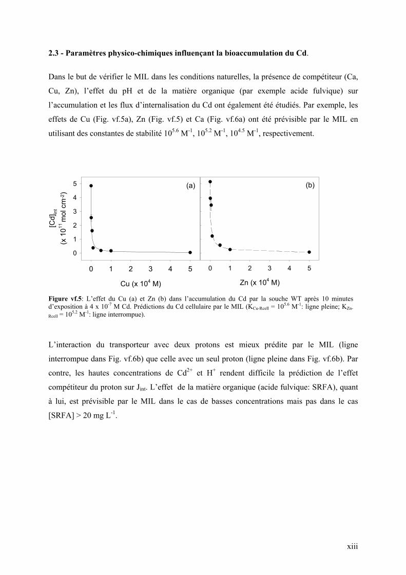
2.3 - Paramètres physico-chimiques influençant la bioaccumulation du Cd.
Dans le but de vérifier le MIL dans les conditions naturelles, la présence de compétiteur (Ca,
Cu, Zn), l’effet du pH et de la matière organique (par exemple acide fulvique) sur
l’accumulation et les flux d’internalisation du Cd ont également été étudiés. Par exemple, les
effets de Cu (Fig. vf.5a), Zn (Fig. vf.5) et Ca (Fig. vf.6a) ont été prévisible par le MIL en
utilisant des constantes de stabilité 105.6 M-1, 105.2 M-1, 104.5 M-1, respectivement.
Cu (x 104 M)
0 1 2 3 4 5
[Cd]
int
(x 1
011 m
ol c
m-2
)
0
1
2
3
4
5
Zn (x 104 M)
0 1 2 3 4 5
(a) (b)
Figure vf.5: L’effet du Cu (a) et Zn (b) dans l’accumulation du Cd par la souche WT après 10 minutes d’exposition à 4 x 10⁻⁷ M Cd. Prédictions du Cd cellulaire par le MIL (KCu-Rcell = 105.6 M-1: ligne pleine; KZn-
Rcell = 105.2 M-1: ligne interrompue).
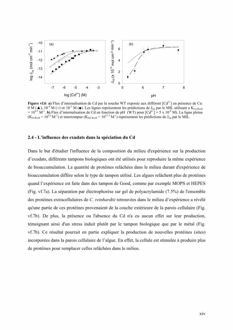
L’interaction du transporteur avec deux protons est mieux prédite par le MIL (ligne
interrompue dans Fig. vf.6b) que celle avec un seul proton (ligne pleine dans Fig. vf.6b). Par
contre, les hautes concentrations de Cd2+ et H+ rendent difficile la prédiction de l’effet
compétiteur du proton sur Jint. L’effet de la matière organique (acide fulvique: SRFA), quant
à lui, est prévisible par le MIL dans le cas de basses concentrations mais pas dans le cas
[SRFA] > 20 mg L-1.
xiv
log [Cd2+] (M)
-7 -6 -5 -4 -3
log
J int (
mol
cm
-2 m
in-1)
-14
-13
-12
-11
-10
pH
5 6 7 8
J int
(x 1
013 m
ol c
m-2
min
-1)
0
2
4
6(a) (b)
Figure vf.6: a) Flux d’internalisation de Cd par la souche WT exposée aux différent [Cd2+] en présence de Ca: 0 M (), 10-4 M () or 10-3 M (). Les lignes représentent les prédictions de Jint par le MIL utilisant a KCa-Rcell = 104.5 M-1. b) Flux d’internalisation de Cd en fonction de pH (WT) pour [Cd2+] = 5 x 10⁻⁸ M). La ligne pleine (KH-Rcell = 105.2 M-1) et interrompue (KH2-Rcell = 1011.3 M-1) représentent les prédictions de Jint par le MIL.
2.4 - L’influence des exudats dans la spéciation du Cd
Dans le but d'étudier l'influence de la composition du milieu d'expérience sur la production
d’exudats, différents tampons biologiques ont été utilisés pour reproduire la même expérience
de bioaccumulation. La quantité de protéines relâchées dans le milieu durant d'expérience de
bioaccumulation diffère selon le type de tampon utilisé. Les algues relâchent plus de protéines
quand l’expérience est faite dans des tampon de Good, comme par exemple MOPS et HEPES
(Fig. vf.7a). La séparation par électrophorèse sur gel de polyacrylamide (7.5%) de l'ensemble
des protéines extracellulaires de C. reinhardtii retrouvées dans le milieu d’expérience a révélé
qu'une partie de ces protéines provenaient de la couche extérieure de la parois cellulaire (Fig.
vf.7b). De plus, la présence ou l'absence du Cd n'a eu aucun effet sur leur production,
témoignant ainsi d'un stress induit plutôt par le tampon biologique que par le métal (Fig.
vf.7b). Ce résultat pourrait en partie expliquer la production de nouvelles protéines (sites)
incorporées dans la parois cellulaire de l’algue. En effet, la cellule est stimulée à produire plus
de protéines pour remplacer celles relâchées dans le milieu.
xv
Temps (min)
0 10 20 30 40 50 60
Pro
téin
es (µ
g cm
-2)
0.2
0.4
0.6
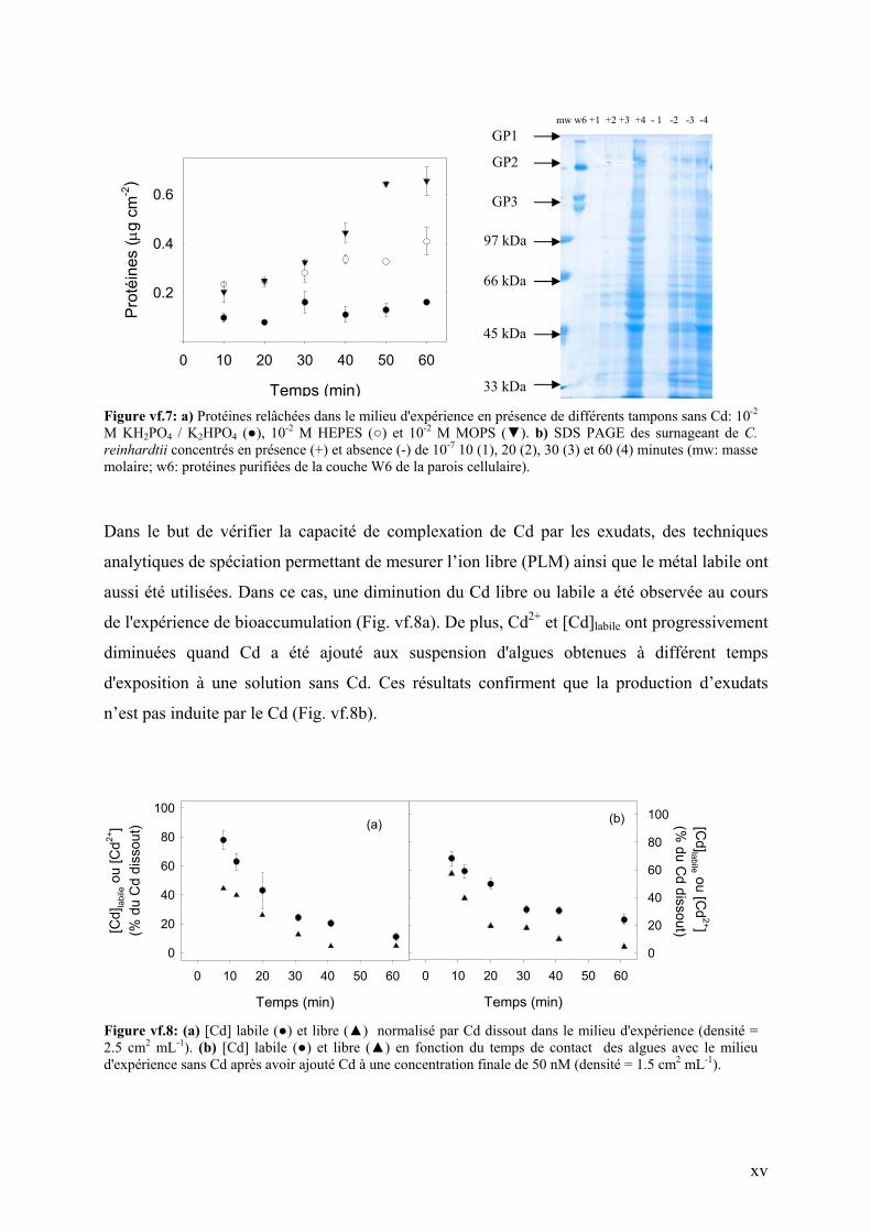
Figure vf.7: a) Protéines relâchées dans le milieu d'expérience en présence de différents tampons sans Cd: 10-2 M KH2PO4 / K2HPO4 (), 10-2 M HEPES () et 10-2 M MOPS (). b) SDS PAGE des surnageant de C. reinhardtii concentrés en présence (+) et absence (-) de 10-7 10 (1), 20 (2), 30 (3) et 60 (4) minutes (mw: masse molaire; w6: protéines purifiées de la couche W6 de la parois cellulaire).
Dans le but de vérifier la capacité de complexation de Cd par les exudats, des techniques
analytiques de spéciation permettant de mesurer l’ion libre (PLM) ainsi que le métal labile ont
aussi été utilisées. Dans ce cas, une diminution du Cd libre ou labile a été observée au cours
de l'expérience de bioaccumulation (Fig. vf.8a). De plus, Cd2+ et [Cd]labile ont progressivement
diminuées quand Cd a été ajouté aux suspension d'algues obtenues à différent temps
d'exposition à une solution sans Cd. Ces résultats confirment que la production d’exudats
n’est pas induite par le Cd (Fig. vf.8b).
Temps (min)
0 10 20 30 40 50 60
[Cd]labile ou [C
d2+]
(% du C
d dissout)
0
20
40
60
80
100(b)
Temps (min)
0 10 20 30 40 50 60
[Cd]
labi
le o
u [C
d2+]
(% d
u C
d di
ssou
t)
0
20
40
60
80
100(a)
Figure vf.8: (a) [Cd] labile () et libre () normalisé par Cd dissout dans le milieu d'expérience (densité = 2.5 cm2 mL-1). (b) [Cd] labile () et libre () en fonction du temps de contact des algues avec le milieu d'expérience sans Cd après avoir ajouté Cd à une concentration finale de 50 nM (densité = 1.5 cm2 mL-1).
33 kDa
45 kDa
66 kDa
97 kDa
GP3
GP2
GP1 mw w6 +1 +2 +3 +4 - 1 -2 -3 -4
xvi
3 - Conclusions
L’adsorption du Cd par les deux souches de C. reinhardtii ne peut pas être régie par les
isothermes de Langmuir. Le processus d'adsorption de Cd se déroule en une étape rapide au
cours de laquelle plus que 50% du Cd est adsorbés en moins de 5 minutes suivie d'une autre
étape plus lente. L'analyse des données cinétiques montre aussi que le processus d'adsorption
semble plutôt être contrôlé par la production de nouveaux sites que par la diffusion ou les
réactions d’échanges ioniques sur la parois cellulaire.
La majorité des résultats présenté dans ce travail permet de mettre en évidence que
l’internalisation du Cd par C. reinhardtii pourra être prédite par les modèles basés sur les
équilibres thermodynamiques des métaux traces près de la surface des miroorganismes. Ceci
implique que le Model de l’Ion Libre pourra être utilisé pour relier les concentrations ou la
vitesse d’accumulation (flux d’internalisation) du Cd par C. reinhardtii aux concentrations de
ce métal dans un système aquatique relativement simple. Même si des déviations du MIL ont
été observées pour des concentrations élevées de matière organique, de Cd et de proton, ces
concentrations ne sont généralement pas représentatives de la majorité des milieux naturelles.
Néanmoins, ce modèle demande à être testé in situ, comme par exemple pour eaux naturelles
présentant des caractéristiques physico-chimiques variées, afin d’assurer son application
générale pour une large gamme de conditions.
1
1 - INTRODUCTION
Water is a crucial compound in the evolution of living organisms since their biochemical
systems are all adapted to function in aqueous media. Five m3 of water are used by a single
individual each day (Moss B.; 1980). Some of this water comes from relatively
uncontaminated upland lakes and rivers but in the lowlands, water must be used and reused
many times as it passed ultimately from atmosphere to land and sea in the hydrological cycle.
The most common water pollutants are high levels of organic matter, plant nutrients,
suspended mineral particles, heavy metals, pesticides, organic chemicals, acids and
radioactive substances.
The concentrations of trace metals in continental waters are primarily controlled by
atmospheric deposition and by weathering processes on soils and bedrock. The atmosphere, in
particular has become a key source for the transfer of trace metals to remote aquatic
ecosystems. The fluxes of trace metals into freshwaters have increased, mainly due to
anthropogenic influences on the important pathways and processes. Domestic and industrial
wastewater, sewage discharge and urban runoff contribute large quantities of metal pollution
to the aquatic environment.
One of the key problems of environmental chemistry and toxicology remains how to best
relate metal exposures to their biological effects. When describing the interaction of trace
metals with aquatic biota, at least three important aspects must be considered: (i) the chemical
speciation of these metals in the external environment, (ii) interactions of the metal with the
biological membrane leading to biological internalization and (iii) the resulting biological
effects. Although, some steady-state models attempt to relate the biological response either to
the free-metal ion concentration in solution, (basis of the Free Ion Activity Model, FIAM,
(Morel F.M.M. and Hering J.G.; 1993; Campbell P.G.C.; 1995)) or to the metal bound to
sensitive sites at the surface of the organism, (basis of the Biotic Ligand Model, BLM, (Playle
R.C.; 1998)), they do not always take into account the complexity of the environmental
processes that are at the interface of physics, chemistry and biology. The work presented here
is an attempt to bring together all of these disciplines in order to improve our understanding of
the important microscopic processes that determine trace metal bioavailability.
2
1.1 - Chemical speciation of metals in natural waters
Chemical speciation is of utmost importance when evaluating the reactivity of a metal in
biological and environmental processes. Trace metals exist in aquatic systems in a variety of
chemical species. Only a small portion of the overall dissolved metal is generally present as
free hydrated cations. Most metal ions form stable complexes by interacting with dissolved
inorganic and organic ligands or inorganic (e.g. colloids) and organic (e.g. phytoplankton,
bacteria) particles. The dissolved inorganic complexes present in natural waters are mainly
chloro-, carbonato-, sulfato-, oxo-, and hydroxo complexes (Boyle E.A., Scalter F. et al.;
1976) whose distribution can generally be well characterized through the use of
thermodynamic models.
Less is known about metal complexation with organic matter of biological origin and with
organic pollutants, although this situation is changing with the development of sensitive metal
speciation techniques. Organic matter contents vary from approximately 0.3 to 3 mg L-1 of
carbon in open seas to 1 to 10 mg L-1 in rivers, lakes and estuaries (Cosovic B., Ciglenecki I.
et al.; 2000). Organic compounds released by living organisms, or resulting from their
decomposition can have a well-characterized structure (e.g., amino acids, hydroxyl acids,
monosaccharides) but more frequently cannot be separated or characterized except into
fractions with similar properties (e.g., fulvic acid, humic acids, polysaccharides, polypetides,
lipids, and proteins). Organic matter appears to play an important role in the chemical
speciation of metal ions in aquatic natural systems. For example, about 50% of dissolved Pb
(Pitts L.C. and Wallace G.T.; 1994) and >90% of dissolved zinc (Bruland K.W.; 1989) is
usually complexed with natural organic ligands in seawaters. In the case of cadmium, the
tendency to form inorganic and organic complexes is relatively small, such that the ratio of
[Cd2+] to total dissolved [Cd] is often approximately 0.5 (Stumm W. and Morgan J.J.; 1996).
Many metal effects on biological species can be explained in terms of the free-ion activity
model (FIAM). Indeed, a dependence of trace metal uptake, phytoplankton growth and
metabolism on the free-ion activity has been observed since the late 70s (Sunda W.G. and
Guillard R.R.L.; 1976; Anderson M.A., Morel F.M.M. et al.; 1978). Nevertheless, complex
calculations and measurements are generally needed in order to determine the concentration
of the free metal in solution, especially in natural waters. For example, for calculations, total
metal concentrations, pH, the concentrations of all ligands and the concentrations of other
3
ionic and molecular species must be known (Borgmann U.; 1982; Simkiss K. and Taylor
M.G.; 1989). Other factors such as the concentration and nature of dissolved and suspended
organic matter and suspended colloidal inorganic matter are important but cannot, as yet, be
included into calculations (Campbell P.G.C.; 1995). A precise determination of metal
speciation is a key to better understanding trace metal availability to biota.
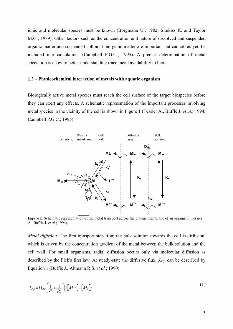
1.2 – Physicochemical interaction of metals with aquatic organism
Biologically active metal species must reach the cell surface of the target biospecies before
they can exert any effects. A schematic representation of the important processes involving
metal species in the vicinity of the cell is shown in Figure 1 (Tessier A., Buffle J. et al.; 1994;
Campbell P.G.C.; 1995).
cell nterior Plasma membrane
Cell wall
Diffusionlayer
Bulksolution
ML ML ML
MZ+ MZ+ MZ+
Ks kint
kd’ ka’
kd
ka
Ln-R----M Ks
DML
DM
Mcell
Figure 1: Schematic representation of the metal transport across the plasma membrane of an organism (Tessier A., Buffle J. et al.; 1994).
Metal diffusion. The first transport step from the bulk solution towards the cell is diffusion,
which is driven by the concentration gradient of the metal between the bulk solution and the
cell wall. For small organisms, radial diffusion occurs only via molecular diffusion as
described by the Fick's first law. At steady-state the diffusive flux, Jdiff, can be described by
Equation 1 (Buffle J., Altmann R.S. et al.; 1990):
[ ] [ ]( )00
11 MMR
DJ zavediff −⋅
+= +
δ (1)
4
where Dave depends on the diffusion coefficient of the metal (M) and its complex (ML), the
stability constant defining the relationship between M and ML, the radius of the
microorganism (R0) and the thickness of the diffusion layer (δ) (Fig. 1). The diffusive flux can
be increased by decreasing the thickness of the boundary layer, by increasing the metal
concentration gradient or by decreasing the size of the microorganism (Eq. 1). The thickness
of the boundary layer (δ) is a function of the movement of the cell relative to the bulk solution
and it is estimated to be 20 to 30 µm for large algal cells (Tessier A., Buffle J. et al.; 1994). In
natural waters, small freely suspended microorganisms move with the bulk solution, so that
no flux enhancement will occur due to fluid motion for the uptake of a typical small solute
(Wilkinson K.J. and Buffle J.; 2004) such as metal ions.
Nature of the cell surface. The metal, after being transferred from the external medium to the
vicinity of the algal cell, will first react with cell wall, which protects the cell interior from its
environment. The plant cell wall is a complex polymeric sheath, consisting of a network of
cellulose microfibrils glued together by a polysaccharide and glycoproteinic matrix. The
polysaccharide and glycoproteinic macromolecules that are the main structural components of
the cell wall have surface groups such as -COOH, -OH, -NH2 (Crist R.H., Oberholser K. et
al.; 1981; Xue H.B., Stumm W. et al.; 1988; Cho D.-Y., Lee S.-T. et al.; 1994; Stumm W.,
Sigg L. et al.; 1994; Kiefer E., Sigg L. et al.; 1997) that, depending on pH, will be ionized. As
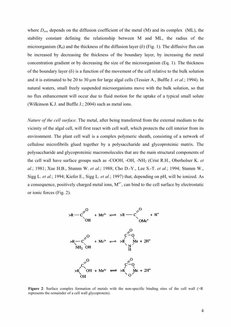
a consequence, positively charged metal ions, Mz+, can bind to the cell surface by electrostatic
or ionic forces (Fig. 2).
Figure 2: Surface complex formation of metals with the non-specific binding sites of the cell wall (>R represents the remainder of a cell wall glycoprotein).
5
The number of cell wall binding sites is assumed to be constant for a given cell at a given life
stage as it depends only on the cell wall structure and its chemical composition. In the
literature of the plant cell growth, two traditional time scales related to cell wall stretching are
known: (i) a long time scale associated with the biosynthesis of wall components, spanning
presumably a period of hours to days and (ii) a short time scale associated with cell expansion
at a given composition spanning a period from seconds to hours (Veytsman B.A. and
Cosgrove D.J.; 1998). The new wall material synthesized by the growing cell and secreted
into the extracellular space is integrated into the extending wall, largely by the noncovalent
mechanism of polymer adhesion, although some covalent cross-linking may occur. For a
unicellular alga such as C. reinhardtii with a fast growth rate, these two time scales cannot be
separated easily. For example, Goodenough and Heuser (Goodenough U.W. and Heuser J.E.;
1985) observed a newly synthesized primitive outer layer of the cell walls of C. reinhardtii <1
hour after the addition of autolysin, a cell wall disrupting agent.
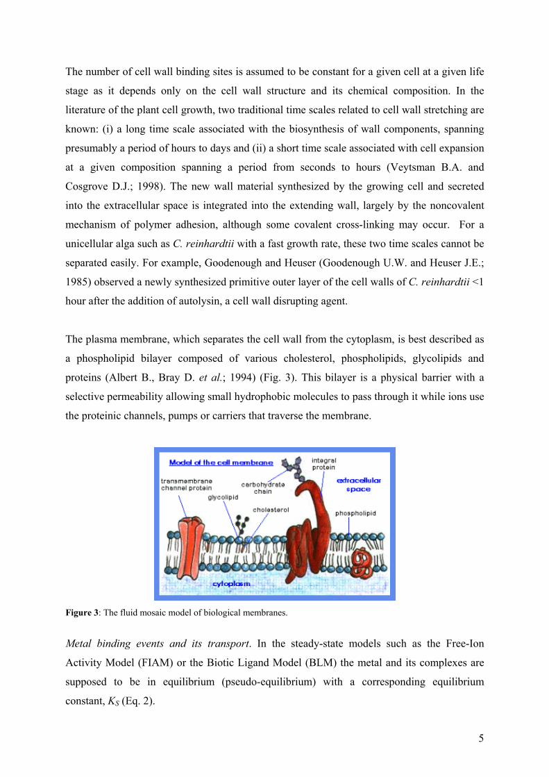
The plasma membrane, which separates the cell wall from the cytoplasm, is best described as
a phospholipid bilayer composed of various cholesterol, phospholipids, glycolipids and
proteins (Albert B., Bray D. et al.; 1994) (Fig. 3). This bilayer is a physical barrier with a
selective permeability allowing small hydrophobic molecules to pass through it while ions use
the proteinic channels, pumps or carriers that traverse the membrane.
Figure 3: The fluid mosaic model of biological membranes.
Metal binding events and its transport. In the steady-state models such as the Free-Ion
Activity Model (FIAM) or the Biotic Ligand Model (BLM) the metal and its complexes are
supposed to be in equilibrium (pseudo-equilibrium) with a corresponding equilibrium
constant, KS (Eq. 2).
6
Mz+ + Ln- ↔ M-L(z-n) (2)
The metal, after being transferred from the external medium to the vicinity of the cell, may
react with non-specific sites such as the cell wall or with cell surface transport sites, Rcell, (see
first reaction in Eq. 3).
ka kint
Mz+ + Rcell ↔ M-Rcell → Mcell
kd
(3)
where ka and kd are the rate constants for the formation and dissociation of the membrane
transport site complex and kint is the first order rate constant for the transfer of the bound
metal from the membrane imbedded transport complex into the cytoplasm. Depending on the
stability of the complex and rate constants, M-L(z-n) can react with Rcell in a ligand exchange
reaction (Eq. 4):
ka'
kint
M-L(z-n) + Rcell ↔ M-Rcell + Ln- → Mcell
kd'
(4)
where kf' and kd
' are the analogous rate constants for ligand exchange. The cell wall binding
sites (non-specific) are physiologically inert such that the binding of the metal to these sites
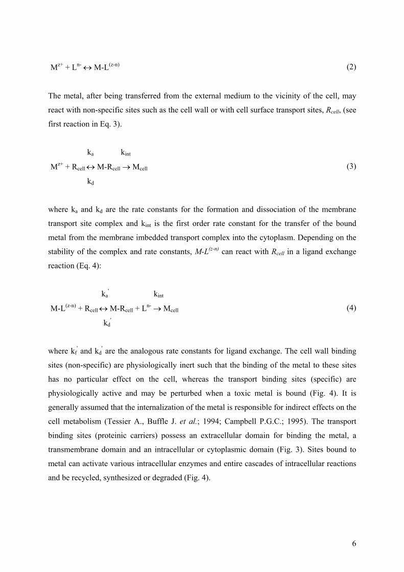
has no particular effect on the cell, whereas the transport binding sites (specific) are
physiologically active and may be perturbed when a toxic metal is bound (Fig. 4). It is
generally assumed that the internalization of the metal is responsible for indirect effects on the
cell metabolism (Tessier A., Buffle J. et al.; 1994; Campbell P.G.C.; 1995). The transport
binding sites (proteinic carriers) possess an extracellular domain for binding the metal, a
transmembrane domain and an intracellular or cytoplasmic domain (Fig. 3). Sites bound to
metal can activate various intracellular enzymes and entire cascades of intracellular reactions
and be recycled, synthesized or degraded (Fig. 4).
7
Figure 4: Levels of complexity of transport binding sites and their location in the cell during metal adsorption and internalization (Lauffenburgar D.A. and Linderman J.J.; 1993).
Metal binding phenomena are influenced by a variety of extrinsic variables that are defined by
the experimental system. Primary among these factors are the temperature and medium
composition, including the pH and ionic strength. Generally speaking, carrier/solute binding
processes are generally exothermic, i.e. ∆H° < 0, although many are driven by positive
entropy changes, ∆S° > 0 (Klotz I.M.; 1985). When ∆S° term dominates, there may be only
minor variation of equilibrium binding with temperature whereas if the ∆H° term dominates,
there will be a strong temperature effect. Interpretation is greatly complicated by the fact that
∆S° and ∆H° vary significantly with temperature, most likely due to changes in three-
dimensional conformation of proteinic carriers. Hence, van’t Hoff plots of the log of
equilibrium dissociation constant (KD) versus 1/T are rarely linear. In addition, the conditional
stability constant for the adsorption decreases as the pH decreases below 7.0 (Kaplan J.; 1985;
Lauffenburgar D.A. and Linderman J.J.; 1993), although it is not clear whether this is due to a
decrease in ka, an increase in kd, or both.
1.3 - Models used to describe the metal adsorption and internalization
Metal adsorption. In the case of a single homogeneous adsorption site, a Langmuir isotherm
can be employed as a simple adsorption model (Eq. 5) (Rhee G.Y. and Thompson P.A.; 1992;
Tessier A., Buffle J. et al.; 1994; Kiefer E., Sigg L. et al.; 1997):
[ ][ ]+−
+
+
Γ=Γ
zads
zeqM MK
M1
max (5)
8
where ΓMeq
and Γmax are the amount and maximum amount of metal ion adsorbed to the
binding sites at equilibrium and Kads is the equilibrium constant for the adsorption (Eq. 6).
[ ][ ][ ]
∆−=−=
+ RTG
MRRMK ads
zcell
cellads exp
(6)
where ∆Gads is the change in the Gibbs free energy of the system that occurs during the
adsorption reaction, which is equal to the difference between the change in the enthalpy of the
system and the change in the product of the temperature times the entropy of the system. In
the case when [Mz+] << 1 / Kads, the Langmuir equation can be simplified to Equation 7:
[ ]+≅Γ zHeqM MK (7)
where KH is the Henry constant (Eq. 8):
adsH KK maxΓ= (8)
There are two important conditions for the validity of the Langmuir isotherm: equilibrium
must exist up to the formation of the monolayer (with 1:1 stoichiometry) and ∆Gads must be
independent of site occupancy, implying that all sites should have an equal activity. In reality,
the tendency to form surface complexes decreases with increased metal loading of the surface
because the metal ions bind first to the surface groups with highest affinity and subsequently
to groups with lower affinities (Xue H.B., Stumm W. et al.; 1988; Buffle J., Altmann R.S. et
al.; 1990). Thus, other types of isotherms are often better able to describe the adsorption
phenomena. For instance, the Freundlich isotherm represents a monolayer adsorption on a
heterogeneous surface (Eq. 9):
ΓMeq = m [L]n (9)
where m and n are fitting constants, which do not necessarily have any physical meaning.
Adsorption models with additional interactions between the adsorbed species lead to more
complex isotherms (e.g. Frumkin-Fowler-Guggenheim isotherm) (Stumm W. and Morgan
J.J.; 1996), however, this type of binding will not be considered here.
9
Metal adsorption by the cell is generally a rapid process that precedes metal internalization.
The kinetics of adsorption are determined mainly by the concentration of the metal active
form (Mz+ or ML) and by the concentration and affinity of binding sites on the cell surface.
The dependence of metal adsorption to the cell wall with time, Г(t), can be given by Equation
10 (van Leeuwen H.P.; 1992):
ττ
τtt
tteq
+−=
+=
ΓΓ
1
111
)(
(10)
where τ is the time that it takes for the metal to diffuse through the diffusion layer and the cell
wall prior to adsorption (Eq. 11).
Cd
H
DRK 0=τ
(11)
It should be noted that the relaxation time increases with microorganism size and decreases
with mobility.
Metal transport. Metal internalization generally is relatively slower than metal adsorption,
and hence considered to be rate-determining with respect to the overall metal uptake process
(Mason A.Z. and Jenkins K.D.; 1995). Under steady-state conditions (local equilibrium
between metal species in the bulk solution and those at the cell surface), the flux (rate) of
metal uptake can be described by the Michaelis-Menten hyperbolic rate law (that was initially
developed for enzyme kinetics and nutrient uptake, Eq. 12) (Pasciak W.J. and Gavis J.; 1974):
[ ][ ]+
+
+= z
M
z
MKMJ
J maxint
(12)
where Jmax (Jmax = kint Rcelltot) is the maximum uptake rate achieved when all of the transport
sites, Rcelltot, are bound by metal and KM is the half-saturation constant equal to the available
metal concentration at which half of the sites are complexed (KM = kd + kint / ka) (Morel
F.M.M.; 1986; Sunda W.G. and Huntsman S.A.; 1992; Sunda W.G. and Huntsman S.A.;
10
1998). Under steady-state conditions, the half-saturation constant is inversely related to the
affinity of the uptake sites for a given metal. It is possible to distinguish between two extreme
cases of steady-state conditions:
(i) When kd >> kint (KM = kd / ka), the uptake rate is slow as compared to the establishment of
all other complex formation equilibria and uptake is controlled by the (thermodynamic)
equilibrium in solution, since the flux of metal ions into the cell is proportional to the product
of kint and M-Rcell (Morel F.M.M. and Hering J.G.; 1993). Therefore, uptake is rate-limiting
in the case of a small kint and a fast mass transport. A large value of kd implies weak binding
of the metal to Rcell since kd = ka / KM (Sunda W.G. and Huntsman S.A.; 1998).
(ii) When kint >> kd (KM = kint / ka), the uptake rate may be controlled by the rate of formation
of the surface complexes, M-Rcell, and by the dissociation of complexes ML in solution
(kinetic control) (Hudson R.J.M.; 1998) or other important processes such as mass transfer
(i.e., diffusive flux). The kinetically controlled situation is favored at low metal
concentrations because the (few) metal ions arriving at the cell surface will readily find a free
transport channel and thus be incorporated into the cell.
Jmax may vary depending on the prior exposure of an algal cell to a given metal ion or its
starvation with respect to the metal (Price N.M., Ahner B.A. et al.; 1994). When the
concentration of metal is much higher than the Michaelis-Menten constant, the sites may
become saturated and the uptake flux becomes independent of [Mz+] (Jint = Jmax). Nonetheless,
in natural waters, where the concentration of heavy metals is generally much lower than KM,
most of the sites will be available for metal binding and thus the uptake rate should be
proportional to the concentration of the free metal (Jint = Jmax [Mz+] / KM), the basis of the
FIAM (Morel F.M.M. and Hering J.G.; 1993). If internalization is faster than the complex
formation reaction of Mz+ with Rcell, the FIAM cannot be valid. Also, when the diffusion of
the metal to the surface is rate-determining, the above proportionality is not assured, since
M-Rcell is practically zero whatever the free-metal concentration in the solution (Hudson
R.J.M. and Morel F.M.M.; 1993).
A summary of the key assumptions of the steady-state uptake models (i.e. FIAM, BLM) are
(Campbell P.G.C.; 1995; Wilkinson K.J. and Buffle J.; 2004):
11
- metal transport toward the cell surface and the surface complexation reactions occur
faster than metal internalization (steady-state)
- plasma membrane is the primary site for metal interaction
- a single 1:1 binding site is involved during metal transport
- the transport sites have an homogeneous distribution of charges and they undergo no
significant modification during the experiment
- the transport sites does not posses regulatory sites
1.4 - Factors influencing metal uptake
Competition. Since metal uptake depends on the transport sites on the surface of the
microorganism, theoretically, a second metal could act antagonistically (compete),
synergistically (reinforce), additively (superimpose), or remain without any effect relative to
the adsorption and uptake of the first metal by a given alga and to the physiological and
morphological changes produced by the first metal in a given alga (Visviki I. and Rachlin
J.W.; 1994). Competition can occur because the metal coordination sites in microorganisms
are never entirely specific for just a single metal and because the competing metals have
similar ionic radius i.e., coordination geometries. The competition may concern nutrient metal
transport sites, active sites of metalloproteins, or intracellular feedback control sites such as
those regulating the number or activity of specific membrane transport proteins (Sunda W.G.
and Huntsman S.A.; 1998). By modifying different simplifying assumptions of FIAM, it is
possible to predict the biological response (i.e., Jint) of a metal, M, in the presence of a second
metal, C, when both metals compete antagonistically for the same transport site (Eq. 13).
1][C][M][M
nz
maxint ++=
−+
−+
−+
RcellCRcellM
RcellMz
KKKJ
J (13)
where [Cn+] is the concentration of a competing metal or proton, KM-Rcell and KC-Rcell are values
of the affinity constants for the interaction of the metal of interest and a competing metal or
proton and Jmax is the maximal uptake flux of the metal of interest.
pH. The acidity of aquatic environments is also a very important abiotic factor affecting metal
uptake, growth, physiology and reproduction of aquatic organisms. Low pH can alter nutrient
uptake (Gensemer R.W., Smith R.E.H. et al.; 1993), facilitate the toxic action of aquatic
12
pollutants (Sunda W.G. and Guillard R.R.L.; 1976; Anderson M.A., Morel F.M.M. et al.;
1978; Rachlin J.W. and Grosso A.; 1991) and modify the composition and richness of algal
communities, resulting in local extinction of sensitive species (Turner L.J., Howell E.T. et al.;
1991; Anderson N.J., Blomqvist P. et al.; 1997). Changes in environmental pH can influence
the uptake of metals in at least two ways: (i) by affecting the metal speciation and (ii) by its
effect on the biological surface uptake sites (Campbell P.G.C. and Stokes P.M.; 1985).
1.5 – The case of cadmium and C. reinhardtii
Cadmium is ubiquitous in our environment. Thanks to its unique chemical and physical
properties, Cd is widely used in alloys, pigments, stabilisers, coatings and, accounting for
almost 70% of its use, in rechargeable nickel-cadmium batteries. Cd is regarded as one of the
most toxic trace elements in the environment because of its easy uptake by plants, its
tendency to accumulate in crops in the food chain and its persistent nature once in the
environment (Dudka S. and Adriano D.C.; 1997). Nearly 60% of the total input of Cd in air
and 30% in water comes from smelting and mining (Nriagu J.O. and Pacyna J.M.; 1988). In
fact, for 1000 kg of mined zinc, 3 kg of Cd are produced (Ohnesorge F.K. and Wilhelm M.;
1991). Fortunately, Cd emissions to the environment have been decreasing significantly since
1960's. This has resulted in decreasing levels of cadmium in air, water and food. The potential
risks to human health have been extensively studied and are now well controlled. Indeed, the
current daily Cd intake of the general population is at the lower end of the range 10-25 µg
day-1, while WHO has set the provisional tolerable daily intake at 60-70 µg day-1 (WHO;
1992).
1.5.1 - Levels of Cd in waters
Cd is a natural, usually minor constituent of surface and groundwater. Levels from 9 x 10-11
M to 3.6 x 10⁻⁷ M have been quoted in the literature depending on specific location and
whether or not total cadmium or dissolved cadmium is measured (Elinder C.-G.; 1985; WHO;
1992; OECD; 1994). Buffle (Buffle J.; 1988) reported a range of 10-13 M to 4 10-10 M for total
cadmium in freshwaters, with an average of 4 10-12 M. Rivers containing excess Cd can
contaminate surrounding land, either through irrigation for agricultural purposes, dumping of
dredged sediments or flooding. It has also been demonstrated that rivers can transport
cadmium for considerable distances, up to 50 km, from the source (WHO; 1992). For river
13
waters, dissolved Cd concentrations of 8.8 x 10⁻¹² M to 1.2 x 10⁻¹⁰ M (Shiller A.M. and Boyle
E.A.; 1987) have been reported. Nonetheless, studies of cadmium contamination in major
river systems over the past twenty to thirty years have conclusively demonstrated that
cadmium levels in these rivers have decreased significantly since the 1960s and 1970s
(Elgersma F., Anderberg B.S. et al.; 1992; Van Assche F.J. and Ciarletta P.; 1992; Cook M.E.
and Morrow H.; 1995; Mukunoki J. and Fujimoto K.; 1996). For example, studies on the
Rhine River Basin from 1973 through 1987 indicated that the point source cadmium
discharges to the Rhine River decreased from 130 to 11 metric tonnes (mt) per year over that
14-year time span, a reduction of over 90% (Elgersma F., Anderberg B.S. et al.; 1992).
Similarly, data on total cadmium and dissolved cadmium at the Dutch/German border over
the period from 1971 to 1987 have shown comparable reductions (van Urk G. and Marquenie
J.M.; 1989).
Cadmium emissions to water arise, in decreasing order of importance, from phosphate
fertilisers, non-ferrous metals production, and the iron and steel industry (Van Assche F.J. and
Ciarletta P.; 1992; OECD; 1994). The average Cd content in the world's oceans has variously
been reported as low as < 4.5 x 10⁻¹¹ M (WHO; 1992; WHO; 1992) and 4.5 x 10⁻¹¹ - 1.8 x 10-
10 M (Jensen A. and Bro-Rasmussen F.; 1992; WHO; 1992; OECD; 1994) to as high as 10-9
M (Cook M.E. and Morrow H.; 1995) and 9 x 10-11 to 9 x 10⁻¹⁰ M (Korte F.; 1983; Elinder C.-
G.; 1985). Higher levels have been noted around certain coastal areas (Elinder C.-G.; 1985)
and the vertical distribution of dissolved cadmium can be characterized by a surface depletion
and deep-water enrichment (Boyle E.A., Scalter F. et al.; 1976; OECD; 1994). This
distribution is assumed to result from the internalization of Cd by phytoplankton in surface
waters and its transport to depth, incorporation into biological debris, and subsequent release.
Cadmium may also enter aquatic systems through weathering and erosion of soils and
bedrock, atmospheric deposition direct discharge from industrial operations, leakage from
landfalls and contaminated sites and the dispersive use of sludge and fertilisers in agriculture.
Much of the cadmium entering fresh waters from industrial sources may be rapidly adsorbed
by particulate matter, and thus sediment may be a significant sink for cadmium emitted to the
aquatic environment (WHO; 1992). Indeed, Cd levels of up to 4.46 10-3 mol kg-1 have been
reported in river and lake sediments (Korte F.; 1983). These quantities are an important
reserve of potential contamination by redissolution. Some data shows that recent sediments in
lakes and streams contain from 1.8 x 10⁻³ to 8 x 10⁻³ mol kg-1 in contrast to the levels of
14
generally less than 8.9 x 10⁻⁴ mol kg-1 found in freshwater sediments (Cook M.E. and Morrow
H.; 1995). Partitioning of cadmium between the adsorbed state and dissolved state is therefore
an important factor as to whether the Cd is able to enter the food chain and affect human
health.
1.5.2 – Metal adsorption
C. reinhardtii cell wall. The cell wall of C. reinhardtii is a seven-layered structure with three
conspicuous layers, W2, W4 and W6 (separated by spaces W3 and W5) composed mainly of
fibers and glycoproteins (Roberts K., Gurney-Smith M. et al.; 1972; Roberts K.; 1974;
Goodenough U.W., Gebhart B. et al.; 1986). The whole wall is approximately 320 nm wide
(Davis R.D. and Plaskitt A.; 1971). The outer cell-wall layers are crystalline and can be
solubilized in chaotropic agents, e.g. NaClO4 and LiCl solutions, while the inner wall layer
has been found to be amorphous and insoluble (Voigt J.; 1985; Voigt J.; 1988). All cell layers
are very similar with respect to the amino acid and sugar composition. The cell wall of
Chlamydomonas does not contain cellulose but consists mainly of hydroxyproline-rich
glycoproteins (HRGP) of high molar mass that have a strong binding affinity for metal ions
(Crist R.H., Martin J.R. et al.; 1994). In fact, the HRGP of the outer-wall are not covalently
cross-linked (crystalline arrays), while those of the inner-wall are covalently cross-linked as in
higher plants. Moreover, a HRGP subgroup, called chimeric extensions, has both fibrous and
globular domains (Kilz S., Waffenschmidt S. et al.; 2000). It is suspected that the former
anchor the protein to the cell wall, while the later mediate the sugar- binding activity. Sugar
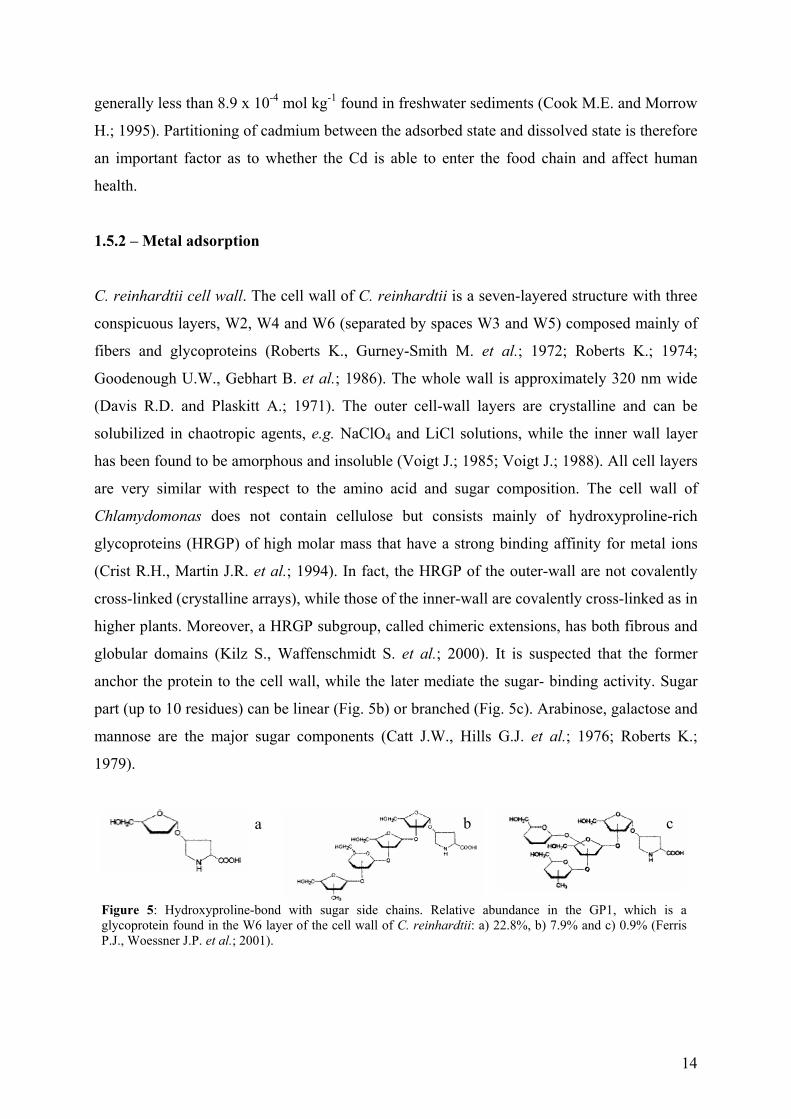
part (up to 10 residues) can be linear (Fig. 5b) or branched (Fig. 5c). Arabinose, galactose and
mannose are the major sugar components (Catt J.W., Hills G.J. et al.; 1976; Roberts K.;
1979).
Figure 5: Hydroxyproline-bond with sugar side chains. Relative abundance in the GP1, which is a glycoprotein found in the W6 layer of the cell wall of C. reinhardtii: a) 22.8%, b) 7.9% and c) 0.9% (Ferris P.J., Woessner J.P. et al.; 2001).
a c b
15
Potentiometric titrations of live C. reinhardtii cells have indicated two types of protonatable
functional groups in the cell wall with average pKa values of 4.9 and 9.1 (I = 10-2 M) (Kiefer
E., Sigg L. et al.; 1997), likely corresponding to carboxylic and amino groups. Adhiya et al.
(Adhiya J., Cai X. et al.; 2002) found similar pKa values of 4.7±0.5 and 9.6±0.3 at I = 10-2 M
and 4.7±1.1 and 9.3±0.3 at I = 10-1 M (KCl) when performing potentiometric titrations on
lyophilized cells. The high ionic strengths appear to lower the effective pKa values by
screening the surface charge so as to reduce the interactions among functional groups thereby
weakening the bonds between the surface sites and H+. Maximal proton binding capacities
were 9.1 x 10⁻⁴ mol g-1 algae (dry wt.) (Kiefer E., Sigg L. et al.; 1997).
Adsorption. The adsorption of heavy metals is affected by the nature (chemical composition
and structure) of the cell wall. For example Cd adsorbed varied greatly for algal species
(arranged in decreasing order of Cd per unit algal surface): C. vulgaris > C. reinhardtii > C
pyrenoidosa (Khoshmanesh A., Lawson F. et al.; 1997). This suggests that C. vulgaris has a
larger number of surface sites and/or a higher affinity for Cd than the other species tested.
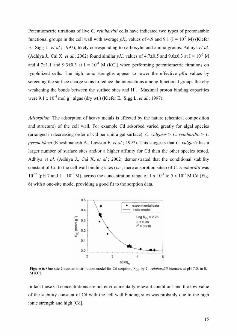
Adhiya et al. (Adhiya J., Cai X. et al.; 2002) demonstrated that the conditional stability
constant of Cd to the cell wall binding sites (i.e., mere adsorption sites) of C. reinhardtii was
102.2 (pH 7 and I = 10-1 M), across the concentration range of 1 x 10⁻⁴ to 5 x 10⁻³ M Cd (Fig.
6) with a one-site model providing a good fit to the sorption data.
Figure 6: One-site Gaussian distribution model for Cd sorption, SCd, by C. reinhardtii biomass at pH 7.0, in 0.1 M KCl.
In fact these Cd concentrations are not environmentally relevant conditions and the low value
of the stability constant of Cd with the cell wall binding sites was probably due to the high
ionic strength and high [Cd].
16
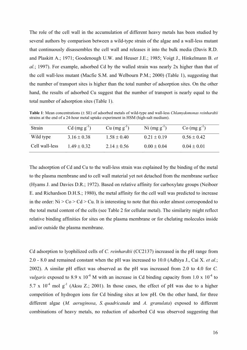
The role of the cell wall in the accumulation of different heavy metals has been studied by
several authors by comparison between a wild-type strain of the algae and a wall-less mutant
that continuously disassembles the cell wall and releases it into the bulk media (Davis R.D.
and Plaskitt A.; 1971; Goodenough U.W. and Heuser J.E.; 1985; Voigt J., Hinkelmann B. et
al.; 1997). For example, adsorbed Cd by the walled strain was nearly 2x higher than that of
the cell wall-less mutant (Macfie S.M. and Welbourn P.M.; 2000) (Table 1), suggesting that
the number of transport sites is higher than the total number of adsorption sites. On the other
hand, the results of adsorbed Cu suggest that the number of transport is nearly equal to the
total number of adsorption sites (Table 1).
Table 1: Mean concentrations (± SE) of adsorbed metals of wild-type and wall-less Chlamydomonas reinhardtii strains at the end of a 24-hour metal uptake experiment in HSM (high-salt medium). Strain Cd (mg g-1) Cu (mg g-1) Ni (mg g-1) Co (mg g-1)
Wild type 3.16 ± 0.38 1.58 ± 0.40 0.21 ± 0.19 0.56 ± 0.42
Cell wall-less 1.49 ± 0.32 2.14 ± 0.56 0.00 ± 0.04 0.04 ± 0.01
The adsorption of Cd and Cu to the wall-less strain was explained by the binding of the metal
to the plasma membrane and to cell wall material yet not detached from the membrane surface
(Hyams J. and Davies D.R.; 1972). Based on relative affinity for carboxylate groups (Neiboer
E. and Richardson D.H.S.; 1980), the metal affinity for the cell wall was predicted to increase
in the order: Ni > Co > Cd > Cu. It is interesting to note that this order almost corresponded to
the total metal content of the cells (see Table 2 for cellular metal). The similarity might reflect
relative binding affinities for sites on the plasma membrane or for chelating molecules inside
and/or outside the plasma membrane.
Cd adsorption to lyophilized cells of C. reinhardtii (CC2137) increased in the pH range from
2.0 - 8.0 and remained constant when the pH was increased to 10.0 (Adhiya J., Cai X. et al.;
2002). A similar pH effect was observed as the pH was increased from 2.0 to 4.0 for C.
vulgaris exposed to 8.9 x 10⁻⁴ M with an increase in Cd binding capacity from 1.0 x 10⁻⁴ to
5.7 x 10⁻⁴ mol g-1 (Aksu Z.; 2001). In those cases, the effect of pH was due to a higher
competition of hydrogen ions for Cd binding sites at low pH. On the other hand, for three
different algae (M. aeruginosa, S. quadricauda and A. granulata) exposed to different
combinations of heavy metals, no reduction of adsorbed Cd was observed suggesting that
17
those metals did not compete for the Cd binding (Guanzon N.G., Nakahara H. et al.; 1995).
Cd adsorption also increased by 15% when the ionic strength decreased from 5 x 10⁻¹ to 5 x
10-3 M (sodium perchlorate was used to adjust the ionic strength) at a fixed pH value (Chen et
al., 1997) as a result of the higher effective value of the stability constant. Na+ and K+, as
typical “hard” cations found in natural waters are not expected to form coordinate complexes
with the negatively charged binding sites of the cell wall of organisms, but rather to be
electrostatically attracted by them screening the negative charge (Nederlof M.M., De Wit
J.C.M. et al.; 1993). It should be mentioned that high salt concentrations can also cause
flocculation of the algal biomass, possibly resulting in diminished availability of reactive
sites. On the other hand, Ca2+ and Mg+, which may form specific, coordinate bounds, might
have even stronger influence on heavy metal ion binding, since they can not only screen the
surface charge but also compete with other metal ions at the surface sites. For exemple, Xue
et al. (Xue H.B., Stumm W. et al.; 1988) examined the reduction of Cd adsorption by C.
reinhardtii in the presence of Ca2+. One mM of Ca2+ was required to reduce binding at 1 µM
Cd2+, implying that the binding of calcium was much weaker than that of cadmium. On the
other hand, since calcium ions are much more abundant in natural waters (seawater, hard
freshwater), this particular type of surface competition may be of some environmental
importance.
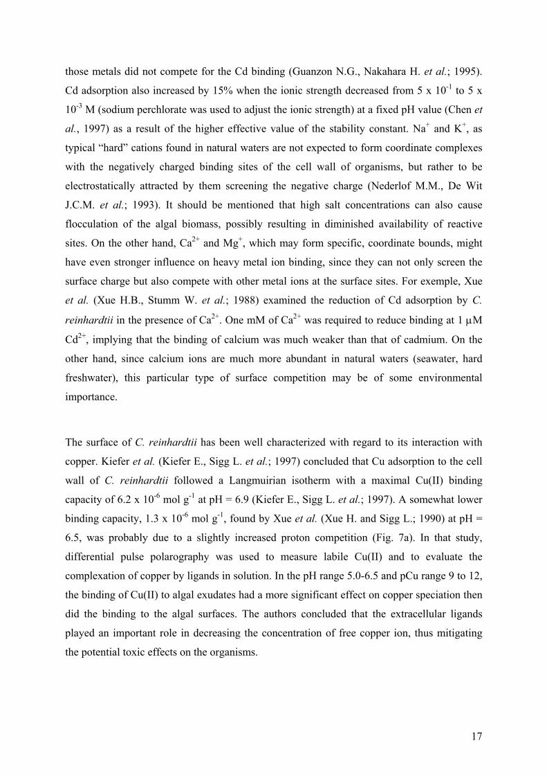
The surface of C. reinhardtii has been well characterized with regard to its interaction with
copper. Kiefer et al. (Kiefer E., Sigg L. et al.; 1997) concluded that Cu adsorption to the cell
wall of C. reinhardtii followed a Langmuirian isotherm with a maximal Cu(II) binding
capacity of 6.2 x 10⁻⁶ mol g-1 at pH = 6.9 (Kiefer E., Sigg L. et al.; 1997). A somewhat lower
binding capacity, 1.3 x 10⁻⁶ mol g-1, found by Xue et al. (Xue H. and Sigg L.; 1990) at pH =
6.5, was probably due to a slightly increased proton competition (Fig. 7a). In that study,
differential pulse polarography was used to measure labile Cu(II) and to evaluate the
complexation of copper by ligands in solution. In the pH range 5.0-6.5 and pCu range 9 to 12,
the binding of Cu(II) to algal exudates had a more significant effect on copper speciation then
did the binding to the algal surfaces. The authors concluded that the extracellular ligands
played an important role in decreasing the concentration of free copper ion, thus mitigating
the potential toxic effects on the organisms.
18
In another study using C. reinhardtii, uranium (U) adsorption to the cell wall was thought to
reach local equilibrium within minutes (<15 min) (Fortin C., Dutel L. et al.; 2004). Uranium
adsorption was correlated with the free uranyl ion concentration in solution but did not appear
to be a saturable process (Fig. 7b). In the both studies, there was no clear evidence from the
experimental points that the saturation was reached at the relatively low metal concentrations
that were used.
Figure 7: a) Cu(II) adsorption to the surface of Chlamydomonas reinhardtii at pH = 6.5 (Xue H. and Sigg L.; 1990). b) Uranium adsorption after 30 min of exposure at various measured total uranium concentrations (Fortin C., Dutel L. et al.; 2004). Lines represented Langmuir isotherms.
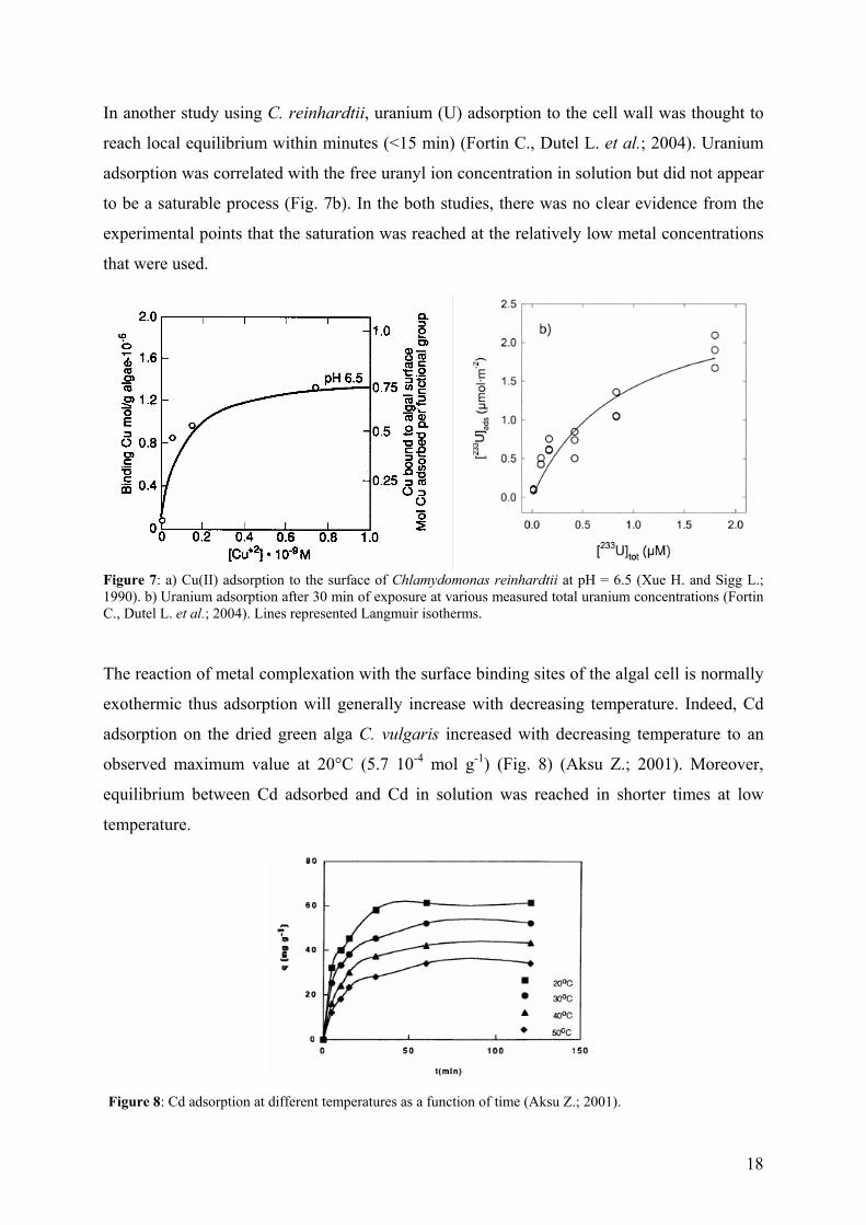
The reaction of metal complexation with the surface binding sites of the algal cell is normally
exothermic thus adsorption will generally increase with decreasing temperature. Indeed, Cd
adsorption on the dried green alga C. vulgaris increased with decreasing temperature to an
observed maximum value at 20°C (5.7 10-4 mol g-1) (Fig. 8) (Aksu Z.; 2001). Moreover,
equilibrium between Cd adsorbed and Cd in solution was reached in shorter times at low
temperature.
Figure 8: Cd adsorption at different temperatures as a function of time (Aksu Z.; 2001).
19
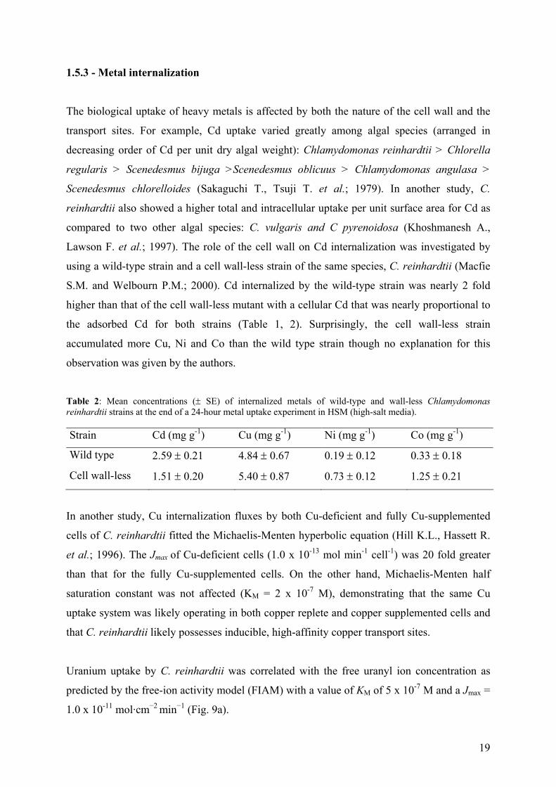
1.5.3 - Metal internalization
The biological uptake of heavy metals is affected by both the nature of the cell wall and the
transport sites. For example, Cd uptake varied greatly among algal species (arranged in
decreasing order of Cd per unit dry algal weight): Chlamydomonas reinhardtii > Chlorella
regularis > Scenedesmus bijuga >Scenedesmus oblicuus > Chlamydomonas angulasa >
Scenedesmus chlorelloides (Sakaguchi T., Tsuji T. et al.; 1979). In another study, C.
reinhardtii also showed a higher total and intracellular uptake per unit surface area for Cd as
compared to two other algal species: C. vulgaris and C pyrenoidosa (Khoshmanesh A.,
Lawson F. et al.; 1997). The role of the cell wall on Cd internalization was investigated by
using a wild-type strain and a cell wall-less strain of the same species, C. reinhardtii (Macfie
S.M. and Welbourn P.M.; 2000). Cd internalized by the wild-type strain was nearly 2 fold
higher than that of the cell wall-less mutant with a cellular Cd that was nearly proportional to
the adsorbed Cd for both strains (Table 1, 2). Surprisingly, the cell wall-less strain
accumulated more Cu, Ni and Co than the wild type strain though no explanation for this
observation was given by the authors.
Table 2: Mean concentrations (± SE) of internalized metals of wild-type and wall-less Chlamydomonas reinhardtii strains at the end of a 24-hour metal uptake experiment in HSM (high-salt media). Strain Cd (mg g-1) Cu (mg g-1) Ni (mg g-1) Co (mg g-1)
Wild type 2.59 ± 0.21 4.84 ± 0.67 0.19 ± 0.12 0.33 ± 0.18
Cell wall-less 1.51 ± 0.20 5.40 ± 0.87 0.73 ± 0.12 1.25 ± 0.21
In another study, Cu internalization fluxes by both Cu-deficient and fully Cu-supplemented
cells of C. reinhardtii fitted the Michaelis-Menten hyperbolic equation (Hill K.L., Hassett R.
et al.; 1996). The Jmax of Cu-deficient cells (1.0 x 10⁻¹³ mol min-1 cell-1) was 20 fold greater
than that for the fully Cu-supplemented cells. On the other hand, Michaelis-Menten half
saturation constant was not affected (KM = 2 x 10⁻⁷ M), demonstrating that the same Cu
uptake system was likely operating in both copper replete and copper supplemented cells and
that C. reinhardtii likely possesses inducible, high-affinity copper transport sites.
Uranium uptake by C. reinhardtii was correlated with the free uranyl ion concentration as
predicted by the free-ion activity model (FIAM) with a value of KM of 5 x 10⁻⁷ M and a Jmax =
1.0 x 10⁻¹¹ mol·cm−2 min−1 (Fig. 9a).
20
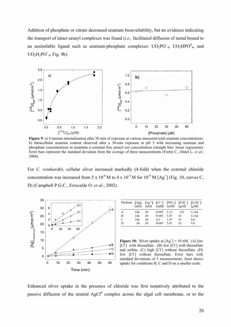
Addition of phosphate or citrate decreased uranium bioavailability, but no evidence indicating
the transport of intact uranyl complexes was found (i.e., facilitated diffusion of metal bound to
an assimilable ligand such as uranium-phosphate complexes: UO2PO−4, UO2HPO0
4, and
UO2H2PO+4, Fig. 9b).
Figure 9: a) Uranium internalization after 30 min of exposure at various measured total uranium concentrations. b) Intracellular uranium content observed after a 30-min exposure at pH 5 with increasing uranium and phosphate concentrations to maintain a constant free uranyl ion concentration (straight line: linear regression). Error bars represent the standard deviation from the average of three measurements (Fortin C., Dutel L. et al.; 2004).
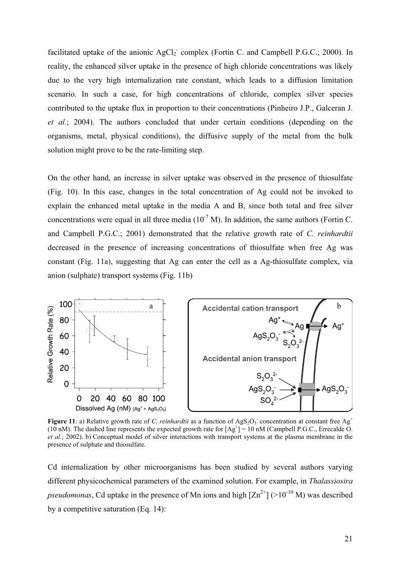
For C. reinhardtii, cellular silver increased markedly (4-fold) when the external chloride
concentration was increased from 5 x 10⁻⁶ M to 4 x 10⁻³ M for 10-8 M [Ag+] (Fig. 10, curves C,
D) (Campbell P.G.C., Errecalde O. et al.; 2002).
Figure 10: Silver uptake at [Ag+] = 10 nM. (A) low [Cl-] with thiosulfate . (B) low [Cl-] with thiosulfate and sulfate. (C) high [Cl-] without thiosulfate. (D) low [Cl-] without thiosulfate. Error bars with standard deviations of 3 measurements. Inset shows uptake for conditions B, C and D on a smaller scale.
Enhanced silver uptake in the presence of chloride was first tentatively attributed to the
passive diffusion of the neutral AgCl0 complex across the algal cell membrane, or to the
b)
[233U]tot (µM)
21
facilitated uptake of the anionic AgCl2- complex (Fortin C. and Campbell P.G.C.; 2000). In
reality, the enhanced silver uptake in the presence of high chloride concentrations was likely
due to the very high internalization rate constant, which leads to a diffusion limitation
scenario. In such a case, for high concentrations of chloride, complex silver species
contributed to the uptake flux in proportion to their concentrations (Pinheiro J.P., Galceran J.
et al.; 2004). The authors concluded that under certain conditions (depending on the
organisms, metal, physical conditions), the diffusive supply of the metal from the bulk
solution might prove to be the rate-limiting step.
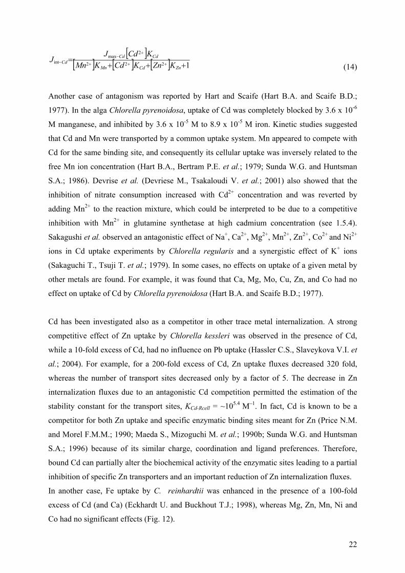
On the other hand, an increase in silver uptake was observed in the presence of thiosulfate
(Fig. 10). In this case, changes in the total concentration of Ag could not be invoked to
explain the enhanced metal uptake in the media A and B, since both total and free silver
concentrations were equal in all three media (10-7 M). In addition, the same authors (Fortin C.
and Campbell P.G.C.; 2001) demonstrated that the relative growth rate of C. reinhardtii
decreased in the presence of increasing concentrations of thiosulfate when free Ag was
constant (Fig. 11a), suggesting that Ag can enter the cell as a Ag-thiosulfate complex, via
anion (sulphate) transport systems (Fig. 11b)
Figure 11: a) Relative growth rate of C. reinhardtii as a function of AgS2O3
- concentration at constant free Ag+ (10 nM). The dashed line represents the expected growth rate for [Ag+] = 10 nM (Campbell P.G.C., Errecalde O. et al.; 2002). b) Conceptual model of silver interactions with transport systems at the plasma membrane in the presence of sulphate and thiosulfate.
Cd internalization by other microorganisms has been studied by several authors varying
different physicochemical parameters of the examined solution. For example, in Thalassiosira
pseudomonas, Cd uptake in the presence of Mn ions and high [Zn2+] (>10-10 M) was described
by a competitive saturation (Eq. 14):
a b
22
[ ][ ] [ ] [ ] 1222
2max
int +++=
+++
+−
−ZnCdMn
CdCdCd KZnKCdKMn
KCdJJ
(14)
Another case of antagonism was reported by Hart and Scaife (Hart B.A. and Scaife B.D.;
1977). In the alga Chlorella pyrenoidosa, uptake of Cd was completely blocked by 3.6 x 10⁻⁶
M manganese, and inhibited by 3.6 x 10⁻⁵ M to 8.9 x 10⁻⁵ M iron. Kinetic studies suggested
that Cd and Mn were transported by a common uptake system. Mn appeared to compete with
Cd for the same binding site, and consequently its cellular uptake was inversely related to the
free Mn ion concentration (Hart B.A., Bertram P.E. et al.; 1979; Sunda W.G. and Huntsman
S.A.; 1986). Devrise et al. (Devriese M., Tsakaloudi V. et al.; 2001) also showed that the
inhibition of nitrate consumption increased with Cd2+ concentration and was reverted by
adding Mn2+ to the reaction mixture, which could be interpreted to be due to a competitive
inhibition with Mn2+ in glutamine synthetase at high cadmium concentration (see 1.5.4).
Sakagushi et al. observed an antagonistic effect of Na+, Ca2+, Mg2+, Mn2+, Zn2+, Co2+ and Ni2+
ions in Cd uptake experiments by Chlorella regularis and a synergistic effect of K+ ions
(Sakaguchi T., Tsuji T. et al.; 1979). In some cases, no effects on uptake of a given metal by
other metals are found. For example, it was found that Ca, Mg, Mo, Cu, Zn, and Co had no
effect on uptake of Cd by Chlorella pyrenoidosa (Hart B.A. and Scaife B.D.; 1977).
Cd has been investigated also as a competitor in other trace metal internalization. A strong
competitive effect of Zn uptake by Chlorella kessleri was observed in the presence of Cd,
while a 10-fold excess of Cd, had no influence on Pb uptake (Hassler C.S., Slaveykova V.I. et
al.; 2004). For example, for a 200-fold excess of Cd, Zn uptake fluxes decreased 320 fold,
whereas the number of transport sites decreased only by a factor of 5. The decrease in Zn
internalization fluxes due to an antagonistic Cd competition permitted the estimation of the
stability constant for the transport sites, KCd-Rcell = ~105.4 M–1. In fact, Cd is known to be a
competitor for both Zn uptake and specific enzymatic binding sites meant for Zn (Price N.M.
and Morel F.M.M.; 1990; Maeda S., Mizoguchi M. et al.; 1990b; Sunda W.G. and Huntsman
S.A.; 1996) because of its similar charge, coordination and ligand preferences. Therefore,
bound Cd can partially alter the biochemical activity of the enzymatic sites leading to a partial
inhibition of specific Zn transporters and an important reduction of Zn internalization fluxes.
In another case, Fe uptake by C. reinhardtii was enhanced in the presence of a 100-fold
excess of Cd (and Ca) (Eckhardt U. and Buckhout T.J.; 1998), whereas Mg, Zn, Mn, Ni and
Co had no significant effects (Fig. 12).
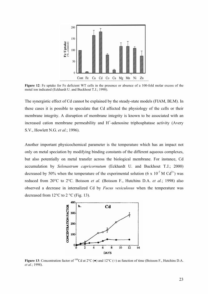
23
Figure 12: Fe uptake for Fe deficient WT cells in the presence or absence of a 100-fold molar excess of the metal ion indicated (Eckhardt U. and Buckhout T.J.; 1998).
The synergistic effect of Cd cannot be explained by the steady-state models (FIAM, BLM). In
these cases it is possible to speculate that Cd affected the physiology of the cells or their
membrane integrity. A disruption of membrane integrity is known to be associated with an
increased cation membrane permeability and H+-adenosine triphosphatase activity (Avery
S.V., Howlett N.G. et al.; 1996).
Another important physicochemical parameter is the temperature which has an impact not
only on metal speciation by modifying binding constants of the different aqueous complexes,
but also potentially on metal transfer across the biological membrane. For instance, Cd
accumulation by Selenastrum capricornutum (Eckhardt U. and Buckhout T.J.; 2000)
decreased by 50% when the temperature of the experimental solution (6 x 10⁻⁷ M Cd2+) was
reduced from 20°C to 2°C. Boisson et al. (Boisson F., Hutchins D.A. et al.; 1998) also
observed a decrease in internalized Cd by Fucus vesiculosus when the temperature was
decreased from 12°C to 2 °C (Fig. 13).
Figure 13: Concentration factor of 109Cd at 2°C () and 12°C () as function of time (Boisson F., Hutchins D.A. et al.; 1998).
24
Changes in temperature would affect membrane fluidity and cellular bioenergetics, both of
which could conceivably affect the carrier-mediated transport of Cd (i.e., kint).
Via its influence on cadmium speciation, pH can influence on ion uptake by microorganisms.
In this case, the effect of pH is due both to competition of hydrogen ions for binding with
ligands and to a competition of the protons for metal binding sites For example, as the pH is
lowered, metal ions are liberated from different complexes, increasing metal activities in
solution and consequently potentially increasing the toxicities. Cultures of Chlorella exposed
to 4.5 x 10⁻⁶ M accumulated twice as much cadmium as the pH was lowered from 8.0 to 7.0,
suggesting that Cd ions were liberated from different complexes (i.e. CdCO3), thus increasing
the activity of Cd2+ in solution (Hart B.A. and Scaife B.D.; 1977).
1.5.4 - Effects of metals on growth and assimilatory pathways
The study of microorganisms such as algae can provide some indications of the toxic effects
of heavy metals on general metabolic processes, in addition to providing an indication of
pollution levels in the environment. Authors in previous studies have constantly found that
high metal concentrations can inhibit algal chlorophyll production and growth or cause
mortality. For example, Co, which normally is an essential component for all living organisms
(synthesis of vitamin B12), partially inhibited growth of C. reinhardtii at a total Co
concentration of 1.7-3.4 x 10⁻⁴ M, while a concentration of 5 x 10⁻⁴ M prevented growth
completely (Lustigman B., Lee L.H. and Khalil A.; 1995). At these concentrations, the major
effect of Co is considered to be the inhibition of chlorophyll synthesis: cobalt ions competed
with iron for active sites on the chlorophyll molecule. Concentrations of 5 x 10⁻⁵-10-4 M of Cd
and Cu also reduced chlorophyll synthesis (Prasad M.N., Drej K. et al.; 1998). Lustigman et
al. (Lustigman B., Lee L.H. et al.; 2000) observed that a concentration of 0.9-2.3 x 10⁻⁴ M of
thallium not only reduced chlorophyll production and growth of C. reinhardtii, but also
influenced the formation of aggregates and cellular debris.
Heavy metals can also compete with essential metals that have crucial roles in the activity of
certain enzymes in metabolic processes. For example, nitrate assimilation in Chlamydomonas
involves three basic steps: (i) the transport across the cytoplasmic membrane, which is
mediated by a specific transporter; (ii) the reduction of nitrate to ammonium, which is
catalyzed by the successive action of nitrate reductase and nitrite reductase (NiR); and (iii) the
25
incorporation of ammonium into carbon skeletons, which is catalyzed by glutamine
synthetase (GS). Concentrations of 10-4 M Cd and Cu separately inhibited nitrate uptake by
the cells (Devriese M., Tsakaloudi V. et al.; 2001), however, after a 50 minute exposure to
metals, nitrate consumption was restored, indicating the development of a detoxification
mechanism by the cells. In this context, is important to mention that these metals have been
shown to induce phytochelatin synthesis in photosynthetic organisms (Rauser W.E.; 1995).
NiR activity was not affected by 10-4 M Cd over 2 days. On the other hand, in the presence of
Cd, GS showed significant inhibition (45%) after 7 hours of incubation. GS activity decreased
quickly when Cd2+ concentrations increased, but this inhibition was suppressed by adding
Mn2+ to the reaction mixture (Devriese M., Tsakaloudi V. et al.; 2001). This result could be
explained on the basis of a competitive inhibition of GS activity by Cd, which in
Chlamydomonas may accumulate in the chloroplast and cytosol (Nagel K., Adelmeier U. et
al.; 1996). The same concentration of Cd did not affect OASTL (O-acetyl-L-
serine(thyol)liase) enzyme activity, which catalyzed the last step of L-cysteine biosynthesis in
the sulphur assimilatory pathway.
Cd toxicity has been tested using different microorganisms. For example, Cain and Allen
(Cain J.R. and Allen R.K.; 1980) investigated the role of the cell wall on Cd (and Hg)
tolerance in C. reinhardtii. They found no difference in toxicity between the wild type and the
cell wall-less mutant: growth was reduced 98% for 4.4 x 10⁻⁶ M and 23% for 4.4 x 10⁻⁵ M Cd
in solution. Using the same strains Macfie et al. (Macfie S.M., Tarmohamed Y. et al.; 1994)
demonstrated that H+ in solution provided some protection against toxic metal ions to algae. Comparing the EC30 values (Table 3), they demonstrated that the reduction of metal toxicity
as a function of solution pH varied among the metals but did not vary among the algal strains.
The differential tolerance was more marked for Co and Cu than for Cd and Ni suggesting that
metal/cell interactions probably are associated with a number of factors including differential
binding site affinities of the metal relative to the cell wall as well as differential uptake
kinetics (Macfie S.M., Tarmohamed Y. et al.; 1994; Macfie S.M. and Welbourn P.M.; 2000).
At higher external metal concentrations, the cell wall offered no further protection, indicating
that the metal-binding sites may have been saturated.
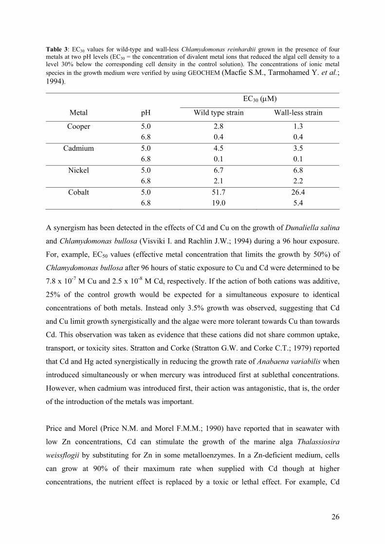
26
Table 3: EC30 values for wild-type and wall-less Chlamydomonas reinhardtii grown in the presence of four metals at two pH levels (EC30 = the concentration of divalent metal ions that reduced the algal cell density to a level 30% below the corresponding cell density in the control solution). The concentrations of ionic metal species in the growth medium were verified by using GEOCHEM (Macfie S.M., Tarmohamed Y. et al.; 1994).
EC30 (µM)
Metal pH Wild type strain Wall-less strain
Cooper 5.0 2.8 1.3 6.8 0.4 0.4
Cadmium 5.0 4.5 3.5 6.8 0.1 0.1
Nickel 5.0 6.7 6.8 6.8 2.1 2.2
Cobalt 5.0 51.7 26.4 6.8 19.0 5.4
A synergism has been detected in the effects of Cd and Cu on the growth of Dunaliella salina
and Chlamydomonas bullosa (Visviki I. and Rachlin J.W.; 1994) during a 96 hour exposure.
For, example, EC50 values (effective metal concentration that limits the growth by 50%) of
Chlamydomonas bullosa after 96 hours of static exposure to Cu and Cd were determined to be
7.8 x 10⁻⁷ M Cu and 2.5 x 10⁻⁸ M Cd, respectively. If the action of both cations was additive,
25% of the control growth would be expected for a simultaneous exposure to identical
concentrations of both metals. Instead only 3.5% growth was observed, suggesting that Cd
and Cu limit growth synergistically and the algae were more tolerant towards Cu than towards
Cd. This observation was taken as evidence that these cations did not share common uptake,
transport, or toxicity sites. Stratton and Corke (Stratton G.W. and Corke C.T.; 1979) reported
that Cd and Hg acted synergistically in reducing the growth rate of Anabaena variabilis when
introduced simultaneously or when mercury was introduced first at sublethal concentrations.
However, when cadmium was introduced first, their action was antagonistic, that is, the order
of the introduction of the metals was important.
Price and Morel (Price N.M. and Morel F.M.M.; 1990) have reported that in seawater with
low Zn concentrations, Cd can stimulate the growth of the marine alga Thalassiosira
weissflogii by substituting for Zn in some metalloenzymes. In a Zn-deficient medium, cells
can grow at 90% of their maximum rate when supplied with Cd though at higher
concentrations, the nutrient effect is replaced by a toxic or lethal effect. For example, Cd
27
uptake rates of Thalassiosira pseudonana decreased 80-fold when free Zn concentration was
increased from 10-11.5 to 10-10 M (Sunda W.G. and Huntsman S.A.; 2000).
A detailed study of the combined effects of pH and cadmium on algal growth was performed
by Rachlin and Grosso (Rachlin J.W. and Grosso A.; 1991). Taking the growth rate of
Chlorella vulgaris as 100% at pH 6.9, they found a reduction to 50% when 8.9 x 10⁻⁷ M of Cd
was added. A reduction of the growth rate (from 100% to 56%) was also found when the pH
was lowered to pH 5. When Cd was added at pH 5, the effects of pH and Cd were partially
additive and the relative growth rate was 44%. Furthermore, a reduction was found when the
pH was raised above 8.3, but at these high pH values, Cd did not produce any additional
effects. This study demonstrated the difficulty of interpreting data on complex environmental
systems.
Drastic pH changes can also influence the physiology and structure of proteins, which
consequently can influence metal bioaccumulation as a result of probable conformational
changes of the metal binding sites. Although ultrastructure is an insensitive indicator of stress,
it can provide some insight on the mechanisms of toxicity and tolerance (Rachlin J.W. and
Grosso A.; 1991; Rachlin J.W. and Grosso A.; 1993). For example, when Chlamydomonas
applanata cultures were exposed to pH 4.4, palmelloid colonies consisting of several
nonmotile vegetative cells surrounded by a common wall, have been observed (Visviki I. and
Santikul D.; 2000). Moreover, single cells of Chlamydomonas in low pH media, tended to be
smaller than controls (Bold H.C. and Wynne M.J.; 1978). In fact, the expulsion of hydrogen
ions from the cytosol to the medium requires considerable expenditure of energy that could
otherwise be used for cellular growth. Similar results have been reported for Gloeochrysis
turfosa exposed to extremely low pHs of 1.3-1.5 (Hargreaves J.W. and Whitton B.A.; 1976).
Cd toxicity tests were performed by Laegreid et al. involving lake water with high and low
organic matter contents. The results for the former were in accord with the FIAM: the
(complexing) organic matter greatly decreased cadmium toxicity (Laegreid M., Alstad J. et
al.; 1983), while for the latter the results contradicted the FIAM: organic matter had the effect
of raising the toxicity of the added cadmium to levels higher than expected from the FIAM. A
tentative explanation, that the organic matter had a large low-molecular-weight fraction which
promoted cadmium uptake was proposed. Simkiss suggested that the charged structure and
relative lipophilicity of organically complexed metals and organic contaminants may also
28
alter rates of transport across the lipid membranes of cells (Simkiss K. and Taylor M.G.;
1989). Similarly, a short-term study involving Selenastrum capricornutum (Errecalde O.,
Seidl M. et al.; 1998) demonstrated that the bioavailability of Cd increased in the order: free
Cd ions < Cd-NTA complexes < Cd-citrate complexes in media containing identical [Cd2+].
This result also contrasted with the FIAM and has not been satisfactorily explained. On the
other hand, metabolites such as alanine decreased the relative growth rate of the green algae,
P. subcapitata (Fig. 14a: dotted and solid lines) (Campbell P.G.C., Errecalde O. et al.; 2002).
The authors suggested that Cd inhibited growth of P. subcapitata by binding to the algal
surface and that this binding involved the formation of a ternary surface complex, L–M–Xcell.
In fact, for such species to contribute to the biological response, either Xcell must represent a
metal-sensitive site at the origin of the biological response (i.e. the metal exerts its biological
effect without crossing the plasma membrane), or L–M must be transported intact across the
plasma membrane. Since no increase in Cd uptake in the presence of alanine was observed
(Fig. 14b), the authors concluded that Xcell must represent a metal-sensitive site.
Figure 14: a) Relative growth rate of P. subcapitata as a function of the free Cd concentration in FRAQNTA and FRAQALA media (growth rate of cadmium-free controls100%). b) Intracellular cadmium taken up by P. subcapitata as a function of time, [Cd2+] = 0.5 mM. () medium buffered by NTA (dashed line); (♦) medium buffered by 0.5 mM L-alanine (dotted line); () medium buffered by 0.5 mM D-alanine (solid line).
The toxicity of heavy metals in solution also depends on the concentration of inorganic
complexes with ligands such as Cl-, CO32-, SO4
2- etc. because they play an important role in
metal speciation. It has been suggested that in some limited cases, ternary complexes may
form at the cell surface prior to metal uptake (Wilkinson K.J., Campbell P.G.C. et al.; 1990;
Campbell P.G.C.; 1995). In such a case, it is possible that the presence of an inorganic ion
29
could increase either the equilibrium adsorption or the reaction rate with sensitive sites.
Wilkinson et al. (Wilkinson K.J. and Campbell P.G.C.; 1993) suggested that ternary Al
surface complexes were present as an equilibrium species contributing to biological response.
1.5.5 - Morphologic changes in the presence of metals
Metal stress modifies not only the activity of the cell but also a variety of cellular parameters
including total cell volume and the nature of the cell wall, vacuoles, lipids, polyphosphate
bodies and periplasmalemmal space. Growth is generally more sensitive to heavy metal stress
than are ultrastructural changes (Visviki I. and Rachlin J.W.; 1992). Changes in ultrastructure
of the cell are important insofar as they increase the understanding of the pathways of metal
toxicity and of algal defences to cation stress. For example, the cell wall volume of
Chlamydomonas busolla (the cell wall is composed of three layers, as in C. reinhardtii) is
shown to decrease in the presence of cadmium (0.025 µM) but the periplasmalemmal space
did not change (Visviki I. and Rachlin J.W.; 1994). This result indicated an absence of
increased osmotic pressure within the cell. The reduction of cell wall volume can be the result
of mechanical stress and may alter the organism’s resistance to cation exposure. The authors
suggested that the increased cellular volume was due to the inhibition of normal cell division
rather than to changes in the cell wall or cell membrane permeability. At the same time the
cells exhibited a 72% increase in the relative vacuolar volume and the development of some
membranous organelles usually found within vacuoles. This was in correlation with the
finding that in higher plants (Zenk M.H.; 1996), phytochelatin-metal complexes are deposited
in vacuoles. On the other hand, a reduction of the cell volume of Chlamydomonas reinhardtii
in the presence of higher total concentrations of Cd and Cu (50-100 µM) was observed
(Prasad M.N., Drej K. et al.; 1998) (Cu being more toxic than Cd), possibly due to the
metabolic cost of metal detoxification (Lawrence S.G., Holoka M.H. et al.; 1989). In another
study, when exposed to sublethal concentrations of cadmium and copper, Chlamydomonas
busolla showed a decrease in its active chloroplast volume but not in the overall chloroplast
volume. This reduction could be an indirect effect of the inhibition of normal cell division.
1.5.6 - Genetic mutation in response to heavy metals
The presence of heavy metals can also induce genetic changes to Chlamydomonas. Collard
and Matagne (Collard J.M. and Matagne R.F.; 1990) isolated resistant clones of
30
Chlamydomonas on TAP agar medium containing 200 µM to 400 µM cadmium, but they
arrived at the conclusion that Cd-resistant cells were not induced by the metal but pre-existed
in the initial population. They also isolated two mutants conferring resistance to cadmium at
concentrations of 600 µM to 900 µM, which were also seen in cell wall-deficient strains,
indicating that they did not involve a modification of the structure or composition of the cell
wall. Hu et. al. (Hu S., Lau K.W.K. et al.; 2001) generated several Cd-resistant mutants (Cd
concentrations in the range of 50-125 µM) and demonstrated that the level of phytochelatin
production was proportional to their capacity for Cd accumulation. One type of mutant
produced a high level of HMW (high-molecular-weight) complexes and also produced and
maintained a higher level of cysteine upon Cd challenge. Various mechanisms allowing the
tolerance of cadmium could be hypothesized, such as decreased uptake, overproduction of
metal-binding ligands, increased extracellular release of organic chelates in solution or finally
sequestration of metal in intracellular insoluble complexes.
For a closer characterization of the genes involved in multiple stress responses, Hanikenne et
al. (Hanikenne M., Matagne R.F. et al.; 2001) used insertional mutagenesis to produce
mutants of this alga which to the contrary, were hypersensitive to Cd and oxidative stress.
They showed that Cd mutants sharply differed in their sensitivities to metal. The minimal
inhibitory Cd concentration ranged from 200 µM to 400 µM in TAP media. Some of them
were exclusively cadmium-sensitive and others presented cross sensitivity with several of the
following agents: Cd, Pb, Cu, Hg, H2O2 and light. Their sensitivity to a broad range of
chemical and physical agents suggested that the disrupted genes could be involved in multiple
stress responses. Such mutants, who have never been isolated in plants, could be of great
interest on account of their pleiotropic character. One of the inactivated genes could also be
involved in the metabolism of phytochelatin, which sequesters the metals by forming an
intracellular complex. The authors concluded that further work would need to be done with
respect to cloning and characterization of the corresponding genes.
Hu et al. (Hu Z., Wong Y.-S. et al.; 2000) transformed a Chlamydomonas reinhardtii strain
(cw10, mt+) with a plasmid containing the metallothionein-like gene (p124MT) from Festuca
ruba on TAP medium. The metal tolerance of the transgenic algae strains (Tr 20B1, Tr 20B2,
Tr 10A3) was compared with the wild type strain in the presence of 650 mM cadmium in agar
medium (Fig. 15). The results showed that the transgenic algae grow much faster than do the
31
wild type strains and that they could also resist higher concentrations of toxic metals. These
results also showed that the expression of foreign metallothionein genes in Chlamydomonas
reinhardtii increased its tolerance to toxic concentrations of cadmium and copper.
Figure 15: Comparison of cadmium tolerance between three transgenic alga strains (Tr 20B1,Tr 20B2,Tr 10A3) and a wild type strain (cc-849).
Siripornadulsil et al. (Siripornadulsil S., Traina S. et al.; 2002) separately introduced two
other genes into the nuclear genome of Chlamydomonas strain (CC-425): HAL2, which
regulates cysteine synthesis, and P5CS gene encoding pyrroline-5-carboxylate, which
regulates proline synthesis in higher plants. They studied growth and binding capacity of cells
expressing these two genes in comparison with a wild-type strain in the presence of a toxic
concentration of cadmium that induces PC synthesis (5 x 10⁻⁵ M) and a low concentration (5 x
10-6 M) that does not induce PC synthesis (Table 4). These authors arrived at the conclusion
that the expression of these genes affected the metal-binding capacity and tolerance of the
algae.
Table 4: Growth and cadmium-binding capacity of cells expressing the HAL2 gene and P5CS gene compared to growth and binding capacity of the wild-type of Chlamydomonas reinhardtii (CC-425). 5 x 10⁻⁵ M Cd 5 x 10⁻⁶ M Cd
CC-425 (HAL2: cysteine) No difference Faster Growth
CC-425 (P5CS: proline) Faster No difference
CC-425 (HAL2: cysteine) 2.4-fold 1.5-fold Cd binding capacity
CC-425 (P5CS: proline) 1.9-fold No difference
The molecular basis of heavy metal uptake and transport in microorganisms and the
understanding of heavy metal detoxification still require elucidation. Cells expressing the
32
HAL2 gene (cysteine) grew faster than the wild type strain at 5 x 10⁻⁶ M Cd suggesting that
cysteine may play the role of complexing the metal at a certain extent. On the other hand, the
same cells at 50 mM Cd showed no difference in growth when compared to the wild type
strain, since at this Cd concentration, phytochelatins were produced.
1.6 - Implications of using algae in bioavailability studies
Exudate production. Algal communities affect trace metal chemistry in natural waters not
only by taking up the metal but also by producing extracellular organic matter with metal
complexing properties. High cell densities and lengthy test durations can result in changes in
chemical speciation, bioavailability and contaminant toxicity during testing. For instance,
laboratory experiments using a 72-h algal growth inhibition bioassay and Cu as a toxicant
underestimated Cu toxicity by a factor of 2 when Cu losses occurring over the 72-h test were
not considered (Simpson S.L., Roland M.G.E. et al.; 2003). A simple model used to predict
the toxicant concentration decline showed that for a toxicant whose concentration declines
exponentially to less than 5% of its original value within 36 h of a 72-h test, the inhibitory
concentration would be underestimated by a factor of 50. In the same study, Cu toxicity
decreased with the increasing initial cell density of the freshwater alga Chlorella sp. and the
temperate species Selenastrum capricornutum (102 to 105 cells mL-1). The authors’ results
indicated that reduced copper toxicity was due to: i) greater Cu adsorption resulting in
depletion of dissolved Cu in solution and ii) exudate production (for the higher cell densities).
These findings suggested that standard laboratory bioassays using 104 to 105 algal cells mL-1
might seriously underestimate metal toxicity in natural waters. The application of cytometry
(Franklin N.M., Stauber J.L. et al.; 2001) or short exposure times (Sunda W.G. and Huntsman
S.A.; 1996; Sunda W.G. and Huntsman S.A.; 1998) may be used to minimize the effect of
exudates on metal speciation.
It has been shown that the nature of exudates depends strongly on the algal species and on
stress factors. For example, anionic polysaccharides that bind metal ions are released by
Chlorella under conditions of high pH (Malis-Arad S. and McGowan R.E.; 1982). In another
study, NaCl induced carbohydrate production more than protein production (Fig. 16) by three
green algae, Scenedesmus quadricauda, Chlorella kessleri, and Raphidocelis subcapitata
(Marsalek B. and Rojickova R.; 1996). The lowest amounts of carbohydrates were produced
during Cd exposure, while the same stress factor led to the highest excretion of proteins by
33
each of the algae. Production of exudates (mainly glutathione and other unidentified thiols) by
E. huxleyi (Leal M.F.C., Vasconcelos M.T.S.D. et al.; 1999), increased with both Cu
concentration and exposure time. For example, a 10 minute exposure to 1.3 x 10⁻⁷ M
dissolved Cu, increased by ~42% the amount of exudates in comparison with what was
observed in pseudoequilibrium (Vasconcelos M.T.S.D. and Leal M.F.C.; 2001).
Vasconcelos et al. (Vasconcelos M.T.S.D., Leal M.F.C. et al.; 2002) demonstrated that a
specific type of algal exudate may favor or inhibit the growth of other algal species. The final
yield of E. huxleyi algae improved in the presence of exudates (glutathione-like compounds)
produced by macroalgae Enteromorpha, but was inhibited by the relatively high concentration
of the exudates (cysteine-like compound) of the diatom P. tricornutum. In fact, additions of
synthetic cysteine and glutathione to E. huxleyi cultures caused changes in growth similar to
those caused by exudates. The mechanisms responsible are still unknown and deserve further
investigation.
Figure 16: The amount of total extracellular proteins (open box) and carbohydrates (filled box) in the medium after 48 hours of cultivation of alga under stress (Marsalek B. and Rojickova R.; 1996).

34
Influence of pH buffers on biological response. Cultures of green algae in unbuffered
synthetic media may undergo pH changes during growth due mainly to the removal of
dissolved CO2 during photosynthesis. As mentioned above, trace metal speciation and its
bioavailability to microorganisms are markedly influenced by pH changes. It is thus essential
to maintain constant the pH in algal culture and exposure media. A control of pH can be
achieved by bubbling CO2 to the media or by adding pH buffers to maintain constant the pH.
Typical buffers maintain pH values close to 7.0 to 8.0 (e.g., phosphate, tris-(hydroxymethyl)-
aminomethane (TRIS)), but may have limited application due to their ability to complex trace
metals or induce toxicity. For instance, phosphates are known to precipitate many polyvalent
cations and they can be a metabolite or an inhibitor in many systems (Good N.E., Winget
G.D. et al.; 1966). For example, increasing phosphate concentrations from 0.54 to 0.84 µg
mL-1, reduced Cd uptake to Chlorella vulgaris by 48% (Khummongkol D., Canterford G.S. et
al.; 1982).
The N-substituted aminosulfonic acids buffers were introduced by Good and collaborators in
1966 (Good N.E., Winget G.D. et al.; 1966). These buffers are increasingly used in biological
and chemical applications. They are zwitterionic buffers that have often been considered to be
more suitable than classical buffers, despite the fact that it is impossible to find a pH buffer
that does not have any physiological or other unrelated effect (Ferguson W.J.,
Braunschweiger K.I. et al.; 1980). Indeed, the toxic potential effects of the buffers (e.g.,
reduction of growth rate, role in exudate production) and their tendency to complex metals
must be carefully examined prior to use. For instance, the growth of Emiliana huxley was
reduced in the presence of 10-2 M N-2-hydroxyethylpiperazine-N’-3-propanesulfonic acid
(HEPPS) (Leal M.F.C., Vasconcelos M.T.S.D. et al.; 1999) or 10-2 M N-2-
Hydroxyethylpiperazine-N’-2-ethanesulfonic acid (HEPES) (Vasconcelos M.T.S.D. and Leal
M.F.C.; 2002). On the other hand, 2.5 10-2 M N-[2-hydroxyethyl]piperazine-N'-[2-
hydroxypropanesulfonic acid] (HEPPSO) favored the growth of Amphidinium carterae,
whereas 2.5 x 10⁻² M piperazine-N,N'-bis[2-hydroxypropanesulfonic acid] POPSO did not
affect growth when compared to a growth media without buffer (Vasconcelos M.T.S.D.,
Almeida C.M.R. et al.; 2000). Both buffers did not affect Cu uptake but significantly
decreased mean cell complexity and size. Moreover, both were able to complex Cu and
probably other metals. Lage et al., (Lage O.M., Vasconcelos M.T.S.D. et al.; 1996) showed
that 2.5 x 10⁻² M HEPES (pH 8.0) increased the toxicity of Cu(II) to the marine dinoflagellate
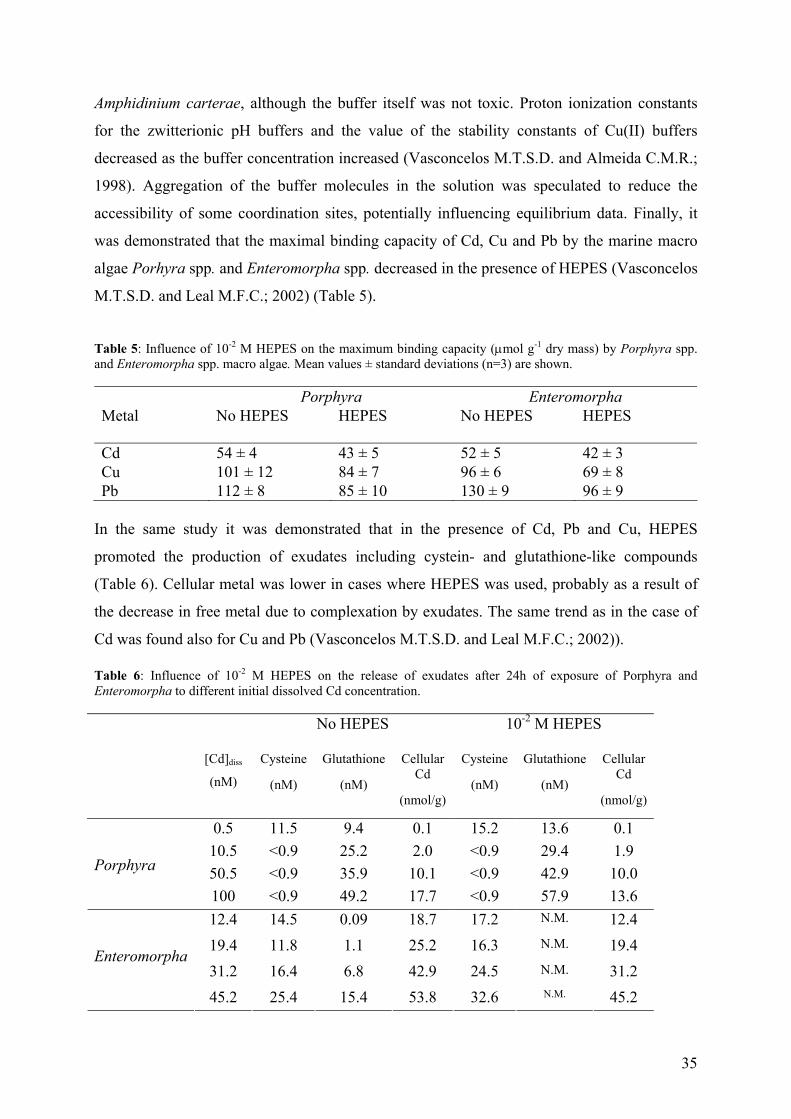
35
Amphidinium carterae, although the buffer itself was not toxic. Proton ionization constants
for the zwitterionic pH buffers and the value of the stability constants of Cu(II) buffers
decreased as the buffer concentration increased (Vasconcelos M.T.S.D. and Almeida C.M.R.;
1998). Aggregation of the buffer molecules in the solution was speculated to reduce the
accessibility of some coordination sites, potentially influencing equilibrium data. Finally, it
was demonstrated that the maximal binding capacity of Cd, Cu and Pb by the marine macro
algae Porhyra spp. and Enteromorpha spp. decreased in the presence of HEPES (Vasconcelos
M.T.S.D. and Leal M.F.C.; 2002) (Table 5).
Table 5: Influence of 10-2 M HEPES on the maximum binding capacity (µmol g-1 dry mass) by Porphyra spp. and Enteromorpha spp. macro algae. Mean values ± standard deviations (n=3) are shown. Porphyra Enteromorpha Metal No HEPES HEPES
No HEPES HEPES
Cd 54 ± 4 43 ± 5 52 ± 5 42 ± 3 Cu 101 ± 12 84 ± 7 96 ± 6 69 ± 8 Pb 112 ± 8 85 ± 10 130 ± 9 96 ± 9
In the same study it was demonstrated that in the presence of Cd, Pb and Cu, HEPES
promoted the production of exudates including cystein- and glutathione-like compounds
(Table 6). Cellular metal was lower in cases where HEPES was used, probably as a result of
the decrease in free metal due to complexation by exudates. The same trend as in the case of
Cd was found also for Cu and Pb (Vasconcelos M.T.S.D. and Leal M.F.C.; 2002)).
Table 6: Influence of 10-2 M HEPES on the release of exudates after 24h of exposure of Porphyra and Enteromorpha to different initial dissolved Cd concentration. No HEPES 10-2 M HEPES
[Cd]diss
(nM)
Cysteine
(nM)
Glutathione
(nM)
Cellular Cd
(nmol/g)
Cysteine
(nM)
Glutathione
(nM)
Cellular Cd
(nmol/g)
0.5 11.5 9.4 0.1 15.2 13.6 0.1 10.5 <0.9 25.2 2.0 <0.9 29.4 1.9 50.5 <0.9 35.9 10.1 <0.9 42.9 10.0
Porphyra
100 <0.9 49.2 17.7 <0.9 57.9 13.6 12.4 14.5 0.09 18.7 17.2 N.M. 12.4 19.4 11.8 1.1 25.2 16.3 N.M. 19.4 31.2 16.4 6.8 42.9 24.5 N.M. 31.2
Enteromorpha
45.2 25.4 15.4 53.8 32.6 N.M. 45.2
36
2 - OBJECTIVES
Metal ions pass across biological membranes, subsequently enhancing or inhibiting the
cellular activities of microorganisms. As a consequence of the exposure of microorganisms to
trace metal ions and, subsequently, their adsorption on the cellular surface (binding of metal
ions to transport sites at the cell surface) and the subsequent transport into the cell, two main
types of response can be observed: (i) short term responses on the order of milliseconds to
minutes (time scale <102 s) and (ii) long term responses (time scale >103 s) such as protein
synthesis, modification of reproductive or growth rate, enzyme inhibition, secretion,
contraction, motility, ultrastructural changes, gene mutation, etc. The latter responses involve
additional molecular interactions (Lauffenburgar D.A. and Linderman J.J.; 1993).
The work was focused on the examination, from a quantitative point of view, of a relationship
between the algal exposure to cadmium and its adsorption and short-term (25 min) biological
response(s). Physicochemical parameters such as the relevant ionic equilibria between the
solution and the surface of the organism, mass transport, etc. and the biological parameters
such as the internalization flux, exudate release, production of cell wall proteins, efflux were
determined and related.
Special attention was paid to equilibrium determinations of the Cd adsorbed to the cell wall
and the Cd dissolved in the bulk solution. The eventual consequences of the non-attainment
of equilibrium were examined within the framework of the FIAM. An estimation of the time
required for Cd adsorption to the cell wall of C. reinhadtii was performed using
experimentally determined thermodynamic constants and estimates based upon the theoretical
kinetic uptake rates (Eigen Wilkens mechanism; Section 4.1). A comparison of adsorbed Cd
and Cd uptake fluxes between the wild type strain and a cell wall-less mutant of C. reinhatdtii
was aimed at distinguishing between cell wall binding sites and Cd transport sites (Section 5,
6). A rigorous examination of Cd speciation was performed using advanced analytical
techniques (i.e. PLM, ASV) in order to verify if algal exudates were released during the short-
term bioaccumulation experiments. In addition, several experiments were designed to
characterize the exudates (Section 5). Since the separation between adsorbed and cellular Cd
was performed using an EDTA extraction, Cd desorption and efflux were discriminated and
an estimation of the formation and dissociation rate constants of Cd with the cell wall binding
sites of C. reinhardtii was also performed (Section 4.1).
37
Finally, an important goal of this research was to evaluate a link between the chemical
speciation of Cd in the bulk media and its corresponding uptake flux by C. reinhardtii by
changing different physicochemical parameters including pH and the presence of ligands
including organic matter, hardness cations and other trace metals. For example, the effect of
the Suwannee River fulvic acid on Cd internalization by C. reinhardtii was investigated in
defined media in which Cd2+ concentrations were measured by the ISE technique (Section 6).
Changes of the overall algal surface charge due to an eventual organic matter adsorption and
to low pH were carefully controlled by electrophoretic mobility measurements.
Since aquatic microorganisms (e.g., algae, bacteria) may be used to investigate the transfer
and uptake of toxic elements along the tropic chain and to monitor contamination levels of
different aquatic environments, a long-term goal associated with this research was to develop
a microorganism that is capable of translating a chemical stimulus into a measurable chemical
signal that is proportional to the bioavailable metal to which it is exposed. Thus, the work
presented here was designed to constitute a first step in obtaining a fundamental
understanding into the biouptake process so that this species could ultimately be used as
biosensor component.
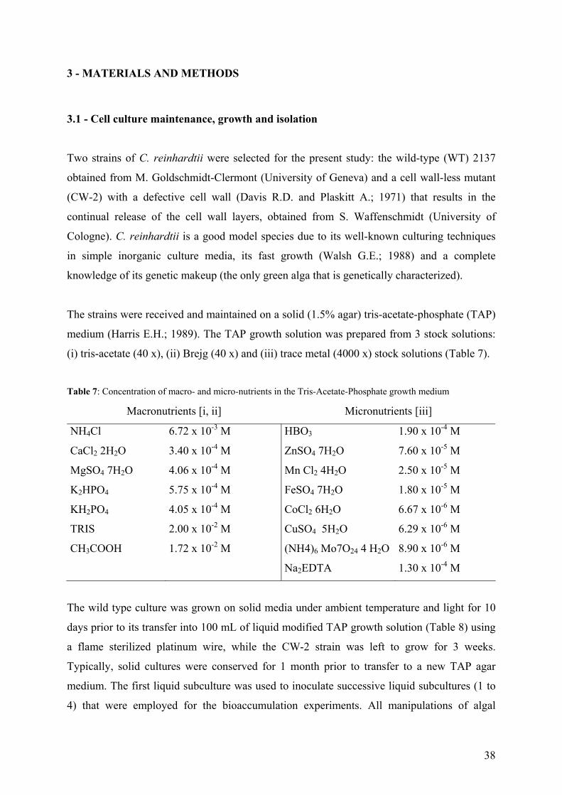
38
3 - MATERIALS AND METHODS
3.1 - Cell culture maintenance, growth and isolation
Two strains of C. reinhardtii were selected for the present study: the wild-type (WT) 2137
obtained from M. Goldschmidt-Clermont (University of Geneva) and a cell wall-less mutant
(CW-2) with a defective cell wall (Davis R.D. and Plaskitt A.; 1971) that results in the
continual release of the cell wall layers, obtained from S. Waffenschmidt (University of
Cologne). C. reinhardtii is a good model species due to its well-known culturing techniques
in simple inorganic culture media, its fast growth (Walsh G.E.; 1988) and a complete
knowledge of its genetic makeup (the only green alga that is genetically characterized).
The strains were received and maintained on a solid (1.5% agar) tris-acetate-phosphate (TAP)
medium (Harris E.H.; 1989). The TAP growth solution was prepared from 3 stock solutions:
(i) tris-acetate (40 x), (ii) Brejg (40 x) and (iii) trace metal (4000 x) stock solutions (Table 7).
Table 7: Concentration of macro- and micro-nutrients in the Tris-Acetate-Phosphate growth medium
Macronutrients [i, ii] Micronutrients [iii]
NH4Cl 6.72 x 10⁻³ M HBO3 1.90 x 10⁻⁴ M
CaCl2 2H2O 3.40 x 10⁻⁴ M ZnSO4 7H2O 7.60 x 10⁻⁵ M
MgSO4 7H2O 4.06 x 10⁻⁴ M Mn Cl2 4H2O 2.50 x 10⁻⁵ M
K2HPO4 5.75 x 10⁻⁴ M FeSO4 7H2O 1.80 x 10⁻⁵ M
KH2PO4 4.05 x 10⁻⁴ M CoCl2 6H2O 6.67 x 10⁻⁶ M
TRIS 2.00 x 10⁻² M CuSO4 5H2O 6.29 x 10⁻⁶ M
CH3COOH 1.72 x 10⁻² M (NH4)6 Mo7O24 4 H2O 8.90 x 10⁻⁶ M
Na2EDTA 1.30 x 10⁻⁴ M
The wild type culture was grown on solid media under ambient temperature and light for 10
days prior to its transfer into 100 mL of liquid modified TAP growth solution (Table 8) using
a flame sterilized platinum wire, while the CW-2 strain was left to grow for 3 weeks.
Typically, solid cultures were conserved for 1 month prior to transfer to a new TAP agar
medium. The first liquid subculture was used to inoculate successive liquid subcultures (1 to
4) that were employed for the bioaccumulation experiments. All manipulations of algal
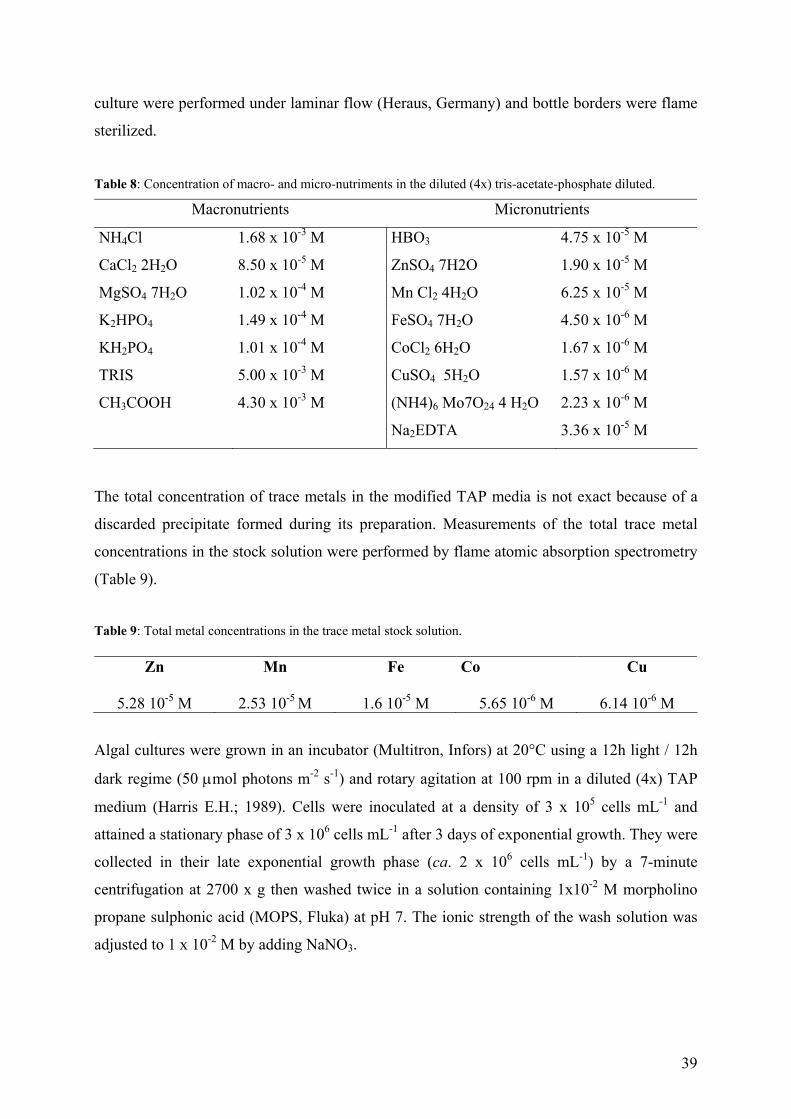
39
culture were performed under laminar flow (Heraus, Germany) and bottle borders were flame
sterilized.
Table 8: Concentration of macro- and micro-nutriments in the diluted (4x) tris-acetate-phosphate diluted.
Macronutrients Micronutrients
NH4Cl 1.68 x 10⁻³ M HBO3 4.75 x 10⁻⁵ M
CaCl2 2H2O 8.50 x 10⁻⁵ M ZnSO4 7H2O 1.90 x 10⁻⁵ M
MgSO4 7H2O 1.02 x 10⁻⁴ M Mn Cl2 4H2O 6.25 x 10⁻⁵ M
K2HPO4 1.49 x 10⁻⁴ M FeSO4 7H2O 4.50 x 10⁻⁶ M
KH2PO4 1.01 x 10⁻⁴ M CoCl2 6H2O 1.67 x 10⁻⁶ M
TRIS 5.00 x 10⁻³ M CuSO4 5H2O 1.57 x 10⁻⁶ M
CH3COOH 4.30 x 10⁻³ M (NH4)6 Mo7O24 4 H2O 2.23 x 10⁻⁶ M
Na2EDTA 3.36 x 10⁻⁵ M
The total concentration of trace metals in the modified TAP media is not exact because of a
discarded precipitate formed during its preparation. Measurements of the total trace metal
concentrations in the stock solution were performed by flame atomic absorption spectrometry
(Table 9).
Table 9: Total metal concentrations in the trace metal stock solution.
Zn Mn Fe Co Cu
5.28 10-5 M 2.53 10-5 M 1.6 10-5 M 5.65 10-6 M 6.14 10-6 M
Algal cultures were grown in an incubator (Multitron, Infors) at 20°C using a 12h light / 12h
dark regime (50 µmol photons m-2 s-1) and rotary agitation at 100 rpm in a diluted (4x) TAP
medium (Harris E.H.; 1989). Cells were inoculated at a density of 3 x 10⁵ cells mL-1 and
attained a stationary phase of 3 x 10⁶ cells mL-1 after 3 days of exponential growth. They were
collected in their late exponential growth phase (ca. 2 x 10⁶ cells mL-1) by a 7-minute
centrifugation at 2700 x g then washed twice in a solution containing 1x10⁻² M morpholino
propane sulphonic acid (MOPS, Fluka) at pH 7. The ionic strength of the wash solution was
adjusted to 1 x 10⁻² M by adding NaNO3.
40
3.2 - Uptake experiments
3.2.1 - Experimental conditions
Cd uptake experiments were performed in the same defined inorganic media that was used for
washing the cells after the addition of known quantities of Cd and metal buffering ligands.
For [Cd2+] below 5 x 10⁻⁷ M, [Cd2+] was buffered using chloride, diglycolic acid, citric acid or
nitrilotriacetic acid (NTA) and 10-6 M or 10-5 M of total cadmium. In order to work under
conditions of ligand excess, ligand concentrations were always 5-1000 times higher than the
total cadmium concentration. Cadmium speciation calculations were performed using Visual
MINTEQ+ (version 2.0 a) or MINEQL+ (version 3.01 a) chemical equilibrium programs
using an ionic strength correction based on the Davies equation. Stability constants were
updated using the NIST stability constant database (National Institute of Standards and
Technology; (Martel A.E. and Smith R.M.; 1998)). In experiments where no ligands were
added, the free cadmium concentration represented 98% of the total Cd in solution. All
aqueous solutions were prepared with MilliQ water (R > 18.2 MΩ cm; TOC < 2 µg L-1). pH
adjustments and solution acidification were made with ultrapure HNO3 (Baker Instra-
Analyzed Reagents) or NaOH (Fluka). In all experiments, Cd was analyzed by flame atomic
absorption spectrometry (AAS) or inductively coupled plasma mass spectrometry (ICP-MS)
depending on the Cd concentration.
Uptake experiments were performed over short periods (25 min) in order to reduce: (i) the
effect of exudate release on [Cd2+] speciation (Kola H., Langlera L.M. et al.; 2004), (ii) the
depletion of [Cd2+] in the bulk solution and (iii) the effect of cellular Cd efflux. Furthermore,
experiments were conducted using relatively low cell surface areas and densities (1-2 cm2 mL-
1 corresponding to 7 x 10⁵-1.4 x 10⁶ cells mL-1). Algal surface areas and average diameters
were determined with a Coulter Multisizer II particle counter with a 50 µm aperture.
Biological and analytical variability was evaluated for each experimental condition by
processing two runs and by repeating the experiment at a later date using a second culture of
C. reinhardtii. For all experiments, the mass balance (sum of the dissolved, adsorbed and
internalized Cd fractions) was relatively constant over time. Data were rejected if significant
deviations of the mass balances were observed over time (>10% for the WT and >15% for the
CW-2 strains).
41
3.2.2 – Adsorbed Cd
Total adsorbed Cd was distinguished from cellular (internalized) Cd using an EDTA
extraction (Hassler C.S., Slaveykova V.I. et al.; 2004) following a 10 minute exposure to
various [Cd2+] in the bulk media. Centrifugation (3 min, 2600 x g) was employed to separate
the algae from the experimental solutions. A small volume (5 mL) of supernatant was filtered
(pore diameter: 1.2 µm) and analyzed for dissolved Cd following acidification (0.4%). The
algal pellet was then washed 2 or 3x (30 mL 10-2 M MOPS, pH 7.0) and resupended for 10
minutes in 20 mL of a 10-3 M EDTA solution (10-2 M MOPS, pH 7.0) in order to extract
surface bond (adsorbed) Cd (Hassler C.S., Slaveykova V.I. et al.; 2004; Kola H. and
Wilkinson K.J.; 2004). Following a final centrifugation, the supernatant was acidified (1%)
and used for the determination of adsorbed Cd. Under these conditions, Cd efflux was
negligible and the EDTA extractible Cd can be attributed uniquely to the adsorbed fraction.
Adsorbed Cd was also analyzed as a function of exposure time. In that case, duplicate 10 mL
aliquots were sampled and centrifuged (2600 x g, 3 min.) 5, 10, 15, 20 and 25 minutes after
the addition of the algae to the experimental medium. As previously, algal pellets were
washed and centrifuged twice prior to the extraction of the adsorbed Cd using 10-3 M
EDTA/10-2 M MOPS.
3.2.3 - Cellular and carrier bound Cd
Cellular Cd was determined by two methods. In the first method 5 mL aliquots of washed
algal concentrate were pipetted into 50 mL polypropylene tubes containing 40 mL of
experimental solution. Algal blanks were obtained by adding 5 mL aliquot of an algal
concentrate to an experimental solution without cadmium. Following incubations of 0 to 25
minutes (5 minute intervals), Cd accumulation was stopped by adding 5 mL of a 10-2 M
MOPS / 10-2 M EDTA (ethylenediamine-tetraacetic acid) solution for 10 minutes. Prior to
EDTA addition, a small volume (5 mL) of the experimental solution was sampled, filtered
(pore diameter: 1.2 µm) and analyzed for dissolved Cd after acidification (0.4%) with ultra
pure HNO3.
C. reinhardtii was isolated from the experimental solutions using centrifugation (3 min, 2600
x g). The algal pellet was generally washed with 30 mL of experimental solution and
centrifuged twice. For total Cd concentrations >10-6 M, 3 washes were performed with the
42
final wash taking place in clean tubes. Following the final centrifugation, the algal pellet was
digested with 100-300 µL of ultrapure HNO3 at 110°C for 2 hours. All thermodynamic
calculations were performed using mean measured values of the dissolved cadmium
concentrations obtained during the uptake phase. Algal surface areas and diameters were
determined for each time point using a Coulter Multisizer II particle counter (50 µm orifice).
Cd internalization fluxes were determined from the slope of non-EDTA extractible Cd as a
function of time in short-term experiments (25 min).
In the second method, algae were exposed to experimental solutions containing Cd for 5, 10,
15, 20 and 25 minutes. Cd accumulation was stopped by adding 1 mL 10-2 M MOPS / 10-2 M
EDTA solution to each 10 mL-tube (final [EDTA] = 10-3 M). After 10 minutes, cells were
centrifuged (2000 x g, 3 min) to separate the pellet from the supernatant. Digestions and Cd
determinations were performed as above. Experiments examining the influence of the EDTA
extraction time on cellular Cd were performed by varying both Cd exposure times (10 and 30
min) and EDTA contact times (2 to 40 min). This method was employed to distinguish carrier
bound from cellular Cd (see section 4). Indeed a consistent intercept of the temporal Cd
accumulation curves that was significantly different from control values was observed and
related to the carrier bound Cd (Slaveykova V.I. and Wilkinson K.J.; 2002; Hassler C.S.,
Slaveykova V.I. and Wilkinson K.J.; 2004).
3.2.4 - Competition experiments
In an initial experiment, 5 mL aliquots of algal concentrate were pipetted into 50 mL
polypropylene tubes with 40 mL of experimental solution (pH 7.0, 10-2 M MOPS) containing
5 x 10⁻⁷ M Cd and 1 x 10⁻⁵ M of a competitor added as nitrate salt. In a second experiment, the
effect of an addition of 1 x 10⁻⁶ to 1 x 10⁻³ M Zn or Cu was examined. In both cases, algae
were allowed to accumulate metal for 10 or 30 minutes. Five mL of the mixture was sampled
in order to measure the concentration of dissolved metal (Cd, competitor). Cellular metal was
determined as above. Cd uptake fluxes were examined in greater detail in the presence of Ca.
In that case, two sets of experiments were performed: (i) [Ca 2+] was varied from 5 x 10⁻⁷ M
to 10-3, while keeping [Cd2+] constant (5 x 10⁻⁷ M) and (ii) [Cd2+] was varied from 5 x 10⁻⁷ M
to 10-3, while keeping [Ca2+] constant (0, 10-4 or 10-3 M).
43
Uptake experiments were also conducted for pH values ranging from 5.0 to 8.0 at low (5 x
10-8 M) and high (7 x 10⁻⁵ M) [Cd2+]. Experimental solutions were buffered with 10-2 M
MOPS between pH 8.0 and 6.0 and 10-2 M MES (2-[N-Morpholino] ethanesulfonic acid,
Sigma) for pH 6.0 and 5.0. In both cases, the ionic strength was adjusted to 1 x 10⁻² M by
adding NaNO3. Cd complexation by the pH buffers was considered to be negligible (Soares
H.M.V.M., Conde P.C.F.L. et al.; 1999).
3.2.5 - Cd uptake fluxes in the presence of a fulvic acid
A standard aquatic fulvic acid isolated from the Suwannee River in Georgia (SRFA) and
having well-characterized physico-chemical properties was obtained from the International
Humic Substances Society. A stock solution of 1 g L-1 was prepared from the lyophilized
SRFA in Milli-Q-water, adjusted to pH 9.0 with a saturated NaOH solution and left overnight
at 4°C to ensure equilibration and hydration before dilution.
[Cd2+] was varied: (i) by increasing [SRFA] from 1 to 30 mg L-1 while keeping the total Cd
concentration constant at 10-6 M, and by varying total Cd while maintaining [SRFA] at 10 mg
L-1. Total Cd was always maintained at ≥ 10-6 M in order to allow for quantification with a
Cd2+ selective electrode (Cd(II)-ISE, model no. 9648BN, Orion Co. using a reference
electrode of Ag/AgCl in 3 M KCl and a 1 M KCl. The electrode responded linearly in the
range 10-8 - 10-5 M [Cd2+] with a near Nernstian slope of –26.8 mV at 20°C. NTA and citrate
were used to obtain the lowest free Cd concentrations. In order to evaluate modifications to
the surface charge of C. reinhardtii in the presence of SRFA, algal electrophoretic mobilities
(EPM) were measured by laser Doppler velocimetry (Zetasizer 2000, Malvern Instrument
Ltd., Worcertershire) following a 25-minute exposure to the SRFA.
Data analysis. Michaelis-Menten constants were obtained by a non-linear regression of
internalization fluxes against free ion concentrations using Eq 12 or 13. The significance of
the constants was evaluated by determining coefficients of determinations (r2) from a plot of
measured versus calculated internalization fluxes.
44
3.2.6 - Cd efflux
Following a 30 minute exposure to 5x 10⁻⁸ -10x 10-5 M of Cd in the MOPS medium, EDTA
was added to sequester adsorbed and dissolved Cd. Algae were then separated by
centrifugation and the pellet was washed three times as above. Algae were resuspended in
100 mL of a Cd free experimental solution with or without 10-3 M EDTA. Every 10 minutes,
duplicate 5 mL samples were centrifuged (2000 x g, 3 min). Both decreases in intracellular
CD and increases to the Cd in the supernatant were quantified in the presence and absence of
EDTA.
3.3 - Lability calculations
The lability of the complexes, L, was calculated using Equation 15 (van Leeuwen H.P.; 1999;
van Leeuwen H.P. and Pinheiro J.-P.; 1999) with values of 3.5 x 10⁻⁴ cm for the cell radius
(R0), 7.3 x 10⁻⁶ cm2 s-1 for the diffusion coefficient of the metal complexes (DCdL) and 109 M-1
s-1 for the association rate constant of Cd(II) (ka).
CdL
d
DkRL 0µ= (15)
The reaction layer thickness (µ) is given by (van Leeuwen H.P.; 1999):
La
CdL
ckD=µ (16)
Where cL is the ligand concentration not bound by Cd, kd is the dissociation rate constant
(cond
ad K
kk = ) and Kcond is the conditional stability constant for the 1:1 Cd complexes at pH =
7.0 and I = 10-2 M. Values of the conditional stability constants employed in the calculations
were 8.0, 4.9, 4.0 and 1.9 for complexes with NTA, diglycolic acid, citric acid and chloride,
respectively. The ratio [Cd]tot / cL varied from 5 to 1000.
45
3.4 - Cd speciation measurements
Algae (1.5 - 2.5 cm2 mL-1) were exposed for 10-60 minutes to an experimental medium with
or without 10–7 M Cd then separated by centrifugation. For exposures without Cd, speciation
measurements were performed on algal supernatants spiked with 50 nM Cd. The supernatant
was divided into two fractions for measurement of Cd2+ by the permeation liquid membrane
(PLM) technique and electrochemically labile Cd by anodic stripping voltammetry (ASV).
3.4.1 - PLM measurements
The diffusion cell used for PLM measurements has been described previously (Parthasarathy
N., Pelletier M. et al.; 2004). A Celgard 2500 (Hoechst) membrane (polypropylene membrane
with a porosity of 45%, a pore size of 0.04 µm and a thickness of 25 µm was impregnated
with a 1/1 (v/v) toluene / phenyl hexane (Fluka) mixture containing 10-1 M 1,10-didecyl 18
crown 6 ether (Kryptofix22DD) and 10-1 M lauric acid (Fluka). Following a rinse with Milli-
Q water to remove excess solvent, a 7.2 cm2 portion of membrane was placed between two
Plexiglas half-cells. The source solution (80 mL) containing Cd (pH = 7.0) and the strip
solution (80 mL) containing 5 x 10⁻⁴ M trans 1,2 diaminocyclohexane tetra acetic acid
(CDTA, Fluka) (pH = 6.4) were placed in two separated compartments of the cell. The
solutions were stirred at 480 rpm. Aliquots of the solutions were sampled periodically for
analysis by ICP MS. In the experimental setup that was employed here, metal transport across
PLM was diffusion controlled so that measured fluxes across the PLM membrane were
directly proportional to the concentration of [Cdz+] in the source solution (Danesi P.R.; 1984).
3.4.2 - ASV measurements
A flow-through electrochemical cell, in which both the working and auxiliary electrodes were
in contact with the inner channel of the cell, was used for the ASV measurements. The
working electrode was a microfabricated square array of 8 Ir based-Hg plated
microelectrodes. Mercury was deposited on the Ir surface by maintaining a potential of –400
mV while circulating a deoxygenated solution of Hg acetate (5 mM) in perchloric acid (50
mM). Electrode radii (4.6 µm ± 0.1) were determined from the total charge accumulated
during Hg deposition. The auxiliary electrode was an Ir microsurface placed on the same
board as the working electrode. The reference electrode was a homemade miniaturized
46
Ag/AgCl/KCl (3 M) electrode with a NaNO3 (0.1 M) bridge. A more detailed description of
the electrochemical system and its operating principles has been published previously (Tercier
M.L. and Buffle J.; 1996; Pei J., Tercier-Waeber M.-L. et al.; 2000). Electrochemical
measurements were performed with a deposition potential of -1350 mV and deposition times
from 2 to 10 minutes depending on the Cd concentration (a linear dependence of peak heights
on deposition times was observed). During the stripping step, the potential was shifted from
-1120 to +100 mV, using a square wave modulation (frequency 150 Hz, scan rate 2 mV and
50 mV pulse amplitude). Well-defined peaks were found around -600 mV and no
contamination from Zn, Pb or Cu was detected in any sample. All samples were degassed
with N2 prior to being pumped through the cell. A measured detection limit of 0.8 nM was
well below the lowest concentration found in the samples (2.8 nM). Coefficients of variation
on replicate measurements (N = 3) were always lower than 5%.
3.5 - Quantification and characterization of algal exudates
3.5.1 - Bradford assay
A Bradford microassay (Kruger N.J.; 1996) was performed to determine protein content. In
order to examine whether Cd influenced the release of cellular proteins, two 10 mL aliquots
of concentrated algal cultures were added to 70 mL of experimental media (10-2 M MOPS)
with or without 10-7 M Cd. Two 6 mL samples (containing 10-15 cm2 mL-1 algae) were
removed and centrifuged (2600 x g, 3 min) following accumulation times of 10 to 60 minutes.
The same procedure was employed for algae washed with 10-2 M N-hydroxyethylpiperazine-
N’-2 ethanesulfonic acid (HEPES) or 10-2 M KH2PO4 / K2HPO4. Protein content was
measured on 100 µL of a supernatant filtered on a 1.2 µm membrane (Millipore) using the
Bradford microassay (detection limit: 10 mg L-1). Standards containing 0 to 80 µg bovine
serum albumin in a final volume of 100 µL (adjusted with Milli-Q water) were prepared in
duplicate from a 1 g L-1 albumin stock solution (Sigma). One mL of Bradford reagent (Sigma)
was added to each microtube containing standards or samples and mixed gently but
thoroughly. The procedure is based on the formation of a complex between the dye, Brilliant
Blue G, and the proteins, which causes a shift in the adsorption maximum from 465 to 595
nm. Absorbance (595 nm) was measured after a 20-minute reaction with 1 mL of Bradford
reagent (Sigma).
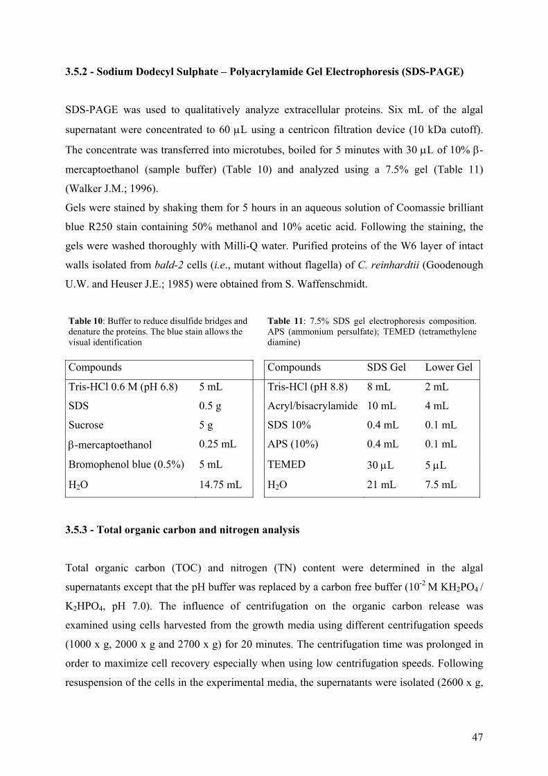
47
3.5.2 - Sodium Dodecyl Sulphate – Polyacrylamide Gel Electrophoresis (SDS-PAGE)
SDS-PAGE was used to qualitatively analyze extracellular proteins. Six mL of the algal
supernatant were concentrated to 60 µL using a centricon filtration device (10 kDa cutoff).
The concentrate was transferred into microtubes, boiled for 5 minutes with 30 µL of 10% β-
mercaptoethanol (sample buffer) (Table 10) and analyzed using a 7.5% gel (Table 11)
(Walker J.M.; 1996).
Gels were stained by shaking them for 5 hours in an aqueous solution of Coomassie brilliant
blue R250 stain containing 50% methanol and 10% acetic acid. Following the staining, the
gels were washed thoroughly with Milli-Q water. Purified proteins of the W6 layer of intact
walls isolated from bald-2 cells (i.e., mutant without flagella) of C. reinhardtii (Goodenough
U.W. and Heuser J.E.; 1985) were obtained from S. Waffenschmidt.
Table 10: Buffer to reduce disulfide bridges and denature the proteins. The blue stain allows the visual identification
Table 11: 7.5% SDS gel electrophoresis composition. APS (ammonium persulfate); TEMED (tetramethylene diamine)
Compounds Compounds SDS Gel Lower Gel
Tris-HCl 0.6 M (pH 6.8) 5 mL Tris-HCl (pH 8.8) 8 mL 2 mL
SDS 0.5 g Acryl/bisacrylamide 10 mL 4 mL
Sucrose 5 g SDS 10% 0.4 mL 0.1 mL
β-mercaptoethanol 0.25 mL APS (10%) 0.4 mL 0.1 mL
Bromophenol blue (0.5%) 5 mL TEMED 30 µL 5 µL
H2O 14.75 mL H2O 21 mL 7.5 mL
3.5.3 - Total organic carbon and nitrogen analysis
Total organic carbon (TOC) and nitrogen (TN) content were determined in the algal
supernatants except that the pH buffer was replaced by a carbon free buffer (10-2 M KH2PO4 /
K2HPO4, pH 7.0). The influence of centrifugation on the organic carbon release was
examined using cells harvested from the growth media using different centrifugation speeds
(1000 x g, 2000 x g and 2700 x g) for 20 minutes. The centrifugation time was prolonged in
order to maximize cell recovery especially when using low centrifugation speeds. Following
resuspension of the cells in the experimental media, the supernatants were isolated (2600 x g,
48
3 min) then acidified with HCl (final concentration 0.1% v/v) and degassed with ultrapure air
(-CO2) prior to measurements on Shimadzu analyzers (Model TOC-5000 or TOC-VCPH/CPN).
3.6 - Algal membrane lipidic permeability and metabolism
The influence of Cd and NTA on C assimilation by C. reinhardtii was determined by
NaH14CO3 short term uptake experiments, while their effect on membrane permeability was
studied by algal incubation in the presence of 14C–D-sorbitol, a neutral molecule that has been
shown to be taken up by passive diffusion without being metabolised (Parent L., Twiss M.R.
et al.; 1996) allowing for an evaluation of algal lipidic membrane integrity.
For the CO2 uptake experiments, the bicarbonate salt (specific activity 7.5 mCi mmol-1,
Anawa, Biomedical Services & Products, Switzerland) was diluted in Milli-Q water and the
pH was adjusted to 9.0. Radiolabeled 14C–D-sorbitol (specific activity of 37 mCi mmol-1,
Anawa) was diluted with 10-2 M non-labelled D-sorbitol. Algae were collected in their mid-
exponential growth phase, centrifuged, washed with 10-2 M MOPS at pH 7.0 and used for the
bioaccumulation experiment. Experimental solutions were labeled separately to a final
concentration of 4 x 10⁻⁵ mCi mL-1 NaH14CO3 or 14C–sorbitol and in various concentrations
of Cd, NTA or their mixtures. One mL of washed algal solution was resuspended in 10 mL of
experimental solution and left to accumulate for 30 minutes. Algae was isolated by
centrifugation (2 min, 2700 x g), washed (2x) with a washing solution containing 10-2 M non-
labeled NaH14CO3 or 14C–sorbitol, adjusted to 1 mL with Milli-Q water and digested with 9
mL Ultima Gold solution (Packard Bioscience). One mL of algal supernatant was also
sampled after the first centrifugation and analyzed for total NaH14CO3 or 14C–sorbitol by
mixing with 9 mL Count Gold (Packard Bioscience) solution. All 14C measurements were
made with a scintillation counter (Beckmann LS 6500). For both NaH14CO3 and 14C–sorbitol
uptake experiments, the measured cellular signal of 14C was normalized to its total measured
concentration in solution. Moreover, in each series, controls in the absence of both NTA and
Cd were included in order to normalize the results among different series.
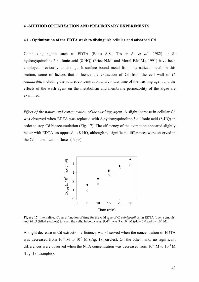
49
4 –METHOD OPTIMIZATION AND PRELIMINARY EXPERIMENTS
4.1 - Optimization of the EDTA wash to distinguish cellular and adsorbed Cd
Complexing agents such as EDTA (Bates S.S., Tessier A. et al.; 1982) or 8-
hydroxyquinoline-5-sulfonic acid (8-HQ) (Price N.M. and Morel F.M.M.; 1991) have been
employed previously to distinguish surface bound metal from internalized metal. In this
section, some of factors that influence the extraction of Cd from the cell wall of C.
reinhardtii, including the nature, concentration and contact time of the washing agent and the
effects of the wash agent on the metabolism and membrane permeability of the algae are
examined.
Effect of the nature and concentration of the washing agent. A slight increase in cellular Cd
was observed when EDTA was replaced with 8-hydroxyquinoline-5-sulfonic acid (8-HQ) in
order to stop Cd bioaccumulation (Fig. 17). The efficiency of the extraction appeared slightly
better with EDTA as opposed to 8-HQ, although no significant differences were observed in
the Cd internalization fluxes (slope).
Time (min)
0 5 10 15 20 25
[Cd]
int (
x 10
11 m
ol c
m-2
)
0
1
2
3
4
Figure 17: Internalized Cd as a function of time for the wild type of C. reinhardtii using EDTA (open symbols) and 8-HQ (filled symbols) to wash the cells. In both cases, [Cd2+] was 3 x 10⁻⁷ M (pH = 7.0 and I = 10-2 M).
A slight decrease in Cd extraction efficiency was observed when the concentration of EDTA
was decreased from 10-4 M to 10-5 M (Fig. 18: circles). On the other hand, no significant
differences were observed when the NTA concentration was decreased from 10-3 M to 10-4 M
(Fig. 18: triangles).
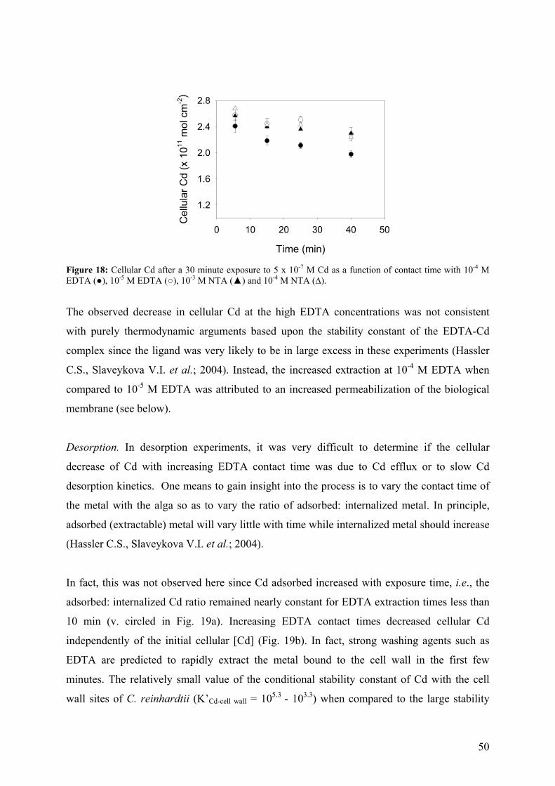
50
Time (min)
0 10 20 30 40 50
Cel
lula
r Cd
(x 1
011 m
ol c
m-2
)
1.2
1.6
2.0
2.4
2.8
Figure 18: Cellular Cd after a 30 minute exposure to 5 x 10⁻⁷ M Cd as a function of contact time with 10-4 M EDTA (), 10-5 M EDTA (), 10-3 M NTA () and 10-4 M NTA (∆).
The observed decrease in cellular Cd at the high EDTA concentrations was not consistent
with purely thermodynamic arguments based upon the stability constant of the EDTA-Cd
complex since the ligand was very likely to be in large excess in these experiments (Hassler
C.S., Slaveykova V.I. et al.; 2004). Instead, the increased extraction at 10-4 M EDTA when
compared to 10-5 M EDTA was attributed to an increased permeabilization of the biological
membrane (see below).
Desorption. In desorption experiments, it was very difficult to determine if the cellular
decrease of Cd with increasing EDTA contact time was due to Cd efflux or to slow Cd
desorption kinetics. One means to gain insight into the process is to vary the contact time of
the metal with the alga so as to vary the ratio of adsorbed: internalized metal. In principle,
adsorbed (extractable) metal will vary little with time while internalized metal should increase
(Hassler C.S., Slaveykova V.I. et al.; 2004).
In fact, this was not observed here since Cd adsorbed increased with exposure time, i.e., the
adsorbed: internalized Cd ratio remained nearly constant for EDTA extraction times less than
10 min (v. circled in Fig. 19a). Increasing EDTA contact times decreased cellular Cd
independently of the initial cellular [Cd] (Fig. 19b). In fact, strong washing agents such as
EDTA are predicted to rapidly extract the metal bound to the cell wall in the first few
minutes. The relatively small value of the conditional stability constant of Cd with the cell
wall sites of C. reinhardtii (K’Cd-cell wall = 105.3 - 103.3) when compared to the large stability
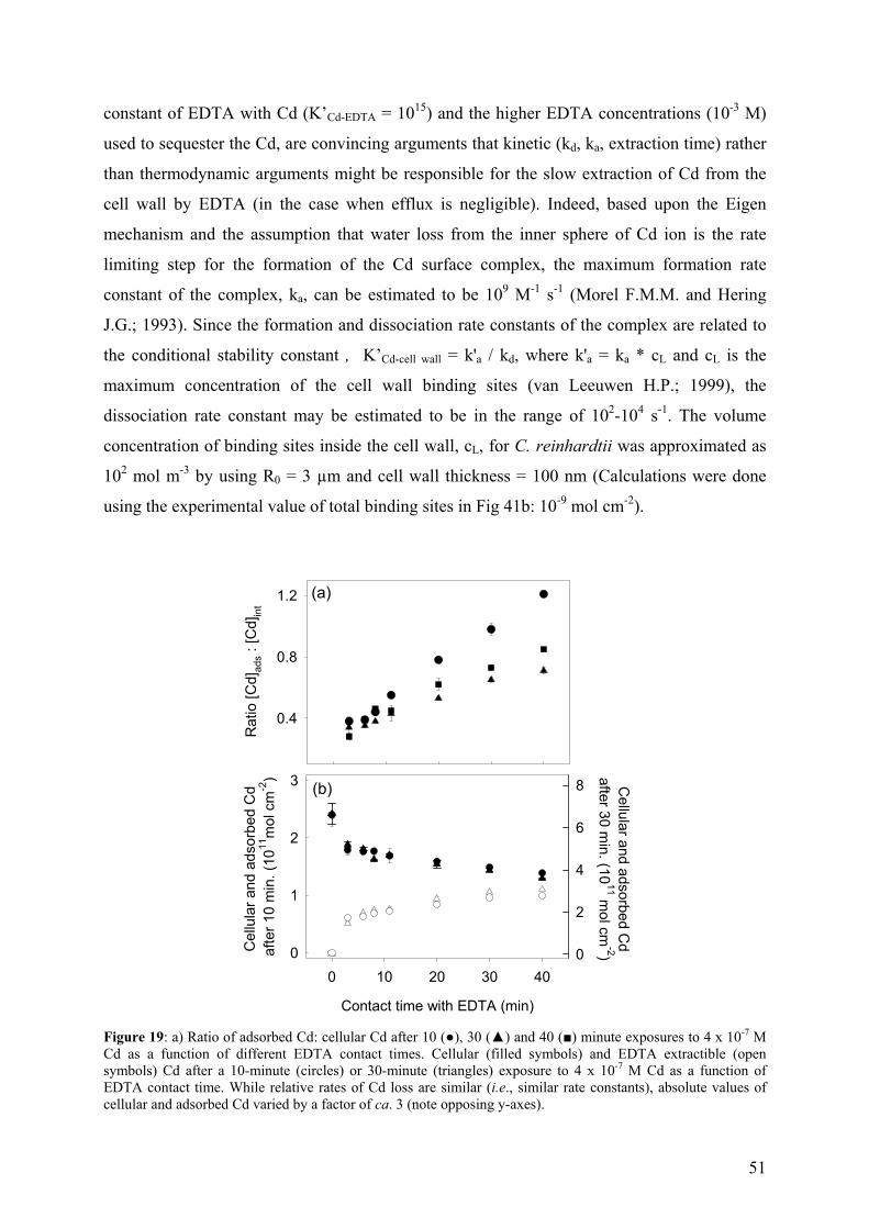
51
constant of EDTA with Cd (K’Cd-EDTA = 1015) and the higher EDTA concentrations (10-3 M)
used to sequester the Cd, are convincing arguments that kinetic (kd, ka, extraction time) rather
than thermodynamic arguments might be responsible for the slow extraction of Cd from the
cell wall by EDTA (in the case when efflux is negligible). Indeed, based upon the Eigen
mechanism and the assumption that water loss from the inner sphere of Cd ion is the rate
limiting step for the formation of the Cd surface complex, the maximum formation rate
constant of the complex, ka, can be estimated to be 109 M-1 s-1 (Morel F.M.M. and Hering
J.G.; 1993). Since the formation and dissociation rate constants of the complex are related to
the conditional stability constant , K’Cd-cell wall = k'a / kd, where k'a = ka * cL and cL is the
maximum concentration of the cell wall binding sites (van Leeuwen H.P.; 1999), the
dissociation rate constant may be estimated to be in the range of 102-104 s-1. The volume
concentration of binding sites inside the cell wall, cL, for C. reinhardtii was approximated as
102 mol m-3 by using R0 = 3 µm and cell wall thickness = 100 nm (Calculations were done
using the experimental value of total binding sites in Fig 41b: 10-9 mol cm-2).
Rat
io [C
d]ad
s : [C
d]in
t
0.4
0.8
1.2
Contact time with EDTA (min)
0 10 20 30 40
Cel
lula
r and
ads
orbe
d C
d af
ter 1
0 m
in. (
1011
mol
cm
-2)
0
1
2
3 Cellular and adsorbed C
dafter 30 m
in. (1011 m
ol cm-2)0
2
4
6
8
(a)
(b)
Figure 19: a) Ratio of adsorbed Cd: cellular Cd after 10 (), 30 () and 40 () minute exposures to 4 x 10⁻⁷ M Cd as a function of different EDTA contact times. Cellular (filled symbols) and EDTA extractible (open symbols) Cd after a 10-minute (circles) or 30-minute (triangles) exposure to 4 x 10⁻⁷ M Cd as a function of EDTA contact time. While relative rates of Cd loss are similar (i.e., similar rate constants), absolute values of cellular and adsorbed Cd varied by a factor of ca. 3 (note opposing y-axes).
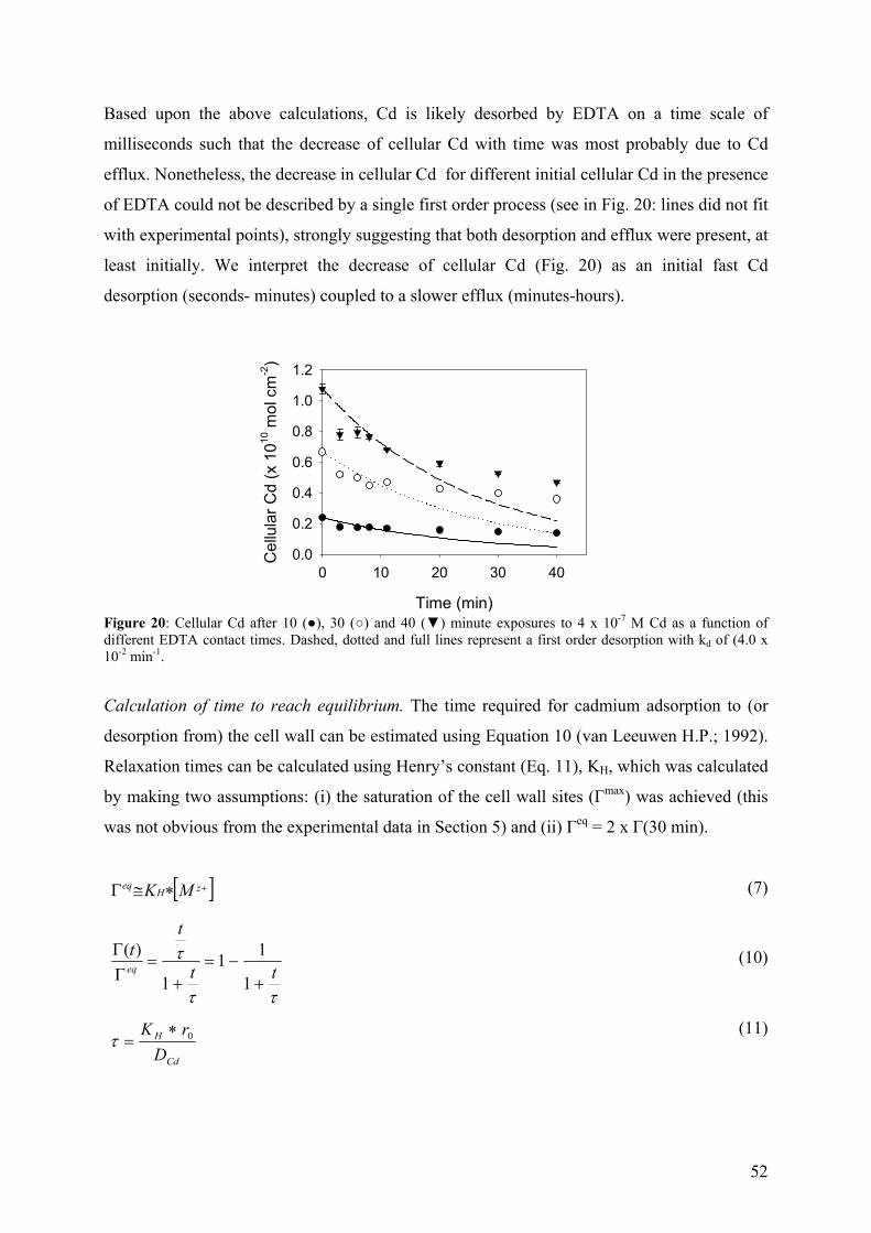
52
Based upon the above calculations, Cd is likely desorbed by EDTA on a time scale of
milliseconds such that the decrease of cellular Cd with time was most probably due to Cd
efflux. Nonetheless, the decrease in cellular Cd for different initial cellular Cd in the presence
of EDTA could not be described by a single first order process (see in Fig. 20: lines did not fit
with experimental points), strongly suggesting that both desorption and efflux were present, at
least initially. We interpret the decrease of cellular Cd (Fig. 20) as an initial fast Cd
desorption (seconds- minutes) coupled to a slower efflux (minutes-hours).
Time (min)
0 10 20 30 40
Cel
lula
r Cd
(x 1
010 m
ol c
m-2)
0.0
0.2
0.4
0.6
0.8
1.0
1.2
Figure 20: Cellular Cd after 10 (), 30 () and 40 () minute exposures to 4 x 10⁻⁷ M Cd as a function of different EDTA contact times. Dashed, dotted and full lines represent a first order desorption with kd of (4.0 x 10⁻² min-1.
Calculation of time to reach equilibrium. The time required for cadmium adsorption to (or
desorption from) the cell wall can be estimated using Equation 10 (van Leeuwen H.P.; 1992).
Relaxation times can be calculated using Henry’s constant (Eq. 11), KH, which was calculated
by making two assumptions: (i) the saturation of the cell wall sites (Гmax) was achieved (this
was not obvious from the experimental data in Section 5) and (ii) Гeq = 2 x Г(30 min).
[ ]+∗≅Γ zHeq MK (7)
ττ
τtt
tteq
+−=
+=
ΓΓ
1
111
)(
(10)
Cd
H
DrK 0∗
=τ (11)
53
It can be noticed that the Гeq is overestimated at any [Cd2+], consequently the calculated times
to reach the equilibrium will be greater than the real values. At [Cd2+] = 10-7 M, KH could be
estimated to be 2 x 10⁻³ m by using Eq. 7 and Гeq = 2 x 10⁻⁷ mol m-2. Thus, the time to reach
90% of equilibrium adsorption was estimated to be 1.8 min by using Eq. 11 and Гmax = 10-5
mol m-2 (see Table 12 for [Cd2+] ranging form 10-9 to 10-5 M).
Table 12: Values of KH, τ and t90% for [Cd2+] ranging form 10-9 M to 10-5 M.
[Cd2+] 10-9 M 10-7 M 10-5 M
KH (m) 2 x 10⁻² 2 x 10⁻³ 2 x 10⁻⁴
τ (min) 2.00 0.20 0.02
t90% (min) 18.00 1.80 0.18
The calculations showed that for [Cd2+] > 10-7 M, 90% of equilibrium adsorption (or 90%
desorption) must be achieved in less than 2 minutes if adsorption was limited by mass
transport, suggesting that adsorptive/desorptive reaction kinetics were fast enough. Moreover,
they were not responsible for the observed absence of an equilibrium between the cell surface
and the bulk solution (see Section 5).
Effect of EDTA on the release of exudates. EDTA was also shown to increase the protein
content of the algal filtrates (Fig. 21a).
Nature of the washing agent
MOPS EDTA DETA TETA
Bioaccumulation time (min)
0 10 20 30 40 50 60 70
Tota
l pro
tein
con
tent
(µ
g cm
-2)
0.0
0.2
0.4
0.6
0.8
1.0(a) (b)
Figure 21: (a) Total protein content in the EDTA extracts as a function of the bioaccumulation time, in the presence () or absence () of Cd. (b) Total protein content in the Cdads extracts after a 20 minute accumulation followed by extractions with MOPS (control), EDTA, diethylen triamine (DETA) and triethylen tetramine (TETA).
54
The protein increase was ca. 2x higher than in the presence of MOPS (Fig. 35b). EDTA likely
destabilizes the proteinic layers by complexing the stabilizer cations Ca and Mg thereby
promoting the release of the cell wall. Protein contents of extracts containing adsorbed Cd,
obtained by the use of two other relatively strong Cd complexing ligands: diethylene and
triethylene tetradiamine were also quantified for comparative reasons. These results showed
that the nature of the extracting agent had an important influence on the exudate release (Fig.
21b).
SDS-PAGE gels of EDTA and NTA extractable Cd showed that the molar masses of the
proteins depended largely on the washing agents. More proteins of molar masses between 45
and 60 kDa were found in the EDTA-extracts, while in the NTA extracts, a sharp band of
proteins was detected at 55 kDa (Fig. 22).
Figure 22: SDS PAGE of EDTA- (a, b) and NTA- (c, d) extracts (containing adsorbed cadmium) after a 10 (a, c) and 20 (b, d) minute contact times (MM: molar mass calibration standard).
The proteins released into the EDTA extracts were similar to those found in the algal filtrates
(10-2 M MOPS) during the uptake experiments (see Section 5), although at higher
concentrations, suggesting that MOPS and EDTA induced similar biological effects to C.
reinhardtii.
Effect of EDTA on CO2 uptake and membrane permeability. Measurements of NaH14CO3
uptake allowed for an assessment of the metabolic effects that are susceptible to affect short-
term uptake due to an induced biological stress on the organism. 14C incorporation increased
with incubation time and no decrease was observed in the presence of 10-3 M EDTA when
compared to measurements made in the absence of EDTA for contact times ranging from 5 to
30 minutes (Fig. 23a).
97 kDa
66 kDa
45 kDa
30 kDaMM a b c d
55
Lipophilic membrane permeability was observed to decrease slightly when increasing the
EDTA contact time (Fig. 23b), but this finding did not appear significant in the
bioaccumulation experiments.
Time (min)
0 10 20 30
Inte
rnal
ized
NaH
14C
O3
(CP
M)
0
500
1000
1500
Time (min)
0 10 20 30
Inte
rnal
ized
14C
-sor
bito
l (C
PM
)
0
2000
4000
6000(a) (b)
Figure 23: Carbon assimilation (a) and sorbitol (b) accumulation by C. reinhardtii exposed to 10-6 M Cd at different contact time with () and without () EDTA.
4.2- Comparison of bioaccumulation methods.
In order to evaluate the validity of the bioaccumulation parameters (i.e. Jint, Cd-Rcell), two
different methods, were used in this study. In Method I, aliquots (5 mL) of algal concentrate
were pipetted into 50 mL polypropylene tubes with 40 mL of experimental solution containing Cd
at t = 0, 6, 12, 18 and 24 min. Cd bioaccumulation was stopped by adding to each tube 5 mL
of a 10-2 M EDTA solution at t = 25, 26, 27, 28 and 29 min, each tube representing an
experimental point (t = 25, 20, 15, 10 and 5 min.). In Method II, algae were exposed to
experimental solutions containing Cd at t = 0 min. Cd accumulation was stopped by adding 1
mL 10-2 M MOPS / 10-2 M EDTA solution to each tube (10 mL) at 5, 10, 15, 20 and 25
minutes.
Cd internalization fluxes determined from the slope of cellular Cd as a function of time were
generally slightly higher (15%) in Method I as compared to Method II (i.e. Method I: 1.8 x
10-12 mol cm-2 min-1, Method II: 1.6 10-12 mol cm-2 min-1) (Fig. 24a, 25).
56
Time (min)
0 5 10 15 20 25
Cel
lula
r Cd
(mol
cm
-2)
0
1
2
3
4
5
Time (min)
0 5 10 15 20 25
Dissolved C
d (nM)
0
100
200
300
400(a) (b)
Figure 24: a) Internalized Cd and b) dissolved Cd as a function of time for wild type strain of C. reinhardtii using the first (open symbols) and second (filled symbols) methods. Initial dissolved Cd was 4 x 10⁻⁷ M (pH = 7.0 and I =10-2 M). The solid (slope = 1.83 x 10⁻¹² mol cm-2 min-1; intercept = –1.10 x 10⁻¹² mol cm-2, R2 = 0.995) and dashed (slope = 1.56 10-12 mol cm-2 min-1; intercept = 5.44 x 10⁻¹² mol cm-2, R2 = 0.997) lines represented linear regressions of the data. The error bars correspond to standard deviations when larger than the symbol size (n = 2).
On the other hand, consistent positive intercepts of the temporal Cd accumulation curves,
which are generally related to the carrier bound Cd, Cd-Rcell (Slaveykova V.I. and
Wilkinson K.J.; 2002; Hassler C.S., Slaveykova V.I. et al.; 2004) were observed when using
Method II, while they were generally negative for [Cd2+] < 10-6 M (i.e. –1.10 x 10⁻¹² mol cm-
2) when using Method I. It is thought that the fact that the algae remained in the concentrated
algal solution for longer times in Method I may explain the negative y-intercepts (Table 13).
Table 13: Residence times of the algae in the algal concentrate prior to the use for the bioaccumulation experiments.
Accumulation time 5 min 10 min 15 min 20 min 25 min
Method I 24 min 18 min 12 min 6 min 0 min
Method II 0 min 0 min 0 min 0 min 0 min
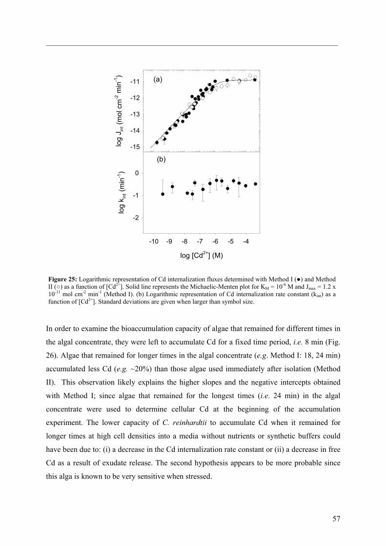
57
log [Cd2+] (M)
-10 -9 -8 -7 -6 -5 -4
log
k int (
min
-1)
-2
-1
0
log
J int (
mol
cm
-2 m
in-1
)
-15
-14
-13
-12
-11 (a)
(b)
Figure 25: Logarithmic representation of Cd internalization fluxes determined with Method I () and Method II () as a function of [Cd2+]. Solid line represents the Michaelic-Menten plot for KM = 10-6 M and Jmax = 1.2 x 10⁻¹¹ mol cm-2 min-1 (Method I). (b) Logarithmic representation of Cd internalization rate constant (kint) as a function of [Cd2+]. Standard deviations are given when larger than symbol size.
In order to examine the bioaccumulation capacity of algae that remained for different times in
the algal concentrate, they were left to accumulate Cd for a fixed time period, i.e. 8 min (Fig.
26). Algae that remained for longer times in the algal concentrate (e.g. Method I: 18, 24 min)
accumulated less Cd (e.g. ~20%) than those algae used immediately after isolation (Method
II). This observation likely explains the higher slopes and the negative intercepts obtained
with Method I; since algae that remained for the longest times (i.e. 24 min) in the algal
concentrate were used to determine cellular Cd at the beginning of the accumulation
experiment. The lower capacity of C. reinhardtii to accumulate Cd when it remained for
longer times at high cell densities into a media without nutrients or synthetic buffers could
have been due to: (i) a decrease in the Cd internalization rate constant or (ii) a decrease in free
Cd as a result of exudate release. The second hypothesis appears to be more probable since
this alga is known to be very sensitive when stressed.
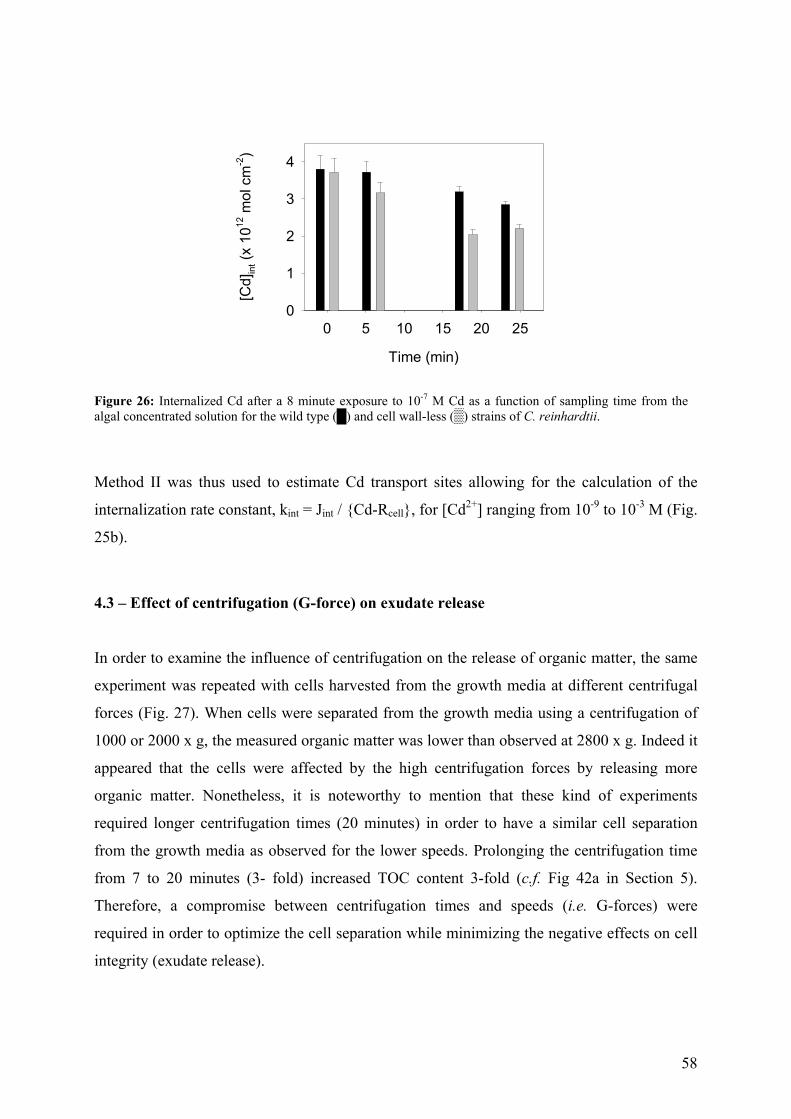
58
Time (min)
0 5 10 15 20 25
[Cd]
int (
x 10
12 m
ol c
m-2
)
0
1
2
3
4
Figure 26: Internalized Cd after a 8 minute exposure to 10-7 M Cd as a function of sampling time from the algal concentrated solution for the wild type () and cell wall-less () strains of C. reinhardtii.
Method II was thus used to estimate Cd transport sites allowing for the calculation of the
internalization rate constant, kint = Jint / Cd-Rcell, for [Cd2+] ranging from 10-9 to 10-3 M (Fig.
25b).
4.3 – Effect of centrifugation (G-force) on exudate release
In order to examine the influence of centrifugation on the release of organic matter, the same
experiment was repeated with cells harvested from the growth media at different centrifugal
forces (Fig. 27). When cells were separated from the growth media using a centrifugation of
1000 or 2000 x g, the measured organic matter was lower than observed at 2800 x g. Indeed it
appeared that the cells were affected by the high centrifugation forces by releasing more
organic matter. Nonetheless, it is noteworthy to mention that these kind of experiments
required longer centrifugation times (20 minutes) in order to have a similar cell separation
from the growth media as observed for the lower speeds. Prolonging the centrifugation time
from 7 to 20 minutes (3- fold) increased TOC content 3-fold (c.f. Fig 42a in Section 5).
Therefore, a compromise between centrifugation times and speeds (i.e. G-forces) were
required in order to optimize the cell separation while minimizing the negative effects on cell
integrity (exudate release).
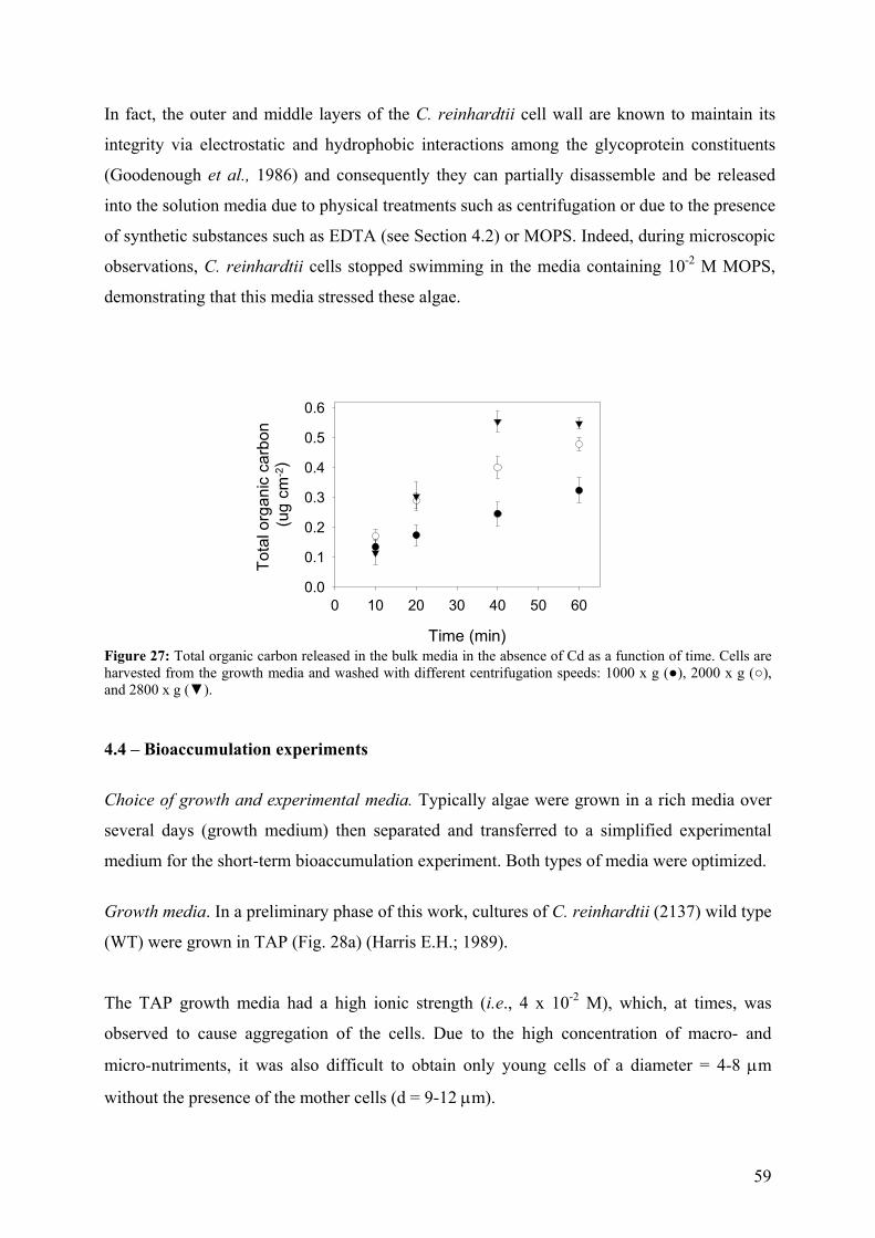
59
In fact, the outer and middle layers of the C. reinhardtii cell wall are known to maintain its
integrity via electrostatic and hydrophobic interactions among the glycoprotein constituents
(Goodenough et al., 1986) and consequently they can partially disassemble and be released
into the solution media due to physical treatments such as centrifugation or due to the presence
of synthetic substances such as EDTA (see Section 4.2) or MOPS. Indeed, during microscopic
observations, C. reinhardtii cells stopped swimming in the media containing 10-2 M MOPS,
demonstrating that this media stressed these algae.
Time (min)
0 10 20 30 40 50 60
Tota
l org
anic
car
bon
(ug
cm-2)
0.0
0.1
0.2
0.3
0.4
0.5
0.6
Figure 27: Total organic carbon released in the bulk media in the absence of Cd as a function of time. Cells are harvested from the growth media and washed with different centrifugation speeds: 1000 x g (), 2000 x g (), and 2800 x g (). 4.4 – Bioaccumulation experiments
Choice of growth and experimental media. Typically algae were grown in a rich media over
several days (growth medium) then separated and transferred to a simplified experimental
medium for the short-term bioaccumulation experiment. Both types of media were optimized.
Growth media. In a preliminary phase of this work, cultures of C. reinhardtii (2137) wild type
(WT) were grown in TAP (Fig. 28a) (Harris E.H.; 1989).
The TAP growth media had a high ionic strength (i.e., 4 x 10⁻² M), which, at times, was
observed to cause aggregation of the cells. Due to the high concentration of macro- and
micro-nutriments, it was also difficult to obtain only young cells of a diameter = 4-8 µm
without the presence of the mother cells (d = 9-12 µm).
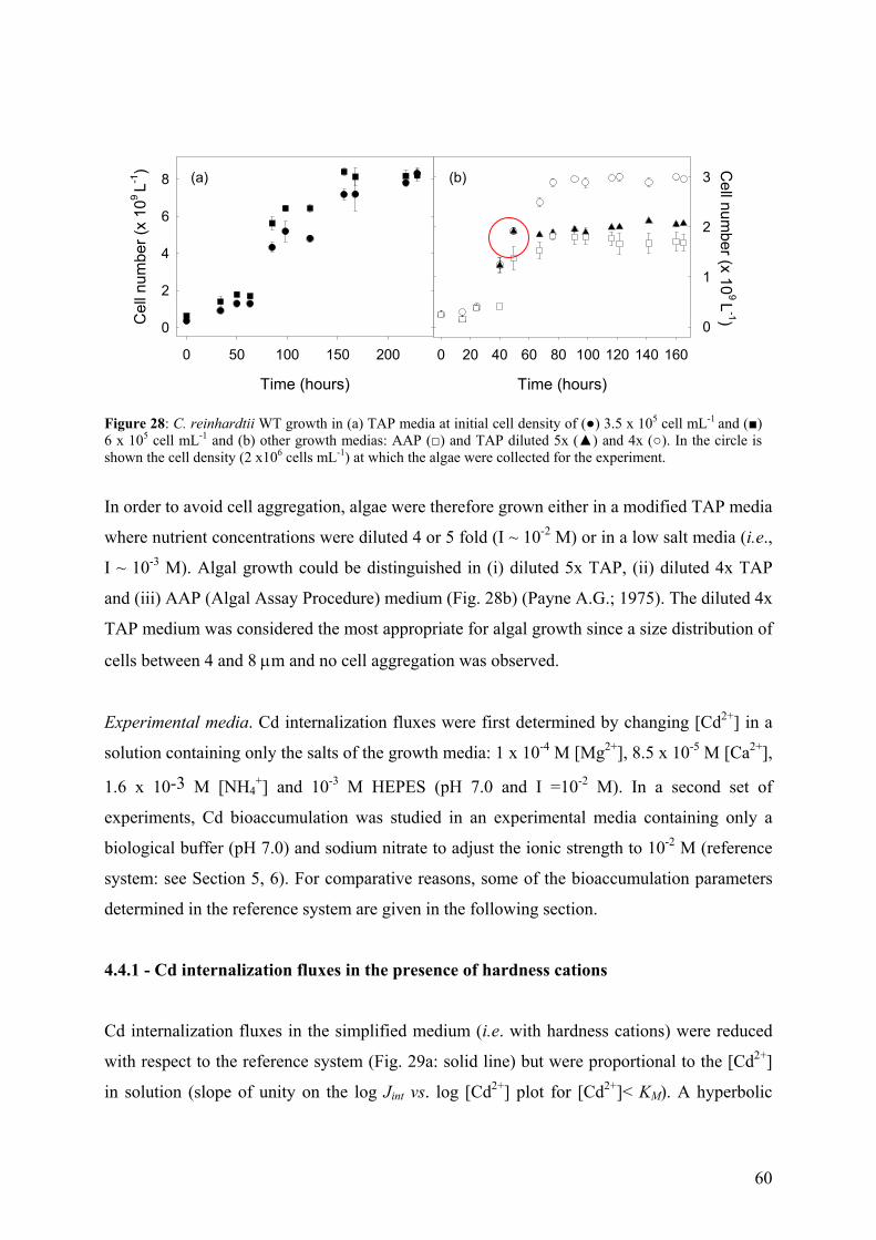
60
Time (hours)
0 20 40 60 80 100 120 140 160
Cell num
ber (x 109 L
-1)
0
1
2
3
Time (hours)
0 50 100 150 200
Cel
l num
ber (
x 10
9 L-1
)
0
2
4
6
8 (a) (b)
Figure 28: C. reinhardtii WT growth in (a) TAP media at initial cell density of () 3.5 x 10⁵ cell mL-1 and () 6 x 10⁵ cell mL-1 and (b) other growth medias: AAP () and TAP diluted 5x () and 4x (). In the circle is shown the cell density (2 x10⁶ cells mL-1) at which the algae were collected for the experiment.
In order to avoid cell aggregation, algae were therefore grown either in a modified TAP media
where nutrient concentrations were diluted 4 or 5 fold (I ~ 10-2 M) or in a low salt media (i.e.,
I ~ 10-3 M). Algal growth could be distinguished in (i) diluted 5x TAP, (ii) diluted 4x TAP
and (iii) AAP (Algal Assay Procedure) medium (Fig. 28b) (Payne A.G.; 1975). The diluted 4x
TAP medium was considered the most appropriate for algal growth since a size distribution of
cells between 4 and 8 µm and no cell aggregation was observed.
Experimental media. Cd internalization fluxes were first determined by changing [Cd2+] in a
solution containing only the salts of the growth media: 1 x 10⁻⁴ M [Mg2+], 8.5 x 10⁻⁵ M [Ca2+],
1.6 x 10⁻³ M [NH4+] and 10-3 M HEPES (pH 7.0 and I =10-2 M). In a second set of
experiments, Cd bioaccumulation was studied in an experimental media containing only a
biological buffer (pH 7.0) and sodium nitrate to adjust the ionic strength to 10-2 M (reference
system: see Section 5, 6). For comparative reasons, some of the bioaccumulation parameters
determined in the reference system are given in the following section.
4.4.1 - Cd internalization fluxes in the presence of hardness cations
Cd internalization fluxes in the simplified medium (i.e. with hardness cations) were reduced
with respect to the reference system (Fig. 29a: solid line) but were proportional to the [Cd2+]
in solution (slope of unity on the log Jint vs. log [Cd2+] plot for [Cd2+]< KM). A hyperbolic
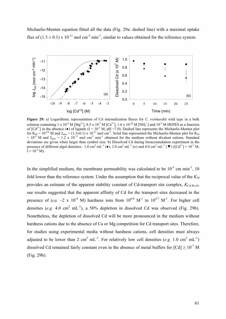
61
Michaelis-Menten equation fitted all the data (Fig. 29a: dashed line) with a maximal uptake
flux of (1.3 ± 0.1) x 10-11 mol cm-2 min-1, similar to values obtained for the reference system.
log [Cd+2] (M)
-10 -9 -8 -7 -6 -5 -4 -3
log
J int
(mol
cm
-2 m
in-1
)
-15
-14
-13
-12
-11
Time (min)
0 5 10 15 20 25
Dis
solv
ed C
d (x
107 M
)
0.0
0.2
0.4
0.6
0.8
1.0
(a) (b)
Figure 29: a) Logarithmic representation of Cd internalization fluxes for C. reinhardtii wild type in a bulk solution containing 1 x 10⁻⁴ M [Mg2+], 8.5 x 10⁻⁵ M [Ca2+], 1.6 x 10⁻³ M [NH4
+] and 10-3 M HEPES as a function of [Cd2+] in the absence () of ligands (I = 10-2 M, pH =7.0). Dashed line represents the Michaelis-Menten plot for KM = 10-4.6 M and Jmax = (1.3±0.1) x 10-11 mol cm-2. Solid line represented the Michaelis-Menten plot for KM = 10-6 M and Jmax = 1.2 x 10⁻¹¹ mol cm-2 min-1 obtained for the medium without divalent cations. Standard deviations are given when larger than symbol size. b) Dissolved Cd during bioaccumulation experiment in the presence of different algal densities: 1.0 cm2 mL-1 (), 2.0 cm2 mL-1 (ο) and 4.0 cm2 mL-1 () ([Cd2+] = 10-7 M, I = 10-2 M).
In the simplified medium, the membrane permeability was calculated to be 10-3 cm min-1, 10
fold lower than the reference system. Under the assumption that the reciprocal value of the KM
provides an estimate of the apparent stability constant of Cd-transport site complex, KCd-Rcell,
our results suggested that the apparent affinity of Cd for the transport sites decreased in the
presence of (ca. ~2 x 10⁻⁴ M) hardness ions from 106.0 M-1 to 104.7 M-1. For higher cell
densities (e.g. 4.0 cm2 mL-1), a 50% depletion in dissolved Cd was observed (Fig. 29b).
Nonetheless, the depletion of dissolved Cd will be more pronounced in the medium without
hardness cations due to the absence of Ca or Mg competition for Cd transport sites. Therefore,
for studies using experimental media without hardness cations, cell densities must always
adjusted to be lower than 2 cm2 mL-1. For relatively low cell densities (e.g. 1.0 cm2 mL-1)
dissolved Cd remained fairly constant even in the absence of metal buffers for [Cd] ≥ 10-7 M
(Fig. 29b).
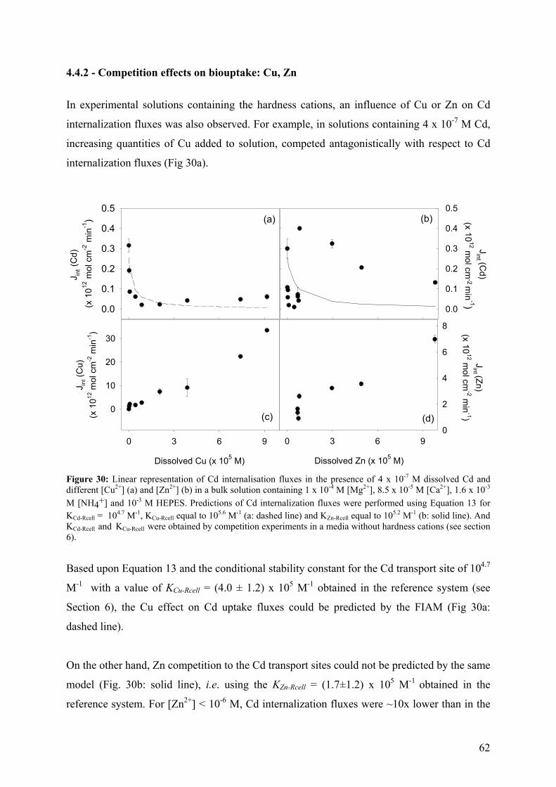
62
4.4.2 - Competition effects on biouptake: Cu, Zn
In experimental solutions containing the hardness cations, an influence of Cu or Zn on Cd
internalization fluxes was also observed. For example, in solutions containing 4 x 10⁻⁷ M Cd,
increasing quantities of Cu added to solution, competed antagonistically with respect to Cd
internalization fluxes (Fig 30a).
J int (
Cd)
(x 1
012 m
ol c
m-2
min
-1)
0.0
0.1
0.2
0.3
0.4
0.5
Jint (C
d) (x 10
12 mol cm
-2 min
-1)
0.0
0.1
0.2
0.3
0.4
0.5(a) (b)
Dissolved Cu (x 105 M)
0 3 6 9
J int (
Cu)
(x 1
012 m
ol c
m-2
min
-1)
0
10
20
30
Dissolved Zn (x 105 M)
0 3 6 9
Jint (Zn)
(x 1012 m
ol cm-2 m
in-1)
0
2
4
6
8
(c) (d)
Figure 30: Linear representation of Cd internalisation fluxes in the presence of 4 x 10⁻⁷ M dissolved Cd and different [Cu2+] (a) and [Zn2+] (b) in a bulk solution containing 1 x 10⁻⁴ M [Mg2+], 8.5 x 10⁻⁵ M [Ca2+], 1.6 x 10⁻³ M [NH4+] and 10-3 M HEPES. Predictions of Cd internalization fluxes were performed using Equation 13 for KCd-Rcell = 104.7 M-1, KCu-Rcell equal to 105.6 M-1 (a: dashed line) and KZn-Rcell equal to 105.2 M-1 (b: solid line). And KCd-Rcell and KCu-Rcell were obtained by competition experiments in a media without hardness cations (see section 6).
Based upon Equation 13 and the conditional stability constant for the Cd transport site of 104.7
M-1 with a value of KCu-Rcell = (4.0 ± 1.2) x 105 M-1 obtained in the reference system (see
Section 6), the Cu effect on Cd uptake fluxes could be predicted by the FIAM (Fig 30a:
dashed line).
On the other hand, Zn competition to the Cd transport sites could not be predicted by the same
model (Fig. 30b: solid line), i.e. using the KZn-Rcell = (1.7±1.2) x 105 M-1 obtained in the
reference system. For [Zn2+] < 10-6 M, Cd internalization fluxes were ~10x lower than in the
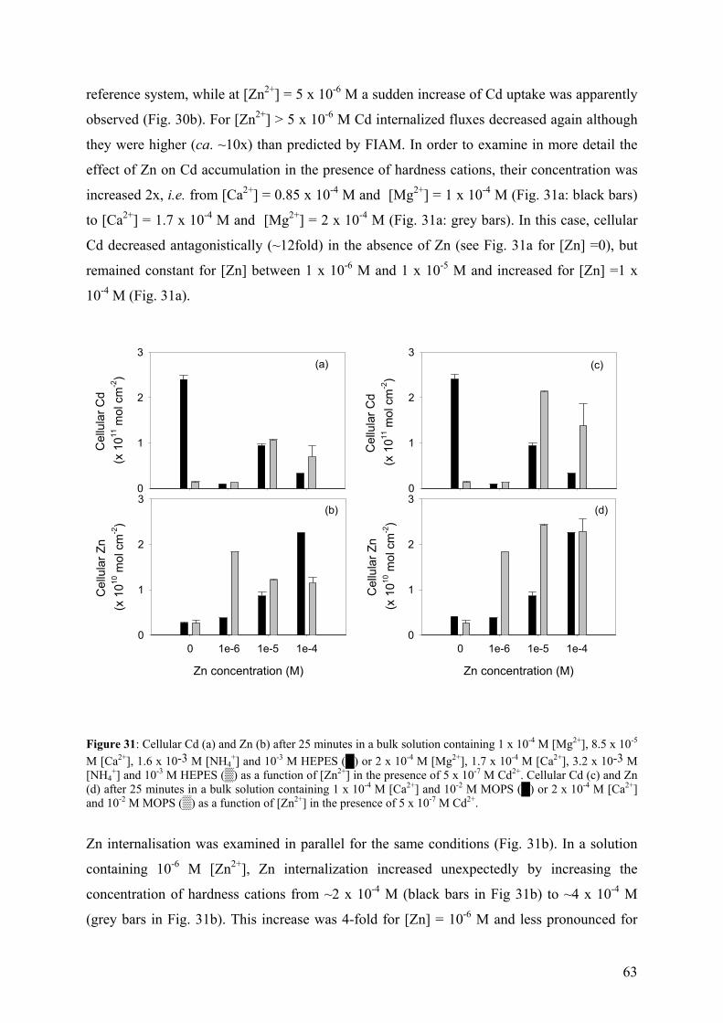
63
reference system, while at [Zn2+] = 5 x 10⁻⁶ M a sudden increase of Cd uptake was apparently
observed (Fig. 30b). For [Zn2+] > 5 x 10⁻⁶ M Cd internalized fluxes decreased again although
they were higher (ca. ~10x) than predicted by FIAM. In order to examine in more detail the
effect of Zn on Cd accumulation in the presence of hardness cations, their concentration was
increased 2x, i.e. from [Ca2+] = 0.85 x 10⁻⁴ M and [Mg2+] = 1 x 10⁻⁴ M (Fig. 31a: black bars)
to [Ca2+] = 1.7 x 10⁻⁴ M and [Mg2+] = 2 x 10⁻⁴ M (Fig. 31a: grey bars). In this case, cellular
Cd decreased antagonistically (~12fold) in the absence of Zn (see Fig. 31a for [Zn] =0), but
remained constant for [Zn] between 1 x 10⁻⁶ M and 1 x 10⁻⁵ M and increased for [Zn] =1 x
10-4 M (Fig. 31a).
Cel
lula
r Cd
(x 1
011 m
ol c
m-2
)
0
1
2
3
Zn concentration (M)
0 1e-6 1e-5 1e-4
Cel
lula
r Zn
(x 1
010 m
ol c
m-2
)
0
1
2
3
Cel
lula
r Cd
(x 1
011 m
ol c
m-2
)
0
1
2
3
Zn concentration (M)
0 1e-6 1e-5 1e-4
Cel
lula
r Zn
(x 1
010 m
ol c
m-2
)
0
1
2
3
(a)
(b)
(c)
(d)
Figure 31: Cellular Cd (a) and Zn (b) after 25 minutes in a bulk solution containing 1 x 10⁻⁴ M [Mg2+], 8.5 x 10⁻⁵ M [Ca2+], 1.6 x 10⁻³ M [NH4
+] and 10-3 M HEPES () or 2 x 10⁻⁴ M [Mg2+], 1.7 x 10⁻⁴ M [Ca2+], 3.2 x 10⁻³ M [NH4
+] and 10-3 M HEPES () as a function of [Zn2+] in the presence of 5 x 10⁻⁷ M Cd2+. Cellular Cd (c) and Zn (d) after 25 minutes in a bulk solution containing 1 x 10⁻⁴ M [Ca2+] and 10-2 M MOPS () or 2 x 10⁻⁴ M [Ca2+] and 10-2 M MOPS () as a function of [Zn2+] in the presence of 5 x 10⁻⁷ M Cd2+.
Zn internalisation was examined in parallel for the same conditions (Fig. 31b). In a solution
containing 10-6 M [Zn2+], Zn internalization increased unexpectedly by increasing the
concentration of hardness cations from ~2 x 10⁻⁴ M (black bars in Fig 31b) to ~4 x 10⁻⁴ M
(grey bars in Fig. 31b). This increase was 4-fold for [Zn] = 10-6 M and less pronounced for
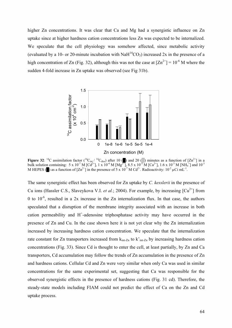
64
higher Zn concentrations. It was clear that Ca and Mg had a synergistic influence on Zn
uptake since at higher hardness cation concentrations less Zn was expected to be internalized.
We speculate that the cell physiology was somehow affected, since metabolic activity
(evaluated by a 10- or 20-minute incubation with NaH14CO3) increased 2x in the presence of a
high concentration of Zn (Fig. 32), although this was not the case at [Zn2+] = 10-6 M where the
sudden 4-fold increase in Zn uptake was observed (see Fig 31b).
Zn concentration (M)
0 1e-8 1e-6 1e-5 5e-5 1e-4
14C
ass
imila
tion
fact
or
(x 1
04 cm
-2)
0.0
0.5
1.0
1.5
Figure 32: 14C assimilation factor (14Cint / 14Cdiss) after 10 () and 20 () minutes as a function of [Zn2+] in a bulk solution containing: 5 x 10⁻⁷ M [Cd2+], 1 x 10⁻⁴ M [Mg2+], 8.5 x 10⁻⁵ M [Ca2+], 1.6 x 10⁻³ M [NH4
+] and 10-3 M HEPES () as a function of [Zn2+] in the presence of 5 x 10⁻⁷ M Cd2+. Radioactivity: 10-2 µCi mL-1.
The same synergistic effect has been observed for Zn uptake by C. kesslerii in the presence of
Cu ions (Hassler C.S., Slaveykova V.I. et al.; 2004). For example, by increasing [Cu2+] from
0 to 10-6, resulted in a 2x increase in the Zn internalization flux. In that case, the authors
speculated that a disruption of the membrane integrity associated with an increase in both
cation permeability and H+-adenosine triphosphatase activity may have occurred in the
presence of Zn and Cu. In the case shown here it is not yet clear why the Zn internalization
increased by increasing hardness cation concentration. We speculate that the internalization
rate constant for Zn transporters increased from kint-Zn to k’int-Zn by increasing hardness cation
concentrations (Fig. 33). Since Cd is thought to enter the cell, at least partially, by Zn and Ca
transporters, Cd accumulation may follow the trends of Zn accumulation in the presence of Zn
and hardness cations. Cellular Cd and Zn were very similar when only Ca was used in similar
concentrations for the same experimental set, suggesting that Ca was responsible for the
observed synergistic effects in the presence of hardness cations (Fig. 31 cd). Therefore, the
steady-state models including FIAM could not predict the effect of Ca on the Zn and Cd
uptake process.
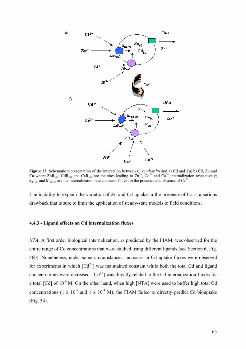
65
Figure 33: Schematic representation of the interaction between C. reinhardtii and a) Cd and Zn; b) Cd, Zn and Ca where ZnRcell, CdRcell and CaRcell are the sites leading to Zn2+, Cd2+ and Ca2+ internalization respectively; kint-Zn and k’int-Zn are the internalization rate constants for Zn in the presence and absence of Ca2+.
The inability to explain the variation of Zn and Cd uptake in the presence of Ca is a serious
drawback that is sure to limit the application of steady-state models to field conditions.
4.4.3 - Ligand effects on Cd internalization fluxes
NTA. A first order biological internalization, as predicted by the FIAM, was observed for the
entire range of Cd concentrations that were studied using different ligands (see Section 6, Fig.
46b). Nonetheless, under some circumstances, increases in Cd uptake fluxes were observed
for experiments in which [Cd2+] was maintained constant while both the total Cd and ligand
concentrations were increased. [Cd2+] was directly related to the Cd internalization fluxes for
a total [Cd] of 10-6 M. On the other hand, when high [NTA] were used to buffer high total Cd
concentrations (1 x 10⁻⁵ and 1 x 10⁻⁴ M), the FIAM failed to directly predict Cd biouptake
(Fig. 34).
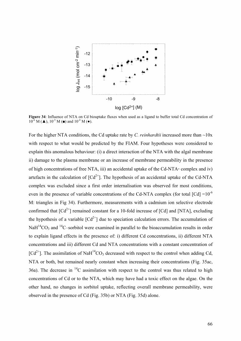
66
log [Cd2+] (M)
-10 -9 -8
log
J int
(mol
cm
-2 m
in-1
)
-15
-14
-13
-12
Figure 34: Influence of NTA on Cd biouptake fluxes when used as a ligand to buffer total Cd concentration of 10-6 M (), 10-5 M () and 10-4 M ().
For the higher NTA conditions, the Cd uptake rate by C. reinhardtii increased more than ~10x
with respect to what would be predicted by the FIAM. Four hypotheses were considered to
explain this anomalous behaviour: (i) a direct interaction of the NTA with the algal membrane
ii) damage to the plasma membrane or an increase of membrane permeability in the presence
of high concentrations of free NTA, iii) an accidental uptake of the Cd-NTA- complex and iv)
artefacts in the calculation of [Cd2+]. The hypothesis of an accidental uptake of the Cd-NTA
complex was excluded since a first order internalisation was observed for most conditions,
even in the presence of variable concentrations of the Cd-NTA complex (for total [Cd] =10-6
M: triangles in Fig 34). Furthermore, measurements with a cadmium ion selective electrode
confirmed that [Cd2+] remained constant for a 10-fold increase of [Cd] and [NTA], excluding
the hypothesis of a variable [Cd2+] due to speciation calculation errors. The accumulation of
NaH14CO3 and 14C–sorbitol were examined in parallel to the bioaccumulation results in order
to explain ligand effects in the presence of: i) different Cd concentrations, ii) different NTA
concentrations and iii) different Cd and NTA concentrations with a constant concentration of
[Cd2+]. The assimilation of NaH14CO3 decreased with respect to the control when adding Cd,
NTA or both, but remained nearly constant when increasing their concentrations (Fig. 35ac,
36a). The decrease in 14C assimilation with respect to the control was thus related to high
concentrations of Cd or to the NTA, which may have had a toxic effect on the algae. On the
other hand, no changes in sorbitol uptake, reflecting overall membrane permeability, were
observed in the presence of Cd (Fig. 35b) or NTA (Fig. 35d) alone.
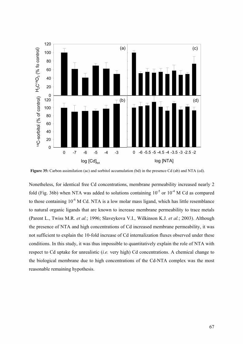
67
H2C
14O
3 (%
fo c
ontro
l)
0
20
40
60
80
100
120
log [Cd]tot
0 -7 -6 -5 -4 -3
14C
-sor
bito
l (%
of c
ontro
l)
0
20
40
60
80
100
120
(a)
(b)
log [NTA]
0 -6 -5.5 -5 -4.5 -4 -3.5 -3 -2.5 -2
(c)
(d)
Figure 35: Carbon assimilation (ac) and sorbitol accumulation (bd) in the presence Cd (ab) and NTA (cd).
Nonetheless, for identical free Cd concentrations, membrane permeability increased nearly 2
fold (Fig. 36b) when NTA was added to solutions containing 10-5 or 10-4 M Cd as compared
to those containing 10-6 M Cd. NTA is a low molar mass ligand, which has little resemblance
to natural organic ligands that are known to increase membrane permeability to trace metals
(Parent L., Twiss M.R. et al.; 1996; Slaveykova V.I., Wilkinson K.J. et al.; 2003). Although
the presence of NTA and high concentrations of Cd increased membrane permeability, it was
not sufficient to explain the 10-fold increase of Cd internalization fluxes observed under these
conditions. In this study, it was thus impossible to quantitatively explain the role of NTA with
respect to Cd uptake for unrealistic (i.e. very high) Cd concentrations. A chemical change to
the biological membrane due to high concentrations of the Cd-NTA complex was the most
reasonable remaining hypothesis.
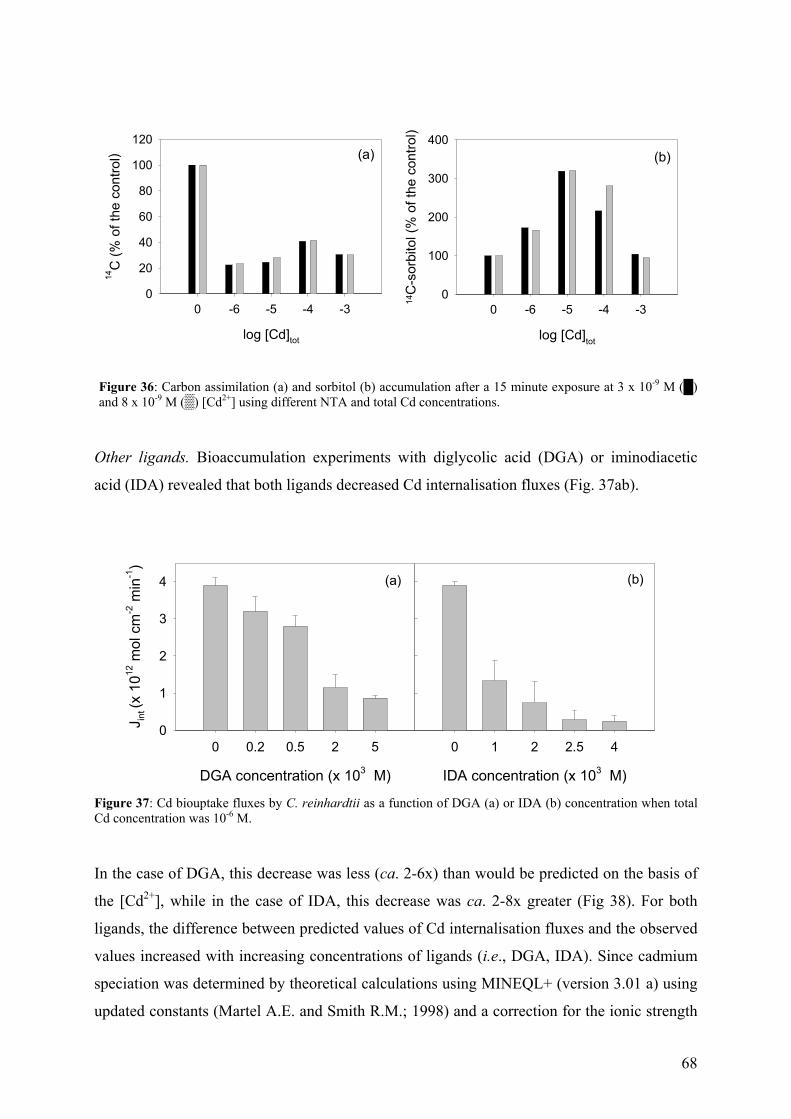
68
log [Cd]tot
0 -6 -5 -4 -3
14C
(% o
f the
con
trol)
0
20
40
60
80
100
120
log [Cd]tot
0 -6 -5 -4 -3
14C
-sor
bito
l (%
of t
he c
ontro
l)
0
100
200
300
400(a) (b)
Figure 36: Carbon assimilation (a) and sorbitol (b) accumulation after a 15 minute exposure at 3 x 10⁻⁹ M () and 8 x 10⁻⁹ M () [Cd2+] using different NTA and total Cd concentrations.
Other ligands. Bioaccumulation experiments with diglycolic acid (DGA) or iminodiacetic
acid (IDA) revealed that both ligands decreased Cd internalisation fluxes (Fig. 37ab).
DGA concentration (x 103 M)
0 0.2 0.5 2 5
J int (
x 10
12 m
ol c
m-2
min
-1)
0
1
2
3
4
IDA concentration (x 103 M)
0 1 2 2.5 4
(a) (b)
Figure 37: Cd biouptake fluxes by C. reinhardtii as a function of DGA (a) or IDA (b) concentration when total Cd concentration was 10-6 M.
In the case of DGA, this decrease was less (ca. 2-6x) than would be predicted on the basis of
the [Cd2+], while in the case of IDA, this decrease was ca. 2-8x greater (Fig 38). For both
ligands, the difference between predicted values of Cd internalisation fluxes and the observed
values increased with increasing concentrations of ligands (i.e., DGA, IDA). Since cadmium
speciation was determined by theoretical calculations using MINEQL+ (version 3.01 a) using
updated constants (Martel A.E. and Smith R.M.; 1998) and a correction for the ionic strength
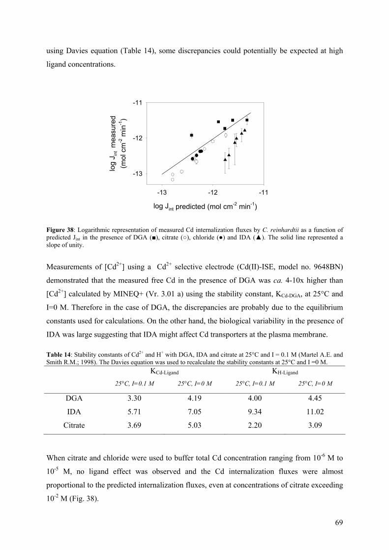
69
using Davies equation (Table 14), some discrepancies could potentially be expected at high
ligand concentrations.
log Jint predicted (mol cm-2 min-1)
-13 -12 -11
log
J int m
easu
red
(mol
cm
-2 m
in-1
)
-13
-12
-11
Figure 38: Logarithmic representation of measured Cd internalization fluxes by C. reinhardtii as a function of predicted Jint in the presence of DGA (), citrate (), chloride () and IDA (). The solid line represented a slope of unity.
Measurements of [Cd2+] using a Cd2+ selective electrode (Cd(II)-ISE, model no. 9648BN)
demonstrated that the measured free Cd in the presence of DGA was ca. 4-10x higher than
[Cd2+] calculated by MINEQ+ (Vr. 3.01 a) using the stability constant, KCd-DGA, at 25°C and
I=0 M. Therefore in the case of DGA, the discrepancies are probably due to the equilibrium
constants used for calculations. On the other hand, the biological variability in the presence of
IDA was large suggesting that IDA might affect Cd transporters at the plasma membrane. Table 14: Stability constants of Cd2+ and H+ with DGA, IDA and citrate at 25°C and I = 0.1 M (Martel A.E. and Smith R.M.; 1998). The Davies equation was used to recalculate the stability constants at 25°C and I =0 M. KCd-Ligand KH-Ligand
25°C, I=0.1 M 25°C, I=0 M 25°C, I=0.1 M 25°C, I=0 M
DGA 3.30 4.19 4.00 4.45
IDA 5.71 7.05 9.34 11.02
Citrate 3.69 5.03 2.20 3.09
When citrate and chloride were used to buffer total Cd concentration ranging from 10-6 M to
10-5 M, no ligand effect was observed and the Cd internalization fluxes were almost
proportional to the predicted internalization fluxes, even at concentrations of citrate exceeding
10-2 M (Fig. 38).
70
5 - CADMIUM INTERACTION WITH CHLAMYDOMONAS REINHARDTII AND ITS
CELL WALL PROTEINS
(Environ. Chem,, 2004, 1, 172-179)
In natural waters, biological systems play an important role in regulating trace metal
concentrations and speciation (e.g. lakes:(Sigg L.; 1987); oceans:(Morel F.M.M. and Price
N.M.; 2003)) by decreasing metal concentrations through adsorption and internalization and
by modifying chemical speciation through release of extracellular organic matter. In order to
better understand the mechanisms by which metals are sequestered, metabolized, stored and
detoxified by aquatic organisms, model unicellular systems have been extensively developed.
Chlamydomonas reinhardtii is a green alga that is often used to study the deleterious effects
of toxic trace metals through measurements of photosynthesis (Nagel K. and Voigt J.; 1995;
Prasad M.N., Drej K. et al.; 1998), growth (Ben-Bassat D., Shelef G. et al.; 1972; Lustigman
B., Lee L.H. and Weiss-Magasic C.; 1995; Weiss-Magasic C., Lustigman B. et al.; 1997),
nutrient uptake inhibition (Devriese M., Tsakaloudi V. et al.; 2001), ultrastructural changes
(Irmer U., Wachholz I. et al.; 1986), etc. Furthermore, the recent availability of the complete
genome for C. reinhardtii (www.biology.duke.edu/genome_chlamy) has given an added
impetus to the use of this organism for metal bioavailability studies, for fundamental
investigations of trace metal effects and for the development of eukaryotic biosensors (Sasaki
T., Kurano N. et al.; 1998; Rubinelli P., Siripornadulsil S. et al.; 2002; Siripornadulsil S.,
Traina S. et al.; 2002; Moseley J.L., Allinger T. et al.; 2002a; Moseley J.L., Page M.D. et al.;
2002b).
Nearly all descriptions of metal uptake and adsorption are equilibrium based models that
exclude biological parameters such as the production of organic exudates, metal efflux or the
synthesis of uptake sites (Mirimanoff N. and Wilkinson K.J.; 2000; Hassler C.S. and
Wilkinson K.J.; 2003). For example, metal adsorption to the surface of microorganisms is
most often described by a single site model (e.g., Langmuir isotherm) with (Plette A.C.C.,
Benedetti M.F. et al.; 1996; Plette A.C.C., Nederlof M.M. et al.; 1999) or without (Crist R.H.,
Oberholser K. et al.; 1988; Tessier A., Buffle J. et al.; 1994; Kiefer E., Sigg L. et al.; 1997) a
charge correction. Nonetheless, at least two important conditions are required for a Langmuir
isotherm to apply: (i) equilibrium is attained between the adsorbed and dissolved metal and
(ii) all surface sites are equally active. In reality, for chemically heterogeneous and multiply
71
charged surfaces, the tendency to form surface complexes decreases with an increased metal
loading of the surface due to both chemical and columbic effects (Buffle J.; 1988; Xue H.B.,
Stumm W. et al.; 1988). The equilibrium assumption implies that the concentration of the free
aquo metal ion in solution will be central to predicting metal adsorption. It is thus important
to quantify the role and nature of the metal binding ligands that are produced and excreted by
aquatic organisms. For exemple, Xue et al. (Xue H. and Sigg L.; 1990) have demonstrated
that the binding of Cu(II) to algal exudates produced by C. reinhardtii had a more significant
effect on Cu speciation than did the adsorption to algal surfaces. In that case, the complexing
capacity of the algal filtrates varied with cell number, cell storage, the duration of the Cu
treatment and the culture conditions. In another example, a 10-fold increase in exudate
production was observed during the stationary growth phase as compared to the log phase of
the green alga Fritschiella tuberose lying (Melkonian M.; 1979). The nature of the exudates
depends strongly on the biological species and the exposure conditions. For Scenedesmus
quadricauda, Chlorella kessleri, and Raphidocelis subcapitata, exposure to Cd increased the
protein to carbohydrate ratio of the exudates whereas exposure to Cl had an opposite effect
(Marsalek B. and Rojickova R.; 1996). Finally, in the case of C. reinhardtii, algae are known
to secrete hydrophilic organic signal molecules that affect quorum sensing regulation in
naturally encountered bacteria (Teplitski M., Chen H. et al.; 2004), even in the absence of an
apparent external stress.
Given that the organism can modify its own metal uptake, the objective of the present study
was to precisely describe Cd adsorption to C. reinhardtii by taking into account the most
important biological, physicochemical and environmental parameters. Several techniques
were combined in order to quantitatively determine free Cd or Cd bound either to
extracellular ligands in the bulk solution or to the cell surface.
5.1 - Results and Discussion
5.1.1 - Cd adsorption by C. reinhardtii WT and CW-2 strains
Metal adsorption to algal cells is generally a rapid process with equilibrium being attained in
the first few minutes of exposure (Bates S.S., Tessier A. et al.; 1982; Hassler C.S.,
Slaveykova V.I. et al.; 2004). Contrary to this expectation, adsorbed Cd for both the wild type
and cell wall-less strains of C. reinhardtii did not attain constant values, but instead increased
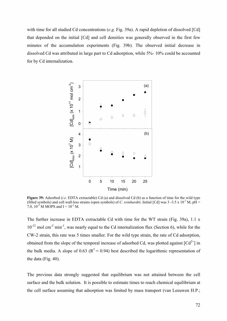
72
with time for all studied Cd concentrations (e.g. Fig. 39a). A rapid depletion of dissolved [Cd]
that depended on the initial [Cd] and cell densities was generally observed in the first few
minutes of the accumulation experiments (Fig. 39b). The observed initial decrease in
dissolved Cd was attributed in large part to Cd adsorption, while 5%- 10% could be accounted
for by Cd internalization.
[Cd]
ads (
x 10
11 m
ol c
m-2
)
0
1
2
3
Time (min)
0 5 10 15 20 25
[Cd]
diss
(x 1
07 M)
1
2
3
4
(a)
(b)
Figure 39: Adsorbed (i.e. EDTA extractable) Cd (a) and dissolved Cd (b) as a function of time for the wild type (filled symbols) and cell wall-less strains (open symbols) of C. reinhardtii. Initial [Cd] was 3 -3.5 x 10⁻⁷ M, pH = 7.0, 10-2 M MOPS and I = 10-2 M.
The further increase in EDTA extractable Cd with time for the WT strain (Fig. 39a), 1.1 x
10-12 mol cm-2 min-1, was nearly equal to the Cd internalization flux (Section 6), while for the
CW-2 strain, this rate was 5 times smaller. For the wild type strain, the rate of Cd adsorption,
obtained from the slope of the temporal increase of adsorbed Cd, was plotted against [Cd2+] in
the bulk media. A slope of 0.63 (R2 = 0.94) best described the logarithmic representation of
the data (Fig. 40).
The previous data strongly suggested that equilibrium was not attained between the cell
surface and the bulk solution. It is possible to estimate times to reach chemical equilibrium at
the cell surface assuming that adsorption was limited by mass transport (van Leeuwen H.P.;
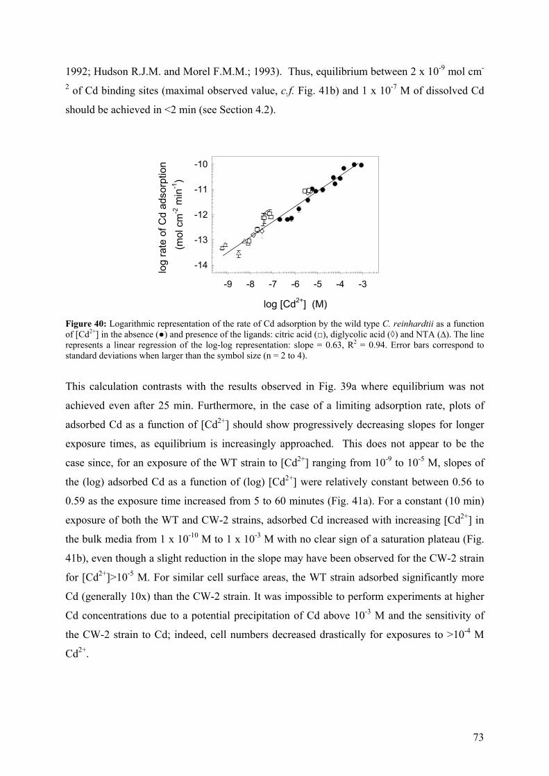
73
1992; Hudson R.J.M. and Morel F.M.M.; 1993). Thus, equilibrium between 2 x 10⁻⁹ mol cm-
2 of Cd binding sites (maximal observed value, c.f. Fig. 41b) and 1 x 10⁻⁷ M of dissolved Cd
should be achieved in <2 min (see Section 4.2).
log [Cd2+] (M)
-9 -8 -7 -6 -5 -4 -3
log
rate
of C
d ad
sorp
tion
(m
ol c
m-2
min
-1)
-14
-13
-12
-11
-10
Figure 40: Logarithmic representation of the rate of Cd adsorption by the wild type C. reinhardtii as a function of [Cd2+] in the absence () and presence of the ligands: citric acid (), diglycolic acid (◊) and NTA (∆). The line represents a linear regression of the log-log representation: slope = 0.63, R2 = 0.94. Error bars correspond to standard deviations when larger than the symbol size (n = 2 to 4).
This calculation contrasts with the results observed in Fig. 39a where equilibrium was not
achieved even after 25 min. Furthermore, in the case of a limiting adsorption rate, plots of
adsorbed Cd as a function of [Cd2+] should show progressively decreasing slopes for longer
exposure times, as equilibrium is increasingly approached. This does not appear to be the
case since, for an exposure of the WT strain to [Cd2+] ranging from 10-9 to 10-5 M, slopes of
the (log) adsorbed Cd as a function of (log) [Cd2+] were relatively constant between 0.56 to
0.59 as the exposure time increased from 5 to 60 minutes (Fig. 41a). For a constant (10 min)
exposure of both the WT and CW-2 strains, adsorbed Cd increased with increasing [Cd2+] in
the bulk media from 1 x 10⁻¹⁰ M to 1 x 10⁻³ M with no clear sign of a saturation plateau (Fig.
41b), even though a slight reduction in the slope may have been observed for the CW-2 strain
for [Cd2+]>10-5 M. For similar cell surface areas, the WT strain adsorbed significantly more
Cd (generally 10x) than the CW-2 strain. It was impossible to perform experiments at higher
Cd concentrations due to a potential precipitation of Cd above 10-3 M and the sensitivity of
the CW-2 strain to Cd; indeed, cell numbers decreased drastically for exposures to >10-4 M
Cd2+.
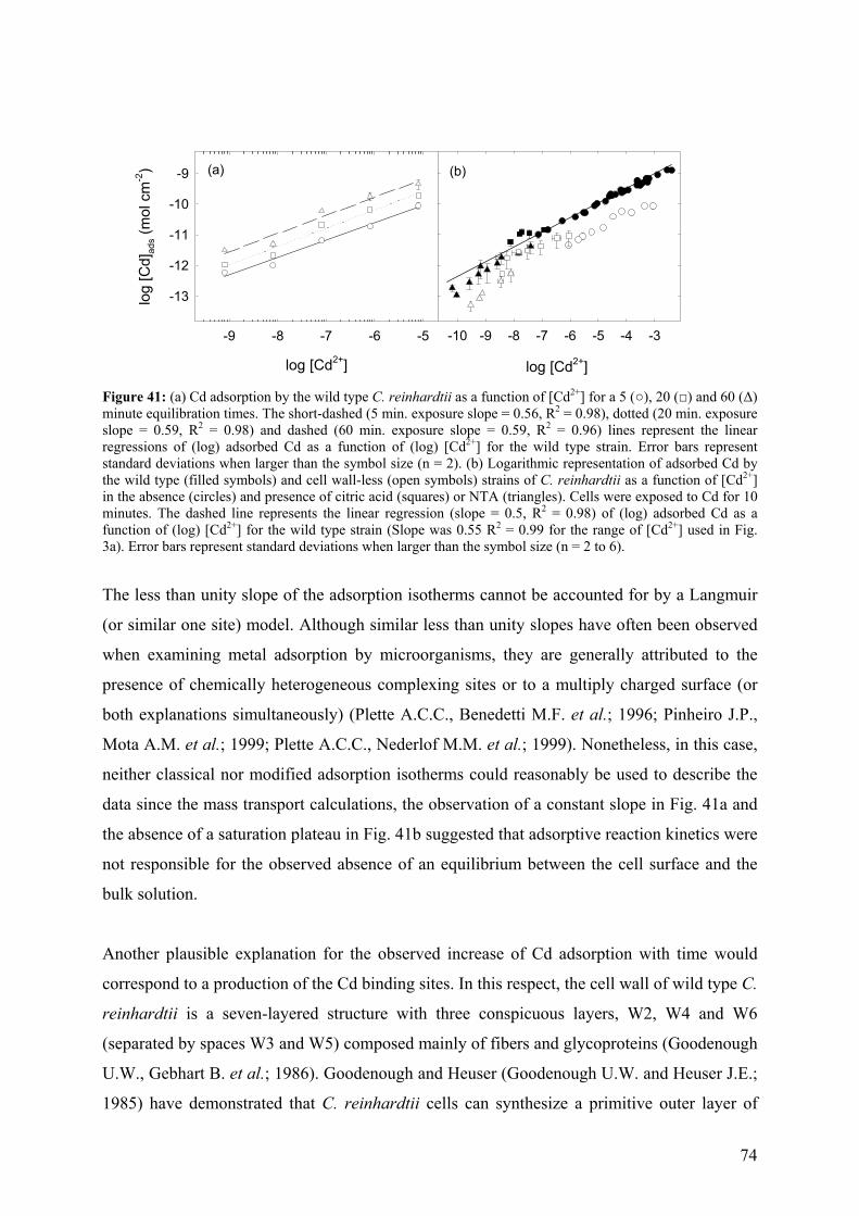
74
log [Cd2+]
-9 -8 -7 -6 -5
log
[Cd]
ads (
mol
cm
-2)
-13
-12
-11
-10
-9 (a)
log [Cd2+]
-10 -9 -8 -7 -6 -5 -4 -3
(b)
Figure 41: (a) Cd adsorption by the wild type C. reinhardtii as a function of [Cd2+] for a 5 (), 20 () and 60 (∆) minute equilibration times. The short-dashed (5 min. exposure slope = 0.56, R2 = 0.98), dotted (20 min. exposure slope = 0.59, R2 = 0.98) and dashed (60 min. exposure slope = 0.59, R2 = 0.96) lines represent the linear regressions of (log) adsorbed Cd as a function of (log) [Cd2+] for the wild type strain. Error bars represent standard deviations when larger than the symbol size (n = 2). (b) Logarithmic representation of adsorbed Cd by the wild type (filled symbols) and cell wall-less (open symbols) strains of C. reinhardtii as a function of [Cd2+] in the absence (circles) and presence of citric acid (squares) or NTA (triangles). Cells were exposed to Cd for 10 minutes. The dashed line represents the linear regression (slope = 0.5, R2 = 0.98) of (log) adsorbed Cd as a function of (log) [Cd2+] for the wild type strain (Slope was 0.55 R2 = 0.99 for the range of [Cd2+] used in Fig. 3a). Error bars represent standard deviations when larger than the symbol size (n = 2 to 6).
The less than unity slope of the adsorption isotherms cannot be accounted for by a Langmuir
(or similar one site) model. Although similar less than unity slopes have often been observed
when examining metal adsorption by microorganisms, they are generally attributed to the
presence of chemically heterogeneous complexing sites or to a multiply charged surface (or
both explanations simultaneously) (Plette A.C.C., Benedetti M.F. et al.; 1996; Pinheiro J.P.,
Mota A.M. et al.; 1999; Plette A.C.C., Nederlof M.M. et al.; 1999). Nonetheless, in this case,
neither classical nor modified adsorption isotherms could reasonably be used to describe the
data since the mass transport calculations, the observation of a constant slope in Fig. 41a and
the absence of a saturation plateau in Fig. 41b suggested that adsorptive reaction kinetics were
not responsible for the observed absence of an equilibrium between the cell surface and the
bulk solution.
Another plausible explanation for the observed increase of Cd adsorption with time would
correspond to a production of the Cd binding sites. In this respect, the cell wall of wild type C.
reinhardtii is a seven-layered structure with three conspicuous layers, W2, W4 and W6
(separated by spaces W3 and W5) composed mainly of fibers and glycoproteins (Goodenough
U.W., Gebhart B. et al.; 1986). Goodenough and Heuser (Goodenough U.W. and Heuser J.E.;
1985) have demonstrated that C. reinhardtii cells can synthesize a primitive outer layer of
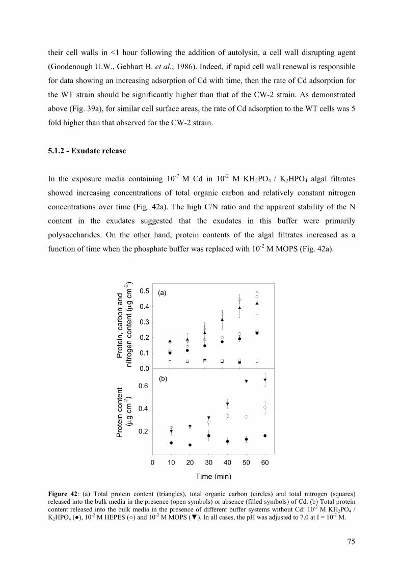
75
their cell walls in <1 hour following the addition of autolysin, a cell wall disrupting agent
(Goodenough U.W., Gebhart B. et al.; 1986). Indeed, if rapid cell wall renewal is responsible
for data showing an increasing adsorption of Cd with time, then the rate of Cd adsorption for
the WT strain should be significantly higher than that of the CW-2 strain. As demonstrated
above (Fig. 39a), for similar cell surface areas, the rate of Cd adsorption to the WT cells was 5
fold higher than that observed for the CW-2 strain.
5.1.2 - Exudate release
In the exposure media containing 10-7 M Cd in 10-2 M KH2PO4 / K2HPO4 algal filtrates
showed increasing concentrations of total organic carbon and relatively constant nitrogen
concentrations over time (Fig. 42a). The high C/N ratio and the apparent stability of the N
content in the exudates suggested that the exudates in this buffer were primarily
polysaccharides. On the other hand, protein contents of the algal filtrates increased as a
function of time when the phosphate buffer was replaced with 10-2 M MOPS (Fig. 42a).
Pro
tein
, car
bon
and
nitro
gen
cont
ent (µ g
cm
-2)
0.0
0.1
0.2
0.3
0.4
0.5
Time (min)
0 10 20 30 40 50 60
Pro
tein
con
tent
(µ
g cm
-2)
0.2
0.4
0.6
(a)
(b)
Figure 42: (a) Total protein content (triangles), total organic carbon (circles) and total nitrogen (squares) released into the bulk media in the presence (open symbols) or absence (filled symbols) of Cd. (b) Total protein content released into the bulk media in the presence of different buffer systems without Cd: 10-2 M KH2PO4 / K2HPO4 (), 10-2 M HEPES () and 10-2 M MOPS (). In all cases, the pH was adjusted to 7.0 at I = 10-2 M.
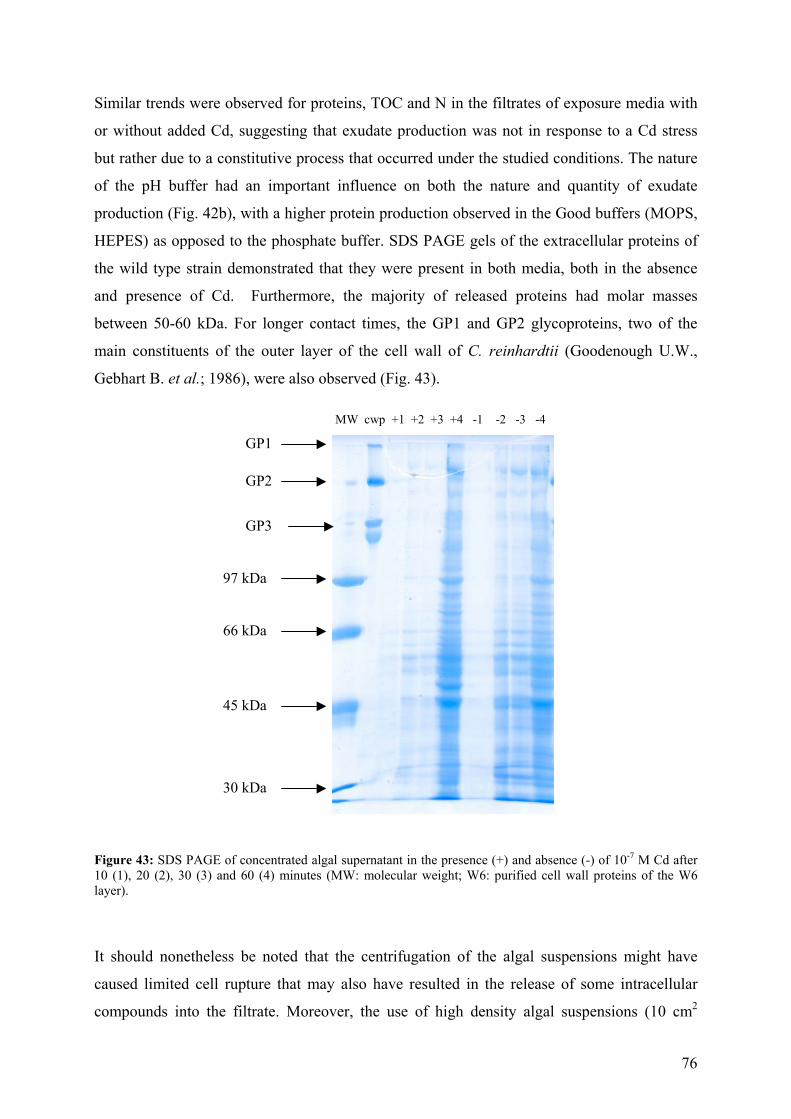
76
Similar trends were observed for proteins, TOC and N in the filtrates of exposure media with
or without added Cd, suggesting that exudate production was not in response to a Cd stress
but rather due to a constitutive process that occurred under the studied conditions. The nature
of the pH buffer had an important influence on both the nature and quantity of exudate
production (Fig. 42b), with a higher protein production observed in the Good buffers (MOPS,
HEPES) as opposed to the phosphate buffer. SDS PAGE gels of the extracellular proteins of
the wild type strain demonstrated that they were present in both media, both in the absence
and presence of Cd. Furthermore, the majority of released proteins had molar masses
between 50-60 kDa. For longer contact times, the GP1 and GP2 glycoproteins, two of the
main constituents of the outer layer of the cell wall of C. reinhardtii (Goodenough U.W.,
Gebhart B. et al.; 1986), were also observed (Fig. 43).
Figure 43: SDS PAGE of concentrated algal supernatant in the presence (+) and absence (-) of 10-7 M Cd after 10 (1), 20 (2), 30 (3) and 60 (4) minutes (MW: molecular weight; W6: purified cell wall proteins of the W6 layer).
It should nonetheless be noted that the centrifugation of the algal suspensions might have
caused limited cell rupture that may also have resulted in the release of some intracellular
compounds into the filtrate. Moreover, the use of high density algal suspensions (10 cm2
GP1
GP3
GP2
97 kDa
30 kDa
66 kDa
45 kDa
MW cwp +1 +2 +3 +4 -1 -2 -3 -4
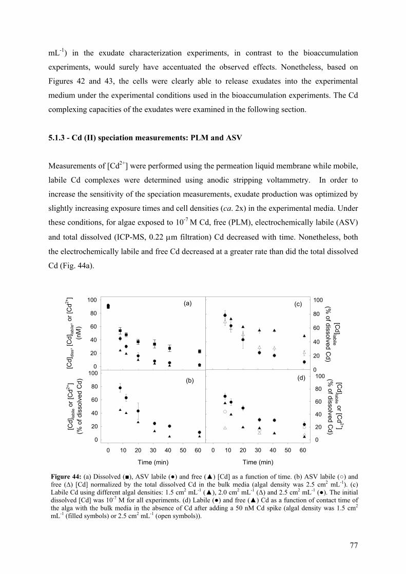
77
mL-1) in the exudate characterization experiments, in contrast to the bioaccumulation
experiments, would surely have accentuated the observed effects. Nonetheless, based on
Figures 42 and 43, the cells were clearly able to release exudates into the experimental
medium under the experimental conditions used in the bioaccumulation experiments. The Cd
complexing capacities of the exudates were examined in the following section.
5.1.3 - Cd (II) speciation measurements: PLM and ASV
Measurements of [Cd2+] were performed using the permeation liquid membrane while mobile,
labile Cd complexes were determined using anodic stripping voltammetry. In order to
increase the sensitivity of the speciation measurements, exudate production was optimized by
slightly increasing exposure times and cell densities (ca. 2x) in the experimental media. Under
these conditions, for algae exposed to 10-7 M Cd, free (PLM), electrochemically labile (ASV)
and total dissolved (ICP-MS, 0.22 µm filtration) Cd decreased with time. Nonetheless, both
the electrochemically labile and free Cd decreased at a greater rate than did the total dissolved
Cd (Fig. 44a).
Time (min)
0 10 20 30 40 50 60
[Cd]labile or [C
d2+]
(% of dissolved C
d)
0
20
40
60
80
100
[Cd]labile
(% of dissolved C
d)
0
20
40
60
80
100(c)
(d)
[Cd]
diss
, [C
d]la
bile, o
r [C
d2+]
(nM
)
0
20
40
60
80
100
Time (min)
0 10 20 30 40 50 60
[Cd]
labi
le o
r [C
d2+]
(% o
f dis
solv
ed C
d)
0
20
40
60
80
100
(a)
(b)
Figure 44: (a) Dissolved (), ASV labile () and free () [Cd] as a function of time. (b) ASV labile () and free (∆) [Cd] normalized by the total dissolved Cd in the bulk media (algal density was 2.5 cm2 mL-1). (c) Labile Cd using different algal densities: 1.5 cm2 mL-1 (), 2.0 cm2 mL-1 (∆) and 2.5 cm2 mL-1 (). The initial dissolved [Cd] was 10-7 M for all experiments. (d) Labile () and free () Cd as a function of contact time of the alga with the bulk media in the absence of Cd after adding a 50 nM Cd spike (algal density was 1.5 cm2 mL-1 (filled symbols) or 2.5 cm2 mL-1 (open symbols)).
78
As expected, the measured concentration of labile Cd was greater than the concentration of
Cd2+ measured by the PLM (Fig. 44ab) due to the dependence of the voltammetric signal on
the concentration of both the free ion and some of its complexes (Pei J., Tercier-Waeber M.-
L. et al.; 2000). The ASV/PLM ratio ranged from near unity values at the beginning of the
experiments to values between 4 and 8 at the end of the accumulation time. The rapid
decrease in both the PLM and ASV signals coupled to the absence of a shift in the potential of
the reoxidation peak in the presence of exudates strongly suggested either that the ligands had
a high affinity for Cd (i.e. kinetically inert complexes), or that the complexes were large and
thus relatively immobile (in the time frame of the voltammetric experiment). Based upon an
estimated stability constant of 103.3 – 105.3 M-1 for the interaction of Cd with the cell wall
(assuming equilibrium and saturation at 2 x 10⁻⁹ mol cm-2, Fig. 41) and the result from the
protein gels, the second explanation would appear more likely.
The effect of algal cell density was also important (Fig. 44c) with a more pronounced
decrease in the ASV labile Cd when the algal density was increased from 1.5 to 2.5 cm2 mL-1.
Indeed, following a 20 minute exposure, 40%-60% of the dissolved Cd was bound in ASV
labile complexes. A similar decrease was observed when Cd was added to the algal
supernatant obtained for cells that were not exposed to Cd (Fig. 44d). Such results support our
previous observation that protein production was a constitutive process rather than a response
to Cd stress.
5.1.4 - Implications for bioaccumulation studies
It is possible to speculate that the nature of the exudates and their observation for nearly all
conditions suggested that the production of exudates by algae and their release into the bulk
media may be important in natural waters in reducing free metal concentrations to low levels
(Town R.M. and Filella M.; 2000). In this study, theoretical models describing metal
adsorption to homogenous or heterogeneous algal surfaces with or without charge correction
(Plette A.C.C., Benedetti M.F. et al.; 1996; Plette A.C.C., Nederlof M.M. et al.; 1999) were
unable to relate Cd adsorption by C. reinhardtii to the speciation of Cd in the bulk media. The
observation of an increase in adsorbed Cd with time suggested that Cd binding sites were
being continuously produced. An important implication of this result is that the extracellular
metal binding sites will never be at simple chemical equilibrium with respect to the bulk
solution (a requirement of both the biotic ligand and free ion activity models). Cd adsorption
79
to C. reinhardtii is thus a dynamic rather than a steady-state process involving a number of
adsorption steps. Because the CW-2 strain is thought to produce the same amount of cell wall
material as the wild type strain (Davis R.D. and Plaskitt A.; 1971), the observed differences in
Cd adsorption for the two strains are likely due to a greater release of the cell wall
components for the CW-2 mutant and a corresponding greater renewal of binding sites for the
WT strain. Indeed, the rate of Cd adsorption for CW-2 strain was 5 times lower than that
observed for the WT strain (Fig. 39a). The observation of a less than unity slope in the (log)
adsorbed Cd - (log) [Cd2+] plot was attributed to cell surface renewal in addition to the more
usual heterogeneity and charge effects (Pinheiro J.P., Mota A.M. et al.; 1999; Wilkinson K.J.
and Buffle J.; 2004). It is important to note that a rapid rate of Cd internalization further
distinguishes C. reinhardtii from several other algal species typically used in bioaccumulation
and toxicity studies. Nonetheless, while the rapid uptake coupled with a continuous
adsorption was likely responsible for the observed depletion of dissolved Cd in the bulk
solution, [Cd2+] was also significantly reduced by the complexation of Cd by the exudates.
While the production of metal binding ligands has been previously observed in response to a
Cd stress (Smiejan A., Wilkinson K.J. et al.; 2003), the continual release of the cell wall
components into the medium under a variety of physicochemical conditions was unexpected,
especially for such short exposure times (Fig. 44).
Finally, although several authors (Twiss M.R., Auclair J.-C. et al.; 2000) advocate the use of
N-substituted aminosulfonic acids buffers such as HEPES for metal bioaccumulation and
toxicity experiments due to their low complexation capacity (Mash H.E., Chin Y.-P. et al.;
2003), exudate production was clearly enhanced in their presence (Vasconcelos M.T.S.D. and
Leal M.F.C.; 2002). On the other hand, while macronutriment buffers such as the phosphate
buffer may have less effect on the cell wall, they are generally not recommended due to their
potential metabolic roles or their ability to precipitate or complex polyvalent cations and thus
reduce biouptake (Khummongkol D., Canterford G.S. et al.; 1982). Based upon these results,
“ideal” pH buffers likely do not exist for metal bioaccumulation or toxicity experiments.
Inevitable effects of the buffers will need to be taken into account and controlled, where
possible, through the reduction of cellular densities, exposure times, and by the selection of
suitable pH buffers. The use of chemostats may also be a useful means to minimize changes
in both metal concentrations (typically verified) and speciation (rarely verified).
80
5.3 – Conclusions
Cd adsorption to living cells, in this case, C. reinhardtii, is a complex, dynamic process that
cannot be described by simple equilibrium models. Slow Cd adsorption dynamics was
attributed to the production of new binding sites, especially for low Cd concentrations. Due
to the continual release of cell wall proteins and the renewal of binding sites on the cell wall,
steady-state conditions were never achieved under the studied conditions. The cell wall was
highly complexing: wild type C. reinhardtii bound 10x more Cd than the cell wall-less strain.
Under the studied conditions, the production and release of cell wall components by algae in
media with or without Cd demonstrated that exudate release was not in response to Cd stress
but rather a constitutive process. The ubiquitous release of the exudates greatly affected Cd
speciation in the exposure media by increasing Cd complexation.
81
6 – CADMIUM UPTAKE BY A GREEN ALGA CAN BE PREDICTED BY
EQUILIBRIUM MODELLING
(Envir. Sci. & Technol., in press)
In environmental systems, only a small proportion of trace metals is found as free hydrated
ions with the majority of the metal being complexed by ligands (Eq. 2) or being sorbed to
surfaces. The physicochemical distribution of a metal among its distinct chemical species i.e.,
metal speciation, greatly affects its bioavailability to aquatic organisms. Biological effects
have generally been attributed to specific chemical reactions of the metal species with
sensitive sites such as carriers (Eq. 3), the subsequent metal internalization flux or
accumulated metal concentrations (for reviews see (Sunda W.G. and Huntsman S.A.; 1998;
Wilkinson K.J. and Buffle J.; 2004; Slaveykova V.I. and Wilkinson K.J.; 2005)).
Mz+ + Ln- ↔ M-L(z-n) (complexation in the bulk media by a ligand, Ln-) (2)
Mz+ + Rcell ↔ M-Rcell (adsorption to transport sites, Rcell, at the cell surface) (3)
M-Rcell → Mcell (internalization of surface bound metal) (3)
One of the key problems of environmental toxicology remains how to best relate metal
exposures to their biological effects. Most research has been based upon the assumption that
local equilibrium is attained between the metal in the bulk solution and that adsorbed to
sensitive sites on the cell surface (Rcell), i.e. metal internalization flux is rate-limiting. In such
a case, the metal induced biological response can be related either to the free-metal ion
concentration in solution, [Mz+], (basis of the Free Ion Activity Model, FIAM, (Morel F.M.M.
and Hering J.G.; 1993; Campbell P.G.C.; 1995)) or to the metal bound to sensitive sites at the
surface of the organism, M-Rcell, (basis of the Biotic Ligand Model, BLM, (Playle R.C.;
1998; Hassler C.S., Slaveykova V.I. et al.; 2004)). Under such steady-state conditions, the
metal internalization flux, Jint, should follow a Michaelis-Menten hyperbolic rate law (Morel
F.M.M. and Hering J.G.; 1993; Sunda W.G. and Huntsman S.A.; 1998) (Eq. 4).
[ ][ ]+
+
+=
zM
z
MKMJ
J maxint
(12)
82
where Jmax is the maximal internalization flux and KM is the half-saturation constant. For
conditions in which [Mz+] >> KM, the sites become saturated and the metal internalization flux
becomes independent of [Mz+] (Jint = Jmax). On the other hand, for [Mz+] << KM, which is the
case for most natural waters, most of the metal transport sites will be available for binding
(Morel F.M.M. and Hering J.G.; 1993) and the uptake rate should be directly proportional to
either the concentration of free metal in solution or to the metal bound to metal transport sites
(Jint = Jmax KM-1 [Mz+] = kint M-Rcell where kint is the internalization rate constant of the
membrane transport process; (Hassler C.S., Slaveykova V.I. et al.; 2004)). Examples for
which internalization is not rate-limiting and the equilibrium models are predicted to fail have
been identified in several cases, including a kinetic limitation of Fe uptake by Thalassiosira
weissflogii (Hudson R.J.M. and Morel F.M.M.; 1990) and a diffusive limitation of Zn or Ag
uptake by green algae (Hassler C.S. and Wilkinson K.J.; 2003; Lee D.-Y., Fortin C. et al.;
2004).
If internalization is rate-limiting such that the uptake at sensitive site on the membrane surface
is at rate-limiting (in agreement with the FIAM and BLM), both the presence of ions that
compete for transport sites and ligands that bind the free metal in solution should decrease
metal biouptake. For example, trace metals may compete with essential metals for active
enzymes, membrane protein sites or biologically active groups (Visviki I. and Rachlin J.W.;
1991) since metal coordination sites in microorganisms are rarely specific for a single metal.
Although competing metals have been shown to act antagonistically, synergistically or
additively (Visviki I. and Rachlin J.W.; 1994), in the equilibrium models, competition is
always assumed to be antagonistic. In such a case, internalization fluxes can be described by
(Galceran J. and van Leeuwen H.P.; 2004; Wilkinson K.J. and Buffle J.; 2004):
1][C][M][M
nz
maxint ++=
−+
−+
−+
RcellCRcellM
RcellMz
KKKJ
J (13)
where [Cn+] is the concentration of a competing metal or proton, KM-Rcell and KC-Rcell are values
of the affinity constants for the interaction of the metal of interest and a competing metal or
proton and Jmax is the maximal metal uptake flux. In addition to direct competition with metal
uptake sites, changes in pH can modify metal speciation in solution, decrease the overall
columbic attraction for the metal or modify the conformation of metal transport proteins
(Campbell P.G.C. and Stokes P.M.; 1985; Stumm W. and Morgan J.J.; 1996; Kleijn J.M. and
83
van Leeuwen H.P.; 2000; Wilkinson K.J. and Buffle J.; 2004). In the case of cadmium,
hydroxo and carbonato complexes can be present in significant quantities above pH 8.0
(Bilinski H., Huston R. et al.; 1976).
Cd speciation is also greatly influenced by the presence of colloidal and organic ligands in
natural waters, including the humic (HA) and fulvic (FA) acids (Xue H. and Sigg L.; 1999;
Ellwood M.J.; 2004). In spite of its role in Cd speciation, there is currently no consensus on
the role of organic matter on Cd bioavailability. Natural organic matter (NOM) has
previously been shown to have stimulatory, negative or negligible effects (Hare L. and Tessier
A.; 1996; Parent L., Twiss M.R. et al.; 1996; Campbell P.G.C., Twiss M.R. et al.; 1997;
Slaveykova V.I., Wilkinson K.J. et al.; 2003; Voets J., Bervoets L. et al.; 2004) on metal
biouptake or toxicity.
The objective of this work was thus to evaluate the influence of Cd speciation, pH and
competing ions on Cd uptake by the unicellular green alga Chlamydomonas reinhardtii. Cd
uptake of a cell wall-less strain of the same species, used in the past to examine the role of
cell wall with respect to metal tolerance (Cain J.R. and Allen R.K.; 1980; Macfie S.M.,
Tarmohamed Y. et al.; 1994; Macfie S.M. and Welbourn P.M.; 2000), was also examined.
Special attention was paid to determining whether existing models of metal biouptake and
toxicity (FIAM, BLM) were appropriate quantitative predictors of the role of solution
physicochemistry on Cd uptake.
6.1 – Results and discussion
6.1.1 - Cd internalization
Due to an important Cd adsorption by the algae (Kola H., Langlera L.M. et al.; 2004) at these
cellular densities, dissolved [Cd] in the non-buffered media decreased by ≤30% for the WT
and ≤20% for the CW-2 strain in the first few minutes of the experiment for dissolved
[Cd]<10-6 M (Fig. 45a). For both strains, a further small decrease of dissolved [Cd] was
observed over the remainder of the experiment due mainly to Cd internalization (Fig. 45b).
For a similar [Cd2+] in the experimental medium (ca. 2 x 10⁻⁷ M), the WT strain accumulated
more Cd than the CW-2 strain (Fig. 45b). For the CW-2 strain, dissolved [Cd] increased for
accumulation times >25 minutes (Fig. 45a), suggesting that Cd was released into the bulk
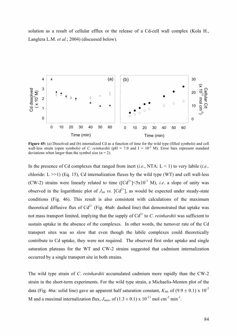
84
solution as a result of cellular efflux or the release of a Cd-cell wall complex (Kola H.,
Langlera L.M. et al.; 2004) (discussed below).
Time (min)
0 10 20 30 40 50 60
Cellular C
d (x 10
11 mol cm
-2)
0
10
20
30
Time (min)
0 10 20 30 40 50 60
Cd
diss
olve
d ( x
107
M)
0
1
2
3
4 (a) (b)
Figure 45: (a) Dissolved and (b) internalized Cd as a function of time for the wild type (filled symbols) and cell wall-less strain (open symbols) of C. reinhardtii (pH = 7.0 and I = 10-2 M). Error bars represent standard deviations when larger than the symbol size (n = 2).
In the presence of Cd complexes that ranged from inert (i.e., NTA: L < 1) to very labile (i.e.,
chloride: L >>1) (Eq. 15), Cd internalization fluxes by the wild type (WT) and cell wall-less
(CW-2) strains were linearly related to time ([Cd2+]<5x10⁻⁷ M), i.e. a slope of unity was
observed in the logarithmic plot of Jint vs. [Cd2+], as would be expected under steady-state
conditions (Fig. 46). This result is also consistent with calculations of the maximum
theoretical diffusive flux of Cd2+ (Fig. 46ab: dashed line) that demonstrated that uptake was
not mass transport limited, implying that the supply of Cd2+ to C. reinhardtii was sufficient to
sustain uptake in the absence of the complexes. In other words, the turnover rate of the Cd
transport sites was so slow that even though the labile complexes could theoretically
contribute to Cd uptake, they were not required. The observed first order uptake and single
saturation plateaus for the WT and CW-2 strains suggested that cadmium internalization
occurred by a single transport site in both strains.
The wild type strain of C. reinhardtii accumulated cadmium more rapidly than the CW-2
strain in the short-term experiments. For the wild type strain, a Michaelis-Menten plot of the
data (Fig. 46a: solid line) gave an apparent half saturation constant, KM, of (9.9 ± 0.1) x 10-7
M and a maximal internalization flux, Jmax, of (1.3 ± 0.1) x 10-11 mol cm-2 min-1.
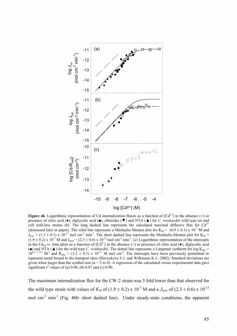
85
log
J int
(mol
cm
-2 m
in-1
)
-15
-14
-13
-12
-11
log
J int
(m
ol c
m-2
min
-1)
-15
-14
-13
-12
-11
(a)
(b)
log [Cd2+] (M)
-10 -9 -8 -7 -6 -5 -4
log
Cd-
Rce
ll (m
ol c
m-2
)
-14
-13
-12
-11
-10 (c)
Figure 46. Logarithmic representation of Cd internalization fluxes as a function of [Cd2+] in the absence () or presence of citric acid (), diglycolic acid (), chlorides () and NTA () for C. reinhardtii wild type (a) and cell wall-less strains (b). The long dashed line represents the calculated maximal diffusive flux for Cd2+ (discussed later in paper). The solid line represents a Michaelis-Menten plot for KM = (0.9 ± 0.1) x 10-7 M and Jmax = (1.3 ± 0.1) x 10-11 mol cm-2 min-1. The short dashed line represents the Michaelis-Menten plot for KM = (1.9 ± 0.2) x 10-7 M and Jmax = (2.3 ± 0.6) x 10-12 mol cm-2 min-1. (c) Logarithmic representation of the intercepts in the Cdint vs. time plots as a function of [Cd2+] in the absence () or presence of citric acid (), diglycolic acid () and NTA () for the wild type C. reinhardtii. The dotted line represents a Langmuir isotherm for log Kads = 106.2 ± 0.1 M-1 and Rmax = (3.2 ± 0.1) x 10-11 M mol cm-2. The intercepts have been previously postulated to represent metal bound to the transport sites (Slaveykova V.I. and Wilkinson K.J.; 2002). Standard deviations are given when larger than the symbol size (n = 2 to 6). A regression of the calculated versus experimental data gave significant r2 values of (a) 0.98, (b) 0.87 and (c) 0.90.
The maximum internalization flux for the CW-2 strain was 5 fold lower than that observed for
the wild type strain with values of KM of (1.9 ± 0.2) x 10-7 M and a Jmax of (2.3 ± 0.6) x 10-12
mol cm-2 min-1 (Fig. 46b: short dashed line). Under steady-state conditions, the apparent
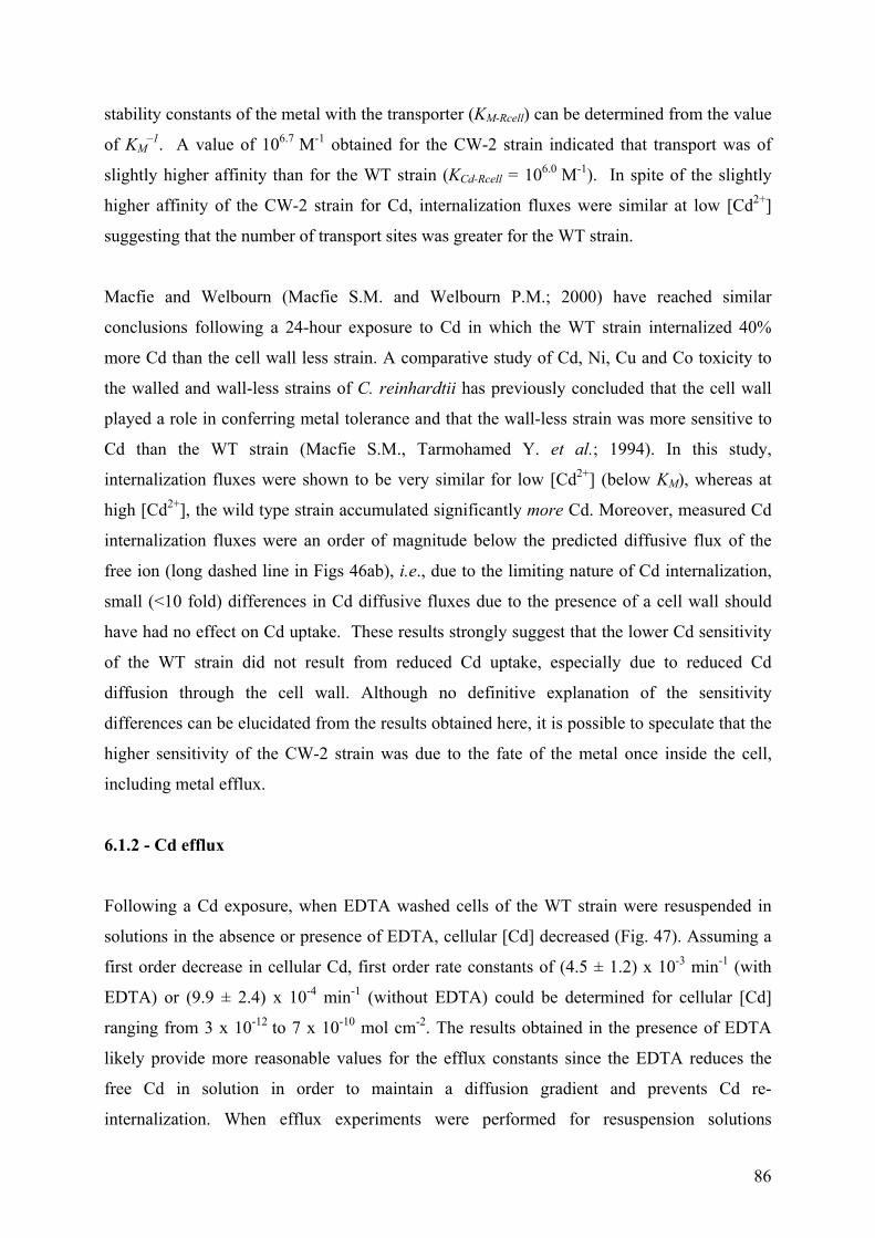
86
stability constants of the metal with the transporter (KM-Rcell) can be determined from the value
of KM–1. A value of 106.7 M-1 obtained for the CW-2 strain indicated that transport was of
slightly higher affinity than for the WT strain (KCd-Rcell = 106.0 M-1). In spite of the slightly
higher affinity of the CW-2 strain for Cd, internalization fluxes were similar at low [Cd2+]
suggesting that the number of transport sites was greater for the WT strain.
Macfie and Welbourn (Macfie S.M. and Welbourn P.M.; 2000) have reached similar
conclusions following a 24-hour exposure to Cd in which the WT strain internalized 40%
more Cd than the cell wall less strain. A comparative study of Cd, Ni, Cu and Co toxicity to
the walled and wall-less strains of C. reinhardtii has previously concluded that the cell wall
played a role in conferring metal tolerance and that the wall-less strain was more sensitive to
Cd than the WT strain (Macfie S.M., Tarmohamed Y. et al.; 1994). In this study,
internalization fluxes were shown to be very similar for low [Cd2+] (below KM), whereas at
high [Cd2+], the wild type strain accumulated significantly more Cd. Moreover, measured Cd
internalization fluxes were an order of magnitude below the predicted diffusive flux of the
free ion (long dashed line in Figs 46ab), i.e., due to the limiting nature of Cd internalization,
small (<10 fold) differences in Cd diffusive fluxes due to the presence of a cell wall should
have had no effect on Cd uptake. These results strongly suggest that the lower Cd sensitivity
of the WT strain did not result from reduced Cd uptake, especially due to reduced Cd
diffusion through the cell wall. Although no definitive explanation of the sensitivity
differences can be elucidated from the results obtained here, it is possible to speculate that the
higher sensitivity of the CW-2 strain was due to the fate of the metal once inside the cell,
including metal efflux.
6.1.2 - Cd efflux
Following a Cd exposure, when EDTA washed cells of the WT strain were resuspended in
solutions in the absence or presence of EDTA, cellular [Cd] decreased (Fig. 47). Assuming a
first order decrease in cellular Cd, first order rate constants of (4.5 ± 1.2) x 10-3 min-1 (with
EDTA) or (9.9 ± 2.4) x 10-4 min-1 (without EDTA) could be determined for cellular [Cd]
ranging from 3 x 10-12 to 7 x 10⁻¹⁰ mol cm-2. The results obtained in the presence of EDTA
likely provide more reasonable values for the efflux constants since the EDTA reduces the
free Cd in solution in order to maintain a diffusion gradient and prevents Cd re-
internalization. When efflux experiments were performed for resuspension solutions
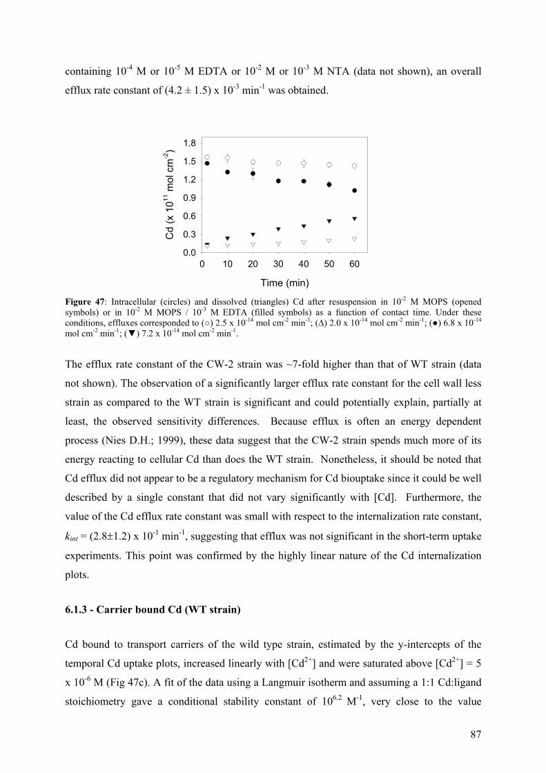
87
containing 10-4 M or 10-5 M EDTA or 10-2 M or 10-3 M NTA (data not shown), an overall
efflux rate constant of (4.2 ± 1.5) x 10-3 min-1 was obtained.
Time (min)
0 10 20 30 40 50 60
Cd
(x 1
011 m
ol c
m-2
)
0.0
0.3
0.6
0.9
1.2
1.5
1.8
Figure 47: Intracellular (circles) and dissolved (triangles) Cd after resuspension in 10-2 M MOPS (opened symbols) or in 10-2 M MOPS / 10-3 M EDTA (filled symbols) as a function of contact time. Under these conditions, effluxes corresponded to () 2.5 x 10⁻¹⁴ mol cm-2 min-1; (∆) 2.0 x 10⁻¹⁴ mol cm-2 min-1; () 6.8 x 10⁻¹⁴ mol cm-2 min-1; () 7.2 x 10⁻¹⁴ mol cm-2 min-1.
The efflux rate constant of the CW-2 strain was ~7-fold higher than that of WT strain (data
not shown). The observation of a significantly larger efflux rate constant for the cell wall less
strain as compared to the WT strain is significant and could potentially explain, partially at
least, the observed sensitivity differences. Because efflux is often an energy dependent
process (Nies D.H.; 1999), these data suggest that the CW-2 strain spends much more of its
energy reacting to cellular Cd than does the WT strain. Nonetheless, it should be noted that
Cd efflux did not appear to be a regulatory mechanism for Cd biouptake since it could be well
described by a single constant that did not vary significantly with [Cd]. Furthermore, the
value of the Cd efflux rate constant was small with respect to the internalization rate constant,
kint = (2.8±1.2) x 10-1 min-1, suggesting that efflux was not significant in the short-term uptake
experiments. This point was confirmed by the highly linear nature of the Cd internalization
plots.
6.1.3 - Carrier bound Cd (WT strain)
Cd bound to transport carriers of the wild type strain, estimated by the y-intercepts of the
temporal Cd uptake plots, increased linearly with [Cd2+] and were saturated above [Cd2+] = 5
x 10-6 M (Fig 47c). A fit of the data using a Langmuir isotherm and assuming a 1:1 Cd:ligand
stoichiometry gave a conditional stability constant of 106.2 M-1, very close to the value
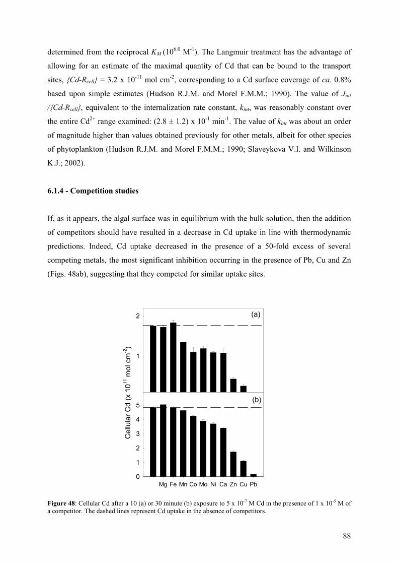
88
determined from the reciprocal KM (106.0 M-1). The Langmuir treatment has the advantage of
allowing for an estimate of the maximal quantity of Cd that can be bound to the transport
sites, Cd-Rcell = 3.2 x 10⁻¹¹ mol cm-2, corresponding to a Cd surface coverage of ca. 0.8%
based upon simple estimates (Hudson R.J.M. and Morel F.M.M.; 1990). The value of Jint
/Cd-Rcell, equivalent to the internalization rate constant, kint, was reasonably constant over
the entire Cd2+ range examined: (2.8 ± 1.2) x 10-1 min-1. The value of kint was about an order
of magnitude higher than values obtained previously for other metals, albeit for other species
of phytoplankton (Hudson R.J.M. and Morel F.M.M.; 1990; Slaveykova V.I. and Wilkinson
K.J.; 2002).
6.1.4 - Competition studies
If, as it appears, the algal surface was in equilibrium with the bulk solution, then the addition
of competitors should have resulted in a decrease in Cd uptake in line with thermodynamic
predictions. Indeed, Cd uptake decreased in the presence of a 50-fold excess of several
competing metals, the most significant inhibition occurring in the presence of Pb, Cu and Zn
(Figs. 48ab), suggesting that they competed for similar uptake sites.
Cel
lula
r Cd
(x 1
011 m
ol c
m-2)
1
2
Mg Fe Mn Co Mo Ni Ca Zn Cu Pb0
1
2
3
4
5
(a)
(b)
Figure 48: Cellular Cd after a 10 (a) or 30 minute (b) exposure to 5 x 10⁻⁷ M Cd in the presence of 1 x 10⁻⁵ M of a competitor. The dashed lines represent Cd uptake in the absence of competitors.
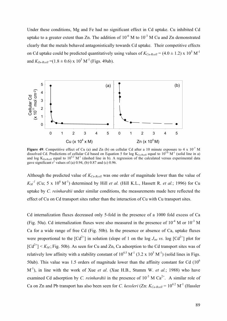
89
Under these conditions, Mg and Fe had no significant effect in Cd uptake. Cu inhibited Cd
uptake to a greater extent than Zn. The addition of 10-6 M to 10-3 M Cu and Zn demonstrated
clearly that the metals behaved antagonistically towards Cd uptake. Their competitive effects
on Cd uptake could be predicted quantitatively using values of KCu-Rcell = (4.0 ± 1.2) x 105 M-1
and KZn-Rcell =(1.8 ± 0.6) x 105 M-1 (Figs. 49ab).
Cu (x 104 x M)
0 1 2 3 4 5
Cel
lula
r Cd
(x 1
011 m
ol c
m-2)
0
1
2
3
4
5
Zn (x 104 M)
0 1 2 3 4 5
(a) (b)
Figure 49: Competitive effect of Cu (a) and Zn (b) on cellular Cd after a 10 minute exposure to 4 x 10⁻⁷ M dissolved Cd. Predictions of cellular Cd based on Equation 5 for log KCu-Rcell equal to 105.6 M-1 (solid line in a) and log KZn-Rcell equal to 105.2 M-1 (dashed line in b). A regression of the calculated versus experimental data gave significant r2 values of (a) 0.94, (b) 0.87 and (c) 0.96.
Although the predicted value of KCu-Rcell was one order of magnitude lower than the value of
KM-1 (Cu; 5 x 10⁶ M-1) determined by Hill et al. (Hill K.L., Hassett R. et al.; 1996) for Cu
uptake by C. reinhardtii under similar conditions, the measurements made here reflected the
effect of Cu on Cd transport sites rather than the interaction of Cu with Cu transport sites.
Cd internalization fluxes decreased only 5-fold in the presence of a 1000 fold excess of Ca
(Fig. 50a). Cd internalization fluxes were also measured in the presence of 10-4 M or 10-3 M
Ca for a wide range of free Cd (Fig. 50b). In the presence or absence of Ca, uptake fluxes
were proportional to the [Cd2+] in solution (slope of 1 on the log Jint vs. log [Cd2+] plot for
[Cd2+] < KM ; Fig. 50b). As seen for Cu and Zn, Ca adsorption to the Cd transport sites was of
relatively low affinity with a stability constant of 104.5 M-1 (3.2 x 10⁵ M-1) (solid lines in Figs.
50ab). This value was 1.5 orders of magnitude lower than the affinity constant for Cd (106
M-1), in line with the work of Xue et al. (Xue H.B., Stumm W. et al.; 1988) who have
examined Cd adsorption by C. reinhardtii in the presence of 10-3 M Ca2+. A similar role of
Ca on Zn and Pb transport has also been seen for C. kessleri (Zn: KCa-Rcell = 104.2 M-1 (Hassler
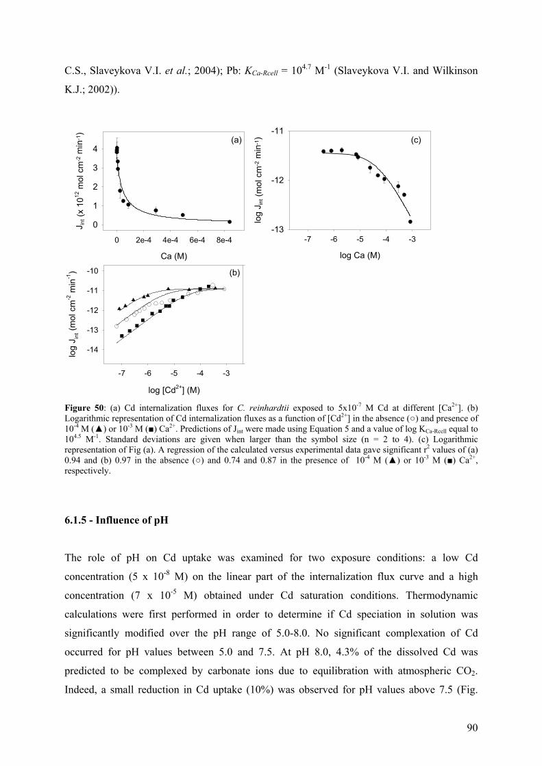
90
C.S., Slaveykova V.I. et al.; 2004); Pb: KCa-Rcell = 104.7 M-1 (Slaveykova V.I. and Wilkinson
K.J.; 2002)).
log [Cd2+] (M)
-7 -6 -5 -4 -3
log
J int (
mol
cm
-2 m
in-1
)
-14
-13
-12
-11
-10
Ca (M)
0 2e-4 4e-4 6e-4 8e-4
J int (
x 10
12 m
ol c
m-2
min
-1)
0
1
2
3
4(a)
(b)
log Ca (M)
-7 -6 -5 -4 -3
log
J int (
mol
cm
-2 m
in-1
)
-13
-12
-11(c)
Figure 50: (a) Cd internalization fluxes for C. reinhardtii exposed to 5x10⁻⁷ M Cd at different [Ca2+]. (b) Logarithmic representation of Cd internalization fluxes as a function of [Cd2+] in the absence () and presence of 10-4 M () or 10-3 M () Ca2+. Predictions of Jint were made using Equation 5 and a value of log KCa-Rcell equal to 104.5 M-1. Standard deviations are given when larger than the symbol size (n = 2 to 4). (c) Logarithmic representation of Fig (a). A regression of the calculated versus experimental data gave significant r2 values of (a) 0.94 and (b) 0.97 in the absence () and 0.74 and 0.87 in the presence of 10-4 M () or 10-3 M () Ca2+, respectively.
6.1.5 - Influence of pH
The role of pH on Cd uptake was examined for two exposure conditions: a low Cd
concentration (5 x 10⁻⁸ M) on the linear part of the internalization flux curve and a high
concentration (7 x 10⁻⁵ M) obtained under Cd saturation conditions. Thermodynamic
calculations were first performed in order to determine if Cd speciation in solution was
significantly modified over the pH range of 5.0-8.0. No significant complexation of Cd
occurred for pH values between 5.0 and 7.5. At pH 8.0, 4.3% of the dissolved Cd was
predicted to be complexed by carbonate ions due to equilibration with atmospheric CO2.
Indeed, a small reduction in Cd uptake (10%) was observed for pH values above 7.5 (Fig.
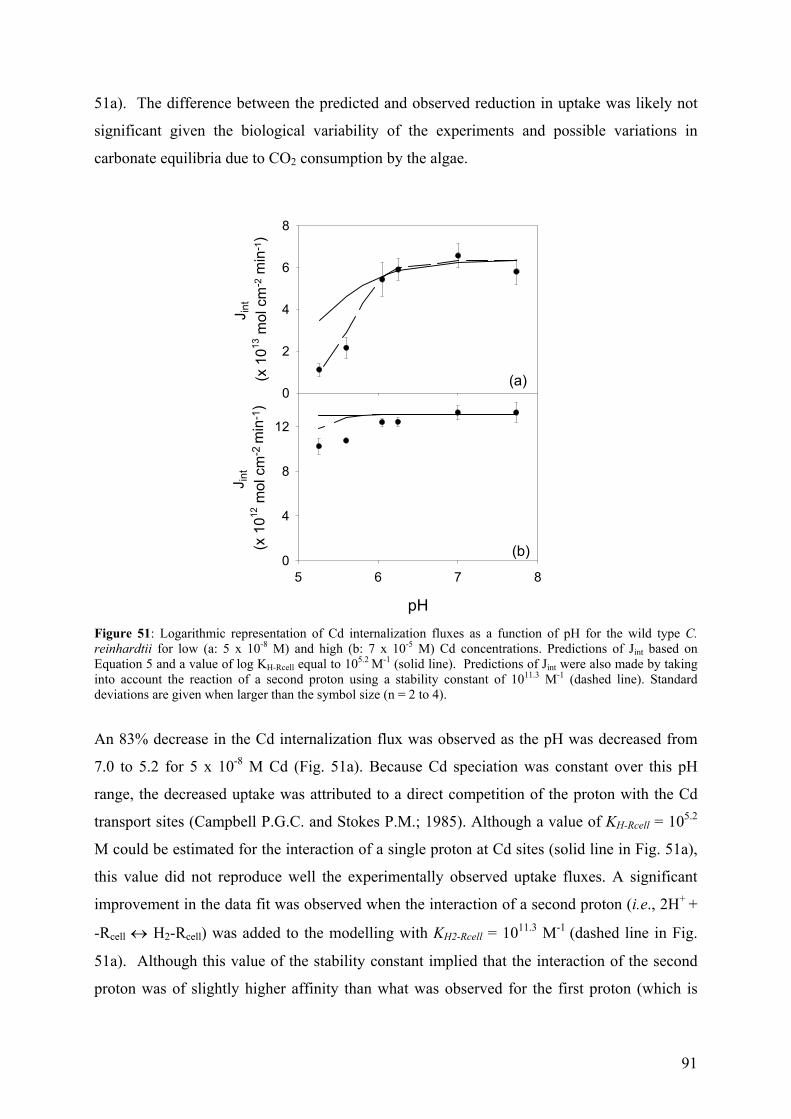
91
51a). The difference between the predicted and observed reduction in uptake was likely not
significant given the biological variability of the experiments and possible variations in
carbonate equilibria due to CO2 consumption by the algae.
J int
(x 1
013 m
ol c
m-2
min
-1)
0
2
4
6
8
pH
5 6 7 8
J int
(x 1
012 m
ol c
m-2
min
-1)
0
4
8
12
(a)
(b)
Figure 51: Logarithmic representation of Cd internalization fluxes as a function of pH for the wild type C. reinhardtii for low (a: 5 x 10⁻⁸ M) and high (b: 7 x 10⁻⁵ M) Cd concentrations. Predictions of Jint based on Equation 5 and a value of log KH-Rcell equal to 105.2 M-1 (solid line). Predictions of Jint were also made by taking into account the reaction of a second proton using a stability constant of 1011.3 M-1 (dashed line). Standard deviations are given when larger than the symbol size (n = 2 to 4).
An 83% decrease in the Cd internalization flux was observed as the pH was decreased from
7.0 to 5.2 for 5 x 10⁻⁸ M Cd (Fig. 51a). Because Cd speciation was constant over this pH
range, the decreased uptake was attributed to a direct competition of the proton with the Cd
transport sites (Campbell P.G.C. and Stokes P.M.; 1985). Although a value of KH-Rcell = 105.2
M could be estimated for the interaction of a single proton at Cd sites (solid line in Fig. 51a),
this value did not reproduce well the experimentally observed uptake fluxes. A significant
improvement in the data fit was observed when the interaction of a second proton (i.e., 2H+ +
-Rcell ↔ H2-Rcell) was added to the modelling with KH2-Rcell = 1011.3 M-1 (dashed line in Fig.
51a). Although this value of the stability constant implied that the interaction of the second
proton was of slightly higher affinity than what was observed for the first proton (which is
92
unlikely), the constants were reasonable given the number of data points and similar to
literature values of the (single) proton interaction obtained from titration experiments (104.9 M-
1, (Kiefer E., Sigg L. et al.; 1997)).
On the other hand, for [Cd] resulting in saturation of the transport sites (i.e. [Cd2+] = 7 x 10⁻⁵
M), the above proton affinity constants greatly underpredicted the observed 17% decrease in
Cd uptake flux at pH 5.2 (Fig. 51b). Although low pH or high [Cd2+] can reduce the overall
(negative) algal surface charge, thereby reducing the electrostatic component of the
interaction with the algal surface (Slaveykova V.I. and Wilkinson K.J.; 2003), this does not
appear to be the case here since an overprediction, rather than an underprediction of the
proton effect would have been expected. Furthermore, electrophoretic mobility (EPM)
measurements of the algal cells, reflective of the overall surface potential of the algae, were
fairly constant, (-9 ± 3) x 10-9 m2 V-1 s-1 between pH 5 and 8. We hypothesize that the
anomaly at low pH and high [Cd] may have been due to an allosteric change in the Cd
transport proteins, with a resulting modification of the apparent binding constant. The
effective change in Cd affinity for the transport sites, reflected by a decrease of the membrane
permeability (Jint/[Cd2+]), demonstrated a potential important limitation of the BLM and
FIAM models.
6.1.6 - Role of a fulvic acid in Cd uptake fluxes
In the presence of 10 mg L-1 SRFA, Cd internalization fluxes (filled triangles in Fig. 52) were
reduced with respect to a system without fulvic acid but were similar to values observed in the
reference system (solid line in Fig. 52). The addition of 1-30 mg L-1 SRFA to an
experimental solution containing 1 x 10⁻⁶ M initial Cd also progressively reduced Cd
internalization fluxes by C. reinhardtii with respect to the system without SRFA (open
squares in Fig. 52). Based upon ISE measured concentrations of [Cd2+], only internalization
fluxes acquired at high [SRFA] (i.e., 20-30 mg L-1) were slightly higher than those predicted
from experiments performed in the absence of fulvic acid (solid line in Fig. 52). The data are
not conclusive but suggest that high concentrations of SRFA may have had a direct influence
on the cell, possibly by increasing membrane permeability due to the adsorption of NOM on
the biological surface (Campbell P.G.C., Twiss M.R. et al.; 1997; Vigneault B., Percot A. et
al.; 2000; Slaveykova V.I., Wilkinson K.J. et al.; 2003). Nonetheless, unlike those papers, no
significant differences were observed among electrophoretic mobility measurements made on
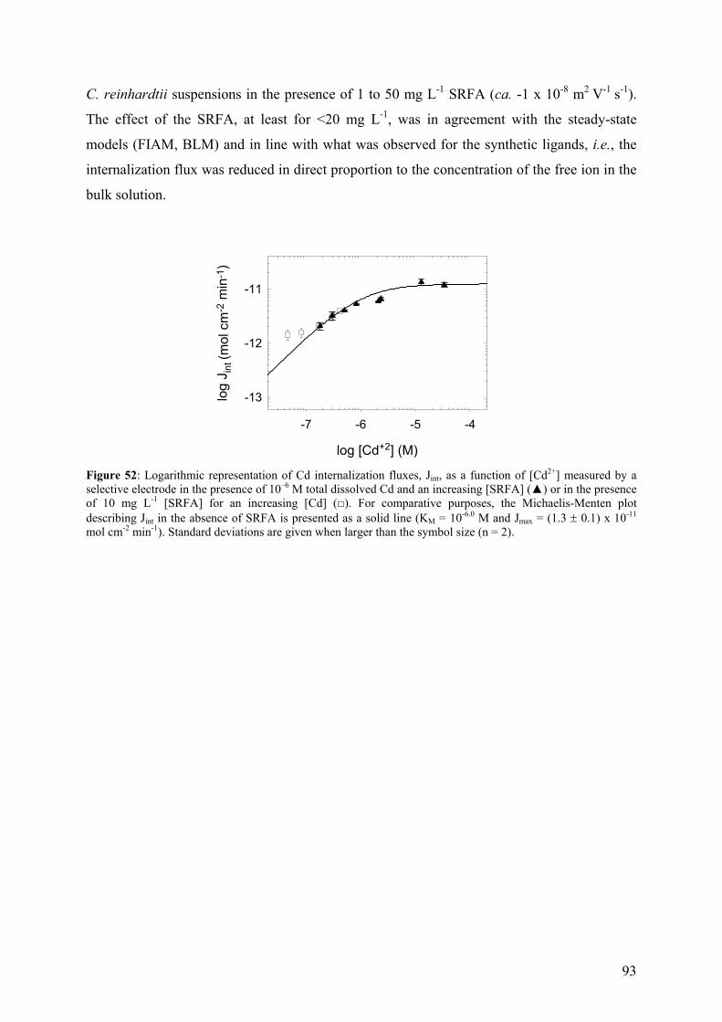
93
C. reinhardtii suspensions in the presence of 1 to 50 mg L-1 SRFA (ca. -1 x 10⁻⁸ m2 V-1 s-1).
The effect of the SRFA, at least for <20 mg L-1, was in agreement with the steady-state
models (FIAM, BLM) and in line with what was observed for the synthetic ligands, i.e., the
internalization flux was reduced in direct proportion to the concentration of the free ion in the
bulk solution.
log [Cd+2] (M)
-7 -6 -5 -4
log
J int (
mol
cm
-2 m
in-1
)
-13
-12
-11
Figure 52: Logarithmic representation of Cd internalization fluxes, Jint, as a function of [Cd2+] measured by a selective electrode in the presence of 10–6 M total dissolved Cd and an increasing [SRFA] () or in the presence of 10 mg L-1 [SRFA] for an increasing [Cd] (). For comparative purposes, the Michaelis-Menten plot describing Jint in the absence of SRFA is presented as a solid line (KM = 10-6.0 M and Jmax = (1.3 ± 0.1) x 10-11 mol cm-2 min-1). Standard deviations are given when larger than the symbol size (n = 2).
94
7 – SUMMARY AND CONCLUSIONS
The Free Ion Activity Model is one of the most employed steady-state models for relating
metal speciation in solution with metal bioavailability and toxicological effects on the aquatic
biota. This study highlighted the advantages of this model to predict metal bioavailability by
using unicellular green alga.
A rigorous study of Cd uptake (i.e., adsorption, internalization) by a walled and a wall-less
strain of Chlamydomonas reinhardtii grown in TAP medium was performed. Free Cd
concentrations were examined in the absence and presence of ligands forming both labile and
inert hydrophilic complexes, from 10-11 M to 10-3 M. Cadmium adsorption by the walled and
wall-less strains of Chlamydomonas reinhardtii was not predicted by a single site
(Langmuirian) model. Indeed, no saturation of the cell wall was observed, even for Cd
concentrations in excess of 5 x 10⁻³ M. A continual production of Cd binding sites appeared to
be responsible for the observed increase of Cd adsorption with time. SDS PAGE and
measurements of the protein content of algal supernatants demonstrated that organic matter
was released by the algae, both in the presence and absence of Cd demonstrating that exudate
release was not in response to Cd stress but rather a constitutive process. Both the nature (e.g.
polysaccharides, proteins) and the quantity of exudate production were influenced by the
physicochemistry of the external medium (i.e., pH buffer). Moreover, measurements using the
permeation liquid membrane (PLM) and anodic stripping voltammetry (ASV) demonstrated
that dissolved cadmium was rapidly complexed by the organic exudates produced by the
algae. Due to the continual production of the binding sites on the cell wall, the equilibrium
between Cd adsorbed to the cell wall sites and Cd in solution (steady-state) was never
achieved under the studied conditions. Nevertheless, this result did not have any implication
in the applicability of the FIAM for Cd internalization, since transporters (specific sites) are
responsible for the internalization and not the cell wall binding sites (non-specific sites).
A first order biological internalization, as predicted by the free ion activity model (FIAM),
was observed for the entire range of Cd concentrations studied for both strains. A linear
relationship with [Cd2+] in solution was seen also for the transporter bound Cd. The maximum
Cd internalization flux, Jmax, for the walled strain was 5-fold higher (1.3x10⁻¹¹ mol cm-2 min-1)
than for the CW-2 strain (2.3x10⁻¹² mol cm-2 min-1) and was not influenced by the presence of
competitors such as Ca in the experimental solution. The conditional stability constant for the
95
adsorption of Cd to transport sites of the CW-2 strain was 5-fold higher (106.7 M-1) than for the
wild type strain (106 M-1) demonstrating the higher affinity of the CW-2 strain for Cd and
suggesting that the number of transport sites was greater for the WT strain. Due to the
demonstrated limiting nature of Cd internalization, differences smaller than 10-fold in Cd
diffusive fluxes due to the presence of a cell wall should have had no effect on Cd uptake.
Competition experiments demonstrated that Mo, Mn, Cu, Co, Zn, Ni, Ca and Pb competed, at
least partially, for the Cd binding sites while no inhibition was observed for similar
concentrations of Mg and Fe. Stability constants for the competitive binding of Ca, Zn and Cu
to the Cd transport site were determined to be 104.5 M-1, 105.2 M-1 and 105.6 M-1, respectively.
Protons also appeared to compete with Cd uptake sites as uptake could generally be predicted
quantitatively in their presence and KH2-Rcell was determined to be 1011.3 M-1. Finally, in the
presence of low concentrations (<20 mg L-1) of Suwannee River fulvic acid, Cd internalization
fluxes could be predicted from [Cd2+], in accordance with the FIAM.
The vast majority of the results presented above indicated that Cd uptake by C. reinhardtii can
be best predicted by models based upon the thermodynamic equilibria of trace metals at the
surface of the microorganism. This would suggest that the FIAM or BLM models would be
reasonable tools for evaluating Cd bioavailability to C. reinhardtii in freshwaters. Although
slight deviations from the steady-state models were observed for high concentrations of
organic matter, Cd or protons, these concentrations are generally not seen in the majority of
freshwaters. Nonetheless, due to the strong interaction of trace metals, hardness metals and
protons at the biological surface, the knowledge of a large number of stability constants and
physicochemical parameters will be necessary to quantitatively predict uptake (and thus
biological effects) in natural aquatic systems.
96
8- REFERENCES
Adhiya J., Cai X., Sayre R. T. and Traina S. J. Binding of aqueous cadmium by the
lyophilized biomass of Chlamydomonas reinhardtii. Colloids Surf. A: Physicochem.
Eng. Aspects. 2002, 210, 1-11.
Aksu Z. Equilibrium and kinetic modelling of cadmium (II) biosorption by C. vulgaris in a
batch system: effect of temperature. Sep. Sci. Technol. 2001, 21, 285-294.
Albert B., Bray D., Lewis J., Raff M., Roberts K. and Watson J. D., Eds; in Molecular
Biology of the Cell; Garland Publishing: London; 1994.
Anderson M. A., Morel F. M. M. and Guillard R. R. L. Growth limitation of a coastal diatom
by low zinc ion activity. Nature 1978, 276, 70-1.
Anderson N. J., Blomqvist P. and Renberg I. An experimental and palaeoecological study of
algal responses to lake acidification and limiting in three central Swedish lakes. Eur. J.
Phycol. 1997, 32, 35-48.
Avery S. V., Howlett N. G. and Radice S. Copper toxicity towards Saccharomyces cerevisiae:
dependence on plasma membrane fatty acid composition. Appl. Environ. Microbiol.
1996, 62, 3960-3966.
Bates S. S., Tessier A., Campbell P. G. C. and Buffle J. Zinc adsorption and transport by
Chlamydomonas variabilis and Scenedesmus subspicatus (Chlorophyceae) grown in
semicontinuous culture. J. Phycol. 1982, 18, 521-9.
Ben-Bassat D., Shelef G., Gruner N. and Shuval H. I. Growth of Chlamydomonas in a
medium containing mercury. Nature 1972, 240, 43-44.
Bilinski H., Huston R. and Stumm W. Determination of the stability constants of some
hydroxo and carbonato complexes of lead(II), copper(II), cadmium(II), and zinc(II) in
dilute solutions by anodic stripping voltammetry and differential pulse polarography.
Anal. Chim. Acta 1976, 84, 157-164.
Boisson F., Hutchins D. A., Fowler S. W., Fisher N. S. and Teyssie J.-L. Influence of
temperature on the accumulation and retention of 11 radionuclides by the marine alga
Fucus vesiculosus (L.). Mar. Poll. Bull. 1998, 35, 313-321.
Bold H. C. and Wynne M. J.; in Introduction to the Algae: Structure and Reproduction;
Englewood Cliffs: New York; 1978.
Borgmann U.; Metal speciation and toxicity of free metal ions to aquatic biota in Aquat.
Toxicol.; Nriag J. O., Eds.; Wiley Interscience: New York; 1982.
97
Boyle E. A., Scalter F. and Edmond J. M. On the marine geochemistry of cadmium. Nature
1976, 263, 42-44.
Bruland K. W. Complexation of zinc by natural organic ligands in the central North Pacific.
Limnol. Oceanogr. 1989, 34, 269-285.
Buffle J.; Complexation properties of homologous complexants and choice of measuring
methods in Complexation reaction in aquatic systems: An analytical approach, Ellis
Horwood: Chichester; 1988.
Buffle J., Altmann R. S., Filella M. and Tessier A. Complexation by natural heterogeneous
compounds: Site occupation distribution functions, a normalized description of metal
complexation. Geochim. Cosmochim. Acta 1990, 54, 1535-53.
Cain J. R. and Allen R. K. Use of cell wall-less mutant strains to assess the role of the cell
wall in cadmium and mercury tolerance by Chlamydomonas reinhardtti. Bull.
Environ. Contamin. Toxicol. 1980, 25, 797-801.
Campbell P. G. C.; Interactions between trace metals and aquatic organisms: a critique of the
free-ion activity model in Metal Speciation and Bioavailability in Aquatic Systems;
Tessier A. and Turner D., Eds.; John Wiley & Sons: Chichester; 1995.
Campbell P. G. C., Errecalde O., Fortin C., Hiriart-Baer V. P. and Vigneault B. Metal
bioavailability to phytoplankton-applicability of the biotic ligand model. Comp.
Biochem. Physiol. Part C: Toxicol. Pharmacol. 2002, 133C, 189-206.
Campbell P. G. C. and Stokes P. M. Acidification and toxicity of metals to aquatic biota. Can.
J. Fish. Aquat. Sci. 1985, 42, 2034-2049.
Campbell P. G. C., Twiss M. R. and Wilkinson K. J. Accumulation of natural organic matter
on the surfaces of living cells: implications for the interaction of toxic solutes with
aquatic biota. Can. J. Fish. Aquat. Sci. 1997, 54, 2543-2554.
Catt J. W., Hills G. J. and Roberts K. A structural glycoprotein, containing hydroxproline,
isolated from the cell wall of Chlamydomonas reinhardtii. Planta 1976, 131, 165-171.
Cho D.-Y., Lee S.-T., Park S.-W. and Chung A.-S. Studies on the biosorption of heavy metals
onto Chlorella vulgaris. J. Environ. Sci. Health 1994, A29 (2), 389-409.
Collard J. M. and Matagne R. F. Isolation and genetic analysis of Chlamydomonas reinhardtii
strains resistant to cadmium. Appl. Environ. Microbiol. 1990, 56, 2051-5.
Cook M. E. and Morrow H.; Anthropogenic sources of cadmium in Canada in. National
Workshop on Cadmium Transport into Plants, June 20-21; Ottawa, Ontario, Canada;
1995.
98
Cosovic B., Ciglenecki I., Vilicic D. and Ahel M. Distribution and seasonal variability of
organic matter in a small eutrophicated salt lake. Estuar. Coast. Shelf Sci. 2000, 51,
705-715.
Crist R. H., Martin J. R., Carr D., Watson J. R. and Clarke H. J. Interaction of metals and
protons with algae. 4. Ion exchange vs adsorption models and a reassessment of
Scatchard plots; ion-exchange rates and equilibria compared with calcium alginate.
Environ. Sci. Technol. 1994, 28, 1859-1866.
Crist R. H., Oberholser K., Schwartz D., Marzoff J., Ryder D. and Crist D. R. Interactions of
metals and protons with algae. Environ. Sci. Technol. 1988, 22, 755-60.
Crist R. H., Oberholser K., Shank N. and Nguyen M. Nature of bonding between metallic ions
and algal cell walls. Environ. Sci. Technol. 1981, 15, 1212-1217.
Danesi P. R. Separation of metal species by supported liquid membranes. Sep. Sci. Technol.
1984, 19, 857-94.
Davis R. D. and Plaskitt A. Genetic and structural analysis of cell-wall formation in
Chlamydomonas reinhardtii. Genet. Res. Camb. 1971, 17, 33-43.
Devriese M., Tsakaloudi V., Garbayo I., Leon R., Vilchez C. and Vigara J. Effect of heavy
metals on nitrate assimilation in the eukaryotic microalga Chlamydomonas reinhardtii.
Plant Physiol. Biochem. 2001, 39, 443-448.
Dudka S. and Adriano D. C. Environmental impacts of metal ore mining and processing: A
review. J. Environ. Qual. 1997, 26, 590-602.
Eckhardt U. and Buckhout T. J. Iron assimilation in Chlamydomonas reinhardtii involves
ferric reduction and is similar to strategy I higher plants. J. Exp. Bot. 1998, 49, 1219-
1226.
Eckhardt U. and Buckhout T. J. Analysis of the mechanism of iron assimilation in
Chlamydomonas reinhardtii: a model system for strategy I plants. J. Plant Nut. 2000,
23, 1797-1807.
Elgersma F., Anderberg B. S. and Stigliani W. M.; Emission factors for aqueous industrial
cadmium emissions in the Rhine river basin; A historical reconstruction for the period
1970-1988 in. Edited Proceedings Seventh International Cadmium Conference; New-
Orleans; 1992.
Elinder C.-G.; Cadmium and Health: A Toxicological and Epidemiological Appraisal; Boca
Raton, Florida, USA; 1985.
Ellwood M. J. Zinc and cadmium speciation in subantarctic waters east of New Zealand. Mar.
Chem. 2004, 87, 37-58.
99
Errecalde O., Seidl M. and Campbell P. G. C. Influence of a low molecular weight metabolite
(citrate) on the toxicity of cadmium and zinc to the unicellular green alga Selenastrum
capricornutum: an exception to the free-ion model. Wat. Res. 1998, 32, 419-429.
Ferguson W. J., Braunschweiger K. I., Braunschweiger W. R., Smith J. R., McCormick J. J.,
Wasmann C. C., Jarvis N. P., Bell D. H. and Good N. E. Hydrogen ion buffers for
biological research. Anal. Biochem. 1980, 104, 300-310.
Ferris P. J., Woessner J. P., Waffenschmidt S., Kilz S., Drees J. and Goodenough U. W.
Glycosylated polyproline II rods with kinks as a structural motif in plant
hydroxyproline-rich glycoproteins. Biochem. 2001, 40, 2978-2987.
Fortin C. and Campbell P. G. C. Silver uptake by the green alga Chlamydomonas reinhardtii
in relation to chemical speciation: influence of chloride. Environ. Toxicol. Chem.
2000, 19, 2769-2778.
Fortin C. and Campbell P. G. C. Thiosulfate enhances uptake by a green alga: role of anion
transporters in metal uptake. Environ. Sci. Technol. 2001, 35, 4000.
Fortin C., Dutel L. and Garnier-Laplace J. Uranium complexation and uptake by a green alga
in relation to chemical speciation: the importance of the free uranyl ion. Environ.
Toxicol. Chem. 2004, 23, 974-981.
Franklin N. M., Stauber J. L. and Lim R. P. Development of flow cytometry-based algal
bioassays for assessing toxicity of copper in natural waters. Environ. Toxicol. Chem.
2001, 20, 160-170.
Galceran J. and van Leeuwen H. P.; Dynamic of biouptake processes. The role of transport,
adsorption and internalization in Physicochemical Kinetics and Transport at
Biointerfaces; van Leeuwen H. P. and Köster W., Eds.; John Wiley & Sons:
Chichester; 2004.
Gensemer R. W., Smith R. E. H. and Duthie H. C. Comparative effects of pH and aluminium
on silica-limited growth and nutrient uptake in Asterionella ralfsii var. americana
(Bacillariophyceae). J. Phycol. 1993, 29, 36-44.
Good N. E., Winget G. D., Winter W., Connolly T. N., Izawa S. and Singh R. M. M.
Hydrogen ion buffers for biological research. Biochem. 1966, 5, 467-77.
Goodenough U. W., Gebhart B., Mecham R. P. and Heuser J. E. Crystals of the
Chlamydomonas reinhardtii cell wall: polymerization, depolymerization, and
purification of glycoprotein monomers. J. Cell Bio. 1986, 103, 405-17.
100
Goodenough U. W. and Heuser J. E. The Chlamydomonas cell wall and its constituent
glycoproteins analyzed by the quick-freeze, deep-etch technique. J. Cell Bio. 1985,
101, 1550-68.
Guanzon N. G., Nakahara H. and Nishimura K. Accumulation of copper, zinc, cadmium, and
their combinations by three freshwater microalgae. Fisheries Sci. 1995, 61, 149-156.
Hanikenne M., Matagne R. F. and Loppes R. Pleiotropic mutants hypersensitive to heavy
metals and to oxidative stress in Chlamydomonas reinhardtii. FEMS Microbiol. Lett.
2001, 196, 107-111.
Hare L. and Tessier A. Predicting animal cadmium concentrations in lakes. Nature 1996, 380,
430-432.
Hargreaves J. W. and Whitton B. A. Effect of pH on growth of acid stream algae. J. Phycol.
1976, 11, 215-223.
Harris E. H.; Culture and Storage Methods in The Chlamydomonas sourcebook- a
comprehensive guide to biology and laboratory use; Academic Press: San Diego;
1989.
Hart B. A., Bertram P. E. and Scaife B. D. Cadmium transport by Chlorella pyrenoidosa.
Environ. Res. 1979, 18, 327-35.
Hart B. A. and Scaife B. D. Toxicity and bioaccumulation of cadmium in Chlorella. Environ.
Res. 1977, 14, 401-13.
Hassler C. S., Slaveykova V. I. and Wilkinson K. J. Discriminating between intra- and
extracellular metals using chemical extractions. Limnol. Oceanogr. Methods 2004, 2,
237–247.
Hassler C. S., Slaveykova V. I. and Wilkinson K. J. Some fundamental (and often
overlooked) considerations underlying the free ion activity and biotic ligand models.
Environ. Toxicol. Chem. 2004, 23, 283-291.
Hassler C. S. and Wilkinson K. J. Failure of the biotic ligand and free-ion activity models to
explain zinc bioaccumulation by Chlorella kesslerii. Environ. Toxicol. Chem. 2003,
22, 620-626.
Hill K. L., Hassett R., Kosman D. and Merchant S. Regulated copper uptake in
Chlamydomonas reinhardtii in response to copper availability. Plant Physiol. 1996,
112, 697-704.
Hu S., Lau K. W. K. and Wu M. Cadmium sequestration in Chlamydomonas reinhardtii.
Plant Sci. 2001, 161, 987-996.
101
Hu Z., Wong Y.-S. and Nora F. Y. T.; Expression of the metallothionein-like gene from
Festuca rubra in Chlamydomonas reinhardtii. in. Ninth International Conference on
the Cell and Molecular Biology of Chlamydomonas (Chlamy2000); Amsterdam; 2000.
Hudson R. J. M. Which aqueous species control the rates of trace metal uptake by aquatic
biota? Observations and predictions of non-equilibrium effects. Sci. Tot. Environ.
1998, 219, 95-115.
Hudson R. J. M. and Morel F. M. M. Iron transport in marine phytoplankton: kinetics of
cellular and medium coordination reactions. Limnol. Oceanogr. 1990, 35, 1002-1020.
Hudson R. J. M. and Morel F. M. M. Trace metal transport by marine microorganisms:
implications of metal coordination kinetics. Deep-Sea Res. 1993, 40, 129-150.
Hyams J. and Davies D. R. The induction and characterization of cell wall mutants of
Chlamydomonas reinhardtii. Mutation Res. 1972, 14, 381-389.
Irmer U., Wachholz I., Schaefer H. and Lorch D. W. Influence of lead on Chlamydomonas
reinhardtii Danegard (Volvocales, Chlorophyta): accumulation, toxicity and
ultrastructural changes. Environ. Experim. Bot. 1986, 26, 97-105.
Jensen A. and Bro-Rasmussen F. Environnemental contamination in Europe. Rev. Environ.
Contamin. Toxicol. 1992, 125, 101-181.
Kaplan J.; Patterns Receptor behaviour and function in Mechanisms of Receptor Regulation;
Poste G. and Crooke S. T., Eds.; Plenum Press: New York; 1985.
Khoshmanesh A., Lawson F. and Prince I. G. Cell surface area as a major parameter in the
uptake of cadmium by unicellular green microalgae. Chem. Eng. J. 1997, 65, 13-19.
Khummongkol D., Canterford G. S. and Fryer C. Accumulation of heavy metals in unicellular
algae. Biotechnol. Bioeng. 1982, 24, 2643-2660.
Kiefer E., Sigg L. and Schosseler P. Chemical and spectroscopic characterization of algae
surfaces. Environ. Sci. Technol. 1997, 31, 759-764.
Kilz S., Waffenschmidt S. and Budzikiewicz H. Mass spectrometric analysis of
hydroxyproline glycans. J. Mass Spectrom. 2000, 35, 689-697.
Kleijn J. M. and van Leeuwen H. P.; Electrostatic and electrodynamic properties of biological
interphases in Physical Chemistry of Biological Interfaces; Baszkin A. and Norde W.,
Eds.; Marcel Dekker: New York; 2000.
Klotz I. M. Ligand-receptor interactions: facts and fantasies. Q. Rev. Biophys. 1985, 18, 227-
259.
102
Kola H., Langlera L. M., Parthasarathy N. and Wilkinson K. J. Cadmium adsorption by
Chlamydomonas reinhardtii and its interaction with the cell wall proteins. Environ.
Chem. 2004, 1, 172-179.
Kola H. and Wilkinson K. J. Cadmium uptake by a green algae can be predicted by
equilibrium modelling. Environ. Sci. Technol. 2004, in press.
Korte F. Ecotoxicology of cadmium: general overview. Ecotoxicol. Environ. Saf. 1983, 7, 3-8.
Kruger N. J.; The Bradford Method for Protein Quantification in Protein Protocols
Handbook; Walker J. M., Eds.; Humana Press: Totowa, New York; 1996.
Laegreid M., Alstad J., Klaveness D. and Seip H. M. Seasonal variation of cadmium toxicity
toward the alga Selenastrum capricornutum Printz in two lakes with different humus
content. Environ. Sci. Technol. 1983, 17, 357-61.
Lage O. M., Vasconcelos M. T. S. D., Soares H. M. V. M., Osswald J. M., Sansonetty F.,
Parente A. M. and Salema R. Suitability of the pH buffers 3-[N,N-bis(hydroxyethyl)
amino]-2-hydroxypropanesulfonic acid and N-2-hydroxyethylpiperazine-N'-2-
ethanesulfonic acid for in vitro copper toxicity studies. Arch. Environ. Contamin.
Toxicol. 1996, 31, 199-205.
Lauffenburgar D. A. and Linderman J. J.; Receptors: Models for Binding, Trafficking and
Signalling; Oxford University Press, Inc.: New York; 1993.
Lawrence S. G., Holoka M. H. and Hamilton R. D. Effects of cadmium on a microbial food
chain, Chlamydomonas reinhardtii and Tetrahymena vorax. Sci. Tot. Environ. 1989,
87-88, 381-95.
Leal M. F. C., Vasconcelos M. T. S. D. and van den Berg C. M. G. Copper-induced release of
complexing ligands similar to thiols by Emiliania huxleyi in seawater cultures. Limnol.
Oceanogr. 1999, 44, 1750-1762.
Lee D.-Y., Fortin C. and Campbell P. G. C. Influence of chloride on silver uptake by two
green algae, Pseudokirchneriella subcapitata and Chlorella pyrenoidosa. Environ.
Toxicol. Chem. 2004, 23, 1012-1018.
Lustigman B., Lee L. H. and Khalil A. Effects of nickel and pH on the growth of Chlorella
vulgaris. Bull. Environ. Contamin. Toxicol. 1995, 55, 73-80.
Lustigman B., Lee L. H., Morata J. and Khan F. Effect of thallium on the growth of Anacystis
nidulans and Chlamydomonas reinhardtii. Bull. Environ. Contamin. Toxicol. 2000, 64,
565-573.
Lustigman B., Lee L. H. and Weiss-Magasic C. Effects of cobalt and pH on the growth of
Chlamydomonas reinhardtii. Bull. Environ. Contamin. Toxicol. 1995, 55, 65-72.
103
Macfie S. M., Tarmohamed Y. and Welbourn P. M. Effects of cadmium, cobalt, copper, and
nickel on growth of the green alga Chlamydomonas reinhardtii: the influences of the
cell wall and pH. Arch. Environ. Contamin. Toxicol. 1994, 27.
Macfie S. M. and Welbourn P. M. The cell wall as a barrier to uptake of metal ions in the
unicellular green alga Chlamydomonas reinhardtii (Chlorophyceae). Arch. Environ.
Contamin. Toxicol. 2000, 39, 413-419.
Maeda S., Mizoguchi M., Ohki A. and Takeshita T. Bioaccumulation of zinc and cadmium in
freshwater alga, Chlorella vulgaris. Part I toxicity and accumulation. Chemosphere
1990b, 21, 953-963.
Malis-Arad S. and McGowan R. E. Alkalinity-induced aggregation in Chlorella vulgaris. II.
Changes in the cell wall during the cell cycle. Plant Cell Physiol. 1982, 23, 11-17.
Marsalek B. and Rojickova R. Stress factors enhancing production of algal exudates. A
potential self-protective mechanism? Z. Naturforsch. C: J. Biosci. 1996, 51, 646-650.
Martel A. E. and Smith R. M.; NIST; Version 5.0; Texas University: 1998.
Mash H. E., Chin Y.-P., Sigg L., Hari R. and Xue H. Complexation of copper by zwitterionic
aminosulfonic (Good) buffers. Anal. Chem. 2003, 75, 671-677.
Mason A. Z. and Jenkins K. D.; Metal detoxification in aquatic organisms in Metal Speciation
and Bioavailability in Aquatic Systems; Tessier A. and Turner D., Eds.; John Wiley
and Sons: Chichester; 1995.
Melkonian M. Release of dissolved organic substances by the green alga Fritschiella tuberosa
Iying. (Chaetophorineae) during different growth phases. Z. Pflanzenphysiol. 1979, 94,
125-33.
Mirimanoff N. and Wilkinson K. J. Regulation of Zn accumulation by a freshwater gram-
positive bacterium (Rhodococcus opacus). Environ. Sci. Technol. 2000, 34, 616-622.
Morel F. M. M. Trace metals-phytoplankton interactions: an overview. Elsevier
Oceanography Series 1986, 43, 177-89.
Morel F. M. M. and Hering J. G., Eds; in Principles and Applications of Aquatic Chemistry;
John Wiley & Sons: Chichester; 1993.
Morel F. M. M. and Price N. M. The biogeochemical cycles of trace metals in the oceans.
Science 2003, 300, 944-947.
Moseley J. L., Allinger T., Herzog S., Hoerth P., Wehinger E., Merchant S. and Hippler M.
Adaptation to Fe-deficiency requires remodelling of the photosynthetic apparatus.
Embo J. 2002a, 21, 6709-6720.
104
Moseley J. L., Page M. D., Alder N. P., Eriksson M., Quinn J., Soto F., Theg S. M., Hippler
M. and Merchant S. Reciprocal expression of two candidate di-iron enzymes affecting
photosystem I and light-harvesting complex accumulation. Plant Cell 2002b, 14, 673-
688.
Moss B.; The uses of waterways; Pollution, waste disposal and water supply in Ecology of
fresh waters; Publications B. S., Eds.; Billing & Sons Ltd: Oxford, London,
Edinburgh, Boston, Melbourne; 1980.
Mukunoki J. and Fujimoto K.; Collection and recycling of used Ni-Cd batteries in Japan in.
Sources of cadmium in the environment, Inter-Organisation Programme for the Sound
Management of Chemical (IOMC); Paris, France; 1996.
Nagel K., Adelmeier U. and Voigt J. Subcellular distribution of cadmium in the unicellular
green alga Chlamydomonas reinhardtii. J. Plant Physiol. 1996, 149, 86-90.
Nagel K. and Voigt J. Impaired photosynthesis in a cadmium-tolerant Chlamydomonas
mutant strain. Microbiol. Res. 1995, 150, 105-110.
Nederlof M. M., De Wit J. C. M., Van Riemsdijk W. H. and Koopal L. K. Determination of
proton affinity distributions for humic substances. Environ. Sci. Technol. 1993, 27,
846-856.
Neiboer E. and Richardson D. H. S. The replacement of nondescript term "heavy metal" by a
biologically and chemically significant classification of metal ion. Environ. Pollut.
1980, 1, 3-26.
Nies D. H. Microbial heavy metal resistance. App. Microbiol. Biotechnol. 1999, 51, 730-750.
Nriagu J. O. and Pacyna J. M. Quantitative assessment of worldwide contamination of air,
water and soils by trace metals. Nature 1988, 333, 134-139.
OECD; (Organization for Economic Co-operation and Development) in. Risk Reduction
Monograph No. 5 : Cadmium; Paris, France; 1994.
Ohnesorge F. K. and Wilhelm M.; Zinc in Metals and their compounds in environment;
Merian E., Eds.; VCH: New York; 1991.
Parent L., Twiss M. R. and Campbell P. G. C. Influences of natural dissolved organic matter
on the interaction of aluminium with the microalga Chlorella: A test of the free-ion
model of trace metal toxicity. Environ. Sci. Technol. 1996, 30, 1713-20.
Parthasarathy N., Pelletier M. and Buffle J. Permeation liquid membrane for trace metal
speciation in natural waters. Transport of liposoluble Cu(II) complexes. J. Chrom. A
2004, 1025, 33-40.
105
Pasciak W. J. and Gavis J. Transport limitation of nutrient uptake in phytoplankton. Limnol.
Oceanogr. 1974, 19, 881-888.
Payne A. G. Responses of the three test algae of the algal assay procedure. Bottle test. Wat.
Res. 1975, 9, 437-45.
Pei J., Tercier-Waeber M.-L. and Buffle J. Simultaneous determination and speciation of zinc,
cadmium, lead, and copper in natural water with minimum handling and artefacts by
voltammetry on a gel-integrated microelectrode array. Anal. Chem. 2000, 72, 161-171.
Pinheiro J. P., Galceran J. and Van Leeuwen H. P. Metal speciation dynamics and
bioavailability: Bulk depletion effects. Environ. Sci. Technol. 2004, 38, 2397-2405.
Pinheiro J. P., Mota A. M. and van Leeuwen H. P. On lability of chemically heterogeneous
systems Complexes between trace metals and humic matter. Colloids Surf. A:
Physicochem. Eng. Aspects 1999, 151, 181-187.
Pitts L. C. and Wallace G. T. Lead deposition in the shell of the bivalve, Mya arenaria: an
indicator of dissolved lead in seawater. Estuar. Coast. Shelf Sci. 1994, 39, 93-104.
Playle R. C. Modelling metal interaction at fish gills. Sci. Tot. Environ. 1998, 219, 147-163.
Plette A. C. C., Benedetti M. F. and van Riemsdijk W. H. Competitive binding of protons,
calcium, cadmium, and zinc to isolated cell walls of a gram-positive soil bacterium.
Environ. Sci. Technol. 1996, 30, 1902-10.
Plette A. C. C., Nederlof M. M., Temminghoff E. J. M. and Van Riemsdijk W. H.
Bioavailability of heavy metals in terrestrial and aquatic systems: a quantitative
approach. Environ. Toxicol. Chem. 1999, 18, 1882-1890.
Prasad M. N., Drej K., Skawinska A., Strzalka K. and Stralka K. Toxicity of cadmium and
copper in Chlamydomonas reinhardtii wild-type (WT 2137) and cell wall deficient
mutant strain (CW 15). Bull. Environ. Contamin. Toxicol. 1998, 60, 306-11.
Price N. M., Ahner B. A. and Morel F. M. M. The equatorial Pacific Ocean: grazer-controlled
phytoplankton populations in an iron-limited ecosystem. Limnol. Oceanogr. 1994, 39,
520-34.
Price N. M. and Morel F. M. M. Cadmium and cobalt substitution for zinc in a marine diatom.
Nature 1990, 344, 658-60.
Price N. M. and Morel F. M. M. Colimitation of phytoplankton growth by nickel and
nitrogen. Limnol. Oceanogr. 1991, 36, 1071-7.
Rachlin J. W. and Grosso A. The effects of pH on the growth of Chlorella vulgaris and its
interactions with cadmium toxicity. Arch. Environ. Contamin. Toxicol. 1991, 20, 505-
8.
106
Rachlin J. W. and Grosso A. The growth response of the green alga Chlorella vulgaris to
combined divalent cation exposure. Arch. Environ. Contamin. Toxicol. 1993, 24, 16-
20.
Rauser W. E. Phytochelatins and related peptides. Structure, biosynthesis and function. Plant
Physiol. 1995, 109, 1141-49.
Rhee G. Y. and Thompson P. A. Sorption of hydrophobic organic contaminants and trace
metals on phytoplankton and implications for toxicity assessment. J. Aquatic Ecosys.
Health 1992, 1, 175-191.
Roberts K. Crystalline glycoprotein cell walls of algae: their structure, composition and
assembly. Philos. Trans. R. Soc. 1974, 268, 129-146.
Roberts K. Hydroxyproline: Its asymmetric distribution in a cell wall glycoprotein. Planta
1979, 146, 275-279.
Roberts K., Gurney-Smith M. and Hills G. J. Structure, composition and morphogenesis of
the cell wall of Chlamydomonas reinhardtii. I. Ultrastructural and preliminary
chemical analysis. J. Ultrastruct. Res. 1972, 40, 599-613.
Rubinelli P., Siripornadulsil S., Fei G.-R. and Sayre R. T. Cadmium- and iron-stress-inducible
gene expression in the green alga Chlamydomonas reinhardtii: evidence for H43
protein function in iron assimilation. Planta 2002, 215, 1-13.
Sakaguchi T., Tsuji T., Nakajima A. and Horikoshi T. Accumulation of cadmium by green
microalgae. Eur. J. Appl. Microbiol. Biotechnol. 1979, 8, 207-15.
Sasaki T., Kurano N. and Miyachi S. Cloning and characterization of high-CO2-specific
cDNAs from a marine microalga, Chlorococcum littorale, and effect of CO2
concentration and iron deficiency on the gene expression. Plant Cell Physiol. 1998,
39, 131-8.
Shiller A. M. and Boyle E. A. Variability of dissolved trace metals in the Mississippi river.
1987, 51, 3273-3277.
Sigg L. Surface chemical aspects of the distribution and fate of metal ions in lakes. Aquat.
Surf. Chem. 1987, 319-49.
Simkiss K. and Taylor M. G. Metal fluxes across the membranes of aquatic organisms. Rev.
Aqua. Sci. 1989, 1, 173-88.
Simpson S. L., Roland M. G. E., Stauber J. L. and Batley G. E. Effect of declining toxicant
concentrations on algal bioassay endpoints. Environ. Toxicol. Chem. 2003, 22, 2073-
2079.
107
Siripornadulsil S., Traina S., Verma D. P. S. and Sayre R. T. Molecular mechanisms of
proline-mediated tolerance to toxic heavy metals in transgenic microalgae. Plant Cell
2002, 14, 2837-2847.
Slaveykova V. I. and Wilkinson K. J. Physicochemical aspects of lead bioaccumulation by
Chlorella vulgaris. Environ. Sci. Technol. 2002, 36, 969-975.
Slaveykova V. I. and Wilkinson K. J. Effect of pH on Pb biouptake by the freshwater alga
Chlorella kesslerii. Environ. Chem. Lett. 2003, 1, 185-189.
Slaveykova V. I. and Wilkinson K. J. Predicting the bioavailability of metals and metal
complexes: Critical review of the biotic ligand model. Environ. Chem. 2005, in press.
Slaveykova V. I., Wilkinson K. J., Ceresa A. and Pretsch E. Role of fulvic acid on lead
bioaccumulation by Chlorella kesslerii. Environ. Sci. Technol. 2003, 37, 1114-1121.
Smiejan A., Wilkinson K. J. and Rossier C. Cd bioaccumulation by a freshwater bacterium,
Rhodospirillum rubrum. Environ. Sci. Technol. 2003, 37, 701-706.
Soares H. M. V. M., Conde P. C. F. L., Almeida A. A. N. and Vasconcelos M. T. S. D.
Evaluation of n-substituted aminosulfonic acid pH buffers with a morpholinic ring for
cadmium and lead speciation studies by electroanalytical techniques. Anal. Chim. Acta
1999, 394, 325-335.
Stratton G. W. and Corke C. T. The effect of cadmium ion on the growth, photosynthesis, and
nitrogenase activity of Anabaena inaequalis. Chemosphere 1979, 8, 277-282.
Stumm W. and Morgan J. J.; Aquatic Chemistry: Chemical Equilibria and Rates in Natural
Waters; John Wiley & Sons: Chichester; 1996.
Stumm W., Sigg L. and Sulzberger B.; The role of coordination at the surface of aquatic
particles in Chemical and biological regulation of aquatic systems; Buffle J. and De
Vitre R. R., Eds.; Lewis: New York; 1994.
Sunda W. G. and Guillard R. R. L. The relationship between cupric ion activity and the
toxicity of cooper to phytoplankton. J. Mar. Res. 1976, 34, 511-529.
Sunda W. G. and Huntsman S. A. Relationship among growth rate, cellular manganese
concentrations and manganese transport kinetics in estuarine and oceanic species of
the diatom Thalassiosira. J. Phycol. 1986, 22, 259-270.
Sunda W. G. and Huntsman S. A. Feedback interactions between zinc and phytoplankton in
seawater. Limnol. Oceanogr. 1992, 37, 25-40.
Sunda W. G. and Huntsman S. A. Antagonisms between cadmium and zinc toxicity and
manganese limitation in a coastal diatom. Limnol. Oceanogr. 1996, 41, 373-387.
108
Sunda W. G. and Huntsman S. A. Control of Cd concentrations in a coastal diatom by
interactions among free Ionic Cd, Zn, and Mn in seawater. Environ. Sci. Technol.
1998, 32, 2961-2968.
Sunda W. G. and Huntsman S. A. Interactions among Cu2+, Zn2+, and Mn2+ in controlling
cellular Mn, Zn, and growth rate in the coastal alga Chlamydomonas. Limnol.
Oceanogr. 1998, 43, 1055-1064.
Sunda W. G. and Huntsman S. A. Processes regulating cellular metal accumulation and
physiological effects: phytoplankton as model systems. Sci. Tot. Environ. 1998, 219,
165-181.
Sunda W. G. and Huntsman S. A. Effect of Zn, Mn, and Fe on Cd accumulation in
phytoplankton: Implications for oceanic Cd cycling. Limnol. Oceanogr. 2000, 45,
1501-1516.
Teplitski M., Chen H., Rajamani S., Gao M., Merighi M., Sayre R. T., Robinson J. B., Rolfe
B. G. and Bauer W. D. Chlamydomonas reinhardtii secretes compounds that mimic
bacterial signals and interfere with quorum sensing regulation in bacteria. Plant
Physiol. 2004, 134, 137-146.
Tercier M. L. and Buffle J. Antifouling membrane-covered voltammetric microsensor for in
situ measurements in natural waters. Anal. Chem. 1996, 68, 3670-3678.
Tessier A., Buffle J. and Campbell P. G. C.; Uptake of trace metals by aquatic organisms in
Chemical and Biological Regulation of Biological Systems; Buffle J. and Devitre R.
R., Eds.; Lewis: New York; 1994.
Town R. M. and Filella M. Dispelling the myths: is the existence of L1 and L2 ligands
necessary to explain metal ion speciation in natural waters? Limnol. Oceanogr. 2000,
45, 1341-1357.
Turner L. J., Howell E. T., Summerby M., Hesslein R. H., Findlay D. L. and Jackson M. B.
Changes in epilithon and epiphyton associated with experimental acidification of a
lake to pH 5. Limnol. Oceanogr. 1991, 36, 1390-1405.
Twiss M. R., Auclair J.-C. and Charlton M. N. An investigation into iron-stimulated
phytoplankton productivity in epipelagic Lake Erie during thermal stratification using
trace metal clean techniques. Can. J. Fish. Aquat. Sci. 2000, 57, 870.
Van Assche F. J. and Ciarletta P.; Cadmium in the environment: Levels, trends and critical
pathways in. Edited Proceedings Seventh International Cadmium Conference; New
Orleans, 1992.
van Leeuwen H. P.; in Environmental Particles. Lewis: Boca Raton; 1992.
109
van Leeuwen H. P. Metal speciation dynamics and bioavailability: inert and labile complexes.
Environ. Sci. Technol. 1999, 33, 3743-3748.
van Leeuwen H. P. and Pinheiro J.-P. Lability criteria for metal complexes in micro-electrode
voltammetry. J. Electroanal. Chem. 1999, 471, 55-61.
van Urk G. and Marquenie J. M. Environmental behaviour of cadmium: Who are at risk and
why. Met. Environ. 1989, 11, 456-459.
Vasconcelos M. T. S. D. and Almeida C. M. R. Electrochemical study of proton ionization,
copper(II) complexation and surfactant properties of piperazine-N-N'-bis[2-
hydroxypropanesulfonic acid] pH buffer. Comparison with other N-substituted
aminosulfonic acids pH buffers. Anal. Chim. Acta 1998, 369, 115-122.
Vasconcelos M. T. S. D., Almeida C. M. R., Lage O. M. and Sansonetty F. Influence of
zwitterionic pH buffers on the bioavailability and toxicity of copper to the alga
Amphidinium carterae. Environ. Toxicol. Chem. 2000, 19, 2542-2550.
Vasconcelos M. T. S. D. and Leal M. F. C. Adsorption and uptake of Cu by Emiliania huxleyi
in natural seawater. Environ. Sci. Technol. 2001, 35, 508-515.
Vasconcelos M. T. S. D. and Leal M. F. C. Influence of N-2-hydroxyethylpiperazine-N'-2-
ethanesulfonic acid pH buffer on the biological response of marine algae. Environ.
Toxicol. Chem. 2002, 21, 404-412.
Vasconcelos M. T. S. D., Leal M. F. C. and van den Berg C. M. G. Influence of the nature of
the exudates released by different marine algae on the growth, trace metal uptake and
exudation of Emiliania huxleyi in natural seawater. Mar. Chem. 2002, 77, 187-210.
Veytsman B. A. and Cosgrove D. J. A model of cell wall expansion based on thermodynamics
of polymer networks. Biophys. J. 1998, 75, 2240-2250.
Vigneault B., Percot A., Lafleur M. and Campbell P. G. C. Permeability changes in model and
phytoplankton membranes in the presence of aquatic humic substances. Environ. Sci.
Technol. 2000, 34, 3907-3913.
Visviki I. and Rachlin J. W. The toxic action and interactions of copper and cadmium to the
marine alga Dunaliella minuta, in both acute and chronic exposure. Arch. Environ.
Contamin. Toxicol. 1991, 20, 271-275.
Visviki I. and Rachlin J. W. Ultrastructural changes in Dunaliella minuta following acute and
chronic exposure to copper and cadmium. Arch. Environ. Contamin. Toxicol. 1992,
23, 420-425.
110
Visviki I. and Rachlin J. W. Acute and chronic exposure of Dunaliella salina and
Chlamydomonas bullosa to copper and cadmium: effects on growth. Arch. Environ.
Contamin. Toxicol. 1994, 26, 149-153.
Visviki I. and Rachlin J. W. Acute and chronic exposure of Dunaliella salina and
Chlamydomonas bullosa to copper and cadmium: effects on ultrastructure. Arch.
Environ. Contamin. Toxicol. 1994, 26, 154-162.
Visviki I. and Santikul D. The pH tolerance of Chlamydomonas applanata (Volvocales,
Chlorophyta). Arch. Environ. Contamin. Toxicol. 2000, 38, 147-151.
Voets J., Bervoets L. and Blust R. Cadmium bioavailability and accumulation in the presence
of humic acid to the zebra mussel, Dreissena polymorpha. Environ. Sci. Technol.
2004, 38, 1003-1008.
Voigt J. Extraction by lithium chloride of hydroxyproline-rich glycoproteins from intact cells
of Chlamydomonas reinhardtii. Planta 1985, 164, 379-89.
Voigt J. The lithium-chloride-soluble cell-wall layers of Chlamydomonas reinhardtii contain
several immunologically related glycoproteins. Planta 1988, 173, 373-384.
Voigt J., Hinkelmann B. and Harris E. H. Production of cell wall polypeptides by different
cell wall mutants of the unicellular green alga Chlamydomonas reinhardtii. Microbiol.
Res. 1997, 152, 189-198.
Walker J. M.; SDS Polyacrylamide Gel Electrophoresis of Proteins in The Protein Protocols
Handbook; Walker J. M., Eds.; Humana Press: Totowa, New York; 1996.
Walsh G. E. Principles of toxicity testing with marine unicellular algae. Environ. Toxicol.
Chem. 1988, 7, 979-87.
Weiss-Magasic C., Lustigman B. and Lee L. H. Effect of mercury on the growth of
Chlamydomonas reinhardtii. Bull. Environ. Contamin. Toxicol. 1997, 59, 828-833.
WHO; Environmental Health Criteria 134- Cadmium in. International Programme on
Chemical Safety (IPCS) Monograph; 1992.
WHO; Natural occurrence and sources of environmental contamination in. Cadmium -
Environmental Aspects; 1992.
Wilkinson K. J. and Buffle J.; Critical evaluation of physicochemical parameters and
processes for modelling the biological uptake of trace metals in environmental aquatic
systems in Physicochemical Kinetics and Transport at Biointerfaces; van Leeuwen H.
P. and Köster W., Eds.; John Wiley & Sons: Chichester; 2004.
111
Wilkinson K. J. and Campbell P. G. C. Aluminium bioconcentration at the gill surface of
juvenile Atlantic salmon in acidic media. Environ. Toxicol. Chem. 1993, 12, 2083-
2095.
Wilkinson K. J., Campbell P. G. C. and Couture P. Effect of fluoride complexation on
aluminium toxicity towards juvenile Atlantic salmon (Salmo salar). Can. J. Fish.
Aquat. Sci. 1990, 47, 1446-1452.
www.biology.duke.edu/genome_chlamy.
Xue H. and Sigg L. Binding of copper(II) to algae in a metal buffer. Wat. Res. 1990, 24, 1129-
1136.
Xue H. and Sigg L. Comparison of the complexation of Cu and Cd by humic or fulvic acids
and by ligands observed in lake waters. Environ. Sci. Technol. 1999, 5, 313-335.
Xue H. B., Stumm W. and Sigg L. The binding of heavy metals to algal surfaces. Wat. Res.
1988, 22, 917-926.
Zenk M. H. Heavy metal detoxification in higher plants - a review. Gene 1996, 179, 21-30.
112
9 – ANNEXES
9.1 - Cadmium
9.1.1 – Cadmium in environment
9.1.1.1 - Cadmium emissions
Cadmium emissions arise from both natural and anthropogenic sources. Emissions occur to
the three major compartments of the environment, i.e. air, water and soil, but there may be
considerable transfer among the compartments after deposition. Emissions to air are generally
considered to be more mobile than those to water, which in turn are considered to be more
mobile than those in soils. Most studies indicate that the vast majority of cadmium emissions,
approximately 80% to 90%, partition initially to soils. The remaining 10% to 20% of
anthropogenic cadmium emissions partition between air and water and depend largely on the
type of source.
The naturally occurring presence of cadmium in the environment results mainly from
continual phenomena such as rock erosion and abrasion, and of singular occurrences, such as
volcanic eruptions. Even though the average natural Cd concentration in the earth's crust is
generally placed between 0.1 and 0.5 µg g-1, much higher levels may accumulate in
sedimentary rocks. Marine phosphates and phosphorites have been reported to contain levels
as high as 500 µg g-1 (WHO; 1992; Cook M.E. and Morrow H.; 1995). The weathering and
erosion of parent rocks result in the transport of large quantities of Cd by rivers, recently
estimated at 15000 metric tones (mt) per year, of cadmium transport to the world's oceans
(WHO; 1992; OECD; 1994). Volcanic activity is also a major natural source of cadmium
release to the atmosphere with estimates of deposition of as high as 820 mt per year (Nriagu
J.O.; 1980; Nriagu J.O.; 1989; WHO; 1992; OECD; 1994). Forest fires have also been
reported as a natural source of cadmium air emissions with estimates ranging from 1 to 70 mt
emitted to the atmosphere each year (Nriagu J.O.; 1980). The average atmospheric
concentration of Cd is estimated to be between 0.1 to 5.0 ng m3.
The existing best available technology now captures more than 99% of incinerator emissions,
greatly reducing some of the problems of Cd contamination due to incinerate of waste
containing cadmium. With regards to the end-of-life disposal of products containing
113
cadmium, it should be emphasized that, in many of its applications, cadmium is embedded in
a product matrix and is thus not directly bioavailable. Over long periods, much of the Cd
released from waste products will be transformed to a stable chemical form (oxide or
sulphide) and thus returning to its original state found in nature.
9.1.1.2 - Cadmium in air
Ambient air Cd concentrations have generally been estimated to range from 0.1 to 5 ng m3 in
rural areas, from 2 to 15 ng m3 in urban areas, and from 15 to 150 ng m3 in industrialized
areas (Eide D. and Guerinot M.L.; WHO; 1992; OECD; 1994) although some much lower
values have been noted in extremely remote areas and some much higher values have been
recorded near uncontrolled industrial sources. Cd air concentrations may be elevated in
certain industrial settings, but these exposures are generally closely controlled today by
national occupational exposure standards. Historically, average exposure levels and regulatory
permissible exposure limits have decreased markedly in the past 40 years in recognition of the
importance of cadmium inhalation on human health and with the significant improvements in
air pollution control technology over that period (Elinder C.-G.; 1985; WHO; 1992).
Occupational exposure standards which were formerly set at 100 to 200 µg m3 are now
specified at 2 to 50 µg m3 along with requirements to maintain biological indicators such as
Cd-in-blood and Cd-in-urine below certain levels to assure no adverse human health effects
from cadmium occupational exposure (ATSDR; 1997). Tobacco leaves naturally accumulate
and concentrate relatively high levels of Cd, and therefore smoking of tobacco is an important
source of air cadmium exposure for smokers. It has been reported that one cigarette contains
about 0.5 - 2 µg Cd and that about 10% of the cadmium content is inhaled when the cigarette
is smoked (Elinder C.-G.; 1985; WHO; 1992). Smokers generally exhibit significantly higher
cadmium body burdens than non-smokers.
9.1.1.2 - Cadmium in soils
Cadmium in soils is derived from both natural and anthropogenic sources. Igneous and
metamorphic rocks tend to show lower values of Cd with respect to its natural abundance,
ranging from 0.02 to 0.2 µg g-1 whereas sedimentary rocks have much higher values, from 0.1
to 25 µg g-1 (Cook M.E. and Morrow H.; 1995). Zinc, lead and copper ores, which are mainly
sulphides and oxides, contain even higher levels, 200 to 14 000 µg g-1 for zinc ores and
around 500 ppm for typical lead and copper ores. The raw materials for iron and steel
production contain approximately 0.1 to 5.0 µg g-1 Cd, while those for cement production
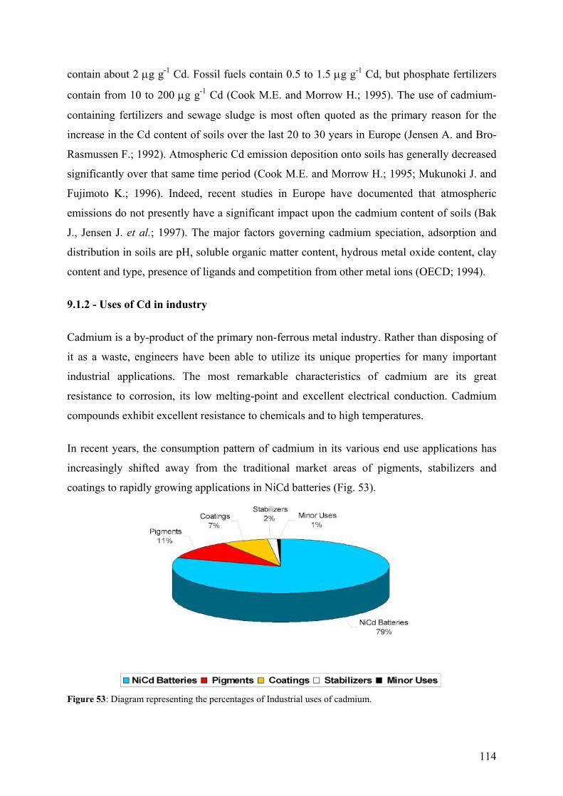
114
contain about 2 µg g-1 Cd. Fossil fuels contain 0.5 to 1.5 µg g-1 Cd, but phosphate fertilizers
contain from 10 to 200 µg g-1 Cd (Cook M.E. and Morrow H.; 1995). The use of cadmium-
containing fertilizers and sewage sludge is most often quoted as the primary reason for the
increase in the Cd content of soils over the last 20 to 30 years in Europe (Jensen A. and Bro-
Rasmussen F.; 1992). Atmospheric Cd emission deposition onto soils has generally decreased
significantly over that same time period (Cook M.E. and Morrow H.; 1995; Mukunoki J. and
Fujimoto K.; 1996). Indeed, recent studies in Europe have documented that atmospheric
emissions do not presently have a significant impact upon the cadmium content of soils (Bak
J., Jensen J. et al.; 1997). The major factors governing cadmium speciation, adsorption and
distribution in soils are pH, soluble organic matter content, hydrous metal oxide content, clay
content and type, presence of ligands and competition from other metal ions (OECD; 1994).
9.1.2 - Uses of Cd in industry
Cadmium is a by-product of the primary non-ferrous metal industry. Rather than disposing of
it as a waste, engineers have been able to utilize its unique properties for many important
industrial applications. The most remarkable characteristics of cadmium are its great
resistance to corrosion, its low melting-point and excellent electrical conduction. Cadmium
compounds exhibit excellent resistance to chemicals and to high temperatures.
In recent years, the consumption pattern of cadmium in its various end use applications has
increasingly shifted away from the traditional market areas of pigments, stabilizers and
coatings to rapidly growing applications in NiCd batteries (Fig. 53).
Figure 53: Diagram representing the percentages of Industrial uses of cadmium.
115
The major intentional uses of cadmium are (i) NiCd batteries, (ii) Cd pigments, (iii) Cd
stabilizers, (iv) Cd coatings, (v) Cd alloys and (vi) Cd electronic compounds such as cadmium
telluride (CdTe), while the major classes of products where cadmium is present as an impurity
are non-ferrous metals (zinc, lead and copper), iron and steel, fossil fuels (coal, oil, gas, peat
and wood), cement, and phosphate fertilizers (Cook M.E. and Morrow H.; 1995). Those uses
are discussed in greater details below:
(i) Cd hydroxide is used as one of the electrode materials in NiCd batteries which have
extensive applications in the railroad and aircraft industry for starting and emergency power
and in consumer applications such as cordless power tools, cellular telephones, camcorders,
portable computers, portable household appliances and toys. NiCd batteries are cost-effective,
well suited for high power applications, and have high cycle lives and excellent low
temperature and high temperature performance relative to other battery chemistries.
(ii) Cd pigments (i.e. cadmium sulphide, cadmium sulphoselenide) produce intense colourings
such as yellow, orange and red, and are well known pigments in artists' colours, plastics,
glasses, ceramics and enamels. They are well known for their ability to withstand high
temperature and high pressure without chalking or fading, and therefore are used in
applications where high temperature or high pressure processing is required (Cook M.E.;
1994).
(iii) Cadmium-bearing stabilizers retard the degradation processes in polyvinylchloride (PVC)
which occur upon exposure to heat and ultraviolet light. These stabilizers contain organic
cadmium salts, usually carboxylates such as cadmium laureate or cadmium stearate, which are
incorporated into PVC before processing and which arrest any degradation during subsequent
processing and ensure a long service life.
(iv) Cadmium coatings are utilized on steel, aluminium, and certain other non-ferrous metal
fasteners and moving parts to provide the best available combination of corrosion resistance,
particularly in salt and alkali media, and lubricity or low coefficient of friction. They are also
employed in many electrical or electronic applications where a good combination of corrosion
resistance and low electrical resistivity are required. In addition, cadmium coatings exhibit
excellent plating characteristics on a wide variety of substrates, have good galvanic
comparability with aluminium and are readily solderable (Morrow H.; 1996).
116
(v) Cadmium alloys include (a) alloys in which cadmium is present in small amount to
improve the physical, mechanical or electrochemical properties of copper, tin, lead or zinc-
based alloys or (b) low melting point fusible alloys and high melting point joining alloys.
Other minor uses of cadmium include cadmium telluride and cadmium sulphide in
photovoltaic cells, and other semiconducting cadmium compounds in a variety of electronic
applications.
(vi) Photovoltaic cells, based on thin-films of either cadmium telluride (CdTe) or cadmium
sulphide (CdS), generate an electric current when light fails upon them. These devices are
used in solar cells and have an important role in the development of cost-effective solar power
installations. Cadmium sulphide photoconductive cells are used in photographic exposure
meters for cameras where the CdS cell acts as a light-sensitive variable resistor. Cadmium
mercury telluride is also an important semiconductor for infrared imaging systems in defence,
space and search and rescue applications.
Cadmium impurities in various products vary widely. For example, the Cd content of the raw
materials used in cement production are generally in the range of 2 µg g-1 and cadmium levels
in fossil fuels vary from 0,1 to 1.5 µg g-1. However, phosphate fertilizers may contain from 10
to 200 µg g-1 Cd and the phosphate rock from which the phosphate fertilizer is derived
contains 3 to 90 µg g-1. Cadmium unintentionally present in iron and steel and most non-
ferrous alloys is generally below 0.1% and in some specifications are required to be well
below that level (Cook M.E. and Morrow H.; 1995). While the cadmium impurity levels in
these products are generally low, the amounts produced are high and thus total cadmium from
this class of materials may be significant.
9.1.3 - Implications for the future
Cadmium will invariably be present in our society, either in useful products or in controlled
wastes. Today, its health effects are well understood and well regulated so that there is no
need to restrict or ban cadmium products, which, in any event, contribute so little to human
cadmium exposure as to be virtually insignificant. Nickel-cadmium batteries are essential and
irreplaceable in many industrial and consumer applications, particularly those requiring high
power, long cycle lives, and good high or low temperature performance. Rechargeable NiCd
117
batteries can replace thousands of primary non-rechargeable batteries, and thus significantly
reduce the total amount of waste. The materials in recyclable NiCd batteries can be more than
99% recovered for reuse in the production of new NiCd batteries (Morrow H. and Keating J.;
1997). NiCd batteries are also making important contributions to the development of the
electric car market in Europe, thereby contributing to the improvement of urban air quality.
Cadmium pigments and stabilizers are important additives in certain specialized plastics,
glasses, ceramics and enamels to achieve bright colours along with long service lives, even in
very demanding applications. From an ecological point of view, it is important to develop and
maintain functional products with long service lives, once again to minimize the input into the
world's waste stream. It should also be emphasized that cadmium in these applications is in a
chemically very stable, highly insoluble form, and embedded in the product's matrix (Cook
M.E.; 1994).
Cadmium coated components, likewise, provide outstanding corrosion resistance along with
low friction coefficient, low electrical resistivity, good galvanic comparability, good plating
coverage, ability to coat a wide variety of surfaces, and good brazeability and solderability
(Morrow H.; 1996). For these reasons, cadmium coated products are preferred for a wide
range of critical and safety related applications in the aerospace, electrical, defence, mining,
nuclear and offshore industries. In addition, cadmium coating wastes and products are easily
recycled.
9.1.4 - Cd exposure and human health
It has been well established that excess cadmium exposure produces adverse health effects on
human beings. For virtually all chemicals, adverse health effects are noted at sufficiently high
total exposures. For certain elements such as copper and zinc, which are essential to human
life, a deficiency as well as excess, can cause adverse health effects. Cadmium is not regarded
as essential to human life. The relevant questions with regard to cadmium exposure are the
total exposure levels and the principal factors, which determine the levels of cadmium
exposure and the adsorption rate of the ingested/inhaled cadmium by the individual.
118
9.1.4.1 - Principal factors determining levels of human exposure
Humans normally absorb cadmium into the body either by (i) ingestion or (ii) inhalation.
Dermal exposure (uptake through the skin) is generally not regarded to be of significance
(Lawerys R.R.; 1986). It is widely accepted (WHO; 1992) that approximately 2% to 6% of
the cadmium ingested is actually taken up into the body. Factors influencing cadmium
absorption are the chemical form in which cadmium is present in the food and the iron status
of the exposed individual. In contrast, from 30% to 64% of inhaled cadmium is absorbed by
the body, with some variation as a function of chemical form, solubility and particle size of
the material inhaled. Thus, a greater proportion of inhaled cadmium is retained by the body
than when cadmium is taken in by ingestion. Moreover, a model for human cadmium intake
(Van Assche F.J.; 1998) has estimated that ingestion accounts for 95% of total cadmium
intake in a non-smoker, while for a smoker, this model estimates that roughly 50% of their
cadmium intake arises from cigarettes with the balance due to ingestion and the low levels of
cadmium naturally present in ambient air. Thus, the principal determinants of human
cadmium exposure today are smoking habits and diet.
9.1.4.2 - Human intake of Cd
(i) A majority of cadmium that enters the body by ingestion comes from terrestrial foods, i.e.,
from plants grown in soil or meat from animals, which have ingested plants. It has been
estimated that 98% of ingested cadmium comes from terrestrial foods, while only 1% comes
from aquatic foods such as fish and shellfish, and 1% arises from cadmium in drinking water
(Van Assche F.J.; 1998). Many studies have thus attempted to establish the average daily
cadmium intake resulting from foods. In general, these studies show that the average daily
diet for non-smokers living in uncontaminated areas is at present at the low end of the range
of 10 to 25 µg of Cd per day (Elinder C.-G.; 1985; WHO; 1992; OECD; 1994; 1997). This
general trend is confirmed by decreasing blood cadmium levels in the general population in
several countries during this time period (Ducoffre G., Claeys F. et al.; 1992).
The World Health Organisation (WHO) has established a provisional tolerable weekly intake
(PTWI) for cadmium at 7 µg kg-1 of body weight (WHO; 1992). This PTWI weekly value
corresponds to a daily tolerable intake level of 70 µg of cadmium for the average 70-kg man
and 60 µg of cadmium per day for the average 60-kg woman. Clearly, the daily cadmium
intake for the general population from food is well below the guidelines established by the
119
WHO. The average daily cadmium intake for the general population in the Western World has
shown a distinct downward trend from 1970 through 1992 (Van Assche F.J. and Ciarletta P.;
1992), a reduction that is presumed to be due to the marked decreases in direct atmospheric
deposition of cadmium onto crops and soils. Other studies have suggested that, over the
timeframe of 1980 - 1985, levels of cadmium intake have been relatively constant (OECD;
1994). At an absorption rate of 5% from ingestion, the average person is believed to retain
about 0.5 to 1.0 µg of cadmium per day from food.
(ii) Cd inhalation is a far smaller contributor to total cadmium body burden except, as
previously noted, in the cases of smokers or some highly exposed workers in the past. Today,
the inhalation route is well controlled in the occupational setting, and is well-controlled from
point sources such as those, which directly pertain to the non-ferrous, cadmium or cadmium
products industries. On the other hand, ambient air emissions from fossil fuel power
generation plants, the iron and steel industry and other major industries where cadmium may
be present as low concentration impurities, may be substantial because the volumes of the
waste gases generated are substantial.
Some recent studies have demonstrated that foods which are naturally enriched in cadmium
are also enriched in substances which inhibit the uptake of cadmium into the body (Reeves
P.G. and Vanderpool R.A.; 1997). Indeed, Vahter et al. (Vahter M., Berglund M. et al.; 1996)
have suggested that overall nutritional status is a more important determinant of cadmium
uptake into the body than is the actual amount of cadmium ingested. For example, women
subsisting upon a vegetarian diet and with reduced iron stores have increased uptake of
ingested cadmium. For these women, iron deficiency is a more important determinant of
cadmium uptake than is the actual amount of cadmium ingested.
Smokers absorb amounts of cadmium comparable to those from food, about 1 to 3 µg of
cadmium per day, from the smoking of cigarettes. It has been reported that one cigarette
contains about 1 to 2 µg of cadmium and that about 10% of the cadmium content is inhaled
when the cigarette is smoked (WHO; 1992). Cigarette construction, the use of filters and
variations in the cadmium contents of tobaccos could decrease cadmium exposure, but in
general cigarette smoking is a habit, which can more than double the average person's daily
120
cadmium intake. Cigarette smokers who are also occupationally exposed may increase their
total cadmium intake even further.
9.1.4.3 - Human health effects of Cd
The kidney is the critical target organ for the general population as well as for occupationally
exposed populations. Cadmium is known to accumulate in the human kidney for a relatively
long time, from 20 to 30 years, and, at high doses, is also known to produce health effects on
the respiratory system and has been associated with bone disease. Most of the available
epidemiological information on cadmium has been obtained from occupationally exposed
workers or on Japanese populations in highly contaminated areas. Such a situation did occur
in the 1950s and 1960s in Japan where heavy cadmium contamination of rice fields, along
with nutritional deficiencies for iron, zinc and other minerals, led to renal impairment and
bone disease (Itai Itai disease) in exposed populations. Studies utilizing very sophisticated
biomarkers have detected mild alterations in kidney functions at lower levels of exposure
(Buchet J.P., Lauwerys R. et al.; 1990). Most studies have centred on the detection of early
signs of kidney dysfunction and lung impairment in the occupational setting and on the
detection and screening for bone disease in general populations exposed to cadmium-
contaminated rice. More recently, the possible role of cadmium in human carcinogenesis has
also been studied in some detail.
For acute exposure by ingestion, the principal effects are gastrointestinal disturbances such as
nausea, vomiting, abdominal cramps and diarrhea. Acute poisoning by inhalation may lead to
respiratory manifestations such as severe bronchial and pulmonary irritation, subacute
pneumonitis, lung emphysema, and, in the most severe situations, death from pulmonary
oedema may occur (Lawerys R.R.; 1986). Chronic obstructive airway disease has been
associated with long-term high-level occupational exposure by inhalation (WHO; 1992;
OECD; 1994). While there have been suggestions in past studies that such exposure may
cause lung or prostate cancer, more recent epidemiological analyses of cadmium-exposed
cohorts have dismissed the prostate cancer association and indicate that arsenic rather
cadmium may be responsible for the observed increase in lung cancer mortality rates (Sorahan
T., Lister M. et al.; 1995; Sorahan T. and Lancashire R.J.; 1997). In addition, most of the
other data cited as evidence for the carcinogenicity of cadmium relies on studies, which are
confounded by the presence of other carcinogenic substances such as nickel, asbestos or
tobacco smoke as well as arsenic.
121
For chronic cadmium exposure, effects occur mainly on the kidneys, lungs, and bones. A
relationship has been established between cadmium air exposure and proteinuria (an increase
in the presence of low molecular weight proteins in the urine and an indication of kidney
dysfunction) (WHO; 1992; OECD; 1994). Cadmium is known to accumulate in the renal
cortex, and there is evidence that the level of cadmium in the renal cortex associated with
increased urinary excretion is about 200 to 250 µg g-1 (wet weight). Depending upon exposure
level and other sources of cadmium, this level might be reached after 20 years occupational
exposure. However, recent work has demonstrated that these effects are reversible at low
exposure levels once the cadmium exposure has been removed or reduced (Roels H.A., Van
Assche F.J. et al.; 1997). With today's low occupational exposure standards, coupled with
required biological monitoring of cadmium exposure levels (e.g. cadmium-in-blood and
cadmium-in-urine) and kidney function parameters (e.g. B-2 microglobulin, a low molecular
weight protein), there is every assurance that kidney dysfunction or other effects do not
develop in occupationally exposed workers as they did in the past (Roels H.A., Van Assche
F.J. et al.; 1997).
9.2 - Biological interaction of trace metals with Chlamydomonas
The biological response of microorganisms to metals includes: (i) exclusion of the metal from
the cell by the cell membrane; (ii) active transport of the metal from the cell so that toxic
levels of the metal cannot accumulate inside the cell; (iii) sequestration of the metal inside the
cell by chelators; (iv) extracellular sequestration in which the metal is bound to the outside of
the cell and prevented from entering the cell, and (v) chemical modification of the metal to a
form that is less toxic to the cell.
9.2.1 - Unicellular green algae
Single-cell organisms, such as bacteria and protozoa, have been so successful in adapting to a
variety of different environments that they comprise more than half of the total biomass on
earth. Unlike animals, many of these unicellular organisms can synthesize all of the
substances they need from a few simple nutrients. Green algae are eukaryotes that exist as
unicellular, colonial or multicellular forms (Fig. 54). Different species of green algae can be
arranged in order of complexity, illustrating the kind of progression that probably occurred in
the evolution of higher plants and animals (Campbell P.G.C.; 1995). With an evolution
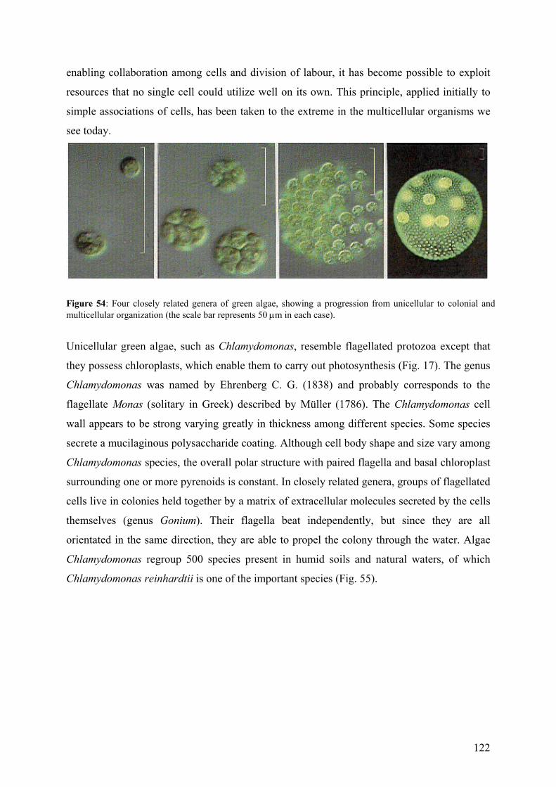
122
enabling collaboration among cells and division of labour, it has become possible to exploit
resources that no single cell could utilize well on its own. This principle, applied initially to
simple associations of cells, has been taken to the extreme in the multicellular organisms we
see today.
Figure 54: Four closely related genera of green algae, showing a progression from unicellular to colonial and multicellular organization (the scale bar represents 50 µm in each case).
Unicellular green algae, such as Chlamydomonas, resemble flagellated protozoa except that
they possess chloroplasts, which enable them to carry out photosynthesis (Fig. 17). The genus
Chlamydomonas was named by Ehrenberg C. G. (1838) and probably corresponds to the
flagellate Monas (solitary in Greek) described by Müller (1786). The Chlamydomonas cell
wall appears to be strong varying greatly in thickness among different species. Some species
secrete a mucilaginous polysaccharide coating. Although cell body shape and size vary among
Chlamydomonas species, the overall polar structure with paired flagella and basal chloroplast
surrounding one or more pyrenoids is constant. In closely related genera, groups of flagellated
cells live in colonies held together by a matrix of extracellular molecules secreted by the cells
themselves (genus Gonium). Their flagella beat independently, but since they are all
orientated in the same direction, they are able to propel the colony through the water. Algae
Chlamydomonas regroup 500 species present in humid soils and natural waters, of which
Chlamydomonas reinhardtii is one of the important species (Fig. 55).
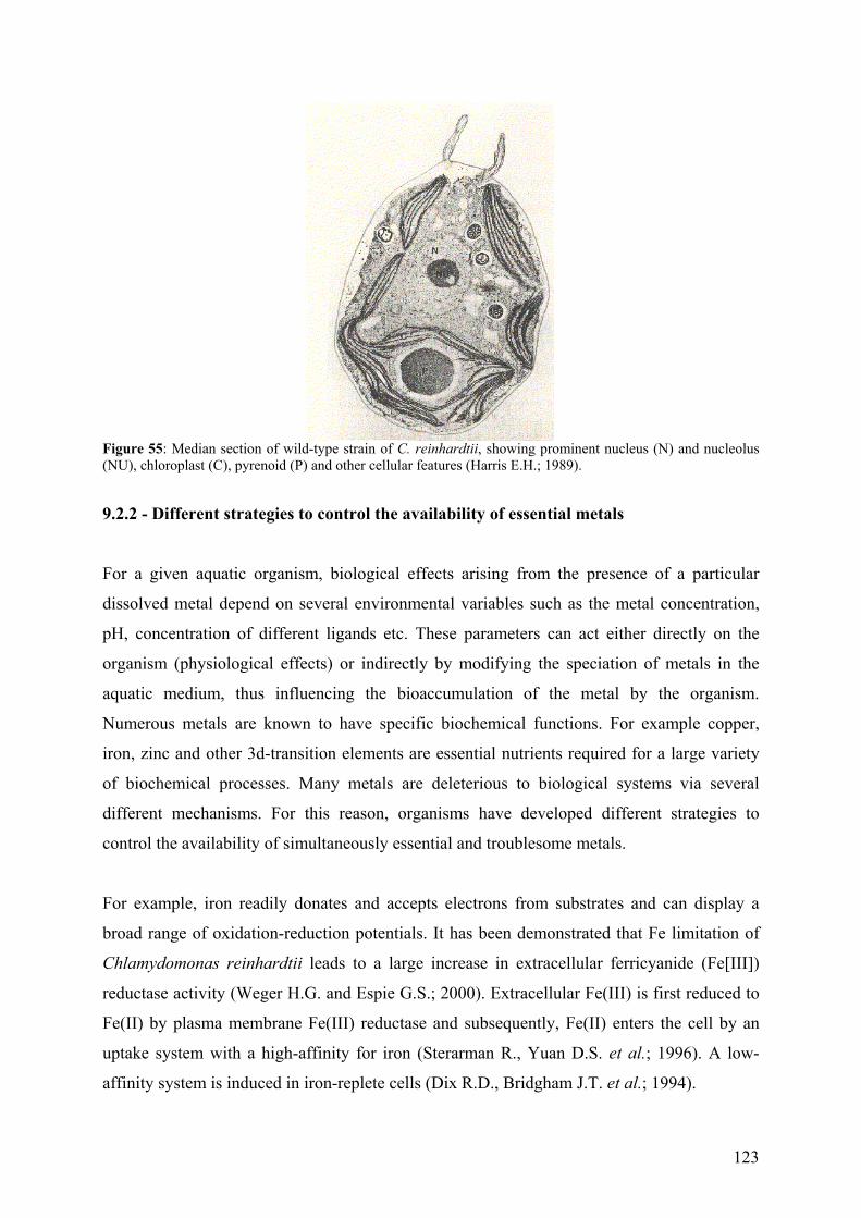
123
Figure 55: Median section of wild-type strain of C. reinhardtii, showing prominent nucleus (N) and nucleolus (NU), chloroplast (C), pyrenoid (P) and other cellular features (Harris E.H.; 1989).
9.2.2 - Different strategies to control the availability of essential metals
For a given aquatic organism, biological effects arising from the presence of a particular
dissolved metal depend on several environmental variables such as the metal concentration,
pH, concentration of different ligands etc. These parameters can act either directly on the
organism (physiological effects) or indirectly by modifying the speciation of metals in the
aquatic medium, thus influencing the bioaccumulation of the metal by the organism.
Numerous metals are known to have specific biochemical functions. For example copper,
iron, zinc and other 3d-transition elements are essential nutrients required for a large variety
of biochemical processes. Many metals are deleterious to biological systems via several
different mechanisms. For this reason, organisms have developed different strategies to
control the availability of simultaneously essential and troublesome metals.
For example, iron readily donates and accepts electrons from substrates and can display a
broad range of oxidation-reduction potentials. It has been demonstrated that Fe limitation of
Chlamydomonas reinhardtii leads to a large increase in extracellular ferricyanide (Fe[III])
reductase activity (Weger H.G. and Espie G.S.; 2000). Extracellular Fe(III) is first reduced to
Fe(II) by plasma membrane Fe(III) reductase and subsequently, Fe(II) enters the cell by an
uptake system with a high-affinity for iron (Sterarman R., Yuan D.S. et al.; 1996). A low-
affinity system is induced in iron-replete cells (Dix R.D., Bridgham J.T. et al.; 1994).
124
Copper is also a well-known example of a metal that is essential for life but is toxic at high
concentrations. In Chlamydomonas reinhardtii, for example Cu has an essential function in
the catalysis of respiration, oxygen transport and photosynthesis. The major copper proteins in
this organism are plastocyanin (which functions in the chloroplast during photosynthesis) and
Cyt oxidase (which functions during respiration in the mitochondrion) (Hill K.L., Hassett R.
et al.; 1996). Garvey et al. (Garvy J.E., Owen H.A. et al.; 1991) showed that the biological
significance of the copper regulation in Chlamydomonas is indicated by the fact that high
concentrations of copper induced deflagellation of the cells. On the other hand, under copper-
deficient conditions, in order to maintain photosynthetic activity, Chlamydomonas reinhardtii
substitutes a heme protein for an otherwise essential copper protein, viz. plastocyanin which is
regulated at the transcriptional level by the availability of Cu (Merchant S., Hill K.L. et al.;
1991). Furthermore, C. reinhardtii cells do not take up copper in great excess over what they
need for the maintenance of copper protein levels, even when it is supplied in excess (Hill
K.L., Hassett R. et al.; 1996). Copper uptake is restricted once the cell has obtained an
amount corresponding to between 1.9x10⁷ and 4.9x10⁷ copper ions per cell, which is about
two to five times the amount needed to saturate the plastocyanin biosynthetic pathway
(plastocyanin is estimated to be 10-fold more abundant than Cys oxidase). Button and
Hostetter (Button K.S. and Hostetter H.P.; 1977) demonstrated that most of the copper sorbed
by Chlamydomonas cells remained bound to the cell wall and probably did not enter the
protoplasm. They arrived at this conclusion by comparing the Cu2+ sorbed on cleaned cell
walls of Chlamydomonas reinhardtii (3.5 nmol Cu2+/106 cells) and live cells (2.6 nmol/106
cells). Furthermore, when live cells were subsequently placed in a Cu-free medium, 59 % of
the adsorbed Cu was released into the medium. Even if copper might be in a nonavailable
chemical form for uptake, cupric reductase activity is regulated; this enzyme seems to be
involved in a reductive copper assimilation pathway.
9.2.3 - Protein induction
Phytochelatins. Phytochelatins are a family of peptides first identified in the yeast
Schizosaccharomyces pombe. They have been found in several fungal species, marine diatoms
and in all plant species examined to date (Rauser W.E.; 1990; Rauser W.E.; 1995) but not in
animal species. Their production in cells is induced by a range of heavy metal ions, including
the cations: Cd, Ni, Cu, Zn, Ag, Hg, and Pb and the anions arsenate and selenite. Cells of
some algae, including Chlamydomonas, also synthesize metal-chelating proteins
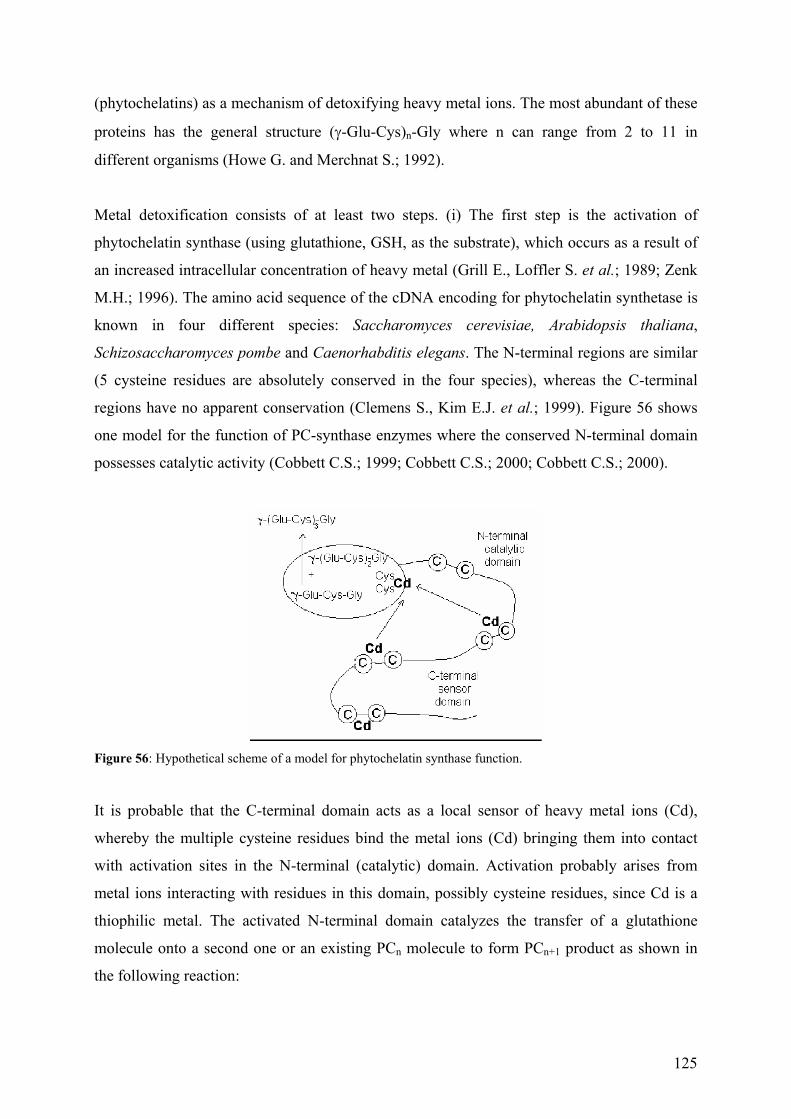
125
(phytochelatins) as a mechanism of detoxifying heavy metal ions. The most abundant of these
proteins has the general structure (γ-Glu-Cys)n-Gly where n can range from 2 to 11 in
different organisms (Howe G. and Merchnat S.; 1992).
Metal detoxification consists of at least two steps. (i) The first step is the activation of
phytochelatin synthase (using glutathione, GSH, as the substrate), which occurs as a result of
an increased intracellular concentration of heavy metal (Grill E., Loffler S. et al.; 1989; Zenk
M.H.; 1996). The amino acid sequence of the cDNA encoding for phytochelatin synthetase is
known in four different species: Saccharomyces cerevisiae, Arabidopsis thaliana,
Schizosaccharomyces pombe and Caenorhabditis elegans. The N-terminal regions are similar
(5 cysteine residues are absolutely conserved in the four species), whereas the C-terminal
regions have no apparent conservation (Clemens S., Kim E.J. et al.; 1999). Figure 56 shows
one model for the function of PC-synthase enzymes where the conserved N-terminal domain
possesses catalytic activity (Cobbett C.S.; 1999; Cobbett C.S.; 2000; Cobbett C.S.; 2000).
Figure 56: Hypothetical scheme of a model for phytochelatin synthase function.
It is probable that the C-terminal domain acts as a local sensor of heavy metal ions (Cd),
whereby the multiple cysteine residues bind the metal ions (Cd) bringing them into contact
with activation sites in the N-terminal (catalytic) domain. Activation probably arises from
metal ions interacting with residues in this domain, possibly cysteine residues, since Cd is a
thiophilic metal. The activated N-terminal domain catalyzes the transfer of a glutathione
molecule onto a second one or an existing PCn molecule to form PCn+1 product as shown in
the following reaction:
126
γ-Glu-Cys-Gly +(γ-Glu-Cys)n-Gly (γ-Glu-Cys)n+1-Gly +Gly
(ii) The second step consists of the complexation and thus inactivation of the metal that has
entered the cell's cytosol by PCn molecules. The enzyme is self-regulated in this respect;
addition of an amount of phytochelatin [as the complexing agent] sufficient to quantitatively
complex the metal immediately stops the ongoing chain elongation reaction (Loeffler S.,
Hochberger A. et al.; 1989; Rauser W.E.; 1999). This interruption can also be achieved by the
use of a metal-chelating agent such as ethylenediaminetetraacetic acid (EDTA) (Zenk M.H.;
1996). Yen et al. (Yen T.-Y., Villa J.A. et al.; 1999) elucidated for the first time the covalent
nature of PC-Cd complexes by nano-electrospray ionization tandem mass spectrometry (nano-
ESI-MS/MS) and capillary liquid chromatography/ electrospray (capillary LC/ESI-MS/MS)
methods. The formation of these complexes involved the cysteine thiol groups of
phytochelatin with the loss of 2H+ for each coordinated Cd2+ ion. The Cd-S distance in the
phytochelatin has been reported for different plant species to be 2.5 Å (Pickering I.J., Prince
R.C. et al.; 1999; Rauser W.E.; 1999).
This sequence of steps confers protection for heavy-metal-sensitive enzymes to which the
toxic metal otherwise would be able to bind. The intracellular pool of these peptides is tightly
regulated in response to the inorganic metal concentration levels that are relevant to
ecosystems (Ahner B.A. and Morel F.M.M.; 1995).
Chlamydomonas reinhardtii has various adaptive mechanisms in order to avoid toxic effects
of metal excess. Gekeler et al. (Gekeler W., Grill E. et al.; 1988) reported that in response to
Cd2+ uptake, Chlamydomonas reinhardtii synthesizes phytochelatins with n-values ranging
from 2 to 4. Howe and Merchant (Howe G. and Merchnat S.; 1992) have extended this
observation by showing that up to 70% of the intracellular Cd2+ ions can be complexed and
inactivated. These Cd-binding peptides have a size of 24 kDa for Chlamydomonas reinhardtii
strains with and without cell walls (Prasad M.N., Drej K. et al.; 1998). The same authors
examined the response of this alga to the addition of micromolar concentrations of Cd, Hg
and Ag, and found that phytochelatin production was induced to the greatest extent by Cd
even at these extremely high concentrations. Hu et. al. (Hu S., Lau K.W.K. et al.; 2001)
characterized two types of PC-Cd-complexes from Cd-challenged Chlamydomonas cells: a
high-molecular-weight (HMW) complex and a low molecular weight (LMW). The HMW
complex appeared first during Cd treatment, supporting the hypothesis of a very quick
127
conversion of the LMW complex to a HMW complex inside the algal cells, which could be an
early sign of metal stress. The same phytochelatin complexes were identified in the marine
macroalgae Kappaphycus alvarezii in the presence of Cd in another study by the same author
(Hu S.; 1998). The PC content in algal fronds was directly correlated with the ambient Cd
concentration as well as exposure time. Murasugi et al. (Murasugi A., Wada C. et al.; 1983)
first showed that an appreciable quantity of acid-labile sulphide was part of the HMW
complex, with much less in the LMW complex. Similar situations were found by Wu in her
studies (Hu S.; 1998; Hu S., Lau K.W.K. et al.; 2001). From different data in vitro has come
the idea that the HMW complex is the LMW complex with added sulphide and Cd (Rauser
W.E.; 1999). Another alga, Chlorella, when exposed to Cd, was found to synthesize a
cytoplasmic protein similar to that mentioned above. Hart and Scaife (Hart B.A. and Scaife
B.D.; 1977) suggested that this type of protein would bind cadmium and prevent it from
diffusing out of the cell.
In many species, the production of phytochelatin in response to Cd appears to be
stoichiometric, but not every toxic metal will induce formation of the detoxifying
phytochelatins. Depending on the species and metal, the extent of phytochelatin induction is
different. In marine phytoplankton, Cd and to a lesser extent Cu and Zn were found to be the
most effective phytochelatin inducers within the range of metal concentrations present in
natural waters (Ahner B.A. and Morel F.M.M.; 1995). In Chlamydomonas reinhardtii strains
with and without cell walls, Prasad et al. (Prasad M.N., Drej K. et al.; 1998) found that
contrary to the above work, copper failed to induce phytochelatin synthesis.
The difference between Cd and other metals may be related to their relative abilities to
activate phytochelatin synthase (Grill E., Loffler S. et al.; 1989), possibly on account of their
different affinities for the groups surrounding the activation site. Different stabilities of the
metal complexes that are formed are another possible explanation (Hayashi Y., Nakagawa
C.W. et al.; 1988). Grill et al. (Grill E., Loffler S. et al.; 1989) observed in vitro that at a
given concentration, Cd was most effective metal for synthase activation. Relative to Cd, the
Ag efficiency was 58%, that of Pb 48%, and that of Zn and Hg <35%.
Other detoxifying proteins. The induction of detoxifying proteins can be metal-specific. Howe
and Merchant (Howe G. and Merchnat S.; 1992) found that unlike their response to Cd and
Hg, Chlamydomonas cells synthesize an inducible sulphur-containing component that has
128
different physical properties from those of the Cd-binding component in response to Ag+. The
synthesis of this component is blocked when cells are pre-treated for 15 hours with
micromolar concentrations of Cu2+. The simplest hypothesis for these competing effects is
that Cu+, as an electronic analogue of Ag+, prevents Ag+ uptake by the cell by competing for a
shared receptor site (at these concentrations copper is not toxic for the alga).
Howe and Merchant (Howe G. and Merchnat S.; 1992) examined the response of the same
algae to the addition of micromolar sublethal concentrations of Cd, Hg and Ag and found that
the response was metal-specific. Within 8 hours of treatment, the cells synthesized and
accumulated peptides consisting solely of glutamic acid, cysteine and glycine (phytochelatin),
which were able to sequester most cadmium and avoid lethality. When Chlamydomonas cells
were cultured in the presence of micromolar concentrations of HgCl2 known to be toxic, GSH
was the principal thiol-containing compound produced at increased levels. Cells began to
recover from Hg2+ toxicity after 48 hours.
The hypothesis that different steps of the phytochelatin biosynthesis pathway are regulated in
a metal-specific manner is supported by the finding that GSH, rather than phytochelatin, is the
primary thiol induced by Hg2+ ions. The high stability of Hg(GSH)2 at physiological pH
suggests that GSH could function as a complexing agent for Hg2+ ions. Although GSH has
been implicated in the detoxification mechanism for Cd2+ (Singhal R.K., Anderson M.E. et
al.; 1987) and Cu2+ in animal cells (Freedman J.H., Ciriolo M.R. et al.; 1989), little attention
has been paid to the possibility that this peptide could serve a similar function in plants. Cd-
GSH complexes are many orders of magnitude less stable than Hg-GSH. Nonetheless,
Hayashi et al. (Hayashi Y., Nakagawa C.W. et al.; 1988) showed that the affinity of Cd2+ to
(γ-Glu-Cys)n-Gly peptides having lengths n = 1 (GSH), 2, 3 increased significantly with
increasing peptide length. The unique potency of Cd2+ to induce phytochelatin production
from GSH may be interpreted as an evolutionary adaptation of the organism to the inability of
GSH to bind Cd2+ ions with the stability required for Cd2+ detoxification.
Chlamydomonas can produce other proteins in the presence of heavy metals when the GSH
pool is depleted. Thioredoxins (TRX) are small ubiquitous proteins found in many organisms
from bacteria to humans. They are characterized by a reactive disulfide-active site (Cys-X-X-
Cys) (Jacquot J.-P. and Lancelin J.M.; 1997). In plants, two isoforms are located in
chloroplasts and are involved in the carbon fixation by photosynthesis. The TRX are able to
129
reduce several key enzymes of the carbon fixation metabolism. Lemaire et al. (Lemaire S.,
Keryer E. et al.; 1999) found that the presence of sublethal concentrations of Hg (1 µM) and
Cd (100 µM) suppressed the activity of the TRX in vitro. This can be explained by the fact
that the active dithiol site of the reduced TRX is a possible target of heavy metal fixation. The
identical concentrations of the same metals lead to an increase in the level of TRX messengers
(mRNA). Thus, induction of TRXs by heavy metals might be an indirect effect mediated by
the depletion of the GSH pool in Chlamydomonas, since upon exposure to metals,
phytochelatins are synthesized and the GSH level drops immediately (Zenk M.H.; 1996). This
finding suggests that one role of TRX could be linked to the detoxification of heavy metals.
Behra found that calmodulin (CaM) (Behra R.; 1993; Behra R.; 1993), a ubiquitous calcium-
binding protein that regulates many cellular processes and stimulates the activity of several
target proteins in Chlamydomonas (Zimmer W.E., Schloss J.A. et al.; 1988), has a potential
role in metal toxicity, since beside calcium, other metals such as Cd, Zn, Al and Pb are
capable of binding to CaM and activating CaM-dependent enzymes. Calcium, having weak,
reversible binding capacities with the oxy-rich domains of the cytosolic protein calmodulin,
has the role of an allosteric switch altering the activity of the latter (Mason A.Z. and Jenkins
K.D.; 1995). Quarmby and Hartzell (Quarmby L.M. and Hartzell H.C.; 1994) demonstrated
that the synthesis of proteins on the flagellar apparatus of Chlamydomonas is coupled with the
production of calmodulin mRNA. CaM action depends on the presence of Ca2+ and is
mediated through the binding of Ca2+-CaM complexes to the respective proteins. These
authors showed two distinct Ca-mediated signal transduction pathways involved in the
deflagellation.
9.3 – Biosensors
A biosensor can be defined as an analytical device incorporating a biological sensing element,
which is either intimately connected to, or integrated with a physicochemical transducer. The
aim of a biosensor is the detection of an environmental signal, usually the presence of a
chemical. Very often, biosensors are constructed in such a way that the signal produced is
proportional to the concentration of a chemical with which the biological element can react.
For the biological components of the biosensor, i.e., the sensing components, three distinct
groups of biological material can be used (Van Der Lelie D., Regniers L. et al.; 1997):
130
1- Behavioural groups, which consist of living higher organisms (e.g. fish, mussels, etc.)
2- Catalytic groups, which includes enzymes, microorganisms and tissues.
3- Noncatalytic groups, which includes antibodies, receptors and nucleic acids.
A number of different transducer technologies can be used to transform the biological signal
into measurable physical signals. These include:
1- Electrochemical elements, which may be amperometric (the measurement of current
flowing at constant potential), potentiometric (the measurement of potential changes at
constant current), or conductimetric (the measurement of changes of conductivity between
two electrodes)
2- Optical elements (the measurement of changes in optical properties, like absorption,
fluorescence, light emission or reflection)
3- Acoustic elements (the measurement of changes in the acoustic properties of the sensor, as
with transducers based on piezoelectric materials)
4- Calorimetric elements (the measurement of small changes in temperature). Note that this
transducer should be a universal one, since almost all biological reactions are exothermic.
A good biomonitoring test must fulfil the following criteria (Persoone G.; 1993):
a)- be replicable in other test laboratories
b)- allow standardization
c)- be as realistic as possible for the ecosystem and the environmental form of the chemical
d)- be economical and uncomplicated
The growing concern about the deterioration of the aquatic environment by groundwater
contamination resulting from domestic sewage and industrial effluents, requires sensitive
online techniques for the effective detection and monitoring of these contaminants. Chemical
analyses, currently performed with sophisticated and expensive equipment such as gas/liquid
chromatography and mass spectroscopy (HPLC, GC/MS), need to be supplemented by
reliable, low-cost techniques providing early-warning systems for effective monitoring.
Microorganisms therefore have a great potential in the construction of biosensors.
131
9.3.1- Microorganisms as a sensing component
A number of advantages are likely to be gained when using intact microorganisms rather than
isolated biological components as the sensing element. Since microorganisms have
mechanisms that enable the regulation of their internal environment, they are usually more
tolerant to assay conditions such as pH, temperature, etc. than an isolated enzyme or protein
(Ramanathan S., Ensor M. et al.; 1997; Ramanathan S., Liu Y. et al.; 1997; Ramanathan S.,
Shi W. et al.; 1997). At the same time, microorganisms have an advantage over current testing
methods in laboratories, since the in-situ monitoring of soils and waters will be possible, thus
allowing one to avoid changes in metal speciation during collection and transport. These
sensors are also less expensive to prepare since the active biological component does not have
to be isolated. However, they have some disadvantages including longer response times than
isolated biological components or the possibility of variations in their responses when
produced from different batches of cells (Karube I. and Chang M.E.; 1991). Culture age,
temperature, cell density and aeration of the culture during the induction of the response can
also affect the response of the sensor. Despite these problems, the use of intact cells is a
promising avenue of research.
Bacterial biosensors. Microorganisms have evolved different mechanisms that allow them to
survive, grow and become resistant to contaminant levels of heavy metals. The expression of
genes responsible for these resistance mechanisms in some cases is induced and regulated by
the presence of a metal specific to the cell’s environment. This specificity is used to construct
biosensors that detect toxic metal. Gene-based biosensors using bacteria as a sensing
component for the specific detection of heavy metals have been developed over the past few
years. Many of the bacterial resistance determinants are inducible by metals, and this
transcriptional regulation is exploited in vivo using genes with a readily determined
phenotype. The genes are organized in operons and carried by resistant bacteria. The promoter
taken from an operon for metal–resistance is fused to a reporter gene that encodes, for
example, for bioluminescent proteins, such as luciferase or beta-galactosidase. The light
produced by the bioluminescent proteins can be measured by photometers, luminometers,
charge-coupled devices and liquid scintillation (Burlage R.S. and Kuo C.-T.; 1994). A scheme
of the sensory process in bacteria is shown in Figure 57a (Ramanathan S., Shi W. et al.;
1997).
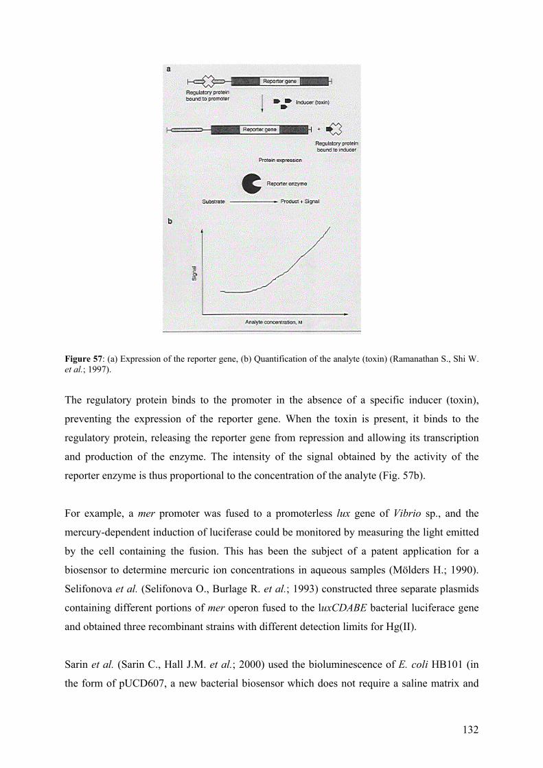
132
Figure 57: (a) Expression of the reporter gene, (b) Quantification of the analyte (toxin) (Ramanathan S., Shi W. et al.; 1997).
The regulatory protein binds to the promoter in the absence of a specific inducer (toxin),
preventing the expression of the reporter gene. When the toxin is present, it binds to the
regulatory protein, releasing the reporter gene from repression and allowing its transcription
and production of the enzyme. The intensity of the signal obtained by the activity of the
reporter enzyme is thus proportional to the concentration of the analyte (Fig. 57b).
For example, a mer promoter was fused to a promoterless lux gene of Vibrio sp., and the
mercury-dependent induction of luciferase could be monitored by measuring the light emitted
by the cell containing the fusion. This has been the subject of a patent application for a
biosensor to determine mercuric ion concentrations in aqueous samples (Mölders H.; 1990).
Selifonova et al. (Selifonova O., Burlage R. et al.; 1993) constructed three separate plasmids
containing different portions of mer operon fused to the luxCDABE bacterial luciferace gene
and obtained three recombinant strains with different detection limits for Hg(II).
Sarin et al. (Sarin C., Hall J.M. et al.; 2000) used the bioluminescence of E. coli HB101 (in
the form of pUCD607, a new bacterial biosensor which does not require a saline matrix and
133
thus allows the evaluation of toxicities of heavy metal chloride complexes at low and
moderate Cl- concentrations) to probe the toxicity of cadmium and lead complexes with
chloride. As the chloride concentration was raised, less Cd2+ or Pb2+ and more CdCl+ or PbCl+
were present. Bioluminescence did not change with increasing chloride concentrations in Cd
solutions, implying that Cd2+ and CdCl+ species were comparable in toxicity.
Bioluminescence decreased with increasing chloride concentrations in Pb solutions, implying
that PbCl+ was more toxic than Pb2+.
Prokaryotes have a simple genome and thus can be easily genetically engineered but they can
also easily modify the trace metal speciation in the test system (Mirimanoff N. and Wilkinson
K.J.; 2000). This is a major disadvantage, if the goal is to measure the bioavailability of
metallic species in the environment. Another drawback is the relatively long induction times
needed to produce a measurable response, usually longer than 30 min. Also, it normally takes
a considerable time for the cells to return to the base-line signal, once they have been
deployed (Ramanathan S., Ensor M. et al.; 1997).
Chlamydomonas strain as a sensor. The unicellular green alga Chlamydomonas reinhardtii
could potentially provide several advantages as the biological component of a biosensor
system. This alga is the genetically and physiologically the best characterized organism
among unicellular eukaryotic algae. Therefore the availability of mutants with specific and
characterized alterations provides the potential for constructing specific test organisms (Xiong
J., Hutchison R.S. et al.; 1997). Furthermore, this alga can be transformed with naked DNA,
opening up the possibility of introducing genes into the organism (Kindle K.L.; 1990).
Schubnell et al. (Schubnell D., Lehmann M. et al.; 1999) have constructed a biosensor for
which pH changes, caused by the uptake or production of CO2 by Chlamydomonas cells, are
monitored on-line by IS-FET (ion-sensitive field effect transistors). This system provides a
rapid and sensitive test system for substances that interfere with either photosynthesis or
cellular metabolism, since metabolic activity reflects changes in pH. Naessens et al.
(Naessaens M., Leclerc J.C. et al.; 2000) proposed an algal biosensor using a fluorescence-
based optical fiber for the determination of herbicides based on kinetic measurements of
chlorophyll-a fluorescence in Chlorella vulgaris, another green alga.
134
Reporter genes encoding for light-emitting proteins such as luciferase (Fuhrmann M., Oertel
W. et al.; 1999) have recently been adapted for expression in Chlamydomonas reinhardtii.
Other potential reporter genes have been suggested by William Zerges (Biology Department,
Concordia University), for example, the protochlorophyllide reductase (POR) gene, which is
responsible for the production of chlorophyll in the dark (Li J. and Timko M.P.; 1996) or the
ars reporter gene (Davies J.P., Weeks D.P. et al.; 1992) encoding a periplasmic enzyme
which cleaves sulphate from aromatic compounds. This latter protein is produced by
Chlamydomonas reinhardtii during sulphur-limited growth. Its activity is easily assayed in
whole cells by adding the chromogenic substrate 5-bromo-4-chloro-3-indolyl sulphate
(XSO4), the product being a blue compound (blue colonies). No arylsulfatase activity is
detectable in untransformed cells grown in sulphur-sufficient medium (Davies J.P., Weeks
D.P. et al.; 1992; Davies J.P., Yildiz F. et al.; 1994). A promoterless radical spoke protein (a
flagellar protein) gene (RSP3) has been used as a reporter gene to identify promoter regions in
the genome of Chlamydomonas reinhardtii activated by the deprivation of nitrogen in the
media (Haring M.A. and Beck C.F.; 1997). The RSP3 gene complements the paralyzed
flagella and thereby restores motility to mutation pf-14 (which lack the radical spoke protein
and thus cannot form flagella or swim). When the RSP3 gene is fused to a promoter induced
by nitrogen deprivation, transformants begin swimming (promoter tagging method).
One of the well-known defence mechanisms of Chlamydomonas in response to heavy metal
stimuli is the production of phytochelatins activated by PC-synthase production, which in turn
is encoded by the PC-synthase reporter gene. The promoter of the PC-synthase gene is a
metal-regulated promoter (Clemens S., Kim E.J. et al.; 1999; Cobbett C.S.; 2000). Since more
than one of the mechanisms mentioned above may be responsible for resistance to a given
metal, different metal-regulated promoters can be activated in Chlamydomonas in the
presence of the heavy metals. Because of the specificity of this regulation, the metal-regulated
promoters can be fused to different reporter genes (promoterless) that produce a strong signal
in Chlamydomonas that will be proportional to the chemical stimulus offered by the
bioavailable metal species to which it is exposed.
9.4 – References (annexes)
Ahner B. A. and Morel F. M. M. Phytochelatin production in marine algae. 2. Induction by
various metals. Limnol. Oceanogr. 1995, 40, 658-65.
135
ATSDR; (Agency for Toxic Substances and Disease Registry) in. Draft Toxicological Profile
for Cadmium; Atlanta, Georgia: Public Health Service, Department of Health &
Human Services; 1997.
Bak J., Jensen J., Larsen M. M., Pritzl G. and Scott-Fordsmand J. A heavy metal monitoring
programme in Denmark. Sci. Tot. Environ. 1997, 207, 179-186.
Behra R. In vitro effects of cadmium, zinc, and lead on calmodulin-dependent actions in
Oncorhynchus mykiss, Mytilus sp., and Chlamydomonas reinhardtii. Arch. Environ.
Contamin. Toxicol. 1993, 24, 21-7.
Behra R. Interaction of cadmium, lead and zinc with calmodulin from rainbow trout, sea
mussels, and a green alga. Sci. Tot. Environ. 1993, 647-53.
Buchet J. P., Lauwerys R., Roels R., Bernard H., Bruaux P., Claeys F., Ducoffre G., De Plaen
P., Staessen J., Amery A., Linjen P., Thijs L., Ronda D., Sartor F., Saint Remy A. and
Nick L. Renal effect of cadmium body burden of the general population. Lancet 1990,
366, 699-702.
Burlage R. S. and Kuo C.-T. Living biosensors for the management and manipulation of
microbial consortia. Annu. Rev. Microbiol. 1994, 48, 291-309.
Button K. S. and Hostetter H. P. Copper sorption and release by Cyclotella meneghiniana
(Bacillariophyceae) and Chlamydomonas reinhardtii (Chlorophyceae). J. Phycol.
1977, 13, 198-202.
Campbell P. G. C.; Interactions between trace metals and aquatic organisms: a critique of the
free-ion activity model in Metal Speciation and Bioavailability in Aquatic Systems;
Tessier A. and Turner D., Eds.; John Wiley & Sons: Chichester; 1995.
Clemens S., Kim E. J., Neumann D. and Schroeder J. I. Tolerance to toxic metals by a gene
family of phytochelatin synthases from plants and yeast. Embo J. 1999, 18, 3325-
3333.
Cobbett C. S. A family of phytochelatin synthase genes from plant, fungal and animal species.
Trends. Plants Sci. 1999, 4, 335-336.
Cobbett C. S. Phytochelatin biosynthesis and function in heavy-metal detoxification. Curr.
Opin. Plant Biol. 2000, 3, 211-216.
Cobbett C. S. Phytochelatins and their roles in heavy metal detoxification. Plant Physiol.
2000, 123, 825-832.
Cook M. E.; Cadmium pigments: When should I use them? in. Inorganic Pigments.
Environmental Issues and Technological Opportunities; London; 1994.
136
Cook M. E. and Morrow H.; Anthropogenic sources of cadmium in Canada in. National
Workshop on Cadmium Transport into Plants, June 20-21; Ottawa, Ontario, Canada
1995.
Davies J. P., Weeks D. P. and Grossman A. R. Expression of the arylsulfatase gene from the
b2-tubulin promoter in Chlamydomonas reinhardtii. Nucl. Acids Res. 1992, 20, 2959-
2965.
Davies J. P., Yildiz F. and Grossman A. R. Mutants of Chlamydomonas with aberrant
responses to sulfur deprivation. Plant Cell 1994, 6, 53-63.
Dix R. D., Bridgham J. T., Broderius M. A., Byersdorfer C. A. and Eide D. J. The FET4 gene
encodes the low-affinity Fe(II) transport protein in S. cerevisiae. J. Biol. Chem. 1994,
269, 26092-26099.
Ducoffre G., Claeys F. and Sartor F. Decrease in blood cadmium levels over time in Belgium.
Arch. Env. Health 1992, 91, 157-161.
Eide D. and Guerinot M. L. Metal ion uptake in eukaryotes. ASM News 63, 199-205.
Elinder C.-G.; Cadmium and Health: A Toxicological and Epidemiological Appraisal; Boca
Raton, Florida, USA; 1985.
Freedman J. H., Ciriolo M. R. and Peicach J. The role of glutathione in copper metabolism
and toxicity. J. Biol. Chem. 1989, 264, 5598-5605.
Fuhrmann M., Oertel W. and Hegemann P. A synthetic gene coding for the green fluorescent
protein (GFP) is a versatile reporter in Chlamydomonas reinhardtii. Plant J. 1999, 19,
353-361.
Garvy J. E., Owen H. A. and Winner R. W. Toxicity of copper to the green alga
Chlamydomonas reinhardtii (Chlorophyceae), as affected by humic substances of
terrestrial and freshwater origin. Aquat. Toxicol. 1991, 19, 89-96.
Gekeler W., Grill E., Winnacker E. L. and Zenk M. H. Algae sequester heavy metals via
synthesis of phytochelatin complexes. Arch. Microbiol. 1988, 150, 197-202.
Grill E., Loffler S., Winnacker E.-L. and Zenk M. H. Phytochelatins, the heavy-metal-binding
peptides of plants, are synthesized from glutathione by a specific y-glutamylcysteine
dipeptidyl transpeptidase (phytochelatin synthase). Proc. Nat. Acad. Sci. 1989, 86,
6838-6842.
Haring M. A. and Beck C. F. A promoter trap for Chlamydomonas reinhardtii: development
of a gene cloning method using 5' RACE-based probes. Plant J. 1997, 11, 1341-1348.
137
Harris E. H.; Culture and Storage Methods in The Chlamydomonas sourcebook- a
comprehensive guide to biology and laboratory use, Eds.; Academic Press: San Diego;
1989.
Hart B. A. and Scaife B. D. Toxicity and bioaccumulation of cadmium in Chlorella. Environ.
Res. 1977, 14, 401-13.
Hayashi Y., Nakagawa C. W., Uyakul D., Imai K., Isobe M. and Goto T. The change of
cadyctin components in Cd-binding peptides from the fission yeast during their
introduction by cadmium. Biochem. Cell Biol. 1988, 66, 288-295.
Hill K. L., Hassett R., Kosman D. and Merchant S. Regulated copper uptake in
Chlamydomonas reinhardtii in response to copper availability. Plant Physiol. 1996,
112, 697-704.
Howe G. and Merchnat S. Heavy metal-activated synthesis of peptides in Chlamydomonas
reinhardtii. Plant Physol. 1992, 92, 127-138.
Hu S. Cadmium sequestration in the marine macroalga Kappaphycus alvarezii. Mol. Marine
Biol. Biotechnol. 1998, 7, 97-104.
Hu S., Lau K. W. K. and Wu M. Cadmium sequestration in Chlamydomonas reinhardtii.
Plant Sci. 2001, 161, 987-996.
Jacquot J.-P. and Lancelin J. M. Thioredoxins: structure and function in plant cells. New
Phytol. 1997, 136, 543-570.
Jensen A. and Bro-Rasmussen F. Environmental contamination in Europe. Rev. Environ.
Contamin. Toxicol. 1992, 125, 101-181.
Karube I. and Chang M. E. Microbial biosensors. Bioprocess Technol. 1991, 15, 267-301.
Kindle K. L. High-frequency nuclear transformation of Chlamydomonas reinhardtii. Proc.
Nat. Acad. Sci. 1990, 87, 1228-1232.
Lawerys R. R.; Health maintenance of workers exposed to cadmium; NY, USA: The
Cadmium Council, Inc.; 1986.
Lemaire S., Keryer E., Stein M., Schepens I., Issakidis-Bourguet E., Gerard-Hirne C.,
Miginiac-Maslow M. and Jacquot J.-P. Heavy-metal regulation of thioredoxin gene
expression in Chlamydomonas reinhardtii. Plant Physiol. 1999, 120, 773-778.
Li J. and Timko M. P. The pc-1 phenotype of Chlamydomonas reinhardtii results from a
deletion mutation in the nuclear gene for NADPH: protochlorophyllide
oxidoreductase. Plant Mol. Biol. 1996, 30, 15-37.
138
Loeffler S., Hochberger A., Grill E., Winnacker E.-L. and Zenk M. H. Termination of the
phytochelatin synthase reaction through sequestration of heavy metals by the reaction
product. FEBS Lett. 1989, 258, 42-46.
Mason A. Z. and Jenkins K. D.; Metal detoxification in aquatic organisms in Metal Speciation
and Bioavailability in Aquatic Systems; Tessier A. and Turner D., Eds.; John Wiley
and Sons: Chichester; 1995.
Merchant S., Hill K. L. and Howe G. Dynamic interplay between two copper-titrating
components in the transcriptional regulation of cys c6. Embo J. 1991, 10, 1383-1389.
Mirimanoff N. and Wilkinson K. J. Regulation of Zn accumulation by a freshwater gram-
positive bacterium (Rhodococcus opacus). Environ. Sci. Technol. 2000, 34, 616-622.
Mölders H. Methods for detecting the presence of mercury using microorganisms with
mercury-enhanced bioluminescence. 1990.
Morrow H.; Questioning the need to develop alternatives for cadmium coatings in.
Proceedings of 2nd Annual Cadmium Alternatives Conference, May 13-15;
Johnstown, Pennsylvania, USA 1996.
Morrow H. and Keating J.; Overview Paper for OECD Workshop on the Effective Collection
and Recycling of Nicel-Cadmium Batteries in. OECD Workshop on the Effective
Collection and Recycling of Nicel-Cadmium Batteries, September 23-25; Lyon,
France; 1997.
Mukunoki J. and Fujimoto K.; Collection and recycling of used Ni-Cd batteries in Japan in.
Sources of cadmium in the environment, Inter-Organisation Programme for the Sound
Management of Chemical (IOMC); Paris, France; 1996.
Murasugi A., Wada C. and Hayashi Y. Occurrence of acid-labile sulfide in cadmium-binding
peptide 1 from fission yeast. J. Biochem. 1983, 93, 661-664.
Naessaens M., Leclerc J. C. and Tran-Minh C. Fiber optic biosensor using Chlorella vulgaris
for determination of toxic compounds. Ecotoxicol. Environ. Saf. 2000, 46, 181-185.
Nriagu J. O., Ed. Cadmium in the Environment: Ecological Cycling in Cadmium in the
atmosphere and in precipitation; John Wiley & Sons: New York; 1980.
Nriagu J. O. A global assessment of natural sources of atmospheric trace metal. Nature 1989,
338, 47-49.
OECD; (Organization for Economic Co-operation and Development) in. Risk Reduction
Monograph No. 5 : Cadmium; Paris, France; 1994.
139
Persoone G.; Criteria for the selection of toxicity tests for application in routine: wishful
thinking versus realism and needs in. Sixth International Symposium, Toxicity
Assessment and on-line Monitoring; Berlin; 1993.
Pickering I. J., Prince R. C., George G. N., Rauser W. E., Wickramasinghe W. A., Watson A.
A., Damerson C. T., Dance I. G., Fairlie D. P. and Salt D. E. X-ray absorption
spectroscopy of cadmium phytochelatin and model systems. Biochim. Biophy. Acta
1999, 1429, 351-364.
Prasad M. N., Drej K., Skawinska A., Strzalka K. and Stralka K. Toxicity of cadmium and
copper in Chlamydomonas reinhardtii wild-type (WT 2137) and cell wall deficient
mutant strain (CW 15). Bull. Environ. Contamin. Toxicol. 1998, 60, 306-11.
Quarmby L. M. and Hartzell H. C. Two distinct, calcium-mediated, signal transduction
pathways can trigger deflagellation in Chlamydomonas reinhardtii. J. Cell Biol. 1994,
124, 807-815.
Ramanathan S., Ensor M. and Daunert S. Bacterial biosensors for monitoring toxic metals.
Trends Biotechnol. 1997, 15, 500-506.
Ramanathan S., Liu Y., Mercer J. and Daunert S.; Sensing analytes with genetically
engineered bacteria in. Book of Abstracts, 213th ACS National Meeting, San
Francisco, April 13-17 1997.
Ramanathan S., Shi W., Rosen B. P. and Daunert S. Sensing Antimonite and Arsenite at the
Subattomole Level with Genetically Engineered Bioluminescent Bacteria. Anal.
Chem. 1997, 69, 3380-3384.
Rauser W. E. Phytochelatins. Ann. Rev. Biochem. 1990, 59, 61-86.
Rauser W. E. Phytochelatins and related peptides. Structure, biosynthesis and function. Plant
Physiol. 1995, 109, 1141-49.
Rauser W. E. Structure and function of metal chelators produced by plants: the case for
organic acids, amino acids, phytin, and metallothioneins. Cell Biochem. Biophys.
1999, 31, 19-48.
Reeves P. G. and Vanderpool R. A. Cadmium burden of men and women who report regular
consumption of confectionery sunflower kernels containing a natural abundance of
cadmium. Environ. Health Prespect. 1997, 105, 1098-1104.
Roels H. A., Van Assche F. J., Oversteyns M., De Groof M., Lauwerys R. R. and Lison D.
Reversibility of microproteinuira in cadmium workers with incipient tubular
dysfunction after reduction of exposure. Amer. J. Lnd. Med. 1997, 31, 645-652.
140
Sarin C., Hall J. M., Cotter-Howelles J., Killham K., Cresser M. S. and Cresser S. Influence
of complexation with chloride on the responses of a Lux-marked bacteria bioassay to
cadmium, copper, lead, and mercury. Environ. Toxicol. Chem. 2000, 19, 259-264.
Schubnell D., Lehmann M., Baumann W., Rott F. G., Wolf B. and Beck C. F. An ISFET-algal
(Chlamydomonas) hybrid provides a system for eco-toxicological tests. Biosens.
Bioelec. 1999, 14, 465-472.
Selifonova O., Burlage R. and Barkay T. Bioluminescent sensors for detection of bioavailable
Hg(II) in the environment. Appl. Environ. Microbiol. 1993, 59, 3083-3090.
Singhal R. K., Anderson M. E. and Meister A. Glutathione, a first line of defence against
cadmium toxicity. FASEB J. 1987, 1, 220-223.
Sorahan T. and Lancashire R. J. Lung cancer mortality in a cohort of workers employed at a
cadmium recovery plant in the US: an analysis with detailed job histories. Occup.
Environ. Med. 1997, 4, 194-201.
Sorahan T., Lister M., Gilthorpe M. and Harrington S. Mortality of copper cadmium alloy
workers with special reference to lung cancer and non-malignant diseases of the
respiratory system, 1946-92. Occup. Environ. Med. 1995, 52, 804-812.
Sterarman R., Yuan D. S., Yarnaguchi-Iwai Y., Klausner R. D. and Dancis A. A permease-
oxidase complex involved in high-affinity iron uptake in yeast. Science 1996, 271,
1552-1557.
Vahter M., Berglund M., Nermell B. and Akesson A. Bioavailability of cadmium from
shellfish and mixed diet in women. Toxicol. Applied Pharmacol. 1996, 136, 332-341.
Van Assche F. J.; A stepwise model to quantify the relative contribution of different
environmental sources to human cadmium exposure in. NiCad "98", September 21-22;
Prague, Czech Republic; 1998.
Van Assche F. J. and Ciarletta P.; Cadmium in the environment: Levels, trends and critical
pathways in. Edited Proceedings Seventh International Cadmium Conference; New
Orleans; 1992.
Van Der Lelie D., Regniers L., Borremans B., Provoost A. and Verschaeve L. The VITOTOX
test, an SOS bioluminescence Salmonella typhimurium test to measure genotoxicity
kinetics. Mutation Res. 1997, 389, 279-290.
Weger H. G. and Espie G. S. Ferric reduction by iron-limited Chlamydomonas cells interacts
with both photosynthesis and respiration. Planta 2000, 210, 775-781.
WHO; Environmental Health Criteria 134- Cadmium in. International Programme on
Chemical Safety (IPCS) Monograph; 1992.
141
Xiong J., Hutchison R. S., Sayre R. T. and Govindjee Modification of the photosystem II
acceptor side function in a D1 mutant (arginine-269-glycine) of Chlamydomonas
reinhardtii. Biochim. Biophys. Acta. 1997, 1322, 60-76.
Yen T.-Y., Villa J. A. and DeWitt J. G. Analysis of phytochelatin-cadmium complexes from
plant tissue culture using nano-electrospray ionization tandem mass spectrometry and
capillary liquid chromatography/electrospray ionization tandem mass spectrometry. J.
Mass Spectrom. 1999, 34, 930-941.
Zenk M. H. Heavy metal detoxification in higher plants - a review. Gene 1996, 179, 21-30.
Zimmer W. E., Schloss J. A., Silflo C. D., Youngblom Y. and Watterson D. M. Structural
organization, DNA sequence, and expression of the calmodulin gene. J. Biologic.
Chem. 1988, 263, 19370-19383.