Les ultrasons pour soigner les maladies mentales ?
Thomas Deffieux, INSERMU979, Physique des Ondes Pour la Médecine
Direction Mickael Tanter
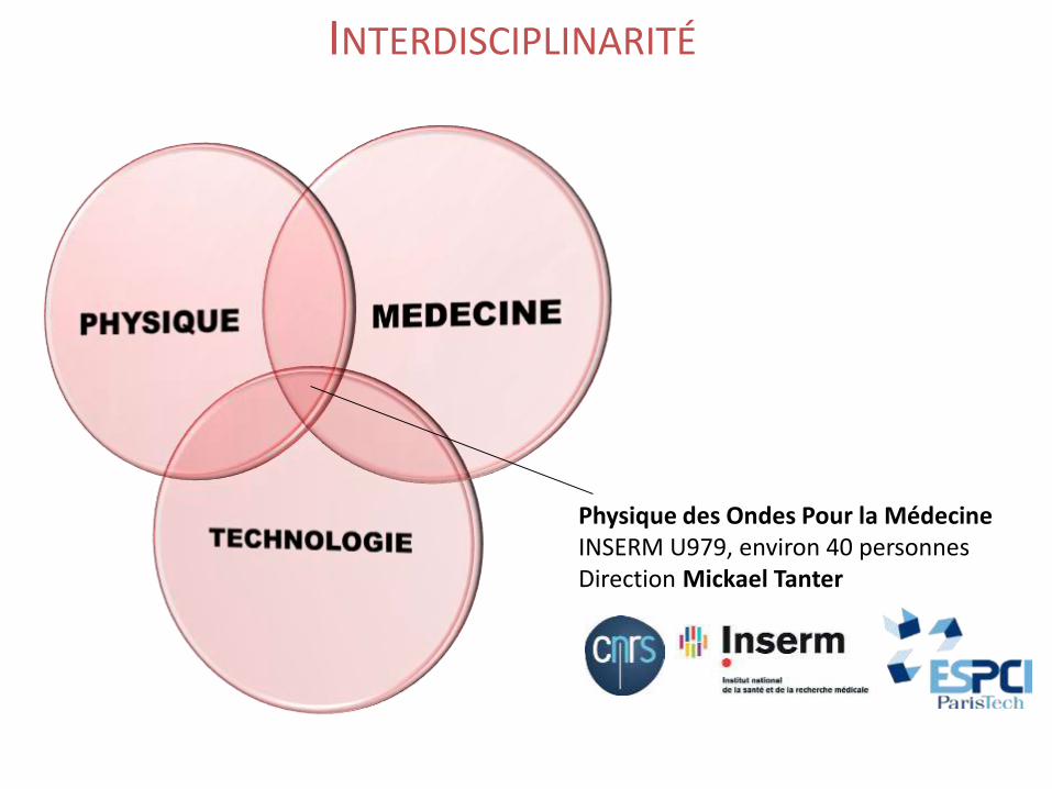
INTERDISCIPLINARITÉ
Physique des Ondes Pour la MédecineINSERM U979, environ 40 personnesDirection Mickael Tanter
Équipe de physiciens !
Application des ultrasons :- à l’imagerie
- à la thérapie
Disclaimer

I. Imagerie fonctionnelle ultrasonore
II. Thérapies par ultrasons focalisés
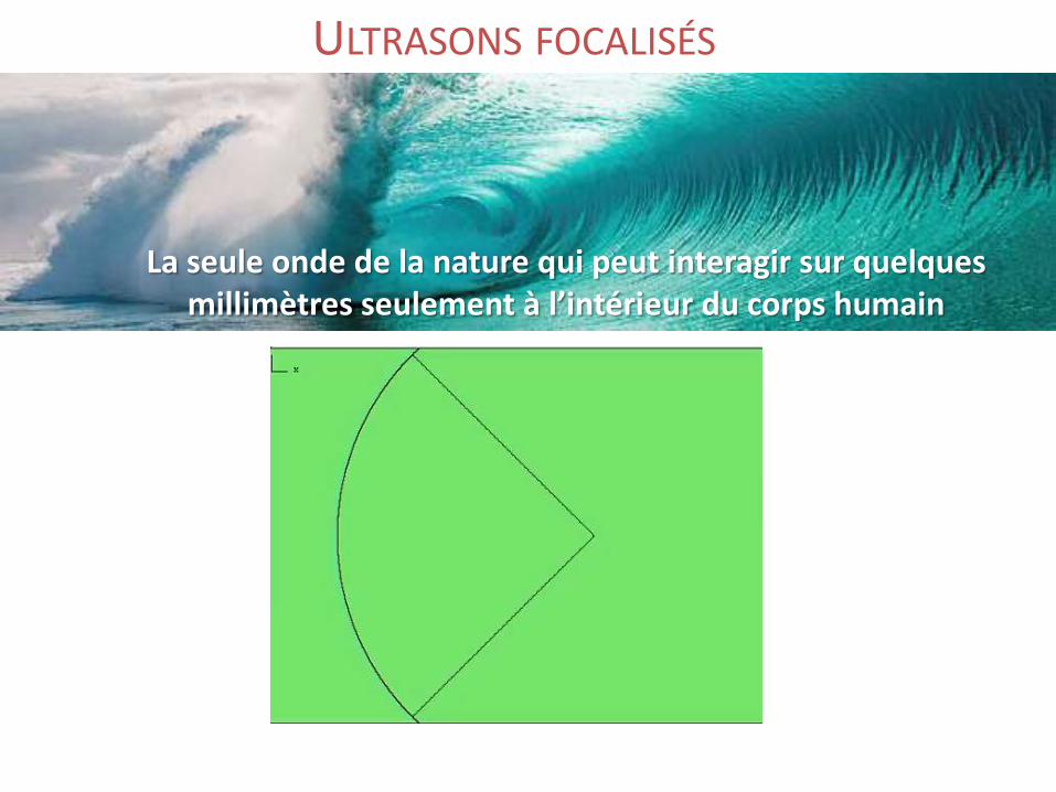
La seule onde de la nature qui peut interagir sur quelques millimètres seulement à l’intérieur du corps humain
ULTRASONS FOCALISÉS
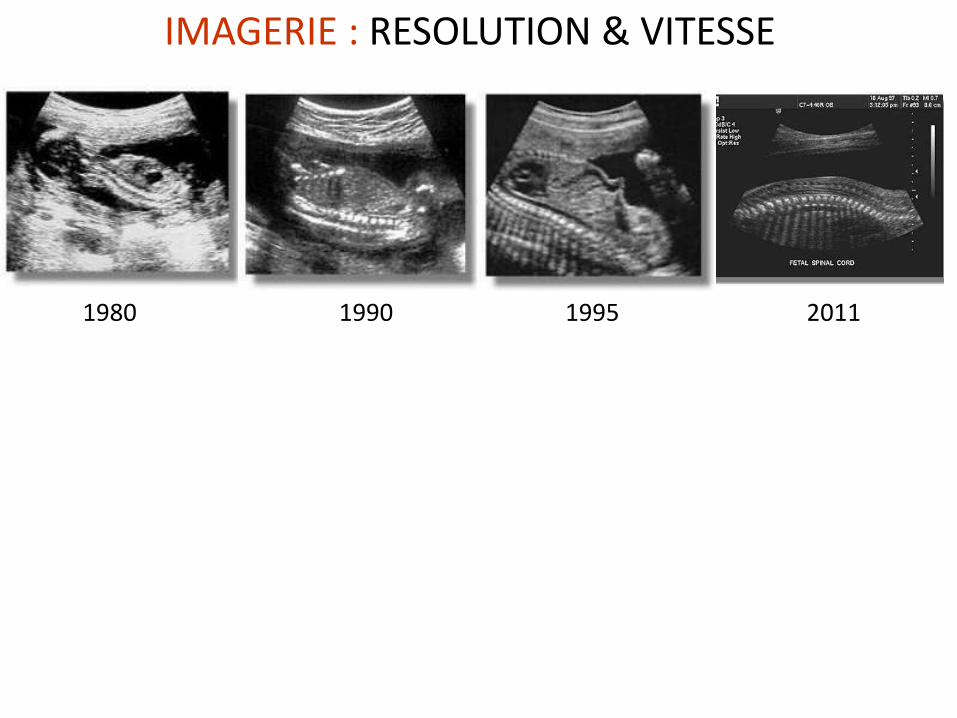
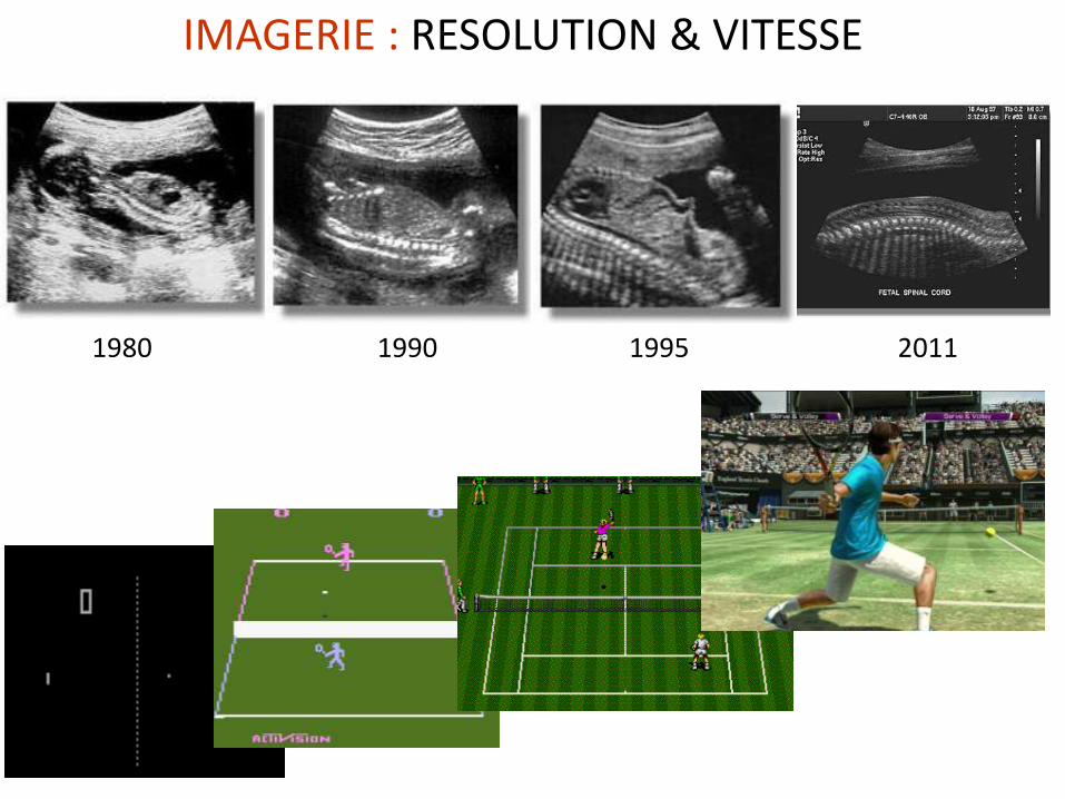
1980 1990 20111995
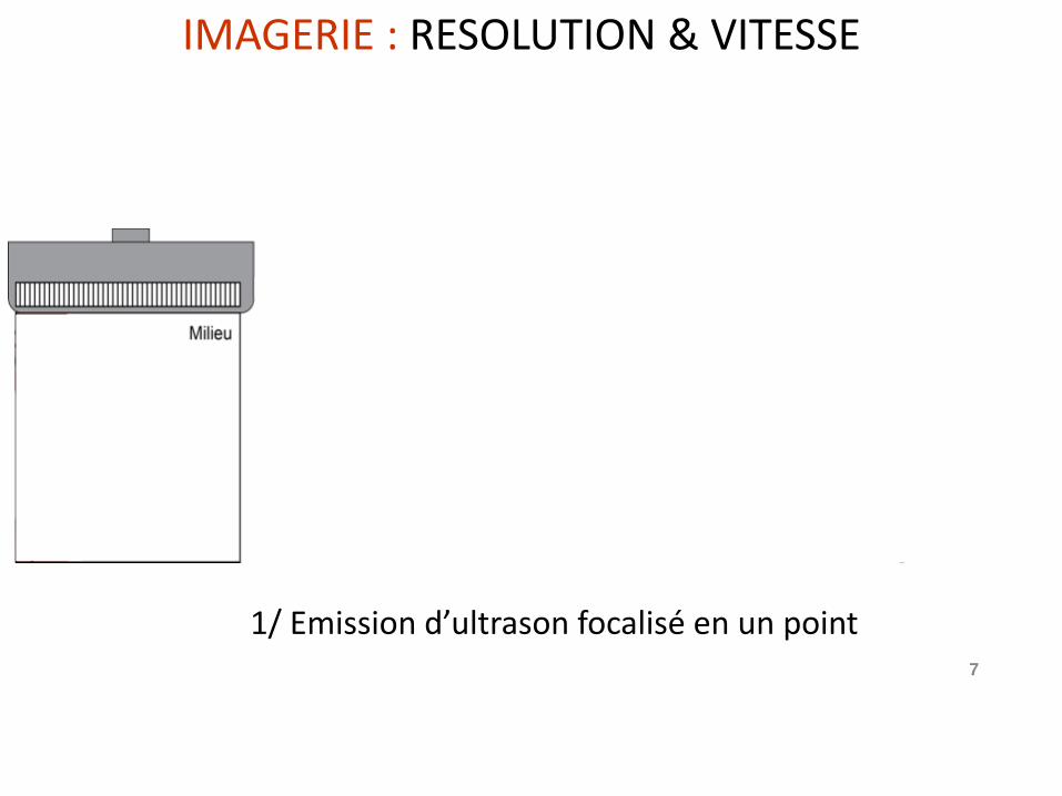
IMAGERIE : RESOLUTION & VITESSE
7
Σ
1/ Emission d’ultrason focalisé en un point
IMAGERIE : RESOLUTION & VITESSE
8
Σ
Conventional ultrasound imaging
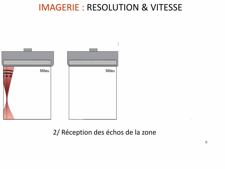
2/ Réception des échos de la zone
IMAGERIE : RESOLUTION & VITESSE
9
Σ
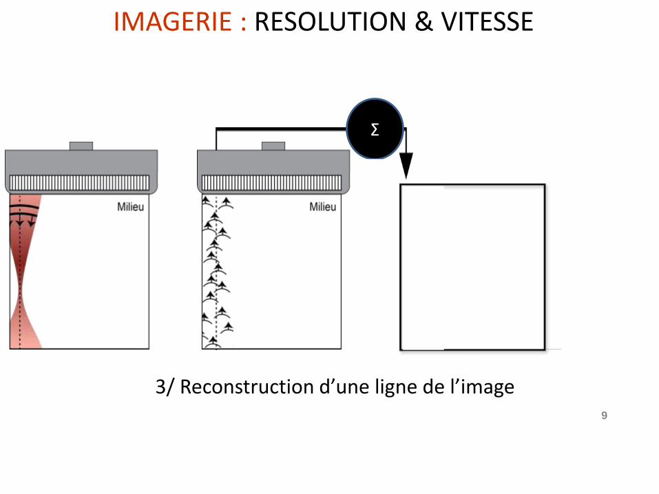
3/ Reconstruction d’une ligne de l’image
IMAGERIE : RESOLUTION & VITESSE
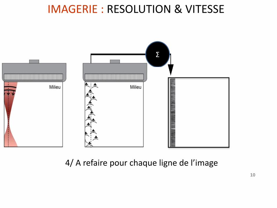
10
Σ
4/ A refaire pour chaque ligne de l’image
IMAGERIE : RESOLUTION & VITESSE
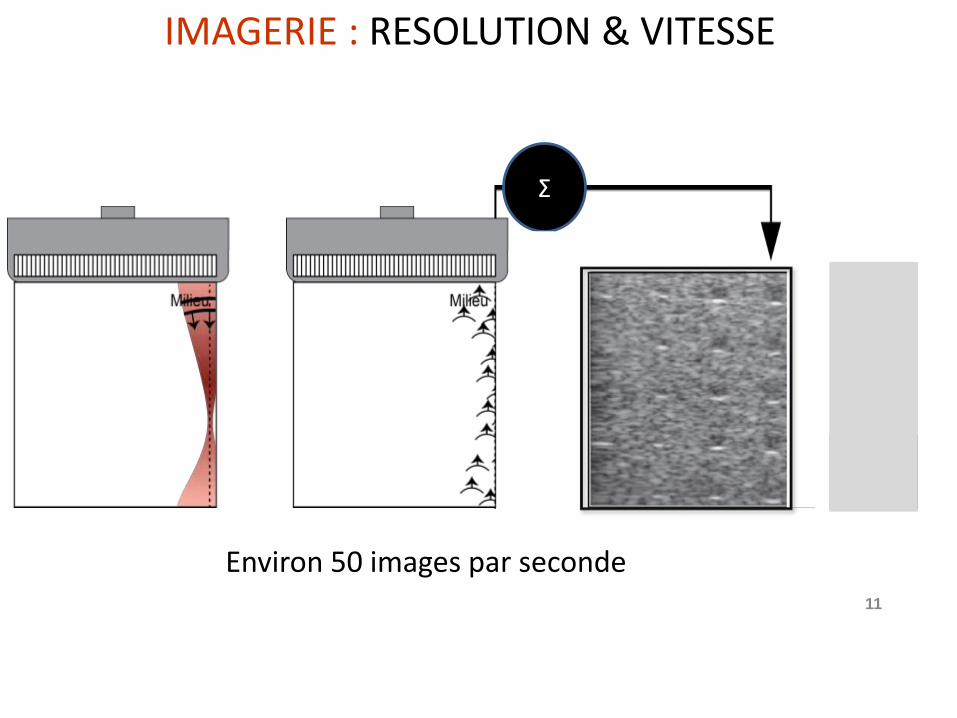
11
Σ
Environ 50 images par seconde
> 5000 images / seconde
50 images /second
> 5000 images / seconde
IMAGERIE : RESOLUTION & VITESSE
IMAGERIE : RESOLUTION & VITESSE
1980 1990 20111995
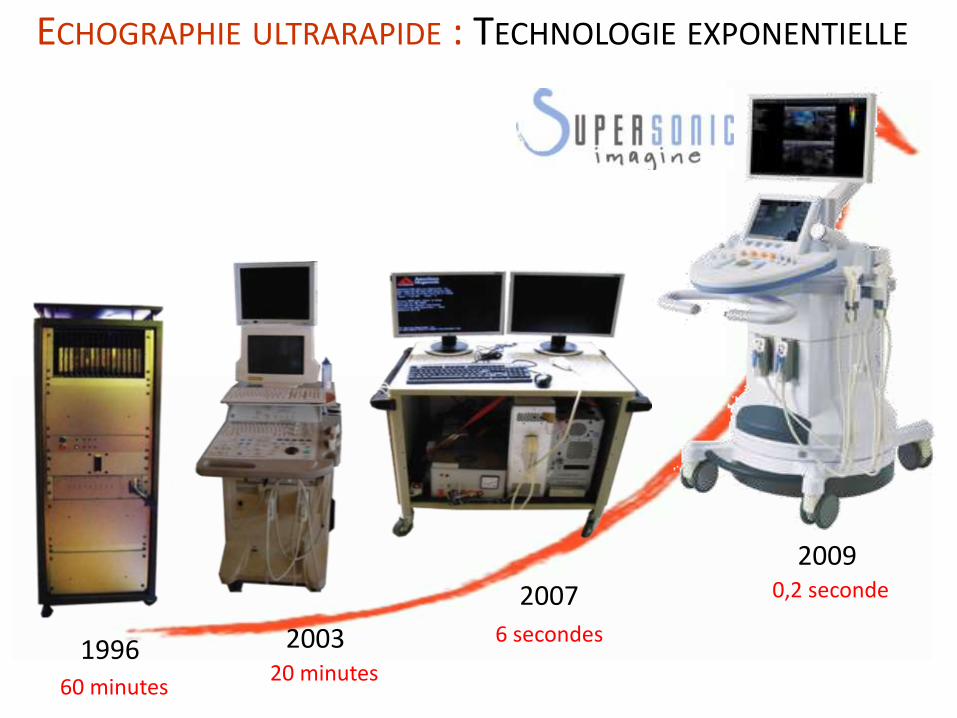
ECHOGRAPHIE ULTRARAPIDE : TECHNOLOGIE EXPONENTIELLE
1996 2003
2007
2009
20 minutes60 minutes
6 secondes
0,2 seconde
Σ
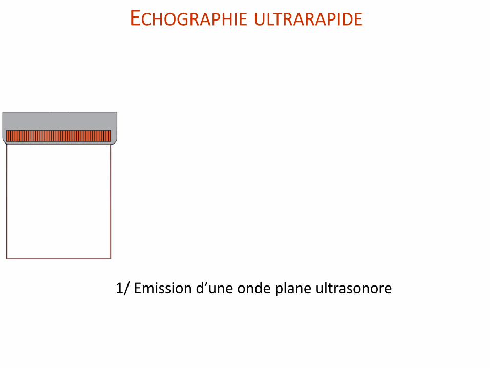
1/ Emission d’une onde plane ultrasonore
ECHOGRAPHIE ULTRARAPIDE
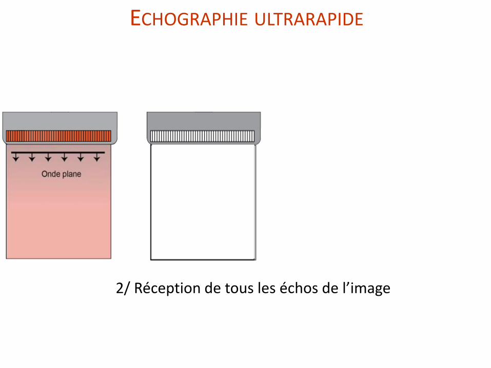
2/ Réception de tous les échos de l’image
ECHOGRAPHIE ULTRARAPIDE
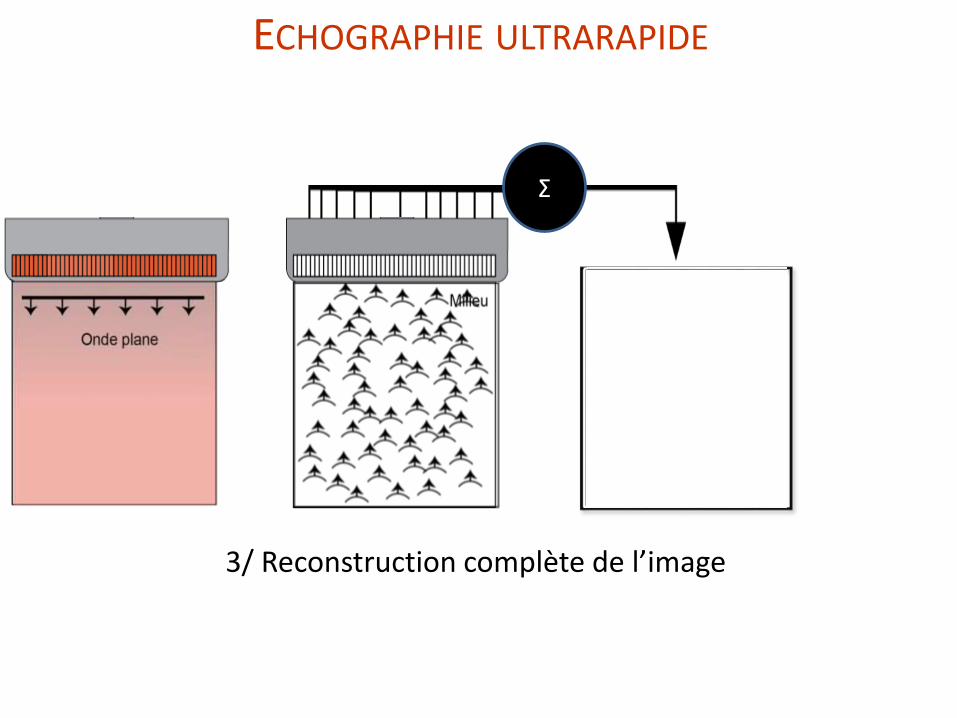
Σ
3/ Reconstruction complète de l’image
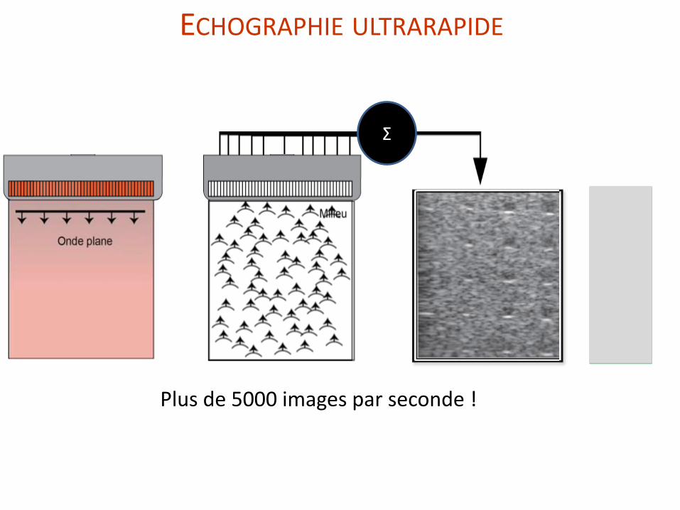
ECHOGRAPHIE ULTRARAPIDE
Σ
Plus de 5000 images par seconde !
> 5000 images / seconde
5000 images /second
> 5000
images / seconde
ECHOGRAPHIE ULTRARAPIDE
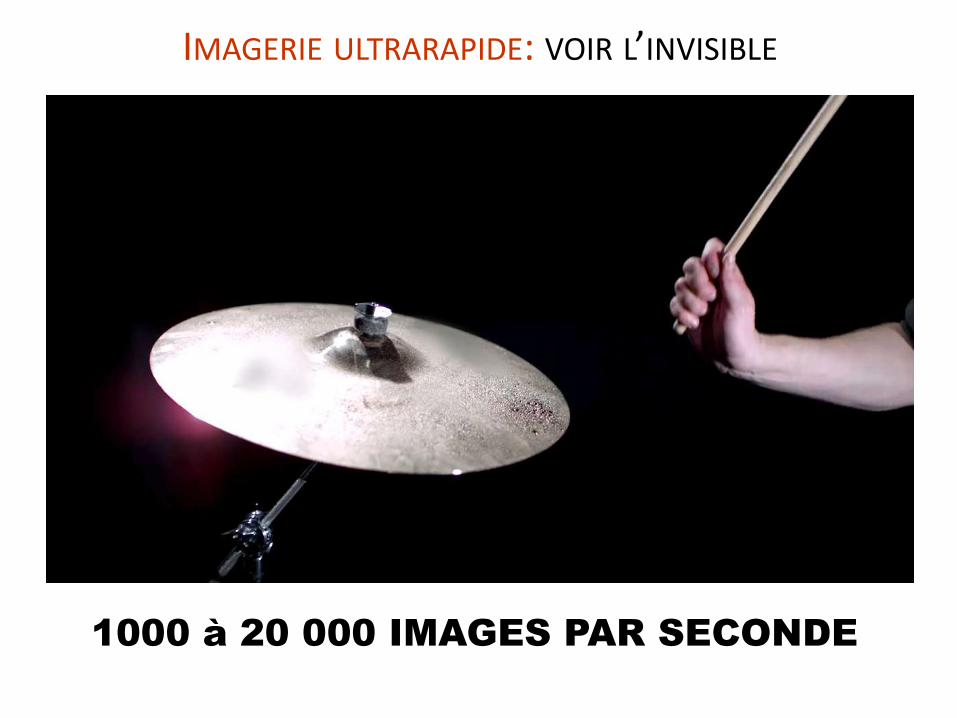
IMAGERIE ULTRARAPIDE: VOIR L’INVISIBLE
1000 à 20 000 IMAGES PAR SECONDE
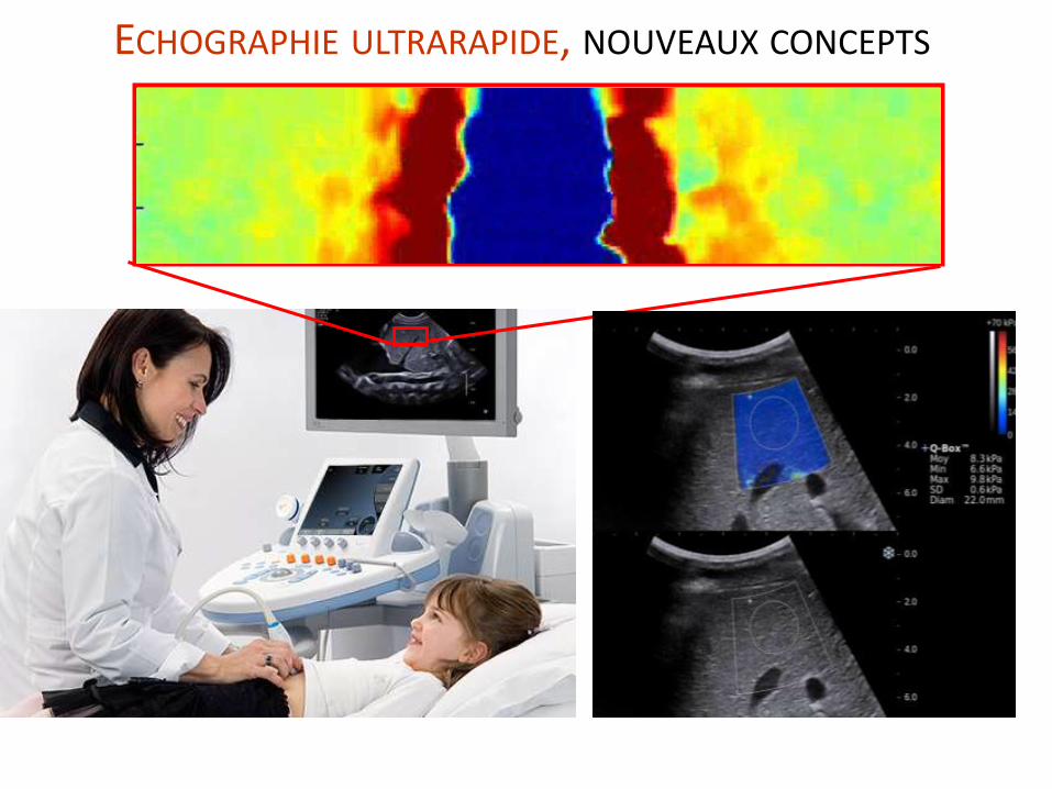
ECHOGRAPHIE ULTRARAPIDE, NOUVEAUX CONCEPTS
DOPPLER ULTRASENSIBLE :Mesurer le flux sanguin très finement
21
Conventional ultrasound imaging
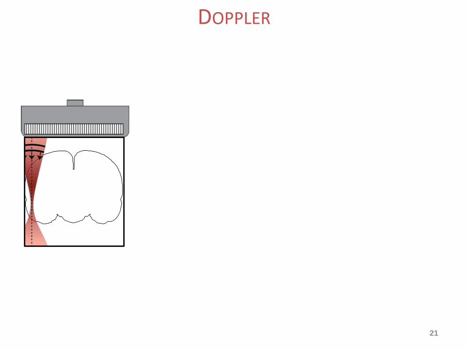
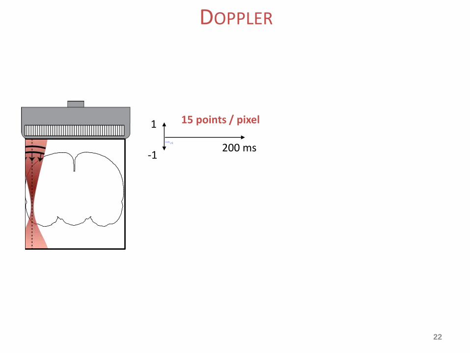
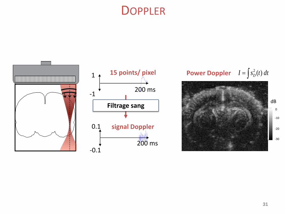
DOPPLER
22
Conventional ultrasound imaging
1
-1
15 points / pixel
200 ms
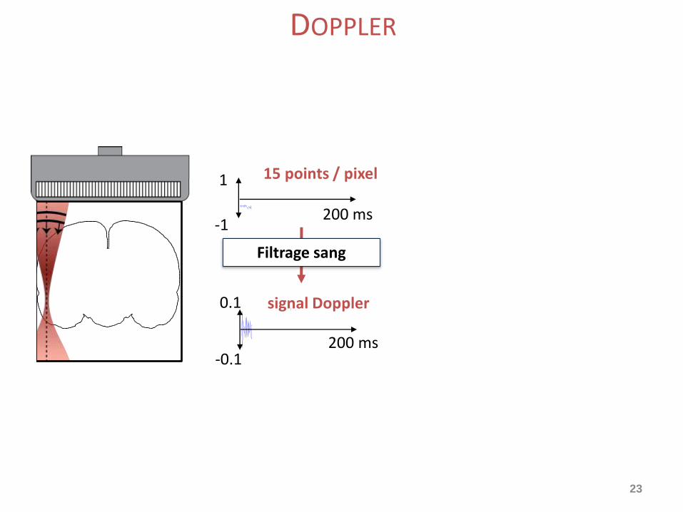
DOPPLER
23
Conventional ultrasound imaging
1
-1200 ms
200 ms
signal Doppler0.1
-0.1
Filtrage sang
15 points / pixel
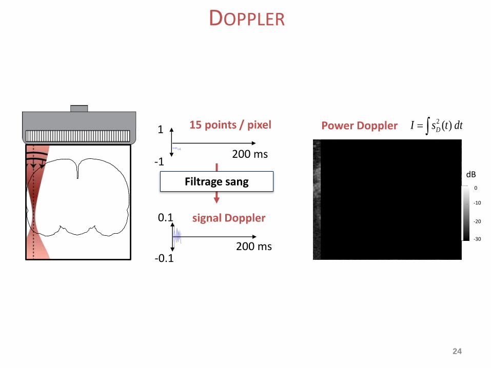
DOPPLER
24
dttsI D )(2Power Doppler
-30
-20
-10
0
dB
1
-1200 ms
200 ms
signal Doppler0.1
-0.1
Filtrage sang
15 points / pixel
DOPPLER

25
Conventional ultrasound imaging
dttsI D )(2Power Doppler
-30
-20
-10
0
dB
1
-1200 ms
200 ms
signal Doppler0.1
-0.1
Filtrage sang
15 points / pixel
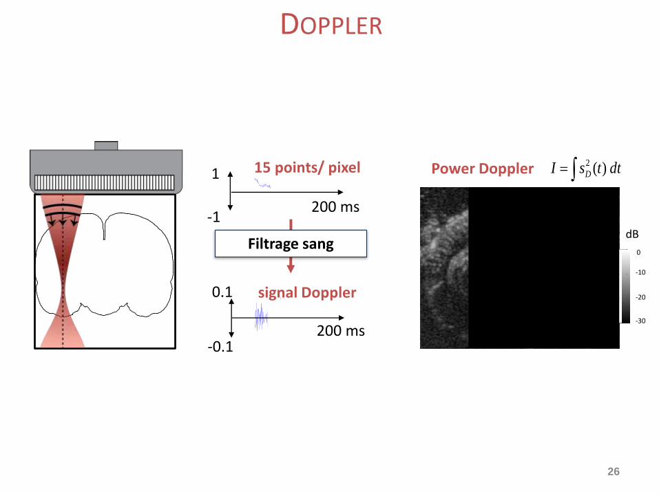
DOPPLER
26
Conventional ultrasound imaging
dttsI D )(2Power Doppler
-30
-20
-10
0
dB
1
-1200 ms
200 ms
signal Doppler0.1
-0.1
Filtrage sang
15 points/ pixel
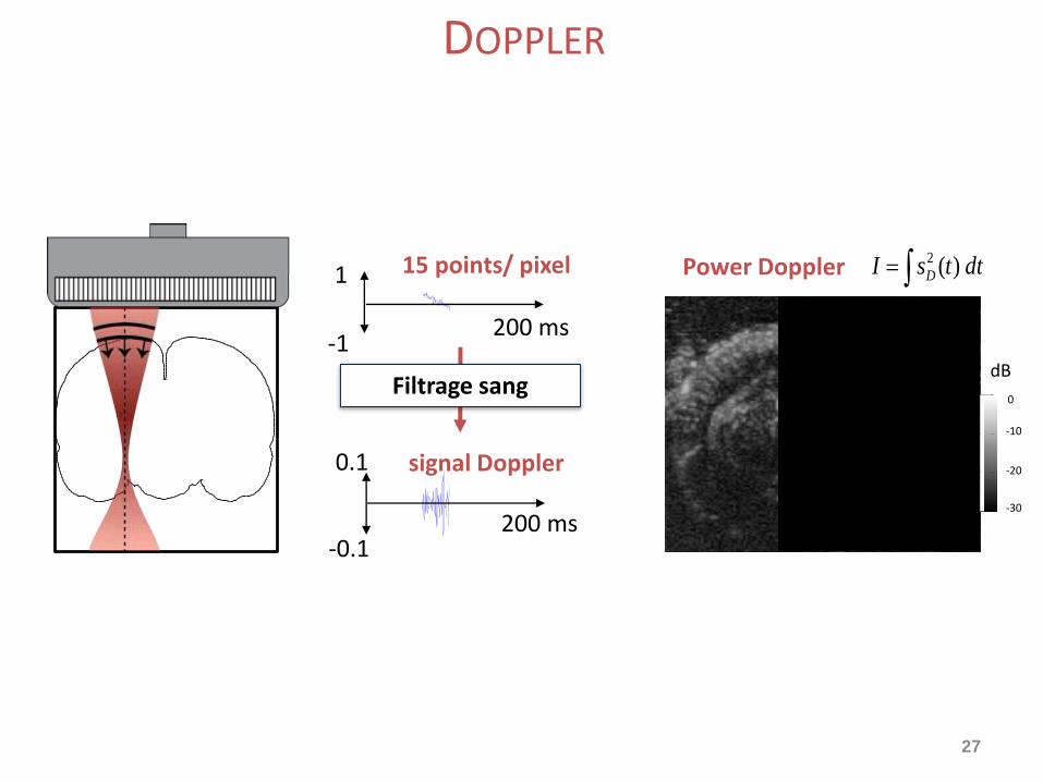
DOPPLER
27
dttsI D )(2Power Doppler
-30
-20
-10
0
dB
1
-1200 ms
200 ms
signal Doppler0.1
-0.1
Filtrage sang
15 points/ pixel
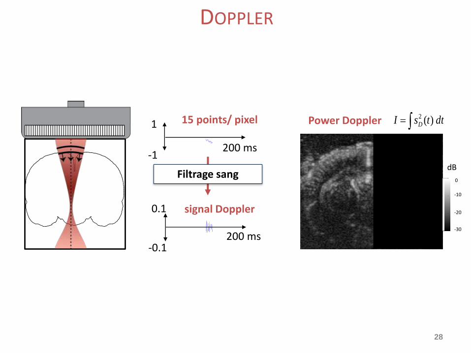
DOPPLER
28
Conventional ultrasound imaging
dttsI D )(2Power Doppler
-30
-20
-10
0
dB
1
-1200 ms
200 ms
signal Doppler0.1
-0.1
Filtrage sang
15 points/ pixel
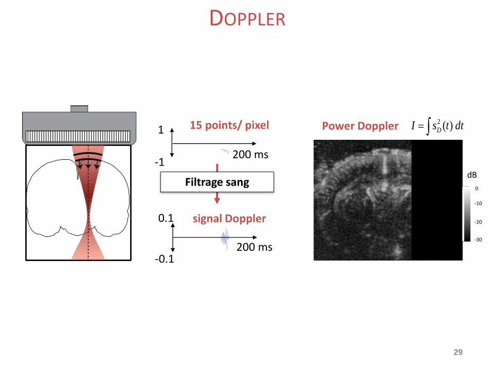
DOPPLER
29
dttsI D )(2Power Doppler
-30
-20
-10
0
dB
1
-1200 ms
200 ms
signal Doppler0.1
-0.1
Filtrage sang
15 points/ pixel
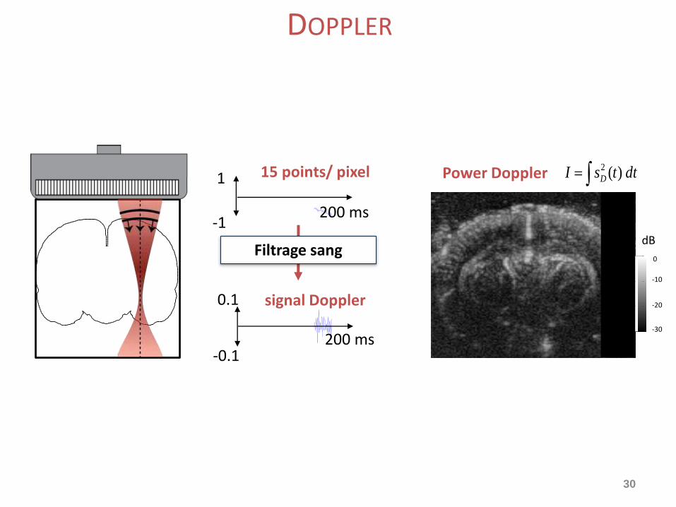
DOPPLER
30
Conventional ultrasound imaging
dttsI D )(2Power Doppler
-30
-20
-10
0
dB
1
-1200 ms
200 ms
signal Doppler0.1
-0.1
Filtrage sang
15 points/ pixel
DOPPLER
31
Conventional ultrasound imaging
dttsI D )(2Power Doppler
-30
-20
-10
0
dB
1
-1200 ms
200 ms
signal Doppler0.1
-0.1
Filtrage sang
15 points/ pixel
DOPPLER
32
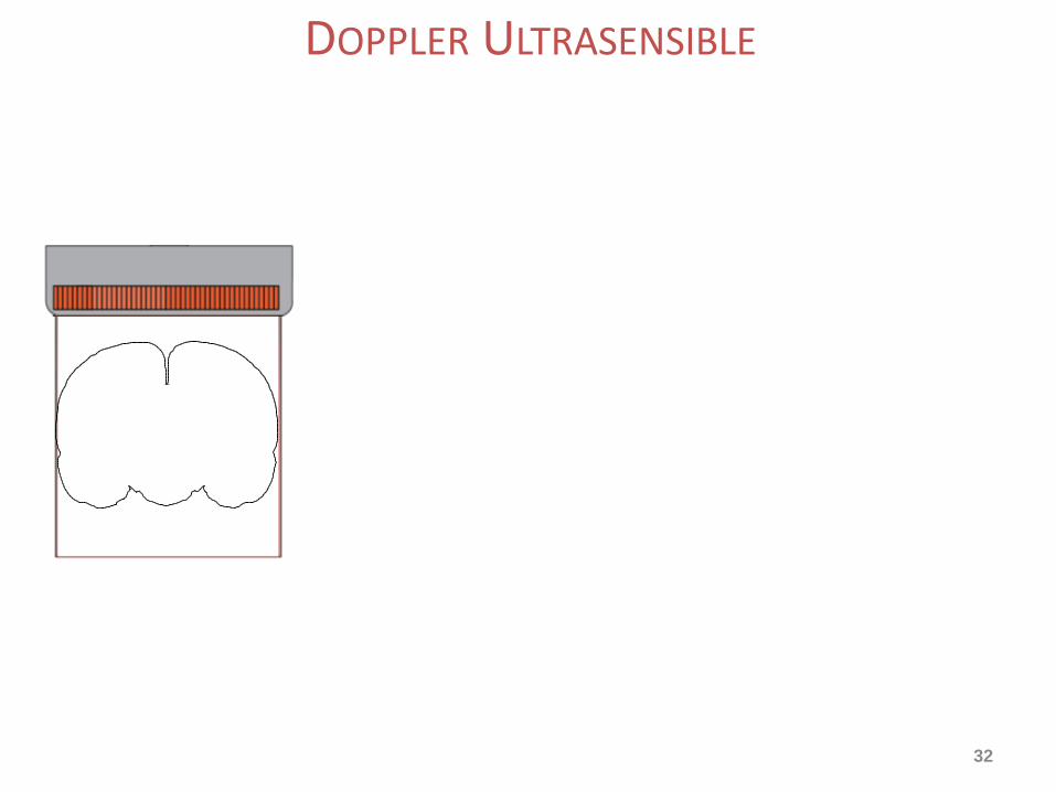
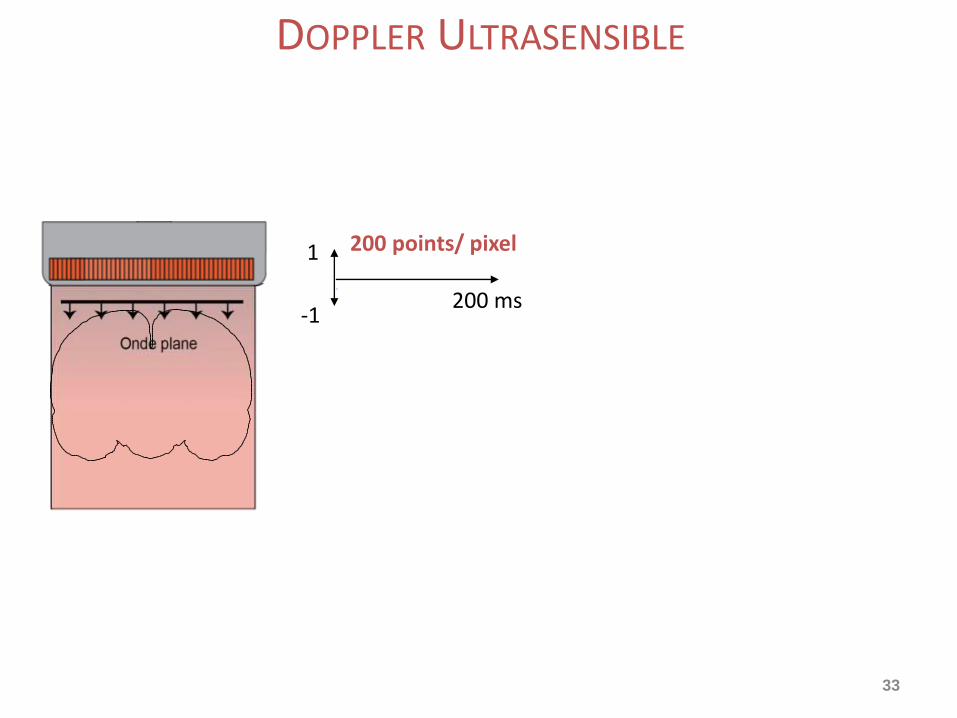
DOPPLER ULTRASENSIBLE
33
1
-1200 ms
200 points/ pixel
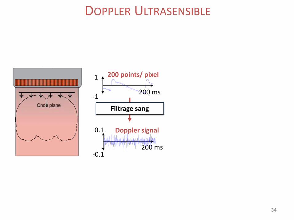
DOPPLER ULTRASENSIBLE
34
1
-1200 ms
200 ms
Doppler signal0.1
-0.1
Filtrage sang
200 points/ pixel
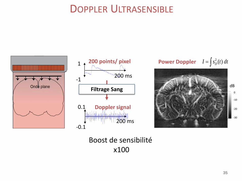
DOPPLER ULTRASENSIBLE
35
dttsI D )(2Power Doppler
-30
-20
-10
0
dB
1
-1200 ms
200 ms
Doppler signal0.1
-0.1
Filtrage Sang
200 points/ pixel
Boost de sensibilité x100
DOPPLER ULTRASENSIBLE
36
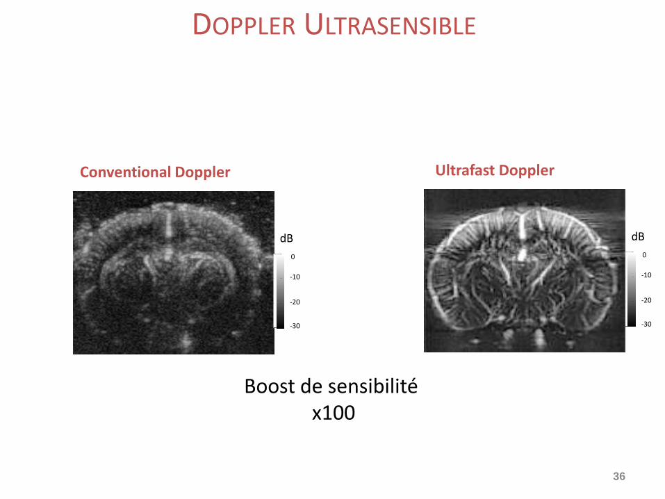
Ultrafast Doppler
-30
-20
-10
0
dB
Conventional Doppler
-30
-20
-10
0
dB
Boost de sensibilité x100
DOPPLER ULTRASENSIBLE
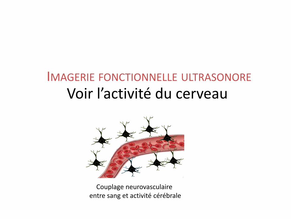
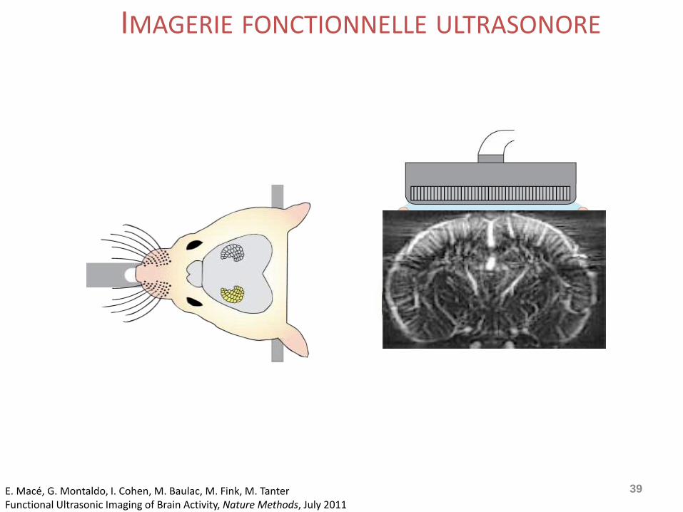
Voir l’activité du cerveauIMAGERIE FONCTIONNELLE ULTRASONORE
Couplage neurovasculaireentre sang et activité cérébrale
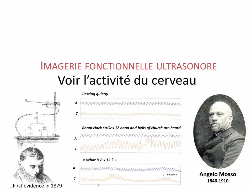
Voir l’activité du cerveauIMAGERIE FONCTIONNELLE ULTRASONORE
Resting quietly
Room clock strikes 12 noon and bells of church are heard
« What is 8 x 12 ? »
Response
A
C
A
C
A
CAngelo Mosso
1846-1910First evidence in 1879
39
Conventional ultrasound imagingImagerie
IMAGERIE FONCTIONNELLE ULTRASONORE
E. Macé, G. Montaldo, I. Cohen, M. Baulac, M. Fink, M. TanterFunctional Ultrasonic Imaging of Brain Activity, Nature Methods, July 2011
40
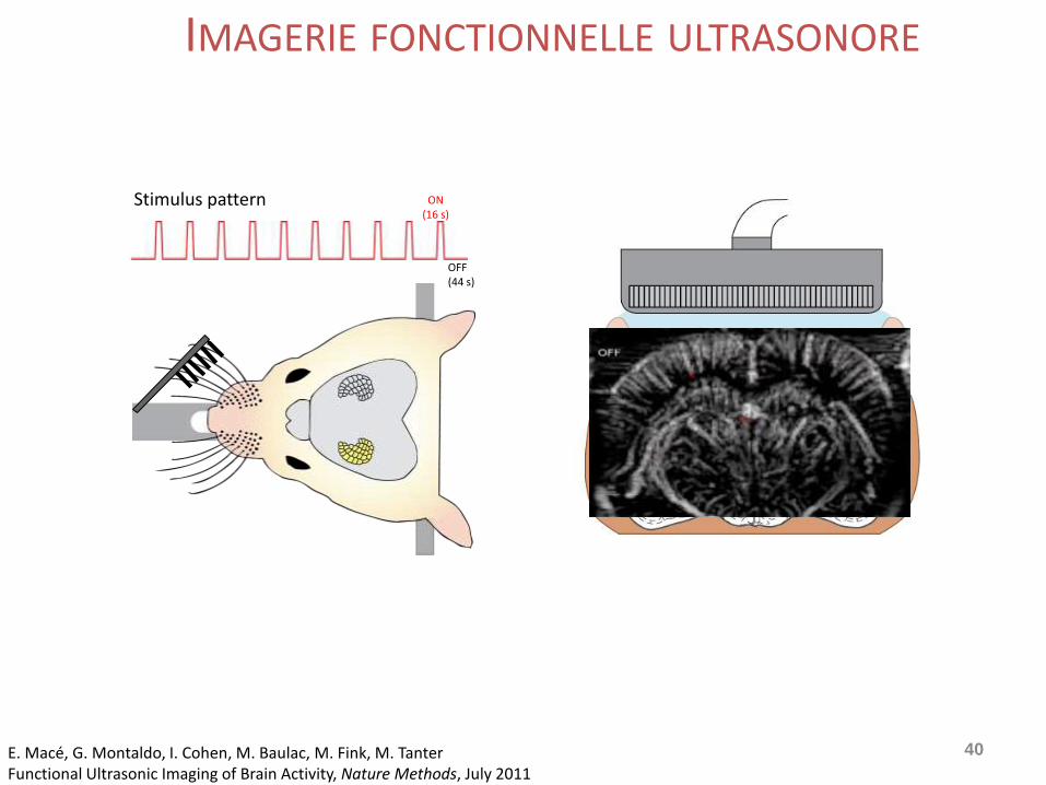
Stimulus pattern ON(16 s)
OFF(44 s)
IMAGERIE FONCTIONNELLE ULTRASONORE
E. Macé, G. Montaldo, I. Cohen, M. Baulac, M. Fink, M. TanterFunctional Ultrasonic Imaging of Brain Activity, Nature Methods, July 2011
41
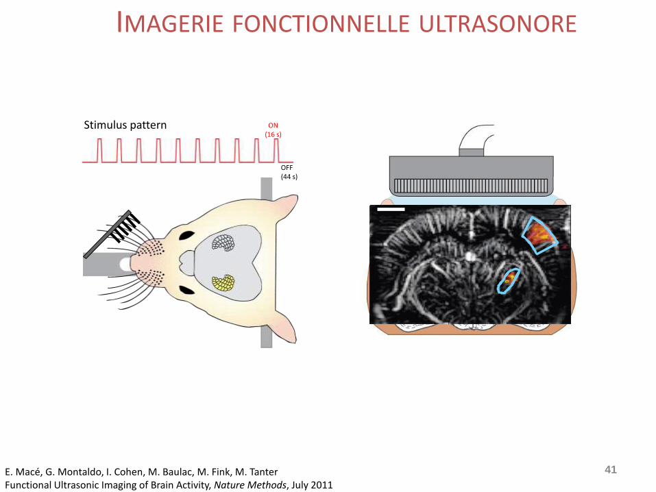
Stimulus pattern ON(16 s)
OFF(44 s)
IMAGERIE FONCTIONNELLE ULTRASONORE
E. Macé, G. Montaldo, I. Cohen, M. Baulac, M. Fink, M. TanterFunctional Ultrasonic Imaging of Brain Activity, Nature Methods, July 2011
42
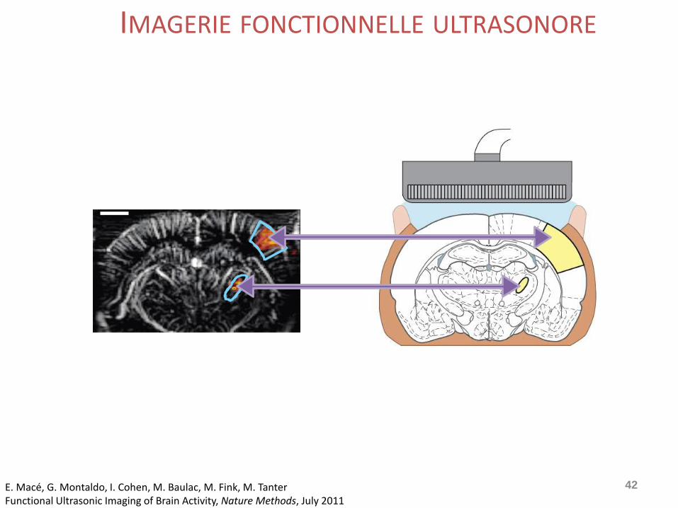
IMAGERIE FONCTIONNELLE ULTRASONORE
E. Macé, G. Montaldo, I. Cohen, M. Baulac, M. Fink, M. TanterFunctional Ultrasonic Imaging of Brain Activity, Nature Methods, July 2011
IMAGERIE FONCTIONNELLE DES ODEURS
B. Osmanski, H. Gurden, G.Montaldo, F. Pain, M. Fink, M. Tanter
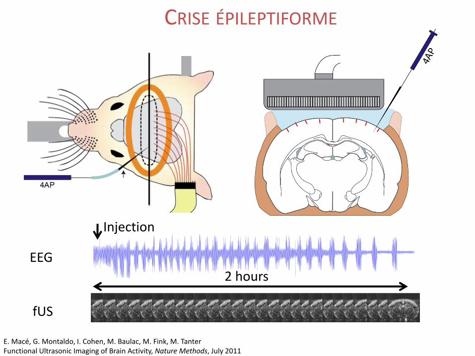
EEG
Injection
2 hours
fUS
CRISE ÉPILEPTIFORME
E. Macé, G. Montaldo, I. Cohen, M. Baulac, M. Fink, M. TanterFunctional Ultrasonic Imaging of Brain Activity, Nature Methods, July 2011
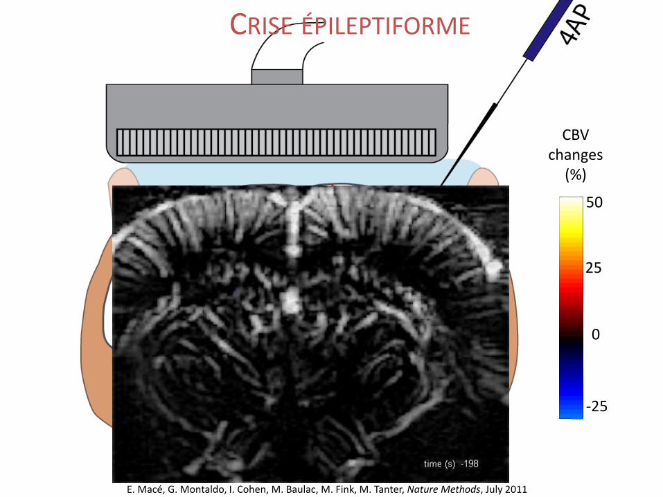
-25
0
25
50
CBV changes
(%)
E. Macé, G. Montaldo, I. Cohen, M. Baulac, M. Fink, M. Tanter, Nature Methods, July 2011
CRISE ÉPILEPTIFORME
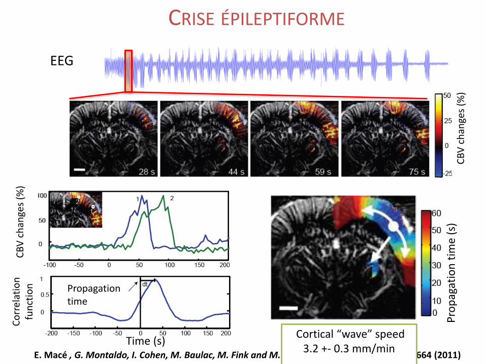
E. Macé , G. Montaldo, I. Cohen, M. Baulac, M. Fink and M. Tanter. Nature Methods, 8, 662-664 (2011)
Cortical “wave” speed3.2 +- 0.3 mm/min
EEG
CB
V c
han
ges
(%)
Pro
pag
atio
n t
ime
(s)
Propagation time
Time (s)
CB
V c
han
ges
(%)
Co
rrel
atio
n
fun
ctio
nCRISE ÉPILEPTIFORME
CONNECTIVITÉ FONCTIONNELLE PAR ULTRASONS
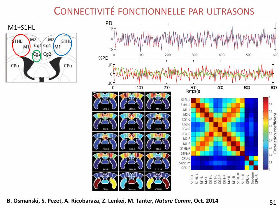
M1+S1HL
51B. Osmanski, S. Pezet, A. Ricobaraza, Z. Lenkei, M. Tanter, Nature Comm, Oct. 2014
0 100 200 300 400 500 60010
0
10
Temps(s)
%PD
CONNECTIVITÉ FONCTIONNELLE PAR ULTRASONS
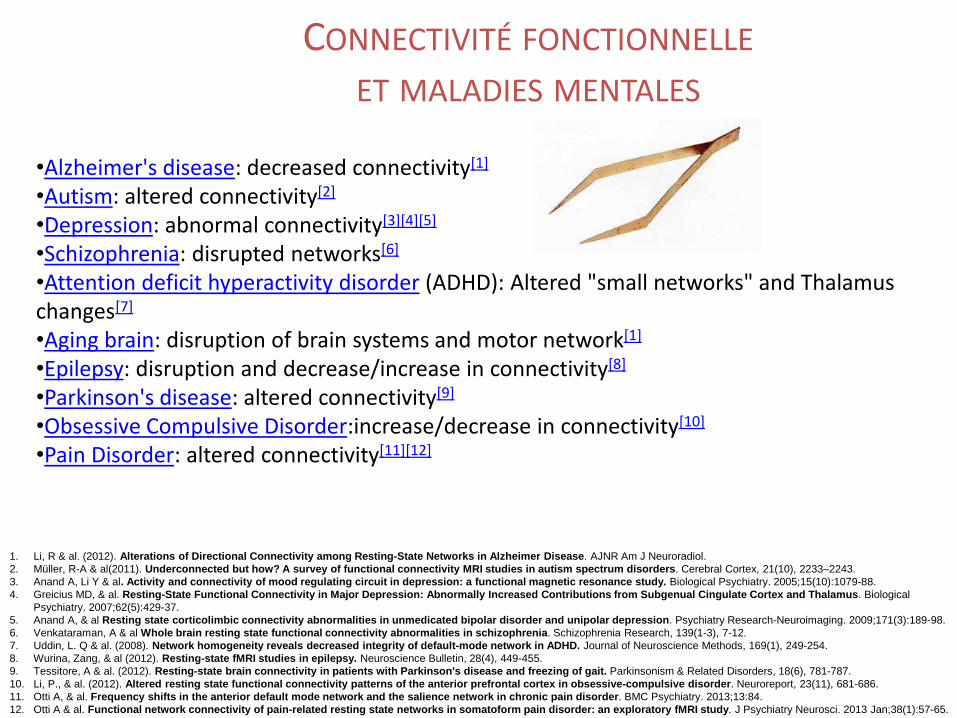
•Alzheimer's disease: decreased connectivity[1]
•Autism: altered connectivity[2]
•Depression: abnormal connectivity[3][4][5]
•Schizophrenia: disrupted networks[6]
•Attention deficit hyperactivity disorder (ADHD): Altered "small networks" and Thalamus changes[7]
•Aging brain: disruption of brain systems and motor network[1]
•Epilepsy: disruption and decrease/increase in connectivity[8]
•Parkinson's disease: altered connectivity[9]
•Obsessive Compulsive Disorder:increase/decrease in connectivity[10]
•Pain Disorder: altered connectivity[11][12]
1. Li, R & al. (2012). Alterations of Directional Connectivity among Resting-State Networks in Alzheimer Disease. AJNR Am J Neuroradiol.
2. Müller, R-A & al(2011). Underconnected but how? A survey of functional connectivity MRI studies in autism spectrum disorders. Cerebral Cortex, 21(10), 2233–2243.
3. Anand A, Li Y & al. Activity and connectivity of mood regulating circuit in depression: a functional magnetic resonance study. Biological Psychiatry. 2005;15(10):1079-88.
4. Greicius MD, & al. Resting-State Functional Connectivity in Major Depression: Abnormally Increased Contributions from Subgenual Cingulate Cortex and Thalamus. Biological
Psychiatry. 2007;62(5):429-37.
5. Anand A, & al Resting state corticolimbic connectivity abnormalities in unmedicated bipolar disorder and unipolar depression. Psychiatry Research-Neuroimaging. 2009;171(3):189-98.
6. Venkataraman, A & al Whole brain resting state functional connectivity abnormalities in schizophrenia. Schizophrenia Research, 139(1-3), 7-12.
7. Uddin, L. Q & al. (2008). Network homogeneity reveals decreased integrity of default-mode network in ADHD. Journal of Neuroscience Methods, 169(1), 249-254.
8. Wurina, Zang, & al (2012). Resting-state fMRI studies in epilepsy. Neuroscience Bulletin, 28(4), 449-455.
9. Tessitore, A & al. (2012). Resting-state brain connectivity in patients with Parkinson's disease and freezing of gait. Parkinsonism & Related Disorders, 18(6), 781-787.
10. Li, P., & al. (2012). Altered resting state functional connectivity patterns of the anterior prefrontal cortex in obsessive-compulsive disorder. Neuroreport, 23(11), 681-686.
11. Otti A, & al. Frequency shifts in the anterior default mode network and the salience network in chronic pain disorder. BMC Psychiatry. 2013;13:84.
12. Otti A & al. Functional network connectivity of pain-related resting state networks in somatoform pain disorder: an exploratory fMRI study. J Psychiatry Neurosci. 2013 Jan;38(1):57-65.
CONNECTIVITÉ FONCTIONNELLE
ET MALADIES MENTALES
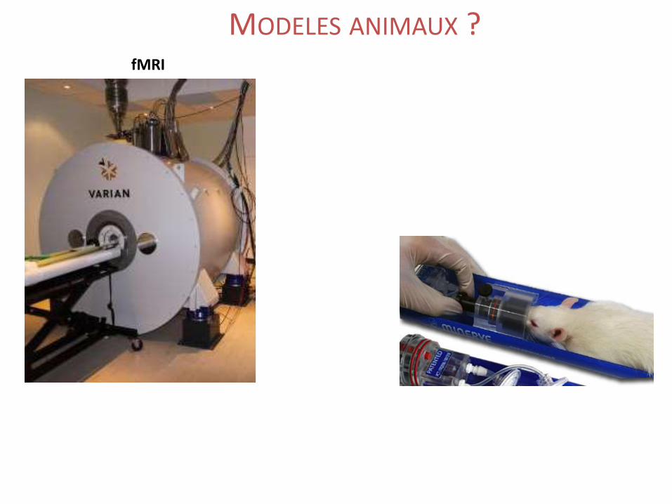
fMRI
MODELES ANIMAUX ?
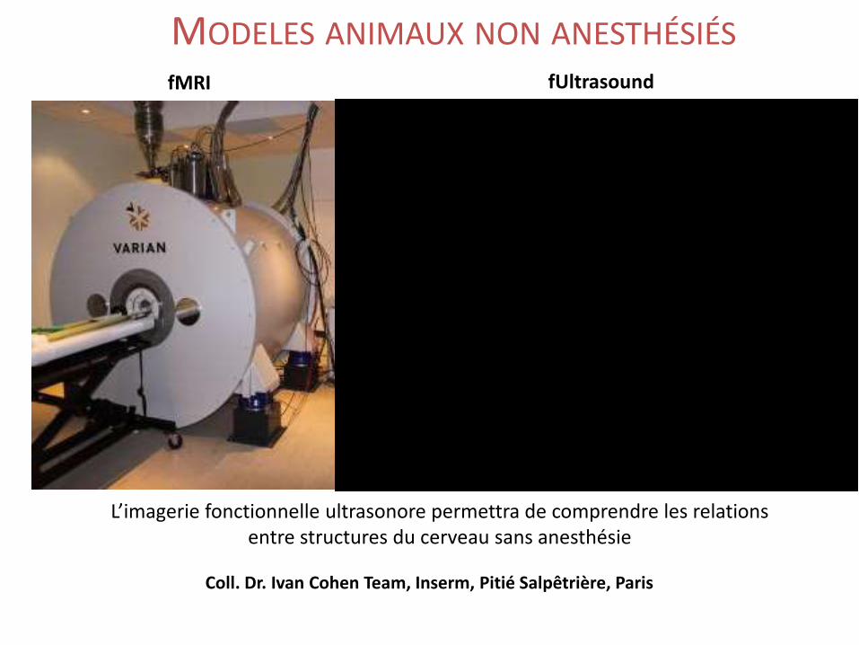
fMRI fUltrasound
Coll. Dr. Ivan Cohen Team, Inserm, Pitié Salpêtrière, Paris
L’imagerie fonctionnelle ultrasonore permettra de comprendre les relations entre structures du cerveau sans anesthésie
MODELES ANIMAUX NON ANESTHÉSIÉS
fUltrasound en clinique ?
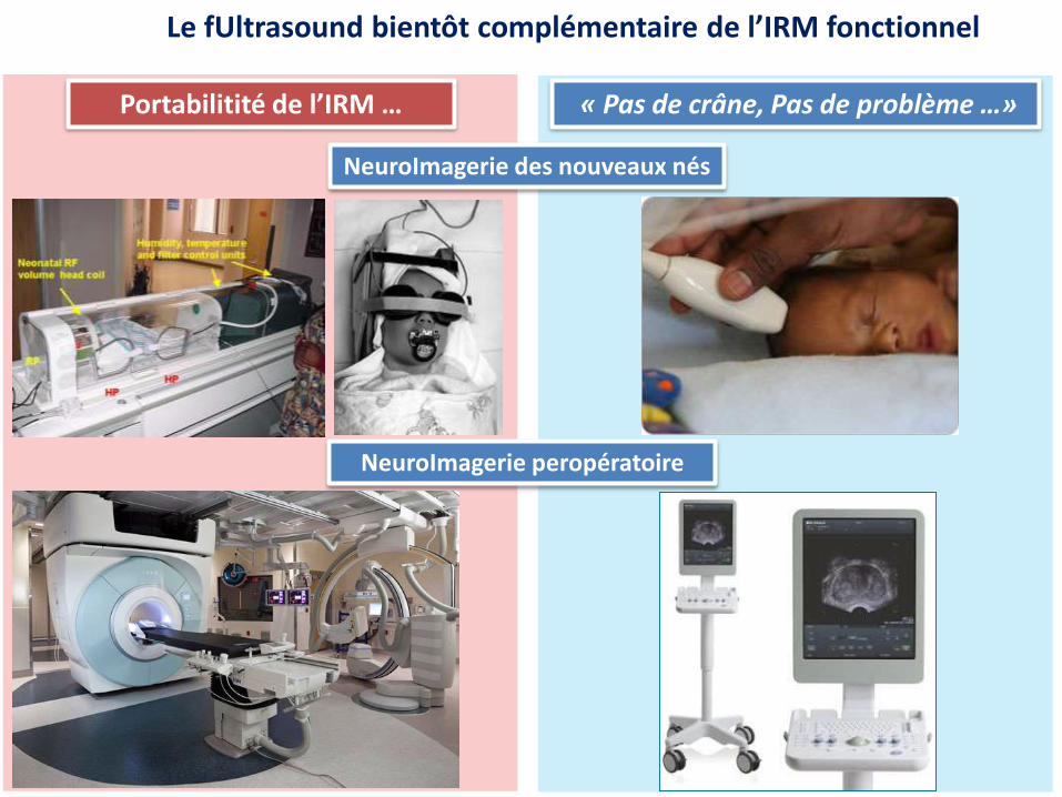
Le fUltrasound bientôt complémentaire de l’IRM fonctionnel
Portabilitité de l’IRM … « Pas de crâne, Pas de problème …»
NeuroImagerie des nouveaux nés
NeuroImagerie peropératoire
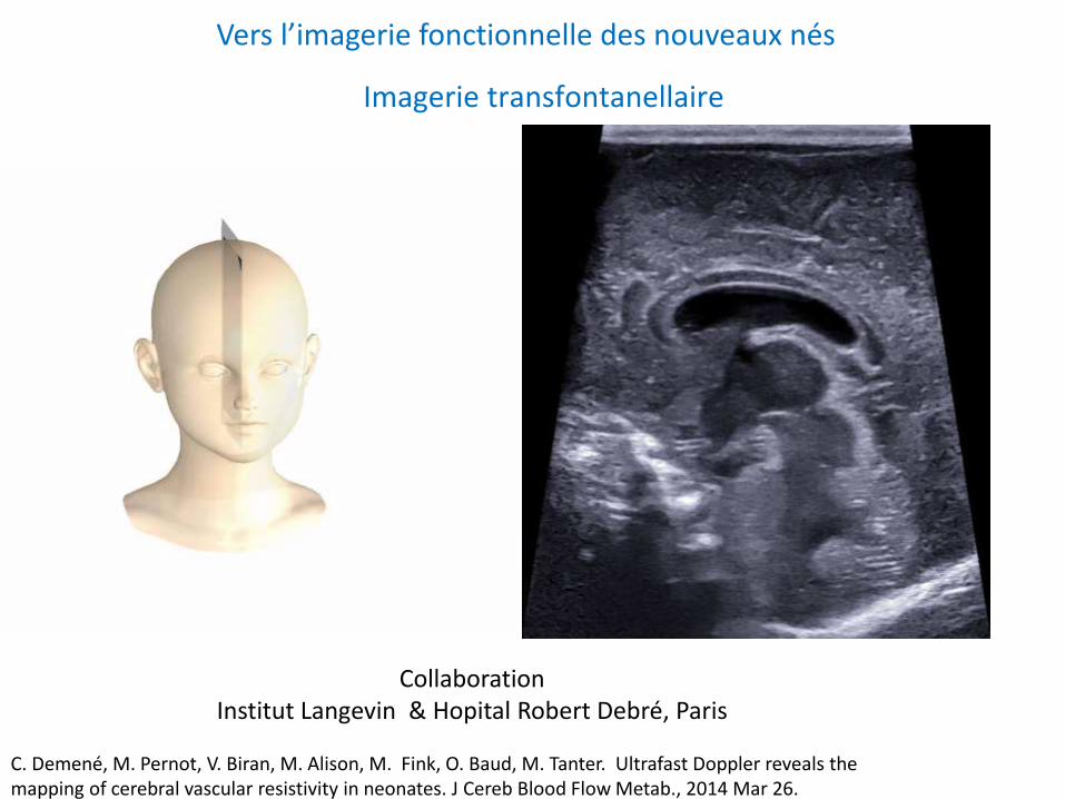
Vers l’imagerie fonctionnelle des nouveaux nés
Collaboration Institut Langevin & Hopital Robert Debré, Paris
C. Demené, M. Pernot, V. Biran, M. Alison, M. Fink, O. Baud, M. Tanter. Ultrafast Doppler reveals the mapping of cerebral vascular resistivity in neonates. J Cereb Blood Flow Metab., 2014 Mar 26.
Imagerie transfontanellaire
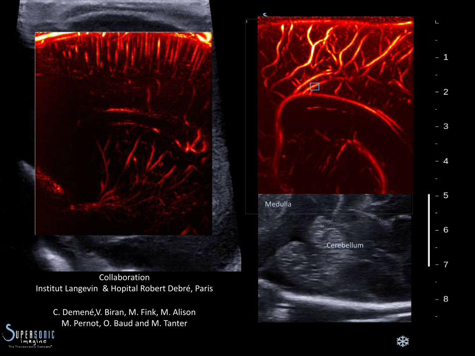
Corpus callosum
Thalamus
Cortex
Cerebellum
Medulla
Collaboration Institut Langevin & Hopital Robert Debré, Paris
C. Demené,V. Biran, M. Fink, M. AlisonM. Pernot, O. Baud and M. Tanter
mm
mm
Ultrafast Doppler Image
0 5 10 15 20 25 30 35
5
10
15
20
25
30
35
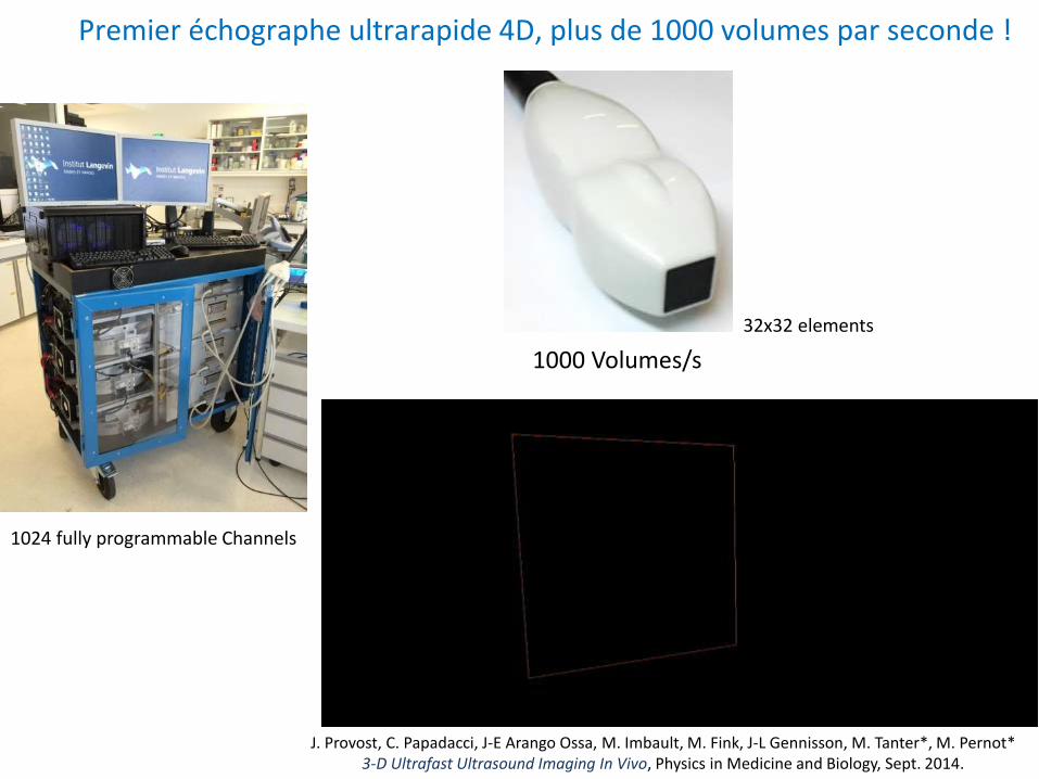
J. Provost, C. Papadacci, J-E Arango Ossa, M. Imbault, M. Fink, J-L Gennisson, M. Tanter*, M. Pernot* 3-D Ultrafast Ultrasound Imaging In Vivo, Physics in Medicine and Biology, Sept. 2014.
32x32 elements
Premier échographe ultrarapide 4D, plus de 1000 volumes par seconde !
1024 fully programmable Channels
1000 Volumes/s
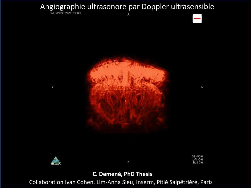
Collaboration Ivan Cohen, Lim-Anna Sieu, Inserm, Pitié Salpêtrière, Paris
Angiographie ultrasonore par Doppler ultrasensible
C. Demené, PhD Thesis
Thérapies ultrasonores
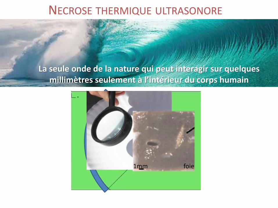
La seule onde de la nature qui peut interagir sur quelques millimètres seulement à l’intérieur du corps humain
NECROSE THERMIQUE ULTRASONORE
foie1mm
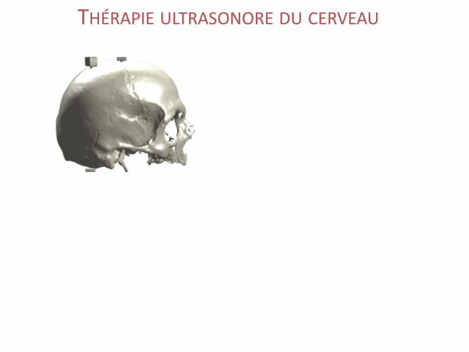
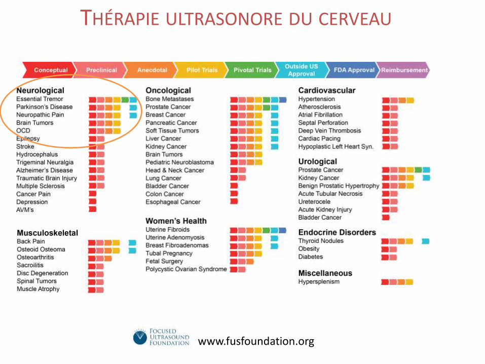
THÉRAPIE ULTRASONORE DU CERVEAU
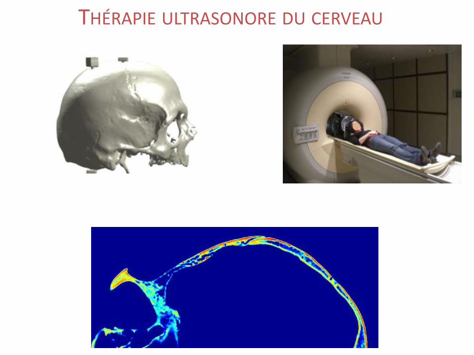
THÉRAPIE ULTRASONORE DU CERVEAU
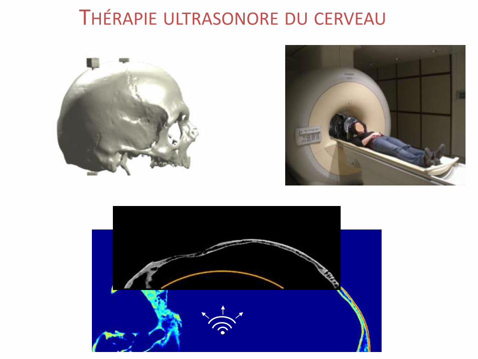
THÉRAPIE ULTRASONORE DU CERVEAU
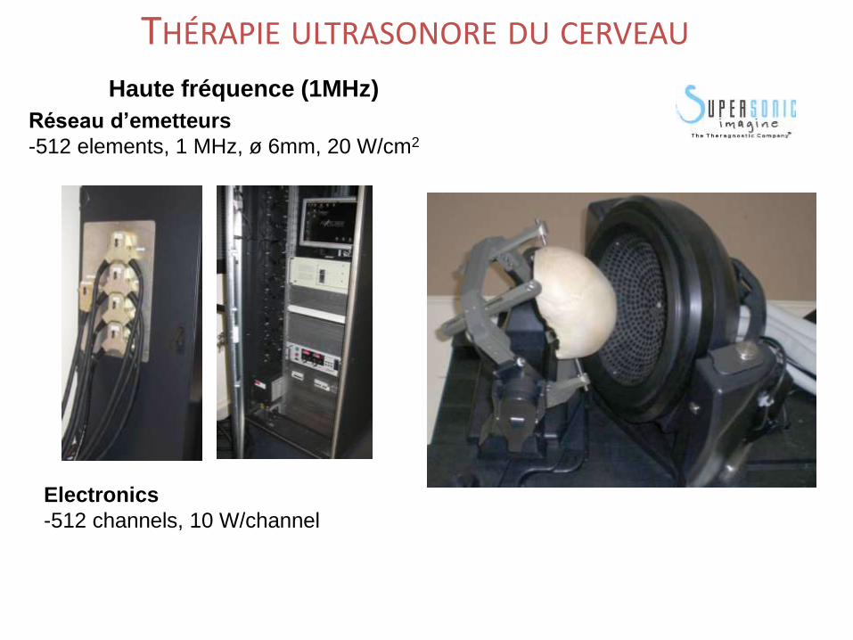
Haute fréquence (1MHz)
Réseau d’emetteurs
-512 elements, 1 MHz, ø 6mm, 20 W/cm2
Electronics
-512 channels, 10 W/channel
THÉRAPIE ULTRASONORE DU CERVEAU
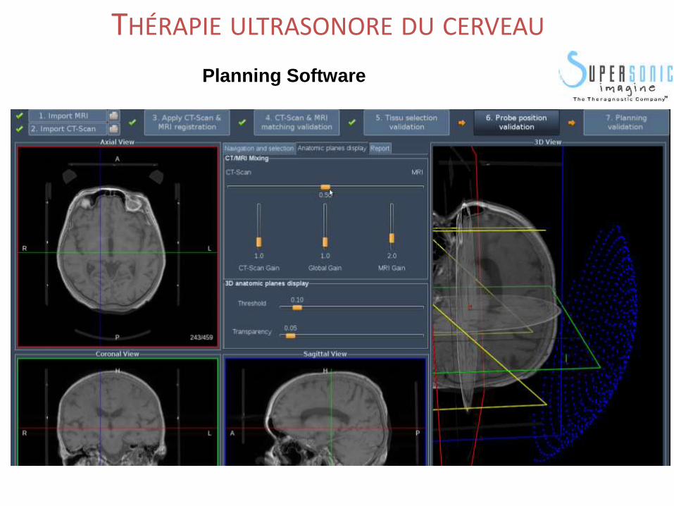
Planning Software
THÉRAPIE ULTRASONORE DU CERVEAU
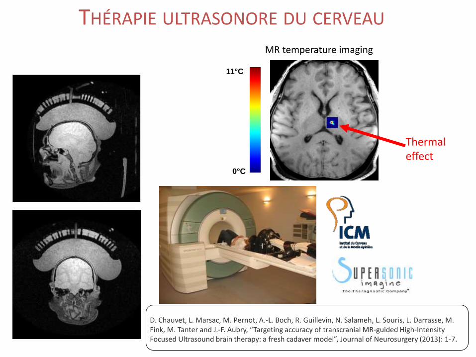
MR temperature imaging
11°C
0°C
D. Chauvet, L. Marsac, M. Pernot, A.-L. Boch, R. Guillevin, N. Salameh, L. Souris, L. Darrasse, M. Fink, M. Tanter and J.-F. Aubry, “Targeting accuracy of transcranial MR-guided High-Intensity Focused Ultrasound brain therapy: a fresh cadaver model”, Journal of Neurosurgery (2013): 1-7.
Thermal effect
THÉRAPIE ULTRASONORE DU CERVEAU
THÉRAPIE ULTRASONORE DU CERVEAU
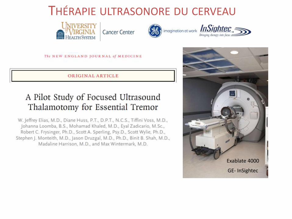
Exablate 4000
GE- InSightec
THÉRAPIE ULTRASONORE DU CERVEAU
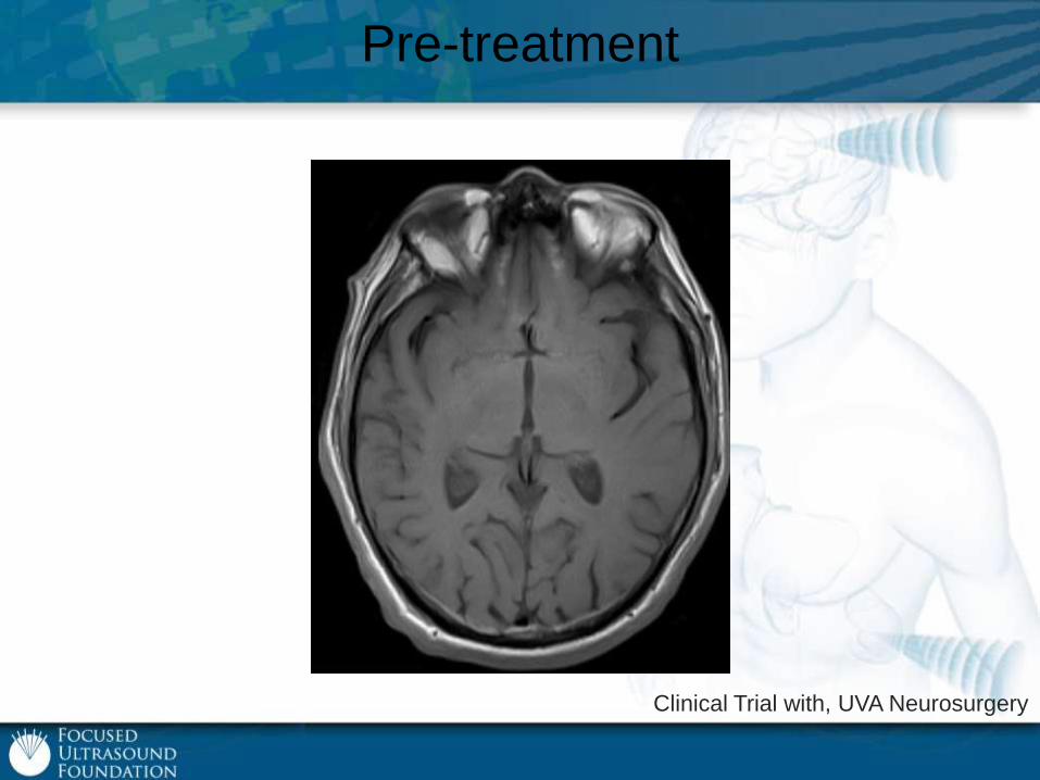
Pre-treatment
Clinical Trial with, UVA Neurosurgery

Targeting
Alias et al, NEJM, 2013
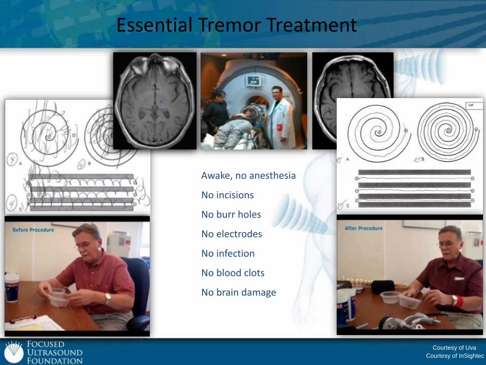
Essential Tremor Treatment
Awake, no anesthesia
No incisions
No burr holes
No electrodes
No infection
No blood clots
No brain damage
Courtesy of Uva
Courtesy of InSightec
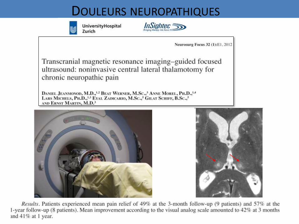
DOULEURS NEUROPATHIQUES
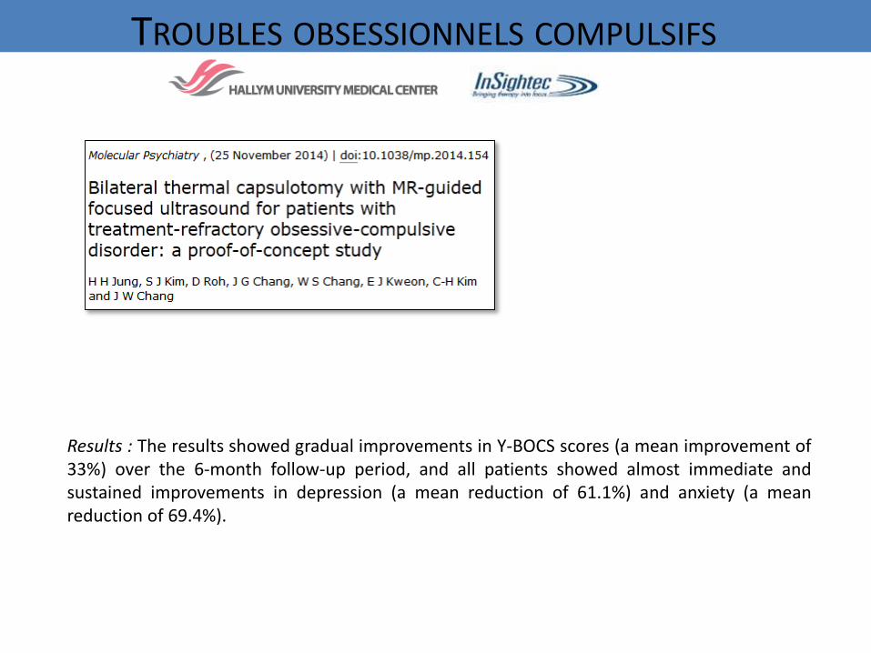
Results : The results showed gradual improvements in Y-BOCS scores (a mean improvement of33%) over the 6-month follow-up period, and all patients showed almost immediate andsustained improvements in depression (a mean reduction of 61.1%) and anxiety (a meanreduction of 69.4%).
TROUBLES OBSESSIONNELS COMPULSIFS
THÉRAPIE ULTRASONORE DU CERVEAU
www.fusfoundation.org
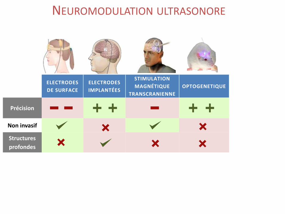
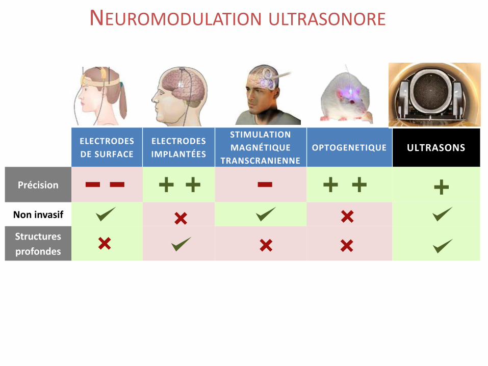
NEUROMODULATION ULTRASONORE
ELECTRODES
DE SURFACE
ELECTRODES
IMPLANTÉES
STIMULATION
MAGNÉTIQUE
TRANSCRANIENNE
OPTOGENETIQUE ULTRASONS
Précision
Non invasif
Structures
profondes
NEUROMODULATION ULTRASONORE
ELECTRODES
DE SURFACE
ELECTRODES
IMPLANTÉES
STIMULATION
MAGNÉTIQUE
TRANSCRANIENNE
OPTOGENETIQUE ULTRASONS
Précision
Non invasif
Structures
profondes
NEUROMODULATION ULTRASONORE
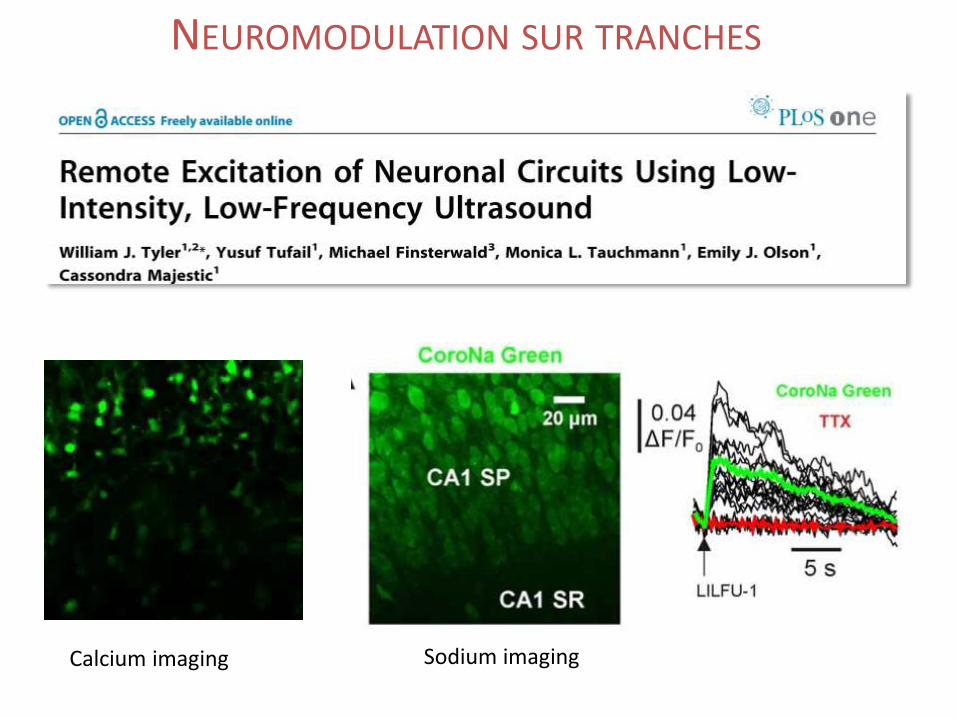
Calcium imaging Sodium imaging
NEUROMODULATION SUR TRANCHES
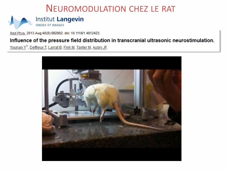
NEUROMODULATION CHEZ LE RAT
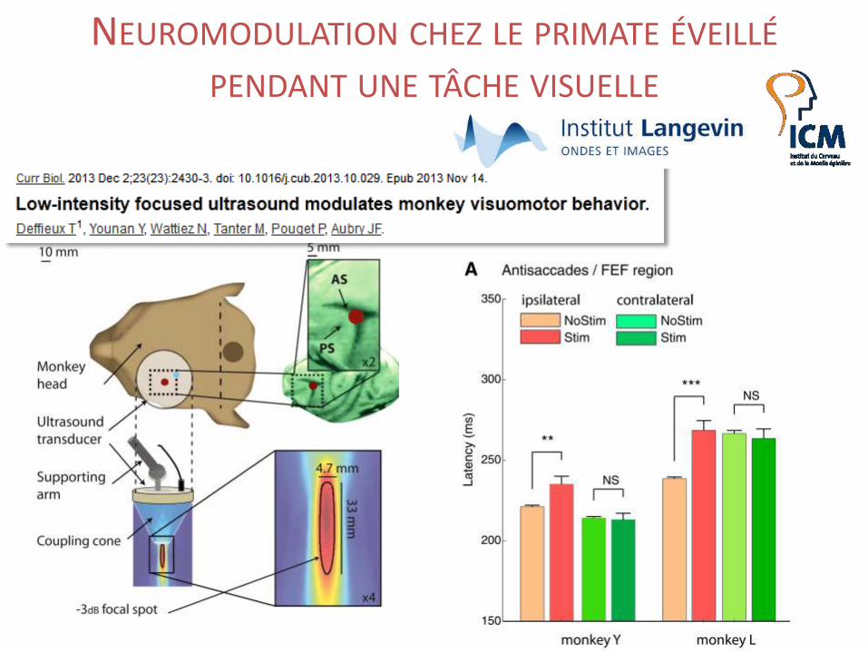
NEUROMODULATION CHEZ LE PRIMATE ÉVEILLÉ
PENDANT UNE TÂCHE VISUELLE
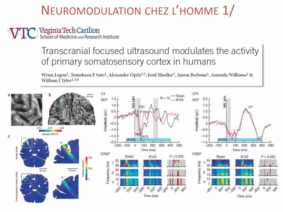
NEUROMODULATION CHEZ L’HOMME 1/
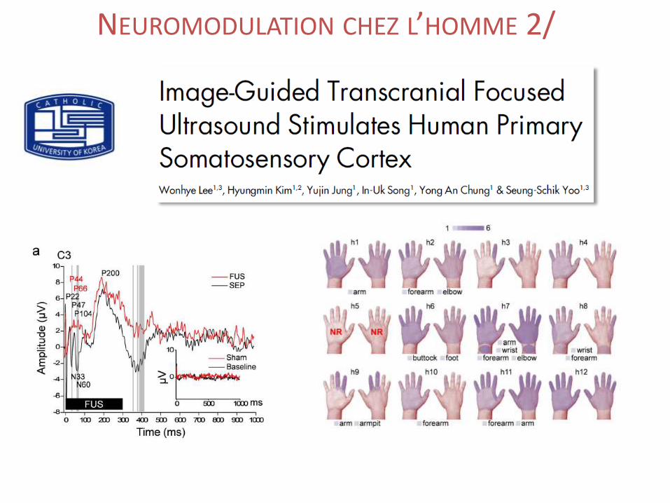
NEUROMODULATION CHEZ L’HOMME 2/
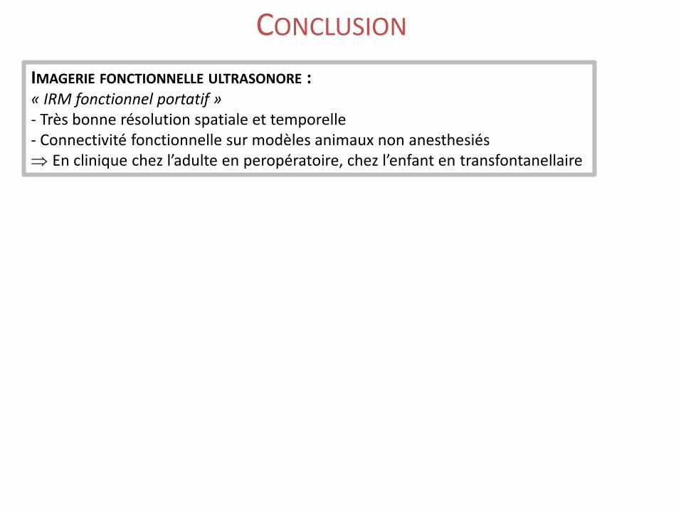
CONCLUSION
IMAGERIE FONCTIONNELLE ULTRASONORE :« IRM fonctionnel portatif »- Très bonne résolution spatiale et temporelle - Connectivité fonctionnelle sur modèles animaux non anesthesiés En clinique chez l’adulte en peropératoire, chez l’enfant en transfontanellaire
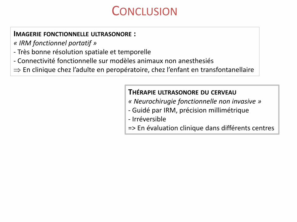
CONCLUSION
IMAGERIE FONCTIONNELLE ULTRASONORE :« IRM fonctionnel portatif »- Très bonne résolution spatiale et temporelle - Connectivité fonctionnelle sur modèles animaux non anesthesiés En clinique chez l’adulte en peropératoire, chez l’enfant en transfontanellaire
THÉRAPIE ULTRASONORE DU CERVEAU
« Neurochirugie fonctionnelle non invasive »- Guidé par IRM, précision millimétrique- Irréversible=> En évaluation clinique dans différents centres
Recommended