Embed Size (px)
Citation preview
An enigmatic filiform organism in the epicardium cavity of a new polycitorid ascidian
Françoise MONNIOT Laboratoire de biologie des invertébrés marins et malacologie,
CNRS D 0699, Muséum national d'Histoire naturelle, 55 rue de Buffon, F-75231 Paris cedex 05 (France)
Monniot F. 1998. — An enigmatic filiform organism in the epicardium cavity of a new polycitorid ascidian. Zoosystema 20 (3) : 429-438.
ABSTRACT In several samples of the ascidian Eudistoma hospitale n.sp. collectée! in the western Pacific Océan and in South Africa, a filiform organism was found implanted on the heart tissue. It is rolled in balls, occupies a large part of the ascidian abdominal cavity, and reaches such a length that it bursts through the branchial sac and finally cornes out at the colony surface with a free tip. Présent in ail zooids and in larvae, always single, this filament remains of an
KEYWORDS unknown nature, despite ail cytochemical and microanalytical methods used a.SC.L 'fnS' to characterize it. This kind of organism has never been recorded in any systematics, & 1
parasitology. other ascidian.
RÉSUMÉ Un énigmatique organisme filiforme de la cavité épicardiale d'une nouvelle espèce d'ascidie polycitoride. Dans tous les spécimens de l'ascidie Eudistoma hospitale n.sp. récoltés dans le Pacifique occidental et en Afrique du Sud, un organisme filiforme est implanté dans les tissus du cœur. Le filament est roulé en pelote et occupe la plus grande partie de la cavité abdominale de son hôte. Il atteint une longueur telle qu'il est expulsé dans la cavité branchiale et que, finalement, son extrémité libre sort de la colonie. Présent dans tous les
MOTS CLÉS zoïdes et les larves, le filament reste d'une nature inconnue malgré les systématique' méthodes de cytologie et de microanalyse utilisées pour le caractériser. Ce parasitologie. type d'organisme n'avait jamais été rencontré dans une ascidie.
Z O O S Y S T E M A • 1 9 9 8 • 2 0 ( 3 ) 429

Monniot F.
I N T R O D U C T I O N
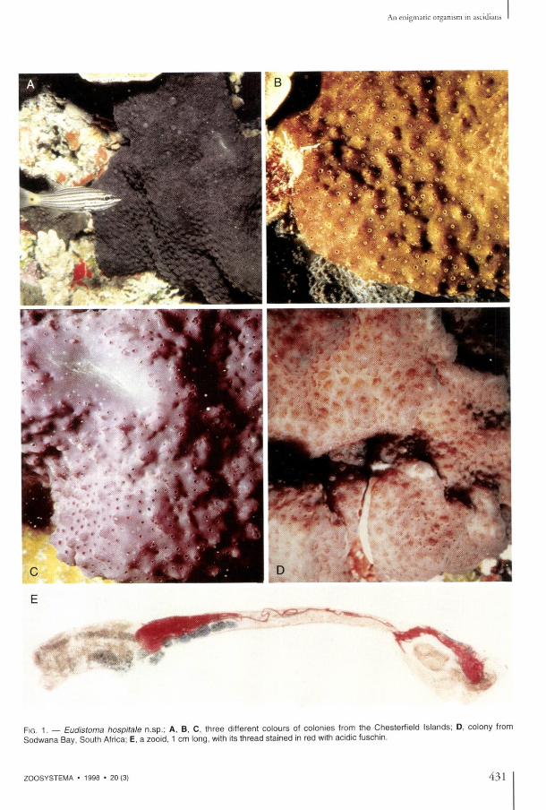
An encrusting colonial ascidian shows curious thin threads located inside the zooids (Fig. 1E) and somet imes coming out to the surface through the oral apertures. This species was col-lected for the first time at the Chesterfield Islands (southwest Pacific) in July 1988, and later in September 1996; it was also collected on Fiji in 1991 and October 1996, and in February 1994 and 1996, it was found in two widely sepa-rated stations in South Africa.
M A T E R I A L
Chesterfield Islands (Laboute coll.). Skeleton, 30 m; Ile Longue, 22 m. Fiji Islands. Nukubalavu, 10 m, 16°45'06S -179°47'83W (Coral Reef Reseach Foundation coll). South Africa. Sodwana Bay, 1 9 m (Schleyer coll.); South Cape coast, intertidal (Glassom coll.).
M E T H O D S
Spécimens fixed in 4 % formaldehyde in sea water were dissected, stained and mounted on slides for gênerai anatomical observations. Sections of 7 um were stained with hemalum-picroindigocarmine or toluidine blue to reveal the morphology of the tissues. Ultrathin sections were observed with an Hitachi 600 électron microscope. Dissected filaments were critical-point dried, coated with gold and examined with a Jeol JSM 840 microscope. Several cytochemical reactions were used (Pearse 1968; Gabe 1968; Ganter & Jolies 1968-1970). They are: - the periodic acid Schiff reaction (PAS) for glu-cids and glycoproteins; - the Lugol's iodine reactive for glycogen; - alcian blue at PH 0.5 and 2.5 for acidic glucids - the alloxane-Schiff reaction for proteins; - the ferrie ferricyanide method (Schmorl's and Lillie processes) for thiols and other reducing groups especially purines and sulphides; - the Massons argentaffin reaction for phénols (tunichrome and analogues); - the coupled tetrazonium reaction of Danielli, using the fast blue B sait for proteins and phénols;
- the l-(4-chloromercuriphenyldiazo)-2-naphtol (mercury orange, red sulphydril reagent) and the 2.2'-dihydroxy-6.6'dinaphtyl disulphide (DDD) methods for thiol groups; - the Lugol's sulfuric acid for cellulose; - the chlor-zinc-iodine for both chitin and cellulose; - the ferrie chlorure-acetic acid for porphyrins. Elementary microanalysis was performed on paraffin sections spread on carbon-coated mylar slides, using a CAMECA MS46 électron microprobe equipped with wavelength dispersing spec-trometers. The diameter of the probe was 1 um. Molecular analysis was carried out on similar sections using a Raman laser microprobe Dilor Microdil 28.
RESULTS
T H E A S C I D I A N
Family P O L Y C I T O R I D A E Michaelsen, 1904 Genus Eudistoma Caullery, 1909
Eudistoma hospitale n.sp.
T Y P E M A T E R I A L . — M N H N A3 Eud 188, Chesterfield Islands.
D E S C R I P T I O N
Ail samples have the same colonial structure: cushions or crusts reaching several decimeters across and up to three centimeters thick. The colour in life is uniform through each colony, but in only one station varies among black, light brown, pink or purple (Fig. 1A -D) . The colony surface is smooth, shiny, sometimes slightly undulated. The zooid oral openings appear as small holes arranged in circles in the center of which the cloacal apertures open, grouped on small protrusions (Fig. 1 A - D ) . The tunic is opaque without any embedded sédiment. The gênerai consistency is firm with an even more résistant surface layer. When fixed, the tunic becomes more translucent and more or less dark grey. It contains abundant vesicular and pigment cells, and vanadocytes. The zooids lie perpendi-cular to the colony surface; the less contracted
430 Z O O S Y S T E M A • 1 9 9 8 • 2 0 ( 3 )
An enigmatic organism in ascidians
Monniot F.
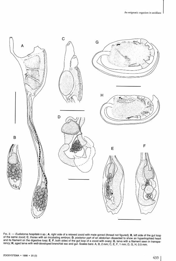
ones easily reach 1 cm in length. The thoracic body wall is black, especially in the anterior part. The abdomens are yellowish, the embryos bright orange, the larvae paler orange. Thèse colours disappear after fixation. The zooids ail have the same characters, whatever their geographical origin. Both siphons are tubu-lar, edged with six rounded lobes, the cloacal siphon longer (Fig. 2A, C) . The endostyle is thick. The thoracic muscles are dense with numerous longitudinal and transverse fibers. Generally, sixteen oral tentacles in two orders of size alternate along two circles. The branchial sac has anteriorly an imperforated area only seen in relaxed zooids. An average of twenty-five elonga-ted stigmata were counted in the first row. The abdomen lies below a constriction of the body. The isodiametric cesophagus is very long (Fig. 2A). The stomach has a generally round shape with sometimes, in the largest individuals, two latéral crests when the gut is empty. The post stomach is cylindrical, separated from the mid-intestine by a constriction. The rectum begins with an annular widening (Fig. 2E, F) which sometimes forms two crescent-shaped caeca. After the curve of the gut loop, the intestine remains parallel to the cesophagus up to the level of the second stigmata row, where the rectum opens via a bilobed anus. Beyond the mid-intestine the rectum has a green colour in life. At the stomach level, the rectum is covered with parallel pyloric tubules converging into a short duct that connects with the posterior part of the stomach. One or two vascular processes prolong the abdomen into the tunic. The heart forms a J-shaped tube, with unequal sides, more or less inflated (Fig. 2 D - F ) . The endothelium of the vessels disappears at a short distance from the heart. On the cardiac tissue, between the ovary and the base of the gut loop, there is an extremely long filiform structure, of unknown nature. This thread begins with a coni-cal root (Figs 2D, E, 3C, D, 4A), then remains the same diameter along its whole length. It is coiled on itself (Figs 2D-F, 4A), and invades the epicardium cavity (Fig. 1 E). Reaching the thorax, it becomes so voluminous and tightly wound that it pierces the wall, enters the branchial sac and finally extends out through the oral siphon.
The gonads have the common structure of the genus. The testis is made of numerous round vesicles (Fig. 2A, B); the ovary is located in the center of the gut loop, below the stomach (Fig. 2F) . In colonies from the Chesterfield Islands, the zooids are ail maie or female; but in ones from Fiji there is a young ovary amidst the testis vesicles and embryos are présent in the cloacal cavity (Fig. 2C). The South African colonies are simultaneously hermaphroditic. One or sometimes two embryos are incubated at the same time. Well-developed larvae were found only in Chesterfield spécimens (Fig. 2G, H) . They remain inside the tip of the oviduct until their release into the sea. The larval trunk is oval, 1 mm long. The tail makes a half turn around it. When younger embryos are orange and translu-cent, the tadpole body wall contains numerous pale opaque cells. The sensory vesicle has the usual otolith and ocellus. Three rows of about fif-teen stigmata apiece are already visible in an incu-bating larva (Fig. 2H), and an embryonic gut lies in the posterior part of the body (Fig. 2 H ) . Anteriorly, the three adhesive papillae, ail in a line, have oval tips. Their thick bases alternate with four foliose vesicles. In addition, there is a vesicle applied on the side of the most dorsal vesicle on each side (Fig. 2H). In aged larvae, a short thread can be seen in transparency amidst the vitellus near the embryonic gut (Fig. 2G) . The présence of this filamentous structure has been ascertained in histological sections. Within the tunic, between functional zooids, traces of degenerated zooids persist with pièces of filaments which are only empty tubes, some with lysed cells. No filaments were found in the tunic outside the zooids.
Eudistoma hospitale differs from other species of the genus only by its larval structure and the constant présence in ail colonies and ail zooids of a filiform organism whence the species name.
T H E F I L A M E N T O U S S T R U C T U R E
A single thread winds about inside the pericardial cavity of each zooid (Figs 1E, 4C) . Its conical basis takes root on the cardiac lining of the ascidian and is externally covered by a cellular layer of the same constitution as the heart's endothelium (Fig. 3C, D). An aperture allowing a com-
432 Z O O S Y S T E M A • 1 9 9 8 • 2 0 ( 3 )
An enigmatic organism in ascidians
FIG . 2. — Eudistoma hospitale n.sp.; A , right side of a relaxed zooid with maie gonad (thread not figured); B , left side of the gui loop of the same zooid; C , thorax with an incubating embryo; D , posterior part of an abdomen dissected to show an hypertrophied heart and its filament on the digestive loop; E , F , both sides of the gut loop of a zooid with ovary; G , larva with a filament seen in transpa-rency; H , aged larva with well-developed branchial sac and gut. Scales bars: A , B , 2 mm; C E F 1 mm- D G H 0 5 mm
Z O O S Y S T E M A • 1 9 9 8 • 2 0 ( 3 )
Monniot F.
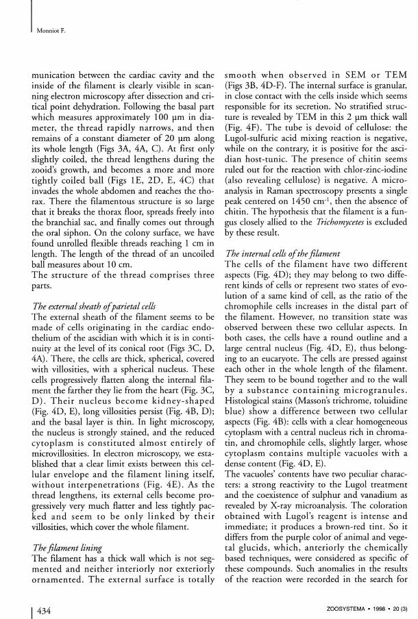
munication between the cardiac cavity and the inside of the filament is clearly visible in scan-ning électron microscopy after dissection and cri-tical point dehydration. Following the basai part which measures approximately 100 um in dia-metet, the thread rapidly narrows, and then remains of a constant diameter of 20 um along its whole length (Figs 3A, 4A, C) . At first only slightly coiled, the thread lengthens during the zooid's growth, and becomes a more and more tightly coiled bail (Figs 1E, 2 D , E, 4C) that invades the whole abdomen and reaches the thorax. There the filamentous structure is so large that it breaks the thorax floor, spreads freely into the branchial sac, and finally cornes out through the oral siphon. On the colony surface, we have found unrolled flexible threads reaching 1 cm in length. The length of the thread of an uncoiled bail measures about 10 cm. The structute of the thread comprises three parts.
The external sheath of pariétal cells
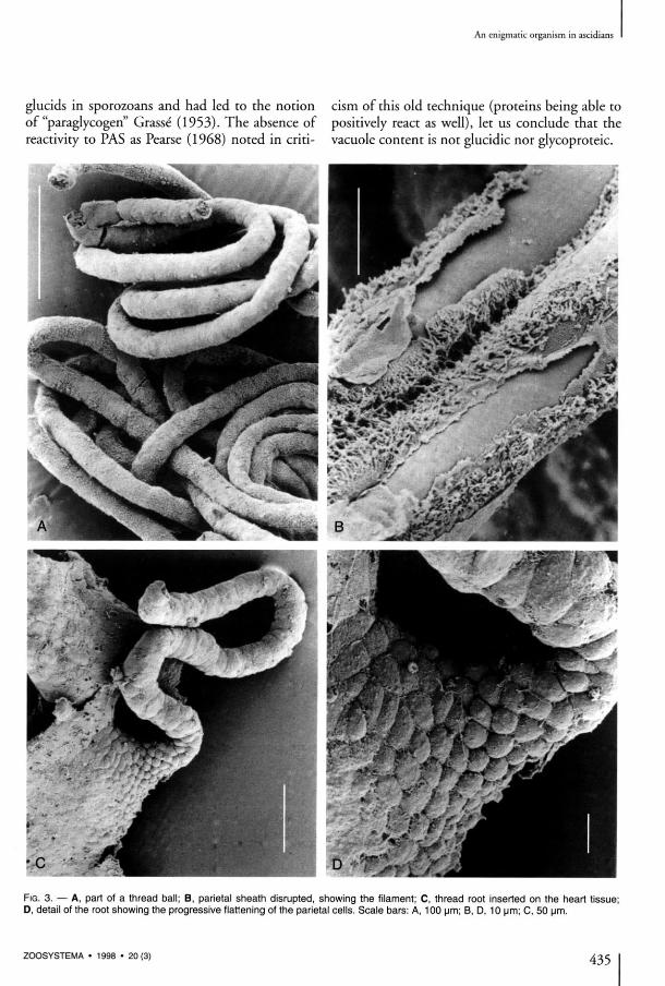
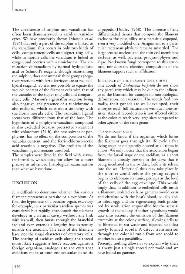
The external sheath of the filament seems to be made of cells originating in the cardiac endo-thelium of the ascidian with which it is in conti-nuity at the level of its conical root (Figs 3C, D, 4A). There, the cells are thick, spherical, covered with villosities, with a spherical nucleus. Thèse cells progressively flatten along the internai filament the farther they lie from the heart (Fig. 3C, D ) . Their nucleus become kidney-shaped (Fig. 4D, E), long villosities persist (Fig. 4B, D); and the basai layer is thin. In light microscopy, the nucleus is strongly stained, and the reduced cytoplasm is constituted almost entirely of microvillosities. In électron microscopy, we esta-blished that a clear limit exists between this cel-lular envelope and the filament lining itself, without interpénétrations (Fig. 4 E ) . As the thread lengthens, its external cells become progressively very much flatter and less tightly pac-ked and seem to be only l inked by their villosities, which cover the whole filament.
The filament lining
The filament has a thick wall which is not seg-mented and neither interiorly nor exteriorly ornamented. The external surface is totally
smooth when obsetved in S E M or T E M (Figs 3B, 4D-F). The internai surface is granular, in close contact with the cells inside which seems responsible for its sécrétion. No stratified structure is revealed by T E M in this 2 um thick wall (Fig. 4F) . The tube is devoid of cellulose: the Lugol-sulfuric acid mixing reaction is négative, while on the contrary, it is positive for the ascidian host-tunic. The présence of chitin seems ruled out for the reaction with chlor-zinc-iodine (also revealing cellulose) is négative. A micro-analysis in Raman spectroscopy présents a single peak centered on 1450 c m 1 , then the absence of chitin. The hypothesis that the filament is a fun-gus closely allied to the Trichomycetes is excluded by thèse resuit.
The internai cells of the filament
The cells of the fdament have two différent aspects (Fig. 4D); they may belong to two différent kinds of cells or represent two states of évolution of a same kind of cell, as the ratio of the chromophile cells increases in the distal part of the filament. However, no transition state was observed between thèse two cellular aspects. In both cases, the cells have a round outline and a large central nucleus (Fig. 4D, E), thus belong-ing to an eucaryote. The cells are pressed against each other in the whole length of the filament. They seem to be bound together and to the wall by a substance conta in ing microgranules . Histological stains (Massons trichrome, toluidine blue) show a différence between two cellular aspects (Fig. 4B): cells with a clear homogeneous cytoplasm with a central nucleus rich in chroma-tin, and chromophile cells, slightly larger, whose cytoplasm contains multiple vacuoles with a dense content (Fig. 4D, E). The vacuoles' contents have two peculiar charac-ters: a strong reactivity to the Lugol treatment and the coexistence of sulphur and vanadium as revealed by X-ray microanalysis. The coloration obtained with Lugol's reagent is intense and immédiate; it produces a brown-red tint. So it differs from the purple color of animal and végétal glucids, which, anteriorly the chemically based techniques, were considered as spécifie of thèse compounds. Such anomalies in the results of the reaction were recorded in the search for
Z O O S Y S T E M A • 1 9 9 8 • 2 0 ( 3 )
An enigmatic organism in ascidians
glucids in sporozoans and had led to the notion cism of this old technique (proteins being able to of "paraglycogen" Grasse (1953). The absence of positively react as well), let us conclude that the reactivity to PAS as Pearse (1968) noted in criti- vacuole content is not glucidic nor glycoproteic.
F I G . 3. — A , part of a thread bail; B , pariétal sheath disrupted, showing the filament; C , thread root inserted on the heart tissue; D , détail of the root showing the progressive flattening of the pariétal cells. Scale bars: A , 1 0 0 um; B , D , 1 0 um; C, 5 0 um.
Z O O S Y S T E M A • 1 9 9 8 • 2 0 ( 3 ) 435
Monniot F.
The coexistence of sulphur and vanadium has often been demonstrated in ascidian vanado-cytes. We have previously shown (Martoja et al. 1994) that only a part of the sulphur is linked to the vanadium; this occurs in only two kinds of cells, compartment cells and signet ring cells, while in morula cells the vanadium is linked to oxygen and coexists with a tunichrome. The élimination of vanadium by normal hydrochloric acid or Schmorl's reagent, though maintaining the sulphur, does not unmask thiol groups (négatives reactions with ferrie ferricyanure or red-sulf-hydril reagent). So it is not possible to equate the vacuole content of the filament cells with that of the vacuoles of the signet ring cells and compartment cells. Massons argentaffin reaction being also négative, the présence of a tunichrome is also excluded, which rules out a similarity with the host's morula cells. The vanadium ligand seems very différent from that of the host. The hypothesis of a porphyrin-vanadium compound is also excluded because protracted tteatment with chloroform (24 h), the best solvent of por-phyrins, has no effect on the composition of the vacuolar content, and the ferrie chlorure-acetic acid reaction is négative. The problem of the vanadium ligand remains unsolved. The samples were fixed in the field with seawa-ter-formalin, which does not allow for a more précise or advanced histological examination than what we have done.
DISCUSSION
It is difficult to détermine whether this curious filament represents a parasite or a symbiont. At first, the hypothesis of a peculiar organ, excretoty for example, in a particular ascidian species was considered but rapidly abandoned: the filament develops in a natural cavity without any link with its wall, then bursts through the branchial sac and even extends a lengthy free extremity outside the ascidian. The cells of the fdament have not the usual characters of excretory cells. The coating of ascidian cells along the thread more likely suggests a host's reaction against a foreign organism, analogous to the cysts that ascidians make around endovascular parasitic
copepods (Dudley 1968). The absence of any differentiated tissues that compose the filament excludes the possibilité of a parasitic copepod, even a very modified one. Assignment to a particular metazoan phylum remains unsettled. The large central nucleus and the thin cell membrane exclude, as well, bacteria, procyanophytes and algae. No known fungi correspond to this structure, nor does the chemical composition of the filament support such an affiliation.
I N F L U E N C E O F T H E F I L A M E N T O N ITS H O S T
The zooids of Eudistoma hospitale do not show any peculiarity which may be due to the influence of the filament, for example no morphological déformation, no castration. The zooids feed nor-mally, their gonads are well-developed, their embryos reach full matutation without monstro-sities. Asexual reproduction is not affected either, as the colonies teach very large sizes compared to other species of the same genus.
T R A N S M I S S I O N M O D E
We do not know if the organism which forms the fdament goes through its life cycle a free living stage or obligatorily housed at ail times in a host. We only notice that the association begins from the larval stage of the ascidian, as a short filament is already présent in the larva that is being incubated in the oviduct, before its release into the sea. "Infection" thus may take place in the mother zooid before the young tadpole begins to elaborate its tunic, perhaps at the level of the cells of the egg envelope. This would imply that, in addition to embedded cells inside a filament, isolated cells or gamètes would exist and circulate with the host's blood, equally able to infect eggs and the regenerating buds produ-ced by strobilation responsible for the asexual growth of the colony. Another hypothesis would take into account the émission of the filament extremity at the colony surface, allowing cells to be liberated in sea water and ingested to infest newly formed zooids. A direct transmission through the colonial tunic from one zooid to another is also a possibillty. Presendy nothing allows us to explain why there is always just a single thread per zooid and we have found no gamètes.
436 Z O O S Y S T E M A • 1 9 9 8 • 2 0 ( 3 )
An enigmatic organism in ascidians
F I G . 4. — A , B , cardiac extremity of a thread in light microscopy (toluidine blue). C , sections of a thread bail contained in the pericar-dium cavity occupying the most part of the abdominal section of a zooid. D , E , sections of a thread in T E M showing pariétal cells with elongated nucleus and microvillies, the tube wall, the internai granular and vacuolated cells. F , détail of the filament wall. Scale bars: A , 50 pm; B , 10 um; C, 100 um; D , 5 um; E , 2 um; F , 1 um.
Z O O S Y S T E M A • 1 9 9 8 • 2 0 ( 3 ) 437
Monniot F.
O T H E R C O M M E N S A L S , S Y M B I O N T S O R PARASITES
K N O W N IN A S C I D I A N S
Organisms living in association with ascidians were reviewed by Monniot (1990). They are very numerous : Vira les , Bacter ia , Flagel la ta , Tr ichomonadina , Rh izopoda , Gregar ina , Coccidia, Haplosporidia, Ciliophora, Nephro-myces, Algae, Cnidaria, Ctenaria, Turbellaria, Annelida, Bryozoa, Nemertina, Mollusca, many Crustacea and especially copepods, and also Pisces.
Aknowledgements I have greatly benefited from the precious know-ledge of R. Martoja in cytology, and I thank him as well for the cytochemistry and microanalyses he has performed as for the thoughtful discussions we had during this study. I am much indebted to T. Newberry for the english language corrections.
REFERENCES
Dudley P. 1968. — A light and électron microscopic study of tissue interactions between a parasitic copepod Scolecodes huntsmani (Henderson) and its host ascidian, Styela gibsii (Stimpson). Journal of Morphology 124 (3): 263-281.
Gabe M. 1968. — Techniques histologiques. Masson éd., Paris, 1113 p.
Ganter P. & Jolies G. 1969-1970. — Histochimie normale et pathologique. Gauthier-Villars, Paris, 1904 p.
Grasse P. P. 1953. — Sous-embranchement des Sporozoaires, volume 1 (2), in Grasse P. P. (éd.), Traité de zoologie. Masson éd., Paris, 545 p.
Martoja R., Gouzerh P. & Monniot F. 1994. — Cytochemical studies of vanadium, tunichromes and related substances in ascidians: possible biolo-gical significance. Oceanography and Marine Biology 32: 531-556.
Monniot C. 1990. — Diseases of Urochordata, volume 3: 569-636, in Kinne O. (éd.), Diseases of marine animais. Biologische Anstalt Helgoland, Hamburg.
Pearse A. G. E. 1968. — Histochemistry, theoretical andapplied. Churchill, London, 1518 p.
Submitted on 10 February 1998; accepted on 6April 1998.
438 Z O O S Y S T E M A • 1 9 9 8 • 2 0 ( 3 )