Embed Size (px)
Citation preview
Bacterial diseases
Alippi A.M.
in
Colin M.E. (ed.), Ball B.V. (ed.), Kilani M. (ed.). Bee disease diagnosis
Zaragoza : CIHEAMOptions Méditerranéennes : Série B. Etudes et Recherches; n. 25
1999pages 31
Article available on line / Article disponible en ligne à l’adresse :
--------------------------------------------------------------------------------------------------------------------------------------------------------------------------
http://om.ciheam.org/article.php?IDPDF=99600235
--------------------------------------------------------------------------------------------------------------------------------------------------------------------------
To cite th is article / Pour citer cet article
--------------------------------------------------------------------------------------------------------------------------------------------------------------------------
Alippi A.M. Bacterial diseases. In : Colin M.E. (ed.), Ball B.V. (ed.), Kilani M. (ed.). Bee disease
diagnosis. Zaragoza : CIHEAM, 1999. p. 31 (Options Méditerranéennes : Série B. Etudes et
Recherches; n. 25)
--------------------------------------------------------------------------------------------------------------------------------------------------------------------------
http://www.ciheam.org/http://om.ciheam.org/
Bacterial diseases
A.M. Alippi Laboratorio de Fitopatologia, Facultad de Ciencias Agrarias y Forestales
Universidad Nacional de La Plata, Calle 60 y 11 8, C.C. 31, 1900 La Plata, Argentina
Introduction
Bacteria are single celled organisms, microscopic in size (less than 1 pm to several pm in length) and lack a defined nucleus, belonging to the Procaryote kingdom. Within the procaryotic organisms, four divisions are recognised, based upon cell wall characteristics (Murray, 1984):
Division I Gracilicutes Gram-negative type cell wall (contains Gram-negative bacteria)
Division Firmicutes Gram-positive type cell wall (contains Gram-positive bacteria)
Division Tenericutes No cell wall (contains class Mollicutes)
Division IV Mendosicutes Procaryotes with unusual walls, membrane lipids, ribosomes and RNA sequences (contains class Archaeobacteria)
Bacteria that affect insects are present in divisions to and can thus be divided into organisms with cell walls (true bacteria and rickettsias) and those without (myeoplasmas and spiroplasmas).
The taxonomy of bacteria has undergone considerable revision during the past two decades. The basic unit of bacterial classification is the species, which may be defined as a collection of strains that have many features in common, differing in these features from other groups of strains. Subspecies represent major divisions within the species taxon.
In honey bees, the two most important diseases of bacterial origin are American Foulbrood and European Foulbrood; they have been studied more than other bee diseases. Septicaemia, powdery scale, half-moon disorder and diseases caused by Spiroplasmas and Rickettsiae have received less consideration because they cause minor economic losses to beekeepers.
American Foulbrood disease
Definition of the disease
American Foulbrood I s an infectious, highly contagious, cosmopolitan disease affecting propupae and due to the multiplication of a sporulating bacterium Paenibacillus larvae ssp. larvae. Symptoms of infection are easily recognisable in freshly dead larvae.
31
CIHEAM - Options Mediterraneennes
Other common names applied to the disease are black brood, ropy brood, foul and diseased brood.
epidemiology
Until 1906 the two foulbrood diseases were not differentiated and the condition was generally referred to as foulbrood. Phillips (1906) used the terms European and American to distinguish the diseases. However the designations did not refer to the geographical distributions but to the areas where they were first investigated scientifically (Shimanuki, 1990). White (1907) demonstrated conclusively that a bacterium that he called Bacillus larvae was the cause of American Foulbrood (AFB) disease by fulfilling
The geographical origin of AFB is unknown, but it is found almost world-wide (Matheson, 1993, 1996). AFB is the most virulent brood disease known in honey bees (Apis mellifera L.). It is one of the few bee diseases capable of killing a colony and poses unique problems for prevention and control because the bacterial spores can remain viable for long periods of time (35 years or more) and survive adverse conditions (Matheson and Reid, 1992). The diseased brood combs are the greatest source of spores, the agents of contamination and dissemination (Shimanuki, 1990; Alippi, 1996). AFB infection is the most unstable of all infections in honey bee communities (Bailey and Ball, 1991). This highly contagious disease is spread between colonies primarily by exchanging infected combs or by allowing diseased colonies to be robbed by bees from~another apiary.
There is no seasonal outbreak of AFB; it occurs at any time of the year when brood is present (Bailey and Ball, 1991), but it is usually diagnosed during the active brood-rearing season.
Etiology
Pathogenic agent. Classification
White (1907, 1920) demonstrated that Bacillus larvae was the cause of AFB and made a full description of the species.
In 1993, Ash and co-workers (Ash et al., 1993) reclassified this species in a newly defined genus, Paenibacillus (from Latin: almost a Bacillus, because it derives from this genus but is phylogenetically distinct), which comprises the group 3 Bacillus spp. as defined by 16 S rRNA analysis.
Recent taxonomic work (Heyndrickx et al., 1996) has shown that one other member of the genus, Paenibacillus pulvifaciens, a weak pathogen of honeybee larvae, should be reclassified as a subspecies of P. larvae. Genomes of the two subspecies are at least 90% homologous. According to these studies, there are 11 species within the genus Paenibacillus, and the current taxonomic position of the causal agent of AFB disease is Paenibacillus larvae ssp. larvae (White) Heyndrickx et al. [Formerly: Paenibacillus larvae (White) Ash et al.].
Spread and transmission
Paenibacillus larvae ssp. larvae spores are highly resistant to desiccation, high temperatures than 10 min), exposure to UV light and also survive contact with classical
disinfectants like 10% formaldehyde solutions for longer than 5 hours.
Spread within-colony
Paenibacillus larvae ssp. larvae infects only the larvae of all castes and not adult bees. Larvae become infected by consuming spores present in their food. A few infected individuals can be removed from the colony by worker bees engaged in nest cleaning duties. As time passes, remaining infected larvae or pupae dry to a scale, which adheres firmly to the bottom of the cell and is very difficult for the bees to remove. One single scale contains about 2,500 million spores. Infection is
32
CIHEAM - Options Mediterraneennes
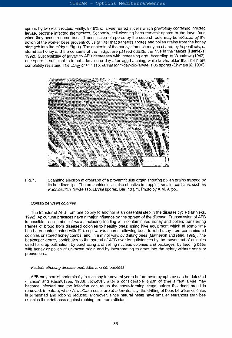
spread by two main routes. Firstly, 8-19% of larvae reared in cells which previously contained infected larvae, become infected themselves. Secondly, cell-cleaning bees transmit spores to the larval food when they become nurse bees. Transmission of spores by the second route may be reduced by the action of the worker bees proventriculus (a filter that transfers spores and pollen grains from the honey stomach into the midgut, Fig. 1). The contents of the honey stomach may be shared by trophallaxis, or stored as honey and the contents of the midgut are passed outside the hive in the faeces (Ratnieks, 1992). Susceptibility of larvae to AFB decreases with increasing age. According to Woodrow (1942), one spore is sufficient to infect a larva one day after egg hatching, while larvae older than 53 h are completely resistant. The LD50 of P. l. ssp. larvae for l-day-old-larvae is 35 spores (Shimanuki, 1990).
Fig. 1. Scanning electron micrograph of a proventriculus organ showing pollen grains trapped by its hair-lined lips. The proventriculus is also effective in trapping smaller particles, such as Paenibacillus larvae ssp. larvae spores. Bar: 1 O pm. Photo by.A.M. Alippi.
Spread between colonies
The transfer of AFB from one colony to another is an essential step in the disease cycle (Ratnieks, 1992). Apicultura1 practices have a major influence on the spread of the disease. Transmission of AFB is possible in a number of ways, including feeding with contaminated honey and pollen; transferring frames of brood from diseased colonies to healthy ones; using hive equipment which at some time has been contaminated with P, 1. ssp. larvae spores; allowing bees to rob honey from contaminated colonies or stored honey combs; and, in a minor way, by drifting bees (Matheson and Reid, 1992). The beekeeper greatly contributes to the spread of AFB over long distances by the movement of colonies used for crop pollination, by purchasing and selling nucleus colonies and packages, by feeding bees with honey or pollen of unknown origin and by incorporating swarms into the apiary without sanitary precautions.
Factors affecting disease outbreaks and seriousness
AFB may persist endemically in a colony for several years before overt symptoms can be detected (Hansen and Rasmussen, 1986). However, after a considerable length of time a few larvae may become infected and the infection can reach the spore-forming stage before the dead brood is removed. In nature, when A. mellifera nests are at a low density, the drifting of bees between colonies is eliminated and robbing reduced. Moreover, since natural nests have smaller entrances than bee colonies their defences against robbing are more efficient.
CIHEAM - Options Mediterraneennes
Pathogenesis
The spores represent the infective form of the disease. Larvae are infected by consuming spores present in their food. Millions of spores are required to infect larvae older than 2 days, but larvae up to 24 hours old become infected with less than 1 0 spores (Woodrow, 1942). The spores germinate in the midgut lumen (pH 6.6) approximately 1 day after ingestion by the larva, giving rise to the vegetative forms (rod stage of P. 1. ssp. larvae). The flagellated rods do not multiply in the lumen of the intestine however, they migrate to the peritrophic membrane, penetrating into the midgut epithelium. The vegetative rods enter the midgut cell by phagocytosis. If some bacteria are destroyed in the phagocytic vacuoles, others survive. After lysis of the invaded cell, the bacteria enter the haemocoel of the host. The rods multiply abundantly in the haemolymph, and then begin to sporulate. The larva dies from a systemic bacteraemia (Davidson, 1973). Apart from the lysis of an invaded midgut cell, no toxin seems to be involved, the disease being a systemic bacteraemia (Tanada and Kaya, 1993).
The conditions for germination of spores are optimal in the youngest larvae, but they soon become unsuitable for vegetative growth, as observed in the in vitro growth of P. 1. ssp. larvae (Bailey and Lee, 1962). Germination of spores occurs at a pH of about 6.6, a temperature of microaerophilic conditions (5-10% CO2 in air). Vegetative cells are unable to multiply in the larval intestine because at this stage the bacteria are aerobic and motile, so they migrate to the epithelium, penetrate into the body cavity and multiply in the haemolymph where aerobic conditions prevail (Bailey and Ball, 1991).
It seems that spore germination is slower in older larvae than in young ones and the vegetative rods may not have time to reach the epithelium and invade the tissues, before being evacuated with the gut contents in the faeces (Bailey and Ball, 1991). Vegetative cells are not infective to other larvae and do not resist desiccation.
Older larvae also have a thicker peritrophic membrane which could constitute an increasing barrier to the movement of P. 1. ssp. larvae vegetative cells. The second barrier is the midgut epithelium. If bacteria successfully penetrate both the peritrophic membrane and the midgut epithelium, the larva can be expected to succumb (Davidson, 1973).
When the bacteria proliferate in the larval tissues before pupation, infected larvae quickly die and spores form, mostly in propupae 11 days after egg hatching. Larvae of 13-1 4 days old contain spores in all their tissues (Bailey and Ball, 1991). After death, the normally white larva becomes dark brown and settles to the bottom of the cell. Its body wall is easy ruptured. The body contents increase in viscosity and adhere to an object, such as a toothpick inserted into the cell, and string out into a sticky thread (ropiness) for a considerable distance. As time passes, a dead larva or pupa dries to a scale that adheres tightly to the cocoon at the base of the cell. -. .
Clinical diagnosis (field symptoms)
AFB has distinctive field symptoms which include characteristics of the brood comb and characteristics of the cell contents (Matheson and Reid, 1992). The general appearance of affected combs are a patchy brood pattern due to the presence of healthy capped brood, uncapped cells containing the remains of diseased larvae, and empty cells.
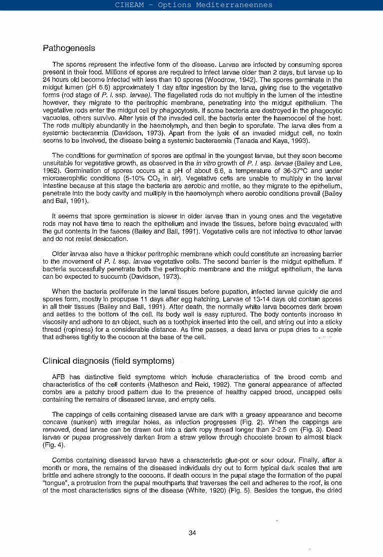
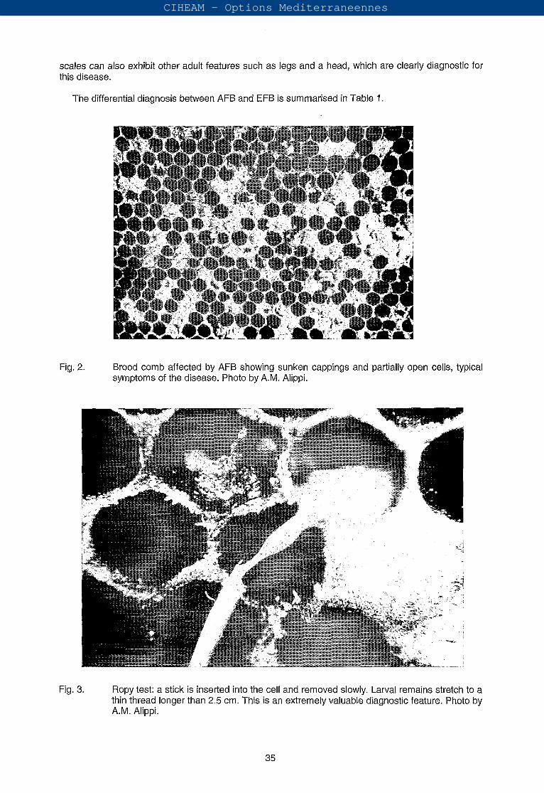
The cappings of cells containing diseased larvae are dark with a greasy appearance and become concave (sunken) with irregular holes, as infection progresses (Fig. 2). When the cappings are removed, dead larvae can be drawn out into a dark ropy thread longer than 2-2.5 cm (Fig. 3). Dead larvae or pupae progressively darken from a straw yellow through chocolate brown to almost black (Fig. 4).
Combs containing diseased larvae have a characteristic glue-pot or sour odour. Finally, after a month or more, the remains of the diseased individuals dry out to form typical dark scales that are brittle and adhere strongly to the cocoons. If death occurs in the pupal stage the formation of the pupal "tongue", a protrusion from the pupal mouthparts that traverses the cell and adheres to the roof, is one of the most characteristics signs of the disease (White, 1920) (Fig. 5). Besides the tongue, the dried
34
CIHEAM - Options Mediterraneennes
scales can also exhibit other adult features such as legs and a head, which are clearly diagnostic for this disease.
The differential diagnosis between AFB and EFB is summarised in Table 1.
Fig. 2. Brood comb affected by AFB showing sunken cappings and partially open cells, typical symptoms of the disease. Photo by A.M. Alippi.
Fig. 3. Ropy test: a stick is inserted into the cell and removed slowly. Larval remains stretch to a thin thread longer than 2.5 cm. This is an extremely valuable diagnostic feature. Photo by A.M. Alippi.
35
CIHEAM - Options Mediterraneennes
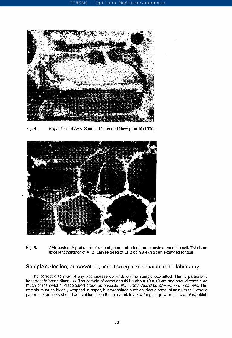
Fig. 4. Pupa dead of AFB. Source: Morse and Nowogrodzki (1990).
Fig. 5. AFB scales. A proboscis of a dead pupa protrudes from a scale across the cell. This is an excellent indicator of AFB. Larvae dead of EFB do not exhibit an extended tongue.
Sample collection, preservation, conditioning and dispatch to the laboratory
The correct diagnosis of any bee disease depends on the sample submitted. This is particularly important in brood diseases. The sample of comb should be about 10 X 10 cm and should contain as much of the dead or discoloured brood as possible. No honey should be present in the sample. The sample must be loosely wrapped in paper, but wrappings such as plastic bags, aluminium foil, waxed paper, tins or glass should be avoided since these materials allow fungi to grow on the samples, which
CIHEAM - Options Mediterraneennes
makes an accurate diagnosis almost impossible. The comb can be mailed in a wooden or stout cardboard box (Shimanuki and Knox, 1991).
~.
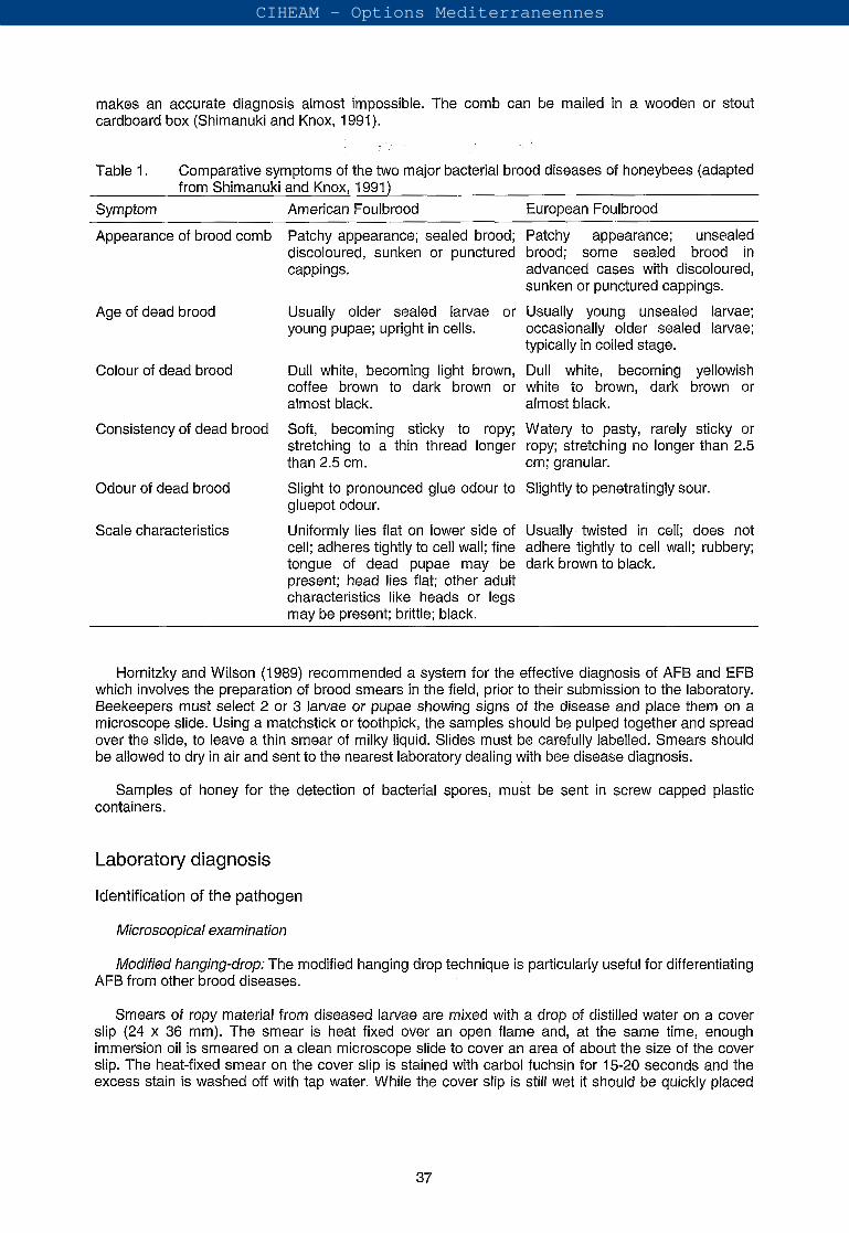
Table 1. Comparative symptoms of the two major bacterial brood diseases of honeybees (adapted
Symptom American Foulbrood European Foulbrood
from Shimanuki and Knox, 1991)
Appearance of brood comb
Age of dead brood
Colour of dead brood
Consistency of dead brood
Odour of dead brood
Scale characteristics
Patchy appearance; sealed brood; Patchy appearance; unsealed discoloured, sunken or punctured brood; some sealed brood in cappings. advanced cases with discoloured,
Usually older sealed larvae or Usually young unsealed larvae; young pupae; upright in cells. occasionally older sealed larvae;
Dull white, becoming light brown, Dull white, becoming yellowish coffee brown to dark brown or white to brown, dark brown or almost black. almost black.
Soft, becoming sticky to ropy; Watery to pasty, rarely sticky or stretching to a thin thread longer ropy; stretching no longer than 2.5 than 2.5 cm. cm; granular.
Slight to pronounced glue odour to Slightly to penetratingly sour. gluepot odour.
Uniformly lies flat on lower side of Usually twisted in cell; does not cell; adheres tightly to cell wall; fine adhere tightly to cell wall; rubbery; tongue of dead pupae may be dark brown to black. present; head lies flat; other adult characteristics like heads or legs may be present; brittle; black.
sunken or punctured cappings.
typically in coiled stage.
Hornitzky and Wilson (1989) recommended a system for the effective diagnosis of AFB and EFB which involves the preparation of brood smears in the field, prior to their submission to the laboratory. Beekeepers must select 2 3 larvae or pupae showing signs of the disease and place them on a microscope slide. Using a matchstick or toothpick, the samples should be pulped together and spread over the slide, to leave a thin smear of milky liquid. Slides must be carefully labelled. Smears should be allowed to dry in air and sent to the nearest laboratory dealing with bee disease diagnosis.
Samples of honey for the detection of bacterial spores, must be sent in screw capped plastic containers.
Laboratory diagnosis
Identification of the pathogen
Microscopical examination
Modified hanging-drop: The modified hanging drop technique is particularly useful for differentiating AFB from other brood diseases.
Smears of ropy material from diseased larvae are mixed with a drop of distilled water on a cover slip (24 X 36 mm). The smear is heat fixed over an open flame and, at the same time, enough immersion oil is smeared on a clean microscope slide to cover an area of about the size of the cover slip. The heat-fixed smear on the cover slip is stained with carbol fuchsin for 15-20 seconds and the excess stain is washed off with tap water. While the cover slip is still wet it should be quickly placed
37
CIHEAM - Options Mediterraneennes
(smear side down) on the slide previously prepared with immersion oil. l h e upper part of the "sandwich" is gently blotted dry with a paper towel.
l h e slide is observed under the microscope using an oil immersion objective, looking for the areas where the water has been trapped between pockets of oil. Only P. l. ssp. larvae spores show a Brownian movement.
The technique can also be performed using a scale instead of ropy material, by leaving a single scale in a drop of distilled water on a coverslip for 2-3 min, then removing it, and fixing and staining the smear as previously described.
This is an extremely valuable diagnostic feature, since spores of Paenibacillus alvei and other species, in a similar mount, generally remain attached to the cover slip and exhibit no motion. Please note however that Brownian movement can be affected by slide preparation. Occasionally debris and other bacteria can exhibit motion and it is then important to take into account the size of the spores.
Single stain: Proceed as in the hanging drop technique but make the smear directly on a microscope slide instead of the cover slip. Heat fix and flood with carbol fuchsin or another suitable bacterial stain for 30 seconds. Wash off the stain and gently blot dry before microscopic examination under an oil immersion objective. This relies solely on differentiating bacteria by their morphology.
Note: If the infection appeared in larvae less than 10 days old, long vegetative rods with coalesced flagella will be present. These flagellar bundles are characteristic of the pathogen.
Immunological techniques
Immunofluorescence: This technique serves to demonstrate the presence of a defined micro- organism in a clinical sample directly (without isolation of the pathogen).
The exposed antigens of the cell wall of a bacterium fixed on a microscope slide will react with specific antibodies prepared against these antigens. As an antibody-covered cell cannot be distinguished from an uncovered cell by light microscopy, antibodies are conjugated with a fluorescent dye and examined by fluorescence microscopy. Otte (1973) developed a direct immunofluorescence technique for the detection of P. l. ssp. larvae by injecting rabbits with bacterial cells from a pure culture. l h e resulting antiserum was collected and conjugated with a fluorochrome dye. Disadvantages reported consisted of difficulties when fixing the smears and some cross reactivity with others spore-forming bacteria. Also it was necessary to use different antibody conjugates for sporal and vegetative forms of the bacterium.
In indirect immunofluorescence non-conjugated specific antibodies, for example produced in a rabbit, react with the bacterial cell wall antigens in the first step. In the second step, the antibody- coated cells are stained with anti-rabbit antibodies labelled with a fluorescent dye, e.g. produced in goat or sheep.
In addition to individual bacterial cells, bacterial colonies on an agar medium can also be stained with fluorescent antibodies.
ELISA: The enzyme-linked-immunosorbent-assay is characterised by using enzymes linked to antibodies to visualise and amplify the serological recognition of homologous antigenic determinants. There are many different types of ELISA, but all use an enzyme-mediated change to indicate the presence of an antigen.
Olsen et al. (1990) developed a diagnostic technique using a monoclonal antibody specific to P. l. ssp. larvae in an indirect ELISA. l h e assay was found to be satisfactory for routine detection of laboratory-grown P. l. ssp. larvae vegetative cells and spores at minimum levels of 1 X cells ml-'. l h e technique was also satisfactory for confirmatory diagnosis of AFB from samples with clinical signs of the disease. Further study is needed to determine whether the assay can detect AFB infections at subclinical levels in colonies.
CIHEAM - Options Mediterraneennes
Microbiological techniques: isolation and precise identification
Brood samples
For the isolation of P. l. ssp. larvae, scales or ropy larval remains (from a single cell) are ' suspended in 5 ml sterile distilled water and kept at room temperature for 10 min, then heat-shocked
10-15 min in order to kill non-spore-forming bacteria. After vortex mixing, 200 pl of the suspension is spread by means of sterile cotton swabs over the surface of the specific culture medium. The plates are incubated at 36-37'" in an atmosphere of 10% CO2 in air (microaerophilic conditions). Routine culture media, such as nutrient agar, will not support the growth of P. l. ssp. larvae. The micro-organism requires media rich in organic growth factors, such as MYPGP agar (Dingman and Stahly, 1983), J-agar (Gordon et al., 1973), brain heart infusion agar fortified with thiamine (BHITA) (Shimanuki and Knox, 1991) or the medium recommended by Bailey and Lee (1962). Also, blood based media are suitable for vegetative growth, such as Columbia agar with 10% sheep's blood (Lloyd, 1986) and SBA (sheep blood agar) (Hornitzky and Karlovskis, 1989). Best sporulation occurs on the medium used by Dingman and Stahly (1 983).
When samples are contaminated with secondary bacterial invaders, mainly Paenibacillus alvei, plates of suitable isolation media containing nalidixic acid at a final concentration of 6 pg ml-' are used (Alippi, 1991).
I Honey
Several methods for the isolation and cultivation of spores of P. l. ssp. larvae from honey have been developed. Hansen (1984a,b) developed a technique to detect P. l. ssp. larvae spores by the direct inoculation of undiluted honey samples previously heated to Shimanuki and Knox (1 988) reported a method that included dialysis, centrifugation, resuspension and heat treatment of honey before inoculating onto BHITA plates. Hornitzky and Clark (1 991) described a procedure which involved the centrifugation of diluted honey samples, heat treatment of the sediment and culture on SBA plates containing 3 pg ml-' nalidixic acid (SBANal) to prevent the development of motile colonies of P. alvei. The incorporation of nalidixic acid into the culture medium inhibits the growth of P. alvei but other Paenibacillus and Bacillus species may overgrow the plates making the correct diagnosis of P. 1. ssp. larvae almost impossible. A semi-selective medium for the detection of P. 1. ssp. larvae spores in severely contaminated honeys was developed in Argentina (Alippi, 1995). The technique involves the dilution of honey samples (1:2) in phosphate buffer, concentration of spores by centrifugation and heat treatment prior to inoculation onto J-agar to which nalidixic acid and pipemidic acid are added. Plates are incubated at in an atmosphere of 5% CO2 in air for up to 7 days. Recently, Nördstrom and Fries (1995) reported the superiority of MYPGP to other media for the detection of low levels of P. l. ssp. larvae spores in honey. They also demonstrated that incubation of plates in an atmosphere of 5% CO2 enhanced the growth considerably.
Adult bees
Hornitzky and Karlovskis (1989) developed a culture technique which provides a rapid means of detecting P. 1. ssp. larvae spores in adult bees that could act as a source of AFB infection for young larvae. Each sample of 30 nurse bees is homogenised in 20 ml sterile PBS for 30 s. The homogenate is filtered and centrifuged and the pellet resuspended in PBS. The samples are heat-shocked and plated onto a suitable culture medium supplemented with nalidixic acid to inhibit the spread of P. alvei colonies.
Spores of P. l. ssp. larvae have also been recovered from contaminated beeswax treated with benzene (Máchová, 1993) and from pollen filtrates (Gochnauer and Corner, 1987).
Identification of the causal agent
P. 1. ssp. larvae vegetative cells are Gram-positive slender rods with a tendency to form chains of variable length, from about 0.5-0.6 pm wide by 2.3-5.0 pm long (Fig. 6). Spores are ellipsoidal in
39
CIHEAM - Options Mediterraneennes
shape, measuring about 0.5-0.7 pm wide by 1.3-1.5 pm long. When stained with carbol fuchsin, the spore walls appear reddish purple while the centres remain clear. Also, f . l. ssp. larvae spores are easily distinguishable from other species by their surface configuration as seen by scanning electron microscopy (SEM) (Alippi, 1991). Spores have a definite smooth surface, are highly refractile and without traces of sporangia (Figs 7 and 8).
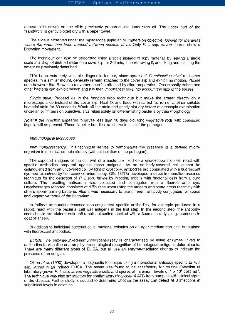
Fig. 6. Scanning electron micrograph of vegetative cells of faenibacillus larvae ssp. larvae, showing peritrichous flagella and flagellar bundles. Bar: 10 pm. Photo by A.M. Alippi.
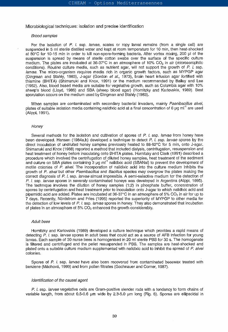
Fig. 7. Free spores of faenibacillus larvae ssp. larvae showing smooth surfaces and without sporangia remnants as seen by scanning electron microscope. Bar: 1 pm. Photo: reproduced with permission from Journal of Apicultura/ Research, 75-80 (1 991), a publication of the International Bee Research Association, 18 North Road, Cardiff, CF1 3BY, UK.
40
CIHEAM - Options Mediterraneennes
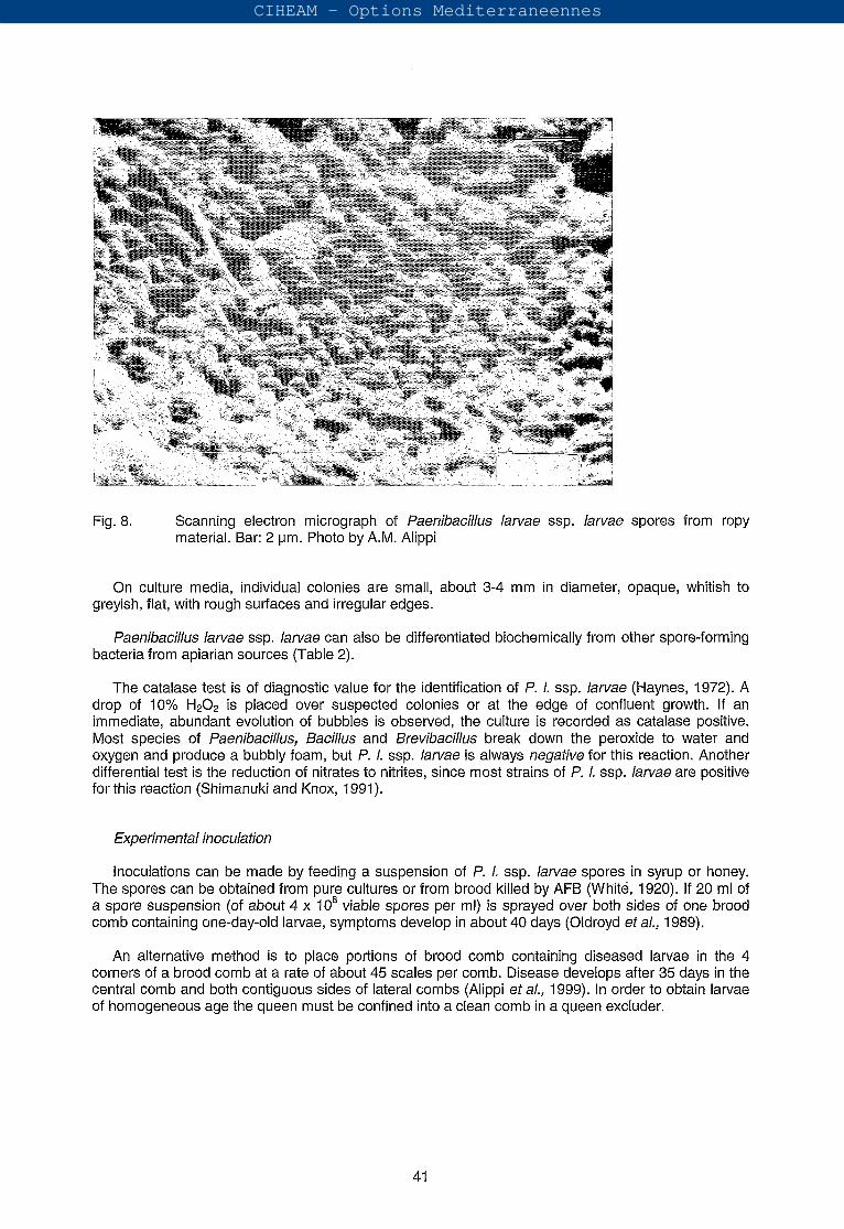
Fig. 8. Scanning electron micrograph of Paenibacillus larvae ssp. larvae spores from ropy material. Bar: 2 pm. Photo by A.M. Alippi
On culture media, individual colonies are small, about 3-4 mm in diameter, opaque, whitish to greyish, flat, with rough surfaces and irregular edges.
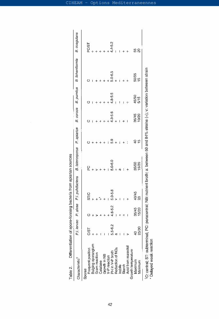
Paenibacillus larvae ssp. larvae can also be differentiated biochemically from other spore-forming bacteria from apiarian sources (Table 2).
The catalase test is of diagnostic value for the identification of P. l. ssp. larvae (Haynes, 1972). A drop of 10% is placed over suspected colonies or at the edge of confluent growth. If an immediate, abundant evolution of bubbles is observed, the culture is recorded as catalase positive. Most species of Paenibacillus, Bacillus and Brevibacillus break down the peroxide to water and oxygen and produce a bubbly foam, but P. l. ssp. larvae is always negative for this reaction. Another differential test is the reduction of nitrates to nitrites, since most strains of P. l. ssp. larvae are positive for this reaction (Shimanuki and Knox, 1991).
Experimental inoculation
Inoculations can be made by feeding a suspension of P. l. ssp. larvae spores in syrup or honey. The spores can be obtained from pure cultures or from brood killed by AFB (Whité, 1920). If 20 ml of a spore suspension (of about 4 X viable spores per ml) is sprayed over both sides of one brood comb containing one-day-old larvae, symptoms develop in about 40 days (Oldroyd et al,, 1989).
An alternative method is to place portions of brood comb containing diseased larvae in the 4 corners of a brood comb at a rate of about 45 scales per comb. Disease develops after 35 days in the central comb and both contiguous sides of lateral combs (Alippi et al., 1999). In order to obtain larvae of homogeneous age the queen must be confined into a clean comb in a queen excluder.
41
CIHEAM - Options Mediterraneennes
42
CIHEAM - Options Mediterraneennes
Routine diagnosis
Fluorescence of a scale: AFB scales fluoresce when examined under ultraviolet light at a wavelength of 360 nm. Pollen and honey also fluoresce.
Ropy test: A stick is inserted into the suspect cell and then withdrawn; the infected larva sticks tenaciously and the contents can be drawn out into a long thread or rope, longer than 2.5 cm (Table 1 and Fig. 3).
Holst milk test: This simple test is based on the fact that high levels of proteolytic enzymes are produced by sporulating P. l. ssp. larvae. The test is conducted by suspending a scale or a smear of a diseased larva in a tube containing 3-4 ml of 1% powdered skimmed milk in water. After incubating for 10-20 min P. l. ssp. larvae spores are present (Shimanuki and Knox, 1991). However, scales may give negative results when they have been in combs that have been fumigated with formaldehyde or paradichlorbenzene, and sometimes for unknown reasons. Also, dead larvae that have not reached the ropy stage do not give a positive reaction (Bailey and Ball, 1991).
Microscopic examination of diseased larvae: The modified hanging drop technique is the most widely used for routine diagnosis of AFB.
Treatments and prophylaxis
Disinfection of contaminated materials
Physical means
Burning ofcolonies: One reliable method of dealing with contaminated colonies is to burn them and bury the remains. Bees from an infected colony should be killed with an insecticide such as the synthetic pyrethroids, or petrol (gasoline) when the foragers have returned and all combs, bees, and hive equipment should be burned in a pit. The ashes should be covered once the fire has burnt out (Morse and Shimanuki, 1990; Matheson and Reid, 1992).
Scorching: Scorching the inner hive parts has been used as a sterilisation method. Empty boxes are stacked up to create a chimney and then petrol-soaked straw is ignited inside the base. Another method is to use a blowtorch for scorching lids, floors and the inside of hive boxes. Although officially recommended in many countries, the method is only partially effective (Matheson and Reid, 1992).
Dipping in paraffin wax: Bees, combs and honey are normally destroyed at the apiary site. Contaminated wooden equipment in good condition (but not including combs or frames) is immersed for 1 O min in paraffin wax This method has been used effectively in New Zealand for the last 50 years (Matheson and Reid, 1992).
Gamma radiation: Gamma radiation from cobalt-60 is a reliable method of sterilising contaminated combs and all wooden equipment. Pollen used for feeding bees and honey used in queen candies can also be treated in this way. Diseased colonies should be free of honey and adult bees must be killed before sending the equipment to the irradiation centre (Matheson and Reid, 1992).
Chemical means: Gas
Ethylene oxide (EtO) sterilises material infected with AFB. However, Et0 is not commercially applied, mainly due to the high cost of treatments, flammability of the gas mixtures used, carcinogenic residues of Et0 and incomplete efficacy of the process (Matheson and Reid, 1992; Ratnieks, 1992). Methyl bromide (MeBr) has also been used to disinfect contaminated equipment, although MeBr is highly neurotoxic.
43
CIHEAM - Options Mediterraneennes
Chemical means: Liquid
Lye bath: A boiling lye solution (450 g of 100% NaOH in 38 litres of water) is useful for the removal of contaminated wax and propolis from frames (after the wax comb has been cut away), bottom boards and supers. They must be completely immersed for 10-20 min (Morse and Shimanuki, 1990).
Sodium hypochlorite: A 2 chlorometric degree solution (one litre of the solution can liberate two chlorine gas litres), mixed with 0.5% of a wetting agent is used for disinfecting hive tools and other small items of equipment (Faucon et al,, 1980).
Ways of saving bees
Shaking bees
This is a little-used way of saving adult bees from an infected colony. The method consists of shaking the bees by transferring the adults (including the queen) to a disease-free nucleus without drawn comb. The honey contaminated with spores carried by the bees, is consumed during comb building (Morse and Shimanuki, 1990). An optional measure that increases the effectiveness of this method is to feed a preventive treatment of antibiotic to the new hive.
Chemotherapy
AFB infected colonies can be treated with antibiotics to suppress disease signs so that the colony can still produce honey. Antibiotics are only effective against vegetative forms; spores are not killed by these drugs. The efficacy of drug treatment varies greatly but chemotherapy becomes economically attractive when the disease is widespread. However, it is not recommended when the incidence of AFB is low and can be contained economically by the destruction of relatively few colonies (Bailey and Ball, 1991).
Oxytetracycline hydrochloride (OTC) and sodium sulfathiazole have been used in many countries for the control of AFB. Tylosin tartrate is also highly effective for the treatment of AFB (Hitchcock et al., 1970; Peng et al., 1996; Alippi et al., 1999).
There are 4 techniques for applying antibiotics: dusting, bulk feeding, extender patties and paperpacks (Morse and Shimanuki, 1990). Dusting application is made by mixing antibiotic powder with fine powdered sugar and sprinkling over the top bars of brood frames at intervals of 4-5 days applications). Bulk feeding is the feeding of medicated sugar syrup to a colony. Extender patties are made of sugar, vegetable oil and antibiotic in a proportion of 7:3:1; they are placed over the top bars of the brood combs and are consumed over a period of 6-8 weeks. A variation of the extender patty is the paper pack which consists of a dry mixture of antibiotic and powdered sugar inside an absorbent paper bag; bees take about 1 week to remove the paper and consume the drug. The antibiotic extender patty is the most effective treatment; the paper pack is less effective, but more effective than dusting. For bulk feeding the preparation of an OTC solution just before use is recommended, because OTC has a tendency to break down in sugar solution (Matheson and Reid, 1992). For more detailed information regarding doses and application consult appropriate sections in Morse and Shimanuki (1990) and Bailey and Ball (1991).
Genetic control of AFB
Selection of appropriate honeybee stock could increase the ability of colonies to resist AFB. Rothenbuhler and co-workers (reviewed by Rothenbuhler, 1964) carried out an extensive investigation of the hygienic behaviour of worker bees engaged in nest cleaning activities. Hygienic bees can be defined as bees with a strong tendency to uncap cells containing dead larvae or pupae and to remove them from the nest. Uncapping and removing behaviours were each found to be under the control of a single genetic locus, and are expressed as recessives. Obviously, the recessive alleles which allow the behaviour to express, occur at low frequencies in natural populations. Natural selection has favoured the spread of the dominant alleles, presumably because the biological cost of the trait
44
CIHEAM - Options Mediterraneennes
exceeds its benefits. Workers of colonies exhibiting the behaviour frequently uncap and remove even healthy larvae. Only in areas of high AFB incidence is the benefit of the hygienic behaviour likely to exceed the cost (Seeley, 1985). In addition, Rothenbuhler and colleagues demonstrated that numerous other hereditary factors contribute towards resistance, such as the rate at which young larvae become resistant with increasing age; the efficiency of adults in filtering P. l. ssp. larvae spores by means of their proventriculus (Fig. 1) and the role of bactericidal factors in the gland secretions of nurse bees (Bailey and Ball, 1991).
Prophylaxis
As beekeepers are probably the major cause of the spread of AFB, they must be motivated to keep disease incidence as low as possible. They can break the transmission cycle in a number of ways (Matheson and Reid, 1992; Alippi, 1996):
(i) Do not incorporate swarms of unknown origin into the apiary. Preferably, leave the colonies in quarantine at a distant site, isolated from other colonies, for at least 3 months before introducing them into the apiary.
(i¡) Buy nucleus colonies, package bees, colonies, queens and queen cells only from registered establishments with the corresponding sanitary certification. In the case of package bees or attendant workers in queen cages it is important to check for possible surface carriage of bacterial spores by the adult bees.
(iii) Do not buy hives or second-hand equipment without checking carefully for AFB.
(¡v) Do not feed bees with honey or pollen of unknown origin (feed sugar syrup or gamma irradiated pollen instead).
(v) During spring and autumn, colonies must be opened and brood combs should be examined for any abnormalities, taking into account that early detection will prevent the disease spreading.
(vi) Be very careful with the management of infected colonies and thoroughly clean all materials that were in contact with them (gloves, hive tools, coveralls, smokers, etc.).
(vii) Do not mix contaminated with clean material.
(viii) Try to avoid drifting or robbing between colonies.
(ix) Do not exchange brood combs between different brood chambers without carefully checking for AFB symptoms.
(X) Do not leave honey or honey combs accessible to bees because robbing is one of the greatest sources of contamination.
(xi) Do not make increase from diseased colonies or suspect ones. First verify their sanitary condition.
(xi) Do not move colonies from apiaries where AFB infection is established.
Efficacy, secondary effects on bees and post-therapeutic residues
A great deal of controversy exists concerning the feeding of antibiotics to colonies for the prevention of AFB. It has been shown that preventive OTC treatments effectively mask all symptoms, with the consequent risk of spreading the disease by the movement of infected materials around apiaries (Oldroyd et al., 1989).
45
CIHEAM - Options Mediterraneennes
In the USA, the only antibiotic that can be legally used is OTC; although sodium sulfathiazole had previously been used for autumn applications, it is no longer approved because the drug is stable in honey for several years.
An inappropriate use of antibiotics may lead to antibiotic resistance of P. l. ssp. larvae strains and honey contaminated with residues of these drugs may reach the market place. Strains of P. l. ssp. larvae resistant to OTC and to sodium sulfathiazole have been reported in Poland (Glinski and Rzedzicki, 1977a,b), Germany (Plagemann, 1991) and Argentina (Alippi, 1996).
In addition, OTC has been reported to be toxic for larvae (Peng et al., 1992) and for adult bees (Alippi et al., 1996a). Tylosin tartrate is an alternative to the use of OTC, because its toxicity for larvae (Peng et al., 1996) or adult bees (Alippi et al., 1999) is negligible. In addition, its degradation time in honey stored in brood combs, as measured by HPLC, is about 60 days which is similar to the rate reported for OTC (Matzuka and Nakamura, 1990).
Recently, natural compounds such as essential vegetable oils (Calderone et al., 1994; Alippi ef al., 1996) and fatty acids (Feldlaufer et al., 1993) have been reported to be effective for limiting the growth of P. 1. ssp. larvae strains in vitro. Further studies are needed in order to determine their effectiveness, appropriate doses and mode of application in honeybee colonies. The use of essential oils and fatty acids for the control of AFB would represent a safe alternative to antibiotics.
European Foulbrood disease
Definition of the disease
European foulbrood is an infectious and contagious disease affecting primarily young larvae (less than 48 h old) but in long established infections, also capped larvae. In the latter case, symptoms are often confused with AFB. However, the causal organism, Melissococcus pluton, does not form spores, and so the disease is believed to be less problematic and often curable.
General epidemiology
EFB attacks the larvae of Apis mellifera (A. m. mellifera; A. m. ligustica; A. m. carnica; A. m. scutellata), A. cerana and A. laboriosa. The disease is found all over the world except for New Zealand (Matheson, 1993, 1996).
Several bacteria may be associated with EFB and most have been considered at one time or another to be the cause of the disease (Bailey, 1983). In 1885, Cheshire and Cheyne published the first description of the disease and believed that the causal agent was Bacillus alvei. White refuted this statement and considered that EFB was caused by a non-spore-forming, uncultivable, Gram-positive organism that he called Bacillus Y and later named Bacillus pluton (Bailey, 1983). After many years of confusion, the question was resolved by Aleksandrova, (1949) who proved that B. pluton was the causal organism of EFB, by fulfilling In 1956 Bailey suggested that the bacterium should be renamed Streptococcus pluton on the basis of the Gram reaction and morphology. (Shimanuki, 1990).
European foulbrood (EFB) is not considered to be a serious disease by most beekeepers. However, in some areas and under certain conditions, EFB has been known to cause severe losses of brood, resulting in lower honey yields. Often, the disease arises in mid to late spring, when colonies are building up to their maximum population (Shimanuki, 1990). Sometimes it is also found in autumn.
Etiology
Pathogenic agent
As Streptococcus pluton differed sufficiently from the type species of the genus, (S. pyogenes), Bailey and Collins (1982) proposed the creation of a new genus, Melissococcus. The current taxonomic position of the causal agent of EFB is Melissococcus pluton (White) Bailey and Collins.
46
CIHEAM - Options Mediterraneennes
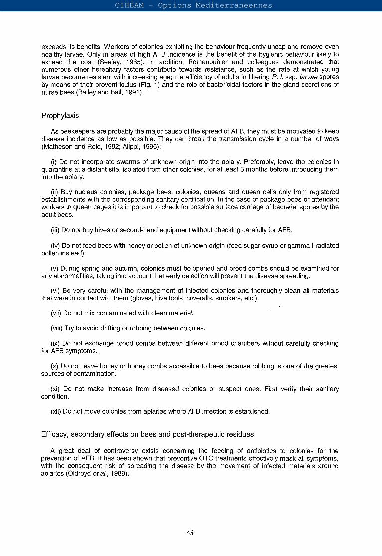
Melissococcus pluton is a Gram-positive, non-spore forming organism. Melissococcus pluton cells are lanceolate cocci occurring singly, in pairs or chains resembling the beads of a rosary, size: 0.5-0.7 pm in width by 1.0 pm in length (Fig. 9). When using a single stain, the cells take up the stain evenly and no unstained areas are detected. In culture, small (up to 1 mm in diameter), white and opaque colonies appear after 4 days of incubation in anaerobic conditions. Melissococcus pluton is very pleomorphic in culture, often being in rod-like forms, particularly when cultures are stored for a few weeks.
Fig. 9. Cells of Melissococcus pluton, the causal agent of EFB from larval remains, as seen by scanning electron microscopy. Bar: 1 pm. Photo: reproduced with permission from Journal Apicultura/ Research, 30(2): 75-80 (1991), a publication of the International Bee Research Association, 18 North Road, Cardiff, CF1 3DY, UK.
Organisms associated with EFB
Melissococcus pluton is generally observed early in the infection cycle before the appearance of the various other bacteria associated with this disease. The secondary invaders that do not cause EFB but influence the odour and consistency of the dead brood include the following:
(i) Lactobacillus eurydice (= Achromobacter eurydice = Bacterium eurydice): Non spore-forming bacterium frequently found in larvae affected by EFB. It is a common inhabitant of the alimentary tract of adult bees and the midgut of healthy larvae but it is much more numerous in larvae infected by M. pluton. The cells are square-ended rods, occurring singly or in chains measuring about 0.4-0.7 pm in width by 0.5-1.4 pm in length. It is Gram-positive in vivo and Gram-variable in culture, which sometimes explains the confusion with other species. It is pleomorphic in culture, taking the form of rods or streptococci, according to its culture medium. It has been grouped with the genera Corynebacterium and Lactobacillus, but its current taxonomic position is still undefined.
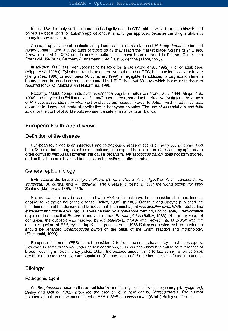
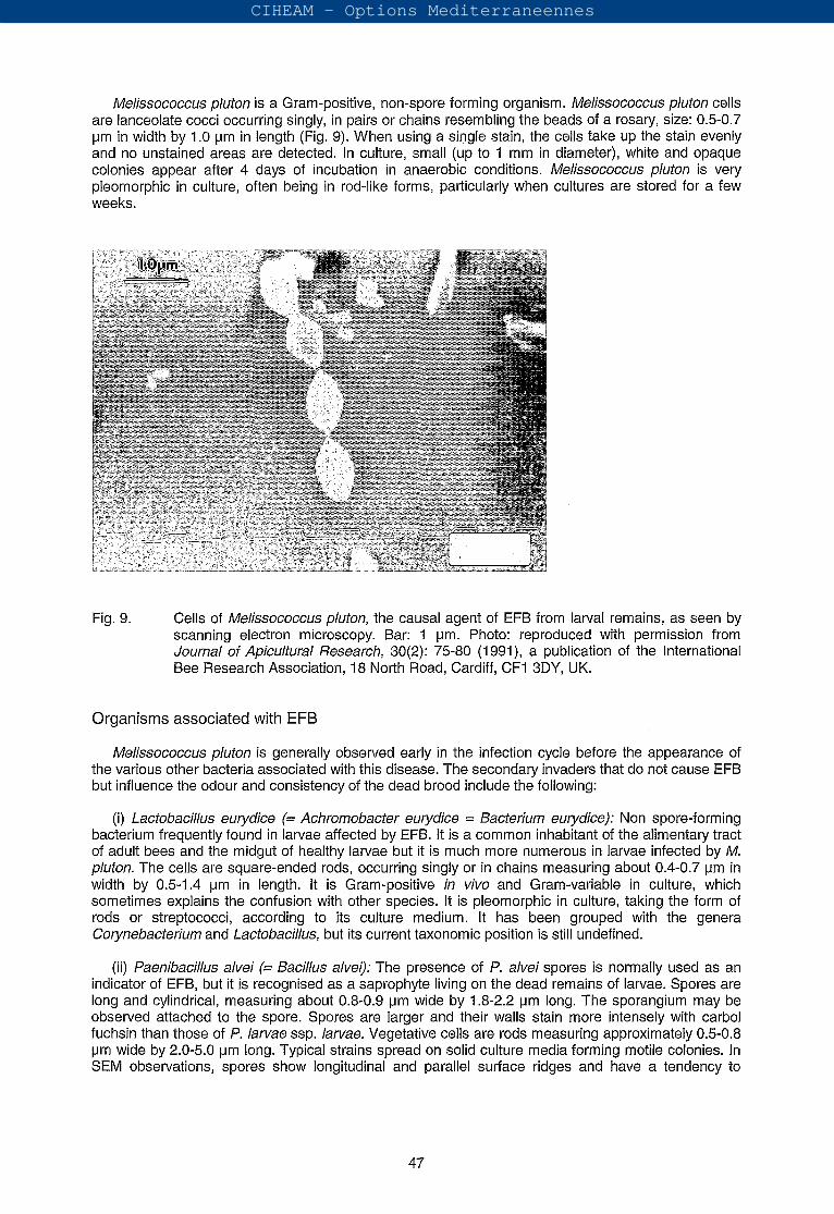
(i¡) Paenibacillus alvei (= Bacillus alvei): The presence of P. alvei spores is normally used as an indicator of EFB, but it is recognised as a saprophyte living on the dead remains of larvae. Spores are long and cylindrical, measuring about 0.8-0.9 pm wide by 1.8-2.2 pm long. The sporangium may be observed attached to the spore. Spores are larger and their walls stain more intensely with carbol fuchsin than those of P. larvae ssp. larvae. Vegetative cells are rods measuring approximately 0.5-0.8 pm wide by 2.0-5.0 pm long. Typical strains spread on solid culture media forming motile colonies. In SEM observations, spores show longitudinal and parallel surface ridges and have a tendency to
47
CIHEAM - Options Mediterraneennes
exhibit a characteristic side-by-side arrangement (Fig. 10). Paenibacillus alvei cultures are Gram- variable and their growth produces an unpleasant odour.
Fig. Spores of Paenibacillus alvei in a typical side-by-side arrangement as seen by SEM. Bar: 2 pm. Photo by A.M. Alippi.
(iii) Brevibacillus laterosporus (= Bacillus laterosporus = Bacillus orpheus): This spore-forming organism is occasionally found in larvae affected by EFB. It has recently been reclassified into the new genus Brevibacillus (Shida et al., 1996). An important feature is the production of a canoe-shaped or C-shaped parasporal body that stains intensely along one side and at both ends and remains attached to the ellipsoidal spore after lysis of the sporangium. Spores measure 1 .O-1.3 pm wide by 1.2-1.6 pm long and the rods 0.5-0.8 pm by 2.0-2.5 pm. Their surface configuration is unique with well-defined branching ridges (Fig. 11). Cultures are Gram-variable.
(¡v) Enterococcus faecalis (= Streptococcus faecalis = Streptococcus apis = Streptococcus liquefaciens): The cells of this Gram-positive organism resemble those of M. pluton; they are ovoid, 0.5-1 .O pm in diameter and occur in pairs or short chains. It causes a sour smell, hence the German name Sauerbrut. Growth occurs on nutrient agar usually within one day, in contrast to M. pluton, which requires special culture media and anaerobic conditions.
(v) Paenibacillus apiarius (= Bacillus apiarius): This spore-forming bacterium is rarely encountered and may not be truly associated with EFB. It was recently reclassified within the genus Paenibacillus (Nakamura, 1996).The spore has a ridged, thick, rectangular coat (Fig. 12) and the remnants of the sporangium remain attached for a long time. Spores are 0.8-1.1 -pm wide by 1.5-2.0 pm long and vegetative cells 0.7-0.8 pm wide by 3.0-5.0 pm long. Gram staining is variable.
disease
The disease becomes a real problem in colonies deficient in proteins. The deficiency can be due not only to a lack of pollen but also to an imbalance between the number of nurse bees and the number of larvae to be fed. The poor nutrition of larvae can also be due to the inability of young bees to produce a normal quantity of royal jelly, when they are infected with sacbrood virus. The disease seems to occur more frequently in certain strains of bees and when the queen is old.
48
CIHEAM - Options Mediterraneennes
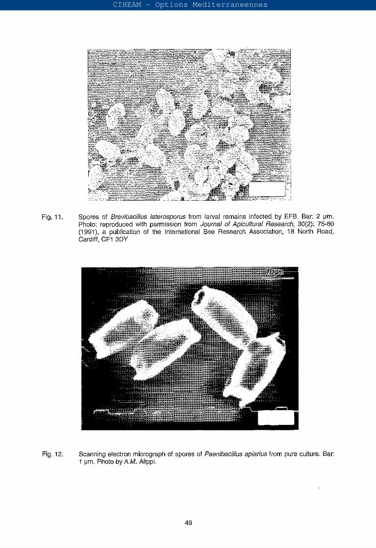
Fig. 11. Spores of Brevibacilhs laterosporus from larval remains infected by EFB. Bar: 2 pm. Photo: reproduced with permission from Journal of Apicultura/ Research, 30(2): 75-80 (1991), a publication of the International Bee Research Association, 18 North Road, Cardiff, CF1 3DY
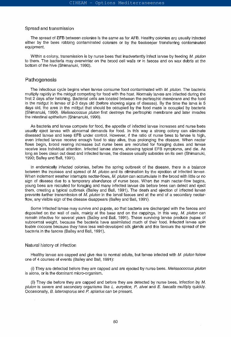
Fig. 12. Scanning electron micrograph of spores of faenibacihs apiarius from pure culture. Bar: 1 pm. Photo by A.M. Alippi.
49
CIHEAM - Options Mediterraneennes
and
The spread of EFB between colonies is the same as for AFB. Healthy colonies are usually infected either by the bees robbing contaminated colonies or by the beekeeper transferring contaminated equipment.
Within a colony, transmission is by nurse bees that inadvertently infect larvae by feeding M. pluton to them. The bacteria may overwinter on the brood cell walls or in faeces and on wax debris at the bottom of the hive (Shimanuki, 1990).
Pathogenesis
The infectious cycle begins when larvae consume food contaminated with M. pluton. The bacteria multiply rapidly in the midgut competing for food with the host. Normally larvae are infected during the first 2 days after hatching. Bacterial cells are located between the peritrophic membrane and the food in the midgut in larvae of 2-3 days old (before showing signs of disease). By the time the larva is 5 days old, the area in the midgut that should be occupied by the food mass is occupied by bacteria (Shimanuki, 1990). Melissococcus pluton first destroys the peritrophic membrane and later invades the intestinal epithelium (Shimanuki, 1990).
As bacteria and larvae compete for food, the appetite of infected larvae increases and nurse bees usually eject larvae with abnormal demands for food. In this way a strong colony can eliminate diseased larvae and keep EFB under control. However, if the ratio of nurse bees to larvae is high, even infected larvae receive enough food to stay alive, thus prolonging the disease. When nectar flows begin, brood rearing increases but nurse bees are recruited for foraging duties and larvae receive less individual attention. Infected larvae starve, showing typical EFB symptoms, and die. As long as bees clean out dead and infected larvae, the disease usually subsides on its own (Shimanuki, 1990; Bailey and Ball, 1991).
In endemically infected colonies, before the spring outbreak of the disease, there is a balance between the increase and spread of M. pluton and its elimination by the ejection of infected larvae. When inclement weather interrupts nectar-flows, M. pluton can accumulate in the brood with little or no sign of disease due to a temporary abundance of nurse bees. When the main nectar-flow begins, young bees are recruited for foraging and many infected larvae die before bees can detect and eject them, creating a typical outbreak (Bailey and Ball, 1991). The death and ejection of infected larvae prevents further transmission of M. pluton in the larval faeces and at the end of a secondary nectar- flow, any visible sign of the disease disappears (Bailey and Ball, 1991).
Some infected larvae may survive and pupate, so that bacteria are discharged with the faeces and deposited on the wall of cells, mainly at the base and on the cappings. In this way, M. pluton can remain infective for several years (Bailey and Ball, 1991). These surviving larvae produce pupae of subnormal weight, because the bacteria have assimilated much of their food. Infected larvae spin feeble cocoons because they have less well-developed silk glands and this favours the spread of the bacteria in the faeces (Bailey and Ball, 1991).
of infection
Healthy larvae are capped and give rise to normal adults, but larvae infected with M. pluton follow one of 4 courses of events (Bailey and Ball, 1991):
(i) They are detected before they are capped and are ejected by nurse bees. Melissococcus pluton is alone, or is the dominant micro-organism.
(i¡) They die before they are capped and before they are detected by nurse bees. Infection by M. pluton is severe and secondary organisms like L. eurydice, P. alvei and faecalis multiply quickly. Occasionally, B. laterosporus and P, apiarius can be present,
50
CIHEAM - Options Mediterraneennes
(iii) They are capped and fail to pupae, but usually void most of their intestinal contents. If P. alvei is present, it multiplies in the larval remains and is found in almost pure culture in the remaining scale.
(¡v) They pupate and form undersized or sometimes normal adults, leaving infective cells of M. phton in their faecal deposits in the brood cell.
Clinical diagnosis (field symptoms)
EFB usually affects young larvae which die while still coiled. They turn yellow at first and then brown, at which time the tracheal system becomes quite visible. Larvae also assume unnatural positions in the cells. The larvae eventually decay to a point where they form dry rubbery scales which are easier to remove than those caused by AFB. The odour of larvae infected with EFB varies with the presence of saprophytes, but it is usually described as a sour smell or like the odour of rotten fish.
Prior to forming a dry scale, the larvae become soft and granular and can be tested for "ropiness" which never occurs with EFB. The ropiness test is a key distinguishing characteristic between EFB and (see also Table 1).
Before they decompose, diseased or dead larvae can be dissected easily on a microscope slide by grasping the cuticle at the centre of the body with two pairs of forceps, which are then pulled apart. The midgut contents are left exposed within the transparent peritrophic membrane which is filled with bacteria in opaque chalk-white clumps (Bailey and Ball, 1991).
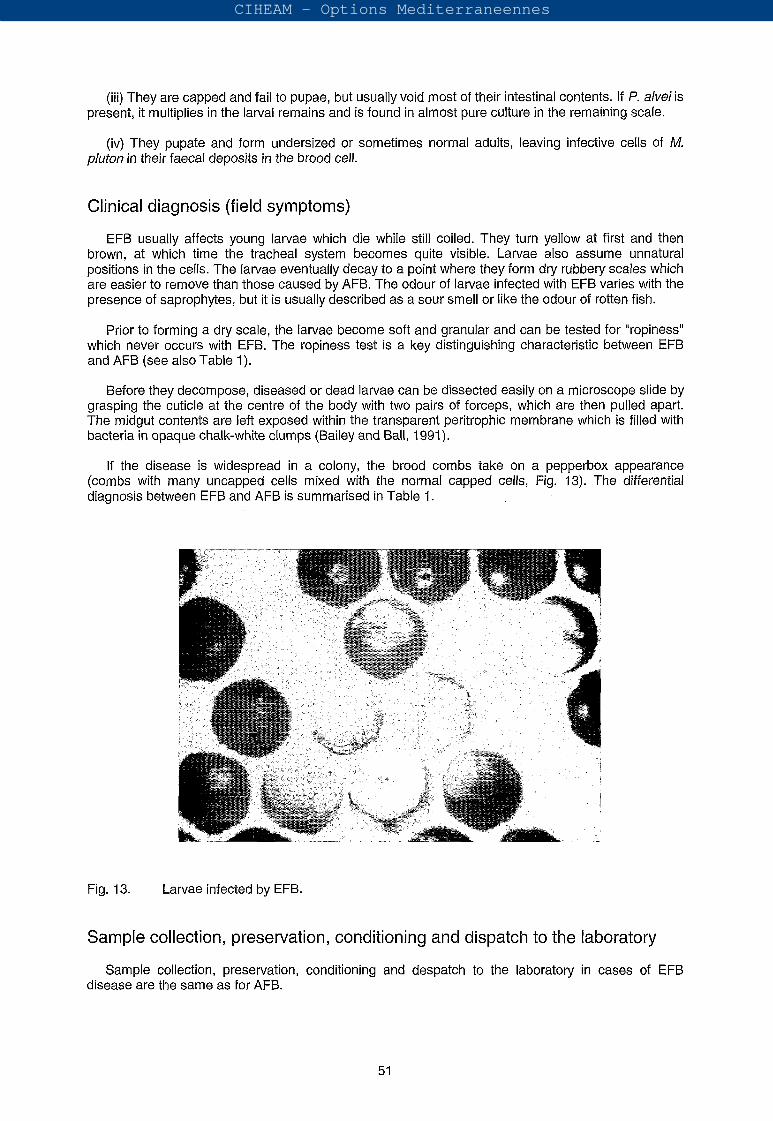
If the disease is widespread in a colony, the brood combs take on a pepperbox appearance (combs with many uncapped cells mixed with the normal capped cells, Fig. 13). The differential diagnosis between EFB and AFB is summarised in Table 1.
Fig. 13. Larvae infected by EFB.
Sample collection, preservation, conditioning and dispatch to the laboratory
Sample collection, preservation, conditioning and despatch to the laboratory in cases of EFB disease are the same as for AFB.
51
CIHEAM - Options Mediterraneennes
Laboratory diagnosis
Identification of the pathogen
Microscopical examination
Negative stain: This technique is routinely used to differentiate M. pluton and other bacteria commonly found as secondary invaders in preparations of diseased larvae with EFB signs. Prepare a turbid suspension of individual diseased larvae in distilled water or leave a scale in distilled water for 5-
min. Place a drop of the suspension on a clean microscope slide and mix with a drop of nigrosin solution (5% nigrosin in distilled water w/v). Spread the smear and heat-fix under a lamp. After the smear is completely dry, it can be examined under an oil immersion objective by placing a drop of immersion oil directly on the smear (without using cover slip). The spores of P. alvei appear bright and the vegetative cells grey, against the dark background of the preparation. Melissococcus pluton cells may not be numerous in smears from infected larvae but they exhibit the typical lanceolate shape and not the elongated rod-like form seen in culture (Allen and Ball,
Immunological techniques
Pinnock and Featherstone developed an ELISA for the detection and quantification of M. pluton in diseased larvae and adult bees. Mm pluton cells from pure cultures were used as the antigen for intravenous injection into rabbits. The animals were bled and the hyperimmune serum was separated and purified to yield the y-globulin fraction. ELISA plates were pre-coated with the y-globulin fraction and clarified homogenates of larvae and adults from apparently healthy colonies extracted in phosphate buffered saline (PBS), were used as antigens. In this way, Pinnock and Featherstone
were able to demonstrate the presence of M. pluton in asymptomatic colonies. The detection threshold in hornogenates of adults and larvae was cells per ml, two orders of magnitude more sensitive than microscopica1 techniques.
Allen and Ball prepared antisera in rabbits in a similar way and the serum was tested against pure cultures of M. pluton by tube agglutination.
Microbiological techniques: isolation and precise identification
It is difficult to isolate M. pluton in culture due to its growth requirements and competition from other bacteria usually present in the sample. It may be isolated from diseased or dead larvae, from their cell cappings or from dry smears of diseased larval midguts on a specific agar medium (Bailey and Ball,
Watery suspensions of the selected material, are streaked onto plates of the special agar. It is recommended to prepare plates for each extract and to incubate one aerobically and the other anaerobically at for 4 days. As M. pluton is a microaerophilic to anaerobic organism that requires
0% CO2, it will grow only on the plates incubated under anaerobic conditions. Small, white, opaque colonies of M. pluton usually appear after 4 days incubation. If E. faecalis is present, it will grow abundantly on the aerobic plates producing small, transparent, grey colonies in h (Bailey and Ball,
It is always necessary to prepare a smear stained with nigrosin from suspected colonies. M. pluton is very pleomorphic in culture, sometimes being in rod-like forms, particularly when cultures are stored for a few weeks.
Routine diagnosis
Microscopical examination of diseased larvae is commonly used for routine diagnosis.
CIHEAM - Options Mediterraneennes
Treatments and prophylaxis
Treatment of EFB is generally less drastic than for AFB. Only in very severe cases must colonies be destroyed by burning, but this is not very effective in reducing disease incidence and it is certainly uneconomical (Bailey and Ball, 1991). Treatment is not required if the infection is slight, because in these cases most colonies can overcome the disease without assistance (Shimanuki, 1990).
The following management techniques reduce the effects of EFB:
(i) Requeening with more resistant stock will clear up the disease. This provides a break in the brood cycle, allowing nurse bees to remove infected larvae and introduces new genetic material. This must be done quickly with as brief a queenless interval as possible.
(i) Addition of a frame of both young and mature brood from a healthy colony and feeding with 1:l sugar syrup. The new brood competes with the infected larvae for the attention of nurse bees and the syrup stimulates production of new brood which also competes for food. In this way, sick larvae are removed earlier, avoiding the dissemination of M. pluton in their faeces.
(iii) Avoid the shortage of pollen when young brood is abundant.
(¡v) Avoid stress. The amount of stress that a colony suffers is correlated with the development of EFB.
Treatment with OTC suppresses the signs of EFB. The methods of application are the same as for AFB, but doses are lower. Wilson (1962) reported the efficacy of erythromycin for the control of EFB. Sodium sulfathiazole has no effect on this disease. For more detailed information regarding doses and application consult appropriate sections in Morse and Shimanuki 990) and Bailey and Ball (1 991).
Prophylaxis is almost the same as that for AFB. Regarding efficacy and secondary effects on bees and post-therapeutic residues, the recommendations given for EFB are equivalent to those for AFB.
Treatment with antibiotics may slow the recovery of colonies by helping infected larvae to survive instead of allowing them die and be removed by nurse bees. In addition, diseased but surviving larvae leave many infective bacteria in the cell in their faeces when they pupate, so the disease usually recurs in treated colonies the following season.
Powdery scale disease
Powdery scale disease is a rare larval disease that has been reported in the USA (Katznelson, 1950). Spores of its causal agent have also been identified in Mexican honey (Hansen, 1984b).
The typical symptom of the disease is the scale that results from the remains of dead larvae, which is dry, powdery and light brown to yellow in colour and extends from the base to the top of the cell. When handled, the scales crumble into a dust, hence the name. The cappings over diseased larvae appear similar to those of larvae infected with AFB or EFB (Shimanuki, 1990).
Katznelson named the spore-forming bacterium associated with this disease Bacillus pulvifaciens (Katznelson, 1950). Recently, it has been reclassified as a subspecies of Paenibacillus larvae, based on the high level of homology of both bacterial genomes (Heyndrickx et al,, 1996). Paenibacillus larvae ssp. pulvifaciens differs from P. l. ssp. larvae in its ability to grow on routine culture media such as nutrient agar. Also, its spores do not exhibit Brownian movement in the modified hanging drop technique.
The bacterium is Gram-positive and when it is first isolated it produces colonies with a reddish to brownish-orange pigment, which is usually lost by subculturing. Vegetative rods are 0.3-0.6 by 1.5-3 um, with rounded ends. Spores are 0.8-1.2 by 1.5-1.8 pm, oval, terminal with remnants of the sporangium which are swollen and spindle-shaped (Katznelson, 1950). The optimum growth temperature is 28 to 30"C, but it also grows at 20°C. Additional biochemical tests are summarised in Table 2.
53
CIHEAM - Options Mediterraneennes
Half-moon disorder
Half-moon disorder (HMD) was described from diseased larvae in New Zealand (Anonymous, 1982). The disease affects larvae of 1-4 days old that die while curled in a half-moon position at the bottom of their cells.
Vandenberg and Shimanuki (1990) isolated and identified Bacillus coagulans strains from HMD affected larvae. The bacterium is a Gram-positive spore-forming rod. Vegetative cells have average dimensions of 0.6 X 3.8 pm. Spores swell the sporangium and are ellipsoidal in shape. Additional characteristics are summarised in Table 2.
Vandenberg and Shimanuki (1990) performed laboratory tests by inoculating larvae with different concentrations of bacterial spores. Results showed that 1 day-old larvae were the most susceptible to infection, especially at doses higher than 2 X 10 cfu per larva, but, in hive tests, inoculation of larvae in their cells failed to produce the disease. They concluded that 5. coagulans is probably not the cause of HMD, but, under certain conditions, it can be pathogenic for young larvae.
HMD appears to be a disorder associated with queens. Attempts to spread HMD by transferring combs with dead worker larvae to healthy colonies have failed, but transferring queens from diseased colonies to healthy ones resulted in the onset of HMD (Vandenberg and Shimanuki, 1990).
Septicaemia
In honey bees, septicaemia refers to any disease caused by the presence of pathogenic bacteria or their toxic products in the haemolymph.
Burnside (1928) described a disease of adult bees caused by a bacterium that he called Bacillus apisepticus. Landerkin and Katznelson (1 959) reclassified B. apisepticus as Pseudornonas apiseptica, which is now considered to be a synonym of Pseudornonas aeruginosa. Septicaemia occurs when stress of a colony increases. The major symptoms are a change in the colour of the haemolymph of adult bees from apple brown to chalky white and a rapid degeneration of muscles. As a consequence of the destruction of the connective tissues of the thorax, legs, wings and antennae, bees fall apart when handled. Affected bees in colonies appear restless, do not feed and appear to be unable to fly. Dead or dying bees also have a putrid odour. It is thought that the bacteria invade via the spiracles (Shimanuki, 1990).
Pseudornonas aeruginosa rods measure 0.5-0.8 by 1.5-3.0 pm; they are Gram-negative and occur singly, in pairs or in short chains. A bacterial smear and Gram stain can be prepared by removing a wing from the thorax and dipping the wing base in a drop of water on a microscope slide. For isolations, the base of a wing can be streaked on medium King B (KBA) (King et al., 1954) or Pseudornonas agar F (Difco) and the plates incubated at in aerobic conditions. On these media, P. aeruginosa produces a yellow-green diffusible pigment that fluoresces under ultraviolet light (wavelength below 260 nm) (Shimanuki and Knox, 1991). For further characterisation of the causal agent, consult appropriate sections in Bergeys Manual of Systematic Bacteriology (Krieg and Holt, 1 984).
Septicaemia can be reproduced in healthy caged bees by preparing a water extract (macerate the equivalent of one suspect bee per ml of water) and inoculating by injection through the thorax (Shimanuki and Knox, 1991) or by dipping them in the water extract (Papadopoulou-Karabela et al., 1992). Bees with septicaemia die within 24 h and exhibit the typical odour and the symptom of disintegration after 48 h.
Streptomycin has been used to control septicaemia in Switzerland, but the development of resistance in some strains of P. aeruginosa has limited its use (Shimanuki and Knox, 1991).
Serratia marcescens and Hafnia alvei have also been reported to cause septicaemia in adult bees and both are transmitted by the mite Varroa jacobsoni (Strick and Madel, 1988; Glinski and Jarosz, 1990).
54
CIHEAM - Options Mediterraneennes
Rickettsial infections
The pathogenic rickettsiae are Gram-negative, obligate intracellular pathogens with typical bacterial cell walls and no flagella. The entomogenous rickettsiae belong to two genera: and Wolbachia (Tanada and Kaya, 1993).
There have been few reports of rickettsial infections in honey bees. Between 1964 and 1967 Wille examined many adult-bees that exhibited a milky haemolymph in which he identified a large number of rickettsial cells measuring 0.1 X 0.3 ym as determined by electron micrographs (Shimanuki, 1990). Bailey and Ball (1991) suggested the use of the term resembling rickettsiae for these micro- organisms.
At present, the existence of rickettsial diseases of honey bees remains in question (Shimanuki, 1990).
Spiroplasmas and mycoplasmas
Spiroplasmas are procaryotes in the class Mollicufes. They lack a rigid cell wall, have helical configurations and are motile with flexuous and twitching movements (Tanada and Kaya, 1993).
Spiroplasmas have been isolated from the haemolymph and guts of insects, from vascular plant fluids and insects that feed on the fluids, and from the surface of flowers and other plant parts.
In the USA, Clark (1 977) found a spiroplasma that severely infects workers and drones. Infection of the honeybee takes play? through the mouth, the microorganisms enter the haemocoel and multiply until there are about 10 ml of blood before the bee dies. They could be seen by phase contrast or dark-field light microscopy under an oil immersion objective in the haemolymph of a diseased adult bee (Bailey and Ball, 1991; Shimanuki and Knox, 1991). Infected bees are sluggish and usually die within a week. The disease is seasonal being most prevalent at the end of the spring and the beginning of the summer. Clark and co-workers (1985) called this organism Spiroplasma melliferum. Spiroplasma rnelliferum measures 0.7-1.2 ym in diameter and its length increases with age and ranges from 2 to more than 10 Fm as seen by electron microscopy (Shimanuki and Knox, 1991).
Another spiroplasma that causes a lethal infection, called May disease, has been named Spiroplasma apis. Symptoms reported in France were dead or moribund flightless bees with swollen abdomens and agitated movements (Mouches et al., 1984). Affected colonies recovered spontaneously about July (Bailey and Ball, 1991). Spiroplasmas are susceptible to tetracycline and can be cultured in very rich media containing bovine serum.
Mycoplasmas are the smallest and simplest self-replicating procaryotes that belong to the class Mollicutes (Tanada and Kaya, 1993). They are Gram-negative rounded forms bounded by a plasma membrane only, and usually non-motile. The typical colony, under appropriate growth conditions, has a fried-egg appearance.
Costa-Leonardo and Silva de Moraes (1985) reported the presence of mycoplasma-like bodies in the ducts of the hypopharyngeal glands of adult honeybees in Brazil.
References
Aleksandrova, L.V. (1949). Growing the causative organism of European foulbrood (5. pluton) in pure culture. In Boleznipchel. Gosudarstvennue lzdatelístovo, Moscow.
Anonymous (1 982). Mystery disease leaves them stumped. N.Z. Beekeeper, 44: 4.
Alippi, A.M. (1991). A comparison of laboratory techniques for the detection of significant bacteria of the honey bee, Apis mellifera, in Argentina. J. Apic. Res., 30: 75-80.
55
CIHEAM - Options Mediterraneennes
Alippi, A.M. (1995). Detection of Bacillus larvae spores in Argentinian honeys by using a semi- selective medium. Microbiologia SEM, 11 : 343-350.
Alippi, A.M. (1996). World News: International Workshop on AFB. Bee World, 77: 112-1 15.
Alippi, A.M., Albo, G.N., Leniz, D., Rivera, l., Zanelli, M.L. and Roca, A.E. (1 999). Comparative study of tylosin, erythromycin and oxytetracycline to control AFB of bees. J. Apic. (In press).
Alippi, A.M., Ringuelet, J.A., Cerimele, E.L., Re, M.S. and Henning, C.P. (1996). Antimicrobial activity of some essential oils against Paenibacillus larvae, the causal agent of AFB disease. Journal of Herbs, Spices and Medicinal Plants, 4: 9-1 6.
Allen, M.F. and Ball, B.V. (1993). The cultural characteristics and serological relationships of isolates of Melissococcus pluton. J. Apic. Res., 32: 80-88.
Ash, C., Priest, F.G. and Collins, D. (1993). Molecular identification of rRNA group 3 bacilli (Ash, Farrow, Walbanks and Collins) using a PCR probe test. Antonie Van Leeuwenhoek, 46: 270-279.
Bailey, L. (1983). Melissococcus pluton, the cause of European foulbrood of honey bees (Apis spp.). J. Appl. Bacteriol, 55:65-69.
Bailey, L. and Ball, B.V. (1991). Honey Bee Pathology. 2nd edn. Academic Press, London.
Bailey, L. and Collins, M. D. (1982). Reclassification of Streptococcus pluton (White) in a new genus Melissococcus, as Melissococcus pluton nom. rev.; comb. nov. J. Appl. Bacteriol, 53: 215-217.
Bailey, L. and Lee, D.C. (1962). Bacillus larvae: its cultivation in vitro and its growth in vivo. J. Gen. Microbiol., 29: 71 1-717.
Burnside, C. E. (1928). A septicemic condition of adult bees. J. Econ. Entomol., 21 : 379-386.
Calderone, N.W., Shimanuki, and Allen-Wardell, G. (1994). An in vitro evaluation of botanical compounds for the control of the honeybee pathogens Bacillus larvae and Ascosphaera apis, and the secondary invader Bacillus alvei. J. Essent. Oil 6: 279-289.
Clark, T.B. (1 977). Spiroplasma sp., a new pathogen in honey bees. J. lnvertebr. Pathol., 29: 112-1 13.
Clark, T.B., Whitcomb, R.F., Tully, J.G., Mouches, C., Saillard, C., Bové, J.M., Wróblewski, H., Carle, P., Rose, D.L., Henegar, R.B. and Williamson, D.L. (1 985). Spiroplasma melliferum, a new species from the honey bee (Apis mellifera). /nt. J. Syst. Bacteriol., 35: 296-308.
Costa-Leonardo, M. and Silva de Moraes, R.L.M. (1985). Presence of mycoplasma-like bodies in the hypopharyngeal glands of Apis mellifera. J. Apic. 24: 255-257.
Davidson, E.W. (1973). Ultrastructure of AFB disease pathogenesis in larvae of the worker bee, Apis mellifera. J. lnvertebr. Pathol.. 21 : 53-61.
Dingman, D.W. and Stahly, D.P. (1983). Medium promoting sporulation of Bacillus larvae and metabolism of medium components. Appl. Environ. Microbiol., 46: 860-869.
Faucon, J.P., Colin, M.E. and Giauffret, A. (1980). Activite in vitro de Bacillus larvae. Rev. Medecine Veterinaire, 131 : 707-71 0.
Feldlaufer, M.F., Knox, D.A., Lusby, W.R. and Shimanuki, (1993). Antimicrobial activity of fatty acids against Bacillus larvae, the causative agent of AFB disease. Apidologie, 24: 95-99.
Glinski, and Jarosz, J. (1990). Serratia marcescens contaminating brood and worker honeybees, contaminates the Varroa jacobsoni mite. J. Apic. 29: 107-1 11.
56
CIHEAM - Options Mediterraneennes
Glinski, Z. and Rzedzicki, J. (1977a). Evaluation of Bacillus larvae to antibiotics and sulphonamides isolated in Poland in 1962-1 971. Polskie Archiwun Weterynarijne, 20(1): 9-1 6.
Glinski, Z. and Rzedzicki, J. (1977b). A comparison of the activity of certain new sulphonamide preparations against Bacillus larvae and other bacilli isolated from honey bees. Polskie Archiw. WeferynarJ., 20(3): 9-1 9.
Gochnquer, T.A. and Corner, J. (1987). Detection and identification of Bacillus larvae in a commercial pollen sample. J. Apic. 13: 264-267.
Gordon, R.E., Haynes, W.C. and Pang, C.H-N. (1973). The Genus Bacillus. Agriculture Handbook No. 427, ARS-USDA, Washington.
Hansen, H. (1984a). Methods for determining the presence of the foulbrood bacterium Bacillus larvae in honey. Dan. J. Plant Soil Sci., 88: 325-328.
Hansen, H. (1984b). The incidence of the foulbrood bacterium Bacillus larvae in honeys retailed in Denmark. Dan. J. Plant Soil Sci., 88: 329-336.
Hansen, H. and Rasmussen, B. (1986). The investigation of honey from bee colonies for Bacillus larvae. Dan. J. Plant Soil Sci., 90: 81 -86.
Haynes, W.C. (1972). The catalase test. An aid in the identification of Bacillus larvae. Amer. Bee J., 111: 130-131.
Heyndrickx, M., Vandemeulebroecke, K., Hoste, B., Janssen, P., Kersters, K., De Vos, P., Logan, N.A., Ali, N. and Kerkeley, R.C.W. (1996). Reclassification of Paenibacillus (formerly Bacillus) pulvifaciens (Nakamura, 1984) Ash et al., 1993, a later subjective synonym of Paenibacillus (formerly Bacillus) larvae (White, 1906) Ash et al., 1994, as a subspecies of P. larvae, with amended descriptions of P. larvae as P. larvae ssp. larvae and P. larvae ssp. pulvifaciens. /nt. J. Syst. Bacteriol., 46: 270-279.
Hitchcock, J.D., Moffet, J.O., Lackett, J.J. and Elliot, J.R. (1970). Tylosin for the control of AFB disease in honey bees. J . Econ. Entomol., 63: 204-207.
Hornitzky, M.A.Z. and Clark, S. (1991). Culture of Bacillus larvae from bulk honey samples for the detection of AFB. J. 30: 13-1 6.
Hornitzky, M.A.Z. and Karlovskis, S. (1989). A culture technique for the detection of Bacillus larvae in honeybees. J . Apic. 28: 11 8-120.
Hornitzky, M.A.Z. and Wilson, S. C. (1989). A system for the diagnosis of the major bacterial brood diseases of honey bees. J. Apic. 28: 191 -1 95.
Katznelson, H. (1950). Bacillus pulvifaciens (n. sp.), an organism associated with powdery scale of honeybee larvae. J. Bacteriol., 59: 153-1 55.
King, E.O., Ward;M.K. and Raney, D.E. (1954). Two simple media for the demonstration of pyocyanin and fluorescein. J. Laboratory Clin. Medicine, 44: 301-307.
Krieg, N.R. and Holt, J.G. (eds) (1984). Bergeys Manual of Systematic Bacteriology. Williams and Wilkins Co., Baltimore.
Landerkin, G.B. and Katznelson, H. (1959). Organisms associated with septicemia in the honeybee, Apis mellifera. Can. J. Microbio/., 5: 169-172.
Lloyd, J.M. (1986). Simplified laboratory diagnosis of American Foulbrood disease. J . Apic. 25: 55-57.
Máchová, (1 993). Resistance of Bacillus larvae in beeswax. Apidologie, 24: 25-31.
57
CIHEAM - Options Mediterraneennes
Matheson, A. (1993). World bee health report. Bee World, 74: 176-212.
Matheson, A. (1 996). World bee health update 1996. Bee World, 77: 45-51.
Matheson, A. and Reid, M. (1992). Strategies for the prevention and control of AFB. Parts I, II and III. Amer. Bee. J., 132(6): 399-402; 133(7): 471-475; 134(8): 534-547.
Matsuka, W.A. and Nakamura, H. (1990). Oxytetracycline residues in honey and royal jelly. J. Apic. 29: 11 2-1 17.
Morse, R.A and Nowogrodzki, R. (eds) (1990). Honey Bee Pests, Predators and Diseases. Cornell University Press, Ithaca, NY.
Morse, R.A. and Shimanuki, H. (1990). Summary of control methods. In Honey Bee Pests, Predators and Diseases, 2nd edn, Morse, R.A and Nowogrodzki, R. (eds). Cornell University Press, USA, pp. 341 -361.
Mouches, C., Bové, J.M. and Albisetti, J. (1984). Pathogenicity of Spiroplasma apis and other spiroplasmas for honeybees in south-western France. Ann. Microbiol., 135: 151 -1 55.
Murray, R.G.E. (1 984). The higher taxa, or, a place for everything ... ? In Bergeys Manual of Systematic Bacteriology, Vol. 2, Krieg, N.R. and Holt, J.G. (eds). Williams and Wilkins Co., Baltimore, pp. 31- 34.
Nakamura, L.K. (1996). Paenibacillus apiarius sp. nov. /nt. J. Syst. Bacteriol., 46: 688-693.
Nördstrom, S. and Fries, (1995). A comparison of media and cultural conditions for identification of Bacillus larvae in honey. J . Apic. 34: 97-1
Oldroyd, B.P., Goodman, R.D., Hornitzky, M.A.Z. and Chandler, D. (1989). The effect on AFB of standard oxytetracycline hydrochloride treatments for the control of EFB of honeybees (Apis mellifera). Aust. J. Agric. Res., 40: 691-697.
Olsen, P.E., Grant, G.A., Nelson, D.L. and Rice, W.A. (1990). Detection of American Foulbrood disease of the honey-bee, using a monoclonal antibody specific to Bacillus larvae in an enzyme- linked immunosorbent assay. Can. J. Microbiol., 36: 732-735.
Otte, E. (1973). Contribution to the laboratory diagnosis of American Foulbrood (AFB) of the honeybee with particular reference to the fluorescent antibody technique. Apidologie, 4: 331-339. (ln German.)
Papadopoulou-Karabela, K., Iliadis, N., Liakos, V. and Bourdzy-Hatzopoulou, E. (1 992). Experimental infection of honeybees by Pseudomonas aeruginosa. Apidologie, 23: 393-397.
Peng, C.Y-S., Mussen, E., Fong, A., Montague, M.A., and Tyler, T. (1992). Effects of chlortetracycline on honey bee worker larvae reared in vitro. J. lnvertebr. Pathol., 60: 127-133.
Peng, C.Y-S., Mussen, E., Fong, A., Cheng, P. Wong, G. and Montague, M.A. (1996). Laboratory and field studies on the effects of the antibiotic tylosin on honey bee Apis mellifera L. (Hymenoptera: Apidae). Development and prevention of AFB disease. J . lnverfebr. 67: 65-71.
Phillips, E.F. (1906). Introduction. In The Bacteria of the Apiary, with Special Reference to Bee Diseases, White, G.F. (ed.). US. Department of Agriculture Technical Series, No. 14.
Pinnock, D.E. and Featherstone, N.E. (1984). Detection and quantification of Melissococcus pluton infection in honeybee colonies by means of enzyme-linked immunosorbent assay. J. Apic. Res., 23: 168-1 70.
Plagemann, O. (1991). Sensitivity of Bacillus larvae to antibiotics and sulphonamides. Tieraztliche Umschau, 46: 24-27. (ln German.)
58
CIHEAM - Options Mediterraneennes
Ratnieks, F.L.W. (1992). American Foulbrood: The spread and control of an important disease of the honey bee. Bee World, 73: 177-1 91.
Rothenbuhler, W.C. (1964). Behavioural genetics of nest cleaning in honeybees. IV. Responses of F2 and back cross generation to disease-killed brood. Amer. Zoologist, 4:111-123.
Seeley, T.D. (1985). Honeybee Ecology. A Study of Adaptation in Social Life. Princeton University Press, New Jersey, USA.
Shida, O., Takagi, H., Kadowaki, K. and Komagata, K. (1996). Proposal for two new genera, Brevibacillus gen. nov. and Aneurinibacillus gen. nov. /nt. J. Syst. Bacteriol., 46: 939-946.
Shimanuki, H. (1990). Bacteria. In Honey Bee Pests, Predators and Diseases, 2nd edn, Morse, R.A and Nowogrodzki, R. (eds). Cornell University Press, USA, pp. 27-47.
Shimanuki, H. and Knox, D.A. (1988). Improved method for the detection of Bacj//us larvae spores in honey. Am. Bee J., 128: 353-354.
Shimanuki, H. and Knox, D.A. (1991). Diagnosis of Honey Bee Diseases. U.S. Department of Agriculture Handbook, No. AH-670.
Strick, H. and Madel, G. (1 988). Transmission of the pathogenic bacterium Hafnia alvei to honeybees by the ectoparasitic mite Varroa jacobsoni. In Africanized honeybees and bee mites, Needham, G.R. et al. (eds). Ellis Horwood, Chichester, pp. 462-466.
Tanada, Y. and Kaya, H.K. (eds) (1993). Insect Pathology. Academic Press Inc., Harcourt Brace Jovanovich Publishers.
Vandenberg, J.D. and Shimanuki, H. (1990). Isolation and characterization of Bacillus coagulam associated with half-moon disorder of honey bees. Apidologie, 21 : 233-241.
White, G.F. (1907). The cause of American Foulbrood. US. Department of Agriculture, Bureau of Entomology, Circular 94.
White, G.F. (1920). American Foulbrood. U.S. Department of Agriculture, Bulletin No. 809.
Wilson, W.T. (1962). Control of European Foulbrood using two erythromycin formulations and yearly disease recurrence. Amer. Bee J., 102(9): 351 -354.
Woodrow, A.W. (1942). Susceptibility of honeybee larvae to individual inoculations with spores of Bacillus larvae. J. Econ. Entomol., 35: 892-895.
59
CIHEAM - Options Mediterraneennes