Embed Size (px)
Citation preview
BK-SFN-HON_V9-160105-Rizzolatti.indd 330 5/7/2016 2:57:39 PM
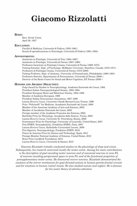
Giacomo Rizzolatti
Born: Kiev, Soviet Union April 28, 1937
Education:Facoltà di Medicina, Università di Padova (1955–1961)Scuola di specializzazione in Neurologia, Università di Padova (1961–1964)
appointmEnts:Assistente in Fisiologia, Università di Pisa (1964–1967)Assistente in Fisiologia, Università di Parma (1967–1969)Professore Incaricato in Fisiologia Umana, Università di Parma (1969–1975)Visiting Scientist, Dept. of Psychology, McMaster University, Hamilton, Canada (1970–1971)Professore di Fisiologia Umana, Università di Parma (1975–2009)Visiting Professor, Dept. of Anatomy, University of Pennsylvania, Philadelphia (1980–1981)Professore Emerito, Dipartimento di Neuroscienze, Università di Parma (2009–)Director of the Brain Center for Social and Motor Cognition, IIT Parma (2009–)
Honors and awards (sElEctEd):Golgi Award for Studies in Neurophysiology, Academia Nazionale dei Lincei, 1982President Italian Neuropsychological Society, 1982–1984President European Brain and Behaviour Society, 1984–1986Member of Academia Europaea, 1989President Italian Neuroscience Association, 1997–1999Laurea Honoris Causa, University Claude Bernard Lyon, France, 1999Prize “Feltrinelli” for Medicine, Accademia Nazionale dei Lincei, 2000Member of the American Academy of Arts and Sciences, 2002Member of Accademia Nazionale dei Lincei, 2002Foreign member of the Académie Francaise des Sciences, 2005,Herlitzka Prize for Physiology, Accademia delle Scienze, Torino, 2005Laurea Honoris Causa, University St. Petersburg, Russia, 2006Grawemeyer Prize for Psychology, University of Louisville, United States, 2007 Prix IPSEN, Neuroplasticity, Fondation IPSEN, Paris, 2007Laurea Honoris Causa, Katholieke Universiteit Leuven, 2009Prix Signoret, Neuropsychology, Fondation IPSEN, 2010Prince de Asturias Prize for Science and Technology, Spain, 2011Foreign Member National Academy of Sciences, United States, 2012Brain Prize Lundbeck Foundation, 2014Laurea Honoris Causa, Università di Sassari, 2015
Giacomo Rizzolatti initially conducted studies on the physiology of sleep and vision. Subsequently, his research concerned mostly the motor cortex. Among his main contributions
is the description of goal-encoding motor neurons and of canonical neurons in area F5, of neurons encoding peripersonal space in area F4, and of the functional properties of
presupplementary motor cortex. He discovered mirror neurons. Rizzolatti demonstrated the existence of the mirror mechanism for goal-directed actions in human parieto-frontal circuits and for emotions in human rostral insula. He also studied autism and neglect. He is famous
for his motor theory of selective attention.
BK-SFN-HON_V9-160105-Rizzolatti.indd 331 5/7/2016 2:57:39 PM
Giacomo Rizzolatti
Kiev and Soviet UnionI was born in Kiev (Soviet Union) in 1937. My father was Italian and my mother Russian. I know very little about my mother’s family, except that it came to Kiev from Odessa. The history of my father’s family is compli-cated. My great-grandfather, Pietro, emigrated from Friuli, a north-eastern region of Italy in the second half of the 19th century. At that time Friuli was a poor region and many “Friulani” were compelled to emigrate. Pietro Rizzolatti emigrated as well. He was, however, a skillful artisan, so he went to Kiev, rather than going to the Far East to construct the Trans-Siberian railway, as many emigrants from Friuli did. He started a small firm special-ized in marble decorations. After a few years his firm grew and he became a respected and considerably rich citizen of Kiev.
Pietro Rizzolatti died a few years before the Soviet revolution. My grandfa-ther Giacomo was born in Kiev. He had a bachelor degree in engineering. After the revolution most of his properties were nationalized, but, being an Italian citizen, he kept part of his house and a small land property in Korostichev, at the time a village, west of Kiev. He decided, therefore, to remain in Kiev in spite of the new regime. He continued to work (as an employee) in his firm.
My grandfather married Maria Galubowska, a lady from a small line of Russian nobility. It was a very fortunate marriage. My grandparents had four children. Three of them decided to become medical doctors. Among these was their eldest son, my father, Pietro. After high school, Pietro applied to the medical school of Kiev, but he was not admitted. He was dubbed (rightly) to be a “bourgeois” and only people of proletarian origin were allowed to go to medical school. Then my father went to the Urals where, for about two years, he worked in a mine. Being now recognized as a proletarian, he started his medical studies.
At the university, Pietro met my mother, Valentina Fedorkova, who also studied medicine. They survived the terrible years of Stalin terror, including the periodical “chistkas,” the collective processes used to iden-tify possible traitors among professors and students. Just before graduating they got married.
Among many defects of the Soviet regime, there was a very positive aspect. At the end of the university the students were ranked according to their marks. The highest ranking students were given the opportunity to choose the jobs they preferred. My father remained at the university as an assistant in anatomy. In that period, on April 28, 1937, I was born.
BK-SFN-HON_V9-160105-Rizzolatti.indd 332 5/7/2016 2:57:39 PM
Giacomo Rizzolatti 333
This happy period for my parents finished very soon. At the end of 1937 all Italian citizens were ordered to go back to Italy. The order came abruptly and had to be executed immediately. Thus, in almost two weeks, my family was sent back to Clauzetto, a village of Friuli of about 1,000 people. Clauzetto was the place my great-grandfather had emigrated from seventy years before.
Early Life in UdineIf the Soviet regime was ferocious, the fascism was, in general, stupid. In our case, the return to Clauzetto was due to a law that forbade citizens to move out of their place of origin. Internal emigration was forbidden. This determined a kind of Catch 22 situation: to change residence some of the family had to find a job, but to have a job one must be resident in the job place. My family was stuck in Clauzetto.
Fortunately, Italians are much more clever than their politicians. Thus, a solution was found by the director of the hospital in Udine. He accepted my father as “Assistente volontario,” that is an assistant without salary. This was a pseudo-job, but sufficient to have the permission to move to Udine.
Udine was at that time a town of about 60,000 inhabitants. A bit provin-cial, but very civilized and proud of its culture, its own language (Friulano), and an ancient cultural heritage. Order, an almost compulsive love for work, and the sense of being a community rendered Udine more similar to a mittleleuropean town than to a typical southern-type Italian city.
The brief period of quiet life in Udine soon finished for my father. He was recruited into the army, but, being a doctor, he was enrolled in the Military Medical School in Florence and exited with the degree of “Sottotenente medico” (lieutenant doctor). A few months later World War II broke out and my father was sent to the Yugoslavian front. Fortunately in 1943, when the Italian army surrendered to Germans, he was in Udine and was not taken as prisoner to Germany.
The first things I remember of my life concern the war period. Following German occupation, Udine was bombed and, even if I also remember some pleasant episodes, those most vivid concern the bombing and the fear expe-rienced during hours spent in the shelters. The last years of the war were particularly difficult for all of us. My father joined the Garibaldi brigade, a leftist partisan group. He continued to practice medicine in his private office and traveled, during the weekends, to the mountain where the Garibaldi brigade had their shelters, to cure wounded and sick partisans.
I was rather a precocious kid. My father, my grandmother, and espe-cially my mother spent a lot of time with me. I learned to play chess when I was five. One of my greatest successes and happiest memories of these dark years was a chess match with a friend of my father, a respected member of Udine chess club. With my great joy and his disappointment, the match terminated with a tie.
BK-SFN-HON_V9-160105-Rizzolatti.indd 333 5/7/2016 2:57:39 PM
334 Giacomo Rizzolatti
I also learned to read very early. So I started school at five. My school years were very pleasant. The post–war era was a great period for most Italians. The economy was growing fast, and in 20 years, Italy changed form a poor agricultural country to one of the richest countries in the word. My family enjoyed this new well-being. My father was a general practitioner with many patients and a good income. Soon after the war Tania, my sister, was born and a few years later my mother decided to stop working to devote all her time to my sister, my father, and myself.
A New Life: The Years in the LyceumI often think that my “conscious” life, a life with a “self,” started when I began my studies in the Ginnasio-Liceo Iacopo Stellini of Udine. The five years spent in this learning institution were my Lehrjahre, the years that formed my adult personality.
The Ginnasio-Liceo Stellini is more that 150 years old. The studies were difficult. The teachers were important personalities in the city, respected by the students and by their parents. A fundamental lesson we learned was that in order to succeed one has to work very hard. We were also taught that we were destined to be the future “ruling class” of the country with privileges and duties. This teaching may sound strange today, but our “Liceo” was very successful in training many students that became very distinguished in their fields. Three of us, students in the same period, are now members of the Accademia dei Lincei, the Italian very selective National Academy. Considering the size of Udine, this has been undoubtedly a great achieve-ment of our school.
I was very lucky because besides good professors, I also had an excep-tionally nice and friendly group of classmates. Besides studying, we had parties, went on bicycle rides, and went to motion pictures together. Our friendships still continue.
I was particularly fond of a girl, during those long years at the Ginnasio-Liceo, always sitting in the first desk near the door. Her name was Leni Bronzin and, in addition to being pretty, she was different from the other girls, because she was broadly interested in reading, music, and culture. With her and a couple of other students, we discussed the books we read and many other issues. At the end of “Liceo,” my link with Leni became very tenuous. I had my life in Padua, where I started Medical School, and Leni was in Venice where she studied foreign languages. At the end of university years, we met again and enjoyed very much being together. We married in 1964, and after more than 50 years, we are still very happy together.
Another strong influence on my cultural development was my cousin Vitale Petrus and his (and my) great friend Tito Maria Maniacco. They were both older than me and, unlike me, very poor students, but both had talents for painting and Tito also for writing. Together they added other cultural
BK-SFN-HON_V9-160105-Rizzolatti.indd 334 5/7/2016 2:57:39 PM
Giacomo Rizzolatti 335
aspects to my Lehrjahre, including a nonconformist attitude toward the conservative politics of those years.
The Medical Studies in PaduaIn 1955 I enrolled in the Medical Faculty of Padua, one of the oldest medi-cal schools and universities in the world. The standard of my professors as teachers was high. They believed in their work or, if you want, in their “mission.” Thus, the studies were very hard.
I liked the new topics that, considering my classical background, were almost completely new for me. Yet, these years have been by far the least exciting of my life. It was passive learning, with little discussion with other students and even less with the professors. I passed hours and hours study-ing in my room in Antonianum, the residential college where I stayed the whole six years of my medical studies. Attending to lectures was not compul-sory and, except for some lessons, I eventually discovered that it was more productive to study at home. As far as the practical medical experience, it was difficult to achieve it at the university hospital because the number of students was relatively high and that of assistants relatively low. I learned much more during the summertime, when I attended the hospital in Udine, where the doctors were very pleased to teach to a university student.
The social life of the students was dominated by the old “goliardic” tradi-tion. Goliardia was historically a free behavior that the students enjoyed during the rigid middle-age society. In modern times, goliardia was reduced to making practical jokes on freshmen. One of the most popular was that of compelling the first-year students to climb the statue of Cavour, a monu-ment located close to the old central building of the university and to take a mock exam from him based on sexual jokes and innuendos. It might seem an amusing event, but, as most other “goliardic ceremonies,” it was repetitive and rather boring.
Things changed radically in my fifth year, when I entered the Neurological Clinic as an “allievo interno.” The director of the clinic was Professor G. B. Belloni, an excellent clinician and a good pathologist of the nervous system. The intellectually dominant figure was, however, Hrayr Terzian. Terzian was Armenian, educated in Venice and in Padua. Terzian divided students and colleagues into two categories: “bravo” and “mona.” This last is a rather vulgar but common word of Venetian dialect meaning “definitely stupid.”
This classification was shocking for a student like me convinced that his professors were great men. Terzian was unfair as far as the medical and teaching capacities of his colleagues were concerned, but right in relation to their scientific merits. During fascism and during war, Italian science remained isolated from the rest of the world and became inferior to that of the other major European countries and the United States. Terzian was an
BK-SFN-HON_V9-160105-Rizzolatti.indd 335 5/7/2016 2:57:39 PM
336 Giacomo Rizzolatti
exception because, at the beginning of his career, he spent time in Pisa in the lab of Professor Moruzzi, one of the few Italian centers of international reputation, and later in Marseilles working with Henry Gastaut.
In 1961, I graduated in medicine with the maximum of marks cum laude. The graduation ceremony, following a tradition going back to the medieval times, took place at night. After the official part of it, started the “goliardic part.” The friends of the newly graduated fellow waited for him in the oldest courtyard of the university and kicked him in the ass, meaning to expel him both practically and symbolically from the free student life.
Studying NeurologySince the beginning of my medical studies, I thought that neurology was the most fascinating branch of medicine. So after getting my MD degree I entered neurology. In the clinic, besides duties in the ward, I was work-ing in the electroencephalography (EEG) laboratory, which was overseen by Terzian. It is curious, considering my subsequent discoveries of mirror neurons, that Terzian was the first to describe with Henry and Yvette Gastaut (Gastaut, Terzian, and Gastaut 1952) the “mu rhythm,” the EEG rhythm that desynchronizes during motor activity and that (we know now) is correlated with mirror neuron activity (see Altschuler et al. 1997).
I liked clinical work. At that time semiology was the basis for neurologi-cal diagnosis. This implied deep knowledge of neurological diseases and a great capacity of reasoning in order to make differential diagnosis. I liked also to talk with patients and to help them within the rather narrow possi-bilities of neurology of that time.
My fellow medical interns were motivated people. Most them studied neurology with the aim of getting a job in a hospital or starting a private practice. A few had the ambition to remain at the university. Among them was Nicola Rizzuto. We graduated in the same year and worked together during our internship in neurology. We became good friends and tried to do some research together. The difficulty of conducting serious research in the clinic was the most disappointing part of our internship. I managed, however, almost by chance, to publish a paper describing two rare cases of self-induced light-sensitive epilepsy, but, this type of anecdotal research was highly unsatisfactory (Ravenna and Rizzolatti 1964).
The limits of this form of clinical research were also clear to Terzian. One day he called me in his office and told me that if I really wanted to be a “true” university professor, I should learn a basic technique that could be applied to neurology. He offered me two opportunities: one was to go to Marseilles to work on epilepsy, the other to learn neurophysiology. I chose neurophysiology.
In 1963, Professor Arnaldo Arduini, the oldest pupil of Professor Giuseppe Moruzzi, obtained a chair in physiology in the University of
BK-SFN-HON_V9-160105-Rizzolatti.indd 336 5/7/2016 2:57:39 PM
Giacomo Rizzolatti 337
Ferrara. Arduini needed somebody to help him and was happy to accept me in his lab. So I moved for six months to Ferrara and began my training in neurophysiology. Arduini would later play an important part in my life when he accepted me as his assistant in Parma.
My first approach to physiology was interesting, but not exciting. Arduini and I recorded mass activity from the pyramid tract and measured it during slow-wave sleep and wakefulness. The results showed a clear pyra-midal activity increase correlated with EEG desynchronization (Arduini and Rizzolatti 1964).
The major weakness of Ferrara was that Arduini was the only physiolo-gist. I spent most of my free time with a nice and friendly group of biochem-ists, or studying neurology and psychiatry in my room. However, I became sufficiently interested in physiology to go back to Arduini the following year to finish the work we started. In the meantime, Arduini moved to Parma, where he had a much larger, but virtually empty institute. I finished with him my work on pyramidal tract and the results were accepted by Professor Belloni as my neurology thesis.
The year 1964 was a crucial year for me. In that year I obtained my degree in neurology. Terzian strongly advised me to continue an academic career, but not in Padua. According to him, I had first to complete my train-ing in neurophysiology in Pisa, which was the best center of neurophysiol-ogy in Italy. Professor Moruzzi, the director of the Institute of Physiology of Pisa, accepted me and found for me a tiny fellowship. This, together with the salary of Leni, who taught English in the middle school, allowed us to marry. We married in March 1964. In September we started our new life in Pisa.
Physiology in Pisa: Sleep StudiesThe Institute of Physiology of Pisa is a large, imposing building located close to the city’s medieval walls. There is a small garden in front of the building and a very large one, almost a small park, behind it. Professor Moruzzi, as it was tradition in those days, lived in the institute. His office, a large room full of books, was located on the second floor. It was there that he received me at my arrival. Professor Moruzzi had an impressive personality that commanded respect. The institute was undoubtedly his institute and every-body working there accepted his ethical values. He had an almost religious idea of science and a scientist, a real scientist, had to devote all of his time and energy to science.
When I arrived to Pisa, Moruzzi was at the apex of success. When still young he realized that the isolation of Italy from the world scientific commu-nity imposed by fascism was a strong handicap for becoming a good scientist. So he moved from Italy to Bruxelles where he worked with Frédéric Bremer and subsequently to Cambridge where he collaborated with Lord Adrian. After Word War II, Moruzzi moved for about one year to Chicago where he
BK-SFN-HON_V9-160105-Rizzolatti.indd 337 5/7/2016 2:57:39 PM
338 Giacomo Rizzolatti
discovered, with Magoun, the arousing effect of the electrical stimulation of brain stem reticular formation. This finding changed radically the way of conceiving sleep and more general brain functions. Back to Italy, Moruzzi created in Pisa a unique center for studying the nervous system.
My interview with Moruzzi went very well. I told him about my limited experience in physiology. He replied not to worry. He decided I was to work with Lamberto Maffei, who could teach me all technical aspects of physiol-ogy. The work of the team formed by Maffei and myself will be supervised directly by him. The decision of Moruzzi to put me in Lamberto’s lab was very successful. Lamberto was only one year my senior, but he had been working in physiology since the beginning of his studies in medicine, and, as Moruzzi told me, he was already an accomplished physiologist. I learned a lot from him. Lamberto was a very practical person, so he organized the lab in such a way that my little expertise in physiology could be useful for our team.
We carried out the experiment on midpontine pretrigeminal cats, a “preparation” in which, following a section of the brain stem, the animal is paralyzed except for vertical eye movements. Pretrigeminal cats do not feel pain and show a predominantly desynchronized EEG. This EEG pattern is frequently interrupted by short episodes of synchronized sleep. The surgery was typically done by Maffo (as we called Lamberto in those times). Once the preparation was ready it was my turn to work. I took care of the animal and, using a micromanipulator, isolated single neurons from the dorsal part of the lateral geniculate body (LGB) and from the optic tract. As soon as a neuron was isolated, Maffo turned on a lamp that generated a sine-wave stimulus, and by using a tiny computer named “CAT,” established the time relation between the stimulus and the responses of the recorded neuron.
We addressed two main issues. The first was the transfer properties of LGB neurons to sinusoidal light stimuli presented at different frequencies. Maffo was very interested in this problem that was a continuation of his previ-ous work on the retina. The second issue was to see how sleep-wakefulness cycles modulated the transfer of visual information.
We worked very hard. The experiments lasted the whole day from 8:30 in the morning till the evening and we ran them every day except on Saturday morning (devoted to reading in the splendid Pisa library) and Sunday (mostly for the family). Occasionally, we were helped by a gentle and quiet medical student, Luigi Cervetto. Our efforts were rewarded. At the end of the year we had seven publications, including a paper in Science (Maffei, Moruzzi, and Rizzolatti 1965) and one in the Journal of Neurophysiology (Maffei and Rizzolatti 1967), at that time a top journal in neuroscience. The most exciting result was the change in information transfer during slow-wave sleep relative to wakefulness. While during wakefulness the modula-tion of the LGB spikes by sinusoidal light gave as an output a sinusoid, this information was completely lost during sleep.
BK-SFN-HON_V9-160105-Rizzolatti.indd 338 5/7/2016 2:57:39 PM
Giacomo Rizzolatti 339
Our work was greatly appreciated by Moruzzi who was constantly informed about the progress of our experiments and helped us in writing the papers. Lamberto and I worked very well together and became very good friends. I liked his quiet leadership and his way to train me giving me progressively more to do in the experiment. Although our careers later diverged, our friendship is still very strong. I was very happy when some years ago Maffo became the Presidente of Accademia dei Lincei, the most prestigious Italian scientific institution.
My first year in Pisa was also very successfully from a social point of view. In front of our lab was that of Franco Magni, just back from the lab of John Eccles, and of Piergiorgio Strata. Strata is a very brilliant scien-tist, whose discussion abilities are as unique as his knowledge of physiology. We immediately became friends and our friendship increased with years. He become later interested in the organization of the university and more generally in those aspects of politics that concerns research. Even now, if I need some advice or desire to discuss an issue concerning research politics, I call Piergiorgio in Turin where he is professor of physiology.
In July 1965, Pietro, our first son, was born in Udine where Leni’s and my family lived. Leni did the birth preparation exercises in Pisa together with Dolly Strata, Piergiorgio’s wife, and Benedetta Cervetto, our student Luigi’s wife, and this made our friendship stronger.
Physiology in Pisa: Corpus Callosum and Superior Colliculus After my successful collaboration with Maffei, I was sure to continue my collaboration with him. Instead, in July, a few days before Pietro’s birth, Moruzzi called me and told me that it would be more appropriate for my education to enlarge my experience and to change lab and topic. His sugges-tion was the lab of Professor Ottavio Pompeiano.
Pompeiano had a good reputation as vestibular system physiologist, which was acquired mostly for his studies done at the Karolinska Institute. I never read his work, but in Pisa he was more famous, among postdocs, for his almost compulsive desire to publish papers and use the coworkers as manpower, than for his research on the vestibular system. Diplomatically, I did not mention these considerations to Moruzzi, but told him that I preferred to work on sleep. In those days, Giovanni Berlucchi was back to Pisa from a sabbatical he spent with Roger Sperry at Caltech. Moruzzi thought to create a new group led by Giovanni. He offered me to join this group and I was happy to accept. Thus in September 1965, I started my work with Giovanni.
Giovanni has been a central person in my scientific and personal life. His love for science, an almost religious sense of duty, and a rich culture outside science rendered his personality unique even among the highly intelligent and motivated young people around Moruzzi. My encounter with
BK-SFN-HON_V9-160105-Rizzolatti.indd 339 5/7/2016 2:57:39 PM
340 Giacomo Rizzolatti
him helped me in my scientific maturation. In addition, Leni and Luisa, Giovanni’s wife, become good friends and, thus, a mere scientific collabora-tion became an enduring family friendship.
The first studies we did together concerned the auditory system and the modulation of the transfer of auditory information to cerebral cortex and cerebellum (Berlucchi, Munson, and Rizzolatti 1966a). These studies, which we carried out with John Munson, a student of Robert Doty, were well accepted by the scientific community. A nice reward was an invitation to Siena where, in addition to a scientific meeting on evoked potentials, there was also a rich social program. This program included the possibility to see the Palio, an exciting rough horse race that goes back to middle age.
Giovanni and I frequently discussed the data of Sperry on the split brain as well as his ideas (which I never fully understood) on consciousness as an emergent property. In the same years, reading the famous paper by Lettvin, Maturana, McCulloch, and Pitts (1968) on the frog eyes and the great papers of Hubel and Wiesel (1959, 1962), I became more and more interested in the visual system. This and the discussions with Giovanni on the role of the corpus callosum in vision led us to record from this structure. This decision was boosted by the arrival in Pisa of Mike Gazzaniga, a coworker of Roger Sperry in the famous split brain experiments, and a good friend of Giovanni.
Mike was a pleasant and witty teammate. Giovanni, Mike, and I worked very well together, and my friendship with Mike became stronger with the years. The first issue we addressed was to study what type of information was transmitted in the posterior third of the corpus callosum. Not surpris-ingly, we found the various types of receptive fields previously described in cortical areas 17, 18, and 19. However, the callosal receptive fields were all located along the vertical meridian of the visual field. We interpreted these data as evidence that the representation of the visual field on the cerebral cortex is on a continuum, the neurons associated with the vertical meridian being the “trait d’union” necessary for bringing together the two half visual fields (Gazzaniga, Berlucchi, and Rizzolatti 1967).
Recording from the fibers of the corpus callosum was not easy and we did many experiments before obtaining good results. In one of his books, Mike Gazzaniga describes one of these initial experiments. He writes that, during a particular experiment, instead of the typical “cracking” sound of action potentials, the loudspeaker started abruptly to transmit the notes of “Yellow submarine.” At this point I would have said: “Finally we are able to record high order information from the corpus callosum.” The first part of the story is true, while the second is a nice embellishment by Mike.
Mike left us very soon. He was offered a position at University of California in Santa Barbara and he accepted it. We were a bit disappointed, but it turned out that a team of two, Giovanni and myself, was sufficient to continue our work on corpus callosum. Our next project was to assess how the information coming via corpus callosum and that arriving from LGB
BK-SFN-HON_V9-160105-Rizzolatti.indd 340 5/7/2016 2:57:39 PM
Giacomo Rizzolatti 341
was integrated in area 17. To study this integration, we split the chiasm and presented the stimuli to each eye of the ipsilateral hemisphere. We found that there were neurons that were driven not only by the ipsilateral eye, through direct geniculo-cortical pathway, but also by the contralateral eye through the corpus callosum. The two monocular receptive fields of a given cell lie in close contact with the vertical meridian. We suggested that this convergence provides the continuity of the cortical visual map across the interhemispheric gap. We sent this paper to Science, where it was accepted after minor revisions (Berlucchi and Rizzolatti 1968). This was my second Science paper emerging from about two years of work in Pisa.
A visual structure that had been neglected for many years in mammal visual physiology was the superior colliculus (SC). An experiment that played a fundamental role in putting the SC in the foreground in vision physiology was a study by Jim Sprague (1966). In this study, he described an enduring hemianopia following a large unilateral visual cortical lesion recovered after a lesion of SC on the intact side.
Jim Sprague was an old friend of Professor Moruzzi. In 1966 he came to Pisa for a sabbatical. Jim belonged to the generation between Moruzzi’s and mine. He was professor at Penn and famous for his contributions on the cerebellum, spinal cord, and brainstem reticular formation. Jim was a great gentleman. Rarely in my life have I found a person so kind and full of consideration for others. In spite of the age gap, we became good friends. Thirteen years later, when I spent a sabbatical at Penn, I appreciated him even more both as a scientist and as a person.
When Jim arrived to Pisa, Pier Lorenzo Marchiafava and I were discuss-ing the possibility of recording single neurons from SC. Actually Pier Lorenzo had already performed a pilot experiment. Pier Lorenzo was a highly moti-vated, witty, intelligent researcher, but somehow, he was (or pretended to be) different from the other pupils of Moruzzi. He liked fast cars and girls, and there were rumors that, once, he made the provocative proposal to Moruzzi to build a swimming pool in the institute. “Just to enjoy ourselves between experiments.” After my departure from Pisa, he published several interesting papers on the retina.
Jim was enthusiastic to join Marchiafava and myself in recording single neurons from SC. We discussed two projects. One was to study the responses of SC neurons to stationary and moving stimuli. We carried out the exper-iments in midpontine pretrigeminal cats (see above). The second project was to inactivate the visual cortex and to see whether cortical inactivation affected neuronal receptive field properties in the SC. While the inactiva-tion project gave rather ambiguous results, the characterization of the SC receptive field properties was very successful. An important observation was that the habituation of neurons’ responses to repeated visual stimuli and that of vertical eye movements (the only motor responses of midpontine pretrigeminal cat) to identical stimuli had the same time course, as also did
BK-SFN-HON_V9-160105-Rizzolatti.indd 341 5/7/2016 2:57:39 PM
342 Giacomo Rizzolatti
the recovery from habituation (dishabituation). In other words there was a clear link between visual attention and SC activity (Marchiafava, Rizzolatti, and Sprague 1968).
At the end of the academic year 1966–1967 my contract with the Institute of Physiology expired. If I could, I would have stayed in Pisa. In addition to the exceptional scientific atmosphere, there was also a lively social life. I already mentioned some of my closest friends, but there were many other interesting and friendly people: Norman Kahn from Columbia; Dick Poppele from Minneapolis; Adrian Morrison from Penn; Giancarlo Carli, who, after a period at Johns Hopkins, became professor of physiology in Siena; and Carlo Marzi, now professor of psychology in Verona.
In the meantime Terzian, my mentor in Padua, has been appointed director of the Clinical Neurology in Cagliari. Thus, in the spring of 1967, Leni and I went to Cagliari to see our future new city and, possibly, to look for a house. Nicola Rizzuto was already there. We were greeted by Terzian with enthusiasm and were guests of Nicola in his apartment. However, when I started to talk with Terzian on the possibility to build a lab for physi-ological experiments, he became very vague, saying that this was something for the future. First, he needed to organize the clinical work. Talking (for hours) with Nicola, I learned that indeed the situation of the clinic was like that of a provincial hospital and no serious research could be carried out there. My visit to the clinic confirmed his view.
Leni and I returned to Pisa very sad. On the one side, I was reluctant to abandon clinical neurology, and on the other, after the years spent in Pisa in touch with “real” research, I did not want to go back to routine clinical work, with only very vague hopes to continue my research in the future. There was also a financial problem. If I was going to refuse Cagliari, I had to find a job. Where? Back to Udine? My links with Padua were severed. The old director Professor Belloni was on the verge of retirement and my mentor Terzian had left for Cagliari.
I decided to talk with Moruzzi, with the hope that maybe he could help me. Moruzzi listened to me very carefully and then said that he could help me because there was an opening in Parma, where an assistant had decided to leave the job, and Arduini, the director of the institute, was look-ing for a replacement. It was a tenure track position and in a few years, if my work was satisfactory, I could become “assistente di ruolo” (assistant professor with tenure). Moruzzi spoke with Arduini, who accepted me with enthusiasm.
Parma: Sleep AgainIn autumn of 1967 I was in Parma. Leni and Pietro remained in Pisa. They joined me the following year. The Institute of Physiology of Parma consisted mostly of walls. The predecessor of Professor Arduini, Professor Pinotti,
BK-SFN-HON_V9-160105-Rizzolatti.indd 342 5/7/2016 2:57:39 PM
Giacomo Rizzolatti 343
was a respiration physiologist who moved to Turin and took away with him all of his instrumentation. So when Arduini became the institute director he found the institute virtually empty.
Arduini was the first pupil of Moruzzi and helped him very much in creating the Pisa Institute. The discovery for which he was most famous was that of the theta rhythm in the hippocampus, a slow rhythm that surpris-ingly appears in the hippocampus during wakefulness. This discovery was done with John D. Green, when Arduini was on sabbatical at the University of California, Los Angeles (UCLA).
Arduini was happy of my return to Parma and helped me very much in my career. First of all, although I was a young assistant, he gave me complete scientific independence, and, very generously, allowed me also to use the best among the few instruments of the institute. Later on, when I started teach-ing, he gave me the Course in Neuroscience, which was much easier for me to teach than other parts of physiology. Arduini was a good organizer and because of his scientific stature and great personal honesty he had a strong influence on faculty. During the years in which I was with him in Parma, his interest for experimental physiology progressively vanished. He was more interested in theoretical problems such as, for example, how to quantify neural activity, and later mostly in philosophical problems.
Besides myself, in the institute, there were nominally two assistants: Ruggero Corazza and Andrea Cavaggioni. However, when I arrived, Corazza was in Boston and Andrea was in Baltimore. An important presence in the institute was Maria Grazia Arduini, Arduini’s wife, who worked as a histolo-gist. She was a superb technician and yet she worked without salary because Arduini thought it was not fair to give a paid position to his wife.
Before coming to Parma I thought that an interesting project in sleep physiology was to extend the study of activity of LGB from slow sleep and wakefulness to the sleep phase characterized by rapid eye movements (REM sleep). In order to do this, I had to learn how to record from behaving cats. I did all the preparatory work in Pisa. So, after a few months in Parma I was able to implant my first cat. Working alone was very hard. The institute’s janitor (Renzo Tebaldi), a very kind man and a good amateur cyclist, was the only person that helped me, when free from his duties. He sometimes even stayed with me some time after his working hours.
The results were coming very slowly. Here Moruzzi helped me again. A young Russian physiologist, Lev Mukhametov, had the permission from his government to come to Italy. He was interested in sleep physiology and to work in one of the groups directed by Moruzzi. Moruzzi told him that in Parma there was Rizzolatti, one of his former pupils, who also spoke Russian and suggested that Lev join me in Parma. Lev accepted and we started a very productive collaboration.
Lev was (and is) an easy-going, practical, and very talented scientist. He already had experience in sleep physiology and was very happy to study
BK-SFN-HON_V9-160105-Rizzolatti.indd 343 5/7/2016 2:57:39 PM
344 Giacomo Rizzolatti
the neuronal activity in behaving animals and to learn this technique. He was a very hard-working person determined to go back home with scientific results obtained abroad. Both of us had no family in Parma. So, except for occasional movies seen after dinner, we spent all our time in the lab record-ing from morning to night, and the results started coming.
In the first study we recorded the spontaneous activity of LGB neurons. As expected, we found a clear difference between spontaneous activity during slow sleep and wakefulness. The new finding was the activity during REM sleep. As soon as EEG, electromyography (EMG), and eye move-ments indicated the occurrence of REM sleep, the neuronal spontaneous activity abruptly changed and was characterized by a well-spaced, unclus-tered discharge, similar to that of wakefulness. Activity in optic tract fibers was not modulated by the behavioral states (Mukhametov, Rizzolatti, and Seitun 1970).
The presence of these different patterns in LGB raised the question of how these patterns may influence the transmission of visual stimuli to the visual cortex. The main obstacle to address this issue was the difficulty of maintaining stable visual stimulation in behaving cats. We solved this problem by paralyzing the intrinsic and extrinsic ocular muscles by cutting cranial nerves III, IV, and VI and the cervical sympathetic trunk. This tech-nique had been devised a couple of years before by Berlucchi, Munson, and myself (Berlucchi et al. 1966b). The visual stimuli were delivered by a lamp cemented to a contact lens inserted on the eye.
The responses were quantified as “absolute response,” that is, the number of spikes during the second starting with stimulus onset, and “relative response,” the same responses referred to background activity. The absolute responses were very large in wakefulness, decreased in the synchronized sleep, and increased again, reaching its maximum, during REM periods of desynchronized sleep. The relative responses, however, were highest during wakefulness. We concluded that, accepting the signal-to-noise ratio as the biologically fundamental measure, the transmission through LGB was impaired during sleep relative to wakefulness by two different mechanisms: (a) a postsynaptic inhibition (pause-burst pattern) during synchronized sleep and (b) a marked increase of spontaneous activity during desynchronized sleep (Mukhametov and Rizzolatti 1969).
These data were published in Archives Italiennes de Biologie, one of the top journals for sleep physiology of that time. Before sending them to the editor, we asked Emilio Bizzi to read them. Emilio is another of Moruzzi’s students. When I was in Pisa, he had already left for the States. He visited Pisa, however, from time to time and during these visits we became friends. Bizzi is especially known for his contributions to motor physiology. However, he also studied sleep and is the discoverer with Brooks of the “ponto-geniculate-cortical waves,” one of the characteristics of the REM sleep (Bizzi and Brooks 1963). At the time of my collaboration with Mukhametov, Emilio
BK-SFN-HON_V9-160105-Rizzolatti.indd 344 5/7/2016 2:57:39 PM
Giacomo Rizzolatti 345
was in Milan. We took advantage of this fact and discussed our findings with him. He was also so kind to correct our English.
The Soviet bureaucracy was unpredictable. Thus, although both Mukhametov and I thought that the possibility for him to come to Italy again was almost zero, he actually got the permission to work in Italy for another period. This allowed us to record from some additional animals and to analyze a series of neurophysiological data, which, according to the histological analysis, were obtained from “nucleus reticularis thalami,” a nucleus that sends its output top-down to other thalamic nuclei. The data revealed that the neuronal discharge pattern in the different phases of sleep-wakefulness cycle was similar to that of the LGB but with some differences. A notable difference was that during REM sleep the activity of nucleus reticularis was more regular than that in the LGB and almost indistinguishable from that observed in nucleus reticularis during wakefulness. We interpreted these findings as support for theories (e.g., Berger 1969) that maintain that REM sleep represents an endogenous stimulation of the nervous system necessary for the setting up of connections between neurons. Only a pattern similar to that present in wakefulness can fulfill this function (Mukhametov, Rizzolatti, and Tradardi 1970).
My collaboration with Lev ended with this paper. These were two years full of scientific rewards and pleasant camaraderie. Also for Lev the years spent in Parma studying sleep in behaving animals were very useful for his career and for his subsequent fundamental studies on the sleep of dolphins and other aquatic mammals (e.g., Mukhametov, Lyamin, and Polyakova 1985).
The work I did in Pisa and in Parma was appreciated by both Moruzzi and Arduini, who suggested that I apply for “Libera docenza.” This title, which corresponds to the German “Privat docent,” gave the title of profes-sor and was a fundamental step for becoming full professor for Italian schol-ars in an academic career. The discussion of my findings with the Members of the Libera Docenza Committee and the subsequent lecture on a topic of human physiology (respiratory mechanics!) went very well. Thus, in May of 1969, I received the title of “Libero Docente in Fisiologia Umana.”
Neuropsychology with Berlucchi and Umiltà When I was still in Pisa, Giovanni Berlucchi and I discussed the possibility to apply our findings concerning the organization of the corpus callosum to humans. There was an old study by Poffenberger (1912) that reported that crossed reaction times to visual stimuli are significantly longer than uncrossed reaction times. We decided to verify these findings, which were not confirmed by other studies (see Smith 1938), and to test, by presenting the stimuli at different distances from the visual field midline, whether the interhemispheric transfer was performed by visual callosal fibers.
BK-SFN-HON_V9-160105-Rizzolatti.indd 345 5/7/2016 2:57:39 PM
346 Giacomo Rizzolatti
We carried out the experiments in Bologna with Carlo Umiltà. Carlo was a young psychologist from Bologna who had been trained in physiology by Arduini. From time to time he was in Parma to work with Arduini on analysis of their data. In one of his visits I proposed to Carlo to carry out in his lab the reaction time experiment that I had discussed with Giovanni. Carlo accepted.
At that time in Bologna there was as a visiting professor Ray Hyman, a psychologist from Eugene, Oregon, famous for his studies on choice reac-tion times and who, after returning to the United States became even more famous for having exposed the paranormal capacities of Uri Geller, a very popular illusionist. He accepted to supervise our work. Finally, some help was given to us also by Woodburn (Woody) Heron, a student of Donald Hebb, who was a visiting professor in Pisa.
Our first experiment confirmed the data of Poffenberger. Visual stimuli presented on one side of the fixation point elicited faster reaction from the ipsilateral than from the contralateral hand. The delay between crossed and uncrossed responses remained constant regardless of the distance of the stimuli from the fixation point (Berlucchi et al. 1971). Thus, the inter-hemispheric transfer was not mediated by the visual part of the corpus callosum.
Our previous neurophysiological findings that only the stimuli near the vertical meridian are encoded by both hemispheres raised the possibility of studying the functions of the two hemispheres independently, by present-ing visual stimuli distant from the vertical meridian. We decided therefore to measure choice reaction times to letters and faces presented to the two hemispheres. The results showed that the reaction times to letters were systematically faster when presented to the right visual field, while the reac-tion times to faces were faster when presented to the left visual field. This effect was independent of the hand used for responding. Our interpreta-tion was that the material presented to the nondominant hemisphere for that material had to be transferred to the dominant effect for recognition. This interhemispheric transfer took more time than that related to the Poffenberger effect (Rizzolatti, Umiltà, and Berlucchi 1971).
These two papers on interhemispheric transfer were both published in Brain. They were among the first papers that demonstrate the possibility of studying functional hemispheric asymmetries in normal human subjects using a simple inexpensive method. They were received with great interest. A demonstration of this interest was the election of Giovanni and, a couple of years later, of myself into the International Neuropsychological Symposium group. This group is essentially a private club that has a fixed number of members, has rigid rules, and meets once a year in Europe, typically on the Mediterranean shore. There were formal presentations and discussions, but the beauty of the club consisted in the hours free from programmed session where people could meet and discuss in a very informal and free way.
BK-SFN-HON_V9-160105-Rizzolatti.indd 346 5/7/2016 2:57:39 PM
Giacomo Rizzolatti 347
The leaders of the group, when I became a member, were Hans Lucas Teuber and Brenda Milner. Other outstanding members were Norman Geschwind, Ennio De Renzi, Henry Hecaen, Amedeo Vignolo, Jacques Paillard, Mortimer Mishkin, Charlie Gross, and Edoardo Bisiach, in other words the most prominent clinical and experimental neuropsychologists of that time. For a young man, it seemed almost incredible to have the possi-bility to meet and discuss with the greatest figures of neuropsychology in a completely informal way.
Hamilton, Ontario After I had moved to Parma, Woody Heron arrived to Pisa from McMaster University where he was professor of psychology. Woody came to Pisa to work with Moruzzi and Berlucchi. Giovanni involved him in one of our reaction time experiments. Thus we met and became friends. Woody was an extremely gentle person. His shyness was legendary. However, when his shyness could be overcome, his great culture and intelligence became imme-diately apparent.
Before coming to Italy, Woody received a considerable grant and invited me to spend one year at McMaster. At the beginning, Leni and I were uncer-tain about what to do. In fact in March 1970, Beatrice, our second child, was born. However, after many hesitations we accepted Woody’s invitation.
We arrived at Toronto after a long flight and an unexpected stop in Amsterdam. Fortunately, Pietro found a small girl sitting close to him on the plane, with whom he played most of the time, and Beatrice slept quietly in a hammock that a steward placed above our heads. Woody was at the airport waiting for us and drove us to our house located in Dundas, a beauti-ful village near Hamilton, not far from McMaster University.
The atmosphere at the Department of Psychology was very friendly, and Woody and some other colleagues helped us in solving the practi-cal difficulties related to our arrival. Unfortunately, Woody’s scientific programs were very vague and, after some time, he suggested that, while he was organizing his experiments, I could do some neurophysiology with a postdoc, Barry Jones, an intelligent young medical doctor who later became a clinical neurologist. Professor R. Pritchard kindly allowed us to use his lab. We studied the functional properties of SC in anesthetized cats, but without success. Although no publication came out from my time at McMaster, the years spent there were very useful to enlarge my vision of science, to learn how research was done in America, and, especially, to learn psychology.
At the time behaviorism was the dominant psychology at McMaster. I attended some lectures on it and read many papers on this topic. I found the ideas of Skinner on behavior, including human behavior, very appeal-ing. Abe Black, who was one of the major figures at McMaster, advised me
BK-SFN-HON_V9-160105-Rizzolatti.indd 347 5/7/2016 2:57:39 PM
348 Giacomo Rizzolatti
how to condition a dog. Thus, I experienced personally the power of operant conditioning.
Thanks to the generosity of Woody, I also had the opportunity of making several trips. First, I visited Montreal and had conversations with Peter and Brenda Milner. Another trip was to Philadelphia, where I visited the lab of Jim Sprague, and to Baltimore, where I met Gian Luigi Poggio and Vernon Mountcastle. I had a long conversation with Poggio who tried to convince me that there was no point in studying the properties of visual areas beyond primary visual cortex (“it is premature”) and a short one with Mountcastle, who showed very little interest in my research. More rewarding was my visit to Eugene, where Carlo Umiltà was on sabbatical. There I met two extraor-dinary persons, Michael Posner and Steve Keele. I had a lot of interactions with them in the following years and, with Michael, also many debates on the mechanisms of attention.
Back to Parma: Superior Colliculus In September 1971, I returned to Parma, where I started my experiments with new coworkers: Marcello Camarda, Larry Grupp, and Michele Pisa. Marcello was a neurologist from Palermo, whose mentor wanted him to learn some basic science. Larry was a Canadian from Toronto. He liked the seminars I gave on SC at McMaster and decided to change his field from psychology to neurophysiology. The third member of the team was Michele Pisa, a Sicilian who already had some training in physiology in Pisa.
My three coworkers were hard-working people, but each of them wanted (when I was not in the lab) to be the leader of the group. Discussion among them was a constant feature of the first year of our collaboration. However, the success of the experiments and my diplomatic skills made the work of the team progressively more pleasant and, at the end, Larry and Marcello even became friends.
The idea behind our experiment was that the SC has a fundamental role in selective attention. Imagine that two visual stimuli requiring incompat-ible responses arrive simultaneously to the animal. It is very plausible that some interactions between them could occur before the command to act is issued. Our hypothesis was that these interactions occur already in the SC. To test this hypothesis we presented a stimulus (S2; typically a black spot) outside the receptive field of the recorded neurons in the moment in which the stimulus (S1) entered its receptive field, triggering a neuronal response. We found that in more than 80 percent of the tested units, presentation of S2 produced a strong inhibition of the neuronal discharge (Rizzolatti, Camarda, Grupp, and Pisa 1974).
I liked this experiment very much. Our paper reporting it was accepted by the Journal of Neurophysiology, without any major criticism, but its success among the visual physiologists has been very limited.
BK-SFN-HON_V9-160105-Rizzolatti.indd 348 5/7/2016 2:57:39 PM
Giacomo Rizzolatti 349
Subsequently, we described the same effect in an extrastriate visual area located in the cat lateral suprasylvian gyrus known as “Clare-Bishop area.” In contrast, a control experiment showed that the effect of S2 on neurons of area 17 was minimal, or completely absent. This finding supported the view that the control of attention was determined by the centers that control eye movements, rather than by those processing visual information for object recognition (Rizzolatti and Camarda 1975).
In addition to these neurophysiological experiments, I continued my neuropsychological research. In both areas I received great help from the presence in the lab of Gus Buchtel. Gus was a very precise and meticulous researcher with an excellent preparation in both physiology and clinical neuropsychology. He gave a more methodological solidity to our experi-ments. He stayed with me a couple of years, then moved to Montreal to work with Brenda Milner and eventually became professor at the University of Michigan in Ann Arbor.
At the beginning of 1975, I presented my curriculum vitae (CV) for becoming full professor of physiology. In September of the same year, the National Committee approved my titles, and in November, the Faculty of Medicine appointed me as professor of human physiology, a position that I have held for more than 35 years.
Monkeys in Parma The appointment as full professor, and the many accompanying invitations to present my data in Italy and abroad, was very rewarding. I remember, for example, with particular pleasure the week I spent in Berlin (actu-ally in Dahlem) with Professor Grusser, or the invitation by Professors Baumgartner and Akert to give a seminar in Zurich, where I was also offered a position.
Yet, I felt that there were other researchers who were more in the fore-front of physiological research than me. I was greatly impressed by the paper by Mountcastle on the organization of the parietal lobe (Mountcastle et al. 1975) as well as by the work of Hyvärinen (1982). I found also very exciting the papers of Bob Wurtz and Mickey Goldberg (1971) on the monkey SC. Their results were similar to mine. Their impact, however, was undoubt-edly much greater. The major difference was that they worked on monkeys, while I ran experiments on cats.
I decided therefore that an advanced research program needed monkeys. Professor Arduini gave me his support. It is almost unbelievable today to see how easy it was to do research on animals in those years. Arduini called the chief veterinarian of Parma and asked him for permission to run experi-ments on monkeys. The chief veterinarian did not see any problems. In response to our inquiry regarding who should be responsible for the welfare of animals, he said: “You! I know everything about cows and pigs, but you,
BK-SFN-HON_V9-160105-Rizzolatti.indd 349 5/7/2016 2:57:40 PM
350 Giacomo Rizzolatti
as medical doctor, are more expert than me on what concerns primates.” Thus, we bought the first monkey, a Macaca fascicularis, in a pet shop and learned how to treat it well. This monkey remained with us for several years as our mascot. Then we imported other monkeys from Germany. A new era began.
The first experiment that we conducted on monkey was carried out with the help of Ivan Pigarev. Ivan arrived in Parma from Moscow. He was (and is still) working in one of the institutes of the Russian Academy of Sciences. Ivan looked like a character from a novel of Dostoyevsky. He is tall, with a long beard, and with a hieratic aspect. Ivan is one of the most creative and original scientists I have ever met. In spite of the difficulties that all Russian scientists faced after the collapse of the Soviet regime, Ivan managed to continue his work, mostly abroad. His finding that the visual cortical areas process signals from visceral organs during sleep is of great interest and, in my opinion, has not received, the attention it deserves.
Our initial experiments on monkeys were carried out using awake cura-rized, to induce paralysis, animals. We explored the caudal part of the frontal lobe with the aim to localize the frontal eye fields (FEFs). In this explora-tion, we found, rostral to the arcuate sulcus, neurons with properties similar to the face neurons previously described by Charlie Gross (Gross, Rocha-Miranda, and Bender 1972) in the temporal lobe (Pigarev, Rizzolatti, and Scandolara 1979). The most interesting results came out, however, when we started to record from the cortex immediately posterior to the arcuate sulcus. Neurons in this premotor region responded to tactile and/or joint stimulation. Hands and mouth were the body parts most abundantly repre-sented. Some neurons exhibited tactile responses that were conditional on the arm location. For example they responded to tactile stimulation of the hand, but only when the hand was close to the mouth. We interpreted these data as evidence that this part of the postarcuate cortex encoded hand, mouth, and especially hand-to-mouth movements (Rizzolatti, Scandolara, Matelli, and Gentilucci 1981a).
The great surprise was, however, the presence of a large number of visual neurons. Unlike neurons in the FEF, which responded to far stimuli, postarcuate neurons responded exclusively to stimuli located close to the skin or within the monkey’s reaching distance. Most of these neurons were bimodal with the visual receptive field spatially related to the tactile field. This was the first description of what we later described as the peripersonal space (Rizzolatti, Scandolara, Matelli, and Gentilucci 1981b).
Philadelphia At the end of September 1980, Leni, Pietro, Beatrice, and I left Parma for Philadelphia. Jim Sprague invited me to spend a sabbatical year as visiting professor in the Department of Anatomy of the University of Pennsylvania.
BK-SFN-HON_V9-160105-Rizzolatti.indd 350 5/7/2016 2:57:40 PM
Giacomo Rizzolatti 351
Leni and children arrived to Heathrow from Milan and I reached them from Brighton. In Brighton there had been a meeting of the European Brain Behaviour Society, where I presented my new data on premotor cortex with great success.
Jim organized our stay in Philadelphia in the best possible way. We had a beautiful house in Bala Cynwyd, a rich community in Lower Merion Township. The house was nicely furnished and had everything we could need, including a piano and many classic records. The children attended excellent public schools completely free.
We had already met Dolores Sprague, Jim’s wife, in Pisa, but it was in Philadelphia that Dolores and Leni became close friends. Dolores and Jim also tried to render our life as pleasant as possible. They introduced us to their friends and also gave us opportunities to appreciate life in Philadelphia.
As in my previous stage abroad, the scientific part of my visit was not successful in terms of publications. I was supposed to work with Larry Palmer and to learn from him to program computers. Unfortunately, I found it rather difficult to communicate with Larry and thus our collabo-ration slightly vanished. Another person I was supposed to work with was Alan Rosenquist. Alan was friendly and very communicative, but constantly busy with administrative duties. Thus, the first months of my stay, the only person I worked with was Jim.
Of course Jim was also extremely busy, yet he found some time for me. He reconstructed for me the cortical lesions and the correspond-ing thalamic degenerations of an experiment I performed in Parma (see below). To see Jim drawing the thalamus and to establish which nuclei were degenerated and which were normal was an aesthetic pleasure. I learned a lot from him.
Later, thanks to Adrian Morrison, an old friend from Pisa, who was professor in the Veterinary School at Penn, I made a connection with people working with the 2-deoxy-glucose technique for measuring neuronal activ-ity. Together with Antonella Antonini, a previous student of Berlucchi working with Jim, we also tried this technique. Furthermore, in the little time he was able to devote to me, Alan Rosenquist showed me an early PET machine and made me acquainted with new possibilities that this technique opened for study of the brain.
I also made several travels, visiting labs and friends both in the United States and in Canada. One of the most interesting was a visit to Duke, where I was a guest of Irv Diamond. I liked his idea that the primary motor and visual areas were a later evolutionary acquisition derived form primi-tive “association” areas. We planned to do some comparative work together using Galago crassicaudatus but, with the exception of a paper on this topic published some years later (Fogassi et al. 1987), I returned to Italy completely absorbed by the monkey motor system.
BK-SFN-HON_V9-160105-Rizzolatti.indd 351 5/7/2016 2:57:40 PM
352 Giacomo Rizzolatti
Parma: Anatomy and Physiology of the Premotor Cortex The decade (1980–1991) that extends from my return to Parma to the discovery of mirror neurons was very productive, in physiology, anatomy, and psychology.
After moving from curarized cats to curarized monkeys, I decided that working on behaving monkeys was the only appropriate way to investigate motor and cognitive functions. Thus, with the help of Ivan Pigarev, who was back to Italy, I shifted from curarized to behaving monkeys. This change was immediately fruitful.
We started studying the response properties of postarcuate neurons. We found that a large percentage of them were bimodal (tactile and visual) and that their visual receptive fields were independent of eye position, remain-ing always in register with the tactile receptive field. This was the first description of neurons that encode visual stimuli in body-part coordinates (Gentilucci, Scandolara, Pigarev, and Rizzolatti 1983).
Prompted by these findings, we decided to see what would be the effect of destruction of the area housing these neurons. With Massimo Matelli and Giuseppe Pavesi, a medical student, we ablated the postarcuate cortex of the macaque monkey. We found motor deficits, such as reluctance to use the contralateral hand, but the most interesting result was the presence of a severe hemi-neglect in both the somatosensory and visual modalities. The visual neglect was limited to the peripersonal space. Control experiments in which FEFs were unilaterally ablated showed a decrease of eye movements contralateral to the lesion and neglect for the far visual space (Rizzolatti, Matelli, and Pavesi 1983).
These data were published without difficulties in Brain. At one point, however, the editor did not like the term “peripersonal.” He wrote to me: “A word formed by a Greek (peri) and a Latin word (personal) is unaccept-able.” He complied, nonetheless, when I responded that many other words in English have Latin and Greek roots (e.g., television).
I was very excited by these new neuropsychological data and started to study the old literature on anatomy of this region. I soon found that the distinction between motor, premotor, and supplementary motor cortices was a tremendous oversimplification and that earlier authors, as for exam-ple the Vogts, described several cytoarchitectonic areas in the agranular frontal cortex.
With Matelli and Luppino, a postdoc working with me (now professor of physiology in Parma), we decided therefore to reinvestigate the organiza-tion of motor cortex by examining the patterns of cytochrome activity in the monkey frontal agranular cortex. We found five distinct areas that we called frontal areas (F) and gave them a number (1 to 5). F1 corresponds to primary motor cortex (M1); ventral premotor cortex is formed by two areas (F4 and F5). In addition, there was an area in the dorsal premotor
BK-SFN-HON_V9-160105-Rizzolatti.indd 352 5/7/2016 2:57:40 PM
Giacomo Rizzolatti 353
cortex (F2) and one on the cortical medial surface (F3). The rostralmost part of the agranular cortex was difficult to characterize. Our later studies, where we used the Nissl method and some histochemical techniques, confirmed our subdivision and showed the existence of two additional areas: F6, rostral to F3, and F7, rostral to F2 (Matelli, Luppino, and Rizzolatti 1985).
Our interest in anatomy was boosted by the arrival in 1983 of Mitchell (Mitch) Glickstein from Oxford for a sabbatical. Mitch is one of those rare persons that you immediately like and our friendship is today as strong as then. Mitch introduced anatomical connectivity in Parma, teaching the horseradish peroxidase (HRP) technique for neuronal labeling and tract trac-ing to Matelli. Connectivity studies are still present in Parma after 30 years.
Mitch was also interested in the history of science. Francesco Gennari (1750–1795), the famous anatomist, had studied in Parma. He was the first to identify the nonhomogenous structure of the human cortex. His find-ing, the presence of a strip (stria) in the visual cortex, is still used to define the primary visual area (striate cortex). Mitch was interested in finding the history of the life of this extremely talented student. He managed to find some interesting data, including the record of Gennari’s birth in the archives of a small village in the mountains near Parma. We published together the results of this research in Trends in Neurosciences (Glickstein and Rizzolatti 1984).
Our mastery of the HRP technique allowed us to trace the connections of the different sectors of the agranular frontal cortex (Matelli, Camarda, Glickstein, and Rizzolatti 1986). By this means, we demonstrated that while there are no connections between the mouth and hand field in the primary motor cortex, such connections are very rich in area F5. We suggested that this finding indicates that F5 does not encode movements but something more complex, which later I called motor acts (Matelli, Camarda, Glickstein, and Rizzolatti 1984).
In the same period, I became interested in the functional properties of areas F4 and F5. On this issue I published twin papers. The first of these (Gentilucci et al. 1988) concerned the general organization of the ventral premotor cortex. By combining single neuron recordings and intracortical microstimulation, we found that the inferior portion of Brodmann’s area 6 is somatotopically organized. The proximal movements are mostly located in area F4, whereas the distal movements are in area F5. The second paper became very important for my future research (Rizzolatti et al. 1988). Here I rejected the concept that the premotor cortex encodes individual move-ments. I showed instead that the firing of F5 neurons correlates with specific goal-related motor acts rather than with single movements made by the monkey. Using the motor acts as the classification criterion, we subdivided F5 neurons into four main classes: “grasping-with-the-hand-and-the-mouth neurons,” “grasping-with-the-hand neurons,” “holding neurons,” and “tearing neurons.” Another very interesting finding was the discovery that
BK-SFN-HON_V9-160105-Rizzolatti.indd 353 5/7/2016 2:57:40 PM
354 Giacomo Rizzolatti
many neurons in F5 responded to visual stimuli. They lacked receptive field, but discharged when there was correspondence between the type of grip encoded by the neuron and the size of the stimulus effective in triggering it. For example, neurons discharging during precision grip became active during the observation of a small object such as a peanut, whereas neurons encoding power grip became active during the observation of a large object like an apple. These types of visuomotor neurons are now known as canoni-cal neurons.
My main coworkers in these studies of F4 and F5 were Massimo Matelli and Maurizio Gentilucci. Additional help was given by Cristiana Scandolara and Marcello Camarda.
Massimo Matelli was, among my students, probably the one to whom I have been able to transmit the religious feeling for science that was my heri-tage from Moruzzi. Massimo had his own happy personal life, but he consid-ered life in the institute and research as something sacred. His was a very good surgeon, and a very great observer, but his real talent was anatomy. His work has been appreciated very much by anatomists and especially by Karl Zilles, with whom Massimo was a good friend. He was also an excellent teacher and a real leader. He died in 2003 from a tumor.
Maurizio Gentilucci is an engineer. He arrived from Pisa to give us technical help. After an initial period in which he was just doing his job, he became interested in research and greatly helped in transforming our lab from a merely visual lab to a motor lab. He became particularly expert in movements kinematics and in the relationships between language and movement. He is now professor of physiology in Parma.
The Mirror Neurons As described above, our approach to the motor system was different from that of most researchers of the motor system. We were not interested in studying movement productions, but in correlating the discharge of premo-tor neurons to the monkey motor behavior (reaching, grasping, holding, etc.). Following our initial studies (see above), we adopted a more quantita-tive approach. For this purpose, we trained monkeys to retrieve objects of different sizes and shapes from a testing box, with a variable delay after their presentation. I carried out this work with a new team formed by four brilliant students: Luciano Fadiga, Leonardo Fogassi, Vittorio Gallese, and Giuseppe Di Pellegrino.
After a few experiments, we observed, to our great surprise, that a rela-tively large proportion of F5 neurons discharged when the monkey observed the experimenter performing specific motor acts, such as grasping food for placing it inside the testing box. Most interestingly, some of these neurons were activated only when the motor act of the experimenter coincided with the monkey motor act encoded by the recorded neuron.
BK-SFN-HON_V9-160105-Rizzolatti.indd 354 5/7/2016 2:57:40 PM
Giacomo Rizzolatti 355
In the winter of 1991, we sent a report on this surprising set of neurons to Nature. Nature rejected it for its “lack of general interest.” Then I sent the paper to Professor Otto Creutzfeldt, who was then editor of Experimental Brain Research. After a few days he called me back saying that, according to him, the paper was of extraordinary interest. Thus the paper “lacking general interest” appeared in 1992 (Di Pellegrino et al. 1992) and, in spite its lack of interest, has been cited (to date) around 2,500 times.
After this first note, we published two papers on the same topic (Gallese, Fadiga, Fogassi, and Rizzolatti 1996; Rizzolatti, Fadiga, Gallese, and Fogassi 1996). In these papers, we described in more details the properties of neurons responding to others’ actions and used for the first time the term mirror neurons. With this term we dubbed those neurons that discharge both when the monkey performs a given motor act and when it observes a similar motor act performed by the experimenter. The viewing of an object even very interest-ing, such as a piece of food, was not effective in activating these neurons. The mirror actions most represented were grasping, manipulating, and placing.
We proposed that mirror neurons represent internally, in the observer, the actions done by others. We also proposed that the fundamental, but by no means only functions of this representation, is to “understand actions done by others.” By the term “understanding others’ actions,” we meant the capacity of an individual to recognize the goal of an observed action, to differentiate it from other actions, and to use this information to act appro-priately. We did not imply a role of mirror neurons in self-consciousness.
Fadiga, Fogassi, and Gallese worked with me many years and each of them, according to their personality, has been very important in develop-ing the concept of the mirror mechanism as a fundamental mechanism in neuroscience as well as in psychology, sociology, and philosophy. They are all now professors of physiology. Giuseppe Di Pellegrino was with us only in the first year of the mirror neuron research. After the publication of the first note on mirror neurons, Giuseppe obtained a job offer from the United States and left us.
The Wonderful Years of Human Frontiers The last decade of twentieth century was characterized by my friendship and collaboration with three scientists: Marc Jeannerod, Michael Arbib, and Hideo Sakata.
The story of our collaborations started in 1989, in Helsinki, where I presented my data on visual responses in area F5. Immediately following my presentation, Hideo Sakata gave a talk on the functional organization of the parietal area, which is now known as area AIP. After describing the func-tional properties of neurons of this area, he concluded that the functional role of AIP was visuomotor transformation for hand action. There was a close similarity with my view on the functional role of area F5.
BK-SFN-HON_V9-160105-Rizzolatti.indd 355 5/7/2016 2:57:40 PM
356 Giacomo Rizzolatti
After the symposium we met and found that our ideas on the functional organization of the cortex were very similar. We therefore considered the possibility of starting a collaboration. One means to make this feasible was to obtain a grant from the Human Frontiers Science Program (HFSP). A condition for this grant was the creation of an international interdisciplin-ary research team. We thought of Marc Jeannerod, as a neuropsychologist, and Michel Arbib, as a computer scientist. We contacted them, and they both accepted. The new-formed team won the HFSP grants three times. This allowed us to work together for nine years.
Our group was very well matched. Hideo was a well-established neuro-scientist, professor at Nihon University, and author of several important publications on the functional properties of parietal cortex. He was also coauthor with Mountcastle of the fundamental paper on motor properties of the parietal lobe (Mountcastle et al. 1975).
Marc Jeannerod started his career as a sleep physiologist with Michel Jouvet. Our friendship began in those old times. Marc was one of the first scientists to understand the importance of the motor system in cognition and was also the deepest theorist in this field. A few years before the begin-ning of our collaboration he elaborated the theory of independent channels for reaching and grasping, which was the basis for our first grant request. He was professor of physiology at Claude Bernard University in Lyon.
Michael Arbib started his career at MIT with Norbert Wiener, the founder of cybernetics, and Warren McCulloch. He became famous at a young age for his papers and books, among which was the very successful Brains, Machines, and Mathematics (1964), which was written when he was only 24 years old. Michael was a good friend of Marc and elaborated Marc’s theory of independent channels for visuomotor transformation in mathe-matical terms. He was (and still is) professor at the University of Southern California (USC) in Los Angeles.
We published all together only one paper where we summarized our views on the cortical mechanism underlying grasping movements. This paper appeared in Trends in Neurosciences in 1995 (Jeannerod, Arbib, Rizzolatti, and Sakata 1995) and still represents a fundamental perspective for understanding the cortical basis of hand grasping.
The major importance, however, of our collaboration consisted in discus-sions and in new ideas, which each group used in their research. We had meet-ings in France, Japan, California, and Italy. Our students took part in them. From these discussions started many collaborations. For example, Luppino and Gallese went to Tokyo to work on AIP. Akira Murata, from Sakata’s lab, came to Italy and did splendid work on canonical neurons (Murata et al. 1997). Marc and I met frequently and Marc also spent a minisabbatical in Parma. The discussions with him helped me a lot in the theorization of the function of mirror neurons. I will describe later the important collabora-tion I had with Arbib. Last, but not least, our meetings were characterized
BK-SFN-HON_V9-160105-Rizzolatti.indd 356 5/7/2016 2:57:40 PM
Giacomo Rizzolatti 357
by a unique friendly atmosphere. Frequently, our wives, Jacqueline, Prue, Harumi, and Leni were also with us and helped in creating the warm friend-ship that was broken only by the recent disappearance of Marc and Hideo.
The Mirror Mechanism in HumansAs soon as we found that the observation of an action done by others acti-vates the premotor cortex of the monkey, we wondered whether the same mechanism could be present in humans. One way to demonstrate it was to use brain-imaging techniques. At the time Ferruccio Fazio, whom I met years before as a student in Moruzzi’s lab, had just established a PET center in the San Raffaele Hospital in Milan. He was happy to collaborate with us and I started there the experiment with him and Daniela Perani, a neurolo-gist working in the center, plus Matelli and Fadiga from my group.
The experiments were carried out in the following way. Students were lying in the scanner and observed one of the experimenters (typically Matelli) grasping objects. In spite of weak sensitivity of the PET technique, we found a clear activation of the caudal part of the inferior frontal gyrus (Rizzolatti et al. 1996). I replicated this experiment a few months later at USC with Scott Grafton, a neurologist and expert in brain imaging, and Michael Arbib (Grafton, Arbib, Fadiga, and Rizzolatti 1996). To see acti-vations of the motor cortex during action observation, using this indirect technique, was a really thrilling experience.
A more complete localization of the action observation circuit was obtained later in an fMRI study I carried out at Jülich, a world-famous German research center (the Forschungszentrum Jülich), with a group of scientists working with Hans-Joachim Freund and Karl Zilles (Buccino et al. 2001). Object-related actions made with different effectors (mouth, hand, and foot) were shown to normal subjects. The observation of these actions determined activation in both the premotor cortex and in the parietal lobe. In the premotor cortex, the activation showed a somatotopic organization similar to the classical motor cortex homunculus. We concluded that when individuals observe an action, an internal replica of that action is generated in both their parietal and premotor cortex.
In 2010, a meta-analysis of 139 fMRI and PET experiments was carried out by Svenja Caspers et al. (Caspers, Zilles, Laird, and Eickhoff 2010). They confirmed our findings, revealing a bilateral network for both hand action observation and imitation. The involved areas were the superior temporal sulcus (STS) region; the inferior parietal lobe plus the adjacent cortex within the intraparietal sulcus; and the premotor cortex and areas 44 and 45.
While we were carrying out our initial brain imaging experiment, Luciano Fadiga suggested that another possible way to assess the presence of mirror neurons in humans was by transcranial magnetic stimulation (TMS). The logic was the following. If the observation of a goal-directed motor act
BK-SFN-HON_V9-160105-Rizzolatti.indd 357 5/7/2016 2:57:40 PM
358 Giacomo Rizzolatti
activates the premotor cortex, it should also activate the primary motor cortex, because of rich connections between premotor and primary motor cortex. Thus, a shock, applied to the motor cortex, which at rest would be insufficient to elicit motor-evoked potentials (MEPs), should elicit a motor response when the subjects observed a motor act performed by the experimenter.
To test this hypothesis we recorded MEPs from hand muscles of subjects while they (a) observed an experimenter grasping three-dimensional (3D) objects; (b) observed an experimenter tracing geometrical figures in the air with his arm; and (c) were instructed to detect the dimming of a small light. This last condition was done as a control of the observer’s attention. The results showed that MEPs significantly increased in the two experimental conditions. Furthermore, the MEP pattern closely reflected the pattern of muscle activity recorded when the subjects executed the observed actions (Fadiga, Fogassi, Pavesi, and Rizzolatti 1995). A large number of TMS stud-ies later confirmed these findings (see Rizzolatti, Cattaneo, Fabbri-Destro, and Rozzi 2014).
A third possible way to demonstrate the existence of mirror neurons in humans is by using EEG or magnetoencephalography (MEG). The activity of human sensorimotor cortex is characterized by a specific cortical rhythm called “mu rhythm,” the characterizing property of which is its reactivity to active movements (Gastaut, Terzian, and Gastaut 1952). Ramachandran and his colleagues reasoned that if the observation of an action would desyn-chronize the mu rhythm, this effect could be used to study the mirror mecha-nism in humans. Their results obtained using EEG recordings showed that this was the case (Altschuler et al. 1997).
I discussed these findings with Riitta Hari, one of the greatest experts in MEG, and we decided to test the “mu” reactivity using this technique. We recorded neuromagnetic activity of the human precentral cortex from volun-teers while they were manipulating a small object and when they were observ-ing another individual performing the same task. The median nerves were stimulated and the post-stimulus rebound of the precentral cortex activity was quantified. Object manipulation suppressed this rebound. Most interestingly, action observation also significantly decreased the rebound. These findings clearly indicated that MEG (and EEG) was a useful tool for understanding the machinery underlying action recognition in humans (Hari et al. 1998).
Space Coding (Area F4) and the Discovery of the Presupplementary Motor Cortex (Area F6)Our investigations of the organization of the premotor areas were not limited to area F5. In the years in which we discovered mirror neurons, we also studied the functional organization of area F4. Among the new findings, of particular interest was the discovery that the extent in depth of visual
BK-SFN-HON_V9-160105-Rizzolatti.indd 358 5/7/2016 2:57:40 PM
Giacomo Rizzolatti 359
receptive fields of F4 neurons was not fixed, but increased with the velocity of stimuli that were moved toward the monkey. We concluded that the velocity-related expansion is due to the necessity to program an effective arm reaching movement toward the approaching stimulus (Fogassi et al. 1996).
Another sector of agranular frontal cortex that we were interested in was the mesial aspect. This part was considered traditionally as a single functional region and called the supplementary motor area (SMA). Our histological investigations of this region indicated that this view was wrong and that this sector was formed by two cytoarchitectonic areas: F3 and F6.
We studied areas F3 and F6 using intracortical microstimulation and single neuron recordings. The data showed that area F3 (the caudal area) contains a complete representation of body movements, with hindlimb located caudally, forelimb centrally, and orofacial movements rostrally. Movements were difficult to elicit from area F6 (the rostral area). However, by using longer durations of microstimulation, body-part movements could be elicited. Most of the evoked movements concerned the forelimb. Many of them mimicked the natural reaching and grasping movements of a monkey (Luppino et al. 1991).
Single neuron recordings showed that neurons in F6 were not influ-enced by how objects were grasped nor by where they were located. The most striking feature of F6 neurons was that their activity increased largely prior to the arm movement. This premovement modulation could start with stimulus presentation, with the saccade triggered by the stimulus, or simply after the monkey fixated a stimulus. We concluded that the onset of activity of F6 neurons signals when the monkey decides to perform the reaching-grasping arm movements (Rizzolatti et al. 1990).
At the end of 1991 I was in Japan for an HFSP meeting and, in this occa-sion, I visited Jun Tanji in Sendai. He showed me his data on F6, and we were happy to find that our data were in good agreement. The following year Tanji published an article in the Journal of Neurophysiology where he called the new mesial area (our area F6) the “presupplementary” motor area (pre-SMA; Matsuzaka, Aizawa, and Tanji 1992). This name is now largely used.
The issue of a possible parcellation of SMA into two areas was addressed by Karl Zilles in humans (Zilles et al. 1996). He found that the distinction between areas F3 and F6, which we found in the monkey, was also valid for humans and that there was a strong neurochemical similarity between human and monkey F3 and F6, respectively. I met Karl first in Cleveland, where there was an important meeting on the organization of motor cortex. It was a beginning of a long friendship and collaboration, which included Matelli and Luppino.
Neuropsychology and the Motor Theory of Attention My interest in neuropsychology started in Pisa. With my new job in Parma, this interest did not decline. In fact, Giovanni Berlucchi and I met frequently
BK-SFN-HON_V9-160105-Rizzolatti.indd 359 5/7/2016 2:57:40 PM
360 Giacomo Rizzolatti
and discussed various issues related to hemispheric specialization, selective attention, and stimulus response compatibility. Other people like Carlo Marzi participated in these discussions and among those working with me were Giovanna Bertoloni and Gian Paolo Anzola, two of the best medical students I ever had. Both subsequently decided to do clinical work.
In those years, I became also convinced that the teaching of some neuropsychological issues could enrich the course of neurophysiology and be useful for medical students. Professor Arduini agreed with me. This was, however, not the opinion of most of our medical colleagues who had a very low opinion of psychologists or at least of Italian psychologists. Arduini and I found a solution to this objection: Carlo Umiltà. Carlo was trained as a medical doctor and his CV showed serious experimental studies rather than vague speculations. Using all his prestige, Arduini was able to convince the most influential members of the Medical Faculty of the validity of our idea, and Carlo was appointed professor of psychology in our faculty.
Carlo Umiltà arrived in Parma in 1978. His arrival gave a new boost to the intellectual life of our institute. He had a good international reputa-tion and this, plus our cognitively oriented neurophysiological experiments, attracted to Parma many visitors interested in our studies. Among them were Michael Posner, Alan Allport, a very young Jon Driver, and Steve Keele. Steve spent a sabbatical in Parma giving us a splendid course on motor systems considered from a psychological point of view.
Carlo had also a lot of charisma. When in 1989 Professor Arduini left Parma to return to Pisa, Carlo was elected director of the institute. He has been an excellent director. It was a sad day for me when he decided to leave Parma for strictly personal (nonscientific) reasons. I lost a great coworker (and an excellent tennis partner).
Carlo and I published together many papers, some also with Lucia Riggio, who came from Padua with Carlo and who is now professor of psychology in my department, and others with Luiz Gawriszewski, a very motivated and rather bizarre Brazilian professor.
My most important contribution in the field of psychology is the “motor theory of spatial attention,” originally conceived with Carlo Umiltà (Rizzolatti, Riggio, Dascola, and Umiltà 1987). Spatial attention is the capac-ity to improve the processing of sensory information coming from a specific space sector. The dominant view on spatial attention in those years was that advanced by Posner (Posner, Snyder, and Davidson 1980), which argues that attention depends on a dedicated supramodal control mechanism that is anatomically distinct from the modality-specific circuits underlying senso-rimotor processing.
In the late 1980s, we published a paper (Rizzolatti, Riggio, Dascola, and Umiltà 1987) that challenged this view. We used a variant of the Posner paradigm. In the Posner paradigm (as most commonly used), subjects are seated in front of a computer screen and fixate at a central point on the
BK-SFN-HON_V9-160105-Rizzolatti.indd 360 5/7/2016 2:57:40 PM
Giacomo Rizzolatti 361
screen, marked by a dot or cross. To the left and the right of the fixation point, there are two boxes. A cue is presented on the screen indicating where the imperative stimulus will be presented. The cue is then removed and the imperative stimulus appears in either the left or right box. The observer must respond to this stimulus immediately after detecting it by pressing a button, without moving the eyes. There are “valid” and “invalid” trials. In valid trials, the imperative stimulus is presented in the area as indicated by the cue. In invalid trials, the stimulus is presented on the side opposite to that indicated by the cue. Typically, a ratio of 80 percent valid trials and 20 percent invalid trials is used. Some trials, called “neutral” trials, have no cues prior imperative stimulus presentation. The comparison of perfor-mance on valid, invalid, and neutral trials allows one to establish whether cues direct attention to a particular area and benefit or hinder the subject performance. Since the participant is not allowed to move their eyes, differ-ences in reaction time between imperative stimuli in the three conditions indicates that the subject oriented covertly their attention.
In our experiment the visual display comprised a central box, where a cueing digit was presented, and four boxes for stimulus presentation. The main result was that when an imperative stimulus was located in the hemifield contralateral to where attention was located, reaction times were longer than when the imperative stimulus and attention were deployed in the same hemifield, even when the distance from the unattended stimu-lus and the cued location was the same in the two hemifield conditions. We called this reaction time delay the “meridian effect” (Rizzolatti, Riggio, Dascola, and Umiltà 1987).
The meridian effect cannot be explained easily by the hypothesis that attention is a control system independent of basic anatomical and physi-ological circuits. There is no reason whatsoever why an anatomical land-mark such as the meridian of the visual field could affect the function of a supramodal control mechanism. In contrast, the meridian effect can be easily accounted for by assuming that attention derives from preparation to move the eyes toward the cued location. When a cue indicates the future location of the imperative stimulus, an eye movement program is prepared toward the expected location. This program specifies the direction and the amplitude of the saccade. If a target does not appear in the cued location, a new eye movement program has to be prepared. This requires first a selec-tion of the saccade direction and then the setting of the saccade amplitude. This determines a “cost“ in reaction time. In contrast, changes in saccade amplitude alone imply only a readjustment of an existing program. The cost is, therefore, less than in the former case.
More direct evidence in favor of the premotor theory of attention came from a series of experiments that we carried out with Boris Sheliga (see Rizzolatti, Riggio, and Sheliga 1994). Boris arrived to my lab from Moscow and spent a couple of years with us before going for good to NIH. Boris is a
BK-SFN-HON_V9-160105-Rizzolatti.indd 361 5/7/2016 2:57:40 PM
362 Giacomo Rizzolatti
very kind person and an extremely skillful scientist. Thanks to him we were able to measure the deviation of vertical saccades during attention tasks.
The basic experimental situation was the following. The visual display comprised three filled and two empty boxes. The three filled boxes were arranged horizontally, and the empty boxes were arranged below and above the central box. The central box was the fixation box. The cue was a thin line attached to this box. If the line was pointing to the left, the imperative stimulus was presented in the left filled box, and if pointing to the right in the right filled box. The imperative stimulus consisted of a white vertical line across one of the filled boxes. As soon as the imperative stimulus was presented, participants had to make a vertical saccade directed to one of the empty boxes, according to a previous verbal instruction. The major difference with standard Posner parading was that the measured variable was not a manual response but a vertical saccade directed to a box located below or above the fixation point. The main result was that, when participants paid attention to a given spatial location, the trajectory of a saccade triggered by an imperative stimulus devi-ated, possibly to prevent the natural tendency to look at the stimulus. Thus, allocating attention to a given position necessarily activates the eye movement system, even if the required ocular movement was a simple vertical saccade.
The predictions of the motor theory were confirmed by neuroimaging experiments. The results showed that spatial attention and eye movements share the same cortical neuronal network (Corbetta et al. 1998; Nobre, Gitelman, Dias, and Mesulam 2000). There is no system of cortical areas activated exclusively by covert attention or by a saccade.
It will be too long to present here the rich neurophysiological evidence that confirmed the motor theory of attention (see Rizzolatti and Craighero 2010). One experiment, however, deserves to be mentioned. In a brilliant electrophysiological study, Moore and Fallah (2001) showed that it is possi-ble to enhance spatial perception by altering oculomotor signals within the brain. The authors trained two monkeys to make manual responses at the detection of a transient dimming of a peripheral visual target and tested the effects of FEF microstimulation on monkeys’ performance. They found that subthreshold stimulation of FEF determined a decrease in the psycho-physical threshold for stimulus detection, only, however, when the target stimulus was positioned in the motor field corresponding to the stimulated point. This finding provided direct evidence that the programming of eye movements leads to the allocation of spatial attention.
The motor theory of attention raised a lot of controversy. In general, the field of psychology did not like it. The destruction of a “mythical” supramo-dal construct, which many psychologists adored, elicited furious reactions. A nice exception to these reactions was that of Michael Posner, with whom we discussed several times the advantages and weaknesses of my theory in constructive terms. We were friends and remained friends in spite of our differences of opinion.
BK-SFN-HON_V9-160105-Rizzolatti.indd 362 5/7/2016 2:57:40 PM
Giacomo Rizzolatti 363
Neglect and PerceptionFor many years the chairman of the Department of Psychology at University of St. Andrews was Malcolm Jeeves. His main scientific interests concerned the corpus callosum. Giovanni Berlucchi and I met him in a Congress on inter-hemispheric relations in Smolenice, near Bratislava, in 1969. Subsequently we obtained a small grant that allowed us to exchange visits. Malcolm was a great organizer. After his appointment as chairman, he hired, in a short period, a collection of brilliant young scientists who later became very famous. Among them were Richard Morris, Melwyn (Mel) Goodale, David Milner, and David Perrett. Thanks to our exchange grant, I met Malcolm many times and became friends with these young researchers from St. Andrew.
In 1995, David and Mel published their now-famous book, The Visual Brain in Action. David sent me a preliminary version for comments. I found the book exceptionally good. I have still a copy of it on my desk in which David wrote: ”For Giacomo. With many thanks for your help.” Yet there was a point in the book that did not convince me: Why is one of the most dramatic perceptual deficits in neuropsychology, visual neglect, not caused by lesions of the ventral cortical stream, which according to Milner and Goodale is responsible for perception? Neglect, in fact, occurs typically after lesions of the right inferior parietal lobe.
Neglect was one of the hot issues of those years. Edoardo Bisiach, one the most famous researchers in that field, suggested that one of his students could spend some time with me. This student was Anna Berti. Anna was a neurologist. She came to Parma in 1989 and remained with me four years while getting her PhD in neuroscience. Anna was an outstanding young scientist with a pleasant, warm personality. I liked to work with her. We wrote together several reviews and I still remember those days as a mere pleasure. With Anna (most of the merit is hers), we did an experiment that I consider fundamental for showing that perception does not necessarily depend on the ventral stream (Berti and Rizzolatti 1992).
The question we addressed was the following: Patients with neglect deny seeing objects in their “blind hemifield,” but can they process the presented objects without visual awareness? To address this problem, we required a patient with a “dense” neglect to respond as fast as possible to target stimuli (pictures of animals and fruits) presented to the normal field by indicating the category of the targets. We then studied the influ-ence of priming stimuli, again pictures of animals or fruits, presented to the neglected field on the responses to targets. There were three different experimental conditions. In the first condition, “Highly Congruent,” the target and prime stimuli belonged to the same category and were physically identical; in the second condition, “Congruent,” the stimuli represented two elements of the same category but were physically dissimilar; in the third condition, “Non-Congruent,” the stimuli represented one exemplar from
BK-SFN-HON_V9-160105-Rizzolatti.indd 363 5/7/2016 2:57:40 PM
364 Giacomo Rizzolatti
each of the two categories of stimuli. Our results showed that the responses were facilitated not only in the Highly Congruent condition, but also in the Congruent one.
This finding suggests that patients with neglect are able to process stim-uli presented to the neglected field to a categorical level of representation, even when they deny the stimulus presence in the affected field. In other words the patients did not perceive stimuli in the neglected field in spite of the fact that the ventral stream was not only anatomically intact, but also functionally intact and able to process stimuli to a categorical level. My view from these experiments is that the ventral stream is a “storage of objects.” For perception to take place, however, the activity of the dorsal stream is necessary (or, to be precise, the activity of the ventral part of the dorsal stream; see Rizzolatti and Matelli 2003). Anna Berti is now professor of psychobiology in Turin. She is a productive scientist still interested in neglect and related phenomena.
The New Century: Personal EventsThe beginning of the new century started with bad news. I used to go to work by bicycle. During the winter of 1999, I noticed that I sometimes had pain in the chest, especially when the road had an upward slope. I mini-mized the fact, thinking of an intercostal pain due to the Parma wet climate. In January 2000, however, I decided to have a cardiologic visit. There was no infarct, but my cardiologist wanted to check my heart more deeply with an angiography. The results were very bad. I had to be operated on as fast as possible. One of the best Italian cardiac surgeons was at San Raffaele hospi-tal, the same hospital where we first demonstrated the existence of mirror neurons. Thanks to Feruccio Fazio, also a professor at San Raffaele, the surgeon visited me in the same afternoon of the day when the angiography was made. He confirmed that the situation was bad and fixed the operation for the beginning of the following week. The operation was rather complex, but I was always convinced of a happy end. This was the case. I was also sure that I could be back at work in a week. This was an optimistic view. Before going back to work, I had to spend one month in a rehabilitation clinic on Lake Garda. It was not a bad period. The improvements were evident day after day, and Leni was all the time with me. It was a kind of strange holi-day after which I was ready to go back to Parma with renewed force and enthusiasm.
My return to the lab was characterized by a series of unexpected recog-nitions. The first was the Premio Feltrinelli, the most prestigious Italian scientific prize, given by the Accademia dei Lincei. This prize was followed, two years later, by my election to this Academy.
In 2000, a few months after my coming back from the hospital, I was called by Eric Kandel who invited me to write the chapter on the motor
BK-SFN-HON_V9-160105-Rizzolatti.indd 364 5/7/2016 2:57:40 PM
Giacomo Rizzolatti 365
system for the new edition of Principles of Neurosciences. I accepted with great pleasure. The writing and rewriting of the chapter was an endless and sometimes unnerving story. Eventually the chapter became three, two of which I wrote with John Kalaska. The chapters are now out in the new (fifth) edition of the book (Kalaska and Rizzolatti 2013; Rizzolatti and Kalaska 2013). They are a bit longish but not bad.
The interest aroused by our discovery of mirror neurons led to another prestigious recognition: The election in 2002 as an associate member of the Neuroscience Research Program, directed by Gerald Edelman. The meet-ings were held in La Jolla, California, in Edelman’s beautiful center known as the Neurosciences Institute. The programs were very interesting as were the conversations with Gerry and the other associates, and especially with Joaquin Fuster, a good friend.
This series of recognitions was followed in 2002 by my election as a foreign honorary member of the American Academy of Art and Sciences and in 2005 to Academie Francaise de Sciences. The ceremony at Cupole, the site of French Academy, was very impressive and rendered even more beautiful by the presence of many members dressed in the famous traditional uniform. Finally, some years later (in 2012) I was elected foreign associate of the U.S. National Academy of Sciences, a very great honor for a foreign scientist.
Action Understanding Macaque monkeys are poor imitators. So since the first papers, we proposed that mirror neurons of monkeys play a role in “understanding” the observed actions. By the term “understanding,” we meant the capacity of an indi-vidual to recognize the goal of an observed action, to differentiate it from other actions, and to use this information to act appropriately. The logic was the following: When the monkey decides to perform a given motor act, the mirror neurons become active. When the monkey observes the same action done by another individual, the same neurons fire. Thus, in both cases, the same motor representation is elicited. Because of this identity, the monkey understands what are the goals of others’ actions.
In order to test this hypothesis we carried out two experiments. In the first experiment, we tested whether F5 mirror neurons were able to recog-nize motor acts from their sounds (Kohler et al. 2002). In the second experi-ment, we asked whether the understanding of a motor act, based on memory cues, could trigger their activity (Umiltà et al. 2001).
In the first experiment, we recorded the activity of mirror neurons when the monkey was observing a motor act characterized by a typical sound (e.g., ripping a piece of paper), and when this sound was heard, but the action producing it was hidden from the monkey’s view. The results showed that many mirror neurons responded to the sound, even if the motor act was not seen (Kohler et al. 2002).
BK-SFN-HON_V9-160105-Rizzolatti.indd 365 5/7/2016 2:57:40 PM
366 Giacomo Rizzolatti
In the second experiment, neurons were tested in two conditions: In one, the monkey saw an object-directed action (“full vision” condition); in the other, the same action was presented but with its final part hidden (“hidden” condition). The results showed that many neurons responding to the observation of grasping in full vision also discharged in the hidden condition. Thus, the meaning of the observed actions and not their visibility triggered activity from mirror neurons (Umiltà et al. 2001).
Imitation If macaque monkeys are poor imitators, certainly this is not true for humans. Thus, considering the properties of mirror neurons, it seems plausible that, when in evolution this function appeared in Homo sapiens, its neural basis was a set of mirror neurons encoding not the goal of an observed action but the elementary movements forming it. This hypothesis was indirectly corroborated by TMS experiments showing that the observation of intransitive movements activates, in the observer, the same muscles that are involved in movement execution (see Rizzolatti, Cattaneo, Fabbri-Destro, and Rozzi 2014).
We believed that more direct evidence in support of the hypothesized involvement of mirror neurons in imitation could be obtained using fMRI. At the end of the last century, however, an appropriate scanner for running human fMRI experiments was lacking in Parma. Fortunately, I found teams of scientists willing to collaborate with me. One team was that led by John Mazziotta at UCLA. In particular I worked with one of his young coworkers, Marco Iacoboni. Marco is an Italian neurologist who went to Los Angeles, about 20 years ago, for a short stay and is still there. He is now professor of psychiatry and biobehavioral sciences at UCLA.
An fMRI experiment that we carried out at UCLA concerned imitation. In the main experimental condition, participants had to observe a moving finger, a cross on a still finger, or a cross on an empty background. They were instructed to lift a finger as soon as possible in response to stimulus presentation. The main result was that the activation of the areas belong-ing to the mirror system was significantly stronger when the participants responded to the observation of the moving finger than when they had to respond to the other stimuli (Iacoboni et al. 1999). These data indicate that a mirror mechanism for imitating simple movements is present in humans. Neuron responses to the observation of simple movements have never been observed in monkey premotor cortex.
The second group with which I collaborated extensively in those years was the team working with Hans-Joachim Freund and Karl Zilles in Duesseldorf and Juelich. Thanks to an exchange grant, one of my students, Giovanni Buccino, went to Duesseldorf to learn functional brain imaging. For many years Buccino has been my main coworker in fMRI studies. He is now professor of physiology at the University of Catanzaro.
BK-SFN-HON_V9-160105-Rizzolatti.indd 366 5/7/2016 2:57:40 PM
Giacomo Rizzolatti 367
With Buccino and Stefan Vogt, who played an important role in the experiment, we investigated whether the mirror mechanism is involved in imitation learning. The participants were students who had never played guitar before. They were asked to imitate guitar chords played by an expert guitarist. There were four conditions: observation of the chords made by the teacher, pause, and execution of the observed chords, and rest. The main result was that during pause, when the students built internally the new chord pattern, the same circuit was activated as during observation, plus the middle frontal cortex (area 46). We proposed that imitation learning occurs in two steps: first, “mirror” activation of motor representations; second, recom-bination, thanks to the prefrontal lobe (area 46), of these motor acts (Buccino et al. 2004). A subsequent study carried out on expert and naive guitarists confirmed the fundamental role of area 46 in combining different motor acts encoded in the mirror circuit in a new motor pattern (Vogt et al. 2007).
Mirrors beyond Neuroscience: Philosophy Although Ramachandran’s famous sentence—“Mirror neurons will do for psychology what DNA did for biology”—somehow predicted that mirror neurons will interest scholars in disciplines other than neuroscience, I was very surprised when I saw an article entitled “Constitution by Movement: Husserl in Light of Recent Neurobiological Findings” (Petit 1999). (Edmund Husserl is the early twentieth-century German philosopher who founded the phenomenology movement.) The article was written by Jean-Luc Petit, a phenomenologist. Jean-Luc invited me first to Strasbourg and then orga-nized with Alain Berthoz a small meeting in Paris. It was a great success. Our ideas became very popular among the phenomenological philosophers.
At approximately the same time, Alvin Goldman, the famous philos-opher (not a phenomenologist), wrote an article with Vittorio Gallese on mirror neurons (Gallese and Goldman 1998). The fact that interest in mirror neurons was not limited to phenomenologists was confirmed by an invitation that I received from Giulio Giorello, one of the most influential Italian philosophers of science. Giorello invited me to give a seminar in his Department in Milan. After the meeting, he suggested that I write a book together with Corrado Sinigaglia, his postdoc who had spent a period in Louvain working in the Husserl Archives.
I was initially a bit reluctant to accept this invitation, but Corrado liked the idea. With my surprise Corrado showed an incredible competence in neuroscience. So the book started and was finished in about one year. The Italian edition, So quello che fai (I Know What You Do) was very success-ful and, even now, is widely sold and read (Rizzolatti and Sinigaglia 2006). Soon after its publication, our book was translated into Spanish and French, and subsequently into English, German, and Russian and, more recently, in other languages including Japanese and Korean.
BK-SFN-HON_V9-160105-Rizzolatti.indd 367 5/7/2016 2:57:40 PM
368 Giacomo Rizzolatti
This collaboration with Corrado started in 2006 and is still going on. We published several articles together and introduced the concept of “under-standing from inside” (Rizzolatti and Sinigaglia 2010). With this expression, we indicated that understanding others through mirror neurons is different from other forms of understanding, such as understanding using inferen-tial reasoning. In the first case, understanding emerges from the fact that others are like you; in the second case, you understand them in the same way as you understand physical phenomena, such as why an apple is falling down from a tree.
LanguageAlready from the early PET experiments (Rizzolatti et al. 1996; Grafton, Arbib, Fadiga, and Rizzolatti 1996), it was clear that the observation of others’ actions activates a sector of Broca’s area. On the basis of this obser-vation, Arbib and I (Rizzolatti and Arbib 1998) argued that this activation indicates a close link between gestures and language and, in more general terms, that it supports the previously advanced notion that human speech derived from human gestures rather than from animals’ calls (see ref. in Armstrong, Stokoe, and Wilcox 1995; Corballis 2002).
We proposed that mirror neurons provide the mechanism that creates a direct link between the sender of a message and its receiver. Thanks to this mechanism, actions done by other individuals become messages that are understood by an observer without any cognitive mediation. The novelty of our article consisted in the fact that we suggested a neurophysi-ological basis for a common nonarbitrary link between communicating individuals.
Our paper was published in Trends in Neurosciences. It was highly successful and received a large number of citations. Yet, other issues subse-quently took my interest. Thus, the study of the relations between language evolution and mirror neurons was pursued by Michael, who went on to publish several important articles on this issue (e.g., Arbib 2005).
A different issue, also related to speech, that attracted my attention was whether the articulatory cortical system is endowed with the mirror mechanism. In other words, when I hear a phoneme is there an activation of the cortical motor areas involved in the production of that phoneme? With Luciano Fadiga and others (Fadiga, Craighero, Buccino, and Rizzolatti 2002), we presented words that, when pronounced, strongly involved tongue movements (e.g., birra) and others than, when pronounced, did not require these movements (e.g., baffo). While subjects listened to these words, we stimulated, using TMS, the mouth field of their motor cortex and recorded MEPs from tongue muscles. We found that listening to words (or even pseudo-words) containing “rr” increased MEPs, while this effect was absent when listening to words containing a double “f.” These results clearly
BK-SFN-HON_V9-160105-Rizzolatti.indd 368 5/7/2016 2:57:40 PM
Giacomo Rizzolatti 369
indicate that the heard phonemes are transformed in their motor equiva-lence as suggested by the motor theory of Alvin Liberman (Liberman and Mattingly 1985).
From Mirror Neurons to the Mirror MechanismIn the first years following the discovery of mirror neurons, our research was focused on the mirror properties of areas involved in the recognition of actions devoid of a clear emotional content (“cold actions”). What about emotions? Are they recognized through a mechanism similar to that of cold actions? In 2002, Christian Keysers, Vittorio Gallese, and myself discussed this issue. Christian was in Parma as a postdoc after getting his PhD with David Perrett in St. Andrews. Christian was very eager to start this research. He was an exceptional coworker and almost unique in his capacity to conclude satisfactorily any initiative he started.
Christian was a good friend of Bruno Wicker from Marseilles who had access to a scanner and was connected with Jane Plailly and Jean-Pierre Royet, both of whom were experts in using odorants as stimuli in psycho-logical experiments. The experiment we carried out consisted of two parts. In part one, participants inhaled odorants producing a strong feeling of disgust; in part two, the same participants observed video clips show-ing the emotional facial expressions of disgust. The most important result was that observing emotional faces and feeling disgust activated the same sites in the anterior insula. Thus, as observing hand actions activates the observer’s motor representation of that action, observing an emotion acti-vates the neural representation of that emotion. This finding suggested a unifying mechanism for understanding the behaviors of others (Wicker et al. 2003). In the following year, we wrote an essay, in which we main-tained that the transformation of sensory information into a motor program was not limited to cold actions, but included emotions (Gallese, Keysers, and Rizzolatti 2004).
Although it is well known that emotions are linked to vegetative and motor behavior, one may wonder whether the rostral part of the insula encodes motor programs. I studied this issue with Vittorio Gallese and two PhD students in Parma: Ahmad Jezzini and Fausto Caruana. We exam-ined the functional properties of the insula using prolonged (3 s) electrical intracortical microstimulation in behaving monkeys. The results showed that the insula is composed of several different functional subdivisions. A sensorimotor field occupies the caudal-dorsal portion of the insula, while, more rostrally, positive and negative ingestive motor behaviors are elicited. Finally, affiliative gestures (lip-smacking) and behaviors indicating distress are evoked by sites in the ventral insula. The most important finding of the experiment was our demonstration that the disgusted motor behavior (facial expression and arm movements for pushing food away) was elicited
BK-SFN-HON_V9-160105-Rizzolatti.indd 369 5/7/2016 2:57:40 PM
370 Giacomo Rizzolatti
following stimulation of a rostral portion of the insula corresponding to that activated in the human brain during the presentation of a disgusting odor-ant or the observation of a disgusted face. Thus, the feeling of disgust occurs when the motor behavior of disgust is elicited (Jezzini et al. 2012).
Intention UnderstandingThe observation of a motor act performed by another individual allows the observer to understand the goal of the observed motor act, but also, typi-cally, the intention behind it. Before my heart surgery, I planned with Marco Iacoboni to perform an fMRI experiment to define the circuits involved in understanding the intention of an observed motor act (i.e., “why” an action is performed). The concept is very simple. If I observe Mary grasping a cup of coffee, I immediately understand her motor intention from the way she grasps it. For example, if Mary grasps the cup using the handle, it is likely that her intention is to drink the coffee. In contrast, if she puts her hand on the top of the cup, her intention is hardly that of drinking coffee. Also the context in which the action is performed may clarify the intention of the observed motor act. If the cup is empty, it is hard to think that Mary’s motor intention is to drink, while if the cup is full, it is likely that her intention is to drink. Note that here we deal with motor intention, not with the reasons behind it (i.e., Mary wants to drink coffee because she is drowsy and wants to wake up).
In order to investigate motor intention, we performed an fMRI experi-ment. Normal subjects watched three kinds of stimuli: grasping actions without a context, context only (scenes containing objects), and grasping performed in two different contexts. In the context-with-action condition, the context suggested the motor intention associated with grasping (drink-ing or cleaning). The result showed that actions embedded in contexts, compared with the other two conditions, yielded a significant signal increase in the posterior part of the inferior frontal gyrus and in the adjacent sector of the ventral premotor cortex where hand actions are represented. Thus, premotor mirror areas, active during the execution and the observation of an action, are also involved in understanding the motor intentions of the observed action. To ascribe a motor intention to another person is an opera-tion that the motor system does automatically through the mirror mecha-nism (Iacoboni et al. 2005).
Monkey experiments confirmed this conclusion at the single neuron level. We trained monkeys to grasp objects with two different intentions: eating or placing using the same grip. Neurons were recorded from inferior parietal lobule (IPL, mostly area PFG), and their discharge during grasping was studied. The results showed that two-thirds of IPL grasping neurons discharge with a different intensity according to the intention of the action in which a grasping motor act was embedded. We termed these “action-constrained neurons.”
BK-SFN-HON_V9-160105-Rizzolatti.indd 370 5/7/2016 2:57:40 PM
Giacomo Rizzolatti 371
In a second experiment, monkeys observed the experimenter grasping an object. Two conditions were used to vary the intention of the grasping movement. If the experimenter had a container on his shoulder, he would put the object into it. If there was no container, the experimenter would bring the object into his mouth. The results showed that the intensity of the discharge of about two-thirds of IPL mirror neurons was modulated by the agent’s (experimenter, in this case) intention. Thus, mirror neurons not only represent the goal of the action, but also the motor intention underly-ing it (Fogassi et al. 2005).
In a subsequent study, Bonini et al. (2010) recorded neurons from F5 applying the same two-intention paradigms. The results showed that F5 also contains “action-constrained” grasping neurons that are active both during action execution and action observation. The authors concluded that the similarities between mirror neuron properties of the two areas indicate that they constitute a functional circuit underlying an observer’s under-standing of the intentions of others.
In 2007, I carried out a further experiment on intention. In that year, Mike Gazzaniga invited me to spend a couple of months as a visiting profes-sor in Santa Barbara. Santa Barbara is a very quiet place and the univer-sity campus near the ocean is the most relaxing place you can imagine. In Santa Barbara, I found Scott Grafton, my old coworker in the first PET experiments on mirror neurons and a prominent figure in brain imaging. The scanner, however, for doing fMRI experiments was not yet operational. Scott was working with a young Swiss researcher, Stephanie Ortigue, who was an expert in EEG. I was impressed by the potential of high-density EEG recordings and by the algorithms that had been elaborated for localizing the EEG sources and to transpose them subsequently onto the MNI brain, the widely used “standard” human brain template provided by the Montreal Neurological Institute.
With Stephanie and Scott, we examined the time course of cortical acti-vations during intention understanding using a 128-channel EEG system. Volunteers saw two-frame video-clips. The first frame showed an object with or without context; the second showed a hand interacting with the object. The volunteers were instructed to understand the intention of the observed actions. We found an initial left hemisphere involvement that we interpreted as related to the understanding of the goal of object-directed motor acts. The successive right hemisphere activation suggested that this hemisphere plays an important role in understanding the motor intention of others (Ortigue, Sinigaglia, Rizzolatti, and Grafton 2010).
During my stay in Santa Barbara, I had a nice surprise. I received an important prize: the Grawemeyer Award in Psychology, which I shared with Leonardo Fogassi and Vittorio Gallese, for the discovery of mirror neurons. I was very happy to receive a prize for psychology, a fact indicating that our attempt to “naturalize” psychological function was appreciated also by
BK-SFN-HON_V9-160105-Rizzolatti.indd 371 5/7/2016 2:57:40 PM
372 Giacomo Rizzolatti
psychologists. The prize gave me also the opportunity to visit Kentucky and to meet the nice friendly people who work at the University of Louisville.
Autism: Early StudiesThe idea that the mirror mechanism could be impaired in autism was advanced in 2001 by Williams et al. (Williams, Whiten, Suddendorf, and Perrett 2001). Even before reading that article, however, I had also thought of a possible link between autism and the mirror mechanism. I made some timid attempts to demonstrate it. These attempts unfortunately clashed with the laziness (even more than with the skepticism) of the medical personnel caring for autistic children in Parma.
However, before my stay in Santa Barbara things changed. First, two of my PhD students, Luigi Cattaneo and Maddalena Fabbri-Destro, found the idea very appealing. Second, the new professor of child psychiatry, Giuseppe Cossu, agreed to collaborate with us on this topic. Third, Cossu found an autism care center in Tuscany where the doctors, Cinzia Pierracini and Annalisa Monti, enthusiastically agreed to collaborate with us.
The approach we used derived from our experiments on motor intention coding in monkeys (Fogassi et al. 2005). We asked typically developing (TD) children and children with autistic syndrome disorder (ASD) to perform two actions. One action consisted of grasping a piece of chocolate and bringing it to the mouth (eating), the other in grasping a piece of paper and putting it into a container (placing). We recorded EMG from mylohyoideus (MH) muscle, one of the muscles involved in the mouth opening. We found that, in TD children, the MH muscle became active in the eating condition as soon as the reaching-to-grasp action started, preceding by about 700 ms the moment at which the hand grasped the piece of chocolate. In contrast, in ASD children, the earliest MH activation was first observed when the hand was already grasping the chocolate. As expected, no MH muscle activa-tion was observed in the placing condition. In other words, the overarching intention to eat the chocolate was not transformed automatically into motor intention. There was therefore in ASD children a fundamental deficit in action organization.
In the second part of this experiment, the conditions were the same as in the first (grasping for eating versus placing), but this time TD and ASD chil-dren did not act but rather observed the experimenter grasping for eating or for placing. The results showed that in TD children, MH muscle was active during the observation of the eating action. In contrast, in children with ASD, the observation of an experimenter grasping and eating chocolate did not elicit any muscular activation. In other words, in ASD children, the observation of others’ actions did not “intrude” into the motor system. We proposed that a basic deficit in ASD consists of impairment of the motor system, which in turn prevents the organization of intentional motor action.
BK-SFN-HON_V9-160105-Rizzolatti.indd 372 5/7/2016 2:57:40 PM
Giacomo Rizzolatti 373
This last deficit is responsible for an impaired capacity of children with ASD to understand the intentions of others (Cattaneo et al. 2007).
We conducted additional experiments to test our hypothesis that chil-dren with ASD suffer from a deficit of intention understanding resulting from dysfunction of the mirror mechanism. In one study another student, Sonia Boria, played an important role (Boria et al. 2009). We tested the capacity of TD and ASD children to report the goal of the observed motor acts (i.e., what the actor was doing) and the intention underlying it (i.e., why he/she was doing it). We found that children with ASD recognize the “what” of the motor acts, but they fail to recognize the “why” (i.e., the intention behind the observed action).
Further evidence for a motor impairment in action organization of ASD children was provided by an experiment in which TD and ASD children were asked to perform two distinct actions. Each action consisted of two motor acts. The first act (grasping a small ball) was identical in both actions; the second (placing the ball in a hole) varied in its difficulty. In fact, in one action, the participant had to place the ball in a small container (difficult task), while in the other action, in a large one (easy task). The results showed that, in TD children, the difficulty of the second action (placing the ball in a small container is more difficult than in a large one) slowed the kinemat-ics of the first motor act, while, in children with ASD, it did not affect it (Fabbri-Destro, Cattaneo, Boria, and Rizzolatti 2009). This indicates that while TD children programmed both the motor acts forming the actions from the outset, children with ASD performed them not as a sequence, but as two independent motor acts.
Some authors considered the fact that children with ASD can recognize the goal of motor acts as evidence against a link between mirror neurons and autism deficits. Our claim, however, was that children with ASD have difficulty in understanding the intention of an observed action, not the motor act forming it. In any event, the novelty of our study was in putting the stress on a deficit in the development of motor organization in children with ASD, and in considering the cognitive impairments, which emerge with age, as a consequence of this motor deficit.
A Political Interlude My research with few exceptions was carried out in Italy in a public university. Can I say that the Ministry of Education, which my university depends upon for support, helped me in my discoveries? The answer is no. On the contrary, our discoveries in Parma occurred in spite of the Italian government. For years, Italy has had no PhD programs and public money for research has been minimal. To survive, the members of my research teams had to find paid employment outside of the university. For years, Matelli, for example, spent his weekends as a substitute of a general practitioner. Luppino and
BK-SFN-HON_V9-160105-Rizzolatti.indd 373 5/7/2016 2:57:40 PM
374 Giacomo Rizzolatti
Gallese spent their nights in the Parma jail as substitutes for the official jail doctors. Fortunately, all my coworkers of those years were MDs; otherwise, no research would have been possible in physiology in Parma.
My team started to have money that allowed me to pay young people only at the end of the last century, thanks to the HFSP and to the European commu-nity. However, in the same year when we discovered the mirror neurons, the Italian government decided to take an “ecological” view on animal experi-ments, and for about one year, we could not continue our experiments on monkeys. Fortunately for us, Berlusconi won the next election and appointed a pharmacologist as vice-minister for health. Animal experiments started again.
Berlusconi was, however, little interested in research and the a priori opposition against him, composed mostly of intellectuals and academics, did not improve the situation. Thus, the academic circles were very happy when in 2006 Prodi won the elections and Fabio Mussi was appointed Minister of Education. Mussi had been a student of the prestigious Scuola Normale Superiore of Pisa, which he left, however, because of his heavy involvement in politics. His view of university was not that of a scholar, but a confused mixture of anti-elite claims and Marxist slogans. He decided that university must be reformed. In the meantime, he stopped the recruitment of new professors and implemented a compulsory retirement for those 70 years old. In Italy retirement was at 75. The result was the abrupt elimination of many of the most prominent academics and the emigration of young people to countries more supportive of academic research.
After two years of inefficient government, Prodi was compelled to resign. Berlusconi won the following election. It was a disaster. Maristella Gelmini, a second-rate lawyer with a very poor schooling curriculum, was appointed as Minister of Education. Although politically conservative, she continued the politics of Mussi against the university professors, which she considered corrupt. She implemented a chaotic reform that made the university domi-nated by a series of complex and idiotic bureaucratic laws.
After about 20 years of normal life, I found myself in a nightmare, forced to retire and with the prohibition to apply for and use government money, in spite of the fact that my research was going very well. Fortunately during this crisis I won a substantial European Research Council (ERC) grant. I was free to use this money in any European university. My rector was happy to allow me to remain in Parma and to use all facilities as before. In addi-tion, thanks to the ERC money, I was able to hire a group of excellent post-doctoral fellows. Thus my situation came back to normality.
Ironically, in the same months in which the Italian government decided to compel me to retire, I received a very friendly letter from Joaquin Fuster, who invited me take the chair in cognitive neuroscience at UCLA that he was planning to endow. I was strongly tempted to accept, but my personal situation was improving. Furthermore, I was very reluctant to leave my growing grandchildren.
BK-SFN-HON_V9-160105-Rizzolatti.indd 374 5/7/2016 2:57:40 PM
Giacomo Rizzolatti 375
Mirror Neurons in Tübingen Scientific interest in the mirror mechanism has grown with the years. A review that I wrote in 2004 (Rizzolatti and Craighero 2004) reached, in a couple of years, hundreds of citations (now it exceeds 5,000 citations). However, most of the work on the mirror mechanism had been carried out in humans. There were only few experiments in animals, where one can explore the mirror mechanism at the single-cell level. Thus, I was very happy when Peter Thier, at the University of Tübingen, proposed that we collaborate with the idea to make more quantitative the previous studies of mirror neurons. Involved in this project were, besides myself and Peter, Fogassi and two young Italian scientists, Vittorio Caggiano and Antonio Casile, who were compelled, because of the situation I described above, to emigrate to Germany.
The collaboration worked very well. In the initial paper, published in Science, we demonstrated that space was a fundamental factor for the acti-vation of mirror neurons in F5. Some neurons could be triggered only if the stimuli were in the peripersonal space, others in extrapersonal space. Some were not space committed (Caggiano et al. 2009). We recently confirmed these results with a larger sample of neurons (Bonini et al. 2014).
In the study with Caggiano et al. (2009), we also investigated whether space-selective neurons encode space in a metric or in an operational format. By metric format we meant that there was a fixed boundary between periper-sonal and extrapersonal spaces, while by operational format, we meant that the space was dynamic and the peri/extrapersonal boundary depended on the actor’s (the monkey, in this experiment) potential to reach the objects. The results showed that about half of the tested space-selective mirror neurons were “operational mirror neurons.” They responded only if the presented objects could be reached by the monkey (Caggiano et al. 2009).
A possible functional role of space-sensitive mirror neurons is that they may set the most appropriate behavioral response according to the loca-tion of the observed action in space. Peripersonal space suggests a possible immediate interaction with the action agent, while extrapersonal space implies a more complex behavioral pattern in order to interact with the acting individual. This interaction could be cooperative or competitive.
Among other interesting results of our collaboration with Tübingen, I think of particular importance are the findings that mirror neurons in area F5 are influenced by the visual perspective of the presented stimuli. Three perspectives were tested: subjective (0°), side (90°), and frontal (180°). The results showed that most mirror neurons were view-dependent, with responses tuned to one or, more frequently, two specific points of view. Among the neurons specific for only one view, there was a slight preference for the subjective view. Finally, a minority of the tested mirror neurons exhibited view-independent responses (Caggiano et al. 2011).
BK-SFN-HON_V9-160105-Rizzolatti.indd 375 5/7/2016 2:57:40 PM
376 Giacomo Rizzolatti
A classical view in visual physiology is that view-independent neurons are generated by convergence of several lower order neurons with view-dependent properties. Thus, one may suggest that view-dependent mirror neurons in F5 represent an intermediate step leading to the formation of the view-independent ones. However, one must remember that mirror neurons are motor neurons, and their output is always the same, regardless of what is the input triggering it. Thus, both view-independent and view-dependent mirror neurons encode action goals irrespective of the details of the observed motor acts. This raises the question of what may be the func-tional meaning of the view-dependent mirror neurons.
An interesting possibility is that the view-dependent mirror neurons send a backward projection, via parietal cortex, to neurons located in the higher order visual areas encoding the same actions that determined the discharge of F5 mirror neurons. This hypothesis would suggest that the understanding of the goal of an action is carried out by mirror neurons, but the details of the observed action are rehearsed, after goal comprehension, by visual neurons. A modulation of bottom-up processing by more abstract representations higher up in the processing hierarchy has been conceptualized in the context of reverse-hierarchy theory by Ahissar and Hochstein (2004). Likewise, this idea forms a central element in theories in computer vision (Ullman 1996).
Leuven and the Monkey Cortical Mirror CircuitryAnother important collaboration of the same period is that with Guy Orban. We met the first time in Erice at the end of the 1970s at a school on vision. The reason I started to collaborate with Guy was that his research team in Leuven had worked out a way to use a clinical scanner for performing fMRI studies in monkeys. It was a cheap and effective method.
Our collaboration started with an experiment, published in Science, in which we demonstrated that the observation of a hand-grasping objects acti-vated four frontal areas—rostral F5, areas 45B, 45A, and 46—in the monkey cerebral cortex. Observation of an individual grasping an object activated, in addition, caudal F5 (Nelissen et al. 2005).
This paper was followed by a more extensive fMRI study of cortical acti-vations of monkey brain during grasping observation. The results showed activation in three main nodes: STS, IPL, and the arcuate region (PFG). A subsequent Region of Interest (ROI) analysis showed that, in IPL, grasping observation activated specifically two cytoarchitectonic areas: areas PFG and AIP. In STS, grasping observation produced stronger activation than the observation of the same stimuli presented statically in all areas of the region. Thus, in order to elucidate which of the STS areas were connected with the parietal areas endowed with mirror properties, we injected tracers in areas PFG and AIP (Nelissen et al. 2011). The results showed that visual action information, encoded in the STS, is forwarded to ventral premotor
BK-SFN-HON_V9-160105-Rizzolatti.indd 376 5/7/2016 2:57:40 PM
Giacomo Rizzolatti 377
cortex (F5) along two distinct anatomical routes. One route connects the upper bank of the STS with area PFG, which projects, in turn, to the premo-tor area F5. The other connects the lower bank of the STS with AIP and then to area F5. It is interesting to note that some of the fibers of this last route originate from the inferior temporal lobe, suggesting that the mirror circuit receives information on object semantics through this route.
Human Studies on the Mirror Mechanism: Parma and Leuven The collaboration with Leuven is still going on. In 2012, Guy Orban moved to Parma. Besides the opportunity to continue our collaboration, Parma offered the possibility of an almost free access to a 3T scanner that has been given to me as a donation by a private foundation (Fondazione Cariparma). Guy also obtained an ERC grant that permitted him to form his own group and increased the critical mass of neuroscientists in Parma.
A theme that we addressed together was the cortical representation of actions made with a tool in human and monkey brains. Human subjects, untrained monkeys, and two monkeys previously trained to use tools, were scanned while they observed grasping hand actions and grasping actions performed with tools (e.g., pliers). The observation of an action, regard-less of how it is performed, activated occipito-temporal, intraparietal, and ventral premotor cortex, bilaterally. In humans, however, the observation of actions performed with tools yielded an additional specific activation of a rostral sector of the left IPL (Peeters et al. 2009).
This IPL location was human specific, as it was not observed in monkey IPL, even after monkeys had become proficient, through extensive training, in using a rake or pliers. Thus, while observing a grasping hand activated similar regions in humans and monkeys, an additional specific sector of IPL devoted to tool use may have evolved in Homo sapiens.
The hypothesis we are testing now is that the whole posterior parietal lobe is organized in terms of motor programs and that action recognition is a rehearsal of these programs. Data from an experiment of Orban where a subject observed another individual climbing (Abdollahi, Jastorff, and Orban 2013), and our very recent common paper on arm actions performed with different goals (Ferri, Rizzolatti, and Orban 2015), appear to support this general principle.
Vitality FormsSome years ago I presented our data on action observation at a psychol-ogy conference. After my talk, I was stopped by Daniel Stern, the famous psychologist and psychoanalyst. He asked me why we have not explored, in addition to the “what” and the “why” of an action, the “how” an action was performed. That is, using his terminology, the “vitality forms” of an action.
BK-SFN-HON_V9-160105-Rizzolatti.indd 377 5/7/2016 2:57:40 PM
378 Giacomo Rizzolatti
I knew some of Daniel’s work, but very superficially. After this meeting I read his classic book The Interpersonal World of the Infant (Stern 1985), and I became interested in the neural basis of vitality forms.
We performed therefore an fMRI experiment in which we assessed the neural correlates of vitality form recognition, presenting participants with videos showing actions executed with two different vitality forms: energetic and gentle. The participants had to focus attention either on the goal of the presented action, or on its vitality form. As expected, in both cases, activa-tions were found in the arm mirror circuit. However, the contrast of “how” versus “what” revealed a specific activation of the dorsocentral insula when participants focused on the vitality form of the action. This insular sector is functionally distinct from that related to emotion indicating that there is a specific part of the insula devoted to vitality forms.
This experiment was performed with the help of Daniel Stern and his wife Nadia Bruschweiler for the theoretical part and of Cinzia Di Dio and Giuseppe Di Cesare, my postdoctoral and graduate students, respectively, for the technical part. A few months after we finished writing the paper, Daniel died. We dedicated the paper to his memory (Di Cesare et al. 2014).
Very recently we continued this work investigating the neural corre-lates of vitality forms in three different tasks: action observation, action imagination, and execution. The main purpose was to see whether the insu-lar sector that became active during recognition is active also during vitality form execution. Conjunction analysis showed that, in all three tasks, there was a common, consistent activation of the dorsocentral sector of the insula. These findings indicate that this part of the insula is a key element of the system that modulates the cortical motor activity, allowing individuals to express their internal states and to understand those of others through a mirror mechanism (Di Cesare, Di Dio, Marchi, and Rizzolatti 2015).
Autism Again Our early study showed that children with ASD are impaired in the organi-zation of their actions and, as a consequence, there is a deficit in the develop-ment of the mirror mechanism (see, in addition to studies mentioned above, Sparaci et al. 2014). Motor deficits, although well known to all people who deal with children with ASD, have been largely neglected by psychologists studying autism. In recent years, however, interest in this aspect of ASD has increased (see Mostofsky et al. 2006), especially because this aspect of the syndrome could lead to an early diagnosis of autism.
Recently, we decided to explore the motor deficits associated with this syndrome in more depth, using the Florida Apraxia Battery. Children with ASD showed low performance in all sections of the test. The greatest deficits were found, however, in pantomime execution both on imitation and verbal command, plus in imitation of meaningless gestures. The most interesting
BK-SFN-HON_V9-160105-Rizzolatti.indd 378 5/7/2016 2:57:40 PM
Giacomo Rizzolatti 379
finding was, however, the presence of a correlation between performance in pantomimes and the severity of problems in social behavior. This finding clearly indicates that the motor deficits are not additional symptoms unre-lated to the core ASD syndrome. On the contrary, they are a fundamental part of ASD (Gizzonio et al. 2015).
Early communication between mother and child occurs, in large part, via action vitality forms. Just before Daniel Stern’s death, we examined with him the recognition of vitality forms in children with ASD. Magali Rochat and Vania Veroni tested the children in the San Miniato Autism Center and in the Centre Hospitalier de Tours. The results showed that, unlike typically developing individuals, individuals with ASD exhibit severe deficits in recognizing vitality forms, and their capacity to appraise them does not improve with age (Rochat et al. 2013). A deficit in vitality form recognition appears, therefore, to be an important trait marker of autism.
Past and FutureI have no deep message to deliver at the end of my autobiography. I was lucky to have loving parents, a wife who has supported me for my entire career, and a nice family with two children and five nice grandchildren. I was also lucky to have good coworkers. I do not like working alone, as it happened to me when I first arrived in Parma. I like, instead, working with people with whom I can share the enthusiasm of discoveries and the disap-pointments of negative days.
I frequently present my data to audiences consisting also of laymen. The interest I have seen expressed by such audiences in the mirror mecha-nism, and the very positive reactions I have witnessed to the notion that we have a mechanism that renders us part of a community rather than isolated individuals, encourage me to continue my research. This encouragement is reinforced by scientific recognitions, such the recent Prince of Asturias Prize and the Brain Prize by the Lundbeck Foundation.
There are three lines that I plan to pursue in the next years. The first is intracerebral recording in humans. Intracerebral recordings are an excep-tional scientific resource. The high-frequency gamma power, which one may monitor using this technique, is a robust electrical signal recorded directly from the brain and directly linked to spiking activity, rather than an indirect measure related to blood flow as fMRI studies. Most importantly, the tempo-ral resolution of intracranial recordings allows very precise measurement of the temporal dynamics underlying the investigated functions. The limita-tions of intracerebral recordings include the difficulty of precisely localizing the positions of the recording leads and difficulty of merging results coming from different patients. We have solved these problems by developing soft-ware to recover the precise location of the leads on the cortical sheet of indi-vidual patients, and to warp the cortical sheet containing these sites to any
BK-SFN-HON_V9-160105-Rizzolatti.indd 379 5/7/2016 2:57:40 PM
380 Giacomo Rizzolatti
human brain template. By this technique, we are able to transform intracra-nial recordings into a unique tool for brain mapping. This technical part has been worked out largely by my postdoc Pietro Avanzini. These experiments will be carried out in collaboration between my department and the surgical center for treatment of epilepsy in Niguarda Hospital in Milan, one of the most advanced centers in this field in the world.
The second line of research that I am going to pursue is to improve and render more efficient the action observation therapy for motor reha-bilitation. Fadiga, Buccino, and myself started to develop this therapy many years ago. The principle is the following. Observing others’ actions activates in the observer the same neural structures that are responsible for the actual action execution. Thus this activation potentiates residual movements and might lead, with practice, to a recovery of lost movements. So far, this approach has been successfully applied in the rehabilitation of upper limb motor functions in chronic stroke patients, in motor recovery of Parkinson’s disease patients, and in children with cerebral palsy (see Buccino 2014). This approach also improves lower limb motor functions in postsurgical orthopedic patients. Up to now this technique has been used in a rather primitive custom-made way. A lot of technical improvements are possible such as controlled kinematics, the use of 3D stimuli, and the choice of stimuli most appropriate and even personalized for individual patients.
Finally, autism. It is now recognized that the real challenge in autism is to make an early diagnosis. Early diagnosis allows one, by using appropri-ate rehabilitation techniques, to achieve results that would be unthinkable if the treatment were delayed. Our data demonstrating that a fundamental deficit in autism is related to an impaired motor organization opens a new perspective for rehabilitation. Finally, the deficits we recently found in the mirror mechanism underlying vitality forms are another very promising avenue for advancing our knowledge about autism and for its rehabilitation. The future is interesting.
References Abdollahi, R. O., Jastorff, J., & Orban, G. A. (2013). Common and segregated process-
ing of observed actions in human SPL. Cerebral Cortex, 23(11), 2734–2753.Ahissar, M., and Hochstein, S. (2004). The reverse hierarchy theory of visual percep-
tual learning. Trends in Cognitive Sciences, 8, 457–464.Altschuler, E. L., Vankov, A., Wang, V., Ramachandran, V. S., & Pineda, J. A. (1997,
November). Person see, person do: human cortical electrophysiological corre-lates of monkey see monkey do cells. Poster session (23.719) presented at the annual meeting of the Society for Neurosciences, New Orleans, LA.
Arbib, M. A. (2005). From monkey-like action recognition to human language: an evolutionary framework for neurolinguistics. The Behavioral and Brain Sciences, 28(2), 105–124; discussion 125–167.
Arbib, M. A. (1964). Brains, Machines, and Mathematics. New York: McGraw-Hill.
BK-SFN-HON_V9-160105-Rizzolatti.indd 380 5/7/2016 2:57:40 PM
Giacomo Rizzolatti 381
Armstrong, D.F., Stokoe, W.C., & Wilcox, S.E. (1995). Gesture and the Nature of Language. Cambridge: Cambridge University Press.
Arduini, A., & Rizzolatti, G. (1964). Desincronizzazione dell’EEG ed attivita’ corticifuga. Riv. Patol. Nerv. Ment., 85, 105–115.
Berger, R. J. (1969). Oculomotor control: a possible function of rem sleep. Psychological Review, 76(2), 144–164.
Berlucchi, G., Heron, W., Hyman, R., Rizzolatti, G., & Umiltà, C. (1971). Simple reaction times of ipsilateral and contralateral hand to lateralized visual stimuli. Brain: A Journal of Neurology, 94(3), 419–430.
Berlucchi, G., Munson, J. B., & Rizzolatti, G. (1966a). Auditory-evoked responses in cats with tenotomized middle ear muscles during sleep. Pflügers Archiv Für Die Gesamte Physiologie Des Menschen Und Der Tiere, 292(1), 80–82.
Berlucchi, G., Munson, J. B., & Rizzolatti, G. (1966b). Surgical immobilization of the eye and pupil, permitting stable photic stimulation of freely moving cats. Electroencephalography and Clinical Neurophysiology, 21(5), 504–505.
Berlucchi, G., & Rizzolatti, G. (1968). Binocularly driven neurons in visual cortex of split-chiasm cats. Science, 159(3812), 308–310.
Berti, A., & Rizzolatti, G. (1992). Visual processing without awareness: evidence from unilateral neglect. Journal of Cognitive Neuroscience, 4(4), 345–351.
Bizzi, E., & Brooks, D. C. (1963). Pontine reticular formation: relation to lateral geniculate nucleus during deep sleep. Science, 141(3577), 270–272.
Bonini, L., Maranesi, M., Livi, A., Fogassi, L., & Rizzolatti, G. (2014). Space-dependent representation of objects and other’s action in monkey ventral premotor grasping neurons. Journal of Neuroscience, 34(11), 4108–4119.
Bonini, L., Rozzi, S., Serventi, F. U., Simone, L., Ferrari, P. F., & Fogassi, L. (2010). Ventral premotor and inferior parietal cortices make distinct contribution to action organization and intention understanding. Cerebral Cortex, 20(6), 1372–1385.
Boria, S., Fabbri-Destro, M., Cattaneo, L., Sparaci, L., Sinigaglia, C., Santelli, E., … Rizzolatti, G. (2009). Intention understanding in autism. PloS One, 4(5), e5596.
Buccino, G. (2014). Action observation treatment: a novel tool in neurorehabilitation. Philosophical Transactions of the Royal Society of London. Series B, Biological Sciences, 369(1644), 20130185.
Buccino, G., Binkofski, F., Fink, G. R., Fadiga, L., Fogassi, L., Gallese, V., … Freund, H. J. (2001). Action observation activates premotor and parietal areas in a somatotopic manner: an fMRI study. European Journal of Neuroscience, 13(2), 400–404.
Buccino, G., Vogt, S., Ritzl, A., Fink, G. R., Zilles, K., Freund, H.-J., & Rizzolatti, G. (2004). Neural circuits underlying imitation learning of hand actions: an event-related fMRI study. Neuron, 42(2), 323–334.
Caggiano, V., Fogassi, L., Rizzolatti, G., Pomper, J. K., Thier, P., Giese, M. A., & Casile, A. (2011). View-based encoding of actions in mirror neurons of area f5 in macaque premotor cortex. Current Biology: CB, 21(2), 144–148.
Caggiano, V., Fogassi, L., Rizzolatti, G., Thier, P., & Casile, A. (2009). Mirror neurons differentially encode the peripersonal and extrapersonal space of monkeys. Science, 324(5925), 403–406.
Caspers, S., Zilles, K., Laird, A. R., & Eickhoff, S. B. (2010). ALE meta-analysis of action observation and imitation in the human brain. NeuroImage, 50(3), 1148–1167.
BK-SFN-HON_V9-160105-Rizzolatti.indd 381 5/7/2016 2:57:41 PM
382 Giacomo Rizzolatti
Cattaneo, L., Fabbri-Destro, M., Boria, S., Pieraccini, C., Monti, A., Cossu, G., & Rizzolatti, G. (2007). Impairment of actions chains in autism and its possible role in intention understanding. Proceedings of the National Academy of Sciences of the United States of America, 104(45), 17825–17830.
Corballis, M.C. (2002). From Hand to Mouth: The Origins of Language. Princeton, NJ: Princeton University Press.
Corbetta, M., Akbudak, E., Conturo, T. E., Snyder, A. Z., Ollinger, J. M., Drury, H. A., … Shulman, G. L. (1998). A common network of functional areas for attention and eye movements. Neuron, 21(4), 761–773.
Di Cesare, G., Di Dio, C., Marchi, M., & Rizzolatti, G. (2015). Expressing our internal states and understanding those of others. Proceedings of the National Academy of Sciences of the United States of America, 112(33), 10331–10335.
Di Cesare, G., Di Dio, C., Rochat, M. J., Sinigaglia, C., Bruschweiler-Stern, N., Stern, D. N., & Rizzolatti, G. (2014). The neural correlates of “vitality form” recognition: an fMRI study: this work is dedicated to Daniel Stern, whose immeasurable contribution to science has inspired our research. Social Cognitive and Affective Neuroscience, 9(7), 951–960.
Di Pellegrino, G., Fadiga, L., Fogassi, L., Gallese, V., & Rizzolatti, G. (1992). Understanding motor events: a neurophysiological study. Experimental Brain Research, 91(1), 176–180.
Fabbri-Destro, M., Cattaneo, L., Boria, S., & Rizzolatti, G. (2009). Planning actions in autism. Experimental Brain Research, 192(3), 521–525.
Fadiga, L., Craighero, L., Buccino, G., & Rizzolatti, G. (2002). Speech listening specifically modulates the excitability of tongue muscles: a TMS study. European Journal of Neuroscience, 15(2), 399–402.
Fadiga, L., Fogassi, L., Pavesi, G., & Rizzolatti, G. (1995). Motor facilitation during action observation: a magnetic stimulation study. Journal of Neurophysiology, 73(6), 2608–2611.
Ferri, S., Rizzolatti, G., & Orban, G. A. (2015). The organization of the posterior parietal cortex devoted to upper limb actions: an fMRI study. Human Brain Mapping, 36(10), 3845–3866.
Fogassi, L., Ferrari, P. F., Gesierich, B., Rozzi, S., Chersi, F., & Rizzolatti, G. (2005). Parietal lobe: from action organization to intention understanding. Science, 308(5722), 662–667.
Fogassi, L., Gallese, V., Fadiga, L., Luppino, G., Matelli, M., & Rizzolatti, G. (1996). Coding of peripersonal space in inferior premotor cortex (area F4). Journal of Neurophysiology, 76(1), 141–157.
Fogassi, L., Gentilucci, M., Luppino, G., Matelli, M., & Rizzolatti, G. (1987). The sensory-motor cortex of Galago Crassicaudatus: anatomical and microstimulation data. Behavioural Brain Research, 26, 216–217.
Gallese, V., Fadiga, L., Fogassi, L., & Rizzolatti, G. (1996). Action recognition in the premotor cortex. Brain: A Journal of Neurology, 119(Pt 2), 593–609.
Gallese, V., & Goldman, A. (1998). Mirror neurons and the simulation theory of mind-reading. Trends in Cognitive Sciences, 2(12), 493–501.
Gallese V., Keysers C. & Rizzolatti G. (2004). A unifying view of the basis of social cognition. Trends Cogn Sci, 8, 396–403.
BK-SFN-HON_V9-160105-Rizzolatti.indd 382 5/7/2016 2:57:41 PM
Giacomo Rizzolatti 383
Gastaut, H., Terzian, H., & Gastaut, Y. (1952). [Study of a little electroencephalographic activity: rolandic arched rhythm]. Marseille Médical, 89(6), 296–310.
Gazzaniga, M.S., Berlucchi, G., & Rizzolatti, G. (1967). Physiological mechanisms underlying transfer of visual learning in corpus callosum of cat. Federation Proceedings, 26, 590.
Gentilucci, M., Fogassi, L., Luppino, G., Matelli, M., Camarda, R., & Rizzolatti, G. (1988). Functional organization of inferior area 6 in the macaque monkey. I. Somatotopy and the control of proximal movements. Experimental Brain Research, 71(3), 475–490.
Gentilucci, M., Scandolara, C., Pigarev, I. N., & Rizzolatti, G. (1983). Visual responses in the postarcuate cortex (area 6) of the monkey that are independent of eye position. Experimental Brain Research, 50(2–3), 464–468.
Glickstein, M., & Rizzolatti, G. (1984). Francesco Gennari and the structure of the cerebral cortex. Trends in Neurosciences, 7, 464–467.
Gizzonio, V., Avanzini, P., Campi, C., Orivoli, S., Piccolo, B., Cantalupo, G., … Fabbri-Destro, M. (2015). Failure in pantomime action execution correlates with the severity of social behavior deficits in children with autism: a praxis study. Journal of Autism and Developmental Disorders, 45(10), 3085–3097.
Grafton, S. T., Arbib, M. A., Fadiga, L., & Rizzolatti, G. (1996). Localization of grasp representations in humans by positron emission tomography. 2. Observation compared with imagination. Experimental Brain Research, 112(1), 103–111.
Gross, C. G., Rocha-Miranda, C. E., & Bender, D. B. (1972). Visual properties of neurons in inferotemporal cortex of the Macaque. Journal of Neurophysiology, 35(1), 96–111.
Hari, R., Forss, N., Avikainen, S., Kirveskari, E., Salenius, S., & Rizzolatti, G. (1998). Activation of human primary motor cortex during action observation: a neuromagnetic study. Proceedings of the National Academy of Sciences of the United States of America, 95(25), 15061–15065.
Hubel, D. H., & Wiesel, T. N. (1959). Receptive fields of single neurones in the cat’s striate cortex. Journal of Physiology, 148, 574–591.
Hubel, D. H., & Wiesel, T. N. (1962). Receptive fields, binocular interaction and func tional architecture in the cat’s visual cortex. Journal of Physiology, 160, 106–154.
Hyvärinen, J. (1982). Posterior parietal lobe of the primate brain. Physiological Reviews, 62(3), 1060–1129.
Iacoboni, M., Molnar-Szakacs, I., Gallese, V., Buccino, G., Mazziotta, J. C., & Rizzolatti, G. (2005). Grasping the intentions of others with one’s own mirror neuron system. PLoS Biology, 3(3), e79.
Iacoboni, M., Woods, R. P., Brass, M., Bekkering, H., Mazziotta, J. C., & Rizzolatti, G. (1999). Cortical mechanisms of human imitation. Science, 286(5449), 2526–2528.
Jeannerod, M., Arbib, M. A., Rizzolatti, G., & Sakata, H. (1995). Grasping objects: the cortical mechanisms of visuomotor transformation. Trends in Neurosciences, 18(7), 314–320.
Jezzini A., Caruana F., Stoianov I., Gallese V. & Rizzolatti G. (2012). Functional organization of the insula and inner perisylvian regions. Proc Natl Acad Sci U S A. 109, 10077–82.
BK-SFN-HON_V9-160105-Rizzolatti.indd 383 5/7/2016 2:57:41 PM
384 Giacomo Rizzolatti
Kalaska, J.F., & Rizzolatti, G. (2013). Voluntary movement: The primary motor cortex. In Kandel, E.R., Schwartz, J.H., Jessell, T.M., Siegelbaum, S.A., Hudspeth, A.J. (Eds). Principles of Neural Science (5th ed., pp. 835–864). New York: McGraw-Hill.
Kohler, E., Keysers, C., Umiltà, M. A., Fogassi, L., Gallese, V., & Rizzolatti, G. (2002). Hearing sounds, understanding actions: action representation in mirror neurons. Science, 297(5582), 846–848.
Lettvin, J.Y., Maturana, H.R., McCulloch, W.S., & Pitts, W.H. (1968). What the frog’s eye tells the frog’s brain. In Corning, W.C., Balaban, M. (Eds). The Mind Biological Approaches to Its Functions (pp. 233–258). New York: Interscience Publishers.
Liberman, A. M., & Mattingly, I. G. (1985). The motor theory of speech perception revised. Cognition, 21(1), 1–36.
Luppino, G., Matelli, M., Camarda, R. M., Gallese, V., & Rizzolatti, G. (1991). Multiple representations of body movements in mesial area 6 and the adjacent cingulate cortex: an intracortical microstimulation study in the macaque monkey. Journal of Comparative Neurology, 311(4), 463–482.
Maffei, L., Moruzzi, G., & Rizzolatti, G. (1965). Geniculate unit responses to sine-wave photic stimulation during wakefulness and sleep. Science, 149(3683), 563–564.
Maffei, L., & Rizzolatti, G. (1967). Transfer properties of the lateral geniculate body. Journal of Neurophysiology, 30(2), 333–340.
Marchiafava, P. L., Rizzolatti, G., & Sprague, J. M. (1968). Studies on corticotectal activity in the unanesthetized mid-pontine cat. Effects of cortical cooling and ablation. Archives Italiennes De Biologie, 106(1), 21–40.
Matelli, M., Camarda, R., Glickstein, M., & Rizzolatti, G. (1984). Interconnections within the postarcuate cortex (area 6) of the macaque monkey. Brain Research, 310(2), 388–392.
Matelli, M., Camarda, R., Glickstein, M., & Rizzolatti, G. (1986). Afferent and efferent projections of the inferior area 6 in the macaque monkey. Journal of Comparative Neurology, 251(3), 281–298.
Matelli, M., Luppino, G., & Rizzolatti, G. (1985). Patterns of cytochrome oxidase activity in the frontal agranular cortex of the macaque monkey. Behavioural Brain Research, 18(2), 125–136.
Matelli, M., Luppino, G., & Rizzolatti, G. (1991). Architecture of superior and mesial area 6 and the adjacent cingulate cortex in the macaque monkey. Journal of Comparative Neurology, 311(4), 445–462.
Matsuzaka, Y., Aizawa, H., & Tanji, J. (1992). A motor area rostral to the supplementary motor area (presupplementary motor area) in the monkey: neuronal activity during a learned motor task. Journal of Neurophysiology, 68(3), 653–662.
Milner, D., & Goodale, M. A. (1995). The Visual Brain in Action. Oxford Psychology Series, No. 27. New York: Oxford University Press.
Moore, T., & Fallah, M. (2001). Control of eye movements and spatial attention. Proceedings of the National Academy of Sciences of the United States of America, 98(3), 1273–1276.
Mostofsky, S. H., Dubey, P., Jerath, V. K., Jansiewicz, E. M., Goldberg, M. C., & Denckla, M. B. (2006). Developmental dyspraxia is not limited to imitation
BK-SFN-HON_V9-160105-Rizzolatti.indd 384 5/7/2016 2:57:41 PM
Giacomo Rizzolatti 385
in children with autism spectrum disorders. Journal of the International Neuropsychological Society, 12(3), 314–326.
Mountcastle, V. B., Lynch, J. C., Georgopoulos, A., Sakata, H., & Acuna, C. (1975). Posterior parietal association cortex of the monkey: command functions for operations within extrapersonal space. Journal of Neurophysiology, 38(4), 871–908.
Mukhametov, L. M., Lyamin, O. I., & Polyakova, I. G. (1985). Interhemispheric asynchrony of the sleep EEG in northern fur seals. Experientia, 41(8), 1034–1035.
Mukhametov, L. M., Rizzolatti, G., & Seitun, A. (1970). An analysis of the spontaneous activity of lateral geniculate neurons and of optic tract fibers in free moving cats. Archives Italiennes De Biologie, 108(2), 325–347.
Mukhametov, L. M., Rizzolatti, G., & Tradardi, V. (1970). Spontaneous activity of neurones of nucleus reticularis thalami in freely moving cats. Journal of Physiology, 210(3), 651–667.
Mukhametov, L., & Rizzolatti, G. (1969). Effect of sleep and waking on flash evoked discharges of lateral geniculate units in unrestrained cats. Brain Research, 13(2), 404–406.
Murata, A., Fadiga, L., Fogassi, L., Gallese, V., Raos, V., & Rizzolatti, G. (1997). Object representation in the ventral premotor cortex (area F5) of the monkey. Journal of Neurophysiology, 78(4), 2226–2230.
Nelissen, K., Borra, E., Gerbella, M., Rozzi, S., Luppino, G., Vanduffel, W., … Orban, G. A. (2011). Action observation circuits in the macaque monkey cortex. Journal of Neuroscience, 31(10), 3743–3756.
Nelissen, K., Luppino, G., Vanduffel, W., Rizzolatti, G., & Orban, G. A. (2005). Observing others: multiple action representation in the frontal lobe. Science, 310(5746), 332–336.
Nobre, A. C., Gitelman, D. R., Dias, E. C., & Mesulam, M. M. (2000). Covert visual spatial orienting and saccades: overlapping neural systems. NeuroImage, 11(3), 210–216.
Ortigue, S., Sinigaglia, C., Rizzolatti, G., & Grafton, S. T. (2010). Understanding actions of others: the electrodynamics of the left and right hemispheres. A high-density EEG neuroimaging study. PloS One, 5(8), e12160.
Peeters, R., Simone, L., Nelissen, K., Fabbri-Destro, M., Vanduffel, W., Rizzolatti, G., & Orban, G. A. (2009). The representation of tool use in humans and monkeys: common and uniquely human features. Journal of Neuroscience, 29(37), 11523–11539.
Petit, J-L. (1999). Constitution by movement: Husserl in light of recent neurobiological findings. In Naturalizing Phenomenology. Stanford, CA: Stanford University Press.
Pigarev, I. N., Rizzolatti, G., & Scandolara, C. (1979). Neurons responding to visual stimuli in the frontal lobe of macaque monkeys. Neuroscience Letters, 12(2–3), 207–212.
Poffenberger, A.T., Jr. (1912). Reaction time to retinal stimulation with special reference to the time lost in conduction through nerve centers. Archives of Psychology, 23, 1–73.
Posner, M. I., Snyder, C. R., & Davidson, B. J. (1980). Attention and the detection of signals. Journal of Experimental Psychology, 109(2), 160–174.
BK-SFN-HON_V9-160105-Rizzolatti.indd 385 5/7/2016 2:57:41 PM
386 Giacomo Rizzolatti
Ravenna, C., & Rizzolatti, G. (1964). Due casi di epilessia fotosensibile autoprovocata. Riv. Pat. Nerv. Ment., 85, 277–286.
Rizzolatti, G., & Arbib, M. A. (1998). Language within our grasp. Trends in Neurosciences, 21(5), 188–194.
Rizzolatti, G., & Camarda, R. (1975). Inhibition of visual responses of single units in the cat visual area of the lateral suprasylvian gyrus (Clare-Bishop area) by the introduction of a second visual stimulus. Brain Research, 88(2), 357–361.
Rizzolatti, G., Camarda, R., Fogassi, L., Gentilucci, M., Luppino, G., & Matelli, M. (1988). Functional organization of inferior area 6 in the macaque monkey. II. Area F5 and the control of distal movements. Experimental Brain Research, 71(3), 491–507.
Rizzolatti, G., Camarda, R., Grupp, L. A., & Pisa, M. (1974). Inhibitory effect of remote visual stimuli on visual responses of cat superior colliculus: spatial and temporal factors. Journal of Neurophysiology, 37(6), 1262–1275.
Rizzolatti, G., Cattaneo, L., Fabbri-Destro, M., & Rozzi, S. (2014). Cortical mechanisms underlying the organization of goal-directed actions and mirror neuron-based action understanding. Physiological Reviews, 94(2), 655–706.
Rizzolatti, G., & Craighero, L. (2010). Premotor theory of attention. Scholarpedia, 5(1), 6311.
Rizzolatti, G., & Craighero, L. (2004). The mirror-neuron system. Annual Review of Neuroscience, 27, 169–192
Rizzolatti, G., Fadiga, L., Gallese, V., & Fogassi, L. (1996). Premotor cortex and the recognition of motor actions. Brain Research. Cognitive Brain Research, 3(2), 131–141.
Rizzolatti, G., Fadiga, L., Matelli, M., Bettinardi, V., Paulesu, E., Perani, D., & Fazio, F. (1996). Localization of grasp representations in humans by PET: 1. Observation versus execution. Experimental Brain Research, 111(2), 246–252.
Rizzolatti, G., Gentilucci, M., Camarda, R. M., Gallese, V., Luppino, G., Matelli, M., & Fogassi, L. (1990). Neurons related to reaching-grasping arm movements in the rostral part of area 6 (area 6a beta). Experimental Brain Research, 82(2), 337–350.
Rizzolatti, G., & Kalaska, J.F. (2013). Voluntary movement: The parietal and premotor cortex. In Kandel, E.R., Schwartz, J.H., Jessell, T.M., Siegelbaum, S.A., Hudspeth, A.J. (Eds). Principles of Neural Science (5th ed., pp. 865–893). New York: McGraw-Hill.
Rizzolatti, G., & Matelli, M. (2003). Two different streams form the dorsal visual system: anatomy and functions. Experimental Brain Research, 153(2), 146–157.
Rizzolatti, G., Matelli, M., & Pavesi, G. (1983). Deficits in attention and movement following the removal of postarcuate (area 6) and prearcuate (area 8) cortex in macaque monkeys. Brain: A Journal of Neurology, 106(Pt 3), 655–673.
Rizzolatti, G., Riggio, L., Dascola, I., & Umiltà, C. (1987). Reorienting attention across the horizontal and vertical meridians: evidence in favor of a premotor theory of attention. Neuropsychologia, 25(1A), 31–40.
Rizzolatti, G., Riggio, L., & Sheliga, B.M. (1994). Space and selective attention. In Umiltà, C., Moscovitch, M. (Eds). Attention and Performance XV (pp. 231–265). Cambridge, MA: MIT Press.
Rizzolatti, G., Scandolara, C., Matelli, M., & Gentilucci, M. (1981a). Afferent properties of periarcuate neurons in macaque monkeys. II. Visual responses. Behavioural Brain Research, 2(2), 147–163.
BK-SFN-HON_V9-160105-Rizzolatti.indd 386 5/7/2016 2:57:41 PM
Giacomo Rizzolatti 387
Rizzolatti, G., Scandolara, C., Matelli, M., & Gentilucci, M. (1981b). Afferent properties of periarcuate neurons in macaque monkeys. I. Somatosensory responses. Behavioural Brain Research, 2(2), 125–146.
Rizzolatti, G., & Sinigaglia, C. (2006). So quel che fai. Il cervello che agisce e i neuroni specchio. X, IX-216. Raffaello Cortina Editore.
Rizzolatti, G., & Sinigaglia, C. (2010). The functional role of the parieto-frontal mirror circuit: interpretations and misinterpretations. Nature Reviews. Neuroscience, 11(4), 264–274.
Rizzolatti, G., Umiltà, C., & Berlucchi, G. (1971). Opposite superiorities of the right and left cerebral hemispheres in discriminative reaction time to physiognomical and alphabetical material. Brain: A Journal of Neurology, 94(3), 431–442.
Rochat, M. J., Veroni, V., Bruschweiler-Stern, N., Pieraccini, C., Bonnet-Brilhault, F., Barthélémy, C., … Rizzolatti, G. (2013). Impaired vitality form recognition in autism. Neuropsychologia, 51(10), 1918–1924.
Smith, F. O. (1938). An experimental study of the reaction time of the cerebral hemispheres in relation to handedness and eyedness. Journal of Experimental Psychology, 22, 75–83.
Sparaci, L., Stefanini, S., D’Elia, L., Vicari, S., & Rizzolatti, G. (2014). What and why understanding in autism spectrum disorders and Williams syndrome: similarities and differences. Autism Research, 7(4), 421–432.
Sprague, J. M. (1966). Interaction of cortex and superior colliculus in mediation of visually guided behavior in the cat. Science, 153(3743), 1544–1547.
Stern, D.N. (1985). The Interpersonal World of the Infant. New York: Basic Books.Ullman, S. (1996). High-Level Vision: Object Recognition and Visual Cognition.
Cambridge, MA: MIT Press.Umiltà, M. A., Kohler, E., Gallese, V., Fogassi, L., Fadiga, L., Keysers, C., &
Rizzolatti, G. (2001). I know what you are doing: a neurophysiological study. Neuron, 31(1), 155–165.
Vogt, S., Buccino, G., Wohlschläger, A. M., Canessa, N., Shah, N. J., Zilles, K., … Fink, G. R. (2007). Prefrontal involvement in imitation learning of hand actions: effects of practice and expertise. NeuroImage, 37(4), 1371–1383.
Wicker B., Keysers C., Plailly J., Royet J. P., Gallese V., et al. (2003). Both of us disgusted in my insula: the common neural basis of seeing and feeling disgust. Neuron, 40, 655–664.
Williams, J. H., Whiten, A., Suddendorf, T., & Perrett, D. I. (2001). Imitation, mirror neurons and autism. Neuroscience and Biobehavioral Reviews, 25(4), 287–295.
Woolsey, C. N., Settlage, P. H., Meyer, D. R., Sencer, W., Pinto Hamuy, T., & Travis, A. M. (1952). Patterns of localization in precentral and “supplementary” motor areas and their relation to the concept of a premotor area. Research Publications: Association for Research in Nervous and Mental Disease, 30, 238–264.
Wurtz, R. H., & Goldberg, M. E. (1971). Superior colliculus cell responses related to eye movements in awake monkeys. Science, 171(3966), 82–84.
Zilles, K., Schlaug, G., Geyer, S., Luppino, G., Matelli, M., Qü, M., … Schormann, T. (1996). Anatomy and transmitter receptors of the supplementary motor areas in the human and nonhuman primate brain. Advances in Neurology, 70, 29–43.
BK-SFN-HON_V9-160105-Rizzolatti.indd 387 5/7/2016 2:57:41 PM