Embed Size (px)
Citation preview
Cas de la semaine # 717 OCTOBRE 2016
Préparé par Dre Valérie Brochu, R5
Dre Laurence Péloquin MD FRCPC
Hopital Notre-Dame - CHUM
Département de radiologieFaculté de médecine
Histoire Clinique
• Homme de 68 ans suivi pour lymphome abdominal
• Statut post 4 cycles de chimiothérapie
68 ans
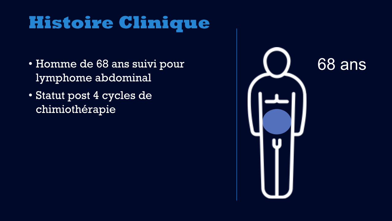
Scan abdomino-pelvien C+
Coupe sélectionnée abdomen moyen
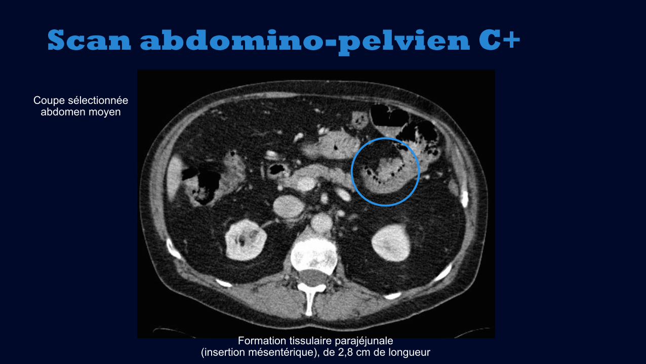
Scan abdomino-pelvien C+
Formation tissulaire parajéjunale(insertion mésentérique), de 2,8 cm de longueur
Coupe sélectionnée abdomen moyen
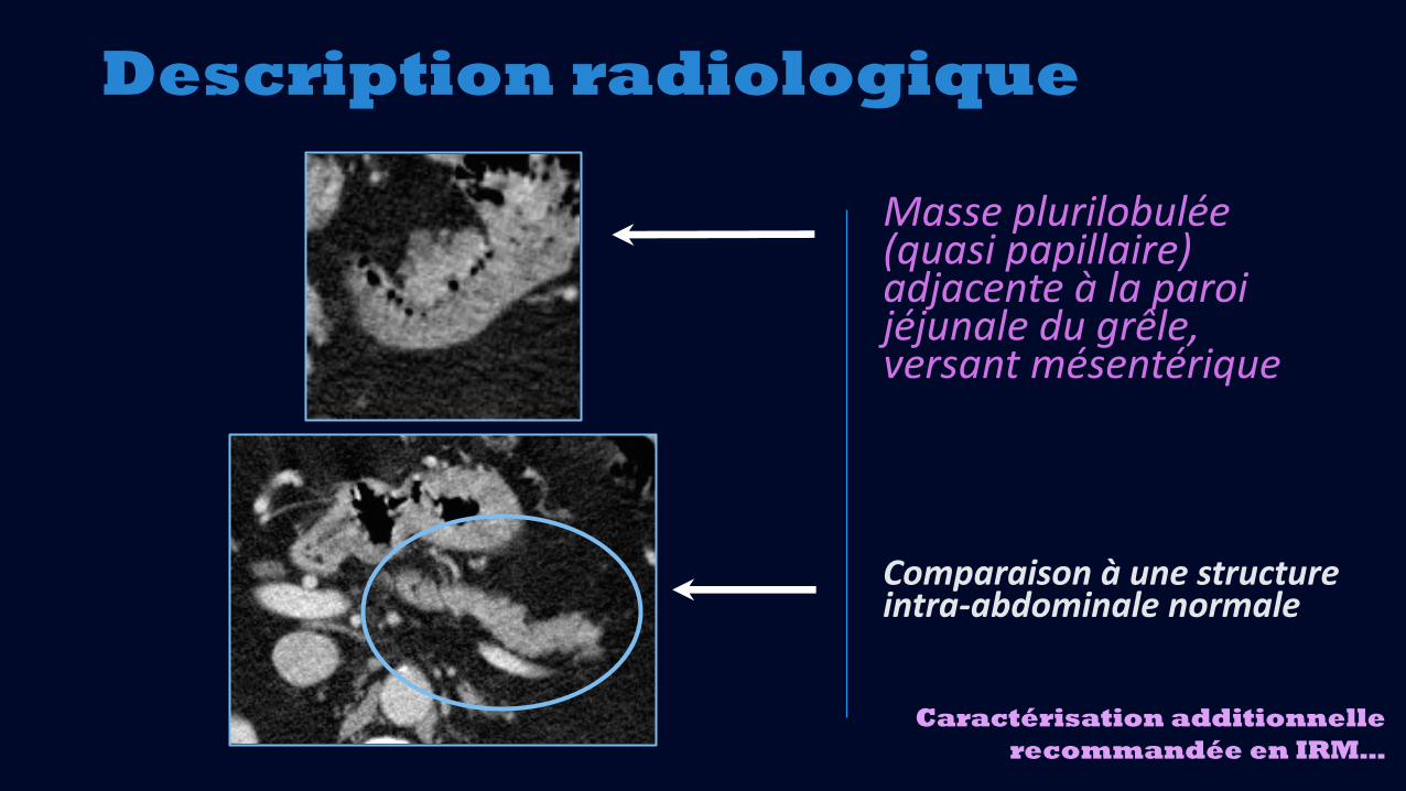
Description radiologique
Caractérisation additionnellerecommandée en IRM…
T2 sans saturation des graisses
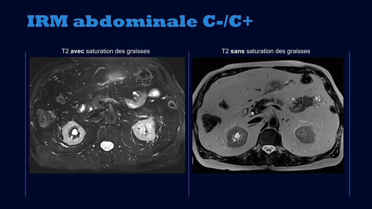
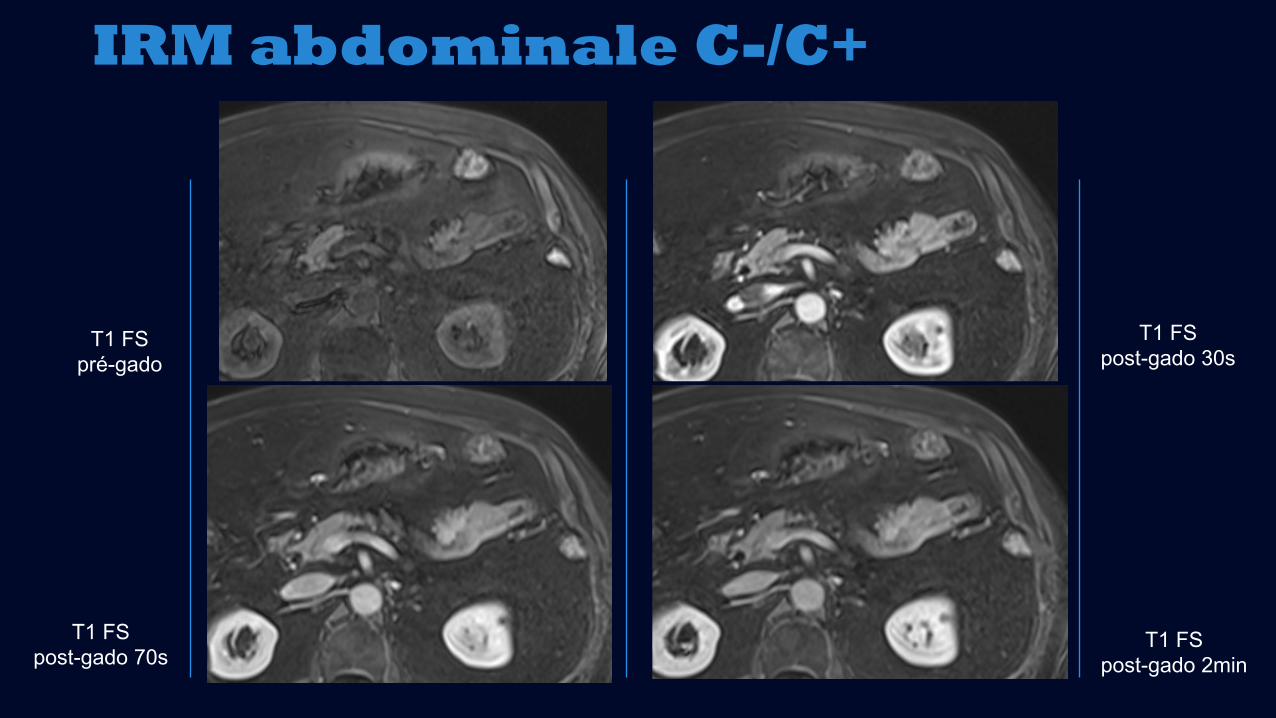
IRM abdominale C-/C+
T2 avec saturation des graisses
T2 sans saturation des graisses
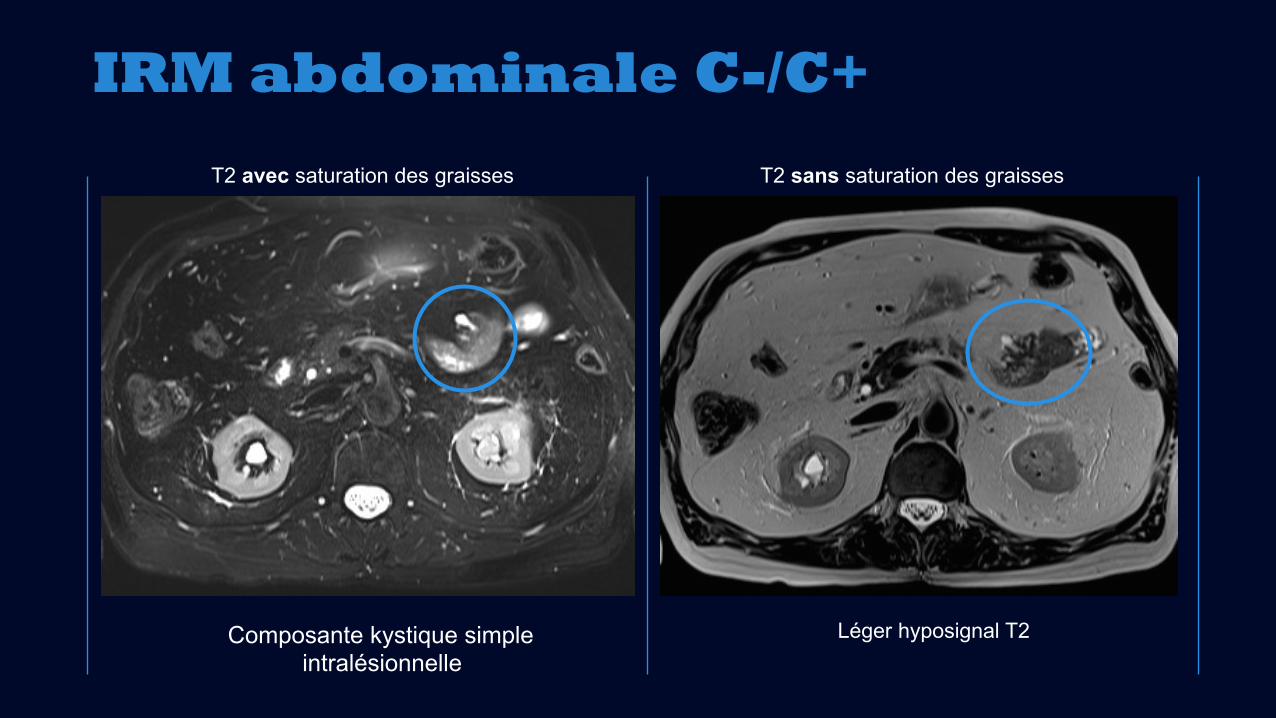
IRM abdominale C-/C+
T2 avec saturation des graisses
Léger hyposignal T2Composante kystique simpleintralésionnelle
IRM abdominale C-/C+
T1 FS pré-gado
T1 FS post-gado 30s
T1 FS post-gado 70s
T1 FS post-gado 2min
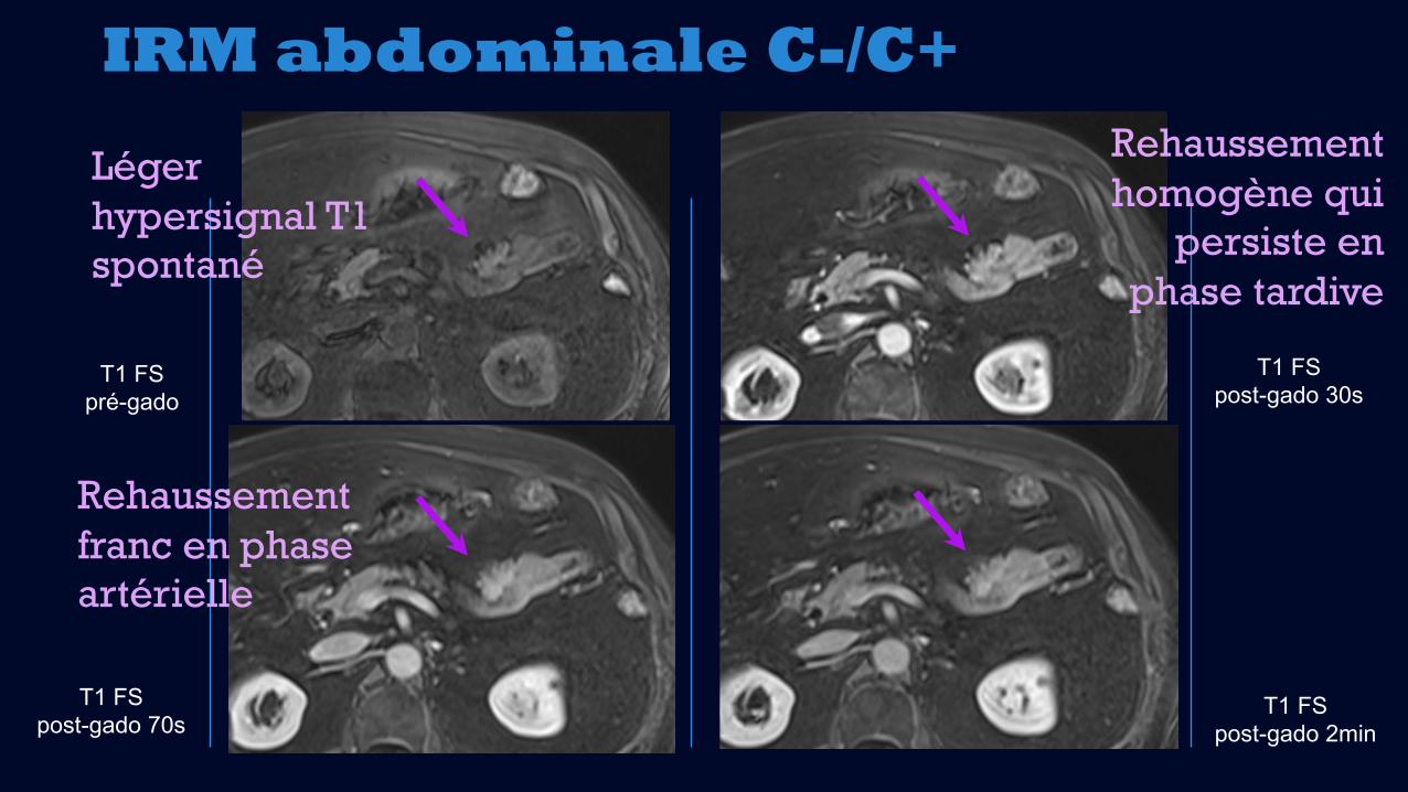
IRM abdominale C-/C+
T1 FS pré-gado
T1 FS post-gado 30s
T1 FS post-gado 70s
T1 FS post-gado 2min
Rehaussementhomogène qui
persiste enphase tardive
Léger hypersignal T1 spontané
Rehaussementfranc en phase artérielle
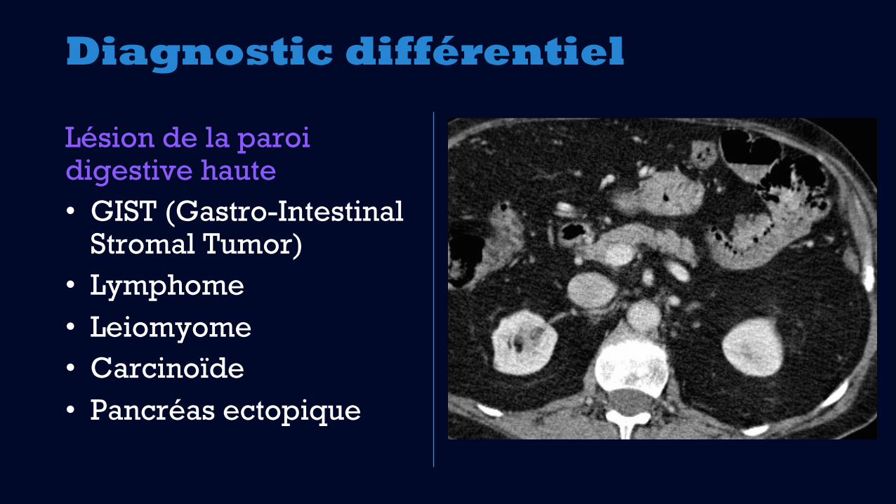
Diagnostic différentiel
Lésion de la paroidigestive haute• GIST (Gastro-Intestinal
Stromal Tumor)• Lymphome• Leiomyome• Carcinoïde• Pancréas ectopique
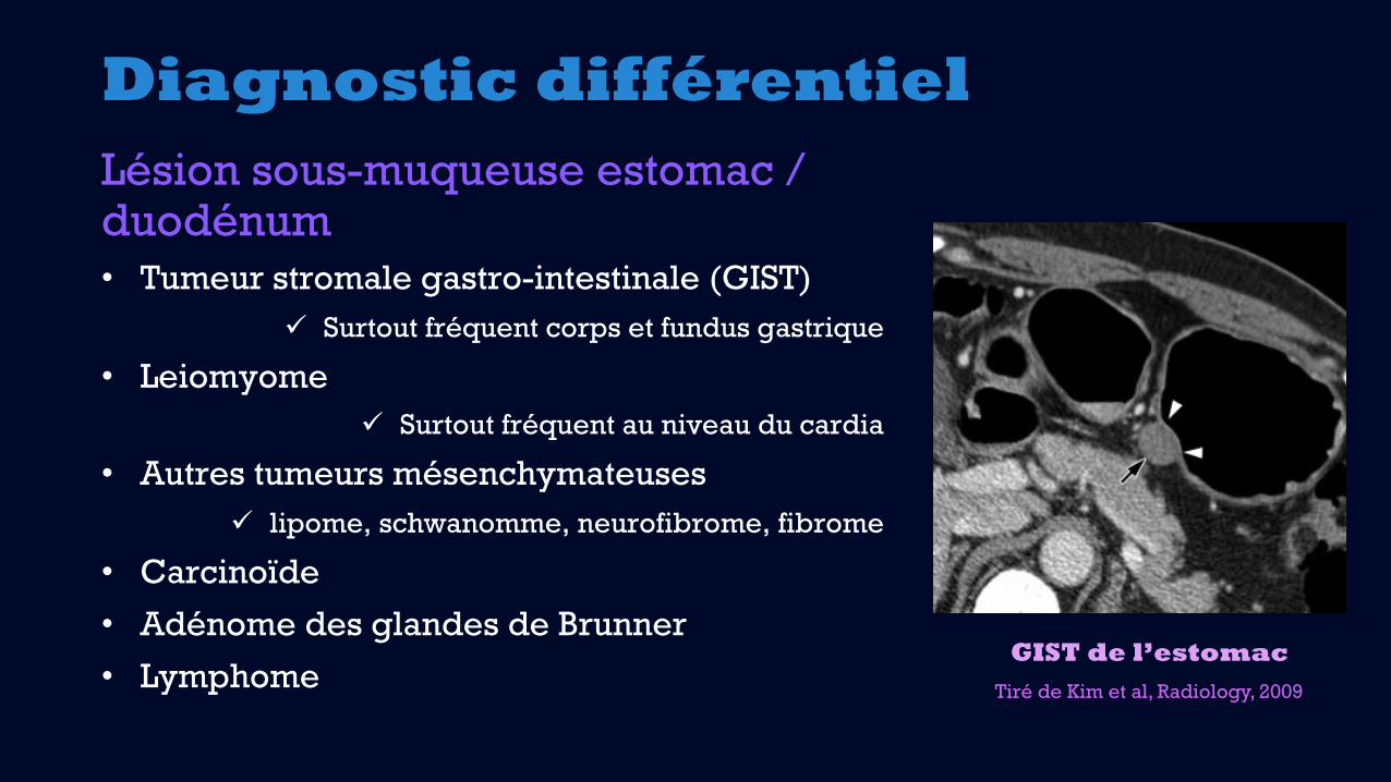
Diagnostic différentielLésion sous-muqueuse estomac / duodénum• Tumeur stromale gastro-intestinale (GIST)
ü Surtout fréquent corps et fundus gastrique
• Leiomyomeü Surtout fréquent au niveau du cardia
• Autres tumeurs mésenchymateusesü lipome, schwanomme, neurofibrome, fibrome
• Carcinoïde
• Adénome des glandes de Brunner
• Lymphome
ectopic pancreas and the two gastricsubmucosal tumors (GIST and leiomy-oma), which yielded a sensitivity of64.3%, a specificity of 82.5%, and anarea under the receiver operatingcharacteristic curve of 0.72.
Radiologic-Pathologic CorrelationIn our study, ectopic pancreases forwhich pathology slides were availablecould be classified into three subtypes ac-cording to their histopathologic composi-tion—that is, predominantly pancreaticacini (three of 11, 27%), predominantlyducts (one of 11, 9%), and mixed (sevenof 11, 64%). All three ectopic pancreaseswith predominantly pancreatic acinishowed greater enhancement and had ahigher CT attenuation value in the portalvenous phase than did the pancreas. Onthe other hand, the one ectopic pancreasthat was predominantly composed ofducts had lower CT attenuation valuesthan the pancreas and even the back mus-cles. The other seven ectopic pancreaseswith mixed composition showed variableCT attenuation values compared with thepancreas (higher attenuation, three[43%]; isoattenuation, one [14%]; lowerattenuation, three [43%]). Representa-tive images are shown in Figures 3, 4,E1 (http://radiology.rsnajnls.org/cgi/content/full/252/1/92/DC1).
Furthermore, the enhancement pat-tern (heterogeneous vs homogeneous)of the ectopic pancreases differed accord-ing to the microscopic composition of theectopic pancreases. The three ectopicpancreases with predominantly pancre-atic acini showed a homogeneous en-hancement pattern. However, all sevenlesions with a mixed composition of aciniand ducts showed a heterogeneous en-hancement pattern.
With regard to the overlying mucosa,among the four ectopic pancreases withprominent enhancement of the overlyingmucosa, two showed marked inflammationof the overlying gastric mucosa, but the re-maining two, in which the diagnosis wasconfirmed only with endoscopic biopsy,had no pathology slides appropriate forevaluating the overlying mucosa (Fig 4).Among the 10 ectopic pancreases withoutprominent enhancement of the overlyingmucosa, the nine with available pathology
Figure 3
Figure 3: Benign GIST in gastric upper body in 61-year-old man. (a) Transverse and (b) coronal CT scansshow a well-defined, homogeneously low-attenuating ovoid mass (arrow in a and arrows in b) in the gastricupper body. The lesion shows a mixed growth pattern. The overlying mucosa (arrowheads) seems to be intact,suggesting a submucosal lesion. However, the overlying mucosa does not prominently enhance comparedwith the adjacent normal mucosa. The LD/SD ratio of this lesion was 1.22 (2.2/1.8 cm).
Figure 4
Figure 4: Ectopic pancreas at gastricantrum in 51-year-old man. (a) Transversecontrast-enhanced CT scan shows anill-defined flat-ovoid 1.4-cm endoluminalsubmucosal mass (arrows) in the anteriorwall of the gastric prepyloric antrum. Thelesion shows heterogeneous enhancementand lower attenuation than back muscle,and the LD/SD ratio was 1.56 (1.4/0.9 cm).Note the prominent enhancement and mildthickening of the overlying mucosal layer(arrowheads). (b, c) Photomicrographsshow marked gastritis of (b) the overlyinggastric mucosa and (c) the mass, whichpredominantly consists of ducts (!). (He-matoxylin-eosin stain; original magnifica-tion, !20 in b and !10 in c.)
GASTROINTESTINAL IMAGING: CT Findings and Differentiation of Ectopic Pancreas Kim et al
Radiology: Volume 252: Number 1—July 2009 ▪ radiology.rsnajnls.org 97
Tiré de Kim et al, Radiology, 2009
GIST de l’estomac
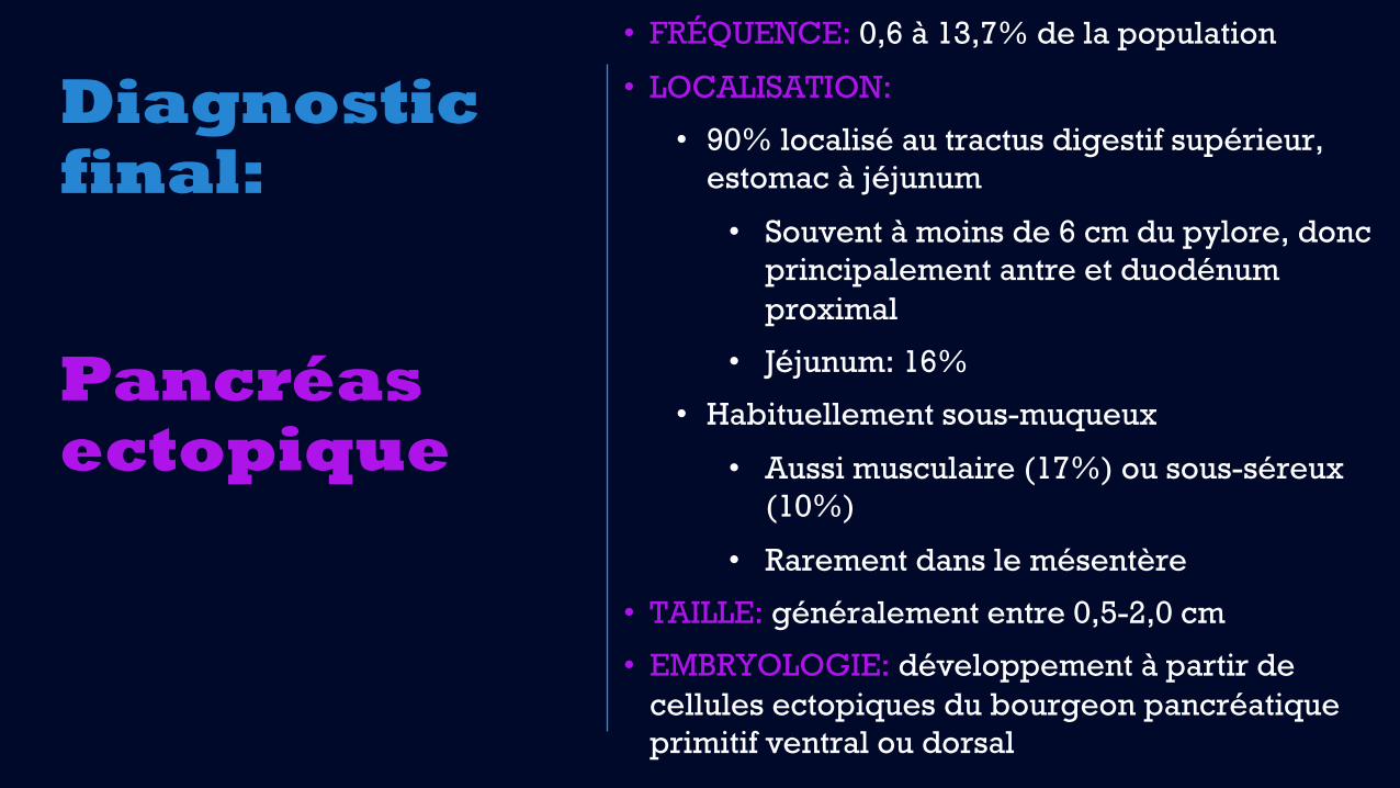
Diagnostic final:
Pancréas ectopique
• FRÉQUENCE: 0,6 à 13,7% de la population
• LOCALISATION:
• 90% localisé au tractus digestif supérieur, estomac à jéjunum
• Souvent à moins de 6 cm du pylore, doncprincipalement antre et duodénumproximal
• Jéjunum: 16%
• Habituellement sous-muqueux
• Aussi musculaire (17%) ou sous-séreux(10%)
• Rarement dans le mésentère
• TAILLE: généralement entre 0,5-2,0 cm
• EMBRYOLOGIE: développement à partir de cellules ectopiques du bourgeon pancréatiqueprimitif ventral ou dorsal
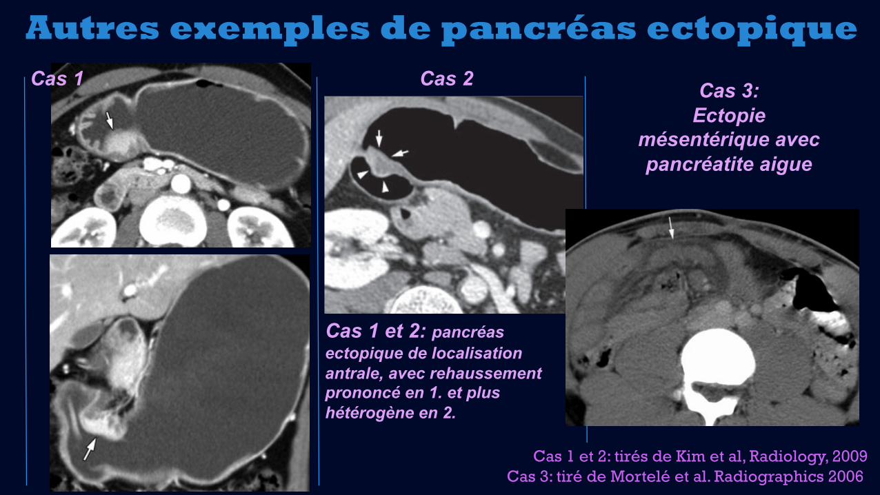
Autres exemples de pancréas ectopique
slides showed mild gastritis (n ! 3) or nomarked abnormality (n ! 6).
Sensitivity and Specificity Values for CTDiagnosisTable 3 summarizes the sensitivity andspecificity values of each significant CTcriterion for differentiating ectopic pan-creas from the two other submucosal tu-mors. Except for prominent enhance-ment of the overlying mucosa, the highestodds ratio was achieved for the border ofthe lesion, followed by the location, theLD/SD ratio, and the growth pattern ofthe lesion.
When at least two of these five crite-ria were used in combination, the sensi-tivity and specificity for diagnosing ec-topic pancreas were 100% (14 of 14)and 82.5% (33 of 40), respectively.When any four or five of these criteria
were used, a specificity of 100% wasachieved (Table 4).
Discussion
Our study results demonstrate that fiveCT findings—that is, prominent enhance-ment of the overlying mucosa, location,LD/SD ratio, growth pattern, and lesionborder—are statistically significant pre-dictors in the differentiation of ectopicpancreas from GIST and leiomyoma. Inaddition, when these CT imaging findingswere used in combination as the diagnos-tic criteria, ectopic pancreas could be dif-ferentiated from submucosal tumors witha high degree of diagnostic accuracy.When the results of our study are appliedin differentiating ectopic pancreas fromother submucosal tumors at CT, they maybe helpful in establishing an accurate di-
agnosis, thereby avoiding unnecessarysurgery or invasive procedures such asendoscopic US in patients with asymp-tomatic ectopic pancreas.
In our study, an LD/SD ratio greaterthan 1.4 was found to be the one of theimportant morphologic CT features of ec-topic pancreas for differentiating it fromother tumors (P " .05). Considering thathistologically, ectopic pancreas of thestomach is not a true neoplasm but is ahamartoma of flat glandular tissue withpancreatic acinar formation and duct de-velopment, (4,7,9,14), it is not surprisingthat ectopic pancreases may have a flat-ovoid shape (high LD/SD ratio) and anill-defined border. Only after intraglandu-lar cyst formation, an ectopic pancreasmay manifest as a large protruding sub-mucosal mass with a rather oval or roundshape (14,16). Furthermore, interest-
Figure 5
Figure 5: Ectopic pancreas in gastric antrum in 45-year-old woman. (a) Transverse and (b) coronal CTscans show an ill-defined ovoid endoluminal submucosal mass (arrow) in the gastric antrum. The lesionshows homogeneous enhancement and higher attenuation than the pancreas, and the LD/SD ratio was 1.05(2.3/2.2 cm).
Figure 6
Figure 6: Graph shows LD/SD ratios (alongy-axis) for ectopic pancreas and the other twosubmucosal tumors (GIST and leiomyoma). When1.4 was used as the cutoff value for the LD/SD ratioof the lesion, a sensitivity of 64.3% and a specific-ity of 82.5% were achieved for the differential di-agnosis of ectopic pancreas and the two gastricsubmucosal tumors.
Table 2
Quantitative Image Analysis Results for Three Types of Gastric Submucosal Tumor
CT Finding Ectopic Pancreas (n ! 14) GIST (n ! 33) Leiomyoma (n ! 7) P Value*
LD (mm) 18.4 # 5.31 28.9 # 6.93 29 # 10.38 . . .SD (mm) 12.6 # 4.68 23.67 # 6.26 23.0 # 9.26 . . .LD/SD ratio 1.52 # 0.37 1.25 # 0.30 1.33 # 0.3 .004CT attenuation value (HU) 106.07 # 30.37 98.1 # 42.11 57.3 # 19.13 .229
Note.—Data are means # standard deviations.
* Between ectopic pancreas and other tumors (GIST and leiomyoma); calculated with Student t test.
GASTROINTESTINAL IMAGING: CT Findings and Differentiation of Ectopic Pancreas Kim et al
98 radiology.rsnajnls.org ▪ Radiology: Volume 252: Number 1—July 2009
ectopic pancreas and the two gastricsubmucosal tumors (GIST and leiomy-oma), which yielded a sensitivity of64.3%, a specificity of 82.5%, and anarea under the receiver operatingcharacteristic curve of 0.72.
Radiologic-Pathologic CorrelationIn our study, ectopic pancreases forwhich pathology slides were availablecould be classified into three subtypes ac-cording to their histopathologic composi-tion—that is, predominantly pancreaticacini (three of 11, 27%), predominantlyducts (one of 11, 9%), and mixed (sevenof 11, 64%). All three ectopic pancreaseswith predominantly pancreatic acinishowed greater enhancement and had ahigher CT attenuation value in the portalvenous phase than did the pancreas. Onthe other hand, the one ectopic pancreasthat was predominantly composed ofducts had lower CT attenuation valuesthan the pancreas and even the back mus-cles. The other seven ectopic pancreaseswith mixed composition showed variableCT attenuation values compared with thepancreas (higher attenuation, three[43%]; isoattenuation, one [14%]; lowerattenuation, three [43%]). Representa-tive images are shown in Figures 3, 4,E1 (http://radiology.rsnajnls.org/cgi/content/full/252/1/92/DC1).
Furthermore, the enhancement pat-tern (heterogeneous vs homogeneous)of the ectopic pancreases differed accord-ing to the microscopic composition of theectopic pancreases. The three ectopicpancreases with predominantly pancre-atic acini showed a homogeneous en-hancement pattern. However, all sevenlesions with a mixed composition of aciniand ducts showed a heterogeneous en-hancement pattern.
With regard to the overlying mucosa,among the four ectopic pancreases withprominent enhancement of the overlyingmucosa, two showed marked inflammationof the overlying gastric mucosa, but the re-maining two, in which the diagnosis wasconfirmed only with endoscopic biopsy,had no pathology slides appropriate forevaluating the overlying mucosa (Fig 4).Among the 10 ectopic pancreases withoutprominent enhancement of the overlyingmucosa, the nine with available pathology
Figure 3
Figure 3: Benign GIST in gastric upper body in 61-year-old man. (a) Transverse and (b) coronal CT scansshow a well-defined, homogeneously low-attenuating ovoid mass (arrow in a and arrows in b) in the gastricupper body. The lesion shows a mixed growth pattern. The overlying mucosa (arrowheads) seems to be intact,suggesting a submucosal lesion. However, the overlying mucosa does not prominently enhance comparedwith the adjacent normal mucosa. The LD/SD ratio of this lesion was 1.22 (2.2/1.8 cm).
Figure 4
Figure 4: Ectopic pancreas at gastricantrum in 51-year-old man. (a) Transversecontrast-enhanced CT scan shows anill-defined flat-ovoid 1.4-cm endoluminalsubmucosal mass (arrows) in the anteriorwall of the gastric prepyloric antrum. Thelesion shows heterogeneous enhancementand lower attenuation than back muscle,and the LD/SD ratio was 1.56 (1.4/0.9 cm).Note the prominent enhancement and mildthickening of the overlying mucosal layer(arrowheads). (b, c) Photomicrographsshow marked gastritis of (b) the overlyinggastric mucosa and (c) the mass, whichpredominantly consists of ducts (!). (He-matoxylin-eosin stain; original magnifica-tion, !20 in b and !10 in c.)
GASTROINTESTINAL IMAGING: CT Findings and Differentiation of Ectopic Pancreas Kim et al
Radiology: Volume 252: Number 1—July 2009 ▪ radiology.rsnajnls.org 97
Cas 1 Cas 3: Ectopie
mésentérique avec pancréatite aigue
Cas 1 et 2: tirés de Kim et al, Radiology, 2009Cas 3: tiré de Mortelé et al. Radiographics 2006
Ectopic PancreasEctopic pancreas occurs in 0.6%–13.7% of thepopulation (32). This ectopic tissue can be foundin the stomach (26%–38% of cases), duodenum(28%–36%), jejunum (16%), Meckel diverticu-lum, or ileum. Rarely, it occurs in the colon,esophagus, gallbladder, bile ducts, liver, spleen,umbilicus, mesentery (Fig 13), mesocolon, oromentum (28,33–35). The ectopic tissue usuallymeasures 0.5–2.0 cm in its largest dimension(rarely up to 5 cm) and is located in the submu-cosa in approximately 50% of cases. Ectopic pan-creas in the gastrointestinal tract is usually asymp-tomatic, although complications such as stenosis,ulceration, bleeding, and intussusception maydevelop (28,34).
Rarely, pancreatic heterotopia can be the resultof (a) tumors developing from one of the cellularelements, including adenocarcinoma and endo-crine tumors; or (b) inflammation characterizedby the presence of fat stranding around the ec-topic pancreas (32).
Pancreatic Agenesis and HypoplasiaTotal agenesis of the pancreas is extremely rareand is incompatible with life (2,36). It is associ-ated with other malformations such as gallbladderaplasia, polysplenia, and fetal growth retardation.The mutation of a developmental protein, IPF1,is responsible for the absence of the gland.
Hypoplasia (partial agenesis) (Fig 14) resultsfrom the absence of the ventral or dorsal anlage.Absence of the dorsal anlage is visualized as ashort or truncated pancreas and can be partial orcomplete. It may be seen as a solitary finding or inassociation with heterotaxia syndromes (28). Par-tial agenesis of the dorsal pancreas is relativelymore common than agenesis of the ventral por-tion, but complete agenesis of the dorsal pancreasis extremely rare (36).
Patients with agenesis of the dorsal pancreasoften present with nonspecific abdominal pain,which may or may not be caused by pancreatitis.Many patients also have diabetes mellitus. Whenagenesis of the dorsal pancreas is suspected, it iscritical to rule out pancreatic carcinoma with up-stream atrophy of the gland (36). Isolated hypo-plasia of the uncinate process has also been re-ported (37).
At imaging, dorsal pancreatic hypoplasia mani-fests as a short, rounded pancreatic head adjacentto the duodenum with absence of the pancreatic
neck, body, and tail. In patients with completeagenesis of the dorsal pancreas, the neck, body,and tail of the pancreas, the duct of Santorini,and the minor duodenal papilla are all absent. Incases of partial agenesis of the dorsal pancreas,the size of the body of the pancreas varies, there isa remnant of the duct of Santorini, and the minorduodenal papilla is present (36).
Congenital Diseases
Pancreas
Congenital Pancreatic Cysts.—Congenitalpancreatic cysts are exceedingly rare. They have afemale predilection and typically manifest as anasymptomatic palpable mass. Patients may alsopresent with epigastric pain, jaundice, and vomit-ing related to the compression of surroundingstructures. These cysts can be single or multipleand are most commonly located in the tail andbody of the pancreas (28). Multiple congenitalcysts are associated with other anomalies, such asvon Hippel–Lindau disease (Fig 15) and hepato-renal polycystic disease (38).
Von Hippel–Lindau Disease.—Von Hippel–Lindau disease is an autosomal dominant disor-der with variable penetrance that is characterizedby retinal angiomas and central nervous systemhemangioblastomas (39). Pancreatic cysts arerelatively common in von Hippel–Lindau disease,and involvement can range from a single cyst to
Figure 13. Ectopic pancreas. CT scan obtained in apatient with abdominal pain shows ectopic pancreatictissue (arrow) within the small bowel mesentery. Sur-gery helped confirm the presence of an inflamed ec-topic pancreas.
724 May-June 2006 RG f Volume 26 ● Number 3
Radio
Gra
phic
s
Cas 2
Cas 1 et 2: pancréas ectopique de localisation antrale, avec rehaussement prononcé en 1. et plus hétérogène en 2.
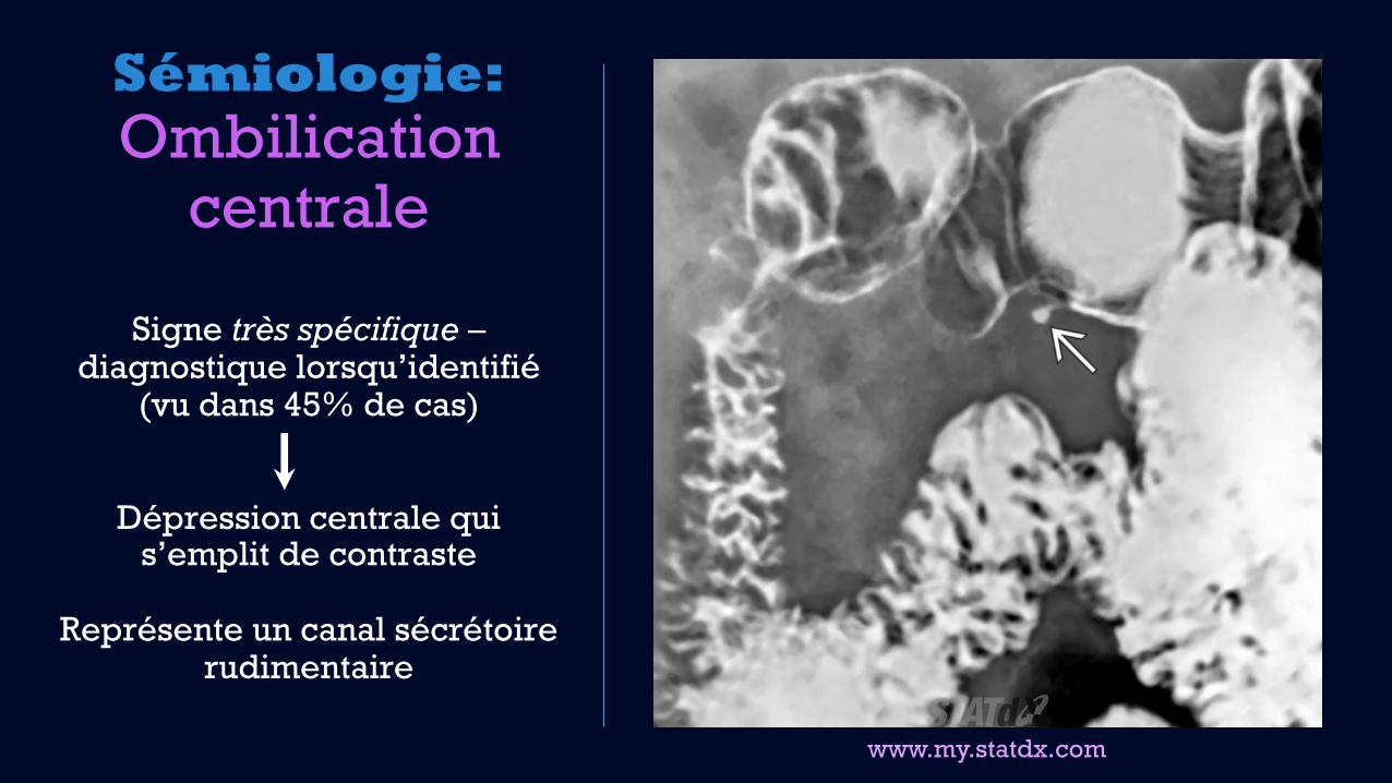
Sémiologie:Ombilication
centrale
Signe très spécifique –diagnostique lorsqu’identifié
(vu dans 45% de cas)
Dépression centrale qui s’emplit de contraste
Représente un canal sécrétoirerudimentaire
www.my.statdx.com
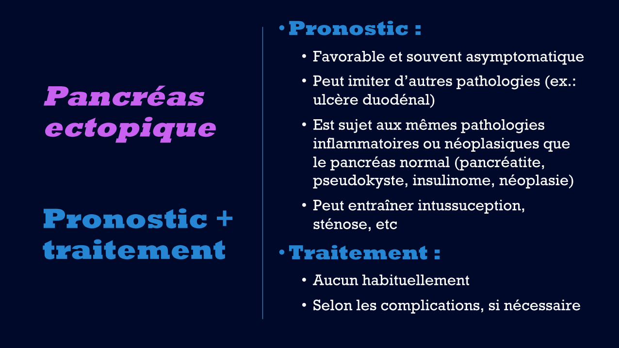
Pancréas ectopique
Pronostic + traitement
• Pronostic : • Favorable et souvent asymptomatique
• Peut imiter d’autres pathologies (ex.: ulcère duodénal)
• Est sujet aux mêmes pathologies inflammatoires ou néoplasiques que le pancréas normal (pancréatite, pseudokyste, insulinome, néoplasie)
• Peut entraîner intussuception, sténose, etc
• Traitement : • Aucun habituellement
• Selon les complications, si nécessaire
Références1. KimJY,LeeJM,KimKW,ParkHS,ChoiJY,KimSH,KimMA,
LeeJY,HanJK,ChoiBI.Ectopic Pancreas:CTFindings withEmphasis onDifferentiation from SmallGastrointestinalStromal Tumor andLeiomyoma.Radiology 2009;252:92-100.
2. Borghei B,Sokhandon F,Shirkhoda A,MorganDE.Anomalies,Anatomic Variants,andSourcesofDiagnosticPitfalls inPancreatic Imaging.Radiology 2013;266:28-36.
3. Statdx.www.mystatdx.com
4. Mortelé KJ,RochaTC,Streeter JL,TaylorAJ.MultimodalityImagingofPancreatic andBiliary Congenital AnomaliesGI.RadioGraphics 2006;26:715-731.
5. Yu J,TurnerMA,FulcherSA,Halvorsen AR.Congenitalanomaliesandnormalvariants ofthepancreticobiliarytractandthepancreas inadults:Part2,Pancreatic ductandpancreas.AmJRoentgenol 2006;187(6):1544-53.