Embed Size (px)
Citation preview

UNNERSITÉ DU QUÉBEC
THÈSE PRÉSENTÉE À
L'UNNERSITÉ DU QUÉBEC À TROIS-RIVIÈRES
COMME EXIGENCE PARTIELLE
DU DOCTORAT EN BIOPHYSIQUE ET BIOLOGIE CELLULAIRES
PAR
LAURENT ADONIS BEKALE
ÉLABORATION D'UNE NOUVELLE MÉTHODE DE PHOTOPROTECTION
DES PORPHYRINES D 'ORIGINE NATURELLE
SEPTEMBRE 2015

Université du Québec à Trois-Rivières
Service de la bibliothèque
Avertissement
L’auteur de ce mémoire ou de cette thèse a autorisé l’Université du Québec à Trois-Rivières à diffuser, à des fins non lucratives, une copie de son mémoire ou de sa thèse.
Cette diffusion n’entraîne pas une renonciation de la part de l’auteur à ses droits de propriété intellectuelle, incluant le droit d’auteur, sur ce mémoire ou cette thèse. Notamment, la reproduction ou la publication de la totalité ou d’une partie importante de ce mémoire ou de cette thèse requiert son autorisation.

UNIVERSITÉ DU QUÉBEC À TROIS-RIVIÈRES
Cette thèse a été dirigée par :
Robert Carpentier Université du Québec à Trois-Rivières
Directeur de recherche, Ph. D . Institution à laquelle se rattache l ' évaluateur
Surat Hotchandani Université du Québec à Trois-Rivières
Codirecteur de recherche, Ph. D . Institution à laquelle se rattache l' évaluateur
Jury d'évaluation de la thèse:
Robert Carpentier, Ph. D . Université du Québec à Trois-Rivières
Prénom et nom, grade Institution à laquelle se rattache l' évaluateur
Surat Hotchandani, Ph. D. Université du Québec à Trois-Rivières Prénom et nom, grade Institution à laquelle se rattache l' évaluateur
Gervais Bérubé, Ph. D. Université du Québec à Trois-Rivières Prénom et nom, grade Institution à laquelle se rattache l ' évaluateur
Subramanyam Rajagopal, Ph. D . University of Hyderabad (lndia)
Prénom et nom, grade Institution à laquelle se rattache l' évaluateur
Daniel Bélanger, Ph. D . Université du Québec à Montréal
Prénom et nom, grade Institution à laquelle se rattache l ' évaluateur
Thèse soutenue le 8 septembre 2015
La difficulté n'est pas de comprendre les idées
nouvelles, mais d'échapper aux idées anciennes.
[John Maynard Keynes J
REMERCIEMENTS
Je remerCIe mes directeurs de thèse, les Drs Robert Carpentier et
Surat Hotchandani, de m'avoir accueilli au sein de leur groupe de recherche.
Je tiens à exprimer ma sincère gratitude au Dr Hotchandani. J'ai vécu mes années
de thèse comme une aventure formidable, jalonnée bien sûr de passages difficiles.
Ma plus belle chance a été de travailler avec le Dr Hotchandani. Il m'est difficile de
trouver les mots pour exprimer toute ma reconnaissance envers Surat Hotchandani. Je le
remercie pour sa très grande disponibilité et pour ses encouragements qui n'ont jamais
cessé. Surat Hotchandani a guidé mes recherches de façon juste, précise et rigoureuse,
tout en m'expliquant chaque facette du métier de chercheur. Il m'a initié au monde de la
recherche en photophysique, photochimie ainsi qu'aux nanosciences. Il a fait en sorte
que je m'y sente bien. Je suis fier d'avoir été parmi ses étudiants en thèse.
J'aimerais également remercier le Dr Saïd Barazzouk, qui m'a initié à la synthèse
des nanoparticules et pour sa contribution à ma formation scientifique.
Mes remerciements vont également à l' ensemble des personnes qui m'ont
accompagné, de près ou de loin, durant ces années et qui ont contribué, directement ou
non, à l'aboutissement de ce travail.
Je terminerai en remercient vivement mes parents Joseph et Augustine Nteme
Eyeghe, ma sœur Merryl et mes frères Dayriss et Aimerick Martial, pour leur soutien et
leurs encouragements. Merci beaucoup.
À Julie Goudreau, mon amour, ta patience et ton attention m'ont été forcément
profitables. Merci d'être là quand il le faut. J'ai dû faire plusieurs sacrifices pour réaliser
cette thèse et tu en as souvent été la victime. Toutefois, c'est maintenant la fin de ce
projet, j'espère que tu es prête pour tous les prochains qui se présenteront à nous!
Je t'aime.
AVANT-PROPOS
Les porphyrines et les métallo-porphyrines sont des composés importants comme
cela a été reflété dans la remise des Prix Nobel de chimie en 1915, 1930, 1961 , 1962,
1965, et 1988 pour les travaux sur les fonctionnalités liées à la porphyrine biologique.
Ces composés sont bien établis comme d' excellents photosensibilisateurs qui absorbent
fortement la lumière dans la région du spectre visible avec une forte réémission par
fluorescence dans le proche infrarouge et génèrent efficacement des espèces réactives de
l'oxygène. Les porphyrines sont importantes à bien des points de vue, y compris pour le
développement de nouvelles technologies innovantes, comme le diagnostic par
fluorescence, le traitement photo dynamique, l'optoélectronique, etc. Dans ce contexte,
l'utilisation de porphyrines naturelles, ou leurs dérivés, est très attrayante, mais leur
photostabilité est loin d'être idéale.
Dans le cadre de mon doctorat, j ' ai élaboré une nouvelle méthode innovatrice de
photoprotection des porphyrines dans le but d ' aider à la mise en œuvre des technologies
de pointe à base de porphyrines naturelles.
Le présent Qocument renferme un exposé détaillé des travaux réalisés au cours des
trois dernières années de recherche sur l 'élaboration de cette approche innovante de
photoprotection. Les résultats y sont présentés dans trois publications scientifiques qui
constituent le corps de ce document, rédigé sous la forme d'insertion de ces articles .
Dans le cadre du premier article intitulé « BeneficiaI role of gold nanoparticles
as photoprotector of magnesium tetraphenylporphyrin» J Mater. Chem., 2012, 22,
2943-2951 (Chapitre In, j'ai effectué l'ensemble de la partie expérimentale, j'ai préparé
et analysé tous les résultats et j'ai rédigé l'intégralité de l'article sous la supervision des
Drs Barazzouk et Hotchandani. l'ai également participé à corriger le manuscrit et
répondu aux interrogations et suggestions des examinateurs.

VI
Dans le cadre du deuxième article : « Enhanced photostability of chlorophyll-a
using go Id nanoparticles as an efficient photoprotector » J Mater. Chem. , 2012, 22,
25316-25324 (Chapitre III), j'ai effectué la majeure partie des expériences
expérimentales, j'ai préparé et analysé tous les résultats et j'ai rédigé l'intégralité de
l'article sous la supervision des Drs Barazzouk et Hotchandani. l'ai également participé
à corriger le manuscrit et répondu aux interrogations et suggestions des examinateurs.
Dans le cadre du troisième article : « Nanosilver Could Usher in Next-Generation
Photoprotective Agents for Magnesium Porphyrins » Part. Part. Syst. Charaet., 2014,
31 , 843-850 (Chapitre IV), j 'ai effectué l'ensemble de la partie expérimentale, j'ai
préparé et analysé tous les résultats et j'ai rédigé l'intégralité de l'article sous la
supervision des Drs Barazzouk et Hotchandani. En qualité d'auteur correspondant, j ' ai
soumis, corrigé le manuscrit et répondu aux interrogations et suggestions des
examinateurs.

RÉsuMÉ
La transition vers l'utilisation de molécules multifonctionnelles comme les porphyrines, issues des écosystèmes aquatiques et forestiers , constituent un enjeu majeur pour notre société, car elle pourrait permettre de répondre aux besoins constamment renouvelés en matériaux de pointe utilisés dans les nouvelles technologies. Cette transition implique cependant de développer des moyens pour améliorer la photostabilité in vitro de ces porphyrines, ce que permettent, dans les organismes vivants, les composés antioxydants comme les caroténoïdes qui les protègent naturellement contre la dégradation par la lumière visible. Dans ce cadre, les travaux présentés dans cette thèse concernent principalement l'élaboration et la mise en place d'une nouvelle stratégie visant à améliorer la photostabilité des porphyrines de magnésium, à partir de nanoparticules de métaux nobles. Ainsi, par l'utilisation de nanoparticules d'or (AuNPs) et d'argent (AgNPs), il a été démontré qu'en bloquant les sites azotés du macro cycle porphyrique dans l'obscurité avant l'illumination, il est possible d'augmenter de manière significative la photostabilité de ces porphyrines. De plus, dans les conditions in vitro, les nanoparticules sont de bien meilleures photoprotecteurs que les molécules antioxydantes, démontrant ainsi l' efficacité de cette approche. La méthode de photoprotection développée dans cette thèse permet non seulement d'augmenter de façon significative la photostabilité des porphyrines, mais aussi induit une coamélioration de l'émission de fluorescence et de la production d'oxygène singulet par photosensibilisation. En un mot, cette thèse jette une nouvelle lumière sur la stratégie de photoprotection des porphyrines et en particulier sur la nécessité de préserver la multifonctionnalité, qui est un caractère extraordinaire de ces molécules et qui constitue le principal facteur recherché pour des applications technologiques.
Mots-clés : photoprotection, porphyrines, nanoparticules d'or, nanoparticules d'argent, augmentation de la fluorescence, augmentation d'espèces réactives de l'oxygène.
TABLE DES MATIÈRES
REMERCIEMENTS ................................ :................................................................ iv
AVANT-PROPOS...................................................................................................... v , ,
RESUME..................................................................................................................... VII
LISTE DES FIGURES .............................................................................................. xii
LISTE DES ACRONYMES, SIGLES ET SYMBOLES ........................................ xiv
CHAPITRE 1 INTRODUCTION GÉNÉRALE .............................................................................. 1
1.1 Mise en contexte : généralités sur les porphyrines et leurs dérivés........... ......... 1
1.2 Structure des porphyrines ................................................................................... 3
1.3 Nomenclature. ...... ..... .. ... ...... ........ .. .. ............ .............. ....................... ... ...... ........ . 4
1.4 Propriétés des porphyrines ................... .. ................................................ .......... .. . 5
1.4.1 Propriétés optiques........... ................................ .. ...... ............ .............. .... . 5
1.4.2 Propriétés photochimiques....... .. ...................... .. ..... .. .............................. 7
1.5 Intérêts et limites des porphyrines endogènes ... .. .. .... ................................ ..... ... . 9
1.5.1 Applications potentielles. .. ......... .... ............................ ... ............. .. .. .. .. .. ... 9
1.6 Problème ................ ... ... ............. .. ......... ... ...... .. ........ .......................................... .. 12
1.7 Revue de la littérature sur les différentes méthodes de photoprotection des porphyrines naturelles et leurs dérivés....... .................... ..... .. .. ... ................... ..... 12
1.8 Présentation du projet de thèse .. ........ .. .. ... ..... .. .... .... .. .... ...... ...... .... ........ .... .... .. .. . 15
1.9 Notions essentielles sur l'augmentation de l'émission de fluorescence d'un fluorophe par un métaL..... ............................................... ..... ............... .... ........... 18
LION otions essentielles sur l'augmentation de la production d'oxygène singulet du photosensibilisateur par un métal ..................... .. ............ ............ .......... ...... ... 21
1.11 Hypothèses de recherche.. ..... ..... ... .. ...................... ..... .... ...... ...... ... .. .... ............... 22
1.12 Objectif général de cette thèse ..... .. ................. ... .... ............................................ . 23
1.13 Objectifs opérationnels .... ............................ .... .... .. .. .... .............. .. .. .. ............ .. .. .. . 23
1.14 Organisation du document....... ...... ... ... ......... ...... ....... ......................................... 23
1.15 Références............. ... .. .......................... .. .. .. ...... ....... ............................................ 24

IX
CHAPITRE II BENEFICIAL ROLE OF GOLD NANOPARTICLES AS PHOTOPROTECTOR OF MAGNESIUM TETRAPHENYLPORPHYRIN ..... 32
2.1 Résumé de l'article ................. ....... ........................................................... ... .. ..... 32
2.2 Premier article scientifique ..... ..... .. ... ... ..... ....... ... .. .. .. .. ..... ...... ... .................... . ... .. 33
Abstract ...... ... ..... .................................................................. ...... .. ...... .. ...... .... ..... 33
Introduction.. ...... .. ...... ........... ..... ... ..... ... ..... ........... ....................................... ... ... . 34
Experimental section ..... ...... ..... . ............ .. ....... .. . .... .............. ..................... ... ....... 35
Materials and methods ............ .. ...................... ... ........ ................ ........ .. .. .. ..... ...... 35
Synthesis of Au nanoparticles.... ...... ......... ..... .. ...... .. ...... .. ...... .. .. .. ...... ... .. 35
Photodegradation measurements ... .. ...... .. .................... ........ .... ...... ......... 36
X-Ray Photoemission Spectroscopy (XPS) ........ ... .. .. .. .. ...... ......... ...... .. .. 36
Percentage photoprotection......................... ... .. .. .... ................................. 37
Determination of the fraction of the light absorbed by MgTPP (5.3 /lM) and AuNPs of varying concentrations (2-130 /lM) in the mixture ofMgTPP and AuNPs ............. .... ..... .. ...... .............. .... .. ...... ...... . 37
Results and discussion .. ..... .. ...... ... ..... ... ..... .. ...... .. ............... ..... ................... ... ..... 39
Conclusions. .. .. .. .. ... ... ... ..... ... ... .. .. ...... ................. .. ..... .. .. ... ..... ... .... .. ............. ..... .. 49
Acknowledgments ...................... ....... .. ...... .. ...... ............. ........ ... ... ... .. ......... ... .. .. . 49
Références.. .. .......... .... .. ...... .. ...... .. ...... ..... ........................ ........................... .. .. .. ... 50
CHAPITRE III ENHANCED PHOTOSTABILITY OF CHLOROPHYLL-A USING GOLD NANOP ARTICLES AS AN EFFICIENT PHOTOPROTECTOR....................... 70
3.1 Résumé de l'article .......... ... ....................... .. ...... .. ...... ... .... ...... ..... ................ .... ... 70
3.2 Deuxième article scientifique ..... ............................. ........................... ... ..... ..... ... 71
Abstract .. ......................... .... ........................ ........ ....... ......................................... 71
Introduction... ............. .. .... .. .. .... .............. ............. ....... ....................... ... .. .... ....... .. 72
Materials and methods ....... .. ...... ........................ .. .... .... ... .. ...... ..... ........ .... .. ......... 73
Materials ... .. ... .. ...... .. ...... .. ...................... .. ...... .. .. .. .. .. ... ... .. ...... .. ...... .. ...... . 73
Synthesis of gold nanoparticles (AuNPs) ...... ............ ............................. 73
Photodegradation measurements ... .. ...... .. .............. ..... ... ................. ... .. ... 74
Absorption and emission spectrometry... ............... .. ...... .. ...... .. .. ....... ...... 74
Transmission electron microscopy (TEM) .................... .. ...... ... ..... ......... 74

x
X-ray photoemission spectroscopy (XPS) ... .. .. ...... .. .. ........................ ..... 75
Percentage photostability ... ...... .. ... .... ... ...................................... ... ....... ... 76
Results ... ...... ....... .. ............... .. ...................................... .. ......... ..................... .. ..... 76
Photodegradation of chlorophyll-a (ChIa)......................... ... ........... .. .... . 76
Photoprotective action of go Id nanoparticles.. ........ ............. ..... .............. 77
X-ray photoelectron spectroscopy (XPS) studies ....................... ............ 80
Comparison of the photoprotective ability of AuNPs with other Chla photoprotectors... ....... .. ....... ....... ... ............. .. ....... ........ .... .... ........ .... 83
Discussion...................................... .. .. .. .. .. ....... ...... .... ........................................ .. 84
Conclusions .................................. .... ............................................ ... ............. ..... . 86
Acknowledgments ............................................................................... ...... ...... ... 87
Références.... ......... ............ ...... .... .... ... ... ... ... ........ ....... .............................. ...... ... .. 87
CHAPITRE IV NANO-SILVER COULD USHER IN NEXT GENERATION PHOTOPROTECTIVE AGENTS FOR NATURAL PORPHYRINS.................. 109
4.1 Résumé de l'article .... .. ... ... ...... .. ...................................... ... ........... ... ........... ....... 109
4.2 Troisième article scientifique ... ...... ........... ................... ... ......... .. .. ...... ....... ....... .. 110
Abstract.. ........... ....... .. ......... ......... ................ ....................................... ... ..... ....... . 110
Experimental Section.. .... ............................. ... ......... ... .. ..... .. ...... .. .............. .... ..... 122
Acknowledgements ....................................................... .............. ......... .......... .... . 125
References ............. .... .. .. ....... .. .... ... ........................................... .. ......................... 125
CHAPITRE V CONCLUSION ET PERSPECTIVES ..................................................................... 146
5.1 Conclusion générale ...... ...................... ........ ...... ....... ..................... ...................... 146
5.2 Bilan des résultats .............. .. ...... .. ... ...... ... .. .. ...... ................. ............... ......... ..... .. . 147
5.3 Contribution de la thèse à l'avancement des connaissances. ....... ......... ..... ...... ... 150
5.4 Perspectives .... ......... ..... .. .. ............................................... .... .. ........ ...... .. ......... .... 150
5.4.1 Analyser systématiquement l'effet de la taille et de la forme des nanoparticules sur l'efficacité photoprotectrice. ....... ....... .. ....... ....... .. ..... 151
5.4.2 Examiner si la méthode de photoprotection mise en œuvre dans cette thèse peut être employée pour l'élaboration des nanoparticules fonctionnelles pour des applications médicales. .... ... ... .... ............ .... ....... 153
5.5 Références............. ...... ...... ... .... .... .. ........................ ............................... .............. 155

Xl
ANNEXE A MÉTHODES EXPÉRIMENTALES, SYNTHÈSE ET INSTRUMENTATION. 157
A.I Synthèse générale des nanoparticules métalliques en solution........... ... .. .. ......... 157
A. 1. 1 Préparation des nanoparticules d'or (AuNPs) ............. ........... .... ......... .... 158
A.1.2 Préparation des nanoparticules d'argent (AgNPs) ..... ....... ............... .... ... 159
A.2 Caractérisation des nanoparticules ......................................... .... ...... ......... .. ...... . 160
A.2.1 Microscopie électronique à transmission (MET)....... .................. ... .... .... 160
A.2.2 Spectrométrie d'absorption UV-visible ......... ............. ..... ............. ... ....... 160
A.2.3 Spectroscopie de photo électrons X (XPS) .................. ..... .. .......... .... .. ..... 161
A.3 Références ..................... ...... ........ .... ... ... ... ........... ...... ... .. ...... ..................... .. ....... . 161

LISTE DES FIGURES
Figure Page
1.1 Structure de quelques porphyrines endogènes ..... ..... .... ................................. 2
1.2 Structure chimique de porphyrine: (a) Porphyrine base libre; (b) Porphyrine base métallée ou métallo-porphyrine; (c) Représentation du système de 18 électrons 1t aromatiques ...... ... ............................ ... ... .... ...... 4
1.3 Système de nomenclature du macrocycle tétrapyrrolique ... ... ....... .... .... ... ..... 5
1.4 Spectres d'absorption et d'émission de la porphyrine H2TPP dans le toluène à 25°C....................................... .. .. ... ... ...... ...... ...... ... ... .. ....... .............. 6
1.5 Diagramme de Jablonski décrivant les transitions non radiatives et radiatives, impliquées lors de l'absorption d'un photon et de sa réémission par luminescence (fluorescence et phosphorescence) ............................ .. .. ... 7
1.6 Illustration schématique de la production des dérivés réactifs de l'oxygène par réaction photochimique des porphyrines .. .. ...... ................. ... ..... .......... .... 8
1.7 Diagramme de Jablonski qui montre qu'on peut inactiver de manière photodynarnique les agents pathogéniques ............ ................... .. ...... .. .. .... .. ... 10
1.8 Mécanisme de photodégradation du tétraphénylporphyrine de magnésium (MgTPP) avec formation d'époxyde .................... .. ..... ......... .. ...... ...... .. ......... 15
1.9 Mécanisme de photodégradation des dérivés de la chlorophylle....... ... .. .... ... 16
1.10 Représentation de la formation d'un plasmon de surface sur une nanoparticule métallique... ... .................. ..... .. ... ... .. ...... .... .. .... ...... .. .. ......... ..... . 19
1.11 Modification du processus d'absorption et de déexcitation par une nanoparticule métallique à proximité d'un fluorophore .............................. .. 21
1.12 Diagramme énergétique de la production d'oxygène singulet photosensibilisée via le couplage plasmon surface-photosensibilisateur ...... 22
5.1 Solutions de nanoparticules d'or de différentes tailles. La différence de taille provoque la différence de couleur.. ...... ... ..... .. ..... ........ ... ...... ... ..... ...... ... 152
5.2 Spectres de diffusion de la lumière par des nanoparticules d'argent de différentes formes ... ...... .. ... ... .. ...... ........ .. ...... .. ...... .. ............................ ..... ...... 152

Xlll
5.3 Représentation schématique de l' agent photoprotecteur magnétique de structure cœur-coquille Fe304@Au .... ... ..... ...... .. ...... ........ ........ .. .. .... .. ........ ... 154
5.4 Schéma illustrant le ciblage in vivo de la tumeur par champ magnétique externe ... ........ .... ...... ... ... ...... .............. ........ .. .... .. .. .. ................... ... .. ...... .. ... ..... . 155

Au
Ag
AgNPs
AuNPs
ChIa
ChI
eV
HOMO
H2TTP
H20 2
K app
Ks
Ksv
mL
mM
MEF
ME10 2
MgTPP
MTPP
nM
nm
NIR
NP
LISTE DES ACRONYMES, SIGLES ET SYMBOLES
Or
Argent
Nanoparticules d'argent
Nanoparticules d'or
Chlorophylle-a
Chlorophylle
Électronvolt
Niveau énergétique le plus haut occupé
Tétraphenyl porphyrine base libre
Peroxyde d'hydrogène
Constante d'association
Constante de formation
Constante de Stem-Volmer
Millilitre (10-3 L)
Millimole (10-3 M)
Augmentation de l'émission de fluorescence par le métal
Augmentation de la génération d'oxygène singulet par le métal
Tétraphénylporphyrine de magnésium
Tétraphénylporphyrine métallique
Nanomole (10-9 M)
Nanomètre (10-9 m)
Proche infrarouge
N anoparticule

XV
O2,- Anion superoxyde
102 Oxygène singulet
302 Oxygène moléculaire
OH· Radical hydroxyle
OLEDs Diodes électroluminescentes organiques
OPV Photovoltaïque organique
PDT Thérapie photo dynamique
IpOrp État fondamental singulet de la porphyrine
3pOrp* Porphyrine en état triplet
IpS État fondamental singulet du photosensibilisateur
IpS* État singulet excité du photosensibilisateur
3PS* État triplet du photosensibilisateur
3porp* État triplet de la porphyrine
PS Photosensibilisateur
~W Tétraalkyl ammonium
ROSs Dérivés réactifs de l'oxygène
TOAB Bromure de tétraoctylammonium
~L Microlitre (10-6 L)
~M Micromole (10-3 M)
À Longueur d'onde

CHAPITRE 1
INTRODUCTION GÉNÉRALE
1.1 Mise en contexte: généralités sur les porphyrines et leurs dérivés
Les porphyrines, les métalloporphyrines et leurs dérivés sont parmi les composés
hétérocycliques aromatiques les plus étudiés de tous les composés macrocycliques 1. Ces
molécules peuvent être distinguées selon leur origine : soit d'origine endogène, si elles
sont synthétisées à l'intérieur d 'un organisme animal ou végétal, soit d'origine exogène,
si elles ne le sont pas (c'est-à-dire qui proviennent des laboratoires). Les porphyrines
entrent dans la composition de nombreux pigments et servent de base dans la formation
de certaines molécules chez les organismes vivants. Les porphyrines endogènes
participent à de nombreux processus physiologiques essentiels, dans bon nombre de
cellules vivantes aussi bien végétales qu'animales (Figure 1.1). À titre d'exemple, l'on
peut citer la cyanocobalarnine, plus connue sous le nom de vitamine B 12, qui est
indispensable au bon développement du corps. Combinées à un ion fer, les porphyrines
animales forment l'hème, un motif nécessaire à la fonction de nombreuses
métalloprotéines : l'hème de l'hémoglobine des globules rouges assure le transport de
l'oxygène. et du dioxyde de carbone dans le sang. Enfin, liées à un ion magnésium, les
porphyrines végétales forment la chlorophylle, indispensable à la photosynthèse. Cette
classe de molécules organiques colorées, interagissent avec la lumière et comptant parmi
les éléments fondamentaux de la vie, ne cesse de piquer la curiosité des chercheurs. Leur
étude nourrit aujourd'hui des applications biotechnologiques innovantes dans des
domaines aussi variés que la médecine, l' agronomie, la microélectronique ou
l'optoélectronique. Les détails sur leurs potentielles applications sont fournis à la
section 1.5.

H'"
Me M. ~ ~N "lr
MeU ,) OH ~ HN 0 NY:r P 1 .... Me 0 "'" \ ' 0 "'\
0- H o
HOCH2
Me
Vitamine B 12
Le groupement hème de l'hémoglobine
chlorophYlle a : --' Rl = ; R2 =Me
chlorophylle b : --'
R l = ; R2 =-CHO
Me
chlorophylle c : ~
Rl= ; R2 = Me
Me Me Me
phytyle=
Chlorophylles
Figure 1.1 Structure de quelques porphyrines endogènes.
2
; RJ = phytyle
; RJ = phytyle
; RJ = phytyle
Me

3
1.2 Structure des porphyrines
Les porphyrines forment une classe de composés cycliques dans lesquels le
macrocycle principal se compose de quatre unités de type pyrrole, reliées entre elles par
des ponts de carbone simple (méthine) (Figure 1.2). Ces macro cycliques tétrapyrroliques
mesurent environ 9 A et possèdent une cavité centrale d'environ 4,2 A de diamètre2. La
cavité centrale de la porphyrine possède une taille idéale pour être occupée par les
métaux de transition de la première série (fer, cobalt, nickel, cuivre, zinc, ect), les ions
métalliques plus volumineux sont localisés au-dessus du macrocycle, ce qui oblige la
porphyrine à adopter une conformation en forme de dôme. Les porphyrines sont des
composés aromatiques possédant 18 électrons 1t conjugués qui leur confèrent une forte
coloration et une fluorescence intrinsèque. Le macrocycle tétrapyrrolique aromatique est
stable thermiquement, chimiquement et biologiquement. Ces composés ont également
l' avantage d'être non toxiques lorsqu ' ils ne sont pas soumis à la lumière3. Toutes les
molécules issues des porphyrines (l'hème, la chlorophylle ou macrocycles dérivés) sont
construites sur le même schéma : un anneau tétrapyrrolique au sein duquel peut être
niché un ion métallique (Figure 1.2).

(
NH
s.sA
# N
4.2 A!
/f Sous-unité pyrrole
(a)
4
)
~ k- Pont méthine
(b)
(c)
Figure 1.2 Structure chimique de porphyrine: (a) Porphyrine base libre; (b) Porphyrine base métallée ou métallo-porphyrine; (c) Représentation du système de 18 électrons 1t aromatiques.
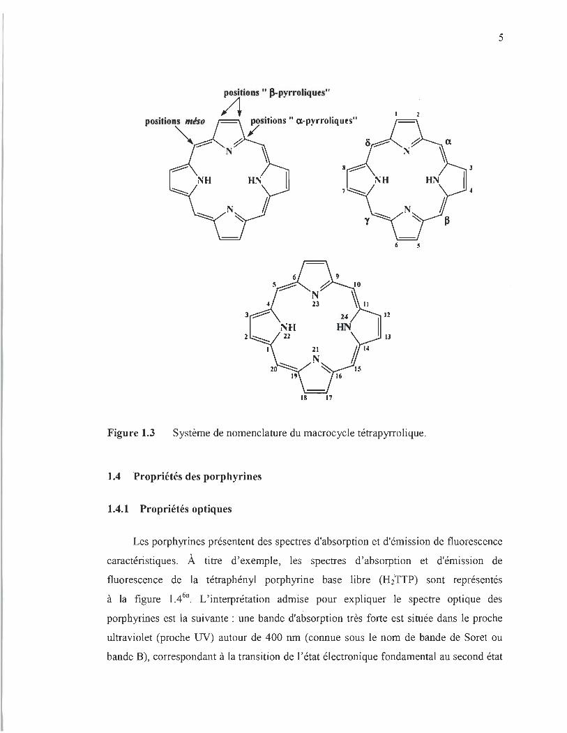
1.3 Nomenclature
Le chimiste allemand Hans Fischer, lauréat du Prix Nobel de chimie en 1930 pour
ses études sur les porphyrines, proposa un système de nomenclature, basé sur un système
de numérotation de 1 à 24, pour désigner les porphyrines substituées sur les positions
~-pyrroliques4. Dans cette nomenclature, les atomes de carbone des positions méthines,
aussi appelés «méso », sont numérotés a, ~, y et (5 et les carbones ~-pyrroliques sont
notés 1, 2, 3, 4, 5, 6, 7 et 8 (Figure l.3). Par la suite, une numérotation systématique du
macrocycle a été adoptée en 1987, afin de simplifier la nomenclature de ces molécules5.
Ainsi, les carbones méso portent alors les numéros 5, 10, 15, 20, les positions a et ~ des
cycles pyrroliques sont notés respectivement 1, 4, 6, 9, Il , 14, 16, 19 et 2, 3, 7, 8, 12, 13,
17, 18 et enfin les numéros 21 , 22, 23, 24 ont été attribués aux quatre atomes d'azote
(Figure l.3).
5
2
6 5
23
24
NH UN 22
21
15
18 17
Figure 1.3 Système de nomenclature du macrocycle tétrapyrrolique.
1.4 Propriétés des porphyrines
1.4.1 Propriétés optiques
Les porphyrines présentent des spectres d'absorption et d'émission de fluorescence
caractéristiques. À titre d'exemple, les spectres d'absorption et d'émission de
fluorescence de la tétraphényl porphyrine base libre (H2·TTP) sont représentés
à la figure 1.46a. L'interprétation admise pour expliquer le spectre optique des
porphyrines est la suivante : une bande d'absorption très forte est située dans le proche
ultraviolet (proche UV) autour de 400 nm (connue sous le nom de bande de Soret ou
bande B), correspondant à la transition de l'état électronique fondamental au second état

6
électronique singulet (S2~SO). Cette bande disparaît lorsque l' aromaticité du
macrocycle est rompue. Dans la région du spectre visible, il existe quatre bandes
supplémentaires autour de 500 à 760 nm dénommées bandes Q, responsables de la
coloration intense de ces composés. Ces bandes d 'absorption sont le résultat de la
transition de l' état · électronique fondamental au premier état électronique singulet
(Sl~SO) . L' intensité des bandes Q des porphyrines est généralement sensible aux
variations de structures du macrocycle. En effet, les bandes Q voient leur intensité
relative varier considérablement, en fonction de la nature et de la position des
substituants sur le macrocycle7. La plupart des porphyrines naturelles, telles que les
chlorophylles, les bactériochlorophylles et leurs dérivés, ont des bandes d 'absorption Q
dans le proche infrarouge entre 650 et 780 nm et plusieurs ont été étudiées par divers
chercheurs8 pour leur utilisation comme agent photosensibilisateur. Par surcroît,
l'excitation des chromophores porphyriques conduit à l'observation de deux fortes
bandes d'émission de fluorescence, situées dans le NIR autour de 670 à 720 nm. Les
interprétations du comportement photophysique des porphyrines sont résumées sur le
diagramme de Jablonski, représenté à la figure 1.5.
1.6 20.0x10³
1.4 S2~SO - - x10Abs
Soret or 8(0,0) y -- Absorbance
1.2 x.J -- Emission 15.0x10³
:!! C 0
1.0 ..,
GJ Q(O,O) ID () (/1
c So S1 Cl ~ ID
of 0.8 10.0x10³ ::J Cl
0 Qy(1 ,O) ID
1/1 S1 So :;-.&l
ct 0.6 ~ ::J (/1
0.4 5.0x10³ ~
0.2
0.0 0.0
400 500 600 700 800
Figure 1.4 Spectres d 'absorption toluène à 25°C6a
.
et d 'émission de la porphyrine H2 TPP dans le

7
82
T2
« 8 1
CI)
~ c:: T1 ro 0 _.-CI)-
-0.0 c:: ~ ::J 0 o CI) ~.o oro
Figure 1.5 Diagramme de Jablonski décrivant les transItions non radiatives et radiatives, impliquées lors de l'absorption d'un photon et de sa réémission par luminescence (fluorescence et phosphorescence) 6b.
1.4.2 Propriétés photochimiques
Non seulement l'excitation des chromophores porphyriques produit généralement
de la photoluminescence (fluorescence ou phosphorescence), mais elle permet
également la production des dérivés réactifs de l'oxygène (en anglais reactive oxygen
species, ROS). comme l'anion superoxyde (02 '-), l'oxygène singulet e02), le radical
hydroxyle (OH'), le peroxyde d'hydrogène H20 2, etc. Lorsque le photosensibilisateur
(PS) à l'état fondamental singulet epS) est stimulé par une lumière correspondant à son
pic d'absorption, il est promu à son état singulet excité (lpS*). Dans cet état, plusieurs
processus de dés excitation peuvent se produire rapidement, afm de ramener le 1 PS * à
son état fondamental IpS. Cependant, le processus de désexcitation indispensable à la
formation des ROSs est la conversion intersystème (ISC pour intersystem crossing), à
l' origine de la formation des PSs à l'état triplet e pS*). Cet état triplet est moins
énergétique que l'état singulet excité et possède une durée de vie beaucoup plus longue

8
que celle de l'état singulet excité (micro/millisecondes au lieu de nanosecondes).
Le 3PS* peut revenir à l'état fondamental en émettant son énergie sous forme d'un
photon (phénomène de phosphorescence), ou bien il peut interagir avec des molécules
abondantes dans son environnement immédiat, tel que l'oxygène. L'interaction du 3PS* avec l'oxygène peut se produire de deux façons distinctes :
• Soit le 3PS * transfère directement un électron, parfois de concert avec un don
de protons à la molécule d'oxygène pour former O2.-, pouvant par la suite
former d'autres ROS comme le OH· et le H20 2 (Figure 1.6). Cette réaction
photochimique est appelée réaction de Type 19.
• Soit le 3PS* transfère son énergie par collision à la molécule d'oxygène.
Compte tenu des règles de sélection qui spécifient que les interactions triplet
triplet sont autorisées, alors que les interactions triplet-singulet sont
interdites \0, le 3PS * peut donc réagir facilement avec l'oxygène moléculaire,
302 l'une des rares molécules de la nature dont l'état fondamental est un état
tripletll . Cette réaction photochimique, connue sous le nom de réaction de
Type II, est à l'origine de la formation du 102 (Figure 1.6)9.
Figure 1.6
relaxation ISC IpS* excited singlet
3pS* triplet state
absorption fluorescence
H.a . 0 : .. OH
1 PS ground singlet state
Illustration schématique de la production des dérivés réactifs de l'oxygène par réaction photochimique des porphyrines9
.

9
Les propriétés optiques et photochimiques évoquées ci-dessus incitent aujourd'hui
les chercheurs à explorer le potentiel des molécules de porphyrines, en vue de
développer des technologies novatrices et performantes pour le génie biomédical,
l'optoélectronique, l' environnement, etc.
1.5 Intérêts et limites des porphyrines endogènes
1.5.1 Applications potentielles
Bien que le rôle biologique et physiologique des porphyrines dans leur milieu
naturel soit bien connu, leur potentiel d'utilisation dans le développement des
biotechnologies commence à peine à être élucidé. Du fait que les maximas des bandes
d'absorption et d'émission des porphyrines naturelles sont situés dans la fenêtre
thérapeutique (typiquement entre 650 et 900 nm), c'est-à-dire là où l' absorption globale
des tissus biologiques est minimale, ils peuvent être utilisés comme agent de contraste
pour l'imagerie optique · par fluorescence. En effet, Fan et ses collègues ont étudié la
possibilité d'utiliser les porphyrines endogènes comme produit de contraste pour
l'imagerie médicale l2. Ils ont démontré que les molécules de chlorophylles, extraites des
feuilles de Chimonanthus salicifoliues, pourraient servir à l'imagerie clinique des
ganglions sentinelles, c'est-à-dire, les premiers ganglions axillaires les plus proches de la
tumeur et donc susceptibles d'être envahis par les cellules cancéreuses. De plus, leurs
résultats des tests de cytotoxicité confirment que les molécules de chlorophylles
présentent une très faible toxicité. Ceci suggère que ces composés abondants dans nos
écosystèmes pourraient potentiellement être utilisés en imagerie médicale.
Il y a également un intérêt croissant pour l'utilisation de photosensibilisateurs
(PSs), à base de porphyrine naturelle, comme produit pour le théranostique (c 'est-à-dire
l'association d'une thérapeutique et d'un test diagnostique) . L'idée derrière cette
application est de tirer avantage de la fluorescence, dans le proche infrarouge des
porphyrines naturelles pour le diagnostic par fluorescence, et de leurs fortes capacités de
génération d'oxygène singulet pour la thérapie photodynamique (PDT). La PDT est un

10
traitement du cancer, cliniquement approuvé, combinant trois ingrédients: un agent
photo dynamique (activé par la lumière) appelé PS, la lumière et l'oxygène. À cet égard,
une équipe de chercheurs de l'Université de Toronto, et leurs collègues de l'Université
de Shanghai ont récemment conçu et synthétisé une sonde théranostique à base d'un
dérivé de la bactériochlorophylle et un agent de ciblage (peptide) 13. ils ont démontré
qu 'en utilisant une telle sonde, il est possible, p~ imagerie de fluorescence de la
bactériochlorophylle, de différencier les tissus sains des tumeurs. De plus, la capacité
qu 'a la bactériochlorophylle à générer l'oxygène singulet par photosensibilisation permet
de détruire sélectivement les cellules cancéreuses ciblées, tout en épargnant les tissus
sains. Ainsi, ces résultats démontrent clairement l'énorme potentiel, encore inexploité
des porphyrines endogènes à servir comme agents multifonctionnels, capable de jouer
un rôle primordial en thénostique, nouvel espoir dans la lutte contre le cancer.
Par ailleurs, la forte capacité de génération d'oxygène singulet des porphyrines
naturelles suscite également un attrait pour le développement de technologies, basées sur
le traitement photodynamique, pour éradiquer les agents pathogéniques, tels que les
bactéries, les levures, les parasites et les champignons 14 (Figure 1.7).
Gram-positive Bacteria
Fungus
Figure 1.7 Diagramme de Jablonski qui montre qu'on peut inactiver de manière photodynarnique les agents pathogéniques14
.
11
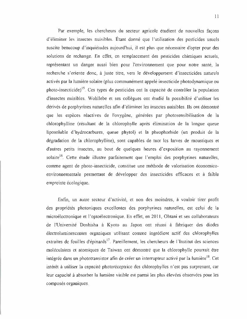
Par exemple, les chercheurs du secteur agricole étudient de nouvelles façons
d 'éliminer les insectes nuisibles. Étant donné que l 'utilisation des pesticides usuels
suscite beaucoup d ' inquiétudes aujourd'hui, il est plus que nécessaire d'opter pour des
solutions de rechange. En effet, en remplacement des pesticides chimiques actuels,
représentant un danger aussi bien pour l'environnement que pour notre santé, la
recherche s' oriente donc, à juste titre, vers le développement d' insecticides naturels
activés par la lumière solaire (plus communément appelé insecticide photodynamique ou
photo-insecticide)l5. Ces types de pesticides ont la capacité de contrôler la population
d'insectes nuisibles. Wohllebe et ses collègues ont étudié la possibilité d 'utiliser les
dérivés de porphyrines naturelles afin d 'éliminer les insectes nuisibles. Ils ont démontré
que les espèces réactives de l'oxygène, générées par photosensibilisation de la
chIorophylline (résultant de la chlorophylle après élimination de la longue queue
liposoluble d ' hydrocarbures, queue phytol) et la pheophorbide (un produit de la
dégradation de la chlorophylline), sont capables de tuer les larves de moustiques et
d'autres petits insectes, au bout de quelques heures d 'exposition au rayonnement
solaire l6. Cette étude illustre parfaitement que l ' emploi des porphyrines naturelles,
comme agent de photo-insecticide, constitue une méthode de valorisation économico
environnementale permettant de développer des insecticides efficaces et à faible
empreinte écologique.
Enfm, un autre secteur d 'activité, et non des moindres, à vouloir tirer profit
des propriétés photoniques excellentes des porphyrines naturelles , est celui de la
rnicroélectronique et l ' optoélectronique. En effet, en 20 Il , Ohtani et ses collaborateurs
de l'Université Doshisha à Kyoto au Japon ont réussi à fabriquer des diodes
électroluminescentes organiques utilisant comme ingrédient actif des chlorophylles
extraites de feuilles d'épinards 17 . Pareillement, les chercheurs de l ' Institut des sciences
moléculaires et atomiques de Taiwan ont démontré que la chlorophylle pourrait être
intégrée dans un phototransistor afm de créer un interrupteur activé par la lumièrel8. Cet
intérêt à utiliser la capacité photoréceptrice des chlorophylles n 'est pas surprenant, car
leur capacité à absorber la lumière visible est parmi les plus élevées observées pour les
composés organiques.

12
Cependant, avant une possible commercialisation ou mIse en oeuvre de telles '
technologies à base de porphyrines naturelles, des avancées concernant leur
photostabilité doivent encore être réalisées. C'est pourquoi les exemples d'applications
présentés ci-dessus ne sont que des démonstrations de faisabilité intéressante, qui
prouvent que l'exploitation des ressources naturelles, en particulier celle provenant des
écosystèmes aquatiques et forestiers, pourrait répondre aux besoins constamment
renouvelés en matériaux de pointe utilisés dans plusieurs technologies modernes.
1.6 Problème
En dépit de leurs caractéristiques photoniques plus que favorables, un
inconvénient majeur avec les porphyrines d'origine végétale (c 'est-à-dire porphyrines de
magnésium) est qu'en raison de leurs niveaux d'énergie HOMO (niveau énergétique le
plus haut occupé) élevés, les porphyrines de magnésium montrent une mauvaise
stabilité, ce qui les rend facilement hydrolysées en solution acide et prompte à la
photodégradation (ou photoblanchiment) sous irradiation lurnineuse l9,20 s' en suit. Le
photoblanchiment est une limite importante des porphyrines naturelles. C'est une
réaction chimique photo-induite et irréversible de destruction des liaisons moléculaires,
responsables des propriétés intéressantes de ces molécules (la capacité d'absorber la
lumière visible, l'émission de fluorescence, la génération d'oxygène singulet, etc.). TI
s'agit essentiellement de réactions d'oxydation avec des radicaux libres provenant de
l'oxygène (principalement l'oxygène singulet). Cet aspect négatif des porphyrines
naturelles fait obstacle à leurs utilisations dans des domaines d'application liés à la
photonique et la photomédecine.
1.7 Revue de la littérature sur les différentes méthodes de photoprotection des porphyrines naturelles et leurs dérivés
Il est généralement admis que les porphyrines se dégradent sous l'action des
interactions entre la porphyrine en état triplet e porp*), qui interagit avec une molécule
de 30 2 (oxygène triplet) pour conduire à de l'oxygène singulet e02). C'est ensuite cette

13
espèce qui réagit avec la molécule de porphyrine à l'état fondamental e Porp) et cause sa
décomposition21. Le mécanisme de photodégradation proposé pour expliquer la
décomposition des colorants organiques, en particulier celle des porphyrines, est résumé
par les équations des réactions photochimiques ci-dessous :
CI) Réaction d'excitation de la porphyrine depuis son état fondamental à son état triplet.
(2) Réaction de la porphyrine état triplet avec oxygène pour produire de l'oxygène singulet.
(3) Décomposition de la porphyrine par l 'oxygène singulet.
Au cours des dernières années, de nombreuses approches ont été explorées afm
d'améliorer la photostabilité de porphyrines naturelles et leurs dérivés. La méthode la
plus courante consiste au remplacement de l'ion central magnésium (Mg2l par d'autres
ions métalliques (par exemple, le palladium), pour former des métalloporphyrines plus
stables22. Cette méthode de photoprotection est particulièrement intéressante afin
d'améliorer non seulement la stabilité, mais aussi la capacité de génération d'oxygène
singulet des porphyrines naturelles, puisque certains métaux lourds (métaux
diamagnétiques) sont connus pour améliorer la conversion intersystème23 ,24. Cependant,
elle a pour inconvénient d 'altérer significativement l'émission de fluorescence de ces
porphyrines25 . Autrement dit, elle ne peut être employée pour augmenter la
photostabilité des porphyrines naturelles, en vue de les utiliser comme agent de contraste
fluorescent pour diagnostiquer un cancer précoce. li fut également constaté que
l' incorporation d'un ou plusieurs groupes attracteurs d'électrons (par exemple, les
sulfonate, les cyano, les halogènes, etc.) conduit à une augmentation prononcée de la
photostabilité, en raison de la diminution de la densité électronique sur le noyau
aromatique, ce qui le rend moins réactif6-29.

14
Une autre approche élégante, afin d'améliorer la photostabilité, consiste à
mcorporer les molécules de porphyrines à l'intérieur d'une nanocapsule (liposome,
micelle, polymère, etc.) de type coeur-coquille30, permettant de conserver la structure
chimique originelle des porphyrines et de réduire leurs interactions avec les 10 2. Par
contre, cette approche possède, sur le plan pratique, deux inconvénients majeurs : (i) les
matériaux constituant la nanocapsule (la coquille) pourraient nuire à l' absorption de
photons par les molécules encapsulées3 1-33
; (ii) les molécules encapsulées doivent être
éjectées de la nanocapsule, afin de pouvoir produire le 10 2. De plus, les 10 2 produites
sont susceptibles de réagir avec les matériaux de la coquille, en particulier les matériaux
polymères, ce qui se traduit souvent par une efficacité photo cytotoxique très inférieure à
celle des molécules non encapsulées34. La complexation de l' ion central avec un ligand
supplémentaire a également été explorée pour améliorer de la photostabilité. En effet,
Matsuo et ses collègues ont démontré qu'une coordination axiale de l'ion métallique peut
améliorer la photostabilité des porphyrines de magnésium35. Enfin, Itoh et ses
collaborateurs ont, pour leur part, utilisé de la silice mésoporeuse pour améliorer la
photostabilité de chlorophylles36. Ces chercheurs ont expliqué que l'effet de la silice
mésoporeuse est similaire à celle des protéines dans la photosynthèse, c'est-à-dire qu ' il
inhibe la formation des espèces réactives de l'oxygène (ROS). Cette méthode n'est donc
pas appropriée pour être utilisée dans des applications, comme la thérapie
photodynamique, le théranostique et la conception de photo-insecticides.
Bien que ces différentes stratégies afin d'améliorer la photostabilité des
porphyrines de magnésium se soient avérées efficaces, par rapport à la stabilité in vivo
de ces molécules (dans les cellules photo synthétiques ), la photostabilité in vitro des
porphyrines de magnésium est encore très faible. De ce point de vue, de nouvelles
méthodes, pour améliorer la photostabilité des porphyrines d'origine naturelle, devraient
être développées. C'est dans la poursuite de cet objectif qu 'ont été réalisés les travaux
décrits dans cette thèse.

15
1.8 Présentation du projet de thèse
Le mécanisme réactionnel détaillé, de la photodégradation de la
tétraphénylporphyrine métallique (MTPP)37-39 des dérivés de chlorophylle40,4 1 et des
bactériochlorophylles42, a été examiné par plusieurs chercheurs. Ces travaux de
recherche ont mis en évidence que la photodégradation des porphyrines est initiée par
l'ajout d'un 10 2 sur les doubles liaisons C=C d'alcènes riches en électrons (réaction de
Diels-Alder, réactions sur les alcènes, 2+2 cyclo-additions) des porphyrines, suivi par
l'ouverture du macrocycle porphyrique. En particulier, Matsuura et ses collègues ont
avancé que le mécanisme réactionnel de photodégradation du tétraphénylporphyrine de
magnésium (MgTPP) passe par la formation d' intermédiaire preroxide zwitterionique 3
ou 4, suivie de la formation d'un époxyde 5, puis de l'ouverture du macrocycle 2
(Figure 1.8)43. Toutefois, du fait de leur très courte durée de vie, ils n'ont pu isoler
d'intermédiaires réactionnels de la photodégradation pour confirmer ce mécanisme.
Ph
Ph
1
or
o Ph H.O OH
~ _H2_0_~~~ ~N'M9,N~ t NH ~'N~
5
~-o.o+ h
N, ,N 1 '\:. Mg' ~
4
Ph
2
Ph
1 roc ' It OUl"t11
Figure 1.8 Mécanisme de photodégradation du tétraphénylporphyrine de magnésium (MgTPP) avec formation d'époxyde43 .

16
Pareillement, il faut souligner que le mécanisme réactionnel, proposé par lturraspe
et ses collègues pour expliquer la photodégradation des dérivés de la chlorophylle, passe
exactement par les mêmes étapes (Figure 1.9)44.
..
O" hl! ..
CO~H:t
.. ~ Xl 7
o ~ j N,Z~ . ~
.... ~ ~
X (a nucleofuge) = CF J Oï or 1- C02CH3
*: - N -----t ..... X , "z~
~)~~~ .. ~
C02CH3
') co+x'
C~H3
Figure 1.9 Mécanisme de photodégradation des dérivés de la chlorophylle44.
Dans les deux mécanismes proposés, on peut clairement constater que l'ouverture
du macrocycle porphyrique implique la participation des doublets d'électrons libres des
atomes d'azote du macrocycle. De plus, de récents travaux sur les propriétés
photochimiques du MgTPP ont révélé que la photodégradation du MgTPP passe par la
formation d'un intermédiaire MgTPP-02, dans lequel une molécule 0 2 est liée aux
atomes d'azotes du macrocycle porphyrique, via des liaisons azote-oxygène (N_0)45,46.
Pris ensemble, ces résultats suggèrent que les atomes d'azote du macrocycle porphyrique
jouent un rôle essentiel dans le processus de photodégradation des porphyrines. D'autre
part, et plus fondamentalement, on se questionne à savoir si empêcher le doublet
d'électrons libres des atomes d'azote du macro cycle de participer à la réaction de

17
photodégradation, ou tout au moins empêcher l'oxygène de se lier à ces sites azotés
pourrait ralentir la photodégradation. Une réponse affirmative à cette interrogation
conduira indubitablement au développement d'une nouvelle stratégie de photoprotection
des porphyrines, basé sur l' inhibition des sites azotés du macrocyc1e par un agent
capable de se lier aux atomes d'azote.
Pour ce faire, j ' ai opté d'utiliser comme ligand, des nanoparticules de métaux
nobles à savoir : les nanoparticules d'or (AuNPs) et d'argent (AgNPS). Parce que les
nanoparticules sont extrêmement petites, elles ont un rapport surface/volume très
important. En raison de cette surface spécifique élevée, les atomes présents en surface
sont plus nombreux et ce sont eux qui participent aux réactions chimiques. De plus, il est
bien connu que la vitesse d'une réaction chimique augmente avec l'étendue de la surface
de contact. Ainsi, les nanoparticules présentent une importante réactivité chimique, ce
qui est l'une des raisons justifiant mon choix d'utiliser ces matériaux. Ma deuxième
raison, d'utiliser des AuNPs et AgNPs, vient du fait que ces nanoparticules font l'objet
d'un intérêt croissant dans des domaines aussi divers que la nanomédecine (l'application
des nanotechnologies au monde médical)47-49, le photovoltaïque50, l' électronique5l,52 ou
la catalyse53,54. Enfin, la troisième et dernière raison, qui explique mon penchant pour les
nanoparticules, est que ces matériaux nanométriques ont, dans certaines conditions, la
capacité d'augmenter l' émission de fluorescence d'un fluorophore , lorsque celui-ci est
situé à proximité de la surface métallique, un phénomène connu sous le nom de
l' augmentation de la fluorescence induite par méta155,56. De plus, il fut également
démontré que la présence des nanoparticules à proximité d'un photosensibilisateur
pourrait accroître la génération des espèces réactives de l'oxygène e02 et radicaux
libresi7.
Le tétraphénylporphyrine de magnésium (MgTPP) et la chlorophylle-a (ChIa) ont
été choisis pour cette étude, comme des porphyrines de magnésium modèles, afm de
tester l' efficacité de notre approche.

18
1.9 Notions essentielles sur l'augmentation de l'émission de fluorescence d'un fluorophe par un métal
L'augmentation de l'émission de fluorescence d'un fluorophe par un métal (MEF
pour Metal-Enhanced Fluorescence) est un effet physique qui se produit lorsqu 'un
fluorophore est situé à une distance nanométrique « 10 nm) d 'une surface métallique58.
Ce processus est caractérisé par un taux d'émission spontanée accrue dans le système
fluorophore-métal. Il est généralement admis aujourd'hui que le MEF est dû à au moins
un des deux mécanismes suivants: (1) l' augmentation du champ électromagnétique
local, lorsque la nanoparticule métallique agit comme un élément concentrateur du
champ électrique local de la lumière, ce qui permet au fluorophore d ' avoir un meilleur
taux d ' absorption de photons59; (2) l' intense champ électrique autour de la nanoparticule
augmente le taux d ' émission radiative, en diminuant le temps de vie à l'état excité de la
molécule 60.
Le premier mécanisme, soit l'amélioration de l'intensité d'émission, à la suite de
l' augmentation du champ incident local sur le fluorophore par le métal, peut être
compris comme suit: sous l ' influence d 'une lumière incidente, le champ électrique est
intensifié autour de la nanoparticule métallique. Ainsi, le champ électrique résultant est
alors égal à la sommation du champ incident (E) et de l' amplification du champ dû au
métal (Ern) . Autrement dit, un fluorophore situé à proximité d 'une nanoparticule reçoit
un champ électrique global amplifié ET (ET = E+Em).
Les propriétés électromagnétiques des nanoparticules métalliques peuvent être
expliquées de façon intuitive par le modèle de Drude des métaux61. Ce modèle traite les
métaux sous forme de matrice d'ions chargés positivement et d'un nuage d'électrons se
déplaçant à travers le potentiel de ces ions (Figure 1.10). Lorsque soumis à un champ
électromagnétique externe, les électrons se déplacent de façon cohérente à l'intérieur du
matériau62. Le déplacement d'électrons génère des forces de rappel électrostatique, où le
nuage d'électrons chargés négativement est attiré vers les ions chargés positivement. La
présence de cette force de rappel, pour les petits déplacements de l'équilibre, provoque
l'onde de densité de charge qui oscillera dans le métal. Cette oscillation, collective et

19
cohérente des électrons à la surface d'une nanoparticule métallique, est appelée plasmon
de surface. Les oscillations dans des nanoparticules sont, bien sûr, localisées, d'où
l'appellation de plasmon de surface localisé (PSLs) , et s'accompagnent d'une
augmentation du champ électromagnétique près de la surface de la particule. Cette
amélioration diminue rapidement avec la distance à partir de la surface.
hamp '1 tri u
Figure 1.10
ph' re m . talliqu
./'
ua d él tr n
Représentation de la formation d'un plasmon de surface sur une nanoparticule métallique 62.
Le champ électrique amplifié localement (ET) produit l'amélioration locale de
fluorescence, en raison du fait que la puissance d'émission de fluorescence (Pfluorophore)
est proportionnelle au flux de photons qui, à son tour, est proportionnel au champ
électrique au carré, en supposant qu'il n'y a aucun effet de saturation dans l'état excité.
Par conséquent, la puissance d'émission de fluorescence peut être donnée par la formule
suivanté3 :
Pjluorop hoe = kn~
et
où k est la section efficace d'absorption du fluorophore, n le flux de photons et Qo le
rendement quantique du fluorophore.
20
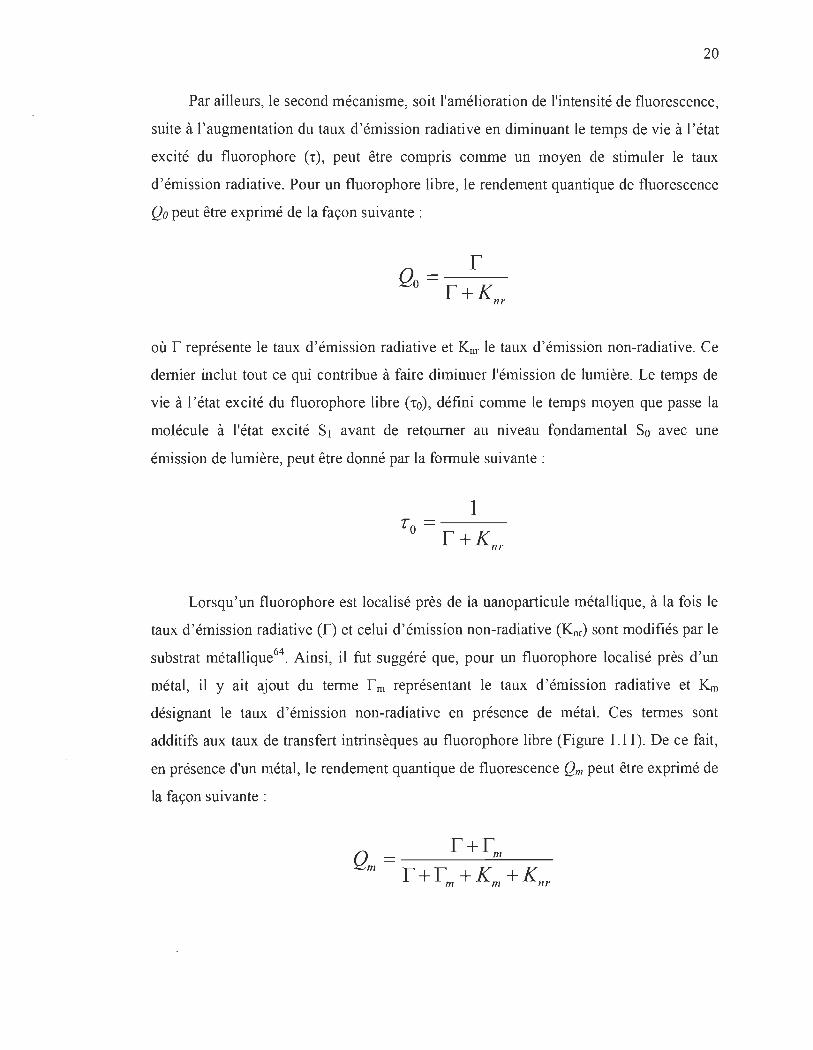
Par ailleurs, le second mécanisme, soit l'amélioration de l'intensité de fluorescence,
suite à l'augmentation du taux d'émission radiative en diminuant le temps de vie à l'état
excité du fluorophore Ct), peut être compris comme un moyen de stimuler le taux
d'émission radiative. Pour un fluorophore libre, le rendement quantique de fluorescence
Qo peut être exprimé de la façon suivante:
r Qo =r K + nr
où r représente le taux d'émission radiative et Knr le taux d'émission non-radiative. Ce
dernier inclut tout ce qui contribue à faire diminuer l'émission de lumière. Le temps de
vie à l'état excité du fluorophore libre ('to), défini comme le temps moyen que passe la
molécule à l'état excité SI avant de retourner au niveau fondamental So avec une
émission de lumière, peut être donné par la formule suivante :
1 '0 =---r+Knr
Lorsqu'un fluorophore est localisé près de la nanoparticule métallique, à la fois le
taux d'émission radiative (f) et celui d'émission non-radiative (Knr) sont modifiés par le
substrat métallique64. Ainsi, il fut suggéré que, pour un fluorophore localisé près d'un
métal, il y ait ajout du terme r m représentant le taux d'émission radiative et Km
désignant le taux d'émission non-radiative en présence de métal. Ces termes sont
additifs aux taux de transfert intrinsèques au fluorophore libre (Figure 1.11). De ce fait ,
en présence d'un métal, le rendement quantique de fluorescence Qm peut être exprimé de
la façon suivante :

21
Et, le temps de vie à l'état excité du fluorophore, localisé à proximité d'un métal
(tm) , est donné par la formule suivante :
1 r = ---------m r + rm + Km + Knr
L'addition d'une composante d'émission non-radiative supplémentaire peut
paraître surprenante mais, en fait, il y a toujours compétition entre le quenching et l' effet
d'amélioration. Toutefois, la dépendance de distance (métal-fluorophore) pour chaque
processus est différente, de sorte que le quenching de fluorescence domine lorsque le
fluorophore est très proche «1 nm) de la surface, tandis que l'amélioration de
fluorescence est prépondérante à de plus grandes distances65,66. Le diagramme de
Jablonski simplifié à la figure 1.11 est une représentation résumant le phénomène
d'augmentation de l'émission de fluorescence par un métal.
E
Figure 1.11
Sans métal Avec métal
" r k nr E EIII r rm ka kn
Modification du processus d'absorption et de déexcitation par une nanoparticule métallique à proximité d'un fluorophore64
.
1.10 Notions essentielles sur l'augmentation de la production d'oxygène singulet du photosensibilisateur par un métal
Il a été montré que d'amélioration du métal ne se limite pas qu'à la fluorescence :
l'amélioration de la production à la fois de l'oxygène singulet e02) et le rendement
quantique de phosphorescence ont été observés67-75
. À ce jour, le mécanisme exact de
l'effet d'amélioration du métal sur la production des ROSs est encore sujet à débat, mais
il apparaît expérimentalement que l' amplification du champ électromagnétique au

22
voisinage des NPs peut jouer un rôle important dans l'amélioration de la production des
ROSs. En effet, il a été démontré que l' interaction, entre le moment dipolaire du
photosensibilisateur et du champ électromagnétique au voisinage des NPs, peut conduire
à une formation accrue de l'état triplet du photosensibilisateur76 et, par le fait même,
améliorer la production de 102 par photosensibilisation.
Alternativement, les NPs pourraient également améliorer la production de 102, en
augmentant la constante de vitesse de la transition 1 /).g ~ 3L g (Figure 1.12), résultant en
une augmentation du rendement quantique de phosphorescence de l'oxygène singuleeo.
En utilisant cette approche, Toftegaard et ses collègues ont réussi à démontrer une
augmentation de la production de 102 par un photosensibilisateur au voisinage des
nanodisques d'or, pour lequel la bande de plasmon de surface correspond à la
phosphorescence de 102 à 1270 nm70.
Energy Sn
hu
Enhanced Excitation
Sa
le
le
le
- Photosensitizers
Oxygen
Enhanced == Energy Transfer le
le 1270 nin Singlet Oxygen Phosphorescence Emission
Figure 1.12 Diagramme énergétique de la production d'oxygène singulet photosensibilisée via le couplage plasmon surface-photosensibilisateur75
.
1.11 Hypothèses de recherche
Si les sites azotés du macrocycle porphyrique sont bloqués dans l'obscurité avant
l'illumination, alors il se produira une augmentation de la photostabilité.

23
1.12 Objectif général de cette thèse
Le principal objectif de cette thèse de doctorat est de développer une stratégie de
photoprotection, reposant sur l'utilisation des nanoparticules de métaux nobles, afm
d'améliorer la stabilité des porphyrines de magnésium.
1.13 Objectifs opérationnels
1) Vérifier que l'inhibition des sites azotés du macrocycle porphyrique peut induire
une photoprotection.
2) Établir que cette méthode de photoprotection pourrait également permettre
d'accroître la stabilité des porphyrines de magnésium, possédant à la fois des
doublets d'électrons libres sur les atomes d'azote du macrocycle et sur les
groupes fonctionnels périphériques qui ne sont pas conjugués au macrocycle,
comme c'est le cas avec les chlorophylles et les bactériochlorophylles.
3) Utiliser cette approche pour démontrer qu' il est possible d'améliorer
simultanément la stabilité, l'émission de fluorescence et la capacité de
génération d'oxygène singulet des porphyrines de magnésium.
1.14 Organisation du document
Cette thèse comporte en tout cinq chapitres. Suite à la présentation au premier
chapitre du contexte, état de l' art sur les différentes stratégies de photoprotection des
porphyrines de magnésium et le projet de thèse, le deuxième chapitre fera état de la
découverte d'une nouvelle stratégie de photoprotection des porphyrines de magnésium.
En effet, dans l'article intitulé « BeneficiaI role of gold nanoparticles as photoprotector
of magnesium tetraphenylporphyrin », il sera démontré que l' inhibition des sites azotés
du macrocycle porphyrique peut induire une photoprotection. Le troisième chapitre
concernera la possibilité de protéger les porphyrines naturelles par cette méthode. Ainsi,
l'article titré « Enhanced photostability of chlorophyll-a using gold nanoparticles as an
24
efficient photoprotector » expliquera les capacités qu'ont les nanoparticules d 'or
(AuNPs) à protéger les molécules de chlorophylles-a, dans les conditions in vitro. À cet
égard, une attention particulière est portée sur la comparaison entre l' efficacité
photoprotectrice des AuNPs et celle des composés antioxydants, lesquelles sont connues
comme étant très efficaces pour protéger les chlorophylles dans la nature. Dans le
chapitre suivant, dont l'article présenté se nomme «Nanosilver Could Usher in Next
Generation Photoprotective Agents for Magnesium Porphyrins », il sera fait mention
d'une possibilité de co améliorer la photostabilité, l ' émission de fluorescence et la
génération d 'oxygène singulet par photosensibilisation des chlorophylles-a. Quant au
dernier chapitre, il s 'applique à présenter une synthèse des résultats et des contributions
scientifiques de l' ouvrage. Nos perspectives sur l'avancement des connaissances et
quelques questions laissées ouvertes par la thèse y seront également soumises. À la fin
du présent document, une annexe explique les procédures expérimentales de synthèse
des nanoparticules développées dans ce travail, ainsi que les méthodes de
caractérisation.
1.15 Références
(1) Kadish, K. M.; Smith, K. M.; Guillard, R. The porphyrin handbook. Eds, Academic press: San Diego, 2000; Vol. 1-10.
(2) Bonnett R. ; Chemical Aspects of Photodynamic Therapy, Gordon and Breach Science Publishers, 2000.
(3) Schaffer, M.; Ertl-Wagner, B.; Schaffer, P. M.; Kulka, u.; Hofstetter, A. ; Duhmke, E.; Jori G. Porphyrins as Radiosensitizing Agents for Solid Neoplasms. Curr. Phar. Des., 2003, 9, 2024-2035 .
(4) Fischer, H. ; Orth, H. Die Chemie des Pyrrols, vol. I-III. Akademische Verlagsgesellschaft 1934, M.B.H., Leipzig.
(5) Dixon H. B. F. , Comish-Bowden A. , Liebecq c., Loening K. L. , Moss G. P., Reedijk J., Velick S. F., Venetianer P., Vliegenthart J. F. G. et al. Nomenclature of tetrapyrroles . Pure Appl. Chem. , 1987, 59, 779-832.
25
(6) (a) Uttamlal, M. ; Holmes-Smith, A S. The excitation wavelength dependent fluorescence of porphyrins. Chem. Phys. Leu. , 2008, 454, 223-228. (b) Zimmermann, J. ; Zeug, A ; Roder, B. A generalization of the Jablonski diagram to account for polarization and anisotropy effects in time-resolved experiments. Phys. Chem. Chem. Phys., 2003,5,2964-2969.
(7) Gouterman, M. Spectra ofporphyrins J. Mol. Spectrosc., 1961, 6, 138-163.
(8) (a) Henderson, B.W.; Sumlin, AB.; Owczarczak, B.L. ; Dougherty, TJ. Bacteriochlorophyll-a as photosensitizer for photodynamic treatment of transplantable murine tumors. J. Photochem. Photobiol. B, 1991, 10, 303-313. (b) Xodo, L. E.; Rapozzi, V. ; Zacchigna, M.; Drioli, S.; Zorzet, S. The Chlorophyll Catabolite Pheophorbide-a as a Photosensitizer for the Photodynamic Therapy. Curr. Med. Chem., 2012, 19, 799-807.
(9) Dai, T; Fuch, B. B.; Coleman, J. J.; Prates, R. A ; Astrakas, C.; St. Denis, T. G.; Ribeiro, M. S.; Mylonakis, E. ; Hamblin, M. R. ; Tegos, G. P. Concepts and princip les of photodynamic therapy as an alternative antifungal discovery platform. Front. Microbiol., 2012, 3, article 120, 1-15.
(10) Schweitzer, C.; Schmidt, R. Physical Mechanisms of Generation and Deactivation ofSinglet Oxygen. Chem. Rev., 2003, 103, 1685-1757.
(11) Valeur, B.; Berberan-Santos, M. N. Molecular Fluorescence: Principles and Applications. 2nd Edition, Wiley VCH, 2013.
(12) Fanl , L. ; WU, Q.; Chu, M. Near infrared fluorescent chlorophyll nanoscale liposomes for sentinel lymph no de mapping. lnt. J. Nanomed., 2012, 7, 3071-3080.
(13) Liu, T W.; Chen, J. ; Burgess, L. ; Cao, W. ; Shi, J. ; Wilson, B.e.; Zheng, G. Multimodal Bacteriochlorophyll Theranostic Agent. Theranostics., 2011, 1, 354-362.
(14) Sperandio, F. F.; Huang, Y-Y.; Hamblin, M. R. Antimicrobial Photodynamic Therapy to Kill Gram-negative Bacteria. Recent Pat. Antiinfect. Drug Discov., 2013, 8, 108-120.
(15) Alves, E.; Faustino, M. A F.; Neves, M. G. P. M. S.; Cunha, A.; Nadais, H. ; Almeida, A Potential applications of porphyrins in photodynarnic inactivation beyond the medical scope. Journal of Photochemistry and Photobiology C: Photochem. Rev. , 2015, 22, 34-57.

26
(16) Wohllebe, S. ; Richter, R. ; Richter, P. ; Hiider, D. P. Photodynamic control of human pathogenic parasites in aquatic ecosystems using chlorophyllin and pheophorbid as photodynamic substances. Parasitol. Res. , 2009, 104, 593-600.
(17) Ohtani, N. ; Kitagawa, N. ; Matsuda, T. Fabrication of Organic Light-Emitting Diodes Using Photosynthetic Pigments Extracted from Spinach. Jpn. J Appl. Phys., 2011, 50, 01BC08.
(18) Chen, S. Y. ; Lu, Y. Y. ; Shih, F. Y. ; Ho, P. H. ; Chen, Y. F.; Chen, C. W.; Chen, Y. T. ; Wang, W. H. Biologically inspired graphene-chlorophyll phototransistors with high gain. Carbon., 2013 , 63 , 23-29.
(19) Bonnett, R. ; Djelal, B. D.; Hamilton, P. A ; Martinez, G.; Wierrani, F.; Photobleaching of 5,10, 15,20-tetrakis(m-hydroxyphenyl)porphyrin (m-THPP) and the corresponding chI orin (m-THPC) and bacteriochlorin(m-THPBC). À comparative study. J Photochem. Photobiol. B. , 1999, 53, 136-143.
(20) Gerola, A P.; Santana, A ; França, P. B.; Tsubone, T. M.; de Oliveira, H. P.; Caetano, W .; Kirnura, E. ; Hioka, N. Effects of Metal and the Phytyl Chain on Chlorophyll Derivatives: Physicochemical Evaluation for Photodynamic Inactivation of Microorganisms. Photochem. Photobiol. , 2011, 87, 884-894.
(21) Sinclair, R. S. ; Martinez, G., photobleaching of sensitisers used in photodynamic therapy. Tetrahedron. , 2001, 57, 9513-9547.
(22) Fiedor, J. ; Fiedor, L. ; Karnrnhuber, N. ; Scherz, A ; Scheer, H. Photodynamics of the bacteriochlorophyll-carotenoid system. 2. Influence of central metal, solvent and beta-carotene on photobleaching of bacteriochlorophyll derivatives. Photochem. Photobiol, 2002, 76, 145-52.
(23) BaU, D. J.; Wood, S. R. ; Vernon, D. 1.; Griffiths, J.; Dubbelman, T. M. AR.; Brown, S.B. The characterisation of three substituted zinc phthalocyanines of differing charge for use in photodynamic therapy. A comparative study of their aggregation and photosensitising ability in relation to mTHPC and polyhaematoporphyrin. J Photochem. Photobiol. B. , 1998, 45, 28-35.
(24) Guldi, D. M.; Mody, T. D. ; Gerasirnchuk, N . N.; Magda, D.; Sessler, J. L. Influence of large metal cations on the photophysical properties of texaphyrin, a rigid aromatic chromophore. J Am. Chem. Soc. , 2000, 122, 8289-8298.
(25) Drzewiecka-Matuszeka, A ; Skalna, A ; Karocki, A ; Stochel, G.; Fiedor, L. Effects of heavy central metal on the ground and excited states of chlorophyll. J Biol. Inorg. Chem. , 2005, 10, 453-62.
27
(26) Dabrowski, J. M.; Amaut, L. G.; Pereira, M. M.; Monteiro, C. J. P.; Urbanska, K ; Simoes, S. ; Stochel G. New halogenated water-soluble chlorin and bacteriochlorin as photostable PDT sensitizers: synthesis, spectroscopy, photophysics, and in vitro photosensitizing efficacy. Chem. Med. Chem., 2010, 5, 1770-1780.
(27) Dabrowski, 1 M.; Urbanska, K ; Amaut, L. G.; Pereira, M. M.; Abreu, A. R ; Simoes, S.; Stochel, G. Biodistribution and photodynamic efficacy of a watersoluble, stable, halogenated bacteriochlorin against melanoma. Chem. Med. Chem. , 2011, 6, 465-475 .
(28) Dabrowski, J. M.; Amaut, L. G. ; Pereira, M. M. ; Urbanska, K; Simoes, S.; Stochel, G.; Cortes, L. Combined effects of singlet oxygen and hydroxyl radical in photodynamic therapy with photostable bacteriochlorins: evidence from intracellular fluorescence and increased photodynamic efficacy in vitro. Free Radical Biol. Med. , 2012, 52, 1188-1200.
(29) Huang, Y Y.; Balasubramanian, T.; Yang, E.; Luo, D. ; Diers, J. R ; Bocian, D. F. ; Lindsey, J. S. ; Holten, D.; Hamblin, M. R Stable synthetic bacteriochlorins for photodynamic therapy: role of dicyano peripheral groups, central metalsubstitution (2H, Zn, Pd), and Cremophor EL delivery. Chem. Med. Chem., 2012, 7, 2155-2167.
(30) Cao, W.; Ng, K K; Corbin, I.; Zhang, Z. ; Ding, L. ; Chen, J.; Zheng, G.; Synthesis and evaluation of a stable bacteriochlorophyll-analog and its incorporation into high-density lipoprotein nanoparticles for tumor imaging. Bioconjug. Chem., 2009, 20, 2023-2031 .
(31) Huang, P.; Li, Z.; Lin, 1 ; Yang, D.; Gao, G.; Xu, c.; Bao, L.; Zhang, c. ; Wang, K ; Song, H.; Hu, H.; Cui, D. Photosensitizer-conjugated magnetic nanoparticles for in vivo simultaneous magnetofluorescent imaging and targeting therapy. Biomaterials. , 2011, 32, 3447-3458.
(32) Chen, Z.L. ; Sun, Y ; Huang. P.; Yang, X. X.; Zhou, X. P. Studies on preparation of photosensitizer loaded magnetic silica nanoparticles and their anti-tumor effects for targeting photodynamic therapy. Nanoscale Res. Lett. , 2009, 4, 400-408 .
(33) Liu, F. ; Zhou, X.; Chen, Z.; Huang, P.; Wang, X. ; Zhou, Y Preparation of purpurin-18 loaded magnetic nanocarriers in cottonseed oil for photodynamic therapy. Mater. Lett. , 2008, 62, 2844-2847.
(34) Sun, Y ; Chen, Z.; Yang, x.; Huang, P.; Zhou, X.; Du, X. Magnetic chitosan nanoparticles as a drug delivery system for targeting photodynamic therapy. Nanotechnology. , 2009, 20, 135102.

28
(35) Ichiki, T.; Matsuo, Y.; Nakamura, E. Photostability of a dyad of magnesium porphyrin and fullerene and its application to photocurrent conversion. Chem. Commun. , 2013, 49, 279-281.
(36) Itoh, T.; Yano, K. ; Inada, Y ; Fukushima, Y Photostabilized Chlorophyll a in Mesoporous Silica: Adsorption Properties and Photoreduction Activity of Chlorophyll-a. J Am. Chem. Soc. , 2002, 124, 13437-13441.
(37) Silva, A. M. S.; Neves, M. G. P. M. S. ; Martins, R. R. L. ; Cavaleiro, J. A. S; Boschi, T. ; Tagliatesta, P. Photo-oxygenation of meso-tetraphenylporphyrin derivatives: the influence of the substitution pattern and characterization of the reaction products. J Porphyrins.Phthalocyanines., 1998, 2, 45-51 .
(38) (a) Smith, K M; Brown, S. B; Troxler, R. F; Lai, J. J. Photo oxygenation of mesotetraphenylporphyrin metal complexs. Photochem. Photobiol, 1982, 36,147-152; (b) Cavaleiro, J. A. S.; Hewlins, M. J. E.; Jackson, A. H. ; Neves, G. P. M. S. Structures of the ring-opened oxidation products from meso-tetraphenylporphyrin. J Chem. Soc., Chem. Commun., 1986, 142-144.
(39) Cavaleiro, 1. A. S.; Neves, M. G. P. S. ; Hewlins, M. J. E. ; Jackson, A. H. The photo-oxidation of meso-tetraphenylporphyrins. J Chem. Soc., Perkin Trans, 1990, 1, 1937-1943.
(40) lturraspe, J.; Gossauer, A. formation of oxoniachlorins on photooxidation of 20-trifluoroactetoxy-and-20-chloro-chlorophyll derivatives. Photochem. Photobiol., 1991, 54, 43-49.
(41) lturraspe, 1. ; Gossauer, A. Dependence of the regioselectivity of photo-oxidative ring opening of the chlorophyll macrocycle on the complexed metal Ion. He/v. Chim. Acta., 1991, 74, 1713-1717.
(42) (a) Kenner, G. W .; Rimmer, J.; Smith, KM.; Unsworth, J. F.Pyrroles and related compounds. Part 39. Structural and biosynthetic studies of the Chlorobiumchlorophylls-660 (bacteriochlorophylls c). Incorporations of methionine and porphobilinogen. J Chem. Soc. Perkin Trans. , 1978, 1, 845-852; (b) Risch, N.; Schormann, A. ; Brockmann, H. Photobilin e. Photooxidation von bacteriochlorophyll-e-derivaten. Tetrahedron. Lett. , 1984, 25, 5993-5996; (c) Brown, S. B. ; Smith, KM.; Bisset, G. M. F.; Troxler, R. F. Mechanism of photooxidation of bacteriochlorophyll c derivatives. J Biol. Chem. , 1980, 255, 8063-8068.
(43) Matsuura, T. ; Inoue, K ; Ranade, A. C.; Saito, I. Photooxygenation of magesium meso-tetraphenylporphyrin. Photochem. Photobiol. , 1980, 31 , 23-26.

29
(44) Zhang, 1. P. ; Zhang, P. Y.; Zhang, Z. ; Chen, G. H.; Han, F.; Wei, X. H. Photochemical reaction between magnesium tetraphenyl porphyrin and oxygen. Chin. Chem. Lett., 2008, 19, 1190-1192.
(45) Zhang, J. ; Zhang, P. ; Zhang, Z.; Wei, X. Spectroscopic and kinetic studies of photochemical reaction of magnesium tetraphenylporphyrin with oxygen. J Phys. Chem. A. , 2009, 113, 5367-5374.
(46) Jena, P. ; Mohanty, S.; Mallick, R ; Jacob, B.; Sonawane, A. Toxicity and antibacterial assessment of chitosan-coated silver nanoparticles on human pathogens and macrophage cells. Int. J Nanomed. , 2012, 7, 1805-1818.
(47) Jeyaraj , M. ; Sathishkumar, G.; Sivanandhan, G. ; MubarakAli, D.; Rajesh, M.; Arnn, R ; Kapildev, G.; Manickavasagam, M.; Thajuddin, N. ; Premkumar, K. ; Ganapathi, A. Biogenic silver nanoparticles for cancer treatrnent: an experimental report. Colloids Surf B. , 2013, 106, 86-92.
(48) Arvizo, R ; Bhattacharya, R. ; MukheIjee, P. Gold nanoparticles: opportunities and challenges in nanomedicine. Expert Opin. drug deliv., 2010, 7, 753-763 .
(49) Boisselier, E.; Astruc, D. Gold nanoparticles in nanomedicine: preparations, imaging, diagnostics, therapies and toxicity. Chem. Soc. Rev. , 2009, 38, 1759-1782.
(50) Stratakis, E.; Kymakis, E. Nanoparticle-based plasmonic organic photovoltaic devices. Mater. Today., 2013, 16, 133-146.
(51) Cui, P. ; Seo, S.; Lee, J. ; Wang, L. ; Lee, E. ; Min, M. ; Lee, H. Nonvolatile memory device using gold nanoparticles covalently bound to reduced graphene oxide. ACS Nano. , 2011, 5, 6826-6833.
(52) Shen, W.; Zhang, X.; Huang, Q.; XU, Q.; Song, W. Preparation of solid silver nanoparticles for inkjet printed flexible electronics with high conductivity. Nanoscale., 2014, 6, 1622-1628.
(53) Xia, Y. ; Yang, H. ; Campbell, C. T. Nanoparticles for Catalysis. Acc. Chem. Res. , 2013, 46, 1671-1672.
(54) Chng, L. L; Erathodiyil, N. ; Ying, J. Y. Nanostructured Catalysts for OrganicTransformations. Acc. Chem. Res., 2013, 46, 1825-1837.

30
(55) Babu, J.; George, J. ; Vanna, R. L. Metal-induced fluorescence lifetime enhancement of quinaldine chromophore on gold nanoparticle surface. New J Chem., 2013, 37,2426-2432.
(56) Mishra, H. ; Mali, B. L.; Karolin, J.; Dragan, A. 1. ; Geddes, C. D. Experimental and theoretical study of the distance dependence of metal-enhanced fluorescence, phosphorescence and delayed fluorescence in a single system. Phys. Chem. Chem. Phys., 2013, 15, 19538-19544.
(57) Karolin, 1. ; Geddes, C. D. Metal-enhanced fluorescence based excitation volumetric effect of plasmon-enhanced singlet oxygen and super oxide generation. Phys. Chem. Chem. Phys., 2013, 15, 15740-15745.
(58) Aslan, K.; Le onenko , Z.; Lakowicz, J. R. ; Geddes, C. D. Annealed Silver-Island Films for Applications in Metal-Enhanced Fluorescence: Interpretation in Terms of Radiating Plasmons. J Fluoresc., 2005, 15, 643-654.
(59) Kümmerlen, J. ; Leitner, A.; Brunner, H. ; Aussenegg, F.R.; Wokaun, A. Enhanced dye fluorescence over sil ver island films: analysis of the distance dependence. Mol. Phys., 1993,80, 1031-1046.
(60) Gersten, 1.; Nitzan, A. Spectroscopic properties of molecules interacting with small dielectric particles. J Chem. Phys., 1981, 75 , 1139-1152.
(61) F. Xie, Ultra-sensitive protein detection by metal-induced fluorescence enhancement, Macquarie University, 2008.
(62) Kelly, K. L. ; Coronado, E.; Zhao, L. L.; Schatz, G. C. The Optical Properties of Metal Nanoparticles: The Influence of Size, Shape, and Dielectric Environment. J Phys. Chem. B., 2002, 107, 668-677.
(63) Le Ru, E. C.; Etchegoin, P. G. Surface-Enhanced Raman Scattering (SERS) and Surface-Enhanced Fluorescence (SEF) in the context of modified spontaneous emission. arXiv preprint physics/0509154, 2005.
(64) Geddes, C. D. ; Gryczynski, 1. ; Malicka, J.; Gryczynski, Z.; Lakowicz,J. R. Metalenhanced fluorescence: potential applications in HTS. Comb. Chem. High Throughput Screen., 2003, 6, 109-117.
(65) Anger, P. ; Bharadwaj, P. ; Novotny, L. Enhancement and Quenching of SingleMolecule Fluorescence. Phys. Rev. Lett., 2006, 96, 113002-1 à 4.

31
(66) Feng, A. L. ; You, M. L. ; Tian, L. ; Singamaneni, S. ; Liu, M.; Duan, Z. ; Lu, T. 1. ; Xu, F.; M. Lin. Distance-dependent plasmon-enhanced fluorescence of upconversion nanoparticles using polyelectrolyte multilayers as tunable spacers. Sei. Rep., 2015, 5: 7779, doi :10.1038/srep07779.
(67) Zhang, Y ; Aslan, K. ; Previte, M. J. R. ; Malyn, S. N.; Geddes, C. D. Metalenhanced phosphorescence: Interpretation in terms of triplet-coupled radiating plasmons. J Phys. Chem. B., 2006, 110, 25108-25114.
(68) Melo, L. S.; Gomes, A S.; Saska, S.; Nigoghossian, K. ; Messaddeq, Y ; Ribeiro, S. J. ; Araujo, R. E. Singlet oxygen generation enhanced by silver-pectin nanoparticles . J Fluorese., 2012, 22, 1633-1638.
(69) Toftegaard, R. ; Arnbjerg, J. ; Cong, H. P. ; Agheli, H. ; Sutherland, D. S.; Ogilby, P. R. Metal nanoparticle-enhanced radiative transitions: Giving singlet oxygen emission a boost. Pure Appl. Chem., 2011, 83 , 885-898.
(70) Toftegaard, R.; Arnbjerg, J.; Daasbjerg, K. ; Ogilby, P. R.; Dmitriev, A ; Sutherland, D. S.; Poulsen, L. Metal-enhanced 1270 nm singlet oxygen phosphorescence. Angew. Chem. Int. Ed. , 2008, 47, 6025-6027.
(71) Zhang, Y. ; Aslan, K. ; Geddes, C. D. Metal-Enhanced Generation ofOxygen Rich Species. John Wiley & Sons, Ine., New Jersey., 2010, 277-293.
(72) Zhang, Y.; Aslan, K. ; Previte, M. 1. R. ; Geddes, C. D. Metal-enhanced singlet oxygen generation: A consequence of plasmon enhanced triplet yields. J Fluorese., 2007, 17, 345-349.
(73) Zhang, Y ; Aslan, K. ; Previte, M. J. R.; Geddes, C. D. Plasmonic engineering of singlet oxygen generation. Proe. Nat! Aead. Sei., 2008, 105, 1798-1802.
(74) Dragan, AI.; Geddes, C.D. Metal-enhanced fluorescence: the role of quantum yield, Qo, in enhanced fluorescence. Appl. Phys. Let!. , 2012,100, p. 093115
(75) Hu, B.; Cao, X.; Nahan, K. ; Caruso, J. ; Tang, H.; Zhang, P.Surface plasmonphotosensitizer resonance coupling: an enhanced singlet oxygen production platform for broad-spectrum photodynamic inactivation of bacteria. J Mater. Chem. B. , 2014, 2, 7073-7081.
(76) Pacioni , N. L. ; Gonzalez-Bejar, M. ; Alarc6n, E.; McGilvray, K. L.; Scaiano, 1. C. Surface Plasmons Control the Dynamics of Excited Triplet States in the Presence ofGold Nanoparticles. J Am. Chem. Soc., 2010, 132, 6298-6299.

CHAPITRE II
BENEFICIAL ROLE. OF GOLD NANOPARTICLES AS PHOTOPROTECTOR OF MAGNESIUM TETRAPHENYLPORPHYRIN
Le contenu de ce chapitre a fait l'objet d'une publication en anglais dans la revue :
Journal of Materials Chemistry, 2012,22,2943-2951.
2.1 Résumé de l'article
Le présent article avance l'hypothèse que si les sites azotés du macrocycle
porphyrique sont bloqués dans l'obscurité avant l'illumination, alors il se produira une
augmentation la photostabilité. Afin de vérifier cette hypothèse, nous avons utilisé des
nanoparticules d'or (AuNPs) pour améliorer la photostabilité du tétraphénylporphyrine
de magnésium (MgTPP). Nos résultats montrent qu'avec une concentration appropriée
de AuNPs, une efficacité de photoprotection d'environ 100 % peut être obtenue. Fait
important, la capacité protectrice des AuNPs est le résultat de leur liaison avec les sites
azotés du macrocycle porphyrique du MgTPP, inhibant par conséquent l'action des
espèces réactives de l'oxygène (ROS) à ces endroits. En outre, la réduction de la
formation d'états singulets excités du MgTPP, à la suite de sa liaison avec AuNPs,
diminue également la production de ROS, ce qui contribue aussi à l'amélioration de la
photostabilité du MgTPP. Par ailleurs, la liaison entre MgTPP et AuNPs peut également
être responsable de la photostabilité induite au MgTPP par les AuNPs dans des
conditions anaérobies. L'utilisation de nanoparticules d'or nous offre donc une nouvelle
stratégie pour améliorer la photostabilité de diverses porphyrines. L'étude présentée
dans cet article a été l'aboutissement du premier objectif opérationnel de cette thèse.

2.2 Premier article scientifique
BeneficiaI role of gold nanoparticles as photoprotector of
magnesium tetraphenylporphyrin
Laurent BekaIé, Saïd Barazzouk and Surat Hotchandani*
Groupe de Recherche en Biologie-Végétale, Université du Québec à Trois-Rivières,
Trois-Rivières, Qc, G9A 5H7, Canada
* To whom correspondence should be addressed. e-mail: [email protected]
Abstract
33
The photoprotection of porphyrins is of immense importance for their utilisation in
several photo-related applications. In the present study, we have employed gold
nanoparticles (AuNPs) to photoprotect a porphyrin, magnesium tetraphenylporphyrin
(MgTPP). Absorption, fluorescence and X-ray photoemission spectroscopie (XPS)
studies of MgTPP under illumination in absence and in presence of AuNPs have been
carried out to assess their effect on the photostability of MgTPP. The results show that
with an appropriate concentration of AuNPs, virtually 100% photoprotection can be
provided to MgTPP. The protecting ability of AuNPs is the result of their binding with
MgTPP at its nitrogen sites, inhibiting, thus, the binding of reactive oxygen species
(ROS) at these sites, known to cause the photodegradation of MgTPP and several other
porphyrins under aerobic conditions. Further, the decreased formation of excited singlet
states of MgTPP, as a result ofits binding with AuNPs, also suppresses the generation of
ROS and improves the photostability of MgTPP. The binding between MgTPP and
AuNPs may also be responsible for the photoprotection provided to MgTPP by AuNPs
under anaerobic conditions too. The use of gold nanoparticles thus offers us a new
strategy to photoprotect various porphyrins.

34
Introduction
Metalloporphyrins, possessing high extinction coefficients in the visible region of
solar spectrum and excellent photoconducting and semiconducting properties, have
found extensive use in the fabrication of various optoelectronic and solar-energy
conversion devices l-5
. Besides these applications, they have also been employed as
photosensitizers in treatment of the tumors (photodynamic therapy)6-S. However, as
useful as they are in both indus trial and medical applications, a major drawback with
porphyrins is that rnany of them tend to photodegrade upon prolonged exposure to
illumination. This is quite undesirable especially in their use in photodynamic therapy as
the bleaching of the sensitizer during illumination may leave the destruction of the tumor
incornplete. Thus for their efficient and durable functioning, it is highly important that
their photostability be increased.
The photodegradation of porphyrins under aerobic conditions is known to be a
photooxidative process, caused by various reactive oxygen species (ROS), namely,
singlet oxygen, superoxide ion and hydrogen peroxide9-l5 which are generated during the
illumination of porphyrins. In a recent study, Zhang et al. l4•l5 reported that during the
photochemical degradation of magnesium porphyrin (MgTPP) in air saturated solution,
an 1: 1 adduct of MgTPP and oxygen, with oxygen bound to the pyrrolenine nitrogen of
MgTPP as N-O-Mg, was formed. It is thus reasonable to assume that if one could
prevent oxygen frorn binding to these nitrogen sites, one would slow down the
photodegradation or, in other words, increase the photostability of porphyrins. One of
the ways to achieve this is to block the se nitrogen sites of porphyrins by sorne harmless
ligand before oxygen has a chance to bind with thern (nitrogens) and cause
photodarnage.
To this end, we have employed gold nanoparticles (AuNPs), that possess a high
affmity for nitrogen atorns, to photoprotect a porphyrin, MgTPP. We present here in tbis
article, the absorption, fluorescence and X-ray photoemission spectroscopic (XPS)
studies of MgTPP under illumination in absence and in presence of AuNPs to assess

35
their effect on the photostability of MgTPP under ambient conditions. It is seen that
AuNPs do provide a huge photoprotection to MgTPP. Further, it should be mentioned
that our primary objective is to study the effect of AuNPs on the photostability of
MgTPP, we have thus not attempted to analyze and identify the products of degradation,
and, therefore, only the comparative study is presented here. To our knowledge, this is
the fIfst report on the photoprotection of porphyrins by AuNPs.
Experimental section
Materials and methods
Magnesium tetraphenylporphyrin monohydrate (MgTPP) was purchased from
Alfa Aesar Co. Hydrogen tetrachloroaurate trihydrate, su lfuri c acid (H2S04),
tetraoctylammonium bromide (TOAB), sodium borohydride (NaBH4), sodium sulfate
(Na2S04) and toluene, of the purest quality available, were purchased from Aldrich
Chemical Co., and were used as received.
Synthesis of Au nanoparticles
The procedure for preparing colloidal gold nanoparticles in an organic medium is a
modified version 16 of that proposed by Brust et al.17 Briefly, an aqueous solution of
hydrogen tetrachloroaurate (III) hydrate ((AuCI4L 6 mL, 0.04 M) was mixed with a
solution of tetraoctylammonium bromide (TOAB) (0.05 M) in 14 mL toluene. The
biphasic mixture was vigorously stirred for 30 min until aIl the (AuC14f was transferred
into the organic layer. An aqueous solution of sodium borohydride (6 mL, 0.4 M) was
added dropwise, and the mixture was stirred for 3h. The organic layer was separated off,
washed twice with a di lute aqueous solution of H2S04 and then washed five times with
deionized water. Finally, the organic layer, containing the colloidal go Id nanopartièles,
was dried ovemight on Na2S04 powder and then filtered. The final concentration of Au
nanoparticles, estimated on the basis of atomic concentration, was 13 mM. 18 The TEM
micrographs of these Au nanoparticles revealed an average particle diameter of 8 om.

36
Photodegradation measurements
The measurements were carried out in al-cm path length quartz cuvette. The
illumination of MgTPP solution (3 mL, 5.3 IlM) in toluene was carried out using
collimated light from a 150W xenon lamp that passed through water filter to absorb heat.
A cut-off filter which allowed wavelengths greater than 400 nm was used. The light
power reaching the sample was - 40 mW/cm2. The solutions were stored in the dark in a
refrigerator when not in use. Ail experiments were carried out in the presence of air.
Absorption spectra were recorded with a Perkin Elmer UV/ Vis spectrophotometer
Lambda 20. The fluorescence spectra were recorded with a FluoroLog-3
spectrofluorometer.
X-Ray Photoemission Spectroscopy (XPS)
XPS was performed on a Kratos Axis Ultra spectrometer (Ivatos Analytical Ltd.,
UK), using a monochromatic Al Ka X-ray source (E = 1486.6 eV) with a power of
225 W, at a take-off angle of 90° relative to the sample surface. 250 ilL of the sample
was placed on an alurninum substrate and dried in vacuum dissicator ovemight to obtain
a thin film. The dried sample was then transferred to the XPS sample holder. The
measurements were made under a high vacuum of 10-9 torr at room temperature. The
surface of the sample was - 20 mm2, and the investigated area was typically 1 x 2 mm2
.
Survey spectra for each sample over a binding energy range of 0 - 1300 eV were an
average of three scans (at three different points) acquired at pass energy of 160 eV and
resolution of 1 eV/step (lens in hybrid mode, which assures maximum sensitivity).
High-resolution C Is, N Is and Au 4f spectra for quantitative measurements of binding
energy and atomic concentration were an average of five scans acquired at a pass energy
of 40 eV and resolution of 0.1 eV per step. The CasaXPS software was used for
background subtraction (Shirley-type), peak integration, fitting and quantitative
chemical analysis. The C Is (C- C) peak at 285 eV was used to calibrate the binding
energy scale. Binding energies values are given at ± 0.2 eV. Gaussian peak profiles were
used for the deconvolution of XPS spectra. The magnesium metal binding energy was

37
not recorded due to the poor signal to background counting ratio caused by the low core
level cross section of Mg 1 s level.
Percentage photoprotection
The percentage photoprotection was calculated using the formula as described by
Claes: 19
(1)
where El , E2, and E3 are, respectively, the concentration (or absorbance) of MgTPP
before irradiation, after irradiation without AuNPs, and after irradiation in the presence
of AuNPs.
Determination of the fraction of the light absorbed by MgTPP (5.3 pM) and AuNPs of varying concentrations (2-130 pM) in the mixture of MgTPP and AuNPs
An example of calculation
1) Quantity of incident light absorbed, labs, by a sample at a given wavelength À. is
equal to:
.Â. _.Â. (l-IO-Aï.. ) 1abs - 10 x
where i~ is the incident illumination intensity at a given wavelength À. and Ai.. is the
absorbance of the sample at that wavelength.
Thus, the light absorbed by AuNPs (2.27 JlM) at 1.= 400nm with absorbance of
2.58 x 10⁻³ is:
e .. =400 = i Â.=400 X '1-1 0-2.58xlO-3 )= 5 92 x 10-3 xi Â.=400 abs 0 ~ . 0

38
2) The total amount of incident light absorbed, IabsCTotal), by the sample fi the
wavelength range (400-900 nm) is thus equal to:
A À=900
Iabs(Total) = f i~ (1 -1 O-AÀ
) dA). A À=400
It should be mentioned that the absorption spectra were recorded with an
increment of 0.1 nm, i.e., the number of wavelengths comprised in the wavelength
region, 400-900 nm, is exactly 5000. Further, to simplify the ca1culations, it has been
assumed that the power output of the lamp at each wavelength in the range 400-900 nm
is constant at a value of io. The total illumination intensity incident on the sample is,
therefore, 5000 io.
Thus the total amount of incident light absorbed, Iabs(Total), taking into account the
absorbance at each wavelength, by AuNPs (2.17 !lM) in the wavelength range of
400-900 nm is:
A Â.=900 ( )
I abs(Total) = io f 1-10- Al dA,,- = 39 io A l =400
3) By analogy, the total amount of incident light absorbed, IabsCTotal), by MgTPP (5.3
JlM) in the wevelength range (400-900 nm) is equal to:
4) The fraction of the incident light absorbed, Fraction (Iabs)AuNPs1 by AuNPs (2 .17 !lM)
in the mixture, over a range interval (400-900 nm) is, therefore equal to:

39
. ( ) Iabs(Total) (AuNP) 39 io FractIon Iabs AuNP = 100 x () ( ) = 100 x = 7.3%
Iabs(Total) AuNP + Iabs(Total) MgTPP 39 io + 494 io
And by inference, the fraction of incident light absorbed by MgTPP (5.3 I!M) in
this mixture over the same wavelength range (400-900 nm) is equal to 92.7%.
It should be mentioned that the assumption of constant io in the wavelengths,
400-900nm, for the sake of calculations does not affect the actual results since it is used
for both MgTPP and AuNPs solutions, and, further, in the end, the fractions, as seen
above, are evaluated in which io eventually gets cancelled.
In Table SI (ESlt) are listed the values of the fraction of the light absorbed by
AuNPs of varying concentrations in a mixture with MgTPP (5 .3 I!M), calculated as
shown above.
Results and discussion
Figure 1 presents the absorption spectra of MgTPP (5.3 I!M) and go Id
nanoparticles (50 I!M) in toluene. The principal absorption peaks of MgTPP (spectrurn
a), namely, the Soret band at 426 nm and the Q bands at 523, 563 and 602 nm are in
agreement with those reported in the literature.15 The Soret band is quite intense, about
20 times as strong as that for Q bands. The 530 nm absorption band (spectrurn b) is
characteristic of the surface plasmon band of AuNPs.20
Figure 2 displays the absorption spectra of MgTPP in toluene as a function of time
of illumination with visible light ()" > 400 nm) under ambient conditions. The choice of
toluene as solvent is due to the fact that AuNPs (to be used for photoprotection studies)
have been prepared in toluene. As seen from the figure, the absorbance of MgTPP
decreases as illumination time increases. In 45 minutes time, about 50% decrease in the
t Supplementary information

40
absorbance of the major Soret band at 426 nm was observed. In about 5 br period,
almost 95% MgTPP, based on its absorbance at 426 nm, seems to have degraded. The
band at 426 nm has practically all but disappeared and there has appeared a very weak
blue-shifted band at 420 nm (spectrum g). The fine structure of Q bands, in the region of
500 to 650 nm, characteristic of MgTPP (inset, spectrum a), is lost, and an increase in
the absorption at 462 and 793 nm is also observed in the photodegraded sample (inset,
spectrum g). This indicates the formation of a new chemical product. These results are in
good agreement with those reported by Zhang et al.15 who studied the photodegradation
of air saturated solution of MgTPP in dichloromethane. The photodegradation is due to
the reaction of oxygen (ROS) with MgTPP during its illumination.
In order to investigate the role of AuNPs as a photoprotector of MgTPP, the
illumination of MgTPP solution (5.3 /lM) in the presence of AuNPs in varying
concentration (2- 130 /lM) was carried out under identical conditions using a 400 nm
cut-off filter. The addition of AuNPs, as is clear from Fig. 3, does seem to pro vide
protection to MgTPP against photodegradation. After 45 minutes of illumination,
MgTPP has .degraded only 15% in presence of the AuNPs (4.3 /lM) compared to the
50% photodegradation in absence of AuNPs, as seen from the decrease in the
absorbance at 426 nm (Figs. 2 & 3). In 5 hrs, MgTPP al one has photodegraded almost
completely, whereas in presence of AuNPs it has degraded only 35%. In other words, a
photoprotection of 65% has been provided to MgTPP by 4.3 /lM AuNPs. The decrease
in photodegradation of MgTPP in presence of AuNPs can also be seen visually in Fig. 4.
While the color of MgTPP solution changes from light pink to light green after
irradiation, the color of MgTPP admixed with AuNPs remains virtually the same after
irradiation, thus indicating clearly the photoprotective action of AuNPs. Further, the
photoprotection is dependent upon the AuNPs concentration. The higher the AuNPs
concentration, the lower is the photodegradation of MgTPP or the greater is the
photoprotection of MgTPP by AuNPs. At AuNPs concentration of 86 /lM, almost 100%
photoprotection is provided (inset, Fig. 3). The percentage photoprotection was
calculated using eq. (1) as described in the Experimental section.

41
It should be mentioned that the enhanced photoprotection provided to MgTPP with
the increase in the concentration of AuNPs is not due to the greater and greater amount
of light being absorbed by AuNPs. It is true that AuNPs do absorb light in the
wavelength region of 400 nm and onward. For example, in a mixture of 5.3 IlM solution
ofMgTPP and 4.3 IlM AuNPs, the relative proportion of the light absorbed by AuNPs is
- 10% (Table SI , Supporting information). This value increases, respectively, to 40 and
56% for 43.3 and 65 IlM AuNPs in their mixture with 5.3 IlM MgTPP. One can,
therefore, argue that the protection provided by AuNPs is because AuNPs absorb a
portion of incident illumination and thus shield MgTPP from deleterious effect of light.
However, such is not the case; because if it were so, one would have already
witnessed a 100% photoprotection of MgTPP with only 22 micromolar solution of
AuNPs since it absorbs 3 times as much light as that absorbed by 4.3 IlM AuNPs (see
Table SI). The results (inset, Fig. 3) show that the photoprotection rendered to MgTPP
by 22 IlM AuNPs is only 72%, which is comparable to 65% provided by 4.3 IlM
solution of AuNPs. Further, how can one, based on the absorption of light by AuNPs,
explain the same degree of photoprotection, i. e., 88%, provided by both the 43.3 and
65 micromolar solutions of AuNPs even though they absorb different amount, i.e., 40
and 56% of the incident illumination, respectively? Obviously, it is not the optical filter
effect of AuNPs, but is rather largely due to the binding between AuNPs and MgTPP
that protects MgTPP by keeping oxygen away, as discussed later in the text.
To exclude the possibility that the tetraoctylammonium bromide (TOAB), a
stabilizer used in the preparation of AuNPs, had any photoprotective role, MgTPP
solution in toluene containing TOAB was irradiated. In this case, MgTPP was not
photoprotected, confirming that it is the AuNPs, rather than TOAB, that provide
photoprotection to MgTPP. Moreover, AuNPs themselves subjected to the irradiation for
same time period (5 hrs) under identical conditions did not show any photochernical
changes.

42
Additionally, AuNPs bring about changes in the absorption spectrum of MgTPP.
A decrease in the absorption at 426 nm and an appearance of a new band at 430 nm, as
the concentration of AuNPs increases, indicates the formation of a complex between
MgTPP and AuNPs (Fig. 5). Similar changes in the bands at 523, 563 and 602 nm are
also visible. The changes in the absorption in Soret (430 nm) and Q band (567 and
608 nm) wavelength regions were used to determine the association constant between
MgTPP and AuNPs employing Benesi-Hilderbrand method?l An average apparent
association constant, Kapp, of 3.95 x 10⁴ M-1 was obtained from the intercept-to-slope
ratio of the double reciprocal plot of 11 ~A vs 1/[AuNPs] (inset, Figs. 5 A,B). It should
be mentioned that since the absorbance of MgTPP is low for Q bands, a higher
concentration of MgTPP has been used to observe meaningful spectral changes in it with
the addition of AuNPs, and obtain a reliable value of Kapp. The high value of Kapp
observed in these experiments suggests a strong binding between AuNPs and MgTPP. A
similar value, ~ 104 M-1, for association constant has also been obtained from
fluorescence quenching experiments of MgTPP in presence of AuNPs (Fig. 6). The high
value of the Stern-Volmer constant, Ksv, (inset, Fig. 6) suggests that the quenching is of
static type, and Ksv represents the association constant22 between MgTPP and AuNPs.
Since, AuNPs possess affinity for the nitro gens, it is reasonable to assume that the
complex is formed as a result of the interaction between the nitro gens of MgTPP and
AuNPs. The binding of organic molecules on metal nanoparticle surfaces via nitrogen
atoms of the molecule has been observed by several authors. For example, using NMR
technique, Kamat et al. 23,24 reported that the complex formation between benzylamine
and AuNPs occurred via interaction of the non-bonding electrons on nitrogen of amino
group of benzylamine with the surface of AuNPs. Kastonis et al.25 also demonstrated the
participation of lone pair of electrons on iminic nitrogen of porphyrin cycle in the
attachrnent of meso-tetradodecylporphyrin molecules on Au(111). Thus, it will not be
far-fetched to say that the binding of MgTPP with AuNPs (or the complex formation
between MgTPP and AuNPs), in the present case, also occurs via the interaction of
non-bonding electrons on nitro gens of MgTPP with the surface of the AuNPs. The

43
photoprotective role of AuNPs could thus be the result of the binding between MgTPP
and AuNPs via nitro gens of MgTPP that prevents oxygen to attach to MgTPP.
In order to verify the interaction between MgTPP and AuNPs, the C 1 sand N 1 s
XPS spectra of MgTPP (before and after irradiation), and the mixture of MgTPP and
AuNPs were recorded. The Au(4f) XPS spectra of AuNPs alone and in its mixture with
MgTPP were also recorded. The experimental atomic composition of different elements
were determined from the XPS survey spectra analysis, and are shown in Table 1. The
important feature of this Table is the increase in the oxygen content (1 %) in irradiated
MgTPP sample which is the result of the photooxidation of the molecule, as discussed
below.
The deconvoluted C1s XPS spectra of both MgTPP (before irradiation) and its
mixture with AuNPs (Figs. 7 a and b) show three peaks: the first at 284.71 eV
corresponds to the aromatic carbon (_C=C_)26-28 from MgTPP; the second at 285 eV is
due to the aliphatic carbon (-C-C-) and -C-H of MgTPP and TOAB (see Fig. Sl),29,3o
while the third, a low intensity, peak at 285.31 eV, appearing in the XPS of MgTPP, is
attributed to the C-N and C=N bonds.28 The corresponding peak due to C-N and
C=N bonds in the mixture of MgTPP and AuNPs, however, appears at a bit higher
binding energy (285 .9 eV). A possible reason for this is that the nitrogen atom in
MgTPP-AuNPs mixture is, as a result of the complex formation between the two, in a
higher oxidized state as discussed below. This increases the electron-withdrawing
character of nitrogen such that the adjacent carbon atom bonded to it becomes more
electropositive.
The Cl s spectrum of the irradiated MgTPP (Fig. 7 c) also shows three peaks,
corresponding to (i) aliphatic C-C and C-H bonds at 285 eV, (ii) C-N bond at 285 .39 eV,
and (iii) a very weak signal at 287.19 eV attributable to carbonyl group, C=O.29,30
Further, the peak at 287 .19 eV is observed only in the irradiated sample of MgTPP as a
result of the photodegradation of MgTPP after its irradiation in an aerobic atmosphere.
44
As far as the N ls spectra are concerned, MgTPP by itself displays, in accordance
with the literature, a main peak at 398.05 eV, arising from the four chemically
equivalent pyrrolenine nitrogen atoms of the metalloporphyrin macrocycle,15,31,32a and a
second less intense peak at 400.36 eV (Fig. 8a). Although, the exact origin of the second
peak is less certain, it is most likely due to a protonated nitrogen, produced as a result of
small degree of demetallation of MgTPP during its prolonged exposure to X-rays in the
course of the experiment. This demetallation seems to be a general feature with
magnesium porphyrins as has been reported by other authors also.31,32
Upon addition of AuNPs to MgTPP, the principal peak at 398.05 eV,
corresponding to the four chemically equivalent pyrrolenine nitro gens in MgTPP, is
found to be shifted about 4.4 eV towards high energy to 402.41 eV (Fig. 8b)). This shift
suggests that the initial chemical environment of the four nitro gens of MgTPP has been
lost, and that a new chemical environment around nitro gens in presence of AuNPs,
giving rise to the N ls signal at 402.41 eV, has been established. This change in N 1s
pattern of MgTPP in presence of AuNPs can be easily understood in terms of the
interaction between nitro gens of MgTPP and AuNPs if we recall that a complex with a
high association constant (104 M-1) is formed between MgTPP and AuNPs as soon as
they are mixed together (Fig. 5).
The emergence of just a single peak in N 1 s spectrum of the mixture of MgTPP
with AuNPs (Fig. 8b) indicates that the four nitro gens of MgTPP in presence of AuNPs
or in the complex are chemically equivalent. Further, the presence of this peak at higher
energy (402.41 eV) suggests that the nitro gens are now in more positively charged or in
higher oxidation state than they were in MgTPP alone. 33 In other words, the nitrogens,
which were in trivalent state in MgTPP with a binding energy of 398.05 eV, have now,
in presence of AuNPs or as a result of the complex formation with AuNPs, been
converted into ammonium quaternary nitro gens (RtNl which are known to possess a
higher binding energy at 402 eV.34-36 This, i.e., the nitro gens acquiring a positive charge
(+ 1), can come about only if the complex formation between MgTPP and AuNPs takes
place via the implication of the non-bonding electrons on nitro gens of MgTPP since

45
nitrogens are already bound to the central metal, Mg. Further, as mentioned above, since
ail four nitrogens of MgTPP, in presence of AuNPs, are chemically equivalent, all these
nitro gens, through their lone pair electrons, will participate in the process of complex
formation with AuNPs, bearing in mind, of course, that the nitro gens are still bound to
Mg. The complex is, thus, MgTPp4+-AuNP; its schematic representation is shown in
Scheme 1. The involvement of non-bonding electrons of nitrogen in complex formation
between MgTPP and AuNPs is consistent with the observations of Kamat et al. 23,24 and
Kastonis et a1. 25, who, respectively, reported the binding of bezylamine and meso
tetradodecylporphyrin with AuNPs via interaction of the non-bonding electrons on
nitrogen of the respective molecules.
One can thus see how the interaction between MgTPP and AuNPs involving the
transfer of non-bonding electrons of nitrogens on MgTPP to AuNPs has led to the
disappearance of the peak at 398 eV of MgTPP and to the appearance of the peak due to
quatemary ammonium nitro gens at 402.41 eV in the XPS of MgTPP in its mixture with
AuNPs (Fig. 8b). A small contribution to the signal at 402.41 eV from the quatemary
nitrogens of free TOAB, ejected to make place for the attachment of MgTPP onto
AuNPs, can not be ignored; TOAB attached to AuNP shows N Is peak at 401.34 eV
(Fig. S2).
The transfer of lone pair electrons from MgTPP to AuNPs is also evident from the
changes observed in the Au(4f) peaks of AuNPs after the addition of MgTPP. The XPS
spectrum of AuNPs alone (Fig. 9a) shows two peaks, namely, 4f712 and 4f512, at binding
energies of 84.06 and 87.65 eV, respectively, corresponding to gold in zero oxidation
state (AUO).37 In its mixture with MgTPP, however; these Au(4f) peaks are shifted to
lower energies (Fig. 9b). This lowering of the Au(4f) binding energy suggests that the
electron density around Au has increased38,39 since it will facilitate the expulsion of the
core electrons. This increase in electron density on Au occurs as a result of the transfer
of electronic charge (lone pair of electrons) from nitrogens of MgTPP to AuNPs during
their binding with each other, corroborating the observations made in N Is spectra of
46
MgTPP in presence of AuNPs (Fig. 8b) where nitrogen has been shown to donate its
electrons to AuNPs to form the MgTPp4+-AuNP complex.
The N ls XPS of degraded MgTPP after 5 hrs of illumination is presented in
Fig. 8c. It shows two peaks of comparable intensity at 400.52 and 398.59 eV, although
at similar energies as that seen for un-irradiated MgTPP (Fig. 8a). In spite of this
resemblance, the origin of these peaks in the two cases is entirely different. In one case
(un-irradiated sample), we are dealing with the structure of MgTPP molecule itself,
whose absorption spectrum (Fig. la), in complete agreement with the literature, shows a
strong Soret absorption band at 426 nm and other somewhat weaker Q bands at 523, 562
and 603 nm. In the case of irradiated or degraded MgTPP, we are dealing with rather
new chemical products with altered molecular structure.12,13 This is evident from the
absorption spectrum of the photodegraded sample (Fig. 2, spectrum g) in which the main
absorption of MgTPP at 426 nm has practically disappeared, and a much weaker band is
visible at 420 nm. In addition, the fme structure of Q bands in the region of 500 to
650 nm is lost and an increase in the absorption at 467 and 793 nm is observed,
confirming the formation of new chemical species as a result of the irradiation of
MgTPP. Further, the intensity of the peak at 400.52 eV in irradiated sample, in
contradistinction to the un-irradiated MgTPP, is almost comparable to that at 398.59 eV.
In light of this, the signal at 400.52 eV in illuminated sample of MgTPP, (Fig. 8c),
has been assigned to the pyrrolenine nitrogen bound to oxygen, present as N-O-Mg in
the photodegraded product. The presence of such a peak at 400.52 eV was also observed
by Zhang et a/. IS in the XPS ofphotochemically degraded MgTPP, who ascribed it to be
due to the formation of a N-O bond. The second peak at 398.59 eV in the XPS of the
degraded sample is assigned to the nitro gens of the photodegraded product of MgTPP, a
dematallized open chain structure. The photoreaction of metalloporphyrins with oxygen
(singlet oxygen) during illumination is known to cause the ring-opening of the porphyrin
cycle, finally resulting in the formation of a demetallized open-chain structure. 12,13,40

47
Now to understand the mechanism underlying the protective role of AuNPs in the
photodegradation of MgTPP, it should be noted that the photodegradation ofporphyrins
is brought about by the reactive oxygen species (ROS), and that during photochemical
degradation of MgTPP, the formation of an adduct of MgTPP and oxygen, with oxygen
bound to the nitrogen of MgTPP as N-O-Mg, takes place (our present study, Fig. 8c, and
ref. 15). In other words, the binding of oxygen to the nitrogens of MgTPP is responsible
for the photodegradation of MgTPP. Thus, the photoprotection imparted to MgTPP by
AuNPs suggests that they have prevented oxygen (ROS) to bind with MgTPP at its
nitrogen sites. One of the ways that AuNPs can do this is that they themselves react with
MgTPP and occupy its nitrogen sites, and thereby deny oxygen the access to the
nitrogens of MgTPP. The formation of a complex as seen in the absorption spectrurn of
MgTPP with the addition of AuNPs tells us that MgTPP is, indeed, bound to AuNPs,
and that the binding between the two takes place via nitrogen atoms of MgTPP is
confirmed by the XPS experiments. The role of AuNPs in photoprotection of MgTPP in
aerobic conditions/medium thus lies in their ability to bind with the nitro gens of MgTPP
before oxygen has a chance to attach itself to these nitro gens and cause photo damage.
One can thus see that both AuNPs and oxygen (ROS) vie for the same nitrogen
sites of MgTPP, although one (AuNPs) to photoprotect it (MgTPP) while the other
(oxygen) out to photodestroy it (MgTPP). Since AuNPs do render photoprotection to
MgTPP, the question arises how AuNPs succeed in keeping oxygen at bay and win the
race of binding with nitro gens? The answer to this is the manner in which oxygen and
AuNPs react with MgTPP. AuNPs, as soon as they are mixed with MgTPP, readily bind
to or react with MgTPP in its ground state, i. e., even in dark prior to illumination, as is
c1ear from the changes observed in the absorption spectrum of MgTPP (Fig. 5). Oxygen,
on the other hand, reacts efficiently with MgTPP only during its illumination (Fig. 8c),
because that is the time that MgTPP is photoexcited and generates triplet states, via
intersystem crossing from its excited singlets. The MgTPP triplets promo.te ground state
triplet molecular oxygen to produce ROS which then react with parent molecule,
MgTPP, to cause its photooxidation and photodegradation. In other words, illumination
is necessary for oxygen to interact with MgTPP.

48
In view of this, AuNPs possess an enormous edge over oxygen with regard to their
interaction with MgTPP. To put it differently, AuNPs, when mixed with MgTPP, have
got sufficient or all the time to react with MgTPP, without any interference from oxygen
or other source, to occupy and saturate practically all the available nitrogen sites of
MgTPP. Thus, when MgTPP is illuminated in presence of AuNPs, oxygen, to its great
surprise and loss, fmds that virtually all or quasi-totality of the nitrogen sites have
already been occupied by AuNPs even before illumination. Consequently, the binding of
oxygen (ROS) with nitro gens of MgTPP is strongly inhibited in presence of AuNPs.
This results in a decreased formation ofN -0 species, which is reflected in the decreased
amount of the photodegradation of MgTPP in presence of AuNPs.
Further, AuNPs not only deprive oxygen (ROS) of the access to nitrogens of
MgTPP, but also help diminish the production of destructive ROS, and provide added
photoprotection to MgTPP. This happens as a result of the decreased formation of the
excited singlet states of MgTPP in presence of AuNPs as is seen from the quenching of
MgTPP fluorescence with addition of AuNPs (Fig. 6). The decreased formation of
MgTPP singlets produce smaller amounts of MgTPP triplets which, in tum, generate
smaller quantities of ROS. The binding between AuNPs and MgTPP via its nitro gens is
thus a key factor in the pro cess of photoprotection. The greater the amount of MgTPP
bound to AuNPs the greater is the degree of photoprotection rendered to MgTPP by
AuNPs, as is seen in the inset of Fig. 3.
The beneficial role of AuNPs as photoprotector of MgTPP can also been seen in
oxygen free solutions of MgTPP (Fig. 10A,B). Vnder anaerobic conditions, the
photodegradation of MgTPP most probably proceeds directly from its excited singlet
or/and triplet states, without the intermediacy of oxygen.9b Although, the exact
mechanism for the photoprotection of MgTPP by AuNPs under anaerobic conditions can
not be elucidated at the present time, the binding of AuNPs with MgTPP, which takes
place as soon as the two are mixed together, might, once again, be the possible reason
for the protection of MgTPP by AuNPs in anaerobic medium too. This binding causes
the fluorescence quenching of MgTPP, i.e., it results in the dissipation of the excited

49
states of MgTPP in presence of AuNPs. Since, the degradation of MgTPP under
anaerobic conditions takes place via its excited states, the smaller quantities of these
states in presence of AuNPs willlead to a diminished photodegradation ofMgTPP.
Conclusions
We have demonstrated that AuNPs protect MgTPP efficiently in solution against
photodegradation. With an appropriate concentration of AuNPs, virtually 100%
photoprotection can be provided to MgTPP. The protecting ability of AuNPs is the result
of their binding with MgTPP at its nitrogen sites, inhibiting, thus, the binding of reactive
oxygen species (ROS) at these sites, known to cause photodegradation of MgTPP and
many other porphyrins under aerobic conditions. This binding also decreases the
formation of excited singlet states of MgTPP, which suppresses the generation of ROS
and further improves the photostability of MgTPP. The same binding may also be
responsible for the photoprotection provided to MgTPP by AuNPs under anaerobic
conditions as weIl. The method of blocking the nitrogen sités of porphyrins thus otIers
us a new strategy and opens up new avenues to increase the photostability of various
porphyrins used in a wide range of industrial (optoelectonic devices) and medical
(photodynamic therapy) applications.
Acknowledgments
The authors thank Prof. Prashant V. Kamat of Radiation Laboratory, University of
Notre Dame, for helpful discussions. The work described herein was supported by the
Natural Sciences and Engineering Research Council of Canada.

50
Références
(1) K. Takahashi, T. Goda, T. Yamaguchi and T. Komura, J Phys. Chem. B. 1999, 103, 4868-4875 .
(2) T. Hasobe, H. Imahori, P.V. Kamat, T.K. Ahn, S.K. Kim, D. Kim, A. Fujimoto, T. Hirakawa and S. Fukuzumi J Am. Chem. Soc. 2005 , 127, 1216-1228.
(3) T. Hasobe, H. Imahori, P.V. Kamat and S.Fukuzumi, J Am. Chem. Soc. 2003 , 125, 14962-14963.
(4) Y. Liu, X. Guo, N. Xiang, B. Zhao, H. Huang, H. Li, P. Shen and S. Tan, J Mater. Chem. 2010, 20, 1140-1146.
(5) H-P. Lu, C-L. Mai, C-Y. Tsia, S-J. Hsu, C-P. Hsieh, C-L. Chiu, C-Y. Yeh and EW-G. Diau, Phys. Chem. Chem. Phys. 2009, 11, 10270-10274.
(6) R. Bonnett, Chem. Soc. Rev. 1995, 24, 19-33.
(7) M.C. DeRosa, andR.J. Crutchley, Coord. Chem. Rev. 2002, 233, 351-37l.
(8) K. Davia, D. King, Y. Hong and S. Swavey, Inorg. Chem. Commun. 2008, 11, 584-586.
(9) (a) M.G.P.M.S. Neves and lA.S. Cavalerio, J Chem. Soc. Perkin Trans. 1990,1, 1937-1943; (b) J.A.S. Cavalerio, H. Gomer, P.S.S. Lacerda, J.G. MacDonald, G. Mark, M.G.P.M.S. Neves, R. Nohr, H.P. Schuchmann, C.V. Sonntag and A.C. Tomé, J Photochem. Photobiol. A. 2001 ,144, 131-140.
(10) (a) J.D. Spikes, Photochem. Photobiol. 1991 , 54, 1079-1092; (b) J.D. Spikes, Photochem. Photobiol. 1992, 55, 797-808.
(11) (a) G.S. Cox and D.G. Whitten, J Am. Chem. Soc. 1982, 104, 516-521 ; (b) M. Krieg and D.G. Whitten, J Photochem. 1984,25, 235-252.
(12) T. Matsuura, K. Inoue, A.c. Ranade, and 1. Saito, Photochem. Photobiol. 1980, 31, 23-26.
(13) K. Smith, T. Brown, R. Troxlers, and J-J. Lai, Photochem. and Photobiol. 1982, 36, 147-152.
51
(14) J.P. Zhang, P.Y. Zhang, Z. Zhang, G.H. Chen, F. Han and X.H. Wei, Chinese Chem. LeU. 2008, 19, 1190-1192.
(15) J. Zhang, P. Zhang, Z. Zhang and X. Wei, J Phys. Chem. A. 2009, 113, 5367-5374.
(16) M. Jakob, H. Levanon, P.Y. Kamat, Nana LeU. 2003, 3, 353-358.
(17) M. Brust, M. Walker, D. Bethell, D.J Schiffrin, and R. Whyman, J Chem. Soc. , Chem. Commun. 1994, (7), 801-802.
(18) V. Subramanian, E.E. Wolf, and PV. Kamat, J Am. Chem. Soc. 2004, 126, 4943-4950.
(19) H. Claes, Biochem. Biophys. Res. Comm. 1960, 3, 585-590.
(20) T. Ung, M. Giersig, D. Dunstan and P. Mulvaney, Langmuir 1997, 13,1773-1782.
(21) H.A. Benesi and J.H. Hildebrand, J Am. Chem. Soc. 1949, 71, 2703-2707.
(22) J. R Lakowicz. Princip les of fluorescence spectroscopy; Springer, New York, 2006, page 282.
(23) K.G. Thomas, J. Zajicek and P.V. Kamat, Langmuir 2002, 18, 3722-3727.
(24) K.G. Thomas and P.V Kamat,. Acc. Chem. Res. 2003, 36, 888-898.
(25) N. Katsonis, J. Vicario, T. Kudemac, J. Visser, M.M. Pollard and BL. Feringa, J Am. Chem. Soc. 2006, 128, 15537-15541.
(26) D. Pante a, H. Darmstadt, S. Kaliaguine and C. Roy, Appl. Surf Sei. 2003, 217, 181-193.
(27) S. Huh, J. Park, K. S. Kim, B. H. Hong, and S. B. Kim, ACS Nana. 2011 , 5, 3639-3644.
(28) F. Vitale, L. Fratoddi, C. Battocchio, E. Piscopiello, L. Tapfer, M. V. Russo, G. Polzonetti and C. Giannini, Nana. Res. Lett. 2011 , 6, 103.
(29) P.V. Galiy, AV. Musyanovych, Funct. Mater. Lett. 2005, 12, 467-475.

52
(30) N. T. T. Tran, T-H. Wang, C-Y. Lin, Y-c. Tsai, C-H. Lai and Y. Tai, Bioconjugate Chem. 2011,22, 1394-140l.
(31) S. Muralidharan and RG. Hayes, J Am. Chem. Soc. 1980, 102, 5106-5107.
(32) a) D.H. Karweik and N. Winograd, Inorg. Chem. 1976, 15, 2336-2342; b) G. Polzonetti, C. Battocchio, A. Goldoni, R. Larciprete, V. Carravetta, R. Paolesse, and M.V. Russo, Chem. Phys. 2004, 297, 307-314.
(33) J. Baltrusaitis, P.M. Jayaweera, V. H. Grassian, J. Baltrusaitis, P.M. Jayaweera and V.H. Grassian, Phys. Chem. Chem. Phys. 2009, 11, 8295-8305 .
(34) J.J. Jack, and D.M. Hercules, Anal. Chem. 1971 , 43,729-736.
(35) S. Virtanen, A. Soininen, V-M. Tiainen, D. Besic, M. Puk, T. Kinnari, J. Salo, R. Trebse, Aj. Trampuz, and Y. T. Konttinen, SOT. 2006, 29, 290-295.
(36) A. Warshawsky, N. Kahana, V. Kampel, 1. Rogachev, R.M. Kautzmann, JL. Cortina and C.H. Sampaio, Macromol. Mater. Eng. 2001 , 286, 285-295.
(37) G. Ertas, U. K. Demirok and S. Suzer, Appl. Surf. Sci. 2005, 249, 12-15.
(38) P. Jiang, J-J. Zhou, R. L. Wang and S-S. Xie, Nanotechnology 2006, 17, 3533-3538.
(39) M. Zhou, B. Wang, Z. Rozynek, Z. Xie, J. O. Fossum, X. Yu and S. Raaen, Nanotechnology 2009, 20, 505606-505615.
(40) K.M. Smith, Porphyrins and Metalloporphyrins; Elsevier Scientific Publishing Company: Amsterdan-Oxford-New York, 1975.

3.0 il
2.5
CU 2.0 U C ca .a 1.5 ... 0 en .a ct 1.0
0.5
400
0.15
a
CU U C 0.10 Il .D io. 0 III .D 0.05 ca:
0.00 450 500 550 600 650
Wavelength, nm
500 600 700
Wavelength, nm
800
53
700
900
Fig. 1 Absorption spectra of (a) 5.3 IlM MgTPP, and (b) 50 IlM Au nanoparticles in toluene. The insert shows an expanded view of these spectra in the region of 450-700 nrn.
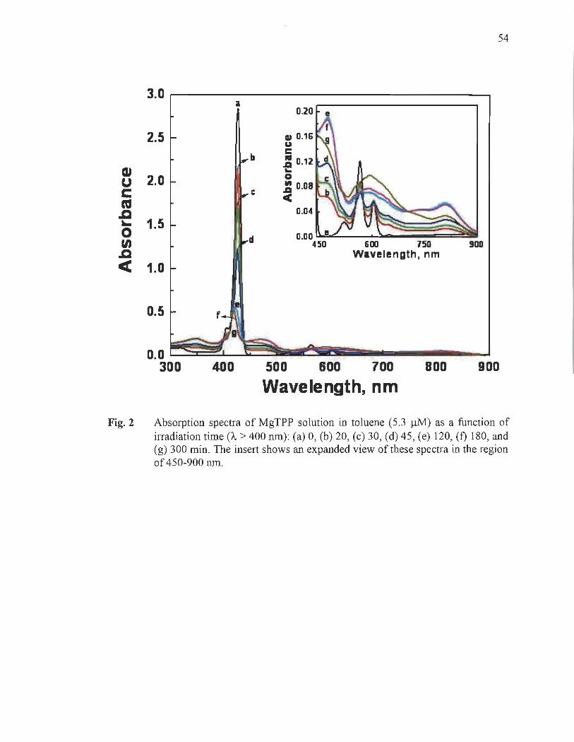
54
3.0 0.20 e
2.5 CD 0.16 u
b c .. d .a 0.12
CU ~
2.0 0 u J 0.08 C c ca CC
oC 0.04 .... 1.5 0
d 0.00 en 450 600 150 900 oC Wavelength, nm
< 1.0
0.5
0.0 ~~~~ ÏiiiIiirII!~ïIIrI:II ...... ___ -.-.J 300 400 500 600 700 800 900
Wavelength, nm
Fig. 2 Absorption spectra of MgTPP solution in toluene (5.3 ~M) as a function of irradiation time (1" > 400 nm): (a) 0, (b) 20, (c) 30, (d) 45, (e) 120, (f) 180, and (g) 300 min. The insert shows an expanded view of these spectra in the region of 450-900 nm.
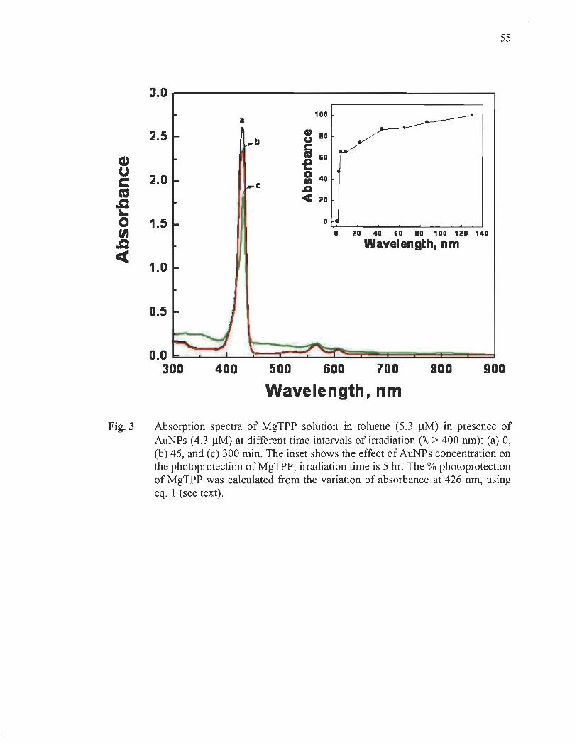
55
3.0
100 il
2.5 cu 10 b u
CU fi 1i0 of! U 2.0 0 40 C 1ft
ca C .a oC CC 20
.... 0 1.5 0
en 0 20 40 1i0 10 100 no 140
oC Wavelength, nm CC
1.0
0.5
400 500 600 700 800 900
Wavelength, nm
Fig. 3 Absorption spectra of MgTPP solution in toluene (5.3 ~M) in presence of AuNPs (4.3 ~M) at different time intervals of irradiation (À > 400 nm): (a) 0, (b) 45, and (c) 300 min. The inset shows the effect of AuNPs concentration on the photoprotection of MgTPP; irradiation time is 5 hr. The % photoprotection of MgTPP was calculated from the variation of absorbance at 426 nm, using eq. 1 (see text) .
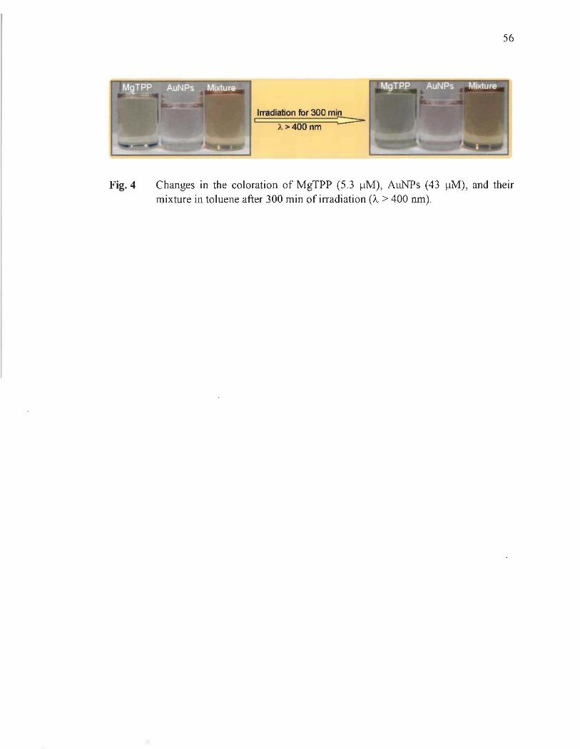
56
Irradiation for 300 min Â. > 400 nm ::::::=:-
Fig. 4 Changes in the coloration of MgTPP (5.3 /lM), AuNPs (43 /lM), and their mixture in toluene after 300 min of irradiation (À > 400 nm).
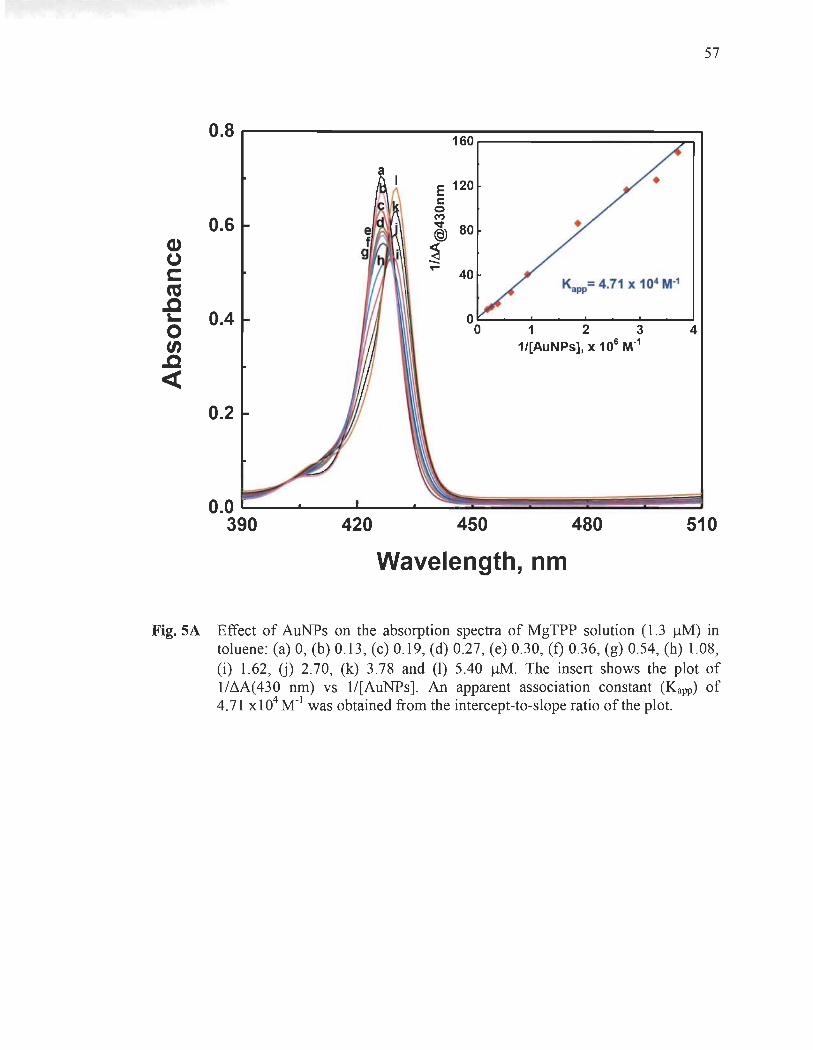
57
0.8 160
a E 120 c
0
0.6 M -.:t 80 @
Cl) ~ (.)
C ....
40 ca Kapp= 4.71 X 10⁴ M-i
.a 0.4 L- 0
0 0 1 2 3 4 tA 1/[AuNPs], X 106 M·i
.a « 0.2
420 450 480 510
Wavelength, nm
Fig. SA Effect of AuNPs on the absorption spectra of MgTPP solution (1.3 !lM) in toluene: (a) 0, (b) 0.13 , (c) 0.19, (d) 0.27, (e) 0.30, (t) 0.36, (g) 0.54, (h) 1.08, (i) 1.62, U) 2.70, (k) 3.78 and (1) 5.40 !lM. The insert shows the plot of 1/~A(430 nm) vs 1/[AuNPs]. An apparent association constant (Kapp) of 4.71 X 10⁴ M-l was obtained from the intercept-to-slope ratio of the plot.

58
0.3 32
E c :;;24
k @
~ 16
(1) 0.2
K"pp= 2.12 x 10⁴ M·l
(.) c: 0.4 0.6 0.8 1.0
ca 40 1/[AuNPs), X 105
M·l
.c ~
0 tn .c
<C 0.1 K.pp= 5.03 x 10⁴ M·l
0.4 0.6 0.8 1.0 1/[AuNPs), x 105
M·l
550 600 650 700
Wavelength, nm
Fig. SB Effect of AuNPs on the absorption spectra of MgTPP solution (1 0 ~M) in toluene: (a) 0, (b) 10.11 , (c) 14.44, (d) 17.33, (e) 20.22, (f) 24.55, (g) 30.33, (h) 33.22, (i) 36.11 , (j) 39 and (k) 43.33 ~M. The inserts show the plots of lIM(567 nm) and II ~A(608 nm) vs 1/[AuNPs].
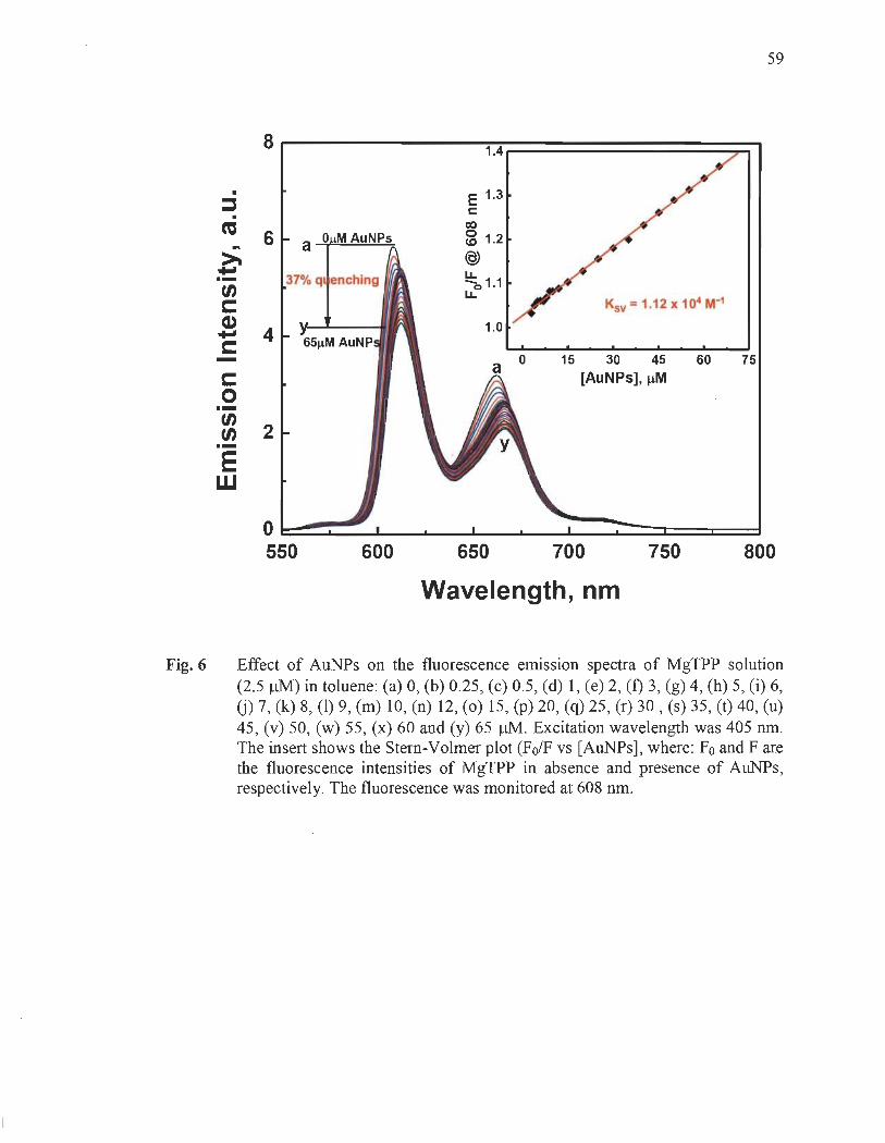
59
8r-----------1A.============~~ .
:::J . CU
... ~ .... .-
-c: o
6
4
~ 2 .-E w
600
E 1.3 c: co ~ 1.2 (§) u.. -0 1.1 u..
1.0
650
o 15 30 45 60 75 [AuNPs), IlM
700 750 800
Wavelength, nm
Fig. 6 Effect of AuNPs on the fluorescence ernission spectra of MgTPP solution (2.5 ~M) in toluene: (a) 0, (b) 0.25, (c) 0.5, (d) 1, (e) 2, (f) 3, (g) 4, (h) 5, (i) 6, (j) 7, (k) 8, (1) 9, (m) 10, (n) 12, (0) 15, (P) 20, (q) 25, (r) 30 , (s) 35, (t) 40, (u) 45, (v) 50, (w) 55, (x) 60 and (y) 65 ~. Excitation wavelength was 405 nm. The insert shows the Stern-Volmer plot (FolF vs [AuNPs], where: Fo and Fare the fluorescence intensities of MgTPP in absence and presence of AuNPs, respectively. The fluorescence was monitored at 608 nm.
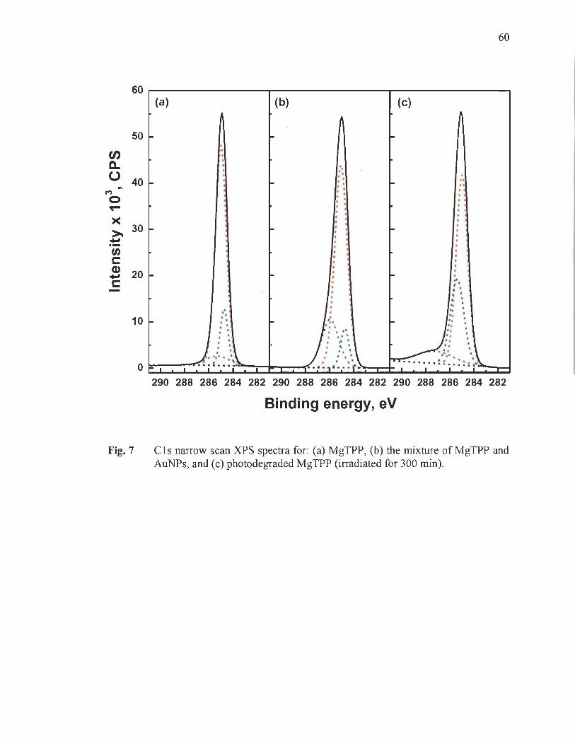
60
60~--------------~--------------~--------------~
en a..
50 ~
o 40 ~ C"')
o
~ 30 ~ .., .-tn c: .! 20 ~ c:
10 ~
(a)
, , , , , , .
, , , , -: , , , , , ,
: ',,~ . -,- .. ', o ~~-I--""'" i ~ ... j"'
(b)
"
n , , , , , , , , , , , , , , , ,
, , , , . ..
" . • • II , ,'
: : ', \ . I , ' . ' .. ' . . ~ . . . · . ," .. ( -
(c)
-
-
-
.. .. , , , , , , . , , , , , , , , , , .. , , . ' , ~ , .. ' .. '
" .
~< 1···· '1" : .; .~ ::: :'i~---.I
290 288 286 284 282 290 288 286 284 282 290 288 286 284 282
Binding energy, eV
Fig. 7 Cls narrow scan XPS spectra for: (a) MgTPP, (b) the mixture of MgTPP and AuNPs, and (c) photodegraded MgTPP (irradiated for 300 min).
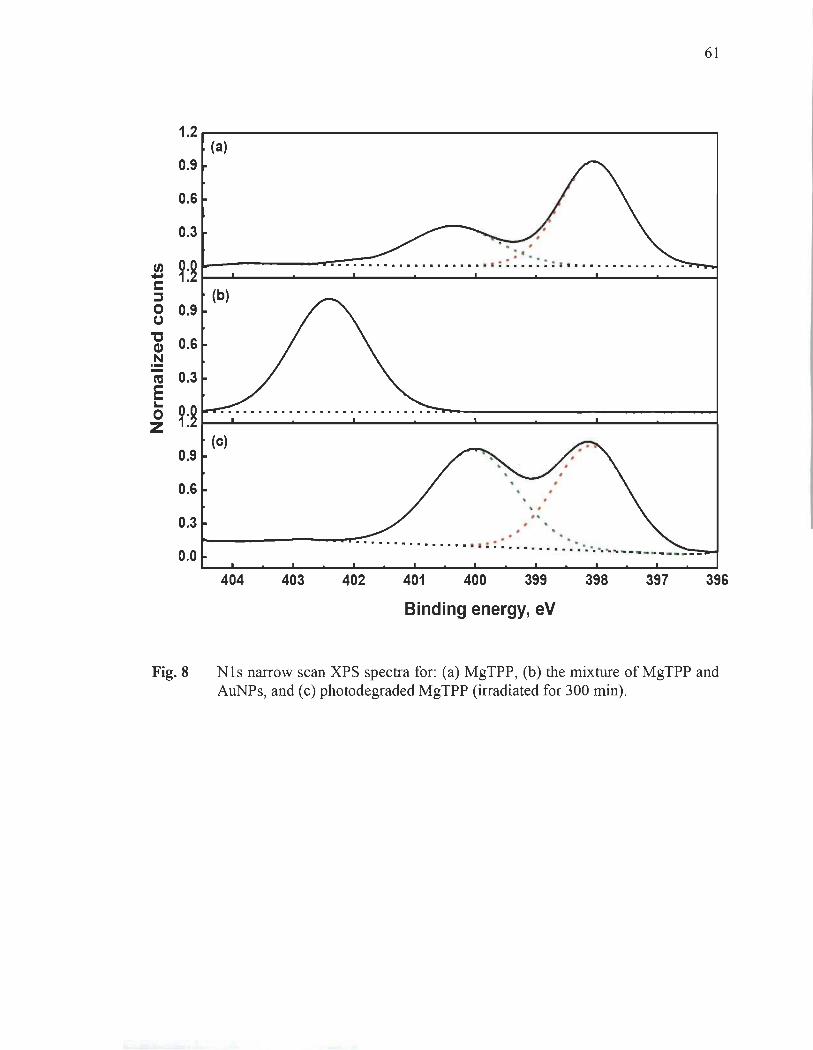
1.2
0.9
0.6
0.3
tA ~:g .... r:: :::s 0 0.9 (.)
"C 0.6 Cl)
.!::! ca 0.3 E '-
~:g 0 z 0.9
0.6
0.3
0.0
(a)
.. l-------r-:.-:". -:-: .. :-:.-:-............... . .. : : : . : : : : 1 • ••••••••••••••••
404 403
. . .. .. . . . . ............... .. : :: ....... . .. ... .. .. .. .......... _---------
402 401 400 399 398 397
Binding energy, eV
61
396
Fig. 8 Nls narrow scan XPS spectra for: (a) MgTPP, (b) the mixture of MgTPP and AuNPs, and (c) photodegraded MgTPP (irradiated for 300 min).

en D.. 0 .. t/) ... C ::l
200
150
100
50
0
(a)
(b)
~ , . .. --......... •• • • 1 ••••• ~~ __ _
o 150 0
100
50 ~ ,
~, fi .... ....•..•.............• ••• : .. ..••••...•.•.•••.•••.•.•.•.......•...•.....
O~ ________ ~ ________ ~ __________ ~ ________ ~~
90 88 86 84 82
Binding energy, eV
62
Fig. 9 Au(4f) narrow scan XPS spectra for: (a) AuNPs and (b) the mixture of AuNPs withMgTPP.

3.---------------------------------,
B 2 rs::: ca .c 10-o t/) .c « 1
a (A)
c
O ~~S-~~~~\~~~~~~~~~ 300 400 500 600 700
Wavelength, nm
63
Fig.lOA Absorption spectra of MgTPP solution in toluene (5 .3 J.lM) in an anaerobic atmosphere in absence AuNPs recorded at different time intervals of irradiation (À > 400 nm): (a) 0, (b) 30, and (c) 300 min.
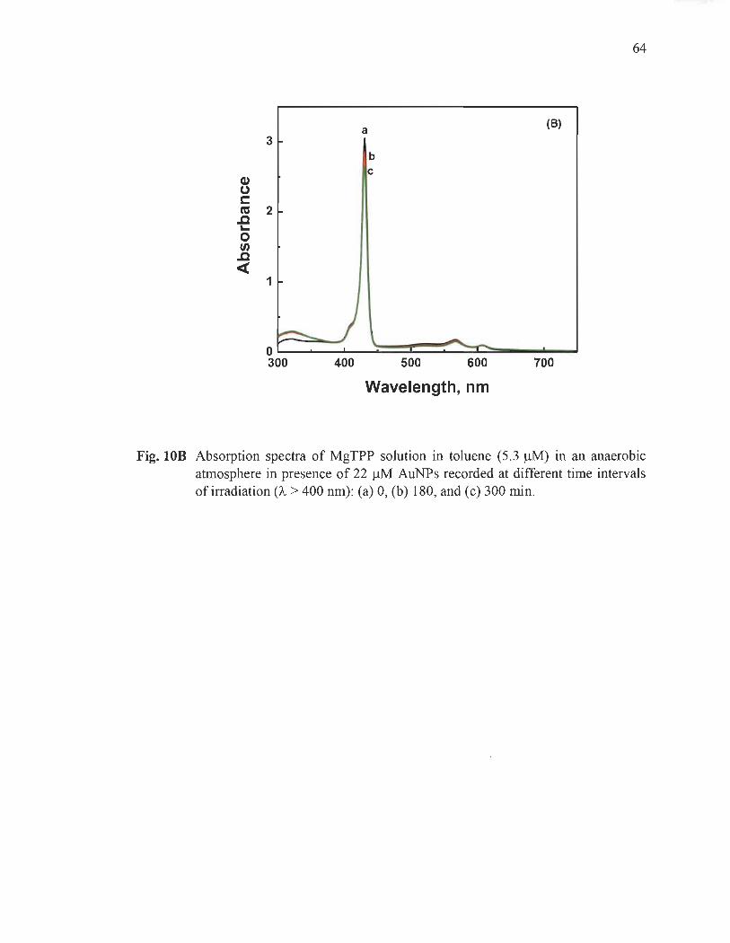
64
a (8)
3 b c
Q) (,) r::::: C'O .c 2 '-0 f/J .c «
1
Wavelength, nm
Fig. lOB Absorption spectra of MgTPP solution in toluene (5.3 ~M) in an anaerobic atmosphere in presence of 22 ~M AuNPs recorded at different time intervals of irradiation (À. > 400 nm): (a) 0, (b) 180, and (c) 300 min.
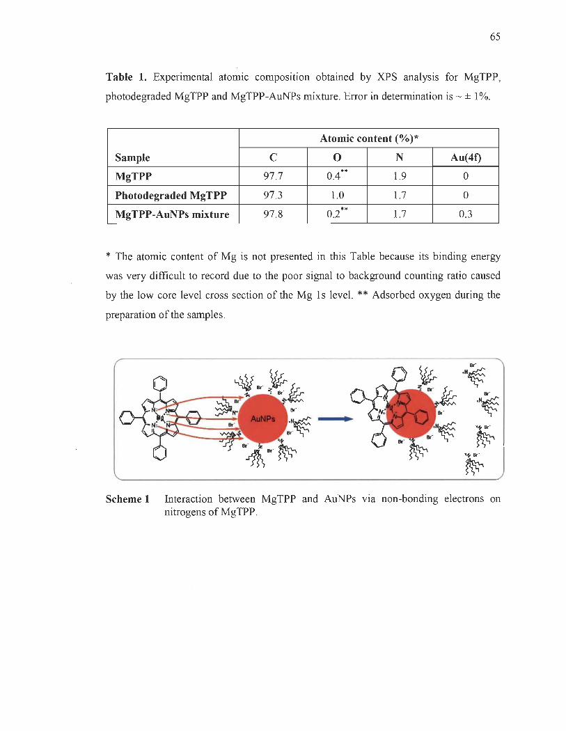
65
Table 1. Experimental atomic composition obtained by XPS analysis for MgTPP,
photodegraded MgTPP and MgTPP-AuNPs mixture. Error in deterrnination is - ± 1 %.
Atomic content (%)*
Sample C 0 N Au(4t)
MgTPP 97.7 0.4 ** 1.9 0
Photodegraded MgTPP 97.3 1.0 1.7 0
MgTPP-AuNPs mixture 97.8 0.2 ** 1.7 0.3
* The atomic content of Mg is not presented in this Table because its binding energy
was very difficult to record due to the poor signal to background counting ratio caused
by the low core level cross section of the Mg Is level. ** Adsorbed oxygen during the
preparation of the sarnples.
•
Scheme 1 Interaction between MgTPP and AuNPs via non-bonding electrons on nitrogens of MgTPP.

SUPPORTING INFORMATION
Beneficiai Role of Gold Nanoparticles as Photoprotector of
Magnesium tetraphenylporphyrin
Laurent Bekalé, Saïd Barazzouk, and Surat Hotchandani*
Groupe de Recherche en Biologie-Végétale, Université du Québec à Trois-Rivières,
Trois-Rivières, Qc, G9A SR?, Canada
* To whom correspondence should be addressed. e-mail: [email protected]
66

67
High-resolution Cls and Nls XPS spectra for gold nanoparticles (AuNPs) are
shown in Fig. SI and S2, respectively. The Cls region of AuNPs comprises three
distinct peaks. The peak appearing at 285 eV, is assigned to C-C and C-H from
tetraoctylammonium bromide (TOAB), the stabilizing agent used in the preparation of
AuNPs. The second peak at 286.07 eV is ascribed to C-N of quatemary amine from
TOAB, while a smaller peak at higher binding energy (287 .17 eV) is probably due to an
impurity.
CI) a..
40
CJ 30 M
Q T""
>< ~ 20 ~ fi) c (1)
ë 10
288 287 286 285
Binding Energy, eV
Fig. SI Cls narrow scan XPS spectrum for AuNPs.
284 283
The N 1 s spectrum of AuNPs (Fig. S2) presents two peaks with an intensity ratio
of about 99:1 , which appear at 401.34 (90.5%) and 402.96 eV (9.5%), respectively. Both
peaks are characteristic of quatemary nitro gens, although, present in two different
chemical environments. The flfSt one at 401.34 eV is assigned to quatemary nitrogen
(Oc14Nl of TOAB adsorbed on the surface of AuNPs 1. The second peak, a weaker one,
at high binding energy 402.96 eV, corresponds most probably to the small quantity of
TOAB which is not in direct contact with gold surface of AuNPs, but rather through the
intermediacy of bromide ions which are adsorbed on gold nanoparticles. This signal is

68
consistent with the work of Bureau et al who reported the presence of a similar peak for
tetramethyl ammonium hydroxide {(CH3)4W , OK) adsorbed on nickel surface2.
2.5....------------------,
en 2.0 0.. U
M o 1.5
>< ~ ~ 1.0 t/) r:::: Cl) -r:::: 0.5
A
OO ~ ....... :-:-•. • ······· ·· ·E:::: .. ::::::: .• ...... .... ······· ..... . . 404 403 402 401 400
Binding energy, eV
Fig. S2 N 1 s narrow scan XPS spectrum for AuNPs.

69
Table SI. Fraction of the incident light absorbed by AuNPs of varying concentration in
the mixture of MgTPP and AuNPs. The concentration of MgTPP in the mixture is
constant at 5.3 /lM, and the totallight absorbed by this solution in the wavelength range
(400-900 nm) is 494 io. The total incident light intensity is 5000 io.
Concentration Amount of incident Total amount of The percentage of of AuNPs (/lM) light absorbed by incident light incident light
AuNPs absorbed by the absorbed by AuNPs mixture in the mixture
(MgTPP + AuNPs)
2.17 39 io 494 io +39 io = 533 io 7.3
4.33 57 io 551 io 10.3
8.67 105 io 599 io 17.3
22 229 io 723 io 31.7
43.3 324 io 818 io 39.6
65 617 io 1111 io 55.6
130 1070 io 1573 io 68.6
References of supporting information
(1) J.R. Pels, F. Kapteijn, J.A. Moulijn, Q. Zhu, and K.M. Thomas, Carbon, 1995, 33, 1641-1653.
(2) C. Bureau, and P.D. Chong, Chem. Phys. Lett, 1997,264,186-19.

CHAPITREITI
ENHANCED PHOTOSTABILITY OF CHLOROPHYLL-A USING GOLD NAN OP ARTICLES AS AN EFFICIENT PHOTOPROTECTOR
Le contenu de ce chapitre a fait l'objet d'une publication en anglais dans la revue :
Journal of Materials Chemistry, 2012, 22, 25316-25324.
3.1 Résumé de l'article
Une étude complète des propriétés photoprotectrices des nanoparticules d'or
(AuNPs) a été effectuée dans ce second article scientifique afin d'établir que la méthode
de photoprotection, précédemment présentée dans le premier article scientifique,
pourrait également permettre d'accroître la stabilité des porphyrines de magnésium plus
complexe que le tétraphénylporphyrine de magnésium (MgTPP), c'est-à-dire, les
porphyrines possédant à la fois des doublets d'électrons libres sur les atomes d'azote du
macrocycle et sur les groupes fonctionnels périphériques qui ne sont pas conjugués au
macrocycle, comme c'est le cas avec les chlorophylles et les bactériochlorophylles. Nos
résultats montrent que la photodégradation de la chlorophylle-a (ChIa) est ralentie en
présence des AuNPs, et une augmentation pouvant atteindre un ordre de grandeur· dans
le temps de demi-vie de ChIa en présence d'AuNPs a été observée. Il a également été
observé que dans des conditions in vitro, les AuNPs sont des agents photoprotecteurs
bien plus efficaces que les molécules comme la ,B-carotène ou de quinones, qui sont
connues pour être très efficaces dans des conditions in vivo (dans les plantes). Enfin, la
capacité protectrice des AuNPs est le résultat de leur liaison avec les sites azotés du
macrocycle porphyrique de la ChIa, et ce, avant la formation des espèces réactives de
l'oxygène (ROS) par photosensibilisation. Ce mécanisme de photoprotection rend les
AuNPs plus efficaces que les carotènes ou des quinones qui offrent une protection à la
ChIa par neutralisation des ROSs après illumination. L'étude présentée dans cet article a
été l'aboutissement du deuxième objectif opérationnel de cette thèse.

3.2 Deuxième article scientifique
Enhanced photostability of chlorophyU-a using gold nanoparticles
as an efficient photoprotector
Saïd Barazzouk:t, Laurent Bekalé:t, and Surat Hotchandani*
Groupe de Recherche en Biologie-Végétale, Université du Québec à Trois-Rivières,
Trois-Rivières, Qc, G9A SR7, Canada
t contributed equally to this work
* To whom correspondence should be addressed. e-mail: [email protected]
Abstract
71
Improving the photostability of chlorophylls is one of the main challenges to facilitate
their indus trial and biotechnological use. In this regard, we have employed go Id
nanoparticles (AuNPs) to photoprotect chlorophyll-a (ChIa). The results show that the
photodegradation of ChIa is slowed down in presence of AuNPs, and an increase of as
much as an order of magnitude in half-life time of Chla in presence of AuNPs has been
observed. It is further seen that under in-vitro conditions, AuNPs are much better
photoprotective agents of Chla than ~-carotene or quinones, which are known to be very
effective in naturalliving conditions (plants) . The protecting ability of ChIa by AuNPs is
the result of their efficient binding with ChIa at its nitrogen sites even in dark, thus,
inhibiting the reaction of reactive oxygen species with ChIa, known to cause its
degradation during illumination. The same property of AuNPs, i.e. , to bind with ChIa in
dark, renders them better photoprotectant than carotene or quinones since these agents
offer protection to ChIa during its illumination.
72
Introduction
The application of biomolecules in various fields of material sCIence such as
biomedical engineering/ optoelectronics,2 etc. , is potentially one of the most important
emerging new technologies. Recently, organic light-emitting diodes (OLEDs) have been
fabricated using horse-heart cytochrome-c.3 This biomolecule is also available in plants.
Among the biomolecules, the chlorophylls are by far the most abundant on the earth.
However, the application of chlorophylls in organic electronic devices such as OLEDs
or organic photovoltaics (OPVs) has a major limitation and that is their high
photodegradation. This is unfortunate since chlorophylls are the best photoreceptors in
nature. Thus, if the photostability of chlorophylls could be significantly amended their
technological application wou Id be enormously improved since the long-term stability of
the molecules is the prerequisite for their commercialization.
Recently, Ohtani et aP have successfully fabricated OLEDs from chlorophylls
(Ch!) extracted from spinach. They found that Chl-OLEDs that were rich in carotenoids
emitted electroluminescent signaIs for much longer time (more than one minute)
compared to less than five seconds for the Chl-OLEDs that were poor in carotenoids.
The long operational time of carotenoids ri ch Chl-OLEDs was attributed to the
antioxidant activities of carotenoids which prevented the destructive photooxidation of
chlorophylls.
While it is interesting to note that the photostability of Chi has been irnproved to
sorne extent by carotenoids, the results also show that carotenoids do not prote ct in-vitro
Chi as effectively as they photoprotect in-vivo Chi, i.e., in green plants. Further,
electorluminescence lifetime of 1 minute for Chl-OLEDs rich in carotenoids do es not
bode well for their commercial success. It is thus highly imperative to search for an
agent that can provide an efficient photoprotection to Chi under in-vitro conditions so
that the utilization of Chis can be exploited in various optoelectronic devices.
In this context, we have recently shown 4 that the use of gold nanoparticules can
help overcome the photodegradation of magnesium tetraphenylporphyrin (MgTPP), a
73
molecule structurally analogous to ChIa. In the present work, we demonstrate that goid
nanoparticles (AuNPs) can aiso increase the photostability of ChIa. The UV-Visible,
fluorescence and X-ray photoelectron spectroscopic studies have been carried out to
assess the effect of AuNPs on the photostability of ChIa. The results show that the
haif-life time of ChIa (4.5 /lM) has been extended from 10 to 100 minutes in presence of
AuNPs (22 /lM) , indeed, quite a significant photoprotection. The protecting ability of
ChIa by AuNPs is the result of their binding with ChIa at its nitrogen sites, inhibiting,
thus, the binding of reactive oxygen species (ROS) at these sites, which are known to
cause the photodegradation of ChIa. It is also seen that AuNPs are much more effective
photoprotective agents of in-vitro ChIa than are the carotenoid moiecule (j3-carotene)
and various quinones.
Materials and methods
Materials
ChIa, hydrogen tetrachioroaurate trihydrate, suifuric acid (H2S04),
tetraoctylammonium bromide (TOAB), sodium borohydride (NaBH4), sodium sulfate
(Na2S04), benzoquinone, 2,6-dichloro-p-benzoquinone (DCBQ), tetramethy-p
benzoquinone (Duroquinone), ,B-carotene and toluene, of the pure st quality available,
were purchased from Aldrich Chemical Co., and were used as received.
Synthesis of go Id nanoparticles (AuNPs)
The procedure for preparing colloidal gold nanoparticles in an organic medium is a
modified version5 of that proposed by Brust et al.6 Briefly, an aqueous solution of
hydrogen tetrachloroaurate (III) hydrate [(AuCI4L 6 mL, 0.04 M)] was mixed with a
solution of tetraoctylarnmonium bromide (TOAB) (0.05 M) in 14 mL toluene. The
biphasic mixture was vigorously stirred for 30 min until all the (AuC14r was transferred
into the organic layer. An aqueous solution of sodium borohydride (6 mL, 0.4 M) was
added, and the mixture was stirred for 3h. The organic layer was separated off, washed

74
twice with a di lute aqueous solution of H2S04 and then washed five times with
deionized water. FinaUy, the organic layer, constituting the colloidal go Id nanoparticles,
was dried ovemight on Na2S04 powder and then filtered. The final concentration of
AuNPs, estimated on the basis of atomic Au concentration, was 13 mM5.
Photodegradation measurements
The measurements were carried out in al-cm path length quartz cuvette. The
illumination of ChIa solution (3 mL, 4.5 /lM) in toluene was carried out using coUimated
light from a 150W xenon lamp that passed through a water filter to absorb heat. A red
cut-off filter which allowed wavelengths greater than 640 nm was used. The light power
reaching the sample was 40 mW.cm-2. AU operations with ChIa solutions were carried
out in either subdued light or green light. The solutions were stored in dark in a
refrigerator when not in use. AU experiments were carried out in the presence of air.
The photodegradation experiments of ChIa in presence of various photoprotective
agents, such as, AuNPs, ~-carotene and quinones (aH in 22 /lM concentration), in order
to assess their capacity to photoprotect ChIa, were also carried out under identical
conditions as those for ChIa aione as described above.
Absorption and emission spectrometry
Absorption spectra were recorded with a Cary 5000 spectrophotometer. The
fluorescence emission spectra were recorded with a FluoroLog-3 spectrofluorometer.
Transmission electron microscopy (TEM)
One drop (5-10 ilL) of AuNPs was deposited onto glow-discharged carbon-coated
electron microscopy grid. The ex cess liquid was absorbed by a piece of filter paper, and
a drop of 2% uranyl acetate negative stain was added before drying. The liquid in excess

75
was wiped off, and the remaining film of stain was allowed to dry. The specimens were
observed using a Philips EM 208S microscope operating at 80 kV.
X-ray photoemission spectroscopy (XPS)
XPS was performed on a Kratos Axis Ultra spectrometer (Kratos Analytical Ltd.,
UK), using a monochromatic Al Ka X-ray source (E = 1486.6 eV) with a power of
225 W, at a take-off angle of 90° relative to the sarnple surface. 250 J.l.L of the sample
(ChIa, 4.5 J.l.M) or (AuNPs, 22 /lM) or their mixture) was dropped onto an aluminum
substrate and dried in a vacuum dessicator ovemight to ob tain a thin film. The dried
sarnples were then transferred to the XPS sample holder. The measurements were made
at room temperature under a high vacuum of 10-9 torr. The surface of the sample was
~ 20 mm2, and the investigated area was typically 1 x 2 mm2
. Survey spectra for each
sample over a binding energy range of 0 - 1300 eV were an average of three scans (at
three different points) acquired at pass energy of 160 eV and resolution of 1 eV /step
(lens in hybrid mode, which assures maximum sensitivity). High-resolution spectra of
C Is, N Is, 0 Is and Au(4f) for quantitative measurements ofbinding energy and atomic
concentration were an average of five scans acquired at a pass energy of 40 eV and
resolution of 0.1 eV/step. Because of the potential degradation of the surface during
X-ray exposure, the spectra were collected in the same order (survey, C Is, Ols, N Is,
Au(4f) such that the amount of exposure to X-rays was equivalent for aIl analyzed
samples. The CasaXPS software was used for background subtraction (Shirley-type),
peak integration, fitting and quantitative chemical analysis. The C Is (C- C) peak at
285 eV was used to calibrate the binding energy scale. Binding energies values are given
as ± 0.2 eV. Gaussian peak profiles were used for spectral deconvolution of C 1 s, Ols,
N Is and Au(4f) spectra. The magnesium metal binding energy was not recorded due to
the poor signal to background counting ratio caused by the low core level cross section
of Mg 1 s level.

76
Percentage photostability
The percentage photostability provided to ChIa by AuNPs was calculated using
the formula as described by Claes : 7
(1)
where El , E2. and E3 are, respectively, the concentration (or absorbance) of ChIa before
irradiation, after irradiation without AuNPs, and after irradiation in the presence of
AuNPs.
Results
Photodegradation of chlorophyll-a (ChIa)
Figure lA presents the absorption spectra of ChIa (4.5 /lM) and gold nanoparticles
(50 /lM) in toluene. The principal absorption peaks of ChIa (spectrum a) in the red at
665 nm, Q band, and at 433 nm in the blue, Soret band, are in agreement with those
reported in the literature,8 and that ChIa is present in monomeric form. 9 The 530 nm
absorption band (spectrum b) is characteristic of the surface plasmon band of gold
nanoparticles (8 nm),lO as is verified by the TEM micrograph of the AuNPs which
revealed an average particle diameter of 8 nm (Fig. lB).
The absorption spectra of ChIa solution in toluene as a function of the time of
illumination with red light (1" > 640 nm) are displayed in Fig. 2. The choice of toluene as
solvent is due to the fact that go Id nanoparticles to be used have been prepared in
toluene. As seen from the figure, there is a progressive decrease of ChIa absorbance as
illumination time increases. In 2 minutes, about 25% decrease in the absorbance of
major absorption bands in the red (665 nm) and blue (433 nm) regions was observed.
On continued exposure, the absorbance at 665 nm decreased 60% in 10 minutes and by
77
80% in 30 minutes. An increase in absorbance at waveiengths above 690 nm and
between 460 and 580 nm was observed. These results are in good agreement with those
reported by Jones and Mackay8 Merzlyak et al. Il and Tregub et al. 12 who studied the
photodegradation of ChIa in micelles, methanoi and various other organic soivents,
respectiveIy.
Noteworthy, however, in the present experiments is the appearance of a band in
near-infra red region at 870 nm arising from the photooxidized product of ChIa whose
intensity increases as illumination time increases. The photodegradation of ChIa in
aerobic conditions, as in many metalloporphyrins, is caused by the reactive oxygen
species (ROS) which are produced during the illumination of ChIa. 13-17
Photoprotective action of go Id nanoparticles
In order to investigate the role of AuNPs as photoprotector of ChIa, the
illumination of ChIa solution (4.5 IlM) in the presence of AuNPs was carried out under
identical conditions using a red eut-off filter providing excitation wavelengths greater
than 640 nm. Since, the absorption maximum of AuNPs in toluene is at 530 nm (Fig. 1),
there was no interference from the direct absorption of the incident Iight (À> 640 nm) by
AuNPs.
The addition of AuNPs, as is c1ear from Fig. 3, do es seem to provide protection to
ChIa against photodegradation. After 30 minutes of illumination, ChIa degraded only
16% in the presence of AuNPs (22 IlM) compared to the 80% photodegradation (at
665 nm) in the absence of AuNPs (Fig. 2). In other words, a photoprotection of 84% has
been provided to ChIa by 22 IlM AuNPs. In addition, the photooxidation product
absorbing at 870 nm totally disappeared. Further, it is found that the degradation of ChIa
is dependent upon the Au nanopartic1es concentration (inset of Fig. 3). That is, the
higher the concentration of AuNPs, the greater is the photoprotection provided to ChIa
against irradiation. Higher concentrations of AuNPs beyond 88 IlM, however, provided

78
no additional protection. The percentage photoprotection was calculated using eq. (1),
described in the Experimental section.
However, to get a meaningful insight into the photostability imparted to ChIa by
AuNPs, the photodegradation kinetics of ChIa was studied, and is presented in Fig. 4,
and described in ESI section. As seen from the inset of the figure, the photodegradation
of ChIa follows a pseudo-first order kinetics with half-life times of 10 and 100 minutes
in absence and in presence of AuNPs, respectively, indicating a tremendous
improvement in the photostability of ChIa in presence of AuNPs. The reaction rate
constants and half-lives are presented in Table 1. The beneficiai role of AuNPs in the
increased photostability of Chia is also possibiy, in part, responsibie for the improved
photoelectrochemical performance of a Chla-AuNPs based solar cell compared to that of
Chia cell as reported earlier. 5
To exclude the possibiiity that the tetraoctyiammonium bromide (TOAB), a
surfactant used in the preparation of AuNPs, had any photoprotective role, a solution of
TOAB (0.05 M) in toluene was added to Chia solution. The ChIa was not
photoprotected, confirming that it is the AuNPs, rather than TOAB, that provide the
photoprotection to Chia. Furthermore, AuNPs themselves subjected to irradiation for
same time period (5-60 min) under identical conditions did not show any photochemical
changes. The photoprotection provided to Chia by AuNPs is due to the binding between
AuNPs and ChIa that prevents ROS from interacting with Chia, similar to that reported
recently by Bekalé et al.4 in case of magnesium tetraphenylporphyrin (MgTPP).
The binding of ChIa with AuNPs is manifested by the changes occurring in the
absorption spectrum of Chia as AuNPs are added (Fig. 5). A decrease in the absorption
of ChIa at - 432 nm and an appearance of a new band at - 437 nm, as the concentration
of AuNPs increases, indicates the formation of Chia-AuNPs complex. A decrease in the
absorption at 665 nm is also visible. The changes in the absorption (M) at 437 nm were
used to determine the association constant between ChIa and AuNPs employing Benesi
Hild~brand method. 18 An apparent association constant, Kapp, of 5.70 x 10⁵ M-1 for

79
Chla-AuNPs complex was obtained from the intercept-to-slope ratio of the double
reciprocal plot of 1/~A vs l/[AuNPs] (inset, Fig. 5). The high value of K app suggests a
strong binding between AuNPs and ChIa. The association constant has also been
obtained from the fluorescence quenching experiments of ChIa in the presence of
AuNPs. Figure 6 presents the fluorescence spectra of ChIa in toluene in the presence of
varying concentration of AuNPs. The fluorescence emission of ChIa exhibits a
maximum at 675 nm, which is characteristic of emission from monomeric ChIa in
nonpolar solvent. Upon addition of gold nanoparticles, one observes a progressive
quenching of the fluorescence of ChIa with increasing amounts of AuNPs. This
quenchmg occurs as a result of the formation of a weakly or non-fluorescent ground
state complex between fluorophore (ChIa) and quencher (AuNPs).
Thus, following the approach described by Lakowicz,19 the dependence of
fluorescence intensity on quencher concentration can be obtained by considering the
association or complex formation constant, Ks, between the fluorophore and quencher,
i.e. , Ks = [F-Q]/[F][Q] , where [F-Q] is the concentration of the complex, and [F] and [Q]
are, respectively, the concentrations of uncomplexed fluorophore and quencher.
Proceeding further as demonstrated in reference 19, one finally ob tains the relation
between fluorescence intensity and concentration of the quencher as follows:
10/1= 1 + Ks [Q] (2)
where 10 and 1 are the fluorescence intensities of ChIa in absence and in presence of
the quencher, AuNPs, respectively.
The ratios of the fluorescence intensity (lo/l) , measured at 675 nm, without and
with AuNPs, were plotted against AuNPs concentration (inset of Fig. 6). From the slope
of the linear plot, a value of ~ 1 04 M-1 for the association constant, Ks, was obtained. This
value, although smaller than that obtained from absorption measurements (inset, Fig. 5),
is, nevertheless, high. A high value of the association constant suggests a Strong

80
interaction between ChIa and AuNPs, which, it shouid be pointed out, aiso heips the
heterogenous charge transfer at Chia-AuNP interface, as has been reported earlier.s
Since AuNPs possess an affinity for nitrogen atoms, the complex formation
between ChIa and AuNPs is the result of the binding of AuNPs with the nitrogens of
ChIa, as described below in XPS studies, and is at the heart of the photoprotection
provided to ChIa by AuNPs.
X-ray photoelectron spectroscopy (XPS) studies
In order to better understand the mechanism by which AuNPs interact with ChIa
and provide it the photoprotection, XPS study of ChIa, AuNPs, and their mixture in a
concentration ration of 1 :5, has been carried out. The Iow resolution (survey) spectra of
these three samples are shown in Fig. S-1 (ESI). AH samples show the spectroscopie
signature of carbon at around 285, nitrogen at - 400, and oxygen at - 532 eV. In
samples containing AuNPs, one can also notice the presence of Au(4f) signal,
corresponding to the fS/2 and f712 components, at binding energies of -352 and 334 eV,
respectively. The magne sium metal binding energy was not recorded due ·to the poor
signal to background counting ratio caused by the low core level cross section of the Mg
ls level. To obtain more XPS information, the high-resolution scans of the XPS spectra
of Cl s, Ols and NI s levels with their respective deconvolutions were obtained, and are
as foHows.
Figure S-2 shows high-resolution spectra of the C Is regions for ChIa and Chla
AuNPs mixture. The most significant contribution to these spectra cornes from the peak
at 285 eV, which corresponds to the aliphatic carbon (C-C) and C-H of Chia and TOAB
(the underline refer to the atom in question).20,21 The signal pertaining to the aromatic
carbons (-C=C-) of ChIa at 284.7 eV is rather hidden in the envelope ofC-1 peak, and is
thus not visible. The contribution of the carbons bound to nitrogen ~-N, C=N) and
those bound to oxygen by single bond (-C-O), peak C-2, appears at 286.1-286.5 eV.20,21
The weak peak, C-3, at 288.9-289.1 eV is attributed to the carbonyl of the ketone

81
(>C=O) and ester (-O-C=O) groups in ChIa (Scheme 1)? 1.22 The fact that the position of
this peak remains unaltered even after the addition of AuNPs is an important indication
that the interaction between Chia and AuNPs does not invoive the nonbonding electrons
of carbonyl oxygen of ketone and ester groups (i.e., >C=O and -O-C=O) of ChIa. This
observation is aiso supported by the 0 Is spectra (vide infra) .
The high-resolution scans of the XPS spectra of Ols Ieveis of ChIa and
Chla-AuNPs with their decomposition into three components are shown in Fig. S-3 . The
0-1 peak at ~ 533.1-533.4 eV is attributed to the ether-type oxygen in ester group
(_0_C=0)23 of ChIa (Scheme 1). The most intense peak, 0-2, at 532.3-532.5 eV has
been assigned to the carbonyi oxygen from ketone (>C=O) and ester groups (-O-C=Q)
of ChIa.23 It is important to point out, once again, that this peak has virtually remained
unchanged after the addition of AuNPs (Fig. S-3 , Chla-AuNPs), confmning, as
mentioned above in Fig. S-2, that the interaction between ChIa and AuNPs does not
implicate the carbonyi groups of ChIa. FinaUy, the 0-3 peak at lower binding energy
~ 530.2- 531 eV, has been attributed to the molecular adsorbed oxygen, O2.
The deconvolution of Nls levels of ChIa, AuNPs and Chla-AuNPs are displayed
in Fig. 7. The main peak, N-l , at binding energy of 398.1-398.9 eV in ChIa (Fig. 7) is
characteristic of the pyrroienine nitrogen atoms of the porphyrin macro cycle of ChIa
(Scheme 1), and is in good agreement with the reported data for ChIa and many other
metalloporphyrins?4-26 A second peak (N-2), a shouider, in the XPS of ChIa alone can
also be seen at ~ 400 eV. Although, the exact origin ofthis peak is less certain, it is most
Iikely due to the protonated nitrogen, produced as a resuit of a smaU degree of
demetallation of ChIa during its exposure to X-rays in the course of the experiment. This
demetallation seems to be a general feature with magnesium porphyrins as has been
reported by many other authors.25-27
The N Is spectrum of AuNPs shown in Fig. 7 presents two peaks (N-3 and N-4),
with an intensity ratio of about 90:10, which appear at 401.34 and 402.96 eV,
respectively. Both peaks are characteristic of quatemary nitro gens, although, present in

82
two different chemical environments. The first one, N-3 , at 401.34 eV is assigned to
quatemary nitrogen (Oct4W) ofTOAB adsorbed on the surface of AuNPs.28 The second
peak N-4, a weaker one, at high binding energy of 402.96 eV, corresponds most
probably to the small quantity of TOAB which is not in direct contact with the surface of
AuNPs, but rather through the intermediacy of bromide ions which are adsorbed on gold
nanoparticles. This signal is consistent with the work of Bureau et al. who reported the
presence of a similar peak for tetramethyl ammonium hydroxide {(CH3)4N+, OK)
adsorbed on nickel surface?9
The N 1 s spectrum of ChIa shows a drastic change after addition of AuNPs
(Fig. 7, Chla-AuNPs). Indeed, the presence of AuNPs causes the disappearance of a
large fraction of the principal peak, N-l , corresponding to the pyrolenine nitro gens in
ChIa at 398.1-398.9 eV, with the simultaneous appearance of a new and intense peak,
N-5, at 402.4 eV. In other words, the pyrolinene nitrogens of ChIa, present initially in a
chemical state represented by a binding energy of 398.1-398.9 eV, find themselves, after
interaction with AuNPs, in a new chemicai state in which the expulsion of the core
electrons has been made rather more difficult, thereby needing comparatively a higher
energy, i. e., 402.4 eV. The requirement of a higher energy suggests that the electron
density around nitro gens of ChIa, represented by N-5 peak, has decreased or that the
nitro gens are relatively in a more positively charged state than they were in ChIa alone,
represented by N-l peak. In other words, the nitro gens in ChIa, with a binding energy of
398.1 eV, have, now, in presence of AuNPs, been converted into positively-charged
tetravalent ammonium quatemary nitro gens (~N°) which are known to possess a
higher binding energy at 402 eV.30-32 This, i.e. , the nitrogens acquiring a positive charge,
occurs as a result of the interaction between ChIa and AuNPs via the transfer of
non-bonding electrons on nitro gens of ChIa, leading to the formation of the complex
Chla-AuNPs as is depicted in Scheme 2.
One can, therefore, say that the complex forrned between ChIa and AuNPs, with a
high association constant of - 105 M-1 (Fig. 5), is the result of their binding with each
other via non-bonding electrons on ChIa nitrogens. This is consistent with our recent

83
work on MgTPP and AuNPs,4 and is also consistent with the observations of Kamat
et al. 33,34 and Kastonis et al. 35, who, respectively, reported the binding of benzylamine
and meso-tetradodecylporphyrin with AuNPs via interaction of the non-bon ding
electrons on nitrogen of the respective molecules.
The comparison of the Au (4f) core-Ievel spectrum of AuNPs with that for the
mixture of AuNPs with ChIa also confirms that the nitro gens of ChIa have donated
electrons to AuNPs during the formation of the Chla-AuNP complex. The Au (4f)
core-Ievel spectrum of AuNPs (Fig. 8) shows two peaks, namely, 4f712 and 4f512, at
binding energies of 84.1 and 87.6 eV, respectively, corresponding to gold in zero
oxidation state (AUO).36 In its mixture with ChIa, however, these Au(4f) peaks are shifted
to lower energies (83.4 and 87.2 eV). This lowering of the Au(4f) binding energy
suggests that the electron density around Au has increased37,38 since it will facilitate the
expulsion of the core electrons. This increase in electron density on Au occurs as a result
of the transfer of electronic charge (lone pair of electrons) from nitro gens of ChIa to
AuNPs during their binding with each other, corroborating the observations made in
N Is spectra of Chla-AuNPs (Fig. 7) where nitrogen has been shown to donate its
electrons to AuNPs to form the Chla-AuNPs complex.
Comparison of the photoprotective ability of AuNPs with other Chia photoprotectors
In order to further assess the advantageous role of gold nanoparticles fi
photoprotection of ChIa, photodegradation experiments of ChIa (4.5 flM) in presence of
other potential ChIa photoprotectants, such as, carotenoid (all-trans ~-carotene) and
quinones (dichlorobenzoquinone, duroquinone, etc.), all in 22 flM solutions in toluene,
were also carried out under identical conditions using a red cut-off filter providing
excitation wavelengths greater than 640 nm where the individual absorption of quinones
and f3-carotene was negligible. These molecules are known to provide photoprotection
to ChIa and, thus, to plants 7,39-42 via several means: (i) They can function as "triplet
valves" by reacting with excited ChIa triplet, and thus prevent ChIa triplet from
promoting oxygen to its singlet excited state to produce ROS, (ii) They can consume the
84
singlet oxygen that is formed, thereby diminishing its quantities, and, thus, its harmful
effects, and (iii) They can directly quench the excited ChIa singlets, which decreases the
intersystem crossing yield of singlet ChIa to triplet ChIa, leading to a decreased ROS
production. The photoprotection of ChIa and, thus, of plants by carotenoids, quinones,
etc., occurs via their reaction with the excited states of ChIa during its illumination.
The present results (Fig. 9) show that Au nanoparticles are a much more effective
(80%) photoprotectant of ChIa in solution than ~-carotene or quinones which provide
photoprotection of only 28 and 9%, respectively. These results confirm the beneficial
role of gold nanoparticles as an effective photoprotective agent for ChIa in vitro
conditions, which is the consequence of the binding of AuNPs with ChIa at its nitrogen
sites prior to the illumination, as discussed below and recently demonstrated in case of
MgTPP,4 in contrast to the photoprotective action of carotenoids and other antioxidants
which provide photoprotection to ChI by their interaction with the excited states of ChI
during illumination.
Discussion
The irreversible photodegradation of chlorophyll (ChI), as fi many
metalloporphyrins, is known to be mainly a photooxidative process, brought about by
various reactive oxygen species (ROS), namely, singlet oxygen, superoxide ion,
etc.39,42-52 Light absorption by ChIa results in the formation of its excited singlets
eChla *). If not readily used photochemically, they get converted into triplet excited
states eChla *). Chlorophyll triplet states are long-lived energetic species which can
easily react with the triplet ground state of oxygen e 02) to produce strongly
photooxidative ROS . These ROS attack the nitrogens of the porphyrin macrocycle of
ChIa and cause its photodegradation.24,53 Therefore, if these nitrogen sites can be
shielded from the attack of ROS by sorne harmless agent, ChIa will be well protected. In
the present case of ChIa admixed with AuNPs, the AuNPs are just doing that. By
binding with the nitrogens of ChIa, as seen in XPS studies (Fig. 7), AuNPs are

85
essentially providing an impregnable golden armor to ChIa against the deadly attack of
ROS which are produced during its illumination.
What is unique with AuNPs as photoprotector of ChIa, is that they readily bind
with the nitro gens of ChIa as soon as they are mixed together, even in dark prior to the
illumination. This is clear from the changes observed in the absorption spectrum of ChIa
as AuNPs are added (Fig. 5). Consequently, the ROS, which are generated during the
illumination of ChIa, find, to their utter dismay, that only very few free nitrogen sites are
available to bind with and cause damage, since most of them have already been occupied
by AuNPs in dark long before the ROS were even produced as a result of the
illumination. The binding of the ROS with nitro gens of ChIa is, thus, strongly inhibited
in presence of AuNPs, which is reflected in the decreased amount of the
photodegradation or in the increased degree of photoprotection of ChIa in presence of
AuNPs (Figs. 3 and 4).
The same trait, i.e., the ability of AuNPs to bind to ChIa in its ground state in dark
prior to the illumination also gives them an added advantage over other conventional
protective agents, such as, carotenoids and quinones. These compounds, as mentioned
earlier, pro vide photoprotection to ChIa by functioning as (i) triplet valves,
(ii) scavengers of ROS, and (iii) quenchers of ChIa excited singlet states to help
dissipate excess energy on ChIa excited singlets. The photoprotection of ChIa by
carotenoids, quinones, etc., thus, occurs via their interaction with the excited states of
ChIa rather than its ground state as is the case with AuNPs. As noted above, oxygen too
reacts with the excited states of ChIa, and pro duces ROS that cause the
photodegradation of ChIa. In a nutshell, the carotenoids react with ChIa excited states so
that lesser and lesser amounts of ROS be produced, while paradoxically oxygen also
reacts with the excited states of ChIa but to produce larger and larger quantities of ROS.
Said otherwise, both carotenoids and oxygen react with the same excited states of ChIa,
although one (carotenoids) to protect it (ChIa) while the other (ROS) to destroy it. It is
thus evident that carotenoids or other antioxidants must compete with oxygen for Chia
excited states to fully assert their role of photoprotectors.

86
In view ofthis, AuNPs possess an enormous edge over carotenoids, quinones, etc.,
vis-à-vis their modus operandi. In fact, the very ability of AuNPs to bind with ChIa in its
ground state in dark gives them a super advantage over these conventional protective
agents. Carotenoids and other antioxidants do not per se bind with ChIa to protect its
vulnerable nitrogen sites from the harrnful attack of ROS. They simply act when it is
already too late, i. e., when ChIa is in excited state eChla\ In contrast, AuNPs, by the
virtue of their simply being in contact with ChIa, even prior to the illumination and with
no competition from other source, react with ChIa to capture and block the nitrogen sites
of ChIa, leaving, thereby, a very little room for ROS to get attached to the nitro gens and
cause photodegradation of ChIa. This makes AuNPs qui te efficient and better
photoprotectors of ChIa in solution than many other well-known photoprotectors
(carotenoids, etc. ) as can be seen from Fig. 9, in that AuNPs provide a huge
photoprotection of - 80% to ChIa, ~-carotene provides a photoprotection of only 28%,
while quinones a meagre protection of9%.
Conclusions
The present work clearly shows that AuNPs, in micromolar concentration, can
protect ChIa efficiently in solution against photodegradation, better than what ~carotene
and quinones can do. The protecting ability of AuNPs is the result of their binding with
ChIa at its nitrogen sites, inhibiting, thus, the binding of reactive oxygen species at these
sites, known to cause the photodegradation of ChIa and many other porphyrins. It is also
shown that an increase of as much as an order of magnitude in half-life of ChIa in
presence of AuNPs can be achieved. Thus, the use of gold nanoparticles as efficient
photoprotector of ChIa offers us new possibilities to increase the photostability of
various porphyrins used in a wide range of industrial (optoelectonic devices such as
OLEDs and photovoltaics) and medical (photodynamic therapy) applications.
87
Acknowledgments
S.B and L.B contributed equally to this work. The authors thank Prof. Prashant V.
Kamat of Radiation Laboratory, University of Notre Dame, for many helpful
discussions. The work described herein was supported by the N atural Sciences and
Engineering Research Council of Canada (NSERC).
Références
(1) R. S. Phadke, Appl. Biochem. Biotechnol. 2001 , 96, 279-286.
(2) H. Tajima, K. Shimatani, T Komino, S. Ikeda, M. Matsuda, Y. Anodo and H. Akiyama, Colloid Surf. A: Physicochem. Eng. Asp., 2006, 284-285, 61-65.
(3) N. Ohtani, N. Kitagawa, T Matsuda, JJAP 2011 , 50, 0IBC08-1-01BC08-3 .
(4) L. Bekalé, S. Barazzouk and S. Hotchandani, J Mater. Chem. 2012, 22, 2943-2951 .
(5) S. Barazzouk, P.V. Kamat and S. Hotchandani, J Phys. Chem. B 2005, 109, 716-723 .
(6) M. Brust, M. Walker, D. Bethell, D.J Schiffrin and R. Whyman, J Chem. Soc., Chem. Commun. 1994, 7, 801-802.
(7) H. Claes, Biochem. Biophys. Res. Comm. 1960, 3, 585-590.
(8) C. E. Jones and R. A. Mackay, J Phys. Chem. 1978, 82, 63-65 .
(9) A. 1. Hoff and J. Amesz, In Chlorophylls; Scheer, H., Ed. ; CRC Press: Boca Raton, FL, 1995; P 726.
(10) V. Subramanian, E. E, Wolf, P. V. Kamat, J Am. Chem. Soc. 2004, 126, 4943-4950.
(11) M.N. Merzlyak, S.l Pogosyan, L. Lekhimean, TV. Zhigolova, lF. Khozina, Z. Cohen and S.S . Khushchev, Rus. J Plant Physiol. 1996, 43, 160-168.
88
(12) S. Tregub, S. Schoch, G. Erago and H. Scheer, J Photochem. Photobiol. À 1996, 98, 51-58.
(13) C. H. Foyer, In Photosynthesis; John Wiley & Sons, New York. 1984; p. 3-13.
(14) A. Dreuw, G. R. Fleming and M.H. Gordon, Biochem. Soc. Trans 2005, 33, 858-862.
(15) F. Fungo, L. Otero, E. Durantini, W. 1. Thompson, J. J. Silber, T. A. Moore, A. L. Moore, D. Gust and L. Sereno Phys. Chem. Chem. Phys. 2003, 5, 469-475.
(16) W. W. Adams III, C.R. Zarter and B. Demmig-Adams, BioSience 2004, 54, 41-49.
(17) T. Golan, P. Müller-Moulé and K. K. Niyogi, Plant, Cel! and Environment 2006, 29, 879-887.
(18) H.A. Benesi and J.H. Hildebrand, J Am. Chem. Soc. 1949, 71, 2703-2707.
(19) Lakowicz, J. R. Principles of fluorescence spectroscopy; Plenum Press: New York, 1993, chapter 8, pp. 282.
(20) N.H. Tran, M.A. Wilson, A.S . Milev, J.R. Bartlett, R.N. Lamb, D. Martin and G.S.K. Kannangara, Advances in Colloid and Interface Science 2009, 145, 23-41 .
(21) N. T. T. Tran, T-H. Wang, C-Y. Lin, Y-c. Tsai, C-H. Lai and Y. Tai, Bioconjugate Chem. 2011 , 22, 1394-1401.
(22) P.V. Galiy, A.Y. Musyanovych, Funct. Mater. Lett. 2005, 12, 467-475.
(23) S. Agraharam, D.W. Hess, P.A. Kohl and S.A.B. Allen, J Electrochem. Soc. 2000, 147,2665-2670.
(24) J. Zhang, P. Zhang, Z. Zhang and X. Wei, J Phys. Chem. A. 2009, 113, 5367-5374.
(25) S. Muralidharan and RG. Hayes, J Am. Chem. Soc. 1980, 102, 5106-5107.
(26) D.H. Karweik and N. Winograd, Inorg. Chem. 1976, 15, 2336-2342.
(27) G. Polzonetti, C. Battocchio, A. Goldoni, R. Larciprete, V. Carravetta, R. Paolesse, andM.V. Russo, Chem. Phys . 2004,297,307-314.

89
(28) J.R. Pels, F. Kapteijn, 1.A. Moulijn, Q. Zhu, and K.M. Thomas, Carbon, 1995, 33, 1641 - 1653.
(29) C. Bureau, and P.D. Chong, Chem. Phys. Lett, 1997, 264, 186-192.
(30) J.1. Jack, and D.M. Hercules, Anal. Chem. 1971 , 43,729-736.
(31) S. Virtanen, A. Soininen, V-M. Tiainen, D. Besic, M. Puk, T. Kinnari, J. Salo, R. Trebse, AJ. Trampuz and Y T. Konttinen, SOT. 2006, 29, 290-295 .
(32) A. Warshawsky, N . Kahana, V. Kampel, 1. Rogachev, R.M. Kautzmann, JL. Cortina and C.H. Sampaio, Macromol. Mater. Eng. 2001 , 286, 285-295.
(33) K.G. Thomas, J. Zajicek and P.v. Kamat, Langmuir. 2002, 18, 3722-3727.
(34) K.G. Thomas and P.V Kamat, Acc. Chem. Res. 2003 , 36, 888-898.
(35) N. Katsonis, J. Vicario, T. Kudernac, 1. Visser, M.M. Pollard and BL. Feringa, J Am. Chem. Soc. 2006, 128, 15537-15541.
(36) G. Ertas, U. K. Dernirok and S. Suzer, Appl. Surf. Sci. 2005, 249, 12-15.
(37) P. Jiang, J-1. Zhou, R. L. Wang and S-S. Xie, Nanotechnology 2006, 17, 3533-3538.
(38) M. Zhou, B. Wang, Z. Rozynek, Z. Xie, J. O. Fossurn, X. Yu and S. Raaen, Nanotechnology 2009, 20, 505606-505615.
(39) Foote, C.S.; Chang, Yc.; Denny, R.W. J Am. Chem. Soc. 1970, 92, 5216-5218.
(40) (a) Miller, N.; Carpentier, R. Photochem. Photobiol. 1991 , 54, 465-472. (b) Bukhov, N.G.; Sridharan, G.; Egorova, E.A. ; Carpentier, R. Biochem. Biophys. Acta 2003, 1604, 115-123. (c) Rajagopal, S. ; Egorova, E.A. ; Bukhov, N.G.; Carpentier, R. Biochem. Biophys. Acta 2003, 1606, 147-152.
(41) Siefermann-Harms D. Biochem. Biophys. Acta 1985, 811, 325-355.
(42) Truscott, T.G. J Photochem. Photobiol. B: Biol. 1990, 6, 359-371.
(43) Apel, K.; Hirt, H. Ann. Rev. Plant Biol. 2004, 55, 373-399.
(44) Joly, D.; Carpentier, R. Biochem. 2007, 46, 5534-5541.

90
(45) Beddard, G.S.; Davidson, R.S. ; Trethewey, K.R. Nature 1977, 267, 373-374.
(46) Havaux, M.; Kloppstech, K. Planta 2001 , 213, 953-966.
(47) Amarie, S.; Standfuss, 1.; Barros, T.; Kühlbrandt, W.; Dreuw, A.; Wachtveitl, 1. J Phys. Chem. B 2007, 111, 3481-3487.
(48) Dreuw, A. ; Fleming, G.R. ; Gordon, M.R. Phys. Chem. Chem. Phys. 2003, 5, 3247-3256.
(49) Demmig-Adarns, B.; Adams III, W.W. Science 2002, 298, 2149-2153.
(50) Kadlecek, P. ; Rank, B. ; Ticha, 1. J Plant Physiol. 2003, 160, 1017-1024.
(51) Ledford, H.K. ; Niyogi, K.K. Plant Cel! Environ. 2005, 28, 1037-1045.
(52) Guo, W.R. ; Tu, C.Y. ; Hu, C.H. J Phys. Chem. B 2008, 112, 12158-12167.
(53) J. P. Zhang, P. y. Zhang, Z. Zhang, G. H. Chen, F. Han and X. H. Wei, Chin. Chem. Lett. , 2008, 19, 1190-1192.

91
0.4 a
Cl) 0.3 (J c:: CU .c ~
0 0.2 rn .c «
0.1
b
0.0 300 400 500 600 700 800
Wavelength, nm
Fig. lA Absorption spectra of (a) 4.5 ~M ChIa and (b) 50 ~M AuNPs (8nm) in toluene.
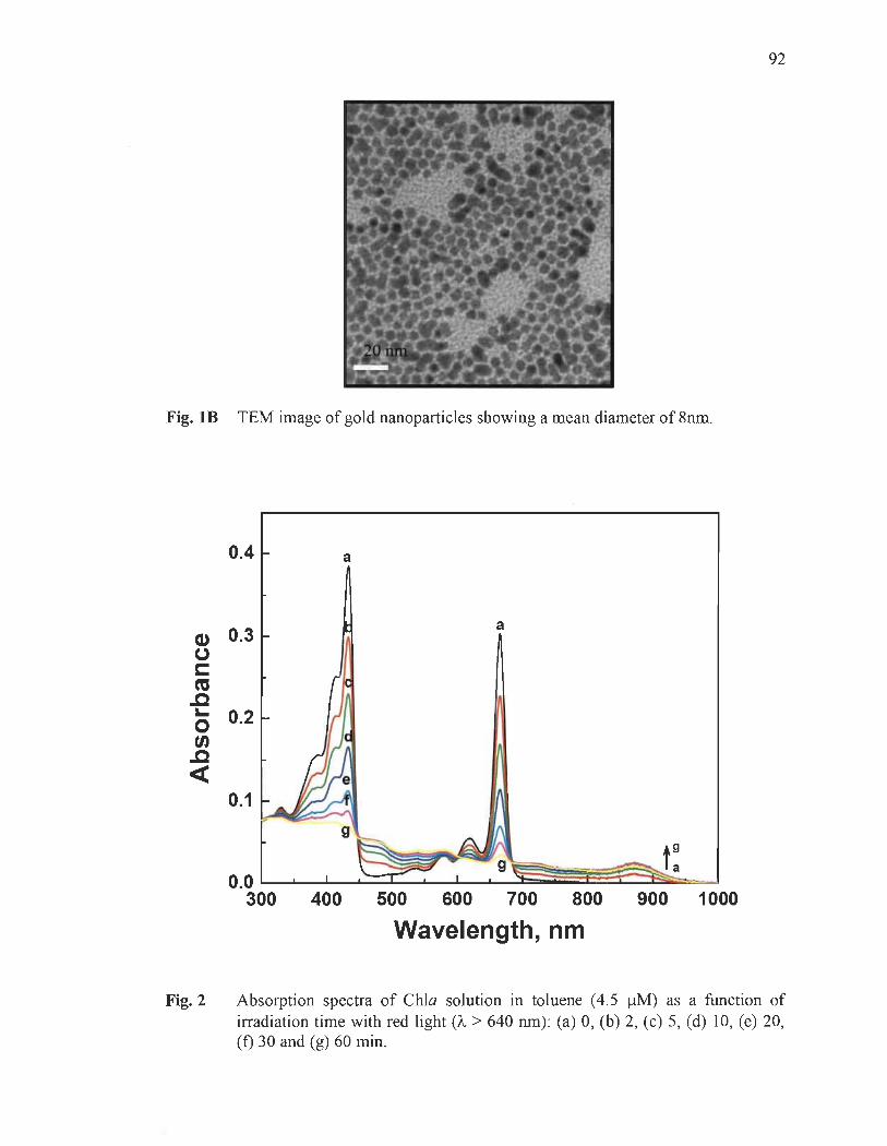
Fig. lB TEM image of gold nanoparticles showing a mean diameter of 8nm.
0.4
CI) 0.3 CJ c: ca .c ~
o en .c <t:
0.2
0.1
a
a
'-~ t: 0.0 LL...--L.......~~~L.......~~~~~~~
300 400 500 600 700 800 900 1000
Wavelength, nm
92
Fig. 2 Absorption spectra of ChIa solution in toluene (4.5 IlM) as a function of irradiation time with red light (Â > 640 nm) : (a) 0, (b) 2, (c) 5, (d) 10, (e) 20, (t) 30 and (g) 60 min.

93
100 'il-
0.4 a Ê 80 c:
1
Il) CD CD 60 @
~ 40
Cl) .CI .'!
(J 0.3 f a ~ 20
1 '0
C ~ Il.
ca 30 60 90 120 150 .c [AuNPsl. "M r.. f 0
0.2 t/) .c «
0.1
0.0 b....-....I....-.....L.....----L...----1_..L.....-..l....---L----I:~_ ........ _ _"_ __ __J
300 400 500 600 700 800 900 1000
Wavelength, nm
Fig. 3 Absorption spectra of ChIa solution in toluene (4.5 J..lM) in the presence of 22 J..lM of AuNPs (8 nm particle size) as a function of irradiation time with red light (À > 640 nm): (a) 0, (b) 5, (c) 10, (d) 20, (e) 30 and (f) 60 min. The inset shows the effect of AuNPs concentration on the photoprotection of ChIa at 665 om; red-light irradiation period is 30 minutes. The % photoprotection of ChIa was calculated from the variation of absorbance at 665 om, using eq. 1.

94
100 5 .-.
4,5 90 ê
1("; 4
e -0 -0 3,5 '""'-= 80 ':i
3 .. li) " 2,5 = \oC :: \oC 70 ,Q 2 L-
@ 0
'" 1,5 ,Q
60 < 1 ~ ~ Cj 0,5
= 50 .s 0
~ 0 0,5 1 1,5 2 2,5 3 -e 40 Irradiation time, h Q f'-)
,.Q 30 < Chla-AuNPs
~ 0 20 Chia
10
0 0 1 2 3 4 - 6 7 8 9 10 ~
Irradiation time, h
Fig. 4 Photostability of ChIa solution in toluene (4.5 J.!M) in absence (a) and presence (b) of 22 /lM of AuNPs as a function of irradiation time with red light (À > 640 nm). The insert shows the first order photodegradation kinetics of ChIa absence and presence of AuNPs (see eq. (4) in SI).

95
250
0.4 200 a
!. E c t-- 150 M ...,.
J @
Cl) ~ 100
0.3 a;:: (.)
!. s::: Kapp= 5.70 X 10⁵ M·l ca .c J 0
0.0 0.5 1.0 1.5 2.0 2.5 3.0 s.. 1/[AuNPs-8nm), x106 M·1
0 tn 0.2 .c «
0.1
400 500 600 700 800 900 1000 1100
Wavelength, nm
Fig. 5. Effect of AuNPs on the absorption spectra of ChIa solution (4.5 IlM) in toluene: (a) 0, (b) 0.36, (c) 0.72, (d) 1.08, (e) l.80, (f) 2.89, (g) 5.05, (h) 7.22, (i) 11.55 and (j) 18.05 IlM. The insert shows the plot of l/M(437 nm) vs l/[AuNPs]. An apparent association constant (Kapp) of 5.70 x 10⁵ M-I was obtained from the intercept-to-slope ratio of the plot.

96
60~------------------------------------
• ::s • ca '"' 45 ~ .... . -
.-r:: o .-t/)
.~ 15 E W
4~------------------~
o 100 200 300 [AuNPs], ~M
650 700 750 800
Wavelength, nm
Fig. 6. Emission spectra of 4.5 !lM ChIa in toluene recorded in the presence of different concentrations of AuNPs: (a) 0, (b) 10.83, (c) 21.67, (d) 32.50, (e) 43.34, (f) 65 .01 , (g) 86.68, (h) 108.35, (i) 130.02, (j) 151.69, (k) 195 .03, (1) 238.37, (m) 281.71 and (n) 325.05 !lM. Excitation wavelength was 433 nm. The insert shows Stern-Volmer plot of the quenching of ChIa by AuNPs. The fluorescence intensity was monitored at 675 nm.
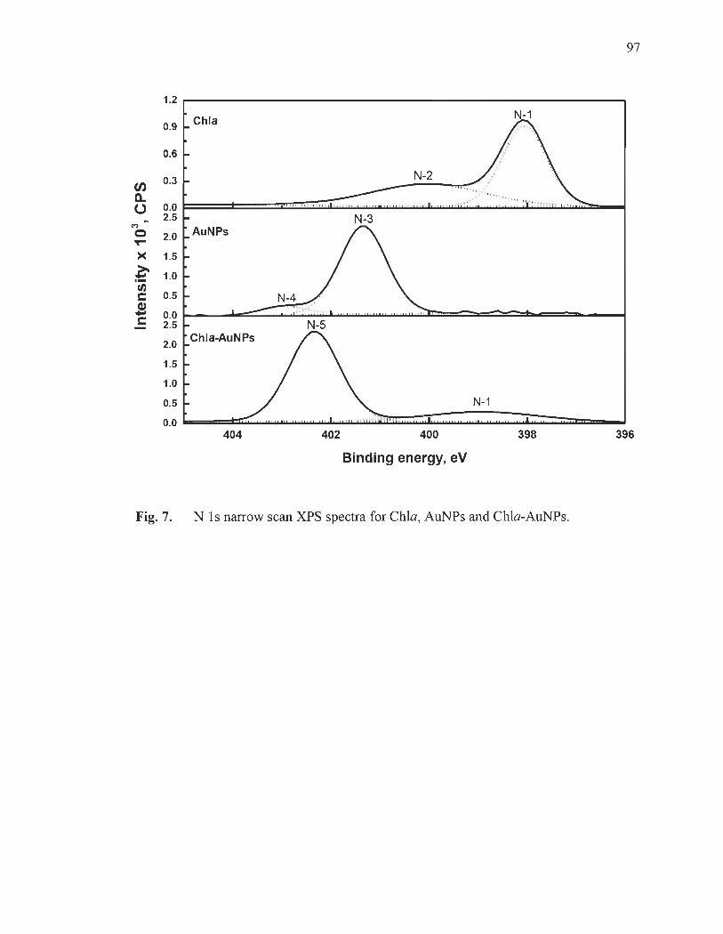
97
1.2
0.9 Chia
0.6
0.3 N-2 en a. ,.' u 0.0
2.5 N-3 C"I
AuNPs 0 2.0 ~
>< 1.5
~ 1.0 t/) C 0.5 N-4 Cl) ... 0.0 C 2.5 N-5
2.0 Chla-AuNPs
1.5
1.0
0.5 N-1
0.0 404 402 400 398 396
Binding energy, eV
Fig. 7. N Is narrow scan XPS spectra for ChIa, AuNPs and Chia-AuNPs,

98
200r-------------------------------------------~
150
100
100
50
AuNPs
Chla-AuNPs
. . .. ...........•....... ~ ...... _ ..... ... :::: ::::~~,~~~-~~--------------------
O~ _____ ~ __________ ~~ _________________ ~ ______________________ ~ _____ ~ __________ ~~
90 88 86 84 82
Binding energy, eV
Fig. 8 Au( 4t) narrow scan XPS spectra for AuNPs and Chla-AuNPs.
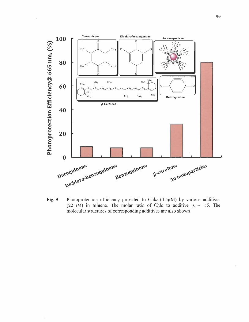
100
80
60
40
20
o
Fig. 9
Duroquinone
HJC
CH) CH)
p..carotene
Dichloro-benzoquinone
o
99
Au nuoparticles
CH; Benzoqumone
Photoprotection efficiency provided to ChIa (4.5/lM) by various additives (22 /lM) in toluene. The molar ratio of ChIa to additive is - 1 :5. The molecular structures of corresponding additives are also shown
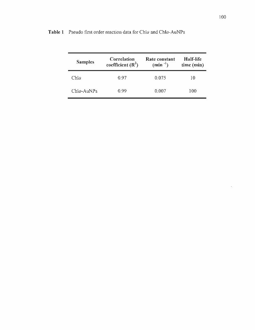
Table 1 Pseudo frrst order reaction data for Chia and Chia-AuNPs
Samples
Chia
Chia-AuNPs
Correlation coefficient (R2
)
0.97
0.99
Rate constant (min -1)
0.075
0.007
Half-life time (min)
10
100
100

101
ChlorophyR-a T etraoctylammonium bromide
Scheme 1 Structure of chiorophyll-a (ChIa) and Tetraoctylammonium bromide (TOAB)

~ +N~
~ ~
Scheme 2 Mechanism of Iigand-exchange reaction between ChIa and AuNPs.
102
SUPPORTING INFORMATION
Enhanced photostability of chlorophyll-a using gold nanoparticles
as an efficient photoprotector
Saïd Barazzouk*, Laurent Bekalé*, and Surat Hotchandani*
103
Groupe de Recherche en Biologie-Végétale, Université du Québec à Trois-Rivières,
Trois-Rivières, Qc, G9A 5H7, Canada
t contributed equally to this work
* To whom correspondence should be addressed. e-mail: [email protected]
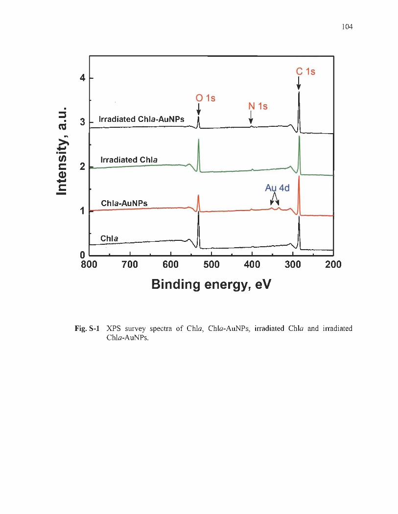
• ::l
• ca ... ~ +' .-(/) s:::: CI) +' s:::: -
4 ~
3 ~ Irradiated Chla-AuNPs
2
1 -
o 800
Irradiated Chia
Chla-AuNPs
Chia
1 1
700 600
C 15
+ 015
t N 15
+ - \J
....
" "'\J
Au4d
A " ... ~
~ -"...i 1 1 1
500 400 300
Binding energy, eV
104
200
Fig. S-1 XPS survey spectra of ChIa, Chia-AuNPs, irradiated ChIa and irradiated Chia-AuNPs.

105
High-resolution Cls and Nls XPS spectra for gold nanoparticles (AuNPs) are
shown in Fig. S-2 and S-3 , respectively. The Cls region of AuNPs comprises three
distinct peaks. The peak appearing at 285 eV, is assigned to C-C and C-H from
tetraoctylammonium bromide (TOAB), the stabilizing agent used in the preparation of
AuNPs. The second peak at 286.07 eV is ascribed to C-N of quatemary amine from
TOAB, while a smaller peak at higher binding energy (287.17 eV) is probably due to an
impurity.
50 Chia C-1 (J) 0- 40 u
M 0 - 30 >< ~ !/) 20 c CI) -c - 10
C-3 0
50 C-1
Chla-AuNPs
(J) 40 0-
U M 0 30 ->< ~ 20 !/) c CI) - 10 c
C-3 0
290 288 286 284 282
Binding energy, eV
Fig. S-2 C 1 s narrow scan XPS spectra for ChIa and Chla-AuNPs.

25~----------------------------~
en 20 c.. o ~ 15
>< ~ :: 10 fi) c: Q) -c: 5
en c.. o~ 10
M o
>< ~ :: fi) 5 c: Q) -c:
Chia 0-2
Chla-AuNPs 0-2
,-- 0-1 .' l' l "" 1
" , , ~ , ~ l i: ! 1 1 ~ \ ' l , ~ , ,
O• _..--.~ " ,,~I " " } " ' :,I ..., Il''1'11 1 1: 1111'''1'111 1 1111 : 111 . 111''11111 '' ~_''' 1l''T'l''oI_....I
535 534 533 532 531 530 529
Binding energy, eV
Fig.8-3 0 Is narrow scan XP8 spectra for Chia and Chla-AuNPs,
106

CI) a.
40
u 30 (")
o
>< ~ 20 ~ tn c: Cl)
ë 10
. .
.. . .. . . . . .
o~==~~~=" =" ~'~==~=='=" ~~~ 289 288 287 286 286 284 283
Binding Energy, eV
Fig. S-4 Cl s narrow scan XPS spectrum for AuNPs.
107

108
KinetÏcs of Photochemical reaction
The photodegradation of ChIa in presence of O2 can be described by following
equation
The photodegradation rate of ChIa was monitored by recording the absorbance at
665 nm as a function of irradiation time, and is:
Where [ChIa] denotes the concentration of ChIa, [02] denotes the concentration of 02,
t is the reaction time, and k is the rate constant. As the [02] remains constant as the
reaction proceeds, the reaction can be considered pseudo-first-order because it depends
on the concentration of [ChIa] only.
Thus, eq. (2) can be written as follows:
_ d[Chla] = k ' [Chla] dt
where k' = k[02]
Solving eq. (3), one obtains eq. (4)
ln([Chla 1) = -k·t + ln([Chla]o)
(3)
(4)
Where [Chla]o is the initial concentration of ChIa and [Chla]t is the concentration of
ChIa at any time t . A plot of ln([Chla 1 )vs t should yield a straight line with a slope of
k'; a typical plot is shown in the inset of Fig. 4.
CHAPITRE IV
NANOSILVER COULD USHER IN NEXT-GENERATION PHOTOPROTECTIVE AGENTS FOR NATURAL PORPHYRINS
Le contenu de ce chapitre a fait l'objet d'une publication en anglais dans la revue:
Particle & Particle Systems Characterization, 2014, 31 , 843-850.
4.1 Résumé de l'article
Une étude sur les propriétés photoprotectrices des nanoparticules d'agent (AgNPs)
a été effectuée dans ce troisième article scientifique afm de démontrer que la méthode de
photoprotection, précédemment présentée dans les deux premiers articles scientifiques
pourrait être utilisée pour améliorer simultanément la stabilité, l'émission de
fluorescence et la capacité de génération d'oxygène singulet des porphyrines de
magnésium. Les résultats présentés dans cet article montrent clairement que l'emploi des
AgNPs (en concentration très faible) est une méthode simple, efficace, rapide et rentable
pour améliorer simultanément la photostabilité, l'émission de fluores?ence et l'efficacité
de production de l'oxygène singulet par photosensibilisation de la chlorophylle-a (ChIa).
De plus, il est également rapporté que les AgNPs dans des conditions in vitro, les AgNPs
sont des agents photoprotecteurs bien plus efficaces que les caroténoïdes. La capacité
protectrice des AgNPs est le résultat de leur liaison avec les sites azotés du macro cycle
porphyrique de la ChIa, et ce, avant la formation des espèces réactives de l'oxygène
(ROS) par photosensibilisation. L'utilisation des AgNPs offre de nouvelles voies pour
améliorer les propriétés photochimiques et photophysiques des porphyrines. Par
conséquent, nous prévoyons que le présent article peut ouvrir la voie pour le
développement d'une nouvelle génération d'agents photoprotecteurs qui pourrait
également jouer un rôle prometteur dans l'imagerie médicale et la thérapie
photodynamique. L'étude présentée dans cet article a été l'aboutissement du troisième et
dernier objectif opérationnel de cette thèse.

4.2 Troisième article scientifique
Nanosilver Could Usher in Next-Generation Photoprotective Agents
for Magnesium Porphyrins
Laurent Bekalé*, Saïd Barazzouk* and Surat Hotchandani*
IlO
Département de chimie, biochimie et physique, Université du Québec à Trois-Rivières,
C. P. 500, Trois-Rivières (Québec), G9A 5H7, Canada
* To whom correspondence should be addressed. e-mail: [email protected];
[email protected]; [email protected]
Abstract
A novel strategy, based on silver nanoparticles (AgNPs), has been developed for
simultaneously improving: 1) photostability, 2) fluorescence emission, and 3) singlet
oxygen generation ability of natural porphyrin (i.e. , chlorophyll (ChI) molecules). The
benefit of AgNPs as photoprotector of ChI is that they spontaneously bind with the
nitrogen atoms of ChI as soon as they are mixed together, even in the dark prior to the
illumination, inhibiting thus the binding of reactive oxygen species (ROS) at nitrogen
sites, known to cause the photodegradation of porphyrins.
Keywords: Nano-Silver, Chlorophyll, Photoprotection, Metal-enhanced fluorescence,
Metal-enhanced singlet oxygen generation.

III
Nanoscale materials have received considerable attention because their structure
and properties differ significantly from those of the atoms and molecules, as well as
those of the bulk materials. [1] For this reason, many researchers continue to explore their
amazing properties of these nanomaterials to address specific needs and problems in a
number of sectors such as microelectronics, materials, environment, energy, healthcare,
etc.[2] Therefore, it is of great interest to investigate how these nano-objects can improve
the photostability of porphyrins such as chlorophylls.
Chlorophyll (ChI), in recent years, has attracted great interest for the
biotechnological applications. Ohtani et al., have recently reported that ChI extracted
from spinach is a promising material for the fabrication of the organic light-emitting
diodes (OLEDs)Y] Moreover, Shao et al., have found that ChI could be used as potent
photosensitizer (photosensitizing agent) in hybrid phototransistors. [4] Further, Fan et al.
have recently discovered that ChI extracted from the leaves of Chimonanthus salicifolius
could have great potential for cancer imaging. [5] Finally, Gilmar et al. have recently
demonstrated the potential of ChIs as possible countermeasure against mosquito
larvae. [6] However, despite the growing efforts to use ChIs as active ingredient in a wide
range of modern biotechnological applications, the in vitro ChI has a crucial
disadvantage, which is its high photoinstability under aerobic conditions. In other words,
the photo-instability of ChIs is one of the primary concems that hinders their utilization
in the majority of the biotechnological areas. Therefore, if the photostability of ChI
could be significantly amended, their technological application would be enormously
improved.
So far, many approaches to enhance the photostability of in vitro chlorophyll have
been explored. Itoh et al. have reported that chlorophyll-a (ChIa), adsorbed on silicate
layers of smectite, showed photos table properties. [7] They have also prepared a
chlorophyll-mesoporous silica conjugate, and suggested that the interaction between ChI
and the supporting silica materials in nano~cale space could increase the photostability
of ChI. [8] In our research team, we decided to approach the problem using noble metal
nanoparticles. We recently employed gold nanoparticles to photoprotect ChIa by
112
suppressmg the binding of reactive oxygen specles (ROS, namely, singlet oxygen,
superoxide ion and hydrogen peroxide) at nitrogen sites of porphyrin macro cycle of
ChIa. [9] Although these materials have been proven to enhance the photostability of
in vitro ChI, they have sorne other limitations in that they have been found to quench the
fluorescence of ChI and also limit its capability to generate singlet oxygen e02). As
such, the above mentioned ChI photoprotective agents do not meet the requirements
when we aim to improve the photostability of in vitro ChIs, especially with an objective
to take advantage of their near-infrared fluorescent properties for imaging, and their
strong singlet oxygen generation abilities for photodynarnic therapy. Therefore, it is
critically important to develop functional materials that could enhance the photostability
of ChIs without compromising their fluorescence emission and their ability to generate
102 so that they can be successfully exploited in various areas of materials science and
biotechnology.
We have selected silver nanoparticles (AgNPs) to assess the possibility of
simultaneously improving these photophysical properties of ChI molecules. There are
three reasons that strongly motivated our choice of AgNPs. First, AgNPs are attractive
owing to their excellent metal-enhanced fluorescence properties (MEF), i.e., increasing
the fluorescence emission of fluorophores close to metallic nanoparticlesYO] Second,
AgNPs are also attractive due to their excellent metal-enhanced singlet oxygen
generation properties (ME10 2), i.e., increasing the singlet oxygen generation efficiency
of the photosensitizers close to metallic nanoparticles. [11] Finally, AgNPs find use in
many fields; the major applications include their uSe in textile engineering, electronics
and optics,P2-16] and, most importantly, in the medical field as bactericidal [17] and
therapeutic agent. [1 8]
In this communication, we report an unprecedented observation that AgNPs are
the potential additives to simultaneously increase the photostability, fluorescence
ernission, as well as singlet oxygen generation capability of in vitro ChIa. The
UV-visible, fluorescence and X-ray photoelectron spectroscopic (XPS) studies have
been carried out to assess the effect of AgNPs on ChIa. The results show that the

113
half-life of ChIa (4.5 IlM) has been extended from 49 to 770 minutes in the presence of
ultralow concentration of AgNPs (0.36 nM). Additionally, it is seen that AgNPs are
much more effective photoprotective agents of in vitro ChIa than are the carotenoid
molecules (i.e., p-carotene and lycopene). The protecting ability of ChIa against
photodegradation by AgNPs is the result of their binding with ChIa at its nitrogen sites,
thus inhibiting the binding of reactive oxygen species (ROS) at these sites, which are
known to cause the photodegradation of ChIa. In addition to their significant
photoprotective performance, we have also observed that AgNPs improve MEF and
ME I 0 2properties of ChIa by 1.2 and 2 times, respectively.
The procedure for preparing silver nanoparticles (AgNPs) in an organic medium is
a modified version of that proposed by Korgel et al.[19) Figure lA shows the UV-visible
absorption spectra of the silver colloids. A localized plasmon resonance peak at around
430 nm was observed for AgNPs, which is in good agreement with that of roughly
spherical AgNPs. [20) The formation of AgNPs was confirmed by the observation of
Ag 3d peaks in XPS (Figure lB). The XPS peaks corresponding to Ag 3d (5/3) and
Ag 3d (2/3) appeared at binding energies of about 368.3 and 374.3 eV, respectively, in
good agreement with bulk sil ver metallic values. [21) Transmission electron rnicroscopy
(TEM) confirmed the existence of spherical Ag nanoparticles (as illustrated in
Figure le). Furthermore, from the analysis of TEM images of 100 nanoparticles, an
average diameter of 5.3 ± 0.76 nm was determined for AgNPs (Figure ID). Figure 2A
presents typical changes in the absorption spectra of ChIa solution in toluene as a
function of irradiation time with red light ()" > 640 nm). The principal absorption peaks
of Chia (see initial spectrum at 0 min of irradiation) in the red at 665 nm (Q band) and at
433 nm in the blue (Soret band) are in good agreement with those reported in the
literature,l22) and that ChIa is present in monomeric form. Photodegradation is
characterized by the decrease in the intensity of both Soret and Q bands on exposure of
ChIa to red Iight. As seen from Figure 2A, there is a quick and progressive decrease of
ChIa absorbance as the irradiation time increases. In 5 minutes, about 10% decrease in
the absorbance ofQ-band in the red (665 nm) was observed. On continued exposure, the
absorbance at 665 nm decreased 36% in 20 minutes and by 73% in 90 minutes. These

114
results of Chla are in good agreement with those reported by Jones and Mackay,(23)
Merzlyak et aL ,(24) and Tregub et aL ,(25) who studied the photodegradation of Chla
in micelles, methanol and various other organic solvents, respectively. In order to
evaluate the effect of AgNPs as a potential photoprotective additive to reduce the
photodegradation of ChIa, the irradiation of ChIa solution (4.5 ~M) in the presence of
AgNPs was carried out under identical conditions as for ChIa alone (irradiation with red
light, À > 640 nm). As is clear from Figure 2B, the addition of AgNPs seems to provide
a remarkable protection to ChIa against photodegradation. Indeed, after 90 minutes of
irradiation, Chla bleached only 10 % in the presence of AgNPs (0.36 nM) compared to
the 73% photodegradation in absence of AgNPs (see Figure 2A). In other words, a huge
photoprotection of - 90% has been provided to ChIa by AgNPs (0.36 nM). The decrease
in photodegradation of ChIa in the presence of AgNPs can also be seen visually
(Figure SI in the Supporting Information). While the color of ChIa solution changes
from light green to clear after irradiation, the color of ChIa admixed with AgNPs
remains virtually the same after irradiation, thus indicating clearly the photoprotective
action of AgNPs. It is worthy to point out that even after a very long time of irradiation,
i.e. , 540 min. a photoprotection ofhigher than 70% has been provided to ChIa by AgNPs
(0.36 nM). This is the first experimental demonstration that highlights the beneficial
effect of AgNPs in increasing enormously the photochemical stability of ChIa.
To get a meaningful insight into the photostability imparted to ChIa by AgNPs, the
photodegradation kinetics of Chla was studied, and is presented in Figure 3, and
described in the supporting information section. The photodegradation kinetics of Chla
in the absence and presence of AgNPs follows pseudo-first order kinetics. It is found
that AgNPs induced a dramatic decrease of the photodegradation rate constant of ChIa,
i.e., from 1.4 x 10⁻² to 9 x10⁻⁴ min-l , resulting into an enormous increase in the half-life
from 49 minutes for free ChIa to 770 minutes for ChIa mixed with AgNPs and
indicating an enormous improvement (i .e., 15714% increase) in the photostability of
ChIa brought about by AgNPs (Table 1).

115
Our findings also reveal that the photoprotection of ChIa is dependent upon the
AgNPs concentration (inset Figure 3). That is, the higher the concentration of AgNPs,
the greater is the photoprotection provided to ChIa against irradiation. However, AgNPs
concentration greater than 0.36 nM did not provide any additional protection. The
percent age photoprotection was calculated using eq. (1), described in Experimental
section. To exclude the possibility that the 1-nonanthiol (NT), a surfactant used in the
preparation of AgNPs, had any photoprotective role, a solution of NT (50 mM) in
toluene was added to ChIa solution. ChIa was not photoprotected, confirming that it is
the AgNPs, rather than NT, that provide the photoprotection to ChIa. Furthermore,
AgNPs themselves subjected to the irradiation for the same time period (540 min) under
identical conditions did not show any photochemical changes.
In order to substantiate the advantage of using AgNPs compared to other
chlorophyll protoprotective agents, such as carotenoids, which have been found to
protect green plants, [26] similar photodegradation experiments of ChIa with red light
irradiation Cf" > 640 nm) were carried out in presence of different carotenoids, i.e.,
,B-carotene and lycopene. It is found that lycopene (22 ~) provides a photoprotection of
only 28% whereas ~-carotene (22 ~) no more than 22 % as shown in Figure 4, which
are about 3 . and 4 times inferior to the photoprotection imparted by the ultralow
concentration of AgNPs (0.36 nM).
Once the role of AgNPs as efficient photoprotector was established, we then
embarked upon the study of the influence of AgNPs on the fluorescence emission of
ChIa. The fluorescence spectra of ChIa (4.5 /lM), AgNPs (0.36 nM), and their mixture
are presented in Figure 5. The samples were excited at 625 nm, right into the red
absorption region of ChIa, where AgNPs have no absorption. An enhancement of ChIa
fluorescence in presence of AgNPs is clearly noticeable. Dividing the fluorescence
intensity of ChIa in presence of AgNPs by that in absence of AgNPs yields an
enhancement factor of 1.2. An increase in fluorescence of similar sm aller magnitude
(l.18 times) has also been reported by other authors. [27b 1
116
It should, however, be mentioned that the observed enhancement of fluorescene of
ChIa in the present study is not due to an additional fluorescence emitted by AgNPs
because they do not exhibit any fluorescence in the fluorescence region of ChIa
(Figure 5). The fluorescence enhancement observed in the CUITent work could be
understood as the result of the modification of local electromagnetic field in the vicinity
of ChIa caused by the localized surface plasmon resonance at the surface of silver
nanoparticles. The plasmonic field of metallic nanoparticle increases the radiative rate of
the fluorophore, leading to an enhancement of the quantum yield of its fluorescence.
This explanation has also been put forward by other researchers with regard to the
fluorescence enhancement of chlorophylls in light-harvesting complexes placed near a
silver metal layer. [27, 28] Further, the fluorescence enhancement factor does not depend
upon the concentration of the AgNPs as shown in the inset of Figure 5.
It is worth mentioning that the photostability of fluorescing species is thought to
be closely linked to MEF phenomenon. [IOc] This is due to the fact that MEF occurs as a
result of the augementation of radiative rate constant of the fluorophore leading to
shorter excited state lifetimes. Accordingly, if the fluorophore spends less time in the
excited state, there is a smaller probability for the reactions between excited fluorophore
and molecular 02 to occur, thereby producing lower amounts of singlet oxygen (known
to cause the photodegradation of the fluorophore) and increasing the photostability. That
is, MEF enchances the photostability of fluorophores as a result of the lower production
of the singlet oxygen e02). However, it is interesting and extremely important to point
out that the enhanced photoprotection provided to ChIa by adding of AgNPs is not due
to MEF phenomenon just described, because in our case, the presence of AgNPs actually
increases the ability of ChIa to generate more 102 which would cause more
photodegration and thus decrease the photostability of ChIa. The photoprotection
provided to ChIa by AgNPs, as mentioned in Introduction, is rather due to the binding of
AgNPs at the nitrogen sites of ChIa that prevents the attachment of 10 2 at the same
nitrogen sites which causes the photodegradation. The effect of AgNPs on the 10 2
generation and its photoprotective action of ChIa are further discussed below.

117
To prove that the presence of AgNPs in ChIa solution promotes the production of
10 2, the aerobic irradiation of ChIa solution (4.5 ~M) in the absence and presence of
AgNPs (0.36 nM) was performed employing 9,10-dimethyl-anthracene (DMA) as an
102 chemical sensor in toluene. This sensor is a highly fluorescent probe with emission
comprising of three bands in the 400-500 nm wavelength region, and reacts efficiently
with 10 2 to form the non-fluorescent DMA 9,10-endoperoxide in many organic
solvents. [29] The Metal-enhanced generation of 10 2 (ME10 2) by ChIa in absence and
presence of AgNPs was measured by following the decrease in the fluorescence
eIlliSSlOn of DMA upon its photosensitized conversion into the corresponding non
fluorescent 9,1 O-endoperoxide (Scheme 1, in the Supporting Information). Figure 6
displays a typical time dependence of the photoinduced decrease in the fluorescence
emission of DMA with increasing irradiation time in presence of ChIa (4.5 ~M) and in
presence of the mixture of ChIa (4.5 ~M) + AgNPs (0.36 nM). From the figure, a 2-fold
enhancement (i.e. , from 5 x 10⁻⁴ S-1 to 9 X 10⁻⁴ S-I) in the photo-oxidation rate of DMA
sensitized by ChIa in presence of AgNPs (i.e. , Chla@AgNPs) can be clearly seen
compared to the photo-oxidation rate of DMA sensitized by ChIa alone under same
irradiation conditions. Since, the decrease in DMA fluorescence is due to its photo
oxidation by 10 2, producing nonfluorescent endoperoxide, the results suggest that the
yield of 10 2, generated by ChIa, is enhanced by a factor of two in the presence of silver
nanoparticles. To confirm that the high efficiency of 102 generation observed in the
mixture of ChIa + AgNPs is, indeed, due to an increase in the photosensitized oxidation
of DMA brought about only by ChIa and not by AgNPs, the photo-production of 102 by
AgNPs in the absence of ChIa was verified. DMA, subjected to the irradiation in the
presence of AgNPs under identical conditions, did not show any decrease in the
fluorescence of DMA (data not shown), indicating that AgNPs alone are not able to
produce 10 2 at aIl.
As 102 is known to play an indispensable role in the photodynamic therapy (PDT),
a medical technique that uses a combination of light and photosensitizing drugs to
produce cytotoxic 102 to induce selective damage to the tumor cells, we envi sion that
AgNPs will play very promising dual roles in PDT: (1) as a photoprotector to increase

118
the photostability of the photosensitizers by increasing their half lives (Fig. 3 and Table
1) so that they can generate 10 2 for longer periods of time, and (2) as a booster of the
cytotoxic 10 2 production via metal- photosensitizer interactions.
At first sight, one may be intrigued by the simultaneous enhancement of both the
fluorescence emission and the yield of 10 2.> since the enhancement in fluorescence, due
to the shortened singlet lifetime of the fluorophore, requires that the yield of 10 2 should
decrease. Both these enhancements can, however, be reconciled if we take the metal
fluorophore (AgNPs and ChIa) interactions into account that lead to a modification of
the local electromagnetic field in the vicinity of ChIa moieties. This same
electromagnetic field can also modify the spin-orbit coupling (a quantum mechanical
process that is responsible for intersystem crossing) in the fluorophore(30) leading to the
increase in its triplet formation and thus the yield of 102. Such contrasting behavior, i.e.,
the enhancement of both the fluorescence emission and the yield of 102, has also been
recently reported by de Melo et al. in case of silver-pectin nanoparticles. (31)
Finally, to understand the ongm of the photoprotective effect of AgNPs, the
interactions between AgNPs and ChIa were studied by UV-vis and X-ray photoemission
spectroscopy (XPS) techniques. With the addition of only a small quantity of AgNPs
(0.36 nM) to ChIa solution, a change in the absorption spectrum of ChIa was observed
(Figure 82). A bathbchromic shift (- 4 nm) of Soret band and a decrease in the intensity
of Q-band at 665 nm can clearly be seen as the concentration of AgNPs increases. These
changes in the UV -vis absorption spectrum of ChIa, induced by AgNPs, indicate the
formation of a complex between ChIa and AgNPs (AgNPs@Chla) through the
pyrrolenine nitrogen atoms of the porphyrin macro cycle of ChIa as shown later.
The changes in the absorption (~A) at 665 nm were used to determine the
association constant between Chia and AgNPs employing Benesi-Hilderbrand
method. (32) An average apparent association constant, Kapp, for Chla@AgNPs complex,
obtained from the intercept-to-slope ratio of the double reciprocal plot of l/~A vs
l/[AgNPs], is 2.6 x 10⁸ M- I (inset of Figure 82B).The high value of K app observed

119
suggests a strong binding between ChIa and AgNPs. Furthennore, based on the fact that
nitrogen atoms possess strong affinity towards Ag nanoparticle surface compared to
oxygen atoms, it is reasonable to assume that the Chla@AgNPs complex is fonned via
pyrrolenine nitrogen atoms of the porphyrin macro cycle of ChIa rather than the oxygen
atoms of the carbonyl group (see structure of ChIa). Several authors have also observed
the preferential interaction of the AgNPs surface towards the nitrogen atoms compared
to oxygen atoms. For example, by comparing the interaction of AgNPs with three free
base porphyrins, i.e. , tetra( 4-aminophenyl) porphyrin, tetrakis(l-methyl-4-pyridionio)
porphyrin tetra(p-toluenesulfonate) and tetrakis( 4-carboxyphenyl)-porphyrin, Murphy
et al. [33] reported that amine shows a stronger interaction with AgNPs surface relative to
the carboxyl groups.
To further support that the complex formation between AgNPs and ChIa occurs
via nitrogen atoms of ChIa, XPS spectra were recorded. The great advantage of the XPS
is its capability of detecting the chemical bonds which are involved in the complex
formation. The XPS spectrum of N Is region of ChIa (Figure S3A) shows two peaks.
The main peak at 398.2 eV corresponds to the pyrrolenine nitrogen atoms of the
porphyrin macro cycle of Chia. The second less intense peak observed at 400 eV, is most
likely due to the protonated nitrogen, produced as a result of a small degree of
demetallation of ChIa during its exposure to X-rays in the course of the experiment.
These observed peaks are in fair agreement with the reported XPS data for ChIa
and other magnesium porphyrins. [9, 34, 35] After adding AgNPs to ChIa solution
(Chla@AgNPs), the principal peak at 398.2 eV disappears while a new peak appears at
402.2 eV (Figure S3B), suggesting that a new chemical environment has been
established. In other words, the tertiary nitro gens in ChIa, with a binding energy of
398.2 eV have now, in the presence of AgNPs, been converted into positively charged
tetravalent ammonium quatemary nitro gens (RtN5+) which are known to possess a higher
binding energy at 402 eV.[9, 34, 35] This result confirms that the Chla@AgNPs complex is
formed via the implication of the lone pair electrons on the pyrrolenine nitrogen atoms
of chlorophyll.

120
To exclude the possibility that the oxygen atoms in carbonyl groups of chlorophyll
molecules are implicated in the formation of the complex Chla@AgNPs, the Ols XPS
spectrum of ChIa was recorded in absence and presence of AgNPs. No significant
change in the position of Ols peak of ChIa was observed upon the addition of AgNPs
(Figure S4), confirming that oxygen atoms of ChIa are not implicated in the
Chia@AgNPs complex formation. Thus, the XPS results conclusively show that the
complex formation occurs via the binding of the nitrogen atoms of Chia to AgNP. This
complex Chla@AgNP consists of 32 molecules of ChIa bound to one Ag nanoparticle as
described in Supporting Information; the schematic representation of the complex is
depicted in Scheme 1.
Since the synthesized I-nonanthiol-stabilized Ag nanoparticles are coated with a
mono layer of surfactant (l-nonanthiol), the formation of complex Chla@AgNP implies
ligand ex change between I-nonanthiol and ChIa on AgNP 's surface. The fixation of
ChIa to AgNP's surface therefore necessitates that I-nonanthiol move out and make
room . for ChIa. Said otherwise, ligand exchange takes place between the incoming
ligand ChIa and I-nonanthiol. This is rendered possible in view of the fact that the
nitrogen atoms of ChIa preferentially coordinate to silver atoms to form strong
coordination bonds, while the sulphur atoms of I-nonanthiol show only weak
interactions with silver atoms. Consequently, I-nonanthiol, initially attached to AgNPs,
is easily replaced by ChIa to yield AgNPs@Chla complex. Previous studies also
bear the same conclusion. For example, using a flexible tetradentate ligands,
1,3-bis(2-pyridylthiol)propane, Xie et al. [36] reported that silver atoms have strong
affmity towards the nitrogen atoms compared to sulfur atoms. Nath et al. [37] also
demonstrated the replacement of the thiol moiety from dodecanethiol (DDT)
encapsulated Ag organosol (Ag-DDT) upon the addition of dodecylamine (DDA) to
form the DDA encapsulated Ag organosoi (Ag-DDA).
In light of these results, one can say that the bene fit of AgNPs as a photoprotector
of ChIa is that they readily bind with the nitro gens of ChIa as soon as they are mixed
with Chia. Consequently, 10 2, which is generated during the illumination of ChIa, fmds,
121
to its great disappointment, that only very few free nitrogen sites are available to bind
with and cause damage, since most of them have already been occupied by AgNPs in
dark long before 10 2 was even produced as a result of the illumination.
This very ability of AgNPs to bind with ChIa in its ground state in dark prior to the
illumination also gives them an added advantage over other convention al protective
agents, such as carotenoids. These molecules provide photoprotection to Chia by
suppressing the formation of 10 2 by functioning as (i) triplet valves, i.e., they react with
the excited triplets of ChIa, and thus prevent ChIa triplets from transferring their energy
to oxygen to produce 10 2, (ii) as scavengers of 10 2, thereby diminishing its quantities,
and, thus, its harmful effects, and (iii) as quenchers of the excited ChIa singlets; this
decreases the intersystem crossing yield of singlet ChIa to triplet ChIa, leading to a
decreased triplet yield of ChIa and, hence, to a decreased 10 2 production.
The photoprotection of ChIa by carotenoids, thus, occurs via their interaction with
the excited states of ChIa which suppresses the production of 102. In other words, the
carotenoids react with the excited states of ChIa so that lesser and lesser amounts of 10 2
are produced, while, paradoxically, oxygen also reacts with the excited states of ChIa
but pro duces larger and larger quantities of 10 2. Thus, both the carotenoids and oxygen
react with the same excited states of ChIa, although one (carotenoids) to protect it (ChIa)
while the other e02) out to destroy it. It is thus evident that carotenoids must compete
with oxygen for the excited states of ChIa to excercise their role as photoprotectors. In
contrast, the photoprotection of ChIa by AgNPs occurs as a result of the binding of
AgNPs with ChIa at its nitrogen sites as soon as they are mixed together even prior to
the illumination. Consequently, AgNPs do not have to compete with any other source,
and thus impart a higher photoprotection to ChIa than do the carotenoids (Fig. 4).
In conclusion, the present work clearly shows that the employment of AgNPs (in
ultra low concentration) is a simple, efficacious, fast, and cost-effective method for
simuitaneously improving the photostability, fluorescence emission and singlet oxygen
generation capacity of in vitro ChIa. It is also noted that AgNPs can prote ct ChIa in

122
solution against photodegradation better than what carotenoids can do. The protective
ability of ChIa by AgNPs is the result of their binding with ChIa at its nitrogen sites,
thus inhibiting the binding of 10 2 at these sites, known to cause the photodegradation of
ChIa and other porphyrins. The use of AgNPs offers new routes to ameliorate the
photochemical and photophysical properties (photostability, fluorescence emission and
singlet oxygen generation capacity) of chlorophylls. We, therefore, envisage that the
present work may pave the way for the development of a new generation of
photoprotective agents which could also play a promising role in imaging and
photodynamic therapy.
Experimental Section
AlI chemicals were purchased from Aldrich Chemical Co., and were used as
received. AlI aqueous solutions were prepared with deionized wateT.
Synthesis of Si/ver Nanoparticles (AgNPs): The procedure for preparing colloidal
silver nanoparticles (AgNPs) in an organic medium is a modified version of that
proposed by Korgel et aL[19j Briefly, an aqueous solution of sodium nitrate (NaN03)
(30 mL, 5 M) was mixed with a solution of tetraoctylammonium bromide (TOAB)
(50 mM) in 50 mL toluene. The biphasic mixture was vigorously stirred for 1 h. An
aqueous solution of silver nitrate (7.5 mL, 30 mM) was added to the organic phase, and
the mixture was stirred for 45 min. The organic layer was separated off, and 189 ilL of
nonanthiol was added to it to cap the silver; the mixture was further stirred for 15 min.
An aqueous solution of sodium borohydride (6.25 mL, 416 mM) was then added
dropwise over 35 min period while stirring vigorously. The stirring was continued for
additional 15 h. The organic layer was extracted and washed 3 times with dilute ethanol
to remove unbounded thiols. Finally, the organic layer, constituting the colloidal silver
nanoparticles, was dried ovemight on Na2S04 powder and then filtered. The fmal
concentration of AgNPs, estimated on the basis of atomic concentration, was 9 mM. The
particle core diameter was estimated by using lMAGEJ® software analysis of the TEM

123
micrographs. The TEM micrographs of these AgNPs revealed an average particle
diameter of 5.3 ± 0.76 nm.
Photodegradation Measurements: The measurements were carried out in al-cm
path length quartz cuvette. The illumination of ChIa solution (3 mL, 4.5 !lM) in toluene
was carried out using collimated light from a 150W xenon lamp that passed through a
water filter to absorb heat. A red cut-off filter that allowed wavelengths greater than
640 nm was used. The light power reaching the sample was 40 mW cm-2. AlI operations
with ChIa solutions were carried out in either subdued light or green light. The solutions
were stored in dark in a refrigerator wh en not in use. AlI experiments were carried out in
the presence of air.
Absorption and Emission Spectrometry: Absorption spectra were recorded with a
Cary 5000 spectrophotometer. The emission spectra were recorded with a FluoroLog-3
spectrofluorometer.
Transmission Electron Microscopy: TEM image of the AgNPs was taken with a
Philips EM 208S microscope on carbon-coated grids and operating at 80 kV.
Singlet Oxygen Sensor: The 9,1O-dimethyl-anthracene (DMA) was used to
evaluate the ability of ChIa and Chla@AgNPs to produce singlet oxygen (10 2). In a
typical experiment, 3 mL toluene solution of Chia (4.5 !lM) containing 0.5 !lM ofDMA,
and 3 mL toluene solution of ChIa (4.5 !lM) containing 0.5 !lM DMA and 0.36 nM
AgNPs were placed in al-cm path length quartz cuvette and illuminated with collimated
light from a 150 W xenon lamp that passed through a water filter to absorb heat. A red
cut-off filter which allowed wavelengths greater than 640 nm was used. The illumination
was carried out under gentle magnetic stirring for different periods oftime (0, 5, 10, 15,
20 s and l , 3, 5, 7, 9, Il , 13, 15, 17 and 20 min).The DMA fluorescence emission was
recorded between 380-550 nm with excitation at "'ex 360 nm. The first-order rate
constant of the photoprocess (photooxidation of DMA) was obtained from the plot of
ln FoIF as a function of the illumination time t; Fo and F represent the fluorescence

124
intensity of DMA at time 0 and time t, respectively. The slope of the linear plot thus
obtained allowed us to calculate the rate constant (k) of the photoprocess.
X-Ray Photoemission spectroscopy: XPS was performed on a Kratos Axis Ultra
spectrometer (Iuatos Analytical Ltd., UK), using a monochromatic Al Ka X-ray source
(E= 1486.6 eV) with a power of225 W, at a take-offangle of 90° relative to the sample
surface. 400 ilL of the sample was dropped on an aluminum substrate and dried in
vacuum dessicator ovemight to obtain a thin film. The dried sample was then transferred
to the XPS sample holder. The measurements were made under a high vacuum of 10-9
torr, at room temperature. The surface of the sample was - 20 mm2, and the investigated
area was typically 1 x 2 mm2. High-resolution spectra of Ag 3d, N ls and 0 ls were an
average of five scans acquired at a pass energy of 40 eV and resolution of 0.1 eV/step,
for quantitative measurements of binding energy. The CasaXPS software was used for
background subtraction (Shirley-type), peak integration, fitting and quantitative
chernical analysis . The C ls (C- C) peak at 285 eV was used to calibrate the binding
energy scale. Binding energies values are given at ± 0.2 eV. Gaussian peak profiles were
used for spectral deconvolution of Ag 3d, Ols and N 1 s spectra.
Percentage Photostability: The percentage photostability was calculated using the
formula as described by Claes:[38]
(1)
where El , E2, and E3 are, respectively, the concentration (or absorbance) of ChIa
before irradiation, after irradiation without AgNPs, and after irradiation in the presence
of AgNPs.
Supporting Information
Supporting Information is available online from the Wiley Online Library or from
the author.

125
Acknowledgements
The work described herein was supported by the N atural Sciences and Engineering
Research Council of Canada (NSERC).
References
[1] a) X. Xue, F. Wang, X. Liu, J Mater. Chem. 2011,21, 13107; b) M. M. Khin, A. S. Nair, V. J. Babu, R. Murugan, S. Rarnakrishna, Energy Environ. Sci. 2012, 5, 8075. b) X. Chen, C. Li, M. Gratzel, R. Kostecki, S. S. Mao, Chem. Soc. Rev. 2012, 41 , 7909. d) E. Roduner, Chem. Soc. Rev. 2006, 35, 583.
[2] R. A. McIntyre, Science progress, 2012, 95, 1.
[3] N. Ohtani, N. Kitagawa, T. Matsuda,JJAP. 2011, 50, 0IBC08-1-01BC08-3.
[4] S. y. Chen, Y. Y. Lu, F. Y. Shih, P. H. Ho, Y. F. Chen, C. W. Chen, Y. T. Chen, W. H. Wang, Carbon. 2013, 63, 23 .
[5] L. Fan, Q. Wu, M. Chu, International Journal ofNanomedicine. 2012, 7, 3071.
[6] G. S. Erzinger, S. Wohllebe, F. Vollrath, S. C. Souza, P. Richter, M. Lebert, D. P. Hader, Parasitol. Res. 2011, 109, 781.
[7] a) Y. Kodera, H. Kageyama, H. Sekine, y. Inada, Biotechnol. Lett. 1992, 14, 119; b) A. Ishii, T. Itoh, H. Kageyarna, T. Mizoguchi, Y. Kodera, A. Matsushima, K. Torii , Y. Inada, Dyes Pigments. 1995, 28, 77; c) T. Itoh, A. Ishii, Y. Kodera, A. Matsushima, M. Hiroto, H. Nishimura, T. Tsuzuki, T. Kamachi, 1. Okura and Y. Inada, Bioconjugate Chem. 1998, 9, 409.
[8] T. Itoh , K. Yano, Y. Inada ,Y. Fukushima, J Am. Chem. Soc. 2002, 124, 13437.
[9] S. Barazzouk, L. Bekalé, S. Hotchandani, J Mater. Chem. 2012, 22, 25316.
[10] a) P. P. Pompa, L. Martiradonna, A. D Torre, F. D Sala, L. Manna, M. De Vittorio, F. Calabi, R. Cingolani, R. Rinaldi, Nature nanotechnology. 2006, l , 126; b) C. D. Geddes, 1. R. Lakowicz, Journal of Fluorescence. 2002, 12, 121. c) 1. R. Lakowicz, Analytical Biochemistry. 2011, 298, 1.
[11] Y. Zhang, K. Aslan, M. J. R. Previte, C. D. Geddes, J Fluoresc. 2007, 17, 345.

126
[12] P. Zhang, C. Shao, Z. Zhang, M. Zhang, J. Mu, Z. Guo, Y. Liu, Nanoscale, 2011 , 3, 3357.
[13] D. A. McFarland, R. P. Van Duyne, Nano Letters, 2003, 3, 1057.
[14] H. Zhu, M. Du, M. Zhang, P. Wang, S. Bao, L.Wang, y. Fu, J. Yao, Biosens Bioelectron. 2013, 49, 210.
[15] A. H. Alshehri, M. Jakubowska, A. Mlozniak, M. Horaczek , D. Rudka, C. Free, J. D. Carey, ACS Appl. Mater. Interfaces , 2012, 4, 7007.
[16] K Liu, S. Qu, X. Zhang, F. Tan, Z. Wang, Nanoscale Research Letters, 2013, 8, 88.
[17] J. Jain, S. Arora, lM. Rajwade, P. Omray, S. Khandelwal, KM. Paknikar, Mol Pharm, 2009, 6, 1388.
[18] K K Wong, S. O. Cheung, L. M. Huang, J. Niu, C. Tao, C. M. Ho, C. M. Che, P. K Tarn, ChemMedChem , 2009, 4, 1129.
[19] B. A. Korgel, S. Fullarn, S. Connolly, D. Fitzmaurice, J Phys. Chem. B. 1998, 102, 8379.
[20] V. Amendola, O. M. Bakr, F. Stellacci, Plasmonics. 2010, 5, 85.
[21] Y. Lai, H. Zhang, K Xie, D. Gong, Y. Tang, L. Sun, C. Lin, Z. Chen, New Journal ofChemistry. 2010, 34, 1335.
[22] l Zvezdanovic, D. Markovic, J Serb. Chem. Soc. 2008, 73, 271.
[23] C. E. Jones, R. A. Mackay, J Phys. Chem. 1978, 82, 63 .
[24] M. N. Merzlyak, S. 1. Pogosyan, L. Lekhimean, T. V. Zhigolova, 1. F. Khozina, Z. Cohen, S. S. Khushchev, Rus. J Plant Physiol. 1996, 43, 160
[25] S. Tregub, S. Schoch, G. Erago, H. Scheer, J Photochem. Photobiol., A. 1996, 98, 51.
[26] a) G. E. Bartley, P. A. Scolnik, The Plant Cel!. 1995, 7, 1027; b) M. A. Montenegro, M. A. Nazareno, E. N. Durantini, C. D. Borsarelli, Photochemistry and Photobiology. 2002, 75 , 353.

127
[27] a) S. Mackowski, S. Wolrmke, A. 1. Maier, T. H. P. Brotosudarmo, H. Harutyunyan, A. Hartschuh, A. O. Govorov, H. Scheer, C. BraI uchle, Nano Leu. 2008, 8,558. b) 1. D. Anatoliy, D. G. Chris, Appl. Phys. LeU. 2012, 100, 093115. c) A. Kadir, R. L. Joseph, D. G. Chris, Anal Bioanal Chem, 2005, 382, 926.
[28] a) D. Kowalska, B. Krajnik, M. Olejnik, M. Twardowska, N. Czechowski, E. Hofmann, S. Mackowski, The Scientific World Journal, 2013, 2013, Article ID 670412. b) A. Kadir, G. Ignacy, M. Joanna, M. Evgenia, L. R. Joseph, D. G. Chris, CUITent opinion in Biotechnology, 2005, 16, 55.
[29] a) E. Albiter, S. Alfaro, M. A. Valenzuela, International Journal of Photoenergy. 2012, 2012, 1. Article ID 987606; b) A. Gomes, E. Fernandes, J. L.F.C. Lima, J. Biochem. Biophys. Methods, 2005, 65, 45 .
[30] a) N. L Pacioni, M. Gonzalez-Bejar, E. Alarcon, K. L. McGilvray, J. C. Scaiano, J. Am. Chem. Soc, 2010, 132, 6298; b) A. E. Cohen, J. Phys. Chem. A, 2009, 113, 11084.
[31] L. S. A. de Melo, A. S. L. Gomes, S. Saska, K. Nigoghossian, Y Messaddeq, S. 1. L. Ribeiro, R. E. de Araujo, J Fluoresc, 2012, 22, 1633.
[32] H. A. Benesi, J. H. Hildebrand, J. Am. Chem. Soc. 1949, 71 , 2703.
[33] S. Murphy, L. Huang, P. V. Kamat, J. Phys. Chem. C. 2011 , 115, 22761.
[34] L. Bekalé, S. Barazzouk, S. Hotchandani, J. Mater. Chem. 2012,22,2943.
[35] a) 1. J. Jack, D. M. Hercules, Anal. Chem. 1971, 43 , 729; b) S. Virtanen, A. Soininen, V. M. Tiainen, D. Besic, M. Puk, T. Kinnari, 1. Salo, R. Trebse, A. 1. Trampuz, Y T. Konttinen, Suomen Ortopediaja Traumatologica. 2006,29,290.
[36] Y-B. Xie, C. Zhang, J.-R. Li, X.-H. Bu, Dalton Trans., 2004, 562.
[37] S. Nath, S. K. Ghosh, S. Kundu, S. Praharaj , S. Panigrahi, T. Pal, Journal of Nanoparticle Research. 2006, 8, 111.
[38] H. Claes, Biochem. Biophys. Res. Commun. 1960, 3, 585.

128
ChllM!'AgNP
I-nonanthiol
Chlorophyll-ll (Chili) .
Scheme 1. Image of the complex consisting of ChIa and AgNP.

129
0.3 A) B) 368.3 eV Ag 3d
0.2S
~ 0.2 .., 374.3 eV Q.
= .. = § of O. IS e ë fil ~ ~
< 0.1 :s O.OS
0 350 ~OO ~5O 500 S50 600 650 00 50 800 366 368 370 372 374 376
Wavelength nm Bending energy, eV
80
70 D) -~ 0 60 -GI SO
~ ~ ~o ... = GI 30 ~ GI
20 ~
10
0 3 ~ S 6 7
Diameter. nm
Figure 1. (A)Absorption spectrum of silver nanoparticles (AgNPs) in toluene (1.47 nM), (B) Ag 3d narrow scan XPS spectrum for AgNPs, (C) Transmission electron microscopie (TEM) image of AgNPs, and (D) size distribution of AgNPs; the mean particle size is 5.3 nm with standard deviation of 0.76 nm.

Q.>
~ = ~ ,.Q ... 0 Vl
,.Q
<
0.35 A)
0.3
0.25
0.2
0.15
0.1
0 .05
0 300 350 400
0.35 0 B)
ë 0.3 = VI \0 0.25 \0
0.05
o
0
450 500 550 600 650
Wavelength, nm
~ 100 ri • 80 S ~ 60 . -! 40 ~
i 20
~ 0
...
V o l 4 6 8 10 12 14 16 1820
[."'-&1'7.), X 10-',1.1\1
130
C hIa
0
700 750 800
ChIa@AgNPs
o
l S .. Omln
300 350 400 450 500 550 600 650 700 750 800
Wavelength, nm
Figure 2. Changes in the ChIa (4.5 )lM) absorption spectra following its exposure to red light irradiation (À. > 640 nrn) in toluene at room temperature: (A) in absence and (B) in presence of AgNPs (0.36 nM). The exposure time periods were: 0, 5, 10, 15, 20, 40, 60, 90, 120 min for ChIa al one and 0, 5, 10, 15, 20, 40, 60, 90, 120, 190, 250, 360, 420, and 540 min for ChIa + AgNPs. The inset shows the effect of AgNPs concentration on the photoprotection of ChIa at 665 nrn; red-light irradiation period is 540 minutes. The % photoprotection of ChIa was calculated from' the variation of absorbance at 665 nrn, using eq. 1.

e c
l/'l \oC \oC
@ ~ y c ~
.Q
"'" Q III
< ~ ~
100 ~------------------------------------~
90
70
60
50
40
30
20
10
0 0
Chla@AgNPs
î 5 ~ 4.5 I,,~ ......--- --.---.-__ ~. le 4 ... ,
1::1 3.5 ... ~ 3 .! 2.5 .. ] 2 < L5
é 1
.5 0.5
.\~ y = -0.0142)(+ 4.6
R' = 0.95
y = -o.OOO9x .. 4.6 R' = 0.88
.Chla
Chia 0 '---'---'----'"--'------'--.... o 75 150 225 300 375 450 S25 600
IrradiatioD time, mm
•
50 100 150 200 250 300 350 400 450 500 550 600
Irradiation time, min
131
Figure 3. Photodegradation of Chia in the absence and presence of AgNPs (0.36 nM) as a function of time under red-iight irradiation (À. > 640 nm). The inset shows the first order photodegradation kinetics of Chia in absence and presence of AgNPs (0.36 nM) (see eq. 4 in SI).

132
100
- 90 ~ ;0
t\ Ps
-. 80 ~ '-i
jl-carOlen
= QI 70 'ëj
lE QI
= 60
Q
':: 50 '-i QI
l ' openc .. Q .. 40 Cl. Q .. j 30 ~
20
10
0
I!-caroteoe Iycopeoe gNP
Figure 4. Comparison of the photo protection efficiency provided to ChIa (4.5 /lM) by various photoprotective agents in toluene: ~-Carotene (22 /lM), Iycopene (22 !lM) and AgNPs (0.36 nM). The moiecular structures of the corresponding photoprotective agents are aiso shown.
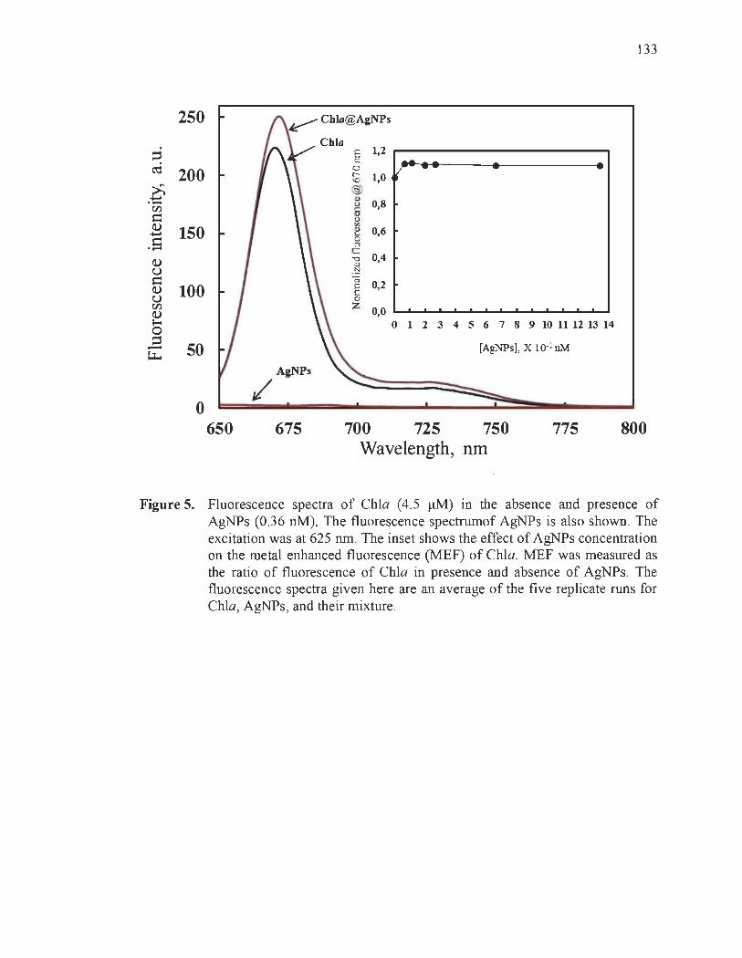
133
250
§ 1,2 ~ ..... ~ 200 0 / - -(- 1,0 <0
~ @) <1>
0,8 .- u en 6 t= u Q) ri>
150 <1> 0,6 ...... '-.s 0 :l
c;::
Q) "0 0,4 <1>
U .:::l s= ro 0,2 Q) 100 E u 0 en Z 0,0 Q) 1-0 o 123 4 5 6 7 8 9 10 II U 13 14 0 ~ 50 [AgNPs], X 10.1 nM ~
A&NJ>s
/ 0
650 675 700 725 750 775 800 Wavelength, nm
Figure 5. Fluorescence spectra of ChIa (4.5 !lM) in the absence and presence of AgNPs (0.36 nM). The fluorescence spectrumof AgNPs is aiso shown. The excitation was at 625 nm. The inset shows the efféct of AgNPs concentration on the metal enhanced fluorescence (MEF) of ChIa. MEF was measured as the ratio of fluorescence of ChIa in presence and absence of AgNPs. The fluorescence spectra given here are an average of the five replicate runs for ChIa, AgNPs, and their mixture.

900
~ œ 750
~ ~ 600 ~ = ~ 450 5 w liI!I
~ 300 l-
.e ~ 150
=
o 380
900
œ 7 0
~ 600 ~ ~ = ~ 450 53 w :; 300 l-
.e ~ 1 0
o 380
Chio + D
480
....... • 0.6
O~--"'----------' o
80
avel ngtb, nm
500 1000 Tlme,s
1500
680
rp +D
480
0.6
0.4 e -= 0.2
ln (F ) = O.OOO9t Rl= O.99
0---"'-----------' o
580
a elengtb nm
soo Tlme.
1000 1500
680
134
Figure 6. Fluorescence spectra of 9,1O-dimethyl-anthracene (DMA): (A) in presence of ChIa, and (B) in presence of ChIa and AgNPs; Àexc = 360 nm. The fluorescence spectra were recorded after red-light irradiation (À > 640 nm) at room temperature for different time intervals: 0, 5, 10, 15,20 s and 1,3, 5, 7, 9, 11 , 13, 15, 17 and 20 min. The insets show the plots of ln (FoIF) as function of irradiation time; Fo and F represent the fluorescence intensity of DMA before and after irradiation, respectively.

135
Table 1. First order kinetic data for irradiated ChIa and ChIa@AgNPs.
Samples Photodegradation kinetic parameters
ChIa Chla@AgNPs
Rate constant (min-1) 1.4 xlO-2 9 x 10⁻⁴
Half-life time, t1l2 (min) 49 770
Correlation coefficient (R 2) 0.94 0.87

SUPPORTING INFORMATION
Nanosilver Could Usher in Next-Generation Photoprotective
Agents for Magnesium Porphyrins
Laurent Bekalé*, Saïd Barazzouk* and Surat Hotchandani*
136
Département de chimie, biochimie et physique, Université du Québec à Trois-Rivières,
C. P. 500, Trois-Rivières (Québec), G9A 5R7, Canada
* To whom correspondence should be addressed. e-mail: [email protected];
[email protected]; [email protected]
(A) Calculation of nanoparticles concentration:
It should be mentioned that 3 mL solution of the mixture of Cha + AgNPs was used for
aU experiments. The concentration of Ag atoms in the stock solution of nanoparticles
was 9 mM. The highest photoprotection imparted to ChIa by AgNPs was achieved after
the addition of7.33 ilL of stock solution of AgNPs to 3 mL ChIa (4.5 !lM) solution.
1) The total number of Ag atoms in 7.33 ilL (9 mM) is given by:
NAg = (9 X 10⁻³ mollL) x (7.33 x10⁻⁶ L) x (6.02 x10²³ mor1) = 3.97 x 10¹⁶
Since, 7.33 ilL of 9 mM AgNPs are added to 3 mL solution of ChIa, the total number
of Ag atoms present in 3 mL solution of the mixture (Cha + AgNPs) will also be 16 3.97 x 10 .
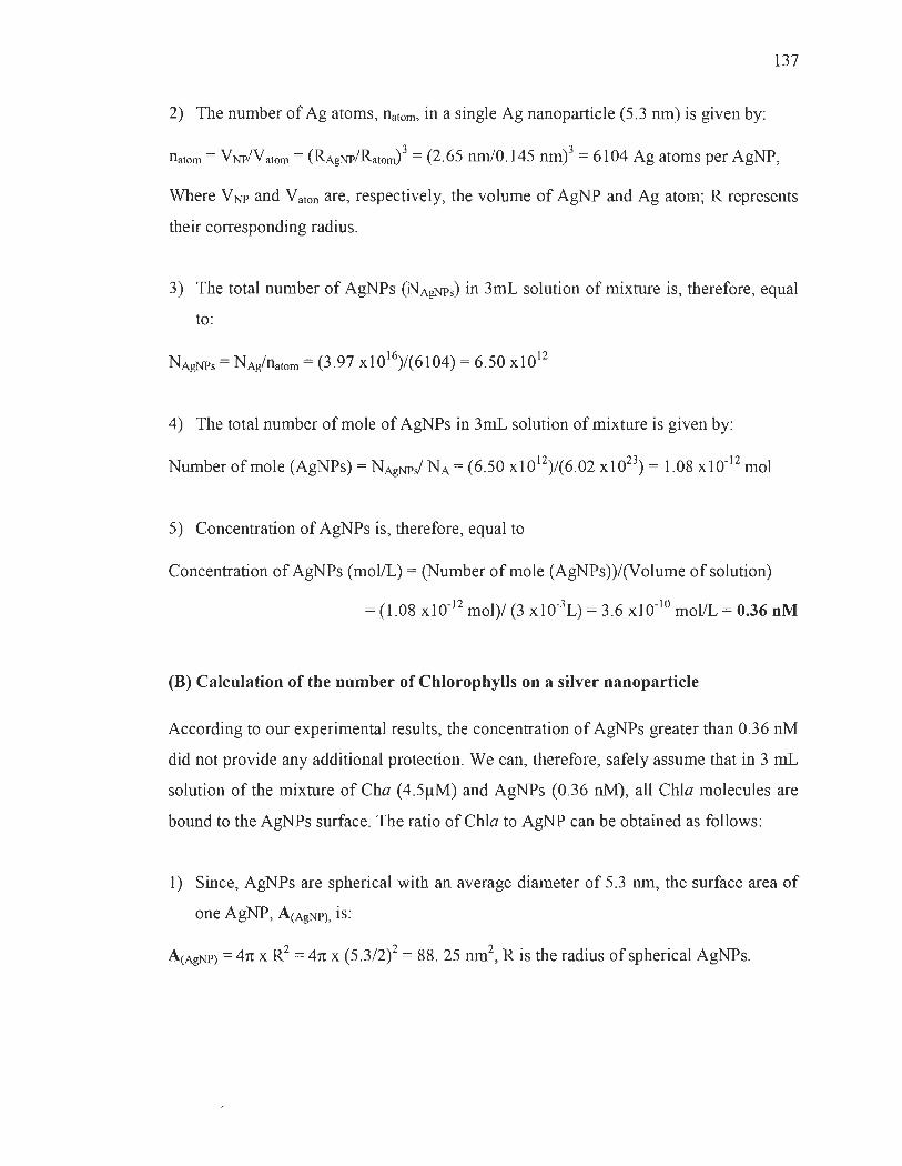
137
2) The number of Ag atoms, natom, in a single Ag nanopartic1e (5.3 nm) is given by:
natom = VNPNatom = (RAgNPlRatom)3 = (2 .65 nmlO.145 nm)3 = 6104 Ag atoms per AgNP,
Where VNP and Vaton are, respectively, the volume of AgNP and Ag atom; R represents
their corresponding radius.
3) The total number of AgNPs (NAgNPs) in 3mL solution of mixture is, therefore, equal
to :
NAgNPs = NAg/natom = (3 .97 X1016)/(61 04) = 6.50 xl012
4) The total number of mole of AgNPs in 3mL solution of mixture is given by:
Number of mole (AgNPs) = N AgNPJ NA = (6.50 Xl012)/(6.02 xl023) = 1.08 xl0-12 mol
5) Concentration of AgNPs is, therefore, equal to
Concentration of AgNPs (mol/L) = (Number ofmole (AgNPs))/(Volume of solution)
= (1.08 Xl0-12 mol)/ (3 xlO-3L) = 3.6 xlO-10 mol/L = 0.36 nM
(B) Calculation of the number of Chlorophylls on a silver nanoparticle
According to our experimental results, the concentration of AgNPs greater than 0.36 nM
did not provide any additional protection. We can, therefore, safely assume that in 3 mL
solution of the mixture of Cha (4 .5IlM) and AgNPs (0.36 nM), aU ChIa molecules are
bound to the AgNPs surface. The ratio of ChIa to AgNP can be obtained as foUows:
1) Since, AgNPs are spherical with an average diameter of 5.3 nm, the surface area of
one AgNP, A(AgNP), is:
A(AgNP) = 4n x R2 = 4n x (5.3/2)2 = 88. 25 nm2, R is the radius of spherical AgNPs.

138
2) The total surface area (AT) provided by 0.36 nM of AgNPs, therefore, is given by:
AT(AgNP) = Total nurnber of AgNPs (NAgNPs) in 3mL solution of mixture multiplied by
the individual area of AgNP, A(AgNP).
Thus, AT(AgNP) = 6.50 xlO I2x 88. 25 nm2 =5.74 x 10¹⁴ nm2
The calculations regarding total number of AgNPs (NAgNPs = 6.50 x10¹²) ln 3mL
solution of mixture has been described above in section (A).
It should be mentioned that the experimental results have revealed that ChIa binds with
AgNP via its nitrogen atoms of porphyrin head while the phytol tail is not involved in
the process of complex formation between AgNP and ChIa, i.e. , Chla@AgNP. It is thus
reasonable to assume that only porphyrin he ad of chlorophyll covers the surface of
AgNPs in the Chla@AgNP complex. According to the literature, the chlorophyll
molecule is depicted as follows. [1]
3) The surface area of one porphyrin head of Cha, A(chl), is, therefore, equal to:
A(chl) = (1.5 nm)2 = 2.25 nm2
4) The total number of ChIa molecules (NChla) ln 3mL solution of mixture
(Cha (4.5IlM) + AgNPs (0.36 nM» is given by:

139
5) The total surface area of porphyrin heads of aH Cha molecules available is given by:
6) The number of Chia molecules per AgNP in 3mL solution ofmixture (Cha (4.5JlM)
+ AgNPs (0.36 nM)) can then be obtained simply by dividing the total area AT(chl)
of chlorophyll molecules by the total surface area furnished by 0.36 nM of AgNPs,
i.e. , AT(AgNP).
Therefore, the number of ChIa moiecules per Ag nanoparticle 1S equal to
(1.83 x 10¹⁶ nrn2)/(5.74 x 1014 nrn2) = 32
In other words, the ratio of ChIa to AgNP is 32 : 1
Kinetics of photochemical reaction:
The photodegradation of Chia in presence of O2 can be described by the following
equation:
(1)
The photodegradation rate of Chia was monitored by recording the absorbance at
665 nrn as a function of irradiation time, and is:
(2)
where [Chia] denotes the concentration of Chia, [02] is the concentration of O2, t is the
reaction time, and k is the rate constant. Since [02] remains constant as the reaction
proceeds, the reaction can be considered pseudo-first-order because it depends on the
concentration of ChIa only.
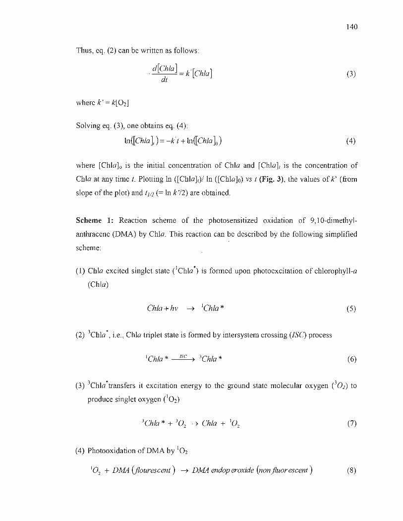
Thus, eq. (2) can be written as follows :
where k ' = k[02]
_ d[Chla] = k' [Chla] dt
Solving eq. (3), one obtains eq. (4):
ln ([Chia J) = -k't + ln ŒChia ]0)
140
(3)
(4)
where [Chla]o is the initial concentration of ChIa and [Chla]r is the concentration of
ChIa at any time t. Plotting ln ([Chla]t)/ ln ([Chla]o) vs 1 (Fig. 3), the values of k' (from
slope of the plot) and 1l/2 (= ln k '/2) are obtained.
Scheme 1: Reaction scheme of the photosensitized oxidation of 9,10-dimethyl
anthracene (DMA) by ChIa. This reaction can be described by the following simplified
scheme:
(1) ChIa excited singlet state eChla *) is formed upon photoexcitation of chlorophyll-a
(ChIa)
ChIa + hv ~ lChla * (5)
(2) 3Chla *, i.e. , ChIa triplet state is formed by intersystem crossing (ISC) process
(6)
(3) 3Chla*transfers it excitation energy to the ground state molecular oxygen e02) to
pro duce singlet oxygen (10 2)
(7)
(4) Photooxidation ofDMA by 102
102 + DMA (fiourescenl) ~ DMA endoperoxide (nonfluorescenl) (8)

141
Figure SI. Image showing the photoprotection provided to ChIa (4.5 ~M) by AgNPs (0.36 nM) under 540 min red-light irradiation (À > 640 nm). As expected, a quick bleaching occurs after exposure of ChIa solution to light. No apparent change in color of ChIa solution is observed wh en AgNPs were added to it.
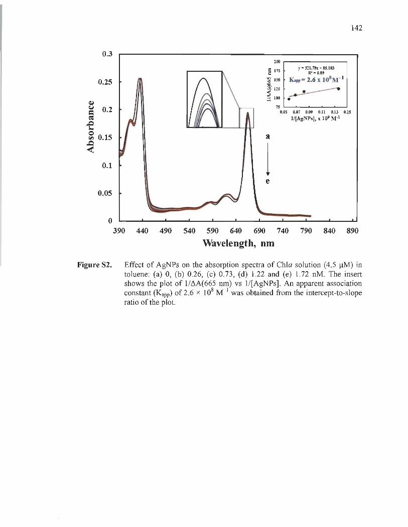
0.3
0.25
~
~ 0.2 ~
-e ~ 0.15
~ 0.1
0.05
o
200 r----------,
'" 175 y -321.78x+8S.l03 ë R'- 0.89
:2 ISO Ka" = 2.6 x l081Vr 1
@) 125
~ 100 •
ft
l e
75 '---"-----'-----'_...o...-....J
0.05 0.07 0.09 0.11 0.13 0.15 1/(AgNPs), x 1()9 1't'I-1
142
390 440 490 540 590 640 690 740 790 840 890
Wavelength, nm
Figure S2. Effect of AgNPs on the absorption spectra of ChIa solution (4.5 ~M) in toluene: (a) 0, (b) 0.26, (c) 0.73, (d) 1.22 and (e) 1.72 nM. The insert shows the plot of 1/M(665 nm) vs 1/[AgNPs]. An apparent association constant (Kapp) of 2.6 x 10⁸ M- 1 was obtained from the intercept-to-slope ratio of the plot.

143
(A) ChIa
396.7 397.7 398.7 399.7 400.7 401.7 Binding energy eV
(B) Cbla@AgNPs
396.7 397.7 398.7 399.7 400.7 401.7 402.7 403.7
Binding energy eV
Figure S3. Nls narrow scan XPS spectra for: (A) ChIa and (B) Chla@AgNPs. The comparison between these spectra supports that nitrogen atoms interact withAgNPs.

144
The high-resolution scans of the XPS spectra of Ols levels of ChIa and
Chla@AgNPs with their decomposition into three components are shown in Figure S-4.
The peak appearing at -530.5 eV has been attributed to the molecular adsorbed oxygen,
O2• The most intense peak at 532.5 eV has been assigned to the carbonyl oxygen from
ketone ()C=O) and ester groups (- O-C=O) of ChIa. Finally, the peak at 533.4 eV is
attributed to the ether-type oxygen in the ester group (-Q-C=O) ofChla.
Binding energy, eV
(B) Ch1a@AgNPs
530 531 531 533 S34 S35
Binding energy, eV
Figure S4. Ols narrow scan XPS spectra for (A) ChIa and (B) Chla@AgNPs.

~ (,J
0.25
0.2
; 0.15 ,.Q :... Q ~ 0.1
< 0.05
o 300 350 400 450 500 550 600 650 700 750 800
Wavelength, nm
Figure S5. Absorption spectrurn of AgNPs (0.36 nM) in toluene.
Références
[1] J. J. Wolken, F. A. Schwartz, 1. Gen Physiol. 1953, 37, 111.
145

CHAPITRE V
CONCLUSION ET PERSPECTIVES
5.1 Conclusion générale
Le but premier de cette thèse de doctorat était de développer une stratégie de
photoprotection, reposant sur l'utilisation des nanoparticules de métaux nobles afin
d'améliorer la stabilité des porphyrines de magnésium. De plus, nous voulions trouver
une façon d'augmenter non seulement la photostabilité, mais aussi les autres propriétés
photophysiques et photochimiques de ces porphyrines, à savoir : l' émission de
fluorescence et la capacité de production d'oxygène singulet. Tous ces efforts visent la
réduction de la photodégradation des porphyrines extraites du milieu naturel, pour la
plupart des porphyrines de magnésium, connues pour être très instables si elles sont
retirées des structures cellulaires hautement organisées qui les protègent naturellement
contre la dégradation par la lumière visible. Le potentiel des porphyrines naturelles
dépasse largement le cadre des processus physiologiques fondamentaux dans les cellules
vivantes et adresse une panoplie d'enjeux scientifiques et technologiques majeurs en lien
avec la biomédicale, l'optoélectronique, le photovoltaïque, la photocatalyse et
l'élaboration de photopesticides. C'est pourquoi le développement d'une stratégie
efficace de photoprotection, dans un contexte de recherche, constitue actuellement un
enjeu de taille pour plusieurs secteurs d'activités.
Je conclus donc cette recherche en abordant successivement la synthèse des
résultats obtenus, la contribution à l'avancement des connaissances et les pistes
possibles d'investigation, nécessaires afin d'approfondir la compréhension de cette
nouvelle méthode de photoprotection.

147
5.2 Bilan des résultats
La présente thèse établit la possibilité d'améliorer la photostabilité des porphyrines
de magnésium par l'inhibition des sites azotés du macrocycle porphyrique. Cette
approche se base sur le résultat d'analyse de différents mécanismes réactionnels
détaillée, postulée dans la littérature pour expliquer la photodégradation des porphyrines
de magnésium. En effet, cette analyse a permis de relever que le doublet libre des
atomes d'azote, ne participant pas à l'aromaticité du macro cycle porphyrique, est sollicité
lors de la réaction de photodégradation des porphyrines de magnésium. Ce bilan a été
particulièrement important comme point de départ, en ce sens qu'il présuppose que, si
les sites azotés (le doublet libre des atomes d'azote) du macro cycle porphyrique sont
bloqués, avant l'illumination, alors il se produira une augmentation de la photostabilité.
Au chapitre II, ce mécanisme de photoprotection fut démontré expérimentalement
par l'utilisation du tétraphénylporphyrine de magnésium (MgTPP) comme porphyrine
modèle, et les nanoparticules d'or (AuNPs) comme agent de blocage des sites azotés du
macrocycle porphyrique. Il a été observé que l' ajout dans l'obscurité, d'une faible
quantité de AuNPs, dans une solution de MgTPP augmentait considérablement la
photostabilité du MgTPP. En effet, avec une concentration appropriée de AuNPs, une
efficacité de photoprotection quasiment de 100 % a pu être obtenue. L'analyse des
interactions entre AuNPs et MgTPP a permis de relever qu' il se forme un complexe
AuNPs@MgTPP après l'addition des nanoparticules. La constance de formation de ce
complexe de l'ordre de 104 M-1 indique que le complex AuNPs@MgTPP est très stable.
De plus, la démonstration établit que le complexe AuNPs@MgTPP est stabilisé par des
liaisons organométalliques directes entre les atomes d'azote (-N) et l'or métallique (Au).
De ce fait, la capacité photoprotectrice des AuNPs réside dans leur faculté à se lier aux
sites d'azote du macrocycle, inhibant ainsi la formation de liaisons entre les espèces
réactives de l'oxygène (ROS) à ces sites d'azote, connues pour causer la
photodégradation de MgTPP et plusieurs autres porphyrines dans des conditions
aérobies. En outre, la diminution de la formation d'états singulets excités du MgTPP, à la
suite de sa liaison avec les AuNPs, supprime également la production de ROS,

148
améliorant considérablement la photostabilité du MgTPP. Ce résultat démontre
clairement l' efficacité de notre approche et met en lumière la possibilité de développer
de nouveaux types d ' agents photoprotecteurs, qui ont comme principale propriété de
bloquer le doublet libre d 'atomes d'azote du macrocycle porphyrique, afin d ' induire une
augmentation de la photostabilité.
Effectivement, il est impossible de prédire, avec ce type d 'agent photoprotecteur,
s' il permettrait également d ' accroître la stabilité des porphyrines de magnésium,
possédant à la fois des doublets d'électrons libres sur les atomes d ' azote du macrocycle
et sur les groupes fonctionnels périphériques, n 'étant pas conjugués au macrocycle.
Toutefois, au chapitre III, nos résultats sur l'emploi de nanoparticules d'or en tant
qu 'agent photoprotecteur pour améliorer la stabilité des chlorophylles-a (ChIa) ont
démontré que la photodégradation de la ChIa est ralentie en présence de AuNPs.
L 'estimation du temps de demi-vie de la ChIa (c'est-à-dire le temps nécessaire pour que
la quantité d 'une substance soit diminuée de moitié de sa valeur initiale) a révélé une
augmentation pouvant atteindre un ordre de grandeur en présence de AuNPs. Nous en
déduisons que le rôle photoprotecteur des AuNPs est bien présent, c'est-à-dire que les
nanoparticules se lient aux atomes d ' azote de la ChIa dans l'obscurité, et ce, malgré la
présence de groupes fonctionnels ester (-COO-R) et cétone (>C=O) porteurs de doublets
libres sur l'oxygène.
Par ailleurs, il est important de mentionner que dans les conditions in vitro, les
AuNPs sont de bien meilleurs photoprotecteurs que les molécules, comme la ~-carotène
ou les quinones, reconnues pour être très efficaces afin de protéger les chlorophylles
dans des conditions in vivo, c'est-à-dire dans les plantes.
Étant donné la nécessité de trouver un procédé multifonctionnel très efficace pour
l'amélioration simultanée de différentes propriétés photoniques des porphyrines
naturelles, la méthode de photoprotection développée dans cette thèse se devrait d ' être
149
ajustée, afin d'augmenter conjointement la photostabilité, l'émission de fluorescence et
la production d'oxygène singulet.
Au chapitre IV, une observation sans précédent démontre que les nanoparticules
d'argent (AgNPs) sont des additifs potentiels pour augmenter simultanément la
photostabilité, l'émission de fluorescence, ainsi que la capacité de production d'oxygène
singulet de ChIa in vitro. L'estimation du temps de demi-vie de la ChIa a révélé une
augmentation pouvant atteindre un ordre de grandeur en présence d'une concentration
d'AgNPs extrêmement faible, c'est-à-dire avec des valeurs de concentration dans la
plage nanomolaire. De ce fait, ils sont actuellement les meilleurs agents photoprotecteurs
connus. En plus de leur performance photoprotectrice surprenante, les AgNPs ont
également permis d'améliorer l'émission de fluorescence et la capacité de production
d'oxygène singulet de la ChIa respectivement de 1,2 et 2 fois. La forte efficacité
photoprotectrice des AgNPs par comparaison aux AuNPs peut-être dû au fait que lors du
processus photoprotection, nous effectuons un échange de ligand entre un tétraakyl
ammonium et une amine tertiaire dans le cas des nanoparticules d'or. Les liaisons
chimiques qui sont brisées (de nature électrostatique) et d'autres qui se contiuent, sont
sensiblement de même force. Par contre, dans le cas des nanoparticules d'argent, nous
brisons une liaison Ag-thiol pour former une liaison Ag-amine. Cette réaction est très
favorable puisque la laison Ag-amine et plus forte que la liaison Ag-thiol. Ainsi, la
réaction d'échange de ligand dans le cas l' argent un plus rapide. Cet aspect peut
expliquer en partie la forte réactivité des nanoparticules d'argent. Toutefois, il est
possible que les nouvelles propriétés émergentes à l'échelle du nanomètre soient aussi
impliquées dans la différence de comportement entre les nanoparticules d'or et d'argent.
Enfm, ces résultats obtenus avec les AgNPs sont exceptionnellement intéressants
et importants, car ils ont fait la preuve de l' amélioration simultanée de différentes
propriétés photoniques des porphyrines. Ils constituent donc une solide raison de
développer de nouveaux agents photoprotecteurs. Ainsi, le présent travail ouvrirait la
VOle au développement d'une nouvelle génération d'agents photoprotecteurs
multifonctionnels, mieux adaptés à des applications des porphyrines naturelles dans le
l
150
théranostique (l'association d'une thérapeutique et d'un test diagnostique), l' imagerie et
la thérapie photo dynamique.
5.3 Contribution de la thèse à l'avancement des connaissances
Eu égard à tout ce qui précède, cette thèse de doctorat contribue, de façon
originale, à l'amélioration des connaIssances relatives à la photoprotection des
porphyrines naturelles, et ce, en exploitant principalement les propriétés des
nanoparticules de métaux nobles. Effectivement, la mise au point d'une méthode de
photoprotection, reposant sur l'utilisation des nanoparticules de métaux nobles pour
accroître la stabilité des porphyrines de magnésium, constitue une première dans le
domaine de la protection des porphyrines contre la dégradation par la lumière. Les
méthodes de photoprotection développées antérieurement s'intéressent davantage à
augmenter la photostabilité, en supprimant tout simplement la production des espèces
réactives de l'oxygène, plutôt que d'augmenter simultanément la photo stabilité,
l'émission de fluorescence et la capacité de production d'oxygène singulet. La
découverte de cette nouvelle stratégie de photoprotection met en lumière la possible
mise au point des photoprotecteurs multifonctionnels, pouvant non seulement pallier le
problème de stabilité, mais aussi renforcer les autres propriétés intéressantes de
porphyrines naturelles.
5.4 Perspectives
Dans le prolongement des résultats obtenus au cours de ce travail de thèse, il sera
intéressant d'approfondir ces études, afin de mieux comprendre, optimiser et étendre
cette méthode de photoprotection. Pour ce faire, la recherche, sur la photoprotection des
porphyrines par des nanoparticules, gagnerait en s' orientant vers deux directions
distinctes.

151
5.4.1 Analyser systématiquement l'effet de la taille et de la forme des nanoparticules sur l'efficacité photoprotectrice
Nous avons démontré dans cette thèse que les nanoparticules d'or (AuNPs) et
d'argent (AgNPs) sont de bons photoprotecteurs, et peuvent ainsi être ulitisés pour
améliorer la photostabilité des porphyrines par l'inhibition des sites azotés du
macrocycle porphyrique. Cette découverte constitue une toute nouvelle possibilité afm
de lutter contre la dégradation rapide des photosensibilisateurs à base de porphyrines
naturelles. Ainsi, elle paverait la voie au développement d'une nouvelle classe d'agents
photoprotecteurs à base de nanoparticules (NPs).
11 est bien établi que la taille et la forme des NPs ont une très grande influence sur
les propriétés finales (activité catalytique, réactivité' chimique, adhésion, etc.)1,2. Cette
caractéristique est un effet du changement de la fraction d'atomes de surface des NPs.
Lorsque la taille de la particule devient plus petite, son rapport surface/volume augmente
considérablement. En outre, pour une nanoparticule de même taille, l'étendue de la
surface dépend de la forme du matériau. Un exemple simple est de comparer une sphère
et un cube ayant le même volume. Le cube a une surface totale plus grande que la
sphère. Pour cette raison, dans la nanoscience, non seulement la taille d'un nanomatériau
est importante, mais également sa forme. L'exemple parfait, pour illustrer l' influence du
changement de rapport surface/volume à l'échelle nanométrique, est d'observer l'effet de
la taille et de la forme des NPs sur les propriétés optiques3,4. Comme le témoigne la
figure 5.1 , selon la taille des AuNPs en suspension, la couleur de la solution varie du
rouge vif (pour des particules de moins de 100· nm), au bleu-violet (pour les particules
les plus grosses).

Figure 5.1
152
Solutions de nanoparticules d'or de différentes tailles. La différence de taille provoque la différence de couleur3
.
De même, Mock et al.4, en élaborant par voie colloïdale des particules d'argent
aux formes variées (sphérique, pentagonale, triangulaire), ont démontré que leur réponse
optique était fortement corrélée à la forme des nanoparticules (Figure 5.2).
Figure 5.2
100
o
Spectres de diffusion de la lumière par des nanoparticules d'argent de différentes formes4
.

153
En vue de l'obtention d'une réactivité optimale des nanoparticules (d'or et
d'argent) afin d'améliorer leur efficacité photoprotectrice, il serait souhaitable
d'examiner les effets de la taille et de la forme sur le pouvoir photoprotecteur des
nanoparticules. Une étude de cette nature revêt une importance particulière, déterminant
ainsi la dimention et la morphologie idéales des nanoparticules photoprotectrices.
5.4.2 Examiner si la méthode de photoprotection mise en œuvre dans cette thèse peut être employée pour l'élaboration des nanoparticules fonctionnelles pour des applications médicales
Les résultats présentés aux chapitres II, III et IV sont encourageants et incitent à de
nouvelles perspectives de recherche intéressantes. Cette stratégie · de photoprotection
permettrait le développement des agents photoprotecteurs magnétiques pouvant être
utilisés en nanomédecine, particulièrement dans la thérapie photodynamique des
cancers. Nous prévoyons que ces nanoparticules joueront un triple rôle dans le
traitement photo dynamique (PDT) :
(i) ils agiront comme photoprotecteurs pour accroître la photostabilité des
photosensibilisateurs à base de porphyrines naturelles, afin de générer des
10 2 pendant de plus longues périodes de temps,
(ii) ils opéreront en tant qu 'amplificateurs additionnels de la production des 102
via des interactions métal-photosensibilisatrices,
(iii) ils se comporteront comme agents de contraste en imagerie par résonance
magnétique (IRM), visualisant clairement les tissus tumoraux, offrant par
surcroît la possibilité de diriger les nanoparticules fonctionnalisées par les
photosensibilisateurs vers des cellules tumorales, plutôt que vers les cellules
sames.
Cette idée propose d'utiliser les nanoparticules d'oxyde de fer
superparamagnétique (Fe304) recouvertes d'une couche d'or, soit une structure

154
cœur-coquille Fe30 4@Au (Figure 5.3). Le revêtement d'or aide à réduire l'agglomération
des particules, améliorant ainsi la biocompatibilité5,6.
Photosensibilisateur à base de phophyrine naturelle
Coquille d ' or (Au)
Cœur d'ol.')'de de fer superparamagnétique
(Fe30 4)
Figure 5.3 Représentation schématique de l'agent photoprotecteur magnétique de structure cœur-coquille Fe30 4@Au.
Récemment, les nanoparticules d'oxyde de fer superparamagnétique (Fe30 4) ont
suscité beaucoup d'intérêt, en raison de leur application potentielle dans le domaine
biomédicaC. Effectivement, les nanoparticules (Fe30 4 NPs) sont utilisées en clinique
humaine, comme agents de contraste d'IRM8.
Actuellement, lors du traitement photodynamique, les photo sensibilisateurs (PSs)
se distribuent dans l'organisme de manière plus ou moins ciblée, c'est-à-dire que le PS
ne sera pas forcément délivré sur les cellules concernées. On doit donc administrer
d' importantes doses de PS, contraignant ainsi une petite proportion d'atteindre
finalement sa cible. L'incorporation d'une propriété magnétique dans des nanoparticules
photo protectrices vise à contourner cet obstacle, rendant le traitement plus spécifique et
plus efficace. Ainsi, les nanoparticules photoprotectrices, magnétiques, faciliteraient la
livraison des PSs, du site d' injection vers les régions tumorales ciblées, se servant d'un
champ magnétique externe (Figure 5.4t
Enfm, la combinaison de l'IRM et de la PDT, sur un même agent thérapeutique,
présente l' avantage de visualiser clairement les tissus tumoraux et de fournir des

155
informations importantes relatives à la réponse thérapeutique au traitement
photodynarnique, et, de ce fait, guider précisément l'irradiation de lumière aux tissus
tumoraux, atteignant ainsi une efficacité maximale du traitement thérapeutique lO-12
.
Lumière Rouge
Champ magnétique e~1:e ... ,e
Photosensibilisatetu· magnétique
Figure 5.4 Schéma illustrant le ciblage in vivo de la tumeur par champ magnétique externe.
5.5 Références
(1) EI-Sayed, M. A. ; Sorne interesting properties of metals confined in time and nanometer space of different shapes. Acc. Chem. Res., 2001 , 34, 257-264.
(2) Xu, R.; Wang, D.; Zhang, J. ; Li, Y. Shape-dependent catalytic activity of silver nanoparticles for the oxidation of styrene. Chem. Asian J, 2006, 1, 888-893.
(3) http://en.wikipedia.org/wiki/Colloidal_gold
(4) Mock, J. J.; Barbic, M.; Smith, D. R. ; Schultz, D. A. ; Schultz, S. Shape effects in plasmon resonance of individual colloidal silver nanoparticles . J Chem. Phys., 2002, 116,6755-6759.
156
(5) Lyon, J. L. ; Fleming, D. A ; Stone, M. B.; Schiffer, P.; Williams, M. E. Synthesis of Fe oxide core/Au shell nanoparticles by iterative hydroxylamine seeding. Nano Lett. , 2004, 4, 719-723 .
(6) Lo, C. K ; Xiao, D.; Choi, M. M. F. Homocysteine-protected gold-coated magnetic nanoparticles: synthesis and characterization. J Mater. Chem. , 2007, 17, 2418-2427.
(7) Hasany, S. F.; Abdurahman, N. H. ; Sunarti, A R. ; Jose, R. Magnetic iron oxide nanoparticles: chemical synthesis and applications review. Curr. Nanosci. , 2013, 9, 561-575.
(8) Corot, C.; Robert, P. ; Idee, J. M.; Port, M. Recent advances in iron oxide nanocrystal technology for medical imaging. Adv. Drug Deliv Rev., 2006, 58, 1471-1504.
(9) Li, Z. W.; Wang, c.; Cheng, L.; Gong, H. ; Yin, S. N.; Gong, Q. F.; Li, Y G. ; Liu, Z. PEG-functionalized iron oxide nanoclusters loaded with chlorin e6 for targeted, NIR light induced, photodynamic therapy. Biomaterials. , 2013, 34, 9160-9170.
(10) Gross, S.; Gilead, A ; Scherz, A ; Neeman, M. ; Salomon, Y. Monitoring photodyilamic therapy of solid tumors online by BOLD-contrast MRI. Nat. Med. , 2003, 9, 1327-1331 .
(11) Vaidya, A.; Sun, Y ; Ke, T. ; Jeong, E. K ; Lu, Z. R. Contrast enhanced MRIguided photodynamic therapy for site-specifie cancer treatment. Magn. Reson. Med., 2006, 56, 761-767.
(12) Vaidya, A ; Sun, Y G.; Feng, Y ; Emerson, L. ; Jeong, E. K; Lu, Z. R. Contrastenhanced MRI-guided photodynamic cancer therapy with a pegylated bifunctional polymer conjugate. Pharm. Res., 2008, 25, 2002-20 Il.

ANNEXE A
MÉTHODES EXPÉRIMENTALES, SYNTHÈSE ET INSTRUMENTATION
A.1 Synthèse générale des nanoparticules métalliques en solution
De façon très générale, on décrit les nanoparticules comme étant des amas
d'atomes de diverses tailles, variant de 1 à 100 nm. En général, la synthèse en solution
(aqueuse ou organique) des nanoparticules métalliques s'effectue principalement
toujours de la même manière, soit par réduction des sels métalliques, dissous en solution
avec un agent réducteur, sous agitation vigoureuse. Une fois réduits, les colloïdes
métalliques à l'état d'oxydation zéro s'associent rapidement entre eux et forment alors
des agglomérats de quelques atomes avant de former, par ajout successif d'atomes, des
nanoparticules de plus en plus grandes (Figure A.1).
Sel métallique
Colloïdes métalliques il l ' état (l ' oxydati ou zél'O
Aggloménlts de quelques atomes Nalloparticule
métallique
Figure A.1 Représentation schématique de la synthèse générale des nanoparticules métalliques en solution.

158
A.1.1 Préparation des nanoparticules d'or (AuNPs)
La procédure de préparation des nanoparticules d'or (AuNPs), dans un solvant
organique, est une version modifiée l de celle proposée par Brnst et al.2, sans addition de
thiols (Figure A.2). Cette méthode permet d'obtenir des nanoparticules monodisperses
en tailles et de formes bien contrôlées. En général, les particules obtenues par cette
méthode ont une ütille inférieure à 10 nm. Sommairement, les AuNPs sont préparés par
une procédure de réduction biphasique (H20 + toluène) de l'acide tétrachloroaurique,
(HAuCI4) en présence d 'un surfactant (le bromure de trétraoctylammonium (CgH17)4NBr
ou TOAB). La phase aqueuse contient du sel d 'or (HAuCI4, 30 mM), tandis que la phase
organique, constituée de toluène, contient l'agent surfactant (TOAB, 0.1 M), étant aussi
l'agent de transfert de phase des ions auriques (AUC4-). Les ions AuC14-, ainsi transférés
dans la phase organique, sont par la suite réduits par addition, goutte à goutte, d'une
solution aqueuse de borohydrure de sodium (NaBH4, 0.4 M), formant ainsi les
nanoparticules métalliques d'or (AuNPs). La phase organique, laquelle contient les
AuNPs, est lavée, puis asséchée. Après filtration, les AuNP de couleur rose-rouge
foncée sont conservées dans l'obscurité à température ambiante et sont très stables
pendant plusieurs mois.
..... -....... ,.....,
:::. .::: Toluène + TOAB
:::. :::.
Agitation
1 :> EAu + AuCI.-
.... .....
..... ....... r--... -'"
.... -.
Toluène + TOAB + AuCI -
....
Eau ..... .....
NaRH. (Réduction) :> Agitation
Figure A.2 Représentation schématique de la synthèse des nanoparticules d'or, par la procédure biphasique de réduction des ions auriques.
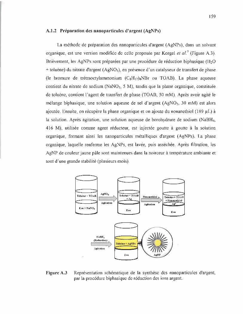
159
A.l.2 Préparation des nanoparticules d'argent (AgNPs)
La méthode de préparation des nanoparticules d'argent (AgNPs), dans un solvant
organique, est une version modifiée de celle proposée par Korgel et al. 3 (Figure A.3).
Brièvement, les AgNPs sont préparées par une procédure de réduction biphasique (H20
+ toluène) du nitrate d'argent (AgN03), en présence d'un catalyseur de transfert de phase
(le bromure de trétraoctylammonium (C8H17)4NBr ou TOAB). La phase aqueuse
contient du nitrate de sodium (NaN03, 5 M), tandis que la phase organique, constituée
de toluène, contient l'agent de transfert de phase (TOAB, 50 mM). Après avoir agité le
mélange biphasique, une solution aqueuse de sel d'argent (AgNO 3 , 30 mM) est alors
ajoutée. Ensuite, on récupère la phase organique et on ajoute du nonanethiol (189 ilL) à
la solution. Après agitation, une solution aqueuse de borohydrure de sodium (NaBH4,
416 M), utilisée comme agent réducteur, est injectée goutte à goutte à la solution
organique, formant ainsi les nanoparticules métalliques d'argent (AgNPs). La phase
organique, laquelle renferme les AgNPs, est lavée, puis asséchée. Après filtration, les
AgNP de couleur jaune pâle sont maintenues dans la noirceur à température ambiante et
sont d'une grande stabilité (plusieurs mois).
-- -...... -" f"" ...... ....... ....
Toluène + TOAB AgNO.
'-----'> Tolu"'~ + TOAB +A~
AgitAtion
Ean + NllNO. Eau
Iloo. ~
NaBH.
(Redurtlon) '" 1 V ~ __ ~~~
Agitation r----""1
".... -...... .-"
-Nonant'lltiol > 1
Agitation
Toluène + TOAB + Nonlll.etloJolI
Ag>
E.u .... ~
Figure A.3 Représentation schématique de la synthèse des nanoparticules d'argent, par la procédure biphasique de réduction des ions argent.

160
A.2 Caractérisation des nanoparticules
La caractérisation des nanoparticules (NPs) est nécessaire, établissant ainsi le
contrôle de la synthèse. La caractérisation morphologique des NPs est faite en utilisant
une variété de techniques différentes, principalement tirées de la science des matériaux.
Les techniques courantes, exploitées dans cette thèse, sont la microscopie électronique à
transmission (MET), la spectroscopie d'absorption UV-visible et la spectroscopie de
photoélectrons X (XPS).
A.2.1 Microscopie électronique à transmission (MET)
La microscopie électronique à transmission est l'outil par excellence, pour
caractériser la morphologie et la taille des NPs. Cette technique de microscopie consiste
à placer un échantillon suffisamment mince sous un faisceau d'électrons, en utilisant un
système de lentilles magnétiques projetant ainsi l'image de l'échantillon sur un écran qui
transforme l'image électronique en image optique. Les effets d'interaction entre les
électrons et l'échantillon donnent naissance à une image, dont la résolution peut atteindre
0,08 nanomètre, soit 1000 fois plus élevées qu'un microscope optique. Le mode
d'imagerie le plus utilisé pour la visualisation des NPs est celui en champ clair. Dans ce
procédé, on emploie un diaphragme, placé dans le plan focal , de manière à sélectionner
uniquement le faisceau transmis en ligne droite par l'échantillon. Donc, seuls les
électrons non diffractés formeront l'image sur l'écran. Conséquemment, les zones de
l'échantillon diffractant fortement le faisceau (zones de fortes densités) apparaissent les
plus sombres.
A.2.2 Spectrométrie d'absorption UV-visible
La spectrométrie d'absorption UV-Vis est une technique utilisant des photons,
dont les longueurs d'onde sont comprises entre le domaine du spectre ultraviolet et celui
du proche infrarouge. Cette technique spectroscopique est grandement utilisée, pour
mettre en évidence la formation des NPs en solution par la mesure de leur spectre
d'absorption.

161
A.2.3 Spectroscopie de photoélectrons X (XPS)
La spectroscopie XPS pennet de mesurer les états électroniques dans un matériau,
par irradiation d'un échantillon avec un rayonnement de rayons X monochromatique et
en analysant les photoélectrons émis. Chaque élément chimique étant caractérisé par un
spectre unique, cette méthode spectroscopique pennet d'analyser précisément la nature
chimique d'un matériau donné, c'est-à-dire qu'on peut détenniner l'état d'oxydation du
matériau, ainsi que son environnement moléculaire (ou environnement chimique). En
XPS, l'identification de l' état chimique d'un élément est obtenue à partir de la mesure
exacte de la position des pics et de leurs séparations en énergie.
A.3 Références
(1) Jakob, M.; Levanon, H. ; Kamat, P.V. Charge Distribution between UV-Irradiated Ti02 and gold nanoparticles: detennination of shift in the ferrni level. Nana Let/., 2003, 3, 353-358.
(2) Brust, M.; Walker, M.; Bethell, D.; Schiffrin, D. J; Whyman, R. Synthesis ofthiolderivatised gold nanoparticles in a two-phase Liquid- Liquid system. J Chem. Soc, Chem. Commun., 1994, 7, 801-802.
(3) Korgel, B. A. ; S. Fullam, Connolly, S.; Fitzmaurice, D. Assembly and selforganization of silver nanocrystal superlattices: ordered "soft spheres". J Phys. Chem. B., 1998, 102, 8379-8388.