Embed Size (px)
Citation preview
AVERTISSEMENT
Ce document est le fruit d'un long travail approuvé par le jury de soutenance et mis à disposition de l'ensemble de la communauté universitaire élargie. Il est soumis à la propriété intellectuelle de l'auteur. Ceci implique une obligation de citation et de référencement lors de l’utilisation de ce document. D'autre part, toute contrefaçon, plagiat, reproduction illicite encourt une poursuite pénale. Contact : [email protected]
LIENS Code de la Propriété Intellectuelle. articles L 122. 4 Code de la Propriété Intellectuelle. articles L 335.2- L 335.10 http://www.cfcopies.com/V2/leg/leg_droi.php http://www.culture.gouv.fr/culture/infos-pratiques/droits/protection.htm
UNIVERSITE HENRI POINCARE, NANCY 12009
THESE
FACULTE DE MEDECINE DE NANCY
NO 10
pour obtenir le grade de
DOCTEUR EN MEDECINE
Présentée et soutenue publiquementdans le cadre du troisième cycle de Médecine Spécialisée
par
Carole PAULUS
le 18 septembre 2009
ETUDE DE REPRODUCTIBILITE DE LALECTURE DE RADIOGRAPHIES DE POIGNETSTRAUMATIQUES SUR PACS, FILM ET PAPIER
Examinateurs de la thèse:
M.A.BLUMM. H. COUDANEM. F. GUILLEMINM.A.PEUTOT
ProfesseurProfesseurProfesseurDocteur en Médecine
PrésidentJugeJugeJuge

UN IVERSITE HEN RI POINCARE, NAN CY 12009
THESE
FAC ULTE DE MEDECINE DE NANC YN"
pour obtenir le grade de
DOCTEUR EN MEDECINE
Présentée et soutenue publiquementdans le cadre du troisième cycle de Médecine Spécialisée
par
Carole PAULUS
le 18 septembre 2009
ETUDE DE REPRODUCTIBILITE DE LALECTURE DE RADIOGRAPHIES DE POIGNETSTRAUMATIQUES SUR PACS, FILM ET PAPIER
Examinateurs de la thèse:
M.A.BLUMM. H. COUDANEM. F. GUILLEMINM.A.PEUTOT
ProfesseurProfesseurProfesseurDocteur en Médecine
PrésidentJugeJ ugeJ uge
UNIV ERSITÉ HENRI POINCARÉ , NAN CY 1
FACULTÉ DE MÉDECINE DE NANCY
l'résident de l'Université: Professeur Jean-Pierre FINANCE
Doyen de la Faculté de Médecine : Professeur Henry COUDANE
Vice Doyen Recherche : Professeur J ean-Lou is GUEANTVice Doyen PédllgugÎ/! : Professeur Anni ck BARRAUD
Vice Doyen Campus : Professeur Marie-Chrtstin e BÉNI~
Assesseu rs :du l' "Cycle :du 2·.... Cycle :du 3<"" Cy cle :Filières professionnalisées :Prospective :FMC/EPP :
M.le Professeur Fra nçois ALLA1\-1. le Professeur J ean-Pierre BRONOWICKIM.le Professeur Pierre-Edouard BOLLAERTM.le Professeur Chr istophe CIIOSE Rû TM.le Professeur Laurent BRESLERM. le Professeur J ean-Dominique DE KOR\\'IN
DOYENS HONORAIRESProfesseur Adrien DUPREZ - Professeur Jean-Bernard DUREUX
Professeur Jacques ROLAND - Professeur Patrick NET f ER
= ======
PROFF-SSEURS HONORAIRES
Pierre ALEXANDRE - Daniel ANTHOINE - AJain BERTRAND - Pierre BEY - Jean BEUR EY - Jacques BORRELLYMichel BOULANGE - Jean-Claude BURDlN - Claude BURLET - Daniel BURNEL - Claude CHARDOT - Jean-Pierre CRANCE
Gérard DEBRY - Jean-Pierre DELAGOUTTE - Emile de LAVERGNE - Jean- Pierre DESCHAMPS - Michel DUCJean DU HEILLE - Adrien DUPREZ - Jean-Bernard DU REUX - Gabriel FAIVRE - Gér ard FIEVE - Jean l'LOQUET
Rnbert FRIS CH - Alain GAUCHER - Pierre GAUCHER - Hubert GERARD - Jean-Marie GILGENKRANTZSimone G1LG ENKRA NTZ , Oliéro GUERCI - Pierre HA RTEMANN - Claude HURIET - Christian JANOT - Jacques LACOST EPierre LAN DES - Alain LARCAN - Marie-Claire LAXENAIRE - Michel LAXENAIRE - Jacques LECLERE - Bernard LEGRAS
Michel MANCIAUX - Jean-Pierre MAILl É - Pierre MATHI EU - Pierre NA BET - Jean-Pierre NICOLAS - Pierre PAYSA NTFrancis PENIN - Gilbert PERCEBOIS - Claude PERRIN - Gu y PETIET - Luc PICARD - Michel PIERSON - Jean-M arie POLU
Jean PREVOT - Antoine RASPILLER - Michel RENARD - Jacques ROLAND - René-Jean ROYER - Pau l SADOULDaniel SCHMITT - Jean SOMMELET - Danièle SO MMELET - Michel ST RICKER - Gilbert THIBAUT - Augusta TREHEUX
Hubert UFFHOLTZ - Gérard VAILLANT - Pau l VERT - Co lette VIDAILHET - Miche l VIDAILH ET - Michel WAYOFFMichel WEBER
= ======
PROFESSEURS DES UNIVERSITÉSPRATICIENS HOSPITALIERS
(Disciplines du Conse il National des Universités)
42'"" Section : MORPHOLOGIE ET MORPHOGENÈS El ère sous-section : (Anatomie)
Professeur Gilles GROS DlDIERProfesseur Pierre LASCOMBES - Professeur Mar c BRAUN
z".. sous-section : (Cytologie et histologie)Professeur Bern ard FOLIGUET
3t n.. sous-section : (Anatomie et cytologie pathologiques)
Professeur Franço is PLENAT - Professeur Jean-Michel VlGNAUD
43'- Sectinn : BIOPHYSIQUE ET IMAGERIE MÉDICALE1 "~ sous-see tlun : (B iophys ique el médecine nucl éaire)
Professeur G illes KARCHER - Professeur Pierre-Yves MARIE - Profe sseur Pierre OLIVIE R2~"" sous-section: (Radiologie el imagerie médicale)
Professeur Denis REGENT - Pro fesseur Michel CLAUDONProfesseur Serge BRACAR D - Professeur Alain BLUM - Profes seur Jacqu es FE LBLlNGER
Professeur René ANXIONNAT
2
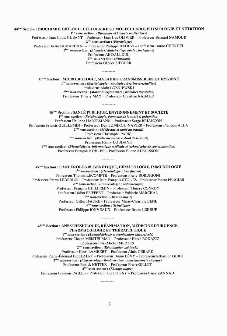
44"" Seclion : lIIOCIIIMIE, BIOLOGIE CELLULAIRE ET MOLÉCULAIRE, l'HYSIOLOGIE ET NUTIUTION. ... sous-section: (lJiochimie et biologie moléculaire)
ProfesseurJean-Louis GUÉANT - ProfesseurJean-Luc OLIVIER- ProfesseurBernard NAMOUR2~mt sous-section : (Physiologie)
Professeur Franço is MAR CHAL - Professeur Philippe HAOUZI - Professeur Bruno CHENU EL3i n
... sons-section : mi%gie Cellulaire (type mixte: biologique)Professeu r Ali DALLOUL4'n... sons-section : (Nutrition)
Professeur Olivier ZIEGIJER
4s'm' Section: MICROBIOLOGIE, MALADIES TRANSMISSIBLES ET HYGIÈNElh. sous-section : (Bact ériolo gie - virologie .. hygi ène hospitali ère}
Professeur Alain LOZNIEWS KI3...... sous-section : (Maladies infectieuses i maladies tropicales)Professeur Thierry MAY - Professeur Christian RABAUD
46" " Section : SANTÉ PUBLIQUE, ENVIRONNEMENT ET SOCIÉTÉI h • sous-section : (Ép idémiologie, économie de la santéet prévention)
Professeur Philippe HARTEMANN - Professeur Serge BRIAN ÇONProfesseur Francis GUILLEMIN - Professeur Deni s ZMIROU-NAVIER - Professeur François AL LA
20"" sous-section: (M édecine et santé au travail)
Professeur Chr istnphe PARIS3f n.. sous-section : (Médecine légale et droit d e la santé)
Professeur Henry COUDANE4'm~ sous-section : {Biostatisiiques, informatique m édicale et technologies de communication)
Professeur François KüHLER - Professeur Éliane ALBUISSON
47'm' Section: CANCÉROLOGIE, GÉNÉTIQUE, HÉMATOLOGIE, IMMUNOLOGIE1h~ sous-section : [H ématoiogie ; transfusion}
ProfesseurThomas LECOMPTE- ProfesseurPierre BORDIGONIProfesseur Pierre LEDERLIN - ProfesseurJean-François STOL'rz - Professeur PierreFEUGIER
2'- sous-section : (Canc érologie ,.radiothérapie)Professeur Françoi s GUILLEMIN - Professeur Thi erry CONROYProfesseur Didier PEIFFERT - Professeur Frédéric MARCHAL
3''''' sons-section: {Immunologie}ProfesseurGilbert FAURE- Professeur Marie-Christine BENE
4"'" sous-section : (Génétique)Professeur Philippe JONVEAUX - Professeur Bruno LEHEUP
4s'm' Seclion : ANESTHÉSIOLOGIE, RÉANIMATION, MÉDECINE D'URGENCE,PHARMACOLOGIE ET THÉRAPEUTIQUE
l h c sous-section: (Anesthésiologie et réanimation chirurgicale)Professeur Claude MEiSTELMAN - Professeur Hervé BOUAZIZ
Professeur Paul-Michel MERTES2'- sous-section : (Réanimation médicale)
Professeur Henri LAMBERT - Professeur Alain GERARDProfesseur Pierre-Édouard BüLLA ERT - Professeur Bruno LÉVY - ProfesseurSébastien GIBOT
3f"" sous-seclion : (pharmacologi e fondamentate ; pharmacologie clinique)
Professeur Patrick NETfER - Professeur Pierre GILLET4....~ sous-section : (Th érap eutiqu e)
ProfesseurFrançois PAILLE- ProfesseurGérard GAY - ProfesseurPelez ZANNAD
3

49·m• Sec tion : l'ATIIOLOGIE NE RV.;USE ET MUSCULAmE, PAT HOLOGIE ~mNTALE,
HAN DICAP cl R~;ltIl UCATIONI ~n sous-secti on : (Neurologie)
Professeur Gérard BARROCHE - Professeur Hervé VESPIGNANIProfessenr Xavier DUCROCQ
l h- sous-sec tion : (N eIlToc/rim rg;e)
Professeur Jean-Claude MARCHAL- Professeur Jean AUQUEProfesseur Thierry CIVIT
3·..... sous-section: {Psych iatrie d 'adultes)Professeur Jean-Pierre KAHN - Professeur RaymundSCHWAN
4''''' sous-section : (Pédopsy chia trie)Professeur Daniel SIBERTIN-BLANC
Sr- sous-section : (Médecine physiqne el de riadaptalion)Professeur Jean-Marie ANDRE - Professeur Jean PAYSANT
50' - Section : l'AT HOLOGIE OSTÉO-ARTICULA IRE, D.:RMATOLOGIE ct CHIRURGIE l' LASTI QUEI h e sous-section : (RI, ,,matologie)
Professeur Jacques POUREL - Professeur Isabelle VALCKENAERE - Professeur Damien LOEUILLE2·me seus-sectlon : {Chirurgie orthopédique et traumatotogique ï
Professeur Daniel MOLEProfesseur Didier MAINARD - Professeur François SIRVEAUX - Professeur Laurent GALOIS
3'''' so us-section : (Dennato- vénéréologie}Professeur Jean-Luc SCHMUTZ - Professeur Annick BARB AUD
4tm" sous-section: (Chirurg ie plastique, reconstruc trice et esthétique)
Professeur François DAP - ProfesseurGilles DAUTEL
51' m. Seetion : PATHOLOGIE CARDlOIŒSPIRATOI RE cl VASCULA IR.;I ~n sous-section: {I'neumotogie}
Professeur Yves MARTINET - Professeur Jean-François CHABOT - Professeur Ari CHAOUAT20
".. sous-sec lion : (Card iolugie)ProfesseurEtienne ALIOT- Professeur Yves JUILLI ERE - Professeur Nicolas SADOUL
Professeur Christian de CHILLOU3~rrw sous-section: (Chirurgie thoracique et cardiovasculaire)
ProfesseurJean-Pierre VILLEMOTProfesseur Jean-Pierre CARTEAUX - Professeur Loïc MACÉ
4h&r sous-section: (Chirurgie vasculaire J. m édecine vasculaire}Professeur Denis WAHL
52.... Section : MALADIES DES AI'PAREILS DIGESTIF cl URINA IR.;I h~ sous-section : (Gastroentérologie; hépato logie)
Professeur Marc-André BIGARDProfesseur lean-Pierre DRONOWI CKI
2·....sous-section : (Chirurgie digesti ve)3'- sous-section : (Néphrologie)
Professeur Michèle KESSLER - Professeur Dominique IIESTIN (Mme) - Professeur Luc FRIMAT4h&r sous-section : (Urologie )
Professeur Philippe MANGIN - Professeur Jacques HUBERT - Professeur Luc CORMIER
S3'm• Section : MÉDECINE INTERNE, GÉRIATRIE et CIIIRURGIE GÉNÉRALE
l'" sous-section: (Médecine inteme}Professeur Denise MONERET-VAUTRIN - Professeur Jean-Dominique DE KO RWIN - Professeur Pierre KAMINSKY
Professeur Athanase BENETOS - Professeur Gisèle KANNY2~n.. sous-section : (Chirurg ie générale)
Professeur Patrick BOISSEL - Professeur Laurent BRESLERProfesseur Laurent BRUNAUD - Professeur Ahmet AYAV
4
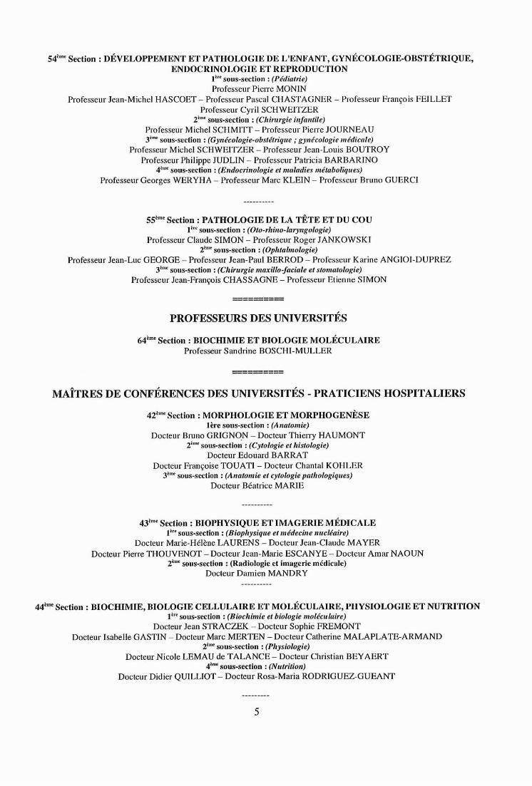
54'm. Seclion : DÉVELOPPEMENT ET PATHOLOGIE DE L'ENFANT, G YNÉCO LOGIE.OIlSTÉTR IQ UE ,ENDOCRINOLOGIE ET REPRODUCTION
t Ort sous-section : (Pédiatrie)
Professeur Pierre MüNINProfesseur Jean-Michel HASCOET - Professeur Pascal CHASTAGNER - Professeur Franço is FEILLET
Professeur Cyril SCHWEITZER2' ''~ sous-section : {Chirurgie inf an tile)
Professeur Michel SCHMITT - Professeur Pierre JOURNEAU) ""'" sous-sec tion : (GYll écolog;e-ob.~/élriqlle ; gynécologie m édi cal e)
Professeur Michel SCHWEITZER - Professeur Jean-Louis 1l0UTROYProfesseur Philippe JUD LJN - Professeur Patricia BARBARINO
4·....sous-section : (Endocrin ologie et maladies métaboliques)Professeur Georges WERYHA - Professeur Marc KLEIN - Professeur Bruno GUERCI
55'm. Section: PATHOLOGIE DE LA TÊTE ET DU COUt Ort sous-sect ion : iOt o-rhino-laryngologie)
Professeur Claude SIMON - Professeur Roger JANKOW SKI2·n
.. sous-sec tion : (Ophta lmo logie)Professeur Jean-Luc GEORGE - Professeur l ean-Paul BERROD - Professeur Karine ANGIOJ-DUPREZ
3'm. sous-sec tion : {Chirurgie maxil1o-faciale et stomatologi e)Professeur Jean-Fra nçois CHASSAGNE - Profes seur Etienne SIMON
= ======
PROFESSEURS DES UNIVERSITÉS
M 'm. Section : mOCHIMIE ET BIOLOGIE MOLÉCU LAIREProfesseur Sandrine BOSCHI-MULLER
= ======
MAÎTRES DE CONFÉRENCES DES UNIVERSITÉS - PRATICIENS HOSPITALIERS
42'm<Section: MORPHOLOGIE ET MORPHOGENÈS El ère sous-section : [Anatom ie)
Docteur Bruno GRIGNON - Docleur Thierry HAUM ONT2~m~ sous-sec tion : (Cytologie et hist ologie)
Docteur Edouard BARRAl'Ducteur FrançoiseTOUATI - Docteur Chantal KOHLER
3~mr sous-section : {Anatomie et cytologie path ologiques)Docteur Béatrice MAR IE
43'm. Section: IIIOPHYSIQUE ET IMAGERIE M ÉDICALEl K~ sous-section : (Biophys ique et m édecin e nucléaire)
Docteur Marie-Hélène LAURENS - Docteur Jea n-Cla ude MAYERDocteur Pierre THOUVENOT - Docteu r Jean-Marie ESCA NYE - Docteur Am ar NAOUN
2~"" sous-seclion : (Rad iologie et im ageri e médi cale)Docteur Dami en MANDRY
44'00. Section: mOCIIIMIE, BIOLOGIE CELLULAIRE ET MOLÉCULAIRE,PHYSIOLOGIE ET NUTRITIONt h e sous- section : (B ioch imie et biologie moléculaire}
Docteur Jean STRACZEK - Docteur Sophie FREMONTDocteur Isabelle GASTlN - Docteur Marc MERTEN - Docteur Catherine MALAPLATE·ARMAND
21"~ sous-section: (Physiologie)Docteur Nicole LEMAU de TALANCE - Docteur Christian BEYAERT
4~mr sous-sect ion : (Nutrition)Docteur Didier QUILLIOT - Doc teur Rusa-Maria RODR IGUE Z-GUEANT
5

4S" " Section : M IC ROIlIOLOGIE, MALADIES T RANSMISS IIILES ET HYGII<:NEl ~n sons-section : (Bact ériologie - Virolog ie ; hygiène hospitali ère)
Docteur Francine MORY - Docteur Véronique VENARD20n
.. scus-sectlon : (Parasito logie et mycologie)Docteur Nelly CONTET-AUDONNEAU - Madame Marie MACHOUART
46'"" Section: SANTÉ PUBLIQUE, ENV IRONNEMENT ET SOCIÉTÉl h. sous-section : (Epidémiologie, économie de la santé et pr évention)
Docteur Alexis HAUTEMAN IÈRE4"· sous-section : (B iostatistioues, informatique médicale el technologies de comm unication
DocteurPierre GILLOIS - Docteur Nicolas lA Y
47'm. Sect ion : CANCÉROLOGIE, GÉNÉTIQUE, HÉMATOLOGIE, IMM UNOLOG IEIon sous-section : (Hématologie ,. transfusion)
Docteur François SCHOONEMAN20n... sous-section: (Canc érologie ; radiothérapie ..cancérologie (type mixte: biologiqu e)
Docteur Lina BEZDETNAYA épouse BOLOTINE3"ru sous-section : {Immunologie)
Docteur Marcelo DE CARVALHO B1TTENCOURT4.... sous-sectlou : (Génétique)
Docteur Christophe PHILIPPE
4S' m, Sect ion: ANESTHEsiOLOGIE, RÉANIMATION, MÉDECINE D' URGENCE ,
PH ARMACOLOGIE ET THÉRAPEUTIQUEI h~ sous-section: {Anesth ésiologie el réanimation chirurg icale)
Docteur Gérard AUDIBERT3~n.. sous-section: (Pharmacologie fondamenta le ; pharmacologie clinique)
Docteur Françoise LAPICQU E - Docteur Marie -Jos é ROYER-MORROT - Docteur Nicolas G AMBIER4~rnt sous-section: (Thérapeutique ; médecine d 'urgence ; add ictologie
Dueteur Patrick ROSSIGNOL
so'm, Section : RHUMATOLOGIEl i'" sous-section : (Rhuma/ologie)Docteur Anne- Christine RAT
S4'm. Scclion : DÉVELOPPEMENT ET PATHOLOGIE DE L ' ENFANT , GYNÉCOLOGIE-OBSTÉTR IQUE,ENDOCRINOLOGIE ET REPRODUCTION
s·..... sous-secüon : (Biologie et médecine du développ ement et de ta reproduction)Docteur Jean -Lou is CORDONNIER
=======
MAÎTRES DE CONFÉRENCES
s'm, section : SCIENCE ÉCONOMIE GÉNÉRALEMonsieur Vincent LHUILLIER
32 tmr section : Chimie Organique, Minérale, Industrie lleMonsieur Franck DALlGAULT
40' m' section : SCIENCES DU MÉDICAMENTMonsieur Jean-François COLLIN
60'm' section: MÉCANIQUE, GÉNIE MÉCANIQUE ET GÉNIE CIVILEMonsieur Alain DURAND
6
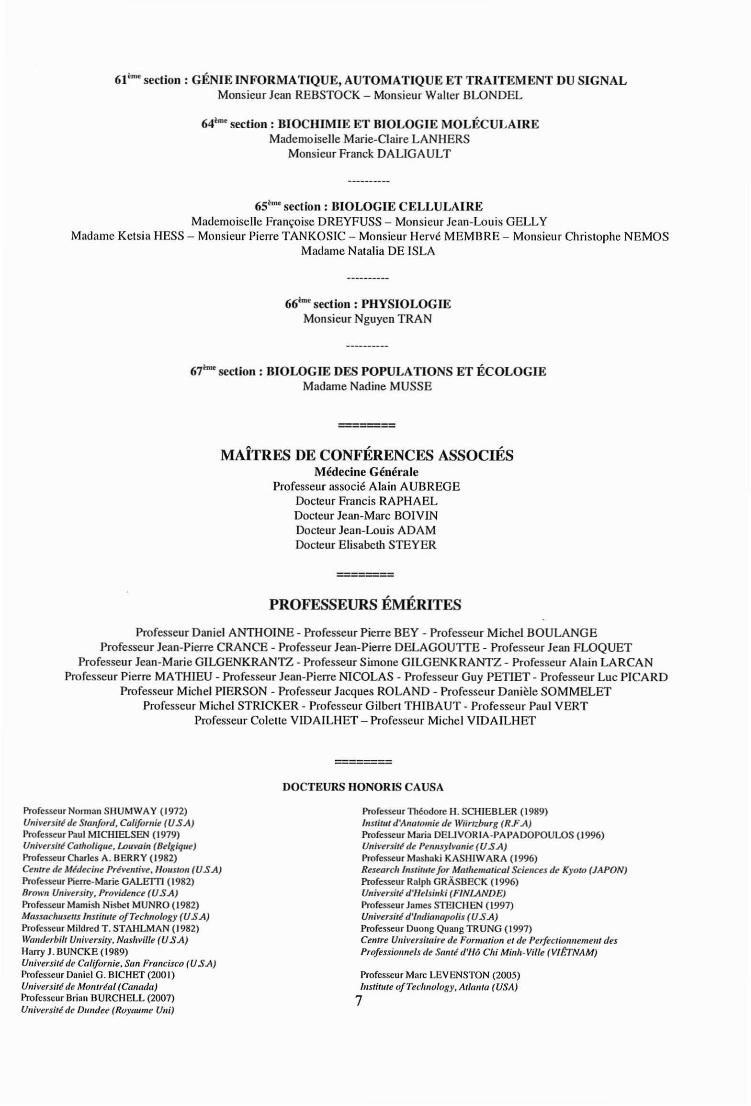
61'· ' section : GÉNIE INFORMATI Q UE, AUTOMATIQUE ET TRAIT~:MENT DU SIGNALMonsieur Jean REBSTOCK - Monsieur Waller BLONDEL
M!m. seclion : IlIOCIIIMIE ET BIOLOGIE MOL~;CULAIRE
Mademoiselle Marie-Claire LANHERSMonsieur Franck DALI GA ULT
6s'm. sec linn : IlIOLOGIE CELLULA IR EMademoiselle Françoise DREYFUSS - Mon sieur Jean-Louis GE LLY
Madame Kct sia HESS - Monsieur Pierre TANKOS IC - Monsieur Hervé MEMBRE - Monsieur Chris tophe NEMOSMadame Natalia DE ISLA
66.... section: PH YSIOLO G IEMonsieur Nguyen TRAN
67...• section : BIO LOG IE DES POPULATIONS ET ÉCOLOGIEMadame Nadine MUSSE
=====
MAÎTRES DE CONFÉRENCES ASSOCIÉSMédecine G énéral e
Professeur associé Alain AUBR EGEDocteur Francis RAPHA ELDocteur Jean-Marc BOIVINDocteur Jean-Loui s ADAMDocteur Elisabeth STE YER
======
PROFESSEURS ÉMÉ RITES
Professeur Daniel ANTHOINE - Professeur Pierre BEY - Professeur Michel BOULANGEProfesseur Jean-Pierre CRANCE - Professeur Jean-Pierre DELAGOUTIE - Professeur Jea n FLOQUET
Pro fesseur Jean-Marie GILGENKRANTZ - Professeur Simone GI LGENKRANTZ - Professeur Alain LAR CA NProfesseur Pierre MATH IEU - Professeur Jean-Pierre NICOLAS - Professeur Guy PETIET - Profe sseur Luc PICARD
Professeur Michel PIERSON - Professeur Jacques ROLAND - Professeu r Danièle SO MMELETProfesseur Michel STRI CKER - l' rofesseur Gilbert THIBAUT - Professeur Paul VERT
Professeur Colet te VIDA ILHET - Professeur Miche l VIDA ILHET
=====
DOCTEURS HONORIS CAUSA
Professeur Norman SHU MWA y (1972)Universite de Stanfo rd, Coli/omie (U SA'Professeur Paul MICHIELSEN (1979)Un;vn ûlé Catholique . Louvain (Belgique)Profe sseur Charles A. BERRY (1982)Centre dt' Jfldui" e Pr éventive , Hou smn (U SA)Professeur Pierre-Marie GALElTI ( 1982)BrowlI UniversiTy, Providence (USA )Profes.seur Mamish Nisbet MUNRO ( 1982)Mcusachuu tu lnstitute ofTechnology (USA)Professeur Mildred T. STAH LM AN (1982)1VlIIlllerbiit University, Nashviile (USA)Harry J . BUNCKE (1989)U'li l'usité de Californ ie, San Francisco (US A)Pro fesseur Daniel G. BIC flET C20UI)Universite de Montr éal (Cmllula)Professeur Brian BURCHELL (2007)Universi t ëde VI II/du (Roytlumt' Ulli}
Professeu r Théodore Il . SCH IEBLER ( 1989)Institut d 'Anatomi e dt' Wiirl, bu rg {RF A)Professeur Maria DELIVOR IA-PAPADOPOULOS (1996)Universtt ëde Pennsylvanie {US.A)Profe sseur Mashak i KASfllWARA (1996)Research Institute for Mathemalical Sc ienc es de K)'OIo {JAPON}Professeur Ralph GRA SBE CK ( 1996)Universite d 'Helsinki {FINLANDE}Professeur James STE ICHEN ( 1997)Univusilé d 'Indianapolis (US A)Professeu r Duo ng Quang TRUNG ( 1997)Centre Universitaire (le Forma tion et de Perfectionnement desProf essionnels (le Sanlé d'lM Ch i Minh -Ville (VlIhNA M)
Professeur Marc LEVENSTON (2005)lns tinue of TedlII% gy ,Arltmta (USA)
7

A notre maître et président de thèse,
Monsieur le Professeur Alain Blum,
Professeur de Radiologie et Imagerie médicale,
Vous nous avez fait l'honneur de nous guider pour cc travail avecenthousiasme et une grande disponibilité.
Votre esprit d 'analyse scientifique et votre dynamisme nous ontimpressionnés et motivés.
L' étendue de vos connaissances est pour nous une source d'admiration etune inspiration dans notre pratique médical e.
Que ce travail soit le témoignage de notre gratitude et de notre profondrespect.
8

A notre maître et juge,
Monsieur le Professeur Henry Coudane,
Professeur de Médecine Légale (Option Clinique),
Doyen de la Faculté de Médecine,
Chevalier dans l'Ordre national de la Légion d'honneur,
Chevalier dans l'Ordre des Palmes Académiques
Nous vous remercions de l'honneur que vous nous faites en acceptant dejuger ce travail et de l'intérêt que vous lui portez.
Veuillez trouver ici l'expression de notre reconnaissance et de notreprofond respect.
9

A notre maître et juge,
Monsieur le Professeu r Francis Guillemin,
Professeur d'Epidémiologie, Economie de la Santé et Prévention
Nous vous remercions de l'honneur que vous nous faites en acceptant dejuger cc travail ct espérons qu'il saura retenir votre intérêt.
Soyez assuré de notre gratitude ct de notre profond respect.
JO
A notre maître et ju ge,
Monsieur le Docteur Alain Peutot,
Vous nous avez fait 1'honneur de vous intéresser à nos travaux ct de venirles juger.
Nous avons eu la chance de bénéficier de votre expérience en imagerieostéo-articulaire au sein de votre service .
Veuillez trouver ici l' expression de notre profonde gratitude.
11

A mes parents,
Votre amour, votre soutien inconditionnel et votre bienveillance m'onttoujours portée. Merci pour tous vos sacrifices et votre infinie gentillesse.
A la mémoire de mes grands-parents Georges et Liselotte et de mongrand-père Jean
A Julie, ma cousine adorée
A ma marraine Bernadette et ma tante Nicole, pour leur affection etleur soutien
A Marylène, ma bouffée d'oxygène
A Julie, pour ton amitié infaillible et si précieuse
A Justine, à notre complicité toujours intacte
A Domitille, un seul regard et tout est dit. . .
A Maeva, mon meilleur souvenir d'anesthésie, pour ton soutien acharné
A Assia, pour ton amitié sincère
A Thomas et Nikias, inimitables et indispensables
A tous mes amis de Nancy, de Strasbourg et d'ailleurs
A l'ensemble du personnel des services de radiologie du CHU de Nancy,de Metz Bonsecours et du Centre Alexis Vautrin.
12

13
A Jonathan, mon amour

71u momea:oftre adinire d e_fj!rcer Iâ !!IédêCli/{!, je promets etje.lure oftrefidè/é aur
lOir dê f'/ÎOllllellr et dê Iâprooité. !MOII pre!!liersouasem dê rétaofir, dêprése/1!er 011 dê
proll/ollvoirIâsalltédanstoussesélèll/m/s, pliysitjlleset II/mtaur; indwidûe!s et SOClol4
.Je respecterai toutes lés persolllles, /éllr autonomie et /éllr volOllté, SOllS aucun«
discnilllilatûJIl selOll /éllr état 011 /élll:r comscttons: .J'tilte/1!lelldraipour lés protéger si
ellés sont q!Jôiofies, vulÎlérao/és ou II/ellacées dâ,lS /éllrtiltégnté ollléllr tÎtlJllIté. !Mêll/e
SOIIS Iâ commmte: .le Ile firai pas IIsa/le dê II/es connaissances comn: lés lOir dê
ftilllnalllté. .J'it!fômœrailéspatients dês MciriollS f!Ilvisa/lées, dê /éllrsmtsonset dê /élll:r
cOllSétjllettces. .Je Ile troll/perOljoll/air létlrco'!ftance et Il~lOlteraipns /épouooïrnénté
dês arronstances pOlirfircer lés consaences: .Je dâllllerai II/es soms d !'tiltÎtlJettt et d
tjllicolltjlle II/e lés dêlllalldêra. .Je Ile II/e fitùseraipas li!fli/{!Ilcerpar fit soydû/laill 011 fit
recnercne dê fit /llOire.
./ldiniredâlls !'titttilllté oespe!J'olllles,.le tairai léssecrets tjlli II/e sont co'!ftés. 'llfflle d
!'tiltétiellrdês!!IairollS,.le respecterailéssecretsdêsfiyers et Ilia COllliiRte Ile sensm pas d
CO/TO!!lpre ks Inœllt:r. .Jefirni tou:pOlirsOIlIâ/lerks sOI#ances. .Je IleprolOllIJernipas
aôllsWell/mt lés a/lollles. .JeIleprov0'1"eTOljoll/air fit morz MfiOérétnettt.
.Je préserverai !'tilMl!mdâllce lIécessaire d fl1cco!!lp/isse!!lellt dê ma IIlirsioll. .Je
Ill?ntreprmdrai nell i"i Mpnsse mes cOlI/pétellces. .Je lés elltretielldrai et lés
peftctiolllleraipollrassurerail !!Iiel/.-( léssenece» tjlli Ille seron: dêlllallMs.
.Jl1pporteraill/on aidêd II/es co'!ftères OIilSitjllii /é1l!J'fill/illés dânsf'ndVerstté.
Q!le lés nOlllllles et II/es co'!fTères 1I/1iCCOrdêllt létlr esttille stje stlir fidè/é d II/es
promesses,tjlle.le soirMsnollorée et !!Iéptirée stf)' Inall'1"e~
14

TA BLE DES MATIERES
In t roduction •••..••.........••.•........ .....••.••••.......••.....•••••••••••.........•.••.•.......•.•...••.........•••.•••. 16
1. Etude technique •..............................••••••••••••••••••••.••••...•••.....••••...•.•..•.......••............• 17
A. Mat ériel et méth odes 17
1. Fantôme et acquisition 17
2. Reproduction et analyse 17
3. Fonction de transfert de modulation 19
R. R ésul ta ts 20
II. Etnde clinique•..•....•..•...... .••....... .•......•••...................•••.•......... •.... ..•.••...•.••••.....•••••••22
A. Ma tériels et méth odes 22
1. Population 22
2. Technique 22
3. Lecture 23
4. Statistiques 23
B. R ésulta ts 24
C. Discussion 26
C oncl us ion 30
Bibliographie 3 1
ANN EX E 34
A r ticle en a ngla is ... .....••..••..•••.... ..•.•••••••••••.••.••.•....•••••••••.........••...•.••...••••••••................ 35

Introduction
Nous ass istons depu is plu sieurs années en radiologie à l'utilisat ion de supports de
communication de plu s en plus variés (fi lm, CD, papier) là où il y a peu de temps
encore le film éta it le seul outil disponible. L' avènement de la lecture des examens sur
écran a dû bousculer quelques habitudes tenaces et apporter la preuve de ses qualités,
notamment en tenne de post-traitement de l' image, pour être finalemen t largement
acceptée.
Par ailleurs, des impérati fs économiques évidents ont stimulé l'utilisat ion très large
des CD et du papier comme support des examens d 'HW et de scanner pour la
communication avec les pratic iens. Ce tte pratiqu e d ésormai s courante a été adoptée
sans difficultés, avant même la réalisation d' études clini ques, finalement peu
nom bre uses [1-3]. En revanche, concernant la radiographie stan dard, l'utilisation du
papier comme support de communication se heurte <i une franc he résistance de la part
des cliniciens, d éj à hostiles à la radiographie numérique [4-6]. Le but de notre travail
a donc été d ' étud ier objectivement la valeur d iagnostique de 3 di fférents supports
couramment utili sés, le film, le papier et le PACS (Picture Archiving and
Com munication System), dans un contexte très spécia lisé: les rad iogra phies du
poignet pour traumatisme.
Notre étude se décompose en 2 parties :
une étude techni que sur fantôme
une étude clinique sur les bi lans radiographiques de 200 patient s
ayant subi un traum atism e du poignet
16

I. Etude technique
Nous avons dans un premier temps réalisé une étude sur un fantôme permettant
d'analyser les performances en termes de contraste et de résolution spatiale d'i mages
éditées sur PACS, film et papier.
A. Matériel et méthodes
1. Fantôme et acquisition
Nous avons utilisé la plaque de test Digi 13 (Figure 1), destinée initialement aux
contrôles de qualité et de constance des systèmes équipés de récepteurs digitaux tels
que les plaques ERLM ( Ecran Radiolumincscent à Mémoire) ou les capteurs plans.
L'acquisition a été réalisée sur un ERLM AGFA avec une unité de numérisation de
type ADC Compact et un post-traitement des images par l' intermédiaire de
paramètres MUSICA. L'image a été centrée afin de ne prendre en considération que
les trois des cinq critères d'évaluation pertinents pour notre étude :1'échelle
d' absorptiométrie de 7 niveaux en cuivre, la mire d'objets pour le faible contraste ct la
mire de résolution.
2. Reproduction et analyse
La radiographie ainsi obtenue (Figure 2) a été éditée selon 3 types dc reprographies :
17

• sur le PACS avec la version 5.3 d'Impax et des stations de travail AGFA bi-
écran ayant une résolution de 1,3 K
• sur film laser en form at 20X25 par l' interm édiaire d 'u ne imprimante Drystar
2000
• sur papier brillant de format A4 avec une qualité de 160g. Deux copies ont été
réalisées sur deux impr imantes différentes:
une imprimante Xerox Workcent re M24 avec une réso lution de
600X600 ppp (imprimante 1)
une imprimante Xerox Phaser 7760GX avec une résolution de
1200X 1200 ppp (imprimante 2).
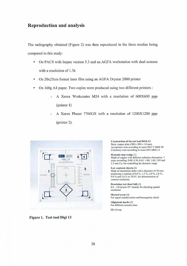
• C onst ruction du IlI GI -13Plaque de base Cil cuivre (300 x 300 x 1.0 mm )Tests d 'ncccptance scion la norme DIN V 6868·58Tcsts de constance scion la lionne DIN 6868 · 13
1:1r'- .0:- mJD ~~
~ DIGI -fJ
...."""/p~~
d;-25·1· Y
O ·· 2O ,O'~0 ..·0 ..·0 ..·
• Echelle de co ntraste dynamiq ue ( 1)En cuivre. avec d ifféren ts absorbe urs derayon nemen t, 7 échelo ns en Cu: 0.00 111111, 0.30mm, 0.30 JU m, 1.00 mm, 1.40 nuu, 1.85 mm cl2.3 mm pour cont rôle r la gamme dynamique.
• O bjets à fai b le co ntra ste (2)En disques d'al umin ium . avec un diam ètre de10 mm, produisant un contraste de 0.8 %. 1.2 %.2.0 % , 2.8 %. 4.0 % ct 5.6 % à 70 kV, pour ladétermination dl: la résolution en contraste.
• Test de résoluti on (3)Mire linéaire orientée il 45°, 0.6 · 5.0 pl/mm
• Zo ne de marqu age (4)Pour le contrôle du signal et de l'h omogénéité
• Marqu eurs d ' ali gn em ent (5)Pour différentes d imensions de cassette
IRA Group
Figure 1. Mire-test Digi 1
18
2
1 : 7 carrés, contraste élevé
2 : 6 disques, faible contraste
3 : mire de résolut ion spatialede 0,6 à 5,0 pl/mm
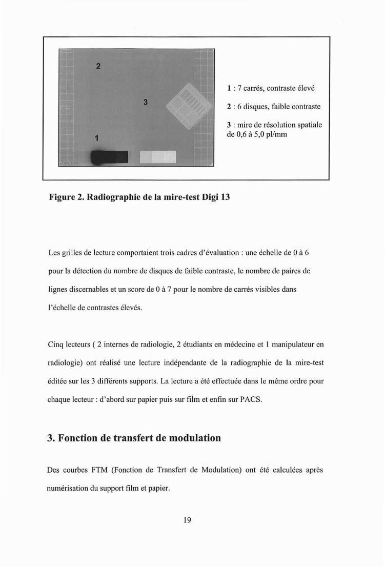
Figure 2. Radiographie de la mire-test Digi 13
Les grilles de lecture comportaient trois cadres d'évaluation : une échelle de 0 à 6
pour la détection du nombre de disques de faible contraste, le nombre de paires de
lignes discernables et un score de 0 à 7 pour le nombre de carrés visib les dans
l'échelle de contrastes élevés.
Cinq lecteurs ( 2 internes de radiologie, 2 étudiants en médecine et 1 manipulateur en
radiologie) ont réalisé une lecture indépendante de la radiographie de la mire-test
éditée sur les 3 différents supports. La lecture a été effectuée dans le même ordre pour
chaque lecteur : d'abord sur papier puis sur film et enfin sur PACS.
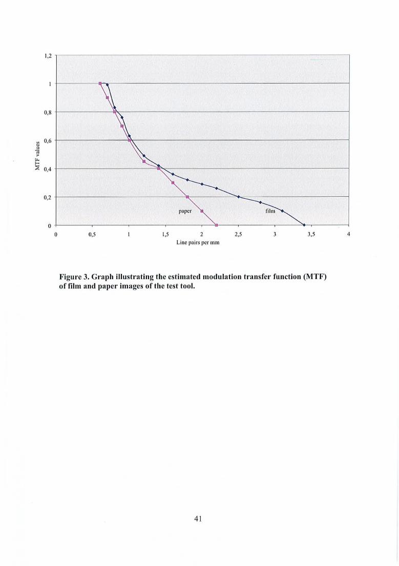
3. Fonction de transfert de modulation
Des courbes FTM (Fonction de Transfert de Modulation) ont été calculées après
numérisation du support film et papier.
19

B. Résultats
Comme l'expose le tableau des résultats (tableau 1), le PACS est supérieur au papier
ct au film pour la résolution en faibles contrastes avec un score moyen de 6/6, le film
étant lui-même supérieur au papier avec un score moyen de 4/6 contre 3,4/6 . Ce
classement est le même pour la performance en résolution spatiale des 3 supports: le
PACS arrive en premier avec une moyenne de 3,52 pl/m m, puis le film avec une
moyenne de 3,22 pl/mm et le papier avec une moyenne de 2, 12 pl/mm. Enfin, il n' y a
pas de différence entre les 3 supports dans la détection des différences élevées de
contraste. Les résultats de cette étude montrent donc une infériorité du papier comme
moyen de reprographie avec une perte non négligeable de sensibilité en terme de
résolution spatiale ct de résolution en faible contraste. L' étude des courbes FTM est
en accord avec cette analyse (figure 3). Notons par ai lleurs qu ' il n' existe aucune
différence entre les deux copies papier issues de deux imprimantes différentes.
Ta bleau I. Résultats de la lecture de la mire-test par cinq lecteurs,trois paramètres d ' étude.
Disques fa ible contraste Paires de ligne s/mm Carrés con traste,élevé
PACS FilmP. ier PACS Film
l'a lierI·PACS Film
Pa lierIl 12 Il 12 Il 12
Lecte ur 1 6 4 4 4 3,7 3,4 2,2 2,2 7 7 7 7
Lecte ur 2 6 4 3 3 3,4 3,1 2,2 2,2 7 7 7 7
Lecteur 3 6 4 4 4 3,4 3,1 2,2 2,2 7 7 7 7
Ledeur4 6 4 3 3 3,7 3, 1 2,0 2,0 7 7 7 7
Lecteur 5 6 4 3 3 3,4 3,4 2,0 2,0 7 7 7 7
Moyenne 6/6 4/6 3,4/6 3,4/6 3,52 3,22 2,12 2,12 7 7 7 7
Il = imprimante J - 12 = imprimante 2
20

1.:
li.' t------- -lh-- - - - - - - - - - - - - - ----------
;; 11./.
-a,
.:!-=:
•••.J
",/
' l . ~
" I----r----~--__--_~_-~----~~,~--~" 0.5 ':.5
Figure 3. Graphique illu strant la Fonction de Transfert deModulation des radiographies dc la mire-test éditées SUl' un SUPPOl'tfilm ct papier.
21

II. Etude clinique
A. Matériels et méthodes
1. Population
Nous avons mené une étude sur 200 patients (82 hommes, 118 femmes, âge moyen
48,3, échelle d'âge 16-95 ans) consécutifs ayant consulté aux urgences entre mars ct
août 2007 pour un traumatisme du poignet. Ces patients ont bénéficié, après un
examen clinique réalisé par un médecin urgentiste, d'un bilan radiograp hique
comportant 4 à 6 incidences du poignet. Lorsque cela était réalisable, en particu lier cn
fonction des douleurs, les 4 incidences systématiques étaient face, profil , berge ulnaire
(incidence en scmi-supination) ct poing serré (serrement actif du poing) selon les
recommandations habituelles [7-10), plus ou moins deux incidences du scaphoïde en
fonction de la suspicion clinique.
2. Technique
L'acquisition des images a été réalisée sur un ERLM AGFA avec une unité de
numérisation de type AOC Compact permettant un post-traitement des images par
l'intermédiaire de paramètres MUSICA, ces paramètres n' étant pas sauvegardés dans
le PACS. Les examens ont été édités selon 3 types de reprographies, à l'instar de
notre étude clinique:
22

•
•
•
sur le PACS avee la version 5.3 d 'Impax et des stations de travail AGFA bi
éeran aya nt une résolution de 1,3 K
sur film laser en format 20X25 par l'in term édiaire d'une impriman te Drystar
2000
sur papier de format A4 avee une qualité de l60g, par une imprimante Xerox
Workcentre M24 ayant une résolut ion de 600X600 ppp .
L'étud e sur fantôme n'ayant pas montré de différenee en fonction du typc
d ' imprimante utili sée, nous n' avons réalisé qu'une seule copie papier de chaque
examen, sur une seule et même imprimante.
3. Lecture
La lecture a été réalisée par 2 lecteurs indépendants en une seule séance par modalit é,
chaque séance étant espacée d 'au moins une semaine. Le lecteur 1 était représenté par
un radiologue expérimenté en imagerie ostéo-articulaire et le lecteur 2 par uri interne
en radiologie aya nt 18 mois d 'expérience en radiologie dont 6 mois en imagerie
ost éo-articulaire, Six catégories de réponses étaient proposées : fracture du radius,
fracture du scaphoïdc, autre fracture, entorse, luxation et RAS s' il n'y avait auc une
lésion. Les associat ions de réponses éta ient possibles.
4. Statistiques
L'étude statistique s 'est attachée à étudier d 'une part la reproductibilité inter
observateur pour les différentes méthodes et d 'autre part la reproductibilité inter
méthode par le calcul du coefficient Kappa [II J.
23
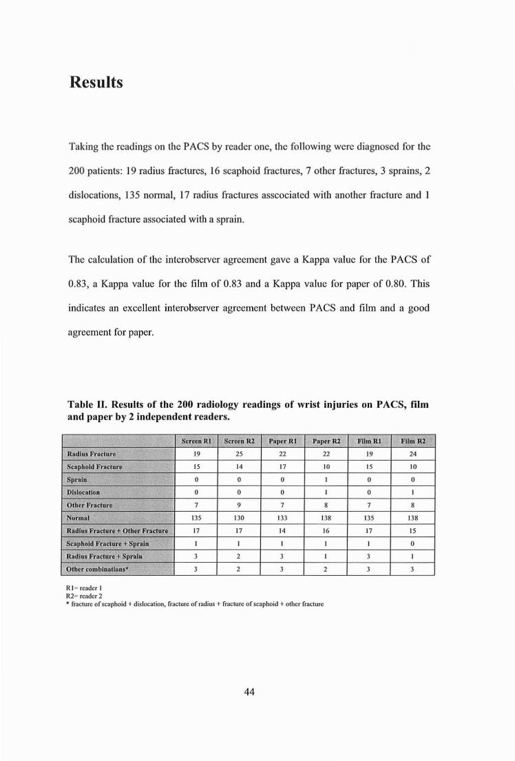
B. Résultats
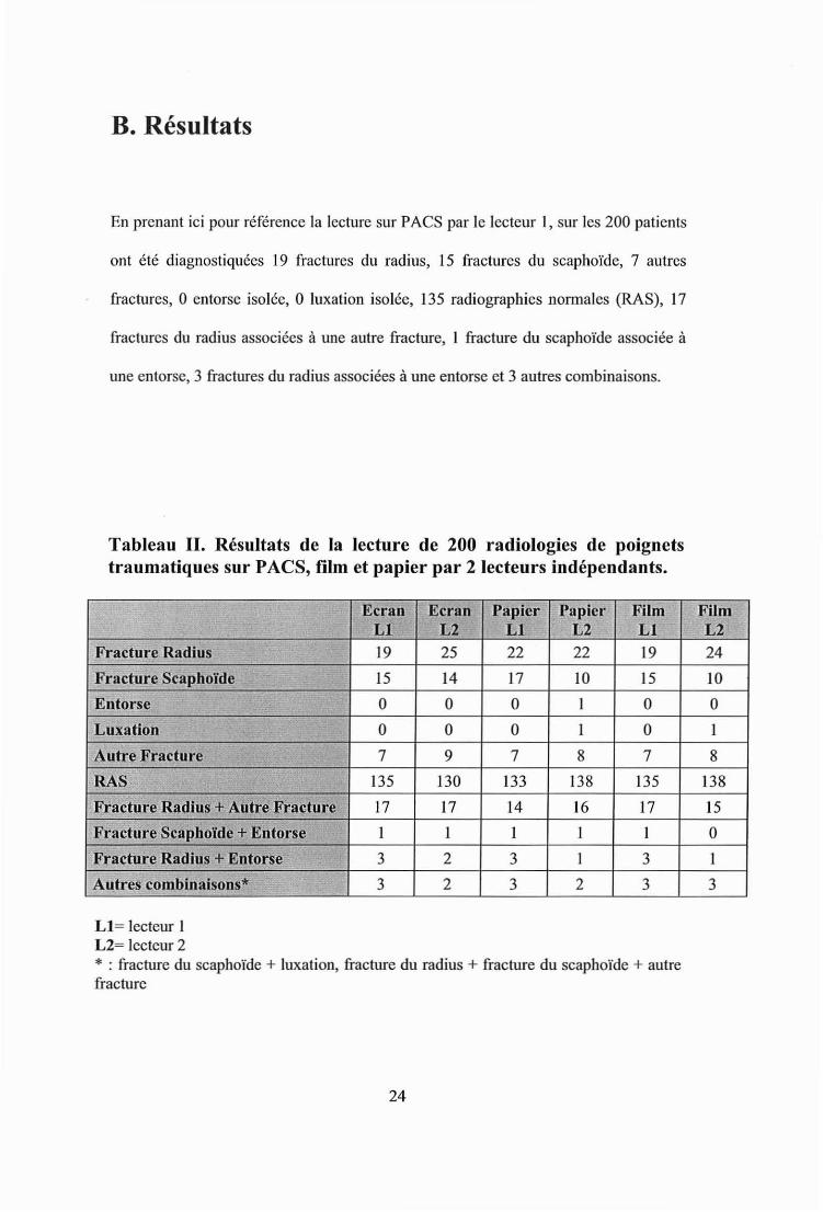
En prenant ici pour r éfére nce la Iccturc sur PACS par Ic Icctcur l , sur Ics 200 patients
ont été diagnostiqu ées 19 fractures du radius, 15 fractures du scaphoïdc, 7 autres
fractures, 0 entorse isolée, 0 luxation isolée, 135 radiographies normalcs (RAS), 17
fractures du radius associ ées à une autre fracture, 1 fracture du scaphoïdc associ ée à
une entorse, 3 fractures du radius assoc iées à une entorse ct 3 au tres com binaisons.
Tableau Il. Résultats de la lecture de 200 radiologies de poignetstraumatiques sur PACS, film et papier par 2 lecteurs indépendants.
Ecran Ecran Papier Papier Film FilmLI L2 LI L2 LI L2
Fracture Radius 19 25 22 22 19 24
FracHire Scaphoïde 15 14 17 JO 15 JO
Entorse 0 0 0 1 0 0
Luxation 0 0 0 1 0 1
Autre Fracture 7 9 7 8 7 8
RAS 135 130 133 138 135 138
Fracture Radius + Autre Fracture 17 17 14 16 17 15
Fracture Scaphoïde + Entorse 1 1 1 1 1 0
Fracture 'Radius + Entorse 3 2 3 1 3 1
Autres combinaisons* 3 2 3 2 3 3
LI = Icctcur 1L2= lecteur 2* : fracture du scaphoïde + luxation, fracture du radius + fracture du scaphoïde + autrefracture
24

Le calcul de la reproductibilité inter-observateur donne un Kappa pour le PACS égal à
0,83, un Kappa pour le film éga l à 0,83 ct un Kapp a pour le papier éga l à 0,80 cec i
faisant la démonstration d 'u ne concordance inter-observateur excellente (PA CS, film)
ou bonne (pap ier).
Le calcul de le reproductibilité inter-méthode donne un Kappa fihnlPACS éga l à 0,99
pour LI ct 0,90 pour L2, un Kappa fihnlpapier égal à 0,89 pour LI et 0,86 pour L2 ct
un Kappa PACS/papi er égal à 0,88 pour LI ct 0,84 pour L2. Ainsi la concordance
inter-méthode est exce llente (tableau IV), quelles que soient les méthodes comparées.
Tableau III. Reproductibilité inter-méthode.
Lecteur 1 Lecteur 2
Kappa film/PACS 0,99 0,90•... ..
"Kappa film/Papier 0,89 0,86
Kappa PACS/papier 0,88 0,84
T ableau IV. Degr é de reproductibilité et valeur de Kappa.
Reproductibilité
Excellente
Bonne
Moyenne
Mauvaise
Très mauvaise
Kappa
> 0 ,8 1
0,80 - 0,61
0,60 - 0 ,21
0,20 - 0 ,0
< 0,0
25
c. Discussion
Il est actuellement largement admis que l'utilisation du PACS comporte de nombreux
avantages comparativement à la lecture traditionnelle sur film comme par exemple
une meilleure accessibilité aux images ct aux compte-rendus, une meilleure gestion
des examens avec moins de pertes d'examens, d'examens non interprétés et une
réduction des coûts [12-14]. Le radiologue dispose également d'outils
supplémentaires (comme le fenêtrage, le zoom) qui facilitent notamment l' étude des
radiographies thoraciques (petits pneumothorax, syndrome interstitiel , micronodulcs)
[15-17] mais ne semblent pas apporter un plus comparativement au film pour la
détection des lésions traumatiques du poignet ct en particulier pour les fractures du
scaphoïde [1 8]. De plus, de nombreu ses études ont montré l' absence de différence
significative de sensibilité diagnostique entre la lecture sur console PACS ct sur film
d' examens radiographiques réalisés aux urgences [19-22]. En revanche, peu d'études
ont comparé le support d'impression papier aux autres modalités [23-25] alors que
celui-ci est de plus en plus utilisé, notamment en scanner ct en IRM.
Les résultats de l' étude que nous avons réalisée sur la mire-test confirment la
perception générale des praticiens d'un e dégradation de la qualité en résolution ct en
contraste de l'impression papier comparativement au PACS ct au film. Cette
différence significative sur la radiographie d'un e mire-test aux caractéristiques
contrôlées ct standardisées ne sc vérifie pas dans la situation clinique réelle que nous
avons étudiée, soit les traumatismes du poignet. En effet , notre étude montre que la
26
nature du support n'a pas d'incidence significative dans la détection des lésions
traumatiques sur des radiographies du poignet, la concordance inter-méthode étant
excellente. Le eorolaire de ces résultats est la substitution théoriquement possible de
ces supports entre eux sans perte de sensibilité diagnostique statistiquement
significative. Néanmoins, la perte de qualité du papier sur la radiographie de la mire
test comparativement aux autres supports ne permet pas d' envisager une substitution
par le papier pour le diagnostic. Les études cherchant à évaluer la qualité des images
radiologiques imprimées sur papier sont peu nombreuses et nous ne pouvons bien sûr
pas élargir les conclusions de notre étude à tous les champs d'activité de la radiologie
standard (thorax, ASP, rachis.. .).
Cette évaluation est dictée par des impératifs financiers qui demandent une gestion
raisonnée des budgets dans tous les domaines d'activité médicale. Cette attitude n'est
bien sûr acceptable qu'à la condition qu'elle n'ait pas de conséquence délétère sur la
prise en charge du patient. L'intention d'utiliser le support papier ne s'envisage que
dans la mesure où les services sont dotés d'un système de lecture sur écran des
examens radiographiques. Prenons l'exemple du fonctionnement du service de
radiologie de l'a ccueil des urgences dans notre institution: pour tout bilan
radiologique, le diagnostic est fait sur écran dans le service de radiologie ou aux
urgences. Une fois que le dossier clinique, biologique ct radiologique est complet et
en fonction du diagnostic : soit le patient est hospitalisé dans un autre service du CHU
où les examens seront également disponibles en ligne sur le réseau interne de
l'hôpital, soit le patient est transféré dans un autre établissement qui n' est pas en
réseau avec le PACS du CHU, soit le patient est sortant. Actuellement, pour tous les
27
patients, chaque examen radiographique est systématiquement édité sur film laser
puisque au moment où le bilan est réalisé, la destination du patient n'est pas encore
connue. Une solution serait de n'imprimer les films que pour les patients qui ne seront
pas hospitalisés au CHU. On peut imaginer alors certains problèmes d'organisation si
les films ne devaient être édités qu' au cas par cas à la demande des cliniciens avant la
sortie du patient, avec forcément des retards ct des oublis. Une autre attitude
économiquement acceptable serait de continuer l'impression systématique de tous les
examens réalisés aux urgences non plus sur film laser mais sur papier, cc support
n'étant pas à visée diagnostique. Il servirait à compléter le dossier du patient destiné
au médecin généraliste ou spécialiste, étayé du courrier de sortie précisant les résultats
des examens et les différentes anomalies cliniques, biologiques et radiographiques
trouvées lors de la consultation aux urgences. Par contre cc support peut être
insuffisant dans certaines situations, par exemple pour un chirurgien orthopédique qui
n' aurait pas accès au réseau PACS du service de radiologie des urgences, dans les cas
d' une hospitalisation hors du CHU d 'origine. Dans cc cas, la gravure d'un CD-Rom
peut être envisagée .
L'objet de l' étude étant représenté par l' examen de radiographies du poignet
traumatique, il est important de garder à l' esprit la sensibilité diagnostique
insatisfaisante de la radiographie standard dans la détection de certaines lésions,
notamment les fractures du scaphoïde [7-10]. Notre étude n'a pas pour but de remettre
en question les stratégies diagnostiques des lésions traumatiques du poignet [26-29]
mais de tenter d' apporter une base de réflexion scientifique à l'enthousiasme des uns
28
et au scepticisme des autres devant une éventuelle utilisation du support papier dans la
transmission des examens de radiographie conventionnelle aux cliniciens.
Cette transmission des images radiographiques sur papier se heurte actuellement à une
forte réticence des cliniciens . Or cette exigence des correspond ants externes ne
semble pas j ustifiée pour de nombreux examens, comme le montre par exemple notre
étude . Evidemment ces propos sont à nuancer en fonction du type d 'examen et du
mode de fonctionnement interne de chaque structure hospitaliè re, mais un
établissement doté d'un PACS avee un bon système de communication entre les
services de radiol ogie et les autres services devrait pouvoir se passer du film et le
remplacer par le papier avec une réduction substantielle des coûts [30-31].
Notre étude comporte un certain nombre de limites à commencer par l'absenc e
d'examen de r éférence (scanner ou IRM par exemple). L'existence d'lm go ld standard
nous aurait en effet permi s d' élargir le champ de l' étude à la sensibilité ct la
spécifici té diagnostique de chaqu e support notamment pour les fractures du scaphoïde
[32]. De plus, il aurait été intéressant d' ajouter un critère d ' évaluati on de la qualité de
l'image selon les observateurs au moment de la lectu re afin d'avoir la corrélation
entre la qualité subjective d'un support ressentie par le lecteur et les résultats
finalement obtenus. Soulignons par ailleurs l'imprécision de la catégorie diagnostique
' autre fracture' qui incluait aussi bien les fractures de l'ulna, les fractures des os du
carpe autre que le scaphoïdc ainsi que les fractures dc la main. La derni ère limit e à
souligner est l'absence d'anonymi sation des examens sur les différents supports, le
29

nom ct l'âge du patient étant toujours visibles. Ccci pouvait d'une part induire un
biais de mémorisation, cependant limité par la lecture dans un ordre aléatoire des 200
dossiers, cn une seule séance par modalité, chaque s éance de lecture étant espaeéc
d'une semaine minimum. D'autre part, notons unc possible influence dc l' âge sur lc
diagnostic évoqué par le lecteur, la prévalence des différentes catégor ies
diagnostiques n'étant pas la même chez l' adult e jeune ou chez le vieillard, cc biais
ayant finalement peu d'importance dans cette étude puisque présent sur les trois types
de support.
Conclusion
Notre étude a donc démontré une excellente reprodu ctibilité inter-méthode entre la
lecture sur film, écran et papier du diagnostic des lésions post-traum atiques du
poignet, permettant d ' envisager la substitution du film par le pap ier pour la
communi cation de l' infonnation. L'étude sur la mire-test confirme néanmoins
l'in fériorité du papier dans la résolution en faibles contrastes, excluant une utilisation
à visée diag nostique.
30

Bibliographie
[1] Reiner BI, Siegel EL, Ilooper FJ. Aeeuraey of interpre tation of CT sea ns:eomparing PACS monitor displays and hard-copy images. AJR Am JRoentgenoI 2002;179(6): 1407-1O.
[2] Beard DY, Hemminger BM, Perry JR , ct al. Interpretation of CT st udies:sing lc-scrccn wo rkstation vers us film alterna tor. Radiology 1993;187(2):5659.
[3] Kundel HL, Polansky M, Dalink a MK, et al. Reliability of soft-copy versushard-copy interpretat ion of emergeney department radiograph s: a pro totypestudy . AJR Am J RoentgenoI 2001 ;177(3) :525-8.
[4] Wade FA, Oliver CW, McBride K. Digital imaging in traum a and orthopaediesurgery: is it worth it? J Bone Joint Surg Br 2000;82(6):79 1-4.
[5] Wilson AJ, Hodge JC. Digiti zed radiographs in skele tal traum a: aperformance eomparison between a digital workstati on and the or iginal filmimages. Radiology 1995;196(2): 565-8.
[6] Wegryn SA, Piraino DW, Richmond BJ , ct al. Comparison of digital andconventional museuloskcletal rad iography: an observer performance study.Radiology 1990; 175(1):225-8 .
[7] Schernberg F. Roentgenographi c examination of the wrist: a sys tematic study .of the normal, lax and injured wri st. Part 2: Stress views. J Hand Surg Br1990;15(2) :220-8.
[8] Blum A, Saucr B, Dctrcill c R, ct a l. The diagnosis of recent seaph oidfractures: revie w of the literature. J RadioI 200 7;88(5 Pt 2):74 1-59.
[9] Temple CL, Ross DC, Bennett JD, Garvin GJ, King GJ, Fabcr KJ.Com parison of sagittal eomputed tomography and plain film radiograph y in aseaphoid frac ture model. J Hand Surg Am 2005;30(3):534-42 .
[10] Demondion X, Boutry N, Khalil C, Colle n A. Plain radiographs of the wristand hand . J Radi oI 200 8;89(5 Pt 2):640-5 1; quiz 52-3.
[II] Roberts C. Modell ing patterns of agreeme nt for nomin al sea les. Stat Med2008;27 (6) :8 10-30.
[12] Siege l EL, Diaeonis JN, Pomerantz S, Allman R, Briscoe B. Making filml essradiology work. J Digit Imaging 1995;8(4):151-5.
[13] Langlotz CP, Even-Shoshan 0, Seshadri SS, et al. A methodology for theeconomie assessment of pieture archiving and communication systems. J DigitJmaging 1995;8(2):95-102.
3 1

[14] Pizcr SM, Beard DY. Medi cal imagc work stations : functions andimplementation, J Digit Imaging 1989;2(4):185-93.
[15] Elam EA, Rchm K, Hillman BJ, Maloney K, Fajardo LL, McNcill K.Efficacy of digital radiography for thc detection of pneumothorax: comparisonwith convcntional ches t radiography. AJR Am J Roentgcnol 1992;158(3):50914.
[16] Thactc FL, Fuhrma n CR, Oliver JH , ct al. Digital radiography andconvcntional imaging of the chcst: a comparison of obse rver performance.AJR Am J Roentgenol 1994;162(3):575-81.
[17] Stcekcl RJ, Batra P, Johnson S, ct al. Comparison of hard- and soft-copydigital ehcst ima ges with different matr ix sizcs for managing coronary eareunit patients. AJR Am J Roentgcnol 1995; 164(4) :837-41.
[18] Khaliq W, Blakeley CJ, Maheshwaran S, Hashemi K, Redman P. Comparisonof a PACS Workstation with Laser Hard Copies for Detecting ScaphoidFraeturcs in the Emergeney Departm ent. J Digit hnaging 2008.
[19] Kundel ilL, Gefter W, Aronchiek J, et al. Accuracy of bcdside chest hardcopy screen-film versus hard- and soft-co py computed radiographs in amcdieal inten sive care unit: receiver operating charaeteristic analysis.Radiology 1997;205(3):859-63.
[20] Weatherbum G, Bryan S, Nicholas A, Coeks R. The effect of a picturearchiving and communications sys tem (PACS) on diagnostic performance inthc acc ident and emergeney department. J Accid Em crg Med 2000; 17(3) :1804.
[21] Ucda K, Iwasaki S, Nagasawa M, ct al. Hard-copy versus soft-copy imagereadin g for detc etion of ureteral stones on abdomina l radiography. Radiat Med2003;21(5):210-3.
[22] Youm ans DC, Don S, lI ildebolt C, Shacke1ford GD, Luker GD, McAl isterWH. Skeletal surveys for child abuse: comparison of interpretation usingdigitized images and sereen-film radi ograph s. AJR Am J Roentgenol1998;171(5) :1415-9.
[23] Ibbott GS, Zhang Y, Mohiuddin M, Ad am s E. Reproduction of radiologieimages on plain paper. Radiographies 1998;18(3):755-60 .
[24] Denslow S. Desktop publi shing and medical imaging: papcr as hardcopymed ium for digital images. J Digit Imaging 1994;7(3) :140-5.
[25] Lyttkens K, Kirkh om T, Kehler M, et al. Eva luation of the image quality ofink-jet printcd paper copies of digital chest radiog raphs as comparcd with film:a receiver operat ing charaeteristic study. J Digit Ima ging 1994;7(2):61-8 .
32

[26] Mack MG, Keim S, Balzer JO , ct al. Clinical impact of MRI in acute wris tfractures. Eur RadioI 2003;13(3) :612-7.
[27] Karantanas A, Dailiana Z, Malizos K. The ro le of MR imaging in scaphoiddisorders. Eur Radio l 2007; 17( Il ):2860-7 1.
[28] Kaewlai R, Avery LL, Asrani AV , Abujudc h 1111, Sackuoff R, Novelline RA.Multidetcctor CT of carpa l injuri es: anatomy, fractures, and frac turedislocations. Radiograph ies 2008;28(6):1771- 84.
[29] Peer R, Lanser A, Giacomuzzi SM, et al. Storage phosphor radi ograph y ofwr ist fractures: a subjective comparison of image qualit y at varyi ng exposurelevels. Eur Radiol 2002; 12(6) :1354-9.
[30] Hilscnrath PE, Smith WL, Berbaum KS, Franken EA, Owen DA. Analysis ofthe cost-effectiveness of PACS. AJR Am J Roentgcnol 1991;156(1): 177-80 .
[31] Singer ME, Appl egate KE. Cos t-effectiveness analysis in radiology.Radiology 200 1;2 19(3) :61 1-20.
[32] Remplik P, Stabler A, Merl T, Roemer F, Bohndorf K. Diagnosis of acutefrac tures of the extremities: comparison of low-ficld MRI and conventionalradiography. Eur Radiol 2004; 14(4):625-30.
33

ANNEXE
34
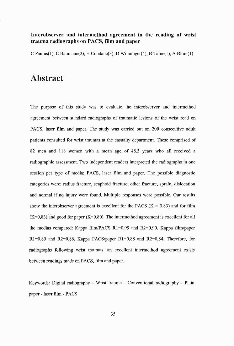
Interobserver and intermethod agreement in the reading of wristtrauma radiographs on PACS, film and paper
C Paulus( 1), C Baumann(2), Il Coudane(3), D Winninger(4), B Taine(l), A Blum(l)
Abstract
The purpose of this study was to evaluate the interobserver and intermethod
agreement between standard radiographs of traumatie lesions of the wrist read on
PACS, laser film and paper. The study was earried out on 200 consecutive adult
patients eonsulted for wrist traumas at the easualty department. These eomprised of
82 men and 118 women with a mean age of 48.3 years who ail reeeived a
radiographie assessment. Two independent readers interpreted the radiographs in one
sess ion per type of media : PACS, laser film and paper. The possible diagnostic
categories were : radius fracture, seaphoid fracture , other fracture, spra in, dislocation
and normal if no injury wcrc found . Multiple responses wcrc possible. Our results
show the interobserver agreement is exce llent for the PACS (K = 0,83) and for film
(K=0,83)"ànd good for paper (K=0,80). The intennethod agreement is excellent for ail
the medias eompared: Kappa filmfPACS Rl =0,99 and R2=0,90, Kappa filmfpaper
R l=0,89 and R2=0,86, Kappa PACS/paper R l =0,88 and R2=0,84. Therefore , for
radiographs following wrist traumas, an exce llent intermethod agreement exis ts
between readings made on PACS, film and paper.
Keywords: Digital radiography - Wrist traum a - Conventiona l radiography - Plain
paper - laser film - PACS
35

Introduction
The medias available in radiology are becoming more and more varied (film , CO,
paper) where previously laser film was the only media available for the
communication of radiologieal images. The introduction of digital radiography has
revolutionised communication betwcen radiologists and c1inicians. This emergence of
readings on electronic displays has forced a change in working methods and has
silenced its initial critics due to its many advantages, notably in terms of image post
processmg so that today the reading of radiological images on screen is largcly
acceptcd .
Obvious economie factors have stimulated the increasing use of CD and paper as
media for the communication of MRI and CT Scan examinations with practitioners.
These widespread changes in practice have been implemented without difficulty and
bcfore a large number of clinical studies were even completed [1-3]. On the other
hand, in conventional radiography the use of paper media suffers strong resistance
from c1inicians, already hostile towards digital radiography [4-6]. The aim of this
study is therefore to look objectively at the diagnostic value of three commonly used
media types, film, paper and PACS (Picture Archiving and Communication System)
in a very specifie context: the radiographs of wrist traumas .
36
Materials and method
The study is fonned of two parts:
A preliminary study using a phantom test tool
A clinieal study of 200 wrist trauma radiographs
A. Technical evaluation
An initial study was carricd out using a phan tom test tool. This allowed an analysis of
the performance in tenns of eontrast and spatial resolution of images viewcd on the
PACS, film and paper.
Phantom and acquisition
The phantom used was a Digi 13 test tool (Figure 1), normally used for quality control
tests and calibration of projection radiography systems with digital image receptors.
The image acquisition was realised using an AGFA DLR with an ADC compact type
digitizer and MUSICA advaneed image proeessing. These paramctcrs were not save d
in the PACS. The image was centrcd to considcr thrce criteria: the dynamic step
wcdgc of 7 levels of copper, low contrast objec ts and the resolution test.
37
Reproduction and analysis
The radiograph y obtained (Figure 2) was then reproduced lt1 the three medias being
compared in this study:
• On PACS with Impa x version 5.3 and an AGFA workstation with dual sc reens
with a reso lution of l.3 k
• On 20x2 5cm format laser film us ing an AGFA Drystar 2000 print er
• On 1GOg A4 paper. Two copies were produ ced using two di fferent printers :
A Xerox Workcentre M24 with a resoiution of GOOXGOO ppp
(printer 1)
A Xerox Phaser 77GOGX with a resolution of 1200X1200 ppp
(printer 2).
1..- "'''' "..~ _.....-
Figure 1. Test too l Digi 13
0 ", 2O ,·O ' ~O·"0 ",0 ",
38
Co ns tr uc tion of 111(' lest 1001 DIG I-13Basic co pper pla te (300 x 300 x 1.0 nuu)Accc ptnnce tests nccording ta norm D1:-J V 6868-58Constaucy tests nccording 10 nonn DI;,,! 68 68- 13
Dynamlc sicp we dg c ( 1)Mode ofcopper w ith diffe ren t radiation ab sorption. 7stcps nscending: 0 .00. 0.30 . 0.6 5. 1.00. 1.40. 1.85 and2.3 1Il111 Cu. fo r controll ing the dynamic range
l .ow con t rast obj ccts (2)Made of aluminium disk s wit h a diarnctc r of 10 mm .produciug il co ntras! oro.s %. 1.2 %, 2.0 %, 2.8 %.4.0 % and 5.6 % al 70 kY, for determination ofcon trast resolution
Reso lution test (lcad foil) (3)0 .6 - 5.0 lp/mm 45(> rotated , for chccking spa tialresolution
Ma rked arc as (4)For sig na l stan da rizntion and hom ogcnit y ch eck
Align mcnllllarks (5)Fo r d ifferent casse tte sites
IBA Gm up

2
1: 7 squares, contrast ascending
32: 6 disks, law cantrast
3 : Spatial resalut ian test of 0,6 ta 5,0 Ip/mm
Figure 2. Radiography of the test tool Digi 13
The table of readings compri sed of three types of evaluation: a scale of 0 to 6 for the
detection of the number of dises of low contrast, the numb er of line pairs visible and a
score of 0 to 7 for the number of squares visib le on the sca le of high contrast.
Five readers cach made indcpendent interpretations of the test radiographs on the
three different medias. These were carried out in the same order by each reader: tirst
on paper, then film and tina lly on the PACS.
MTF measurements
MTF (Modu lation Transfer Funct ion) curves were calculated after digit izing the film
and paper (paper 2) studies (Figure 3).
39
Results
As can be scen from the table of results (Table 1) the PACS proved to be superior to
paper and film for reso lutions in low contrast with an average score of 6/6, the film
was itself superior to paper with an average score of 4/6 against 3.4/6 . This order of
ranking is the same for the three medias in their performance in spatial resolution: the
PACS tirst with a mean value of 3.52 Ip/mm, the film with a mean value of 3.22
pl/mm and then paper with a mean value of 2.12 Ip/mm . Finally, no difference was
found in the performance of the three medi as in the detection of high contras t
difference. The results of this study show paper to be inferior as a reprographi e media
with lower spatial and low contrast reso lution. Observing that no difference was
identitied between the two paper copies produced by the two different printers. MTF
measurements are in agreement with the previous analysis (Figure 3).
Table I. Results of the test tool readings by live readers on the three med ia types.
,Squares of asttndj~g'c:onfrast M: :Lon: eont rast disks Pairs of lines/mm
PACS FilmPa er"
~(;CS Film Pa erPACS Film
Pa er1" PI Pl . Pl Pl 1 . P l P2, .
Rt~'dtr 1 6 4 4 4 3,7 3.4 2,2 2,2 7 7 7 7
Reader 2 6 4 3 3 3,4 3.1 2,2 2.2 7 7 7 7
Reader 3 6 4 4 4 3,4 3,1 2,2 2,2 7 7 7 7
Rt llder4 6 4 3 3 3,7 3, 1 2,0 2,0 7 7 7 7
ReadtrS 6 4 3 3 3,4 3,4 2,0 2.0 7 7 7 7
Anrage 6/6 4/6 3,4/6 3,4/6 J,5 2 3,22 2,12 2,12 7 7 7 7
Pl = printer 1P2 = printcr 2
40
1,2
0,8 t------~__----- - - - - - --- - - - ---- - - - __1
'. 0,6 t------------l~----------------------__1u
"~u,
~ +- - - - - - - - - - - ""'110;,-- - - - - - - - - - - - - - - - - - - - ---10,4
lihnpapcr
0,2 +--------------~Il___-----':_....=----------_____j
43,532,52Lille pairs pcr mm
1,50,5
o +----~---~---~---~---~----~-----'-~-------j
o
Figure 3. G ra ph iIIustratiug th e estima ted modulati on tran sfcr fnnet ion (MTF)of film and pnper images of the test tool.
41

B. Clinical study
Population
We included 200 adult patients (82 men, 118 women, mean age 48.3, from 16-95
years). The patients were eonsulted eonseeutively at the emergeney department during
a 6 month period for wrist traumas. After c1inieal examination by praetitioners in the
easualty these patients reeeived a radiographie assessment including four to six
images of the wrist. When it was possible, taking into aecount the pain experienced,
the four images obtaincd systcmatically were postero-anterior view, lateral view,
semi-supinated oblique VICW and clcnchcd-fist stress view following the usual
guidelines [7-10], then two scaphoid view s (ulnar deviation and wrist extension)
depending upon the suspected diagnosis.
Method
The images were acquired usmg an ERLM AG FA with an ADC compact type
digiti zer allowing post processing of the images through Musica parameters. These
parameters wcrc not saved in the PACS. The examinations were edited using thrce
types of media :
• On PACS with Impax version 5.3 and an AGFA workstation with dual screcns
with a resolution of 1.3k
42

•
• On 20x25cm format laser film using a Drystar 2000 printcr
On 160g A4 papcr using a Xcrox Workccntrc M24 with a resolution of
600X600 ppp
As thc study using a phantom did not idcntify any differences betwccn thc two types
of printcr uscd, only a single paper copy was produccd for cach cxamination using the
samc printer (printer l, Xerox Workcentrc M24) .
Reading
The rcadings werc made by two independent rcaders who intcrprctcd the 200 patients'
radiographs conseentively in one session for each type of media. Thcrc was a period
of at least one week betwccn cach session. Reader number one was a radiologist
experienc ed in musculoskeletal disordcrs and the rcadcr number two was a junior
doctor with eighteen months experience in radiology including six months in
musculoskeletal imaging. Six categories of responsc wcrc possible: radius fracture,
scapho id fracture, other fracture, sprain, dislocation and normal if no injury were
found. Multiple rcsponscs wcre possible.
Statistics
The statistical study was carried out to find the interobserver agreement for the
different methods and the intermethod agreement by calculating the Cohen's Kappa
coefficient [11].
43
Results
Taking the readings on the PACS by readcr one, the following wcrc diagnosed for the
200 patients: 19 radius fractures, 16 scaphoid fractures, 7 other fractures, 3 sprains, 2
dislocations, 135 normal, 17 radius fractures asscociated with another fracture and
scaphoid fracture associated with a sprain.
The calculation of the interobservcr agreement gave a Kappa value for the PACS of
0.83, a Kappa value for the film of 0.83 and a Kappa value for paper of 0.80. This
indicatcs an excellent interobserver agreement between PACS and film and a good
agreement for papcr.
Table II. Resulls of the 200 radiology read ings of wrist injuries on PACS, filmand paper by 2 independent readers.
, Scrce n RI Screen RI Papcr RI Pepe e RZ Film RI Film R2
Radius Fracture 19 25 22 22 19 24
Scaphold Fracture 15 14 17 10 15 10
Spraln 0 0 0 1 0 0
Dislocation 0 0 0 1 0 1
Oth er Fracture 7 9 7 8 7 8
No rmal 135 130 133 138 135 138
Radius Fracture + Other Fracture 17 17 14 16 17 15
Sea phefd Fraeture +Spral n 1 1 1 1 1 0
Radius Fracture + Spratn 3 2 3 1 3 1
O'h ('T ro mbi nai Ion s" .. 3 2 3 2 3 3
RI: rcadcr 1R2: rcadcr 2• fracture of scaphoid .. dislocation, fracture of radius " fracture of scaphoid .. e ther fracture
44

The ealculati on of the intermethod agreemen t gave a Kappa value for film/PACS of
0.99 for RI and 0.90 for R2, a Kappa value film/paper of 0.89 for R I and 0.86 for R2
and a Kappa value PACS/paper of 0.88 for RI and 0.84 for R2. Therefore the
intcrmethod agreemen t is excellent for ail the methods eompared.
Table III. Intermethod agreement.
,', " , "", Rellder 1 l' gO! '~Reaaêr 2 ".,'Kappa fihnfPACS , 0,99 0,90'" 3... . ,'~'
0,89 0,86Kappa film/Paper
Kappa'PACS/paper " 0,88 0,84
Discussion
Today it is widely recognised that the use of PACS brings a number of advantages
when compared to the traditional reading of radiographs on film. For example, easier
image and report aeeess, improved management of examinations with fewer being
lost or un-interpretable and also cost reductions [12-14] . Thcre is also an inereased
range of tools availab le to the radiologist, such as windowing or zoom, which
facilitate most notably the study of thoracic radiographs (small pneumothorax,
alvcolar-intcrstiticl syndrome, mieronodules) [15-17] but do not appear to bring
advantages over film for the deteetion of wrist injuries and in particular scaphoid
fractures [18). Many studies show thore is little difference in the diagnostic accuracy
between interpreting on the PACS or on film radiographs made in the casualty
department [19-22]. On the other hand, few studies have compared printcd paper
45

media to the other medias available [23-25] despite this seeing inereased use, most
notably for CT Seans and MRI.
The results of this study show that the lest 1001 eon firms the practitioncrs' general
pereep lion that there is a reduetion in the resolution quality and in contrast of printed
paper media eompared with PACS and film ( Table 1, Figure 3) . This signi fieant
differenee on the radiography of a lest tool, whieh is eontrolled and standardised, does
not verify the real clinical situation that has been studied in the case of wrist injuries.
Aetually, the sludy shows that the type of media uscd for radiographs of the wrist
does not have a signifieant effeel upon the deteetion of injur ies, with the intermcthod
agreement being excellent. The eorollary of these results is the possibility to
interehange these medias without a signifieant loss in statistieal aeeuraey. However,
the redueed quality of paper, eompared to the other medias, as demonstrated by the
test tool shows that paper should not be used for diagnostic purposes. There arc very
few studies evaluating the quality of radiographieal images printed on paper, thcrcfore
wc arc unable to expand the conclusions of this study to other fields of eonventional
radiogra phy (ehest, abdomen, spine, etc).
This evaluation is dietated by finaneial eonstraints, whieh demand the reasonable
management of budgets in ail areas of medieal aetivity. This approae h is only
acceptable if it does not have a degenerating effeet on patient eare. The idea of using
paper media eould only be envisaged where systems for viewing radiographie images
on sereen arc available. Taking the example of the easualty department radiology
service in our institution where the diagnosis of eaeh radiology examination is made
on sereen . Once the clinical, biologieal and radiologieal examinations are complete
46
the patients eourse depends upon their diagnosis; either the patient is hospitalised
within another department of the University Hospital where their examinations are
available on line via the hospitals internaI network; or the patient is transferred to
another hospital which is not connected to the network with the PACS of the
University Hospital; or the patient is discharged. Currently every patient' s
radiologieal examination is systematieally edited on laser film at the time that the
exam is conducted, due to the destination of the patient at this moment being
unknown. One solution would be to only print the films for those patients who will
not be hospitalised at the University Hospital. In this instance it is possible to imagine
this would raise organisational problems if films should only be printed on a ease-by
ease basis, if demanded by the c1inician before the patient leaves. Here there is likely
to be a strong ehance of delays and missing films. Another economically acceptable
procedure would be to continue the systematic printing of ail examinations made by
the casualty department but to use paper instead of film. This wouId serve to keep
patients files complete for sending to their general practitioners or specialist, whilst
they would be accompanicd by preeise details in the patients report explaining the
examination results and the different clinical, biological and radiographie problems
found following their consultation. In certain eases this media may be unsatisfaetory,
an example bcing for an orthopacdic surgeon who does not have access to the PACS
network of the casua lties radiology department due to being outside the Universi ty
Hospital. In this instance the writing of a CD-Rom could be a praetical solution.
The purpose of the study is reprcsented by the examination of radiographs of wrist
injuries. Il is important to consider the unsatisfaetory accuracy of eonventional
radiography for the diagnosis of eertain lesions, notably scaphoid fraetures [7-10].
47
This study does not set out to question the diagnostic strategies applied for traumatie
lesions of the wrist [26-29] but attempts to bring a basis of seientifie refleetion to the
enthusiasm of some and the seeptieism of others concerning an eventual inerease in
the use of paper media in the communication of eonventional radiography
examinati ons to clinicians.
There is eurrently a strong reluetance among clinieians towards the use of paper
radiography images. This reluetance seems unjustified for many examinations as
demonstrated by this study. Clearly this depends on the type of examination, the
internai structure of eaeh hospital and how it functions . Nevertheless, a hospital using
PACS with a good communication system between radiology and the other
departments should be able to replace film by papcr thus bringing substantial eost
reduetions [30, 3 1].
This study includes a certain number of limits, the first being the absence of a
referenee examination (CT Scan or MRI for example): havin g a 'gold standard'
examination would have allowed the field of study to be enlarged and shown the
diagnosis sensitivity ana speeifieity of eaeh media, espeeially for seaphoid fractures
[32]. In addition it would have been interesting to add an evaluation criteria for the
quality of the images aeeording to the observer at the time of reading in order to
understand the correlation between the subjective quality of a media and the final
results obtained. Il should also be noted the imprecision of the 'othcr fracture'
diagnosis catcgory whieh includcs fractures of the ulna, earpai bone fractures as weil
as scaphoid and hand fractures. The final limit to highlight is the absence of
anonymity in the examinations in their various medias. The names and age of the
48
patients were always visible . This eould lead to a memori sation bias allhou gh the
effeet should be limited due to 200 examinations being read in a single sitting per
media and the reading of dilTerent medias being separa ted by at least one wee k. ln
another way the diagnosis of the reader may be inlluenccd by the patient's age with
the prevalence of certain diagnostic categories not being the same in young adulls as
in the aged. This bias ultimately changes little within this study because it is present in
all three types of media.
ln conclusion this study has demonstrated there is an excellen t intermethod agreement
betwee n readings on film, screen and paper for the diagnosis of wrist traumas. Thus,
plain paper radiograph printing could reasonably be used for the communicatio n of
radiologieal images to practitioners provided it is not to be used for diagnos tic
purposes.
(1 ) Service d'Imagerie Guilloz, CHU de Nancy, Hôpital Central, 29 Avenue du Maréchal deLattre de Tassigny, 54035 Nancy
(2) Service d 'In formatique médicale, Epidémiologie, Statistiques, Hôpital Marin, CHU de Nancy,54037 Nancy
(3) Service de Chirurgie Arthroseopique, Traumatologique cl Orthopédique de l'appareillocomoteur, CHU de Nancy, Hôpital Central, 29 Avenue du Maréchal de Lattre de Tassigny,54035 Nancy
49

References
[1] Rein er BI, Siegel EL, Hooper FJ. Aeeuraey of interpretat ion of CT sea ns:eomparing PACS mon itor displays and hard -eopy images. AJR Am JRoentgenol 2002; 179(6) :1407-1O.
[2] Beard DV, Hemminger BM, Perry JR, ct al. Interpretation of CT studies:single-sereen workstation versus film alterna tor. Radiology 1993;187(2):5659.
[3] Kundel HL, Polan sky M, Dalinka MK , ct al. Reliability of soft-eopy versushard-eopy interpretation of emergeney department radiographs : a prototypestudy. AJR Am J Roentgenol 200 1;177(3):525-8.
[4] Wad e FA, Oliver CW, MeBride K. Digital imaging in trauma and orthopa ediesurgery : is it worth it? J Bone Joint Surg Br 2000;82(6):79 1-4.
[5] Wil son AJ, Hodge JC. Digitized radiographs in ske leta l traum a: aperformance eomparison between a digital workstation and the original filmimages. Radiology 1995;196(2) :565-8.
[6] Wegryn SA , Piraino DW, Richmond BJ, ct a l. Comparison of digital andeonventi onal museuloskeletal radiography: an observ er performance study .Radiology 1990;175(1):22 5-8.
[7] Sehemberg F. Roent geno graphi e examination of the wri st: a sys tematie studyof the normal, lax and injured wrist. Part 2: Stress v iews. J Hand Surg Br1990; 15(2) :220-8.
[8] Blum A, Sauer B, Detreille R, et al. [The diagnosis of reeent scaphoidfractures : review of the literature]. J Radi oI 2007;88(5 Pt 2):74 1-59 .
[9] Temple CL, Ross DC, Benn ett JD, Garvi n GJ , King GJ, Faber KJ.Compariso n of sagittal computed tom ography and plain film rad iography in ascapho id fracture model. J Hand Surg Am 2005;30(3):534-42 .
[10] Demondi on X, Boutry N, Khalil C, Co tten A. [Plain radiographs of the wristand hand] . J RadioI2008;89(5 Pt 2):640-5 1; qu iz 52-3.
[II] Roberts C. Modelling pattern s of agreement for nominal sea les . Stat Med2008;27(6):810-30.
[12] Siegel EL, Diaeon is JN, Pomerantz S, Allman R, Briseoe B. Making filmlessrad iology work . J Digit Imaging 1995 ;8(4) :151-5.
[13] Langlotz CP, Even-Shoshan 0, Seshadri SS, et al. A methodology for theeeonomie assessment of pieture arehiving and communication systems. J DigitImaging 1995;8(2) :95-102.
50

[14] Pizcr SM, Beard DV. Medical image work stations: functions andimplementation. J Digit Imaging 1989;2(4):185-93.
[15] Elam EA, Rchm K, lIillman BJ, Maloncy K, Fajardo LL, McNcill K.Efficacy of digital radiography for the detection of pneumothorax: comparisonwith convcntional chcst radiography. AJR Am J Roentgenol 1992;158(3):50914.
[16] Thacte FL, Fuhrman CR, Oliver JH, et al. Digital radiography andconventional imaging of the chest: a comparison of observer performance.AJR Am J Rocntgcnol 1994;162(3):575-81 .
[17] Stcckel RJ, Batra P, Johnson S, et al. Comparison of hard- and soft-copydigital chest images with different matrix sizes for managing coronary carcunit patients. AJR Am J Roentgenol 1995;164(4) :837-41.
[18] Khaliq W, Blakeley CJ, Mahcshwaran S, Hashemi K, Redman P. Comparisonof a PACS Workstation with Laser Hard Copies for Detecting ScaphoidFractures in the Emergency Department. J Digit Imaging 2008 .
[19] Kundcl HL, Gcftcr W, Aronchick J, ct al. Accuracy of bcdsidc chcst hardcopy scrccn-film versus hard- and soft-copy computed radiographs in amedical intensive care unit: rcceiver operating characteristic analysis.Radiology 1997;205(3):859-63.
[20] Wcathcrburn G, Bryan S, Nicho las A, Cocks R. The cffcct of a picturcarchiving and communications system (PACS) on diagnostic performance inthc accident and emergency dcpartment. J Accid Emerg Med 2000;17(3):1804.
[21] Ueda K, Iwasaki S, Nagasawa M, et al. Hard-copy versus soft-copy imagereading for detection ofureteral stones on abdominal radiography. Radiat Mcd2003;21 (5):210-3.
[22] Youmans DC, Don S, Hildcbolt C, Shackelford GD, Luker GD, McAlisterWH. Skeletal survcys for chiId abuse: comparison of interpretation usingdigitized images and screcn-film radiographs. AJR Am J Roentgcnol1998;171(5):1415-9.
[23] Ibbott GS, Zhang Y, Mohiuddin M, Adams E. Reproduction of radiologieimages on plain paper. Radiographies 1998;18(3):755-60.
[24] Denslow S. Desktop publishing and medical imaging: papcr as hardcopymedium for digital images. J Digit Imaging 1994;7(3): 140-5.
[25] Lyttkens K, Kirkhorn T, Kchlcr M, ct al. Evaluation of the image quality ofink-jet printed paper copies of digital chest radiographs as cornparcd with film:a rcccivcr operating characteristic study . J Digit Imaging 1994;7(2) :61-8.
51

[26] Mack MG, Kcim S, Balzcr JO, et al. Clinical impact of MRl in acute wristfractures. Eur Radiol 2003;13(3):6 12-7.
[27] Karantanas A, Dailiana Z, Malizos K. The role of MR imaging in scaphoiddisordcrs. Eur Radiol 2007;17( Il ):2860-71.
[28] Kaewlai R, Avery LL, Asrani AV, Abujudeh HH, Sacknoff R, Novelline RA.Multideteetor CT of carpal injuries: anatomy, fractures, and fraeturedislocations. Radiographies 2008;28(6):1771-84.
[29] Peer R, Lanser A, Giaeomuzzi SM, et al. Storage phosphor radiography ofwrist fractures: a subjective eomparison of image quality at varying exposurelevels. Eur Radiol 2002; 12(6):1354-9.
[30] Hilsenrath PE, Smith WL, Berbaum KS, Franken EA, Owen DA. Analysis ofthe eost-effeetiveness of PACS. AJR Am J Roentgenol 1991; 156(1):177-80.
[31] Singer ME, Applegate KE. Cost-effeetiveness analysis in radiology.Radiology 2001;219(3):611-20.
[32] Remplik P, Stabler A, Merl T, Roemer F, Bohndorf K. Diagnosis of aeutefractures of the extremities: eomparison of low-field MRI and eonventionalradiography. Eur Radiol 2004; 14(4):625-30.
52
vu
NANCY, le 27 juillet 2009
Le Président de Thèse
Professeur A. BLUM
NANCY, le 30 juillet 2009
Le Doyen de la Faculté de Médecine
Par délégation
Mme le Professeur M.C. BÉNÉ
AUTORISE À SOUTENIR ET À IMPRIMER LA THÈSE
NANCY,24 août 2009
LE PRÉSIDENT DE L'UNIVERSITÉ DE NANCY 1Par délégation
Madame C. CAPDEVILLE-ATKINSON
53

RESUME
Il s'agissait d'étudier la reproductibilité inter-observateur et inter-méthode du diagnostic deslésions post-traumatiques du poignet en radiographie standard sur PACS, film laser et papier.Cette étude incluait 200 patients consécutifs (82 hommes, 118 femmes) de 48,3 ans d'âge moyenayant bénéficié aux urgences d'un bilan radiographique pour traumatisme du poignet. La lecturea été réalisée par 2 lecteurs indépendants en une seule séance par modalité de lecture : PACS,film laser et papier. Les catégories diagnostiques retenues étaient: fracture du radius, fracture duscaphoïde, autre fracture, entorse, luxation, RAS, les associations de réponses étant possibles. Lareproductibilité inter-observateur est excellente pour le PACS (K = 0,83) et le film (K=0,83) etbonne pour le papier (K=0,80). La reproductibilité inter-méthode est excellente quels que soientles supports comparés: Kappa film/PACS L1=0,99 et L2=0,90, Kappa film/papier L1=0,89 etL2=0,86 , Kappa PACS/papier LI =0,88 et L2=0,84En conclusion, pour les radiographies post-traumatiques du poignet, il existe une excellentereproductibilité inter-méthode entre la lecture sur PACS, film et papier.
TITRE EN ANGLAIS: Interobserver and intermethod agreement in the reading of wristtrauma radiographs on PACS, film and paper
ABSTRACT
The purpose of this study was to evaluate the interobserver and intermethod agreement betweenstandard radiographs of traumatic lesions of the wrist read on PACS, laser film and paper. Thestudy was carried out on 200 consecutive adult patients consulted for wrist traumas at thecasualty department. These comprised of 82 men and 118 women with a mean age of 48.3 yearswho ail received a radiographie assessment. Two independent readers interpreted the radiographsin one session per type of media: PACS , laser film and paper. The possible diagnostic categorieswere: radius fracture, scaphoid fracture, other fracture, sprain, dislocation and normal if no injurywere found. Multiple responses were possible. Our results show the interobserver agreement isexcellent for the PACS (K = 0,83) and for film (K=0,83) and good for paper (K=0,80). Theintermethod agreement is excellent for ail the medias compared: Kappa film/PACS RI=0,99 andR2=0,90, Kappa film/paper RI =0,89 and R2=0,86, Kappa PACS/paper RI =0,88 and R2=0,84 .Therefore. for radiographs following wrist traumas, an excellent intermethod agreement existsbetween readings made on PACS, film and paper.
THESE : MEDECINE SPECIALISEE - ANNEE 2009
MOTS-CLEFS: Radiographie numérique - Traumatismes du poignet - Radiographie standard Film - Papier - PACS
INTITULE ET ADRESSE DE L'UFR:
Faculté de Médecine de Nancy9, Avenue de la Forêt de Haye
54505 VANDOEUVRE LES NANCY Cedex