Embed Size (px)
Citation preview
Extended subnanosecond structural dynamicsof myoglobin revealed by Laue crystallographyDominique Bourgeois*†‡, Beatrice Vallone‡§, Alessandro Arcovito§, Giuliano Sciara§, Friedrich Schotte¶,Philip A. Anfinrud¶, and Maurizio Brunori§�
*Institut de Biologie Structurale, Unite Mixte de Recherche 5075, Centre National de la Recherche Scientifique�Commissariat a l’EnergieAtomique�Universite Joseph Fourier, 41 Rue Jules Horowitz, 38027 Grenoble Cedex 1, France; †European Synchrotron Radiation Facility, 6 Rue JulesHorowitz, B.P. 220, 38043 Grenoble Cedex, France; §Dipartimento di Scienze Biochimiche and Istituto Pasteur-Fondazione Cenci Bolognetti, Universita diRoma ‘‘La Sapienza,’’ Piazzale A. Moro 5, 00185 Rome, Italy; and ¶Laboratory of Chemical Physics, National Institute of Diabetes and Digestive andKidney Diseases, National Institutes of Health, Bethesda, MD 20892-0520
Edited by Alan R. Fersht, University of Cambridge, Cambridge, United Kingdom, and approved January 31, 2006 (received for review October 11, 2005)
Work carried out over the last 30 years unveiled the role ofstructural dynamics in controlling protein function. Cavity net-works modulate structural dynamics trajectories and are function-ally relevant; in globins they have been assigned a role in ligandmigration and docking. These findings raised renewed interest fortime-resolved structural investigations of myoglobin (Mb), a sim-ple heme protein displaying a photosensitive iron-ligand bond.Photodissociation of MbCO generates a nonequilibrium popula-tion of protein structures relaxing over a time range extendingfrom picoseconds to milliseconds. This process triggers ligandmigration to matrix cavities with clear-cut effects on the rate andyield of geminate rebinding. Here, we report subnanosecondtime-resolved Laue diffraction data on the triple mutant YQR-Mb[Leu-29(B10)Tyr, His-64(E7)Gln, Thr-67(E10)Arg] that depict thesequence of structural events associated with heme and proteinrelaxation from 100 ps to 316 ns and above. The photodissociatedligand rapidly (<0.1 ns) populates the Xe-binding cavity distal tothe heme. Moreover, the heme relaxation toward the deoxyconfiguration is heterogeneous, with a slower phase (�ns) evidentin these experiments. Damping of the heme response appears toresult from a strain exerted by the E-helix via the CD-turn; Phe-43(CD1), in close contact with heme, opposes tilt until the strain isrelieved. A comparison with crystallographic data on wild-type Mband mutants Leu(29)Phe or Leu(29)Trp suggests that the internalstructure controls the rate and amplitude of the relaxation events.A correlation between structural dynamics as unveiled by Lauecrystallography and functional properties of Mb is presented.
heme proteins � conformational landscape � packing defects � functionalcontrol
S ince the seminal work on myoglobin (Mb) published �30years ago by Austin et al. (1), the role of protein dynamics in
controlling function has become a very active and challengingtheme of research. The concept of conformational substates ofa protein and the description of its energetics in terms of acomplex landscape (2, 3) are fundamental steps forward in ourunderstanding of the relationships between structure and func-tion in proteins. This perspective is based on the notion thatproteins are dynamic structures, selected to optimize thermo-dynamic stability and internal f lexibility. Analysis of the struc-ture of globular proteins revealed that internal packing defectsresult in the formation of cavities (4), which may correspond toreal, thermodynamically costly structural voids; thereby it wasassumed that they have been conserved during evolution toachieve functional tasks.
In Mb, the four cavities shown to bind xenon (5) were assigneda role in ligand migration and docking (6–9). This fact raised theinterest for in-depth investigations of the structural dynamics ofMb, a ‘‘paradigm of complexity’’ (10), using protein engineering,transient spectroscopy, molecular dynamics (MD) simulations,or kinetic crystallography. To answer the general question of‘‘how does the structure of Mb control its function?’’ an efficient
strategy has been to assess how point mutations affect thedynamics of the conformational changes leading from the ligandbound to the deoxy state. With such data at hand, several aspectsof the question may be addressed: can a common pattern ofevents after ligand photodissociation be identified in differentMb variants? Are structural relaxations specific to each variantin either kind or extent? Is the ligand migration pathway throughthe web of internal cavities affected by mutations? Is there acorrelation between the directly observed conformationalchanges and the functional properties of the different variants?
Since the pioneering work on wild-type Mb (wt-Mb) by K.Moffat and his collaborators (11, 12), time-resolved Laue crys-tallography has emerged as a major approach to answering thesequestions. Recently, molecular movies could be produced withhigh spatial resolution and wide temporal sampling (100 ps toms) (13–17). In a study of a triple mutant of sperm whale Mbcalled YQR-Mb [Leu-29(B10)Tyr, His-64(E7)Gln, Thr-67(E10)Arg] (18) it was seen that migration of the photodisso-ciated ligand from the Xe4 cavity on the distal side of the hemeto the Xe1 on the proximal side correlated with larger motionsof the protein and lagged �100 ns in time after photolysis (13,17). Schotte et al. (14), in a study on the Leu-29(B10)Phe Mbmutant (F-Mb), identified several docking sites visited by CO in�1 ns, whose access is largely controlled by conformationalf luctuations of Phe-29. Very recently, time-resolved crystallo-graphic and spectroscopic data (16, 19) on the Leu-29(B10)TrpMb mutant (W-Mb) have revealed that Trp-29 acts as a dynamicplug that closes distal cavities shortly after photolysis; a quan-titative analysis of the transient structural kinetics was success-fully attempted in the ns-to-s time range (16, 19). The transientstructures of YQR-Mb and F-Mb obtained by Laue crystallog-raphy (13, 14) proved to be largely consistent with MD simula-tions (20, 21), cross-validating the results by both techniqueswhen available.
In this article, we extend our investigation by Laue crystal-lography on YQR-Mb to the ps regime. Our data show thatphotodissociated CO populates the distal Xe4 site by 100 ps, atvariance with F-Mb and W-Mb, and that heme tilting displaysheterogeneous kinetics, with a slower contribution extending tons. These observations reinforce the concept of extended struc-tural relaxation in Mb and allow an evaluation of the role of theinternal structure on the dynamics and functional control.
Conflict of interest statement: No conflicts declared.
This paper was submitted directly (Track II) to the PNAS office.
Abbreviations: Mb, myoglobin; wt-Mb, wild-type Mb; YQR-Mb, Mb triple mutant [Leu-29(B10)Tyr, His-64(E7)Gln, Thr-67(E10)Arg]; F-Mb, Leu-29(B10)Phe Mb mutant; W-Mb,Leu-29(B10)Trp Mb mutant; MD, molecular dynamics.
‡D.B. and B.V. contributed equally to this work.
�To whom correspondence should be addressed. E-mail: [email protected].
© 2006 by The National Academy of Sciences of the USA
4924–4929 � PNAS � March 28, 2006 � vol. 103 � no. 13 www.pnas.org�cgi�doi�10.1073�pnas.0508880103
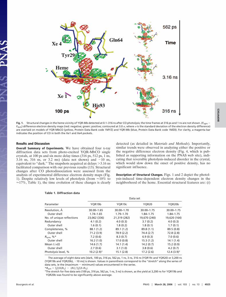
Results and DiscussionOverall Summary of Experiments. We have obtained four x-raydiffraction data sets from photo-excited YQR-MbCO singlecrystals, at 100 ps and six more delay times (316 ps, 512 ps, 1 ns,3.16 ns, 316 ns, or 3.2 ms) (data not shown) and �10 ns,equivalent to ‘‘dark.’’ The snapshots acquired at delays �3.16 nsfacilitated comparison with our previous results (13). Structuralchanges after CO photodissociation were assessed from theanalysis of experimental difference electron density maps (Fig.1). Despite relatively low levels of photolysis (from �10% to�17%, Table 1), the time evolution of these changes is clearly
detected (as detailed in Materials and Methods). Importantly,similar trends were observed in analyzing either the positive orthe negative difference electron density (Fig. 4, which is pub-lished as supporting information on the PNAS web site), indi-cating that reversible photolysis-induced disorder in the crystal,which would slow down the onset of positive density, has nosignificant influence.
Description of Structural Changes. Figs. 1 and 2 depict the photol-ysis-induced time-dependent electron density changes in theneighborhood of the heme. Essential structural features are: (i)
Fig. 1. Structural changes in the heme vicinity of YQR-Mb detected at 0.1–316 ns after CO photolysis; the time frames at 316 ps and 1 ns are not shown. (Flight �Fdark) difference electron density maps (red: negative; green: positive; contoured at 3.0 �, where � is the standard deviation of the electron density difference)are overlaid on models of YQR-MbCO (yellow, Protein Data Bank code 1MYZ) and YQR-Mb (blue, Protein Data Bank code 1MZ0). For clarity, a magenta barindicates the position of CO in both the Xe1 and Xe4 pockets.
Table 1. Diffraction data
Parameter
Data set
YQR19b YQR19c YQR20 YQR20b
Resolution, Å 30.00–1.65 30.00–1.70 30.00–1.75 30.00–1.75Outer shell 1.74–1.65 1.79–1.70 1.84–1.75 1.84–1.75
No. of unique reflections 23,062 (336) 21,319 (282) 19,670 (240) 19,620 (164)Redundancy 4.1 (0.2) 4.0 (0.3) 3.7 (0.2) 4.0 (0.3)
Outer shell 1.6 (0.1) 1.8 (0.2) 1.8 (0.1) 1.7 (0.1)Completeness, % 88.1 (1.2) 89.1 (1.2) 89.8 (1.1) 89.5 (0.8)
Outer shell 71.2 (3.9) 78.9 (2.2) 74.4 (2.7) 72.8 (2.8)Rsym, %* 7.2 (0.6) 8.3 (0.7) 6.9 (0.3) 7.0 (0.6)
Outer shell 16.2 (1.0) 17.0 (0.8) 15.3 (1.2) 14.1 (1.4)Mean I��(I) 14.6 (1.7) 14.1 (1.4) 14.2 (0.7) 15.2 (0.9)
Outer shell 2.7 (0.6) 3.1 (1.0) 3.8 (0.6) 4.2 (0.7)Photolysis level, % 10.2 (2.9)† 15.1 (2.8) 17.2 (2.6) 12.4 (3.9)†
The average of eight data sets [dark, 100 ps, 316 ps, 562 ps, 1 ns, 3 ns, 316 ns (YQR19c and YQR20) or 3,200 ns(YQR19b and YQR20b), �10 ms] is shown. Values in parenthesis correspond to the ‘‘stretch’’ along the series ofdata sets, ie the (maximum � minimum) values encountered in the series.*Rsym � �j�h�Ih, j � �Ih����j�h Ih, j.†The stretch for five data sets (100 ps, 316 ps, 562 ps, 1 ns, 3 ns) is shown, as the yield at 3,200 ns for YQR19b andYQR20b was found to be significantly above average.
Bourgeois et al. PNAS � March 28, 2006 � vol. 103 � no. 13 � 4925
BIO
PHYS
ICS
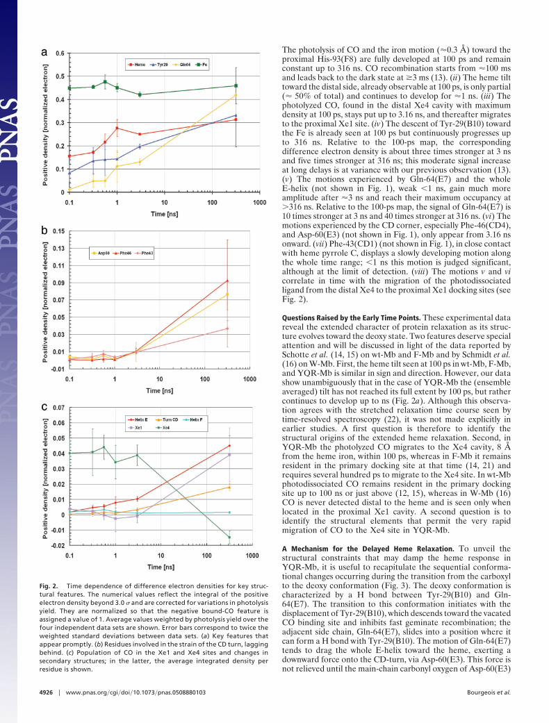
The photolysis of CO and the iron motion (�0.3 Å) toward theproximal His-93(F8) are fully developed at 100 ps and remainconstant up to 316 ns. CO recombination starts from �100 msand leads back to the dark state at �3 ms (13). (ii) The heme tilttoward the distal side, already observable at 100 ps, is only partial(� 50% of total) and continues to develop for �1 ns. (iii) Thephotolyzed CO, found in the distal Xe4 cavity with maximumdensity at 100 ps, stays put up to 3.16 ns, and thereafter migratesto the proximal Xe1 site. (iv) The descent of Tyr-29(B10) towardthe Fe is already seen at 100 ps but continuously progresses upto 316 ns. Relative to the 100-ps map, the correspondingdifference electron density is about three times stronger at 3 nsand five times stronger at 316 ns; this moderate signal increaseat long delays is at variance with our previous observation (13).(v) The motions experienced by Gln-64(E7) and the wholeE-helix (not shown in Fig. 1), weak �1 ns, gain much moreamplitude after �3 ns and reach their maximum occupancy at�316 ns. Relative to the 100-ps map, the signal of Gln-64(E7) is10 times stronger at 3 ns and 40 times stronger at 316 ns. (vi) Themotions experienced by the CD corner, especially Phe-46(CD4),and Asp-60(E3) (not shown in Fig. 1), only appear from 3.16 nsonward. (vii) Phe-43(CD1) (not shown in Fig. 1), in close contactwith heme pyrrole C, displays a slowly developing motion alongthe whole time range; �1 ns this motion is judged significant,although at the limit of detection. (viii) The motions v and vicorrelate in time with the migration of the photodissociatedligand from the distal Xe4 to the proximal Xe1 docking sites (seeFig. 2).
Questions Raised by the Early Time Points. These experimental datareveal the extended character of protein relaxation as its struc-ture evolves toward the deoxy state. Two features deserve specialattention and will be discussed in light of the data reported bySchotte et al. (14, 15) on wt-Mb and F-Mb and by Schmidt et al.(16) on W-Mb. First, the heme tilt seen at 100 ps in wt-Mb, F-Mb,and YQR-Mb is similar in sign and direction. However, our datashow unambiguously that in the case of YQR-Mb the (ensembleaveraged) tilt has not reached its full extent by 100 ps, but rathercontinues to develop up to ns (Fig. 2a). Although this observa-tion agrees with the stretched relaxation time course seen bytime-resolved spectroscopy (22), it was not made explicitly inearlier studies. A first question is therefore to identify thestructural origins of the extended heme relaxation. Second, inYQR-Mb the photolyzed CO migrates to the Xe4 cavity, 8 Åfrom the heme iron, within 100 ps, whereas in F-Mb it remainsresident in the primary docking site at that time (14, 21) andrequires several hundred ps to migrate to the Xe4 site. In wt-Mbphotodissociated CO remains resident in the primary dockingsite up to 100 ns or just above (12, 15), whereas in W-Mb (16)CO is never detected distal to the heme and is seen only whenlocated in the proximal Xe1 cavity. A second question is toidentify the structural elements that permit the very rapidmigration of CO to the Xe4 site in YQR-Mb.
A Mechanism for the Delayed Heme Relaxation. To unveil thestructural constraints that may damp the heme response inYQR-Mb, it is useful to recapitulate the sequential conforma-tional changes occurring during the transition from the carboxylto the deoxy conformation (Fig. 3). The deoxy conformation ischaracterized by a H bond between Tyr-29(B10) and Gln-64(E7). The transition to this conformation initiates with thedisplacement of Tyr-29(B10), which descends toward the vacatedCO binding site and inhibits fast geminate recombination; theadjacent side chain, Gln-64(E7), slides into a position where itcan form a H bond with Tyr-29(B10). The motion of Gln-64(E7)tends to drag the whole E-helix toward the heme, exerting adownward force onto the CD-turn, via Asp-60(E3). This force isnot relieved until the main-chain carbonyl oxygen of Asp-60(E3)
Fig. 2. Time dependence of difference electron densities for key struc-tural features. The numerical values reflect the integral of the positiveelectron density beyond 3.0 � and are corrected for variations in photolysisyield. They are normalized so that the negative bound-CO feature isassigned a value of 1. Average values weighted by photolysis yield over thefour independent data sets are shown. Error bars correspond to twice theweighted standard deviations between data sets. (a) Key features thatappear promptly. (b) Residues involved in the strain of the CD turn, laggingbehind. (c) Population of CO in the Xe1 and Xe4 sites and changes insecondary structures; in the latter, the average integrated density perresidue is shown.
4926 � www.pnas.org�cgi�doi�10.1073�pnas.0508880103 Bourgeois et al.
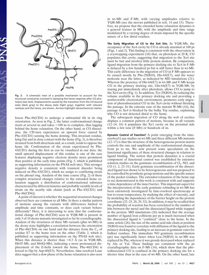
forces Phe-46(CD4) to undergo a substantial tilt in its ringorientation. As seen in Fig. 2, the latter conformational changestarts at several ns and takes �100 ns to complete, thus laggingbehind the heme relaxation. On the other hand, as CO dissoci-ates, the CD-turn experiences an upward force caused byPhe-43(CD1) sensing the heme doming. This invariant residue,lying flat and in close contact with the heme (Fig. 3), is thereforestrained from both directions and, as a result, tends to oppose theheme tilt. Confirmation of the strain experienced by Phe-43(CD1) during the first ns can be visualized in our data: thesmall upward displacement of this residue is one of the rarefeatures displaying negative electron density more prominentthan positive at the early time points (Fig. 5, which is publishedas supporting information on the PNAS web site). This strongernegative density is consistent with transient disorder beinginduced on Phe-43(CD1), which we assign to conflicting strainon the phenyl ring. Analysis of the time course (Fig. 2) of thesecomplex structural changes relative to the extended heme re-laxation suggests a distribution of conformational substatescharacterized by different kinetics and probably variable levels ofstrain on the nearby side chains [such as Phe-43(CD1) andPhe-46(CD4)].
A point of interest is whether the sequential structural changesobserved here are common to all Mbs. Is there a similar patternof motions among the variants with differences limited toamplitude and time constants, or does each variant behavedifferently? Here, it is interesting to note that the conforma-tional change of Phe-46(CD4) seen in YQR-Mb is present inonly 3 of 10 deoxy mutants investigated so far by crystallography.Analysis of the structures of these Mb variants (plus wt-Mb) inthe deoxy state highlights a correlation between the orientationof Phe-46(CD4) on one hand and the distance from the C� ofresidue E7 to the heme iron on the other (Table 2, which ispublished as supporting information on the PNAS web site).When this distance is shorter than �8.3 Å (as in YQR-Mb,H64T-Mb, and H64Q-Mb), indicating a more pronounced dis-placement of the E-helix toward the heme, Phe-46(CD4) isforced to flip by Asp-60(E3). Nevertheless, time-resolved Lauedata suggest that a slow phase of the heme relaxation is also seen
in wt-Mb and F-Mb, with varying amplitudes relative toYQR-Mb (see the movies published in refs. 14 and 15). There-fore, we propose that the extended heme relaxation dynamics isa general feature in Mb, with the amplitude and time rangemodulated by a varying degree of strain imposed by the specificnature of a few distal residues.
The Early Migration of CO to the Xe4 Site. In YQR-Mb, theoccupancy of the Xe4 cavity by CO is already maximal at 100 ps(Figs. 1 and 2). This finding is consistent with the observation ina cryotrapping experiment (18) that, on photolysis at 20 K, COpopulates this cavity, suggesting that migration to the Xe4 sitemust be fast and involves little protein motion. By comparison,ligand migration from the primary docking site to Xe4 in F-Mbis delayed by a few hundred ps but is still faster than in wt-Mb.This early difference in the migration of CO in F-Mb appears tobe caused mostly by Phe-29(B10), His-64(E7), and the watermolecule near the latter, as indicated by MD simulations (21).Whereas the presence of His-64(E7) in wt-Mb and F-Mb keepsCO in the primary docking site, Gln-64(E7) in YQR-Mb, bystaying put immediately after photolysis, allows CO to jump tothe Xe4 cavity (Fig. 1). In addition, Tyr-29(B10), by reducing thevolume available in the primary docking site and providing anonfavorable electrostatic environment, promotes early migra-tion of photodissociated CO in the Xe4 cavity without blockingthe passage. In the extreme case of the mutant W-Mb (16), thepassage to Xe4 is blocked by the bulky Trp residue, explainingwhy CO is not detected distal to the heme.
The subsequent migration of CO along the web of cavitiesdisplays a common pattern of motions, because in all variants(12–14, 16) it populates the Xe1 cavity on the proximal sidewithin a few tens (F-Mb) or hundreds of ns.
Dynamic Control of Function? A point emerging from the time-resolved Laue studies on wt-Mb and three different Mb mutants(12–17) is that the structure of the heterogeneous protein matrixcontrols the rate and amplitude of the conformational changes,from ps to ms. We now present some speculation on thefunctional significance of these structural changes in relation toligand binding. The notion that protein dynamics is a crucialcomponent of functional control was established by extensivesolution studies on the geminate recombination of O2, NO, andCO (1, 2, 23–31). Early geminate recombination of the dissoci-ated ligand was shown to be heterogeneous and was proposed tobe controlled by prosthetic group motions and the specific natureof the pocket residues. The extended relaxation of the heme (upto ns) demonstrated in this work is consistent with and supportsa time dependence of the inner barrier. This important aspect forthe interpretation of the early geminate rebinding in wt-Mb hasbeen extensively investigated by time-resolved spectroscopy atlow to room temperature, by studying the effect of viscosity, andby modeling the dependence of the barrier height on the proteincoordinate (22–25, 28, 29, 32). In addition, it may be recalled thatthe probability of reaction has been correlated to the number ofhits between the metal and the dissociated ligand while trappedin the protein. MD simulations (26) indicated that the averagenumber of ligand�iron collisions per ps is much increased whenthe dissociated ligand is ‘‘confined’’ close to the heme. In thesame article (26), the size of the amino acid side chain at position29(B10) was found to correlate with diffusion of the ligand to theprimary docking site, leading to an increase in geminate rates forbulkier residues. The immediate NO geminate recombination(ps) was significantly faster when Leu-29(B10) in wt-Mb wasreplaced by Phe, whereas it was grossly decreased when replacedby Ala or Val. These findings are consistent with the pscrystallographic data on F-Mb (14), which show that the pho-todissociated CO is confined in the primary docking site for ashorter time than in the case of wt-Mb. On the other hand, late
Fig. 3. A schematic view of a possible mechanism to account for thestructural constraints involved in damping the heme response after CO pho-tolysis (see text). Displacements caused by the transition from the CO-boundstate (dark gray) to the deoxy state (light gray), together with relevantresidues and the heme, are shown. Arrows highlight stereochemical clashes.
Bourgeois et al. PNAS � March 28, 2006 � vol. 103 � no. 13 � 4927
BIO
PHYS
ICS
geminate rebinding was invoked to depend on ligand migrationaway from the distal pocket, based on the effect of filling thecavities by mutations or Xe (30, 31). In fact, for the same Mbvariants the rates of delayed (ns) geminate recombination of NO,reflecting back migration from the protein matrix toward theheme iron, follow an opposite order, suggesting that once NO hasdiffused in the protein, Phe-29(B10) provides a larger stericbarrier to back diffusion. Consistent with these results, theoverall NO dissociation rate constant for F29-Mb was found tobe five times slower than for wt-Mb and �30 times slower thanfor A29-Mb (26), affecting overall ligand affinity by confinementof the (thermally) dissociated NO. Although geminate NOrecombination for YQR-Mb was measured only in the ns range(33), data showed that: (i) the ns photolysis quantum yield forNO is �4%, i.e., much higher than for any other Mb variant,indicating that ps recombination must be negligible, and (ii) thedelayed ns geminate recombination is remarkably slow (�100ns) and is affected by Xe filling the cavities. These findings, whichsuggested immediate migration of the photolyzed ligand awayfrom the distal pocket, are consistent with the very rapid partialmovement of the bulky Tyr-29(B10) toward the heme and theimmediate migration of the ligand into Xe4 seen at 100 ps, asdescribed here.
Is the Extended Mb Relaxation a General Phenomenon? Availabletime-resolved Laue data on photolyzed MbCO (11–16) provethat relaxation to the deoxy state extends over a large dynamicrange in time. These studies provide the structural basis forinterpreting the extensive investigations obtained by transientspectroscopy in solution, showing events extending from ps toms. The huge time range, covering �9 orders of magnitude, hasbeen described by a stretched exponential function reflecting therelaxation of the conformational substates of the protein withinits complex energy landscape (3, 22, 29). The novelty unveiled bythe Laue diffraction data is the direct visualization of thecorresponding structural relaxation events with unprecedentedtime resolution. Here, we have provided evidence that relaxationof residues near the active site [such as Tyr-29(B10), Gln-64(E7),or Phe-43(CD1)], the heme tilting, the collapse of the E-helix,and the coupled strain on the CD corner all are stretched,covering a very large time range. Comparing the structuralrelaxations of the different Mb variants, we observe that theconformational changes are similar in kind but modulated inmagnitude and speed by specific internal mutations, with somereasonable correlation to functional control (see above). Like-wise, the internal migration of photolysed CO is in all variants(including wt-Mb) leading to maximal occupancy of the Xe1cavity by a few hundred ns, but the population of the otherdocking sites en route is heavily modulated by a few distalresidues.
A crucial point to be asked is to what extent is this nonexpo-nential progression of conformational change a general featureof all proteins, and how can we address this question, especiallyfor the faster time regime. In this context, the value of MDsimulations, now extending to tens of ns and applicable to allproteins, is self-evident. Two studies recently published (20, 21)show satisfactory agreement with experiments, which carriesmore advantages, such as (i) features at the limit of reliability inthe diffraction maps may be more confidently assigned to realprotein motions, and (ii) the question related to the propertiesof single molecules can be compared with the average behaviordetected by crystallography. Thus a combined approach mayhelp to solve the fundamental problem of how the extendedconformational relaxation documented by Laue crystallographyrelates to trajectories of individual protein molecules.
The conclusions reached from this Laue time-resolved crys-tallography study strongly support the concepts of conforma-tional substates and energy landscapes in proteins (3, 10) and
affirm the relevance of very fast structural dynamics to biology.The new challenge is to expand on this framework and explorethe role of fast dynamics for other proteins, including thosedisplaying allosteric control.
Materials and MethodsProtein Crystals. YQR-Mb was produced in Escherichia coli froma synthetic gene with the substitution Asp-122–Asn, a conser-vative mutation with negligible functional and structural effects,but one that coaxes the protein to crystallize in space group P6(34). Crystallization conditions under CO were as reported byBourgeois et al. (13).
Laser Photolysis and Data Collection Strategy. YQR-MbCO crystalswere photolysed with light pulses (spectral range 520–560 nm)of �40 �J that had been stretched to several tens of picosecondsby passage through 3 m of multimode optical fiber. A detaileddescription of the laser sources and optical setup is given in ref.15. To extract the excess heat caused by visible light absorptionby the sample, the capillary was bathed in a �10°C gas stream(Oxford Cryosystem 600). The achieved level of photolysis didnot exceed �20% and in some instances was �10%, presumablybecause of the presence of a minor amount of oxidized met-Mb(estimated by optical spectroscopy to be �5%) or a residualmismatch between the pumped and probed volumes of thesample.
Laue diffraction data were acquired on the ID09B beamlineat the European Synchrotron Radiation Facility. The synchro-tron was operated in the so-called four-bunch mode, where fourequally spaced electron bunches circulate in the storage ring.X-ray pulses of �150 ps (full width at half maximum) containing�1010 photons per pulse were generated in a relative bandwidthof �3.5% (full width at half maximum) peaked at 0.79 Å (15).
Four complete time-resolved diffraction data sets were col-lected, each from separate regions of two different large crystals.Each data set was comprised of 31 images spanning 60°. Toimprove the signal-to-noise ratio, each image represented theintegration of 16 pump-probe measurements accumulated at 3Hz on a MarCCD detector (Mar USA, Evanston, IL). Tominimize systematic errors between time points, time was usedas the ‘‘fast variable.’’ At a given crystal orientation, all of thediffraction images at the investigated pump-probe delays werecollected, and then the crystal was rotated and the process wasrepeated. With this approach, f luctuations in photolysis yieldand radiation damage are distributed equally over all time points,thereby minimizing their effects on the observed time-dependent structural changes (14, 15, 35).
Data Analysis. Diffraction data were reduced in an automated wayby using LAUEGEN, PROW, and LSCALE (Table 1) (36). Experi-mental electron density difference maps (Flight � Fdark) weregenerated with CCP4 (37) using phases from a dark-state model(Protein Data Bank code 1MYZ, refined from earlier data onsimilar crystals), after optimization of the difference amplitudeswith a Bayesian statistics-based method (38). To estimate thephotolysis yield, the negative difference electron density featureat the bound CO was integrated within a mask excluding thecontribution of the iron motion. This density was then comparedwith the one obtained from simulated data of equal complete-ness and corresponding to complete photolysis of CO. For alldata set series, the photolysis yield was found to be constant (�1%) over the time range 100 ps to 3.16 ns. To evaluate theprogress of the structural changes with time at different locationsof the structure, the difference (Flight � Fdark) maps wereintegrated above�below 3.0 � with homemade software basedon a 3D connectivity search algorithm. For each Mb atom,difference electron density above�below 3.0 � was searched ata maximum distance of 1.5 Å from the atom. If density was
4928 � www.pnas.org�cgi�doi�10.1073�pnas.0508880103 Bourgeois et al.
found, integration was carried out with the connectivity searchalgorithm, within a limiting cube of 8 Å3. Voxels above�belowthe � threshold could be selected several times (by severalatoms), but were considered only once. Results were thennormalized to the negative density feature at the CO for eachtime delay, which was assigned an arbitrary value of 1.
Figs. 1 and 3 were drawn by using BOBSCRIPT (39) andRASTER3D (40). In Fig. 2, the data shown refer to the weightedaverage behavior of the structural features as assessed fromanalysis of all four data sets. Weights w were taken as w �MAX(0, photolysis yield � 10), so that individual data sets witha photolysis yield �10%, judged to have a too high level of noise,were discarded. Error bars corresponding to twice the weightedstandard deviations between data sets at every time point are
also shown in Fig. 2. However, it must be kept in mind that theseerror estimates include all kinds of systematic deviations fromcrystal to crystal, and therefore give an overly pessimisticoutlook of the data. Because time was used as the fast variable,the time evolution of the electron density for a single crystal isbelieved to be much more reliable than suggested by these errorbars, although no other statistical evaluation is available.
We thank Dr. M. Wolff, ID09, European Synchrotron Radiation Facility.This research was supported in part by Ministero dell’Istruzione, dellUniversita e della Ricerca Grants RBLA03B3KC�004 2003 andRBNE01KXC9 2005 (to M.B.) and the Intramural Research Program ofthe National Institutes of Health, National Institute of Diabetes andDigestive and Kidney Diseases (P.A.A.).
1. Austin, R. H., Beeson, K. W., Eisenstein, L., Frauenfelder, H. & Gunsalus, I. C.(1975) Biochemistry 14, 5355–5373.
2. Frauenfelder, H., Parak, F. & Young, R. D. (1988) Annu. Rev. Biophys. Chem.17, 451–479.
3. Frauenfelder, H., Sligar, S. G. & Wolynes, P. G. (1991) Science 254, 1598–1603.4. Richards, F. M. (1977) Annu. Rev. Biophys. Bioeng. 6, 151–176.5. Tilton, R. F., Jr., Kuntz, I. D., Jr., & Petsko, G. A. (1984) Biochemistry 23,
2849–2857.6. Elber, R. & Karplus, M. (1990) J. Am. Chem. Soc. 112, 9161–9175.7. Carlson, M. L., Regan, R. M. & Gibson, Q. H. (1996) Biochemistry 35,
1125–1136.8. Decatur, S. M., DePillis, G. D. & Boxer, S. G. (1996) Biochemistry 35,
3925–3932.9. Brunori, M. & Gibson, Q. H. (2001) EMBO Rep. 2, 674–679.
10. Frauenfelder, H., McMahon, B. H. & Fenimore, P. W. (2003) Proc. Natl. Acad.Sci. USA 100, 8615–8617.
11. Srajer, V., Teng, T., Ursby, T., Pradervand, C., Ren, Z., Adachi, S., Schildkamp,W., Bourgeois, D., Wulff, M. & Moffat, K. (1996) Science 274, 1726–1729.
12. Srajer, V., Ren, Z., Teng, T. Y., Schmidt, M., Ursby, T., Bourgeois, D.,Pradervand, C., Schildkamp, W., Wulff, M. & Moffat K. (2001) Biochemistry40, 13802–13815.
13. Bourgeois, D., Vallone, B., Schotte, F., Arcovito, A., Miele, A. E., Sciara, G.,Wulff, M., Anfinrud, P. & Brunori, M. (2003) Proc. Natl. Acad. Sci. USA 100,8704–8709.
14. Schotte, F., Lim, M., Jackson, T. A., Smirnov, A. V., Soman, J., Olson, J. S.,Phillips, G. N., Jr., Wulff, M. & Anfinrud, P. A. (2003) Science 300, 1944–1947.
15. Schotte, F., Soman, J., Olson, J. S., Wulff, M. & Anfinrud, P. A. (2004) J. Struct.Biol. 147, 235–246.
16. Schmidt, M., Nienhaus, K., Pahl, R., Krasselt, A., Anderson, S., Parak, F.,Nienhaus, G. U. & Srajer, V. (2005) Proc. Natl. Acad. Sci. USA 102, 11704–11709.
17. Brunori, M., Bourgeois, D. & Vallone, B. (2004) J. Struct. Biol. 147, 223–234.18. Brunori, M., Vallone, B., Cutruzzola, F., Travaglini-Allocatelli, C., Berendzen,
J., Chu, K., Sweet, R. M. & Schlichting, I. (2000) Proc. Natl. Acad. Sci. USA 97,2058–2063.
19. Nienhaus, K., Ostermann, A., Nienhaus, G. U., Parak, F. G. & Schmidt, M.(2005) Biochemistry 44, 5095–5105.
20. Bossa, C., Anselmi, M., Roccatano, D., Amadei, A., Vallone, B., Brunori, M.& Di Nola, A. (2004) Biophys. J. 86, 3855–3862.
21. Hummer, G., Schotte, F. & Anfinrud, P. A. (2004) Proc. Natl. Acad. Sci. USA101, 15330–15334.
22. Jackson, T. A., Lim, M. & Anfinrud, P. (1994) Chem. Phys. 180, 131–140.23. Henry, E. R., Sommer, J. H., Hofrichter, J. & Eaton, W. A. (1983) J. Mol. Biol.
166, 443–451.24. Petrich, J. W., Lambry, J. C., Kuczera, K., Karplus, M., Poyart, C. & Martin,
J. L. (1991) Biochemistry 30, 3975–3987.25. Steinbach, P. J., Ansari, A., Berendzen, J., Braunstein, D., Chu, K., Cowen,
B. R., Ehrenstein, D., Frauenfelder, H., Johnson, J. B., Lamb, D. C., et al.(1991) Biochemistry 30, 3988–4001.
26. Gibson, Q. H., Regan, R., Elber, R., Olson, J. S. & Carver, T. E. (1992) J. Biol.Chem. 267, 22022–22034.
27. Ikeda-Saito, M., Dou, Y., Yonetani, T., Olson, J. S., Li, T., Regan, R. & Gibson,Q. H. (1993) J. Biol. Chem. 268, 6855–6857.
28. Ansari, A., Jones, C. M., Henry, E. R., Hofrichter, J. & Eaton, W. A. (1994)Biochemistry 33, 5128–5145.
29. Hagen, S. J. & Eaton, W. A. (1996) J. Chem. Phys. 104, 3395–3398.30. Scott, E. E. & Gibson, Q. H. (1997) Biochemistry 36, 11909–11917.31. Scott, E. E., Gibson, Q. H. & Olson, J. S. (2001) J. Biol. Chem. 276, 5177–5188.32. Agmon, N. & Hopefield, J. J. (1983) J. Phys. Chem. 79, 2042–2053.33. Brunori, M., Cutruzzola, F., Savino, C., Travaglini-Allocatelli, C., Vallone, B.
& Gibson, Q. H. (1999) Biophys. J. 76, 1259–1269.34. Phillips, G. N., Jr., Arduini, R. M., Springer, B. A. & Sligar, S. G. (1990) Proteins
7, 358–365.35. Ihee, H., Rajagopal, S., Srajer, V., Pahl, R., Anderson, S., Schmidt, M., Schotte,
F., Anfinrud, P. A., Wulff, M. & Moffat, K. (2005) Proc. Natl. Acad. Sci. USA102, 7145–7150.
36. Bourgeois, D., Wagner, U. & Wulff, M. (2000) Acta Crystallogr. D 56, 973–985.37. Collaborative Computational Project (1994) Acta Crystallogr. D 50, 760–763.38. Ursby, T. & Bourgeois, D. (1997) Acta Crystallogr. A 53, 564–575.39. Esnouf, R. M. (1999) Acta Crystallogr. D 55, 938–940.40. Merritt, E. A. & Bacon, D. J. (1997) Methods Enzymol. 277, 505–524.
Bourgeois et al. PNAS � March 28, 2006 � vol. 103 � no. 13 � 4929
BIO
PHYS
ICS