Embed Size (px)
Citation preview
Hindawi Publishing CorporationEvidence-Based Complementary and Alternative MedicineVolume 2013, Article ID 540957, 10 pageshttp://dx.doi.org/10.1155/2013/540957
Research ArticleMetabolic Signatures of Kidney Yang DeficiencySyndrome and Protective Effects of Two Herbal Extractsin Rats Using GC/TOF MS
Linjing Zhao,1,2 Hongbing Wu,1 Mingfeng Qiu,1 Wei Sun,3 Runmin Wei,1
Xiaojiao Zheng,1 Yiting Yang,3 Xue Xin,1 Haimiao Zou,1 Tianlu Chen,4
Jiajian Liu,4 Lina Lu,1 Jing Su,1 Chungwah Ma,3 Aihua Zhao,4 and Wei Jia4
1 School of Pharmacy, Shanghai Jiao Tong University, Shanghai 200240, China2 College of Chemistry and Chemical Engineering, Shanghai University of Engineering Science, Shanghai 201620, China3 Infinitus (China) Company Ltd., Guangzhou 510665, China4Center for Translational Medicine, and Shanghai Key Laboratory of Diabetes Mellitus,Department of Endocrinology and Metabolism, Shanghai Jiao Tong University Affiliated Sixth People’s Hospital,Shanghai 200233, China
Correspondence should be addressed to Chungwah Ma; william [email protected] and Aihua Zhao; [email protected]
Received 25 April 2013; Revised 4 August 2013; Accepted 6 August 2013
Academic Editor: Aiping Lu
Copyright © 2013 Linjing Zhao et al. This is an open access article distributed under the Creative Commons Attribution License,which permits unrestricted use, distribution, and reproduction in any medium, provided the original work is properly cited.
Kidney Yang Deficiency Syndrome (KDS-Yang), a typical condition in Chinese medicine, shares similar clinical signs of theglucocorticoid withdrawal syndrome. To date, the underlying mechanism of KDS-Yang has been remained unclear, especially atthe metabolic level. In this study, we report a metabolomic profiling study on a classical model of KDS-Yang in rats induced byhydrocortisone injection to characterize themetabolic transformation using gas chromatography/time-of-flightmass spectrometry.WKY1, a polysaccharide extract from Astragalus membranaceus and Lycium barbarum, and WKY2, an aqueous extract froma similar formula containing Astragalus membranaceus, Lycium barbarum, Morinda officinalis, Taraxacum mongolicum, andCinnamomum cassia presl, were used separately for protective treatments of KDS-Yang. The changes of serum metabolic profilesindicated that significant alterations of key metabolic pathways in response to abrupt hydrocortisone perturbation, includingdecreased energy metabolism (lactic acid, acetylcarnitine), lipid metabolism (free fatty acids, 1-monolinoleoylglycerol, andcholesterol), gut microbiota metabolism (indole-3-propionic acid), biosynthesis of catecholamine (norepinephrine), and elevatedalanine metabolism, were attenuated or normalized with different degrees by the pretreatment of WKY1 or WKY2, which isconsistent with the observations in which the two herbal agents could ameliorate biochemical markers of serum cortisone,adrenocorticotropic (ACTH), and urine 17-hydroxycorticosteroids (17-OHCS).
1. Introduction
The theory of Yin-Yang is a conceptual framework usedfor observing and analyzing the material world in ancientChina. This philosophical approach has also been adoptedfor the studies of traditional Chinese medicine (TCM) syn-drome and the guidance clinical diagnosis, treatment, andprevention for thousands of years. Kidney Yang DeficiencySyndrome (KDS-Yang) was firstly documented in Huangdi
Neijing, an earliest systematic monograph in TCM. Modernresearch has shown that damages and functional disorders ofhypothalamic-pituitary-target gland axis, including adrenal,thyroid, and gonad, are the main pathological mechanismfor forming KDS-Yang [1]. Over the past half century, morethan 20 experimental models with a variable range of clin-ical manifestations similar to those observed in KDS-Yanghuman have been developed [2]. It is one of classical methodsof establishing KDS-Yang to inject animals with high dose
2 Evidence-Based Complementary and Alternative Medicine
of exogenous glucocorticoid, such as hydrocortisone, whichwould induce adrenocortical insufficiency after abrupt with-drawal administration [2]. This animal model mimicked thepathological state of suppression of hypothalamic-pituitary-adrenal (HPA) axis to some extent in KDS-Yang human andcontributed greatly to important advances in the currentunderstanding of the underlying mechanisms of KDS-Yangas well as treatments [3–5].
Metabolomics, a new omics technique defined as quanti-tative measurement of time-related multiparametric meta-bolic response of multicellular systems to pathophysiologicalstimuli or generic modification [6], has recently developedinto an increasingly important tool and has been successfullyused in revealing the essence of syndrome and therapeuticeffect of TCM.Recently, an increasing number of publicationshave described the applications of metabolomic approach inevaluating the curative effect and mechanism of traditionalmedicine in KDS-Yang [7–10]. Gong et al. [7] investigatedthe metabolic profile of hydrocortisone-induced KDS-Yangin rats and the intervention effects of Morinda officinalisbased on nuclear magnetic resonance (NMR). Lu et al.[8] studied the metabonomic characters of KDS-Yang ratsand the therapeutic effects of Drynaria fortune (Kunze) J.Sm. using ultra-performance liquid chromatography coupledwith mass spectrometry (UPLC/MS). Li et al. [9] describedthe serous metabonomic study on Epimedium brevicornumMaxim. treated KDS rats and its therapeutic basis usingUPLC/MS. Subsequently, an integrated plasma and urinarymetabolomics method by UPLC/MS were also developedto reveal the intervention effects of Epimedium koreanumNakai on KDS-Yang rats [10]. Also, in our previous study,the urinary metabolic profiles using gas chromatographycoupled with mass spectrometry (GC/MS) characterized thebiochemical fingerprints of a physiopathologic status similarto KDS in TCM, and the intervention of Herba Cistanchescould cause a systemic recovery from the hydrocortisone-induced metabolic perturbation in rats [11, 12].
WKY1 and WKY2 are two TCM formulas designed forstrengthening kidney Yang. WKY1 is a crude compoundpolysaccharide extracted fromAstragalus membranaceus andLycium barbarum. The combination of these two medicinalplants as a formula is derived from the Royal Formularyof Yang Spring, which is part of an ancient health preserv-ing book, “The Life Documentary of Emperor Qianlong.”The rationale for the use of this two-plant formula is tostrengthenYang andQiwithAstragalusmembranaceus, whilereplenishing the Yin with Lycium barbarum fruits, accordingto the Royal Formulary. The clinical use of the formula isbelieved to achieve a satisfactory effect of replenishing thekidney deficiency, and therefore, may particularly be effectivefor chronic colitis and diarrhea generally resulting from theKDS-Yang. WKY2 is a mixture of an aqueous extract from asimilar Chinese herbal formula, including Astragalus mem-branaceus, Lycium barbarum,Morinda officinalis, Taraxacummongolicum, and Cinnamomum cassia presl. The additionalthree herbs were used in this formula to further strengthenthe lung and spleen and thus synergize with the first twoherbs to enhance the clinical efficacy of KDS-Yang-derivedconditions such as chronic diarrhea. Based on our pilot study
in dozens of volunteers, the two herbal formulas both showedpromising protective effects against KDS-Yang.
In this study, a serous metabolic profiling approach basedon gas chromatography time-of-flight mass spectrometry(GC/TOF MS) was explored to characterize the metabolicsignature of the KDS-Yang rats. The protective effects ofWKY1 and WKY2 against the metabolic alteration werealso investigated with this strategy. The finding of metabolicpathways will be helpful to understand the essence of KDS-Yang and the underlying mechanism of the two herbaltreatments.
2. Materials and Methods
2.1. Chemicals and Reagents. HPLC grade methanol, chloro-form, and pyridine were purchased from Merck Chemicals(Darmstadt, Germany). Hydrocortisone solution for injec-tion (0.5%) was purchased from Shanghai Xinyi Pharma-ceutical Co. (Shanghai, China). L-2-chlorophenylalanine waspurchased from Intechem Tech. Co. Ltd. (Shanghai, China).BSTFA (1% TMCS), heptadecanoic acid, and methoxyaminewere purchased from Sigma Aldrich (St. Louis, MO, USA).All aqueous solutions were prepared with ultrapure waterproduced by a Milli-Q system (18.2MΩ, Millipore, Bedford,MA, USA).
2.2. Herbal Preparation. The five raw herbs, including Astra-galus membranaceus, Lycium barbarum, Morinda officinalis,Taraxacummongolicum, and Cinnamomum cassia presl, werepurchased fromChinesemainland andwere authenticated onthe basis of morphological and chemical analysis in accor-dance with the Chinese Pharmacopoeia (data not shown).
WKY1 was produced by water extraction and ethanolprecipitation as described previously [13]. In brief, Astragalusmembranaceus and Lycium barbarum mixed at a certainproportionwere boiled in 10-fold water for 1.5 h. At the end of1.5 h boiling, the water extract was collected and the residuewas reboiled in 8-fold water for 1 h.The blending supernatantwas concentrated in a vacuum rotary evaporator, and thenpooled and mixed with ethanol (final concentration 75%v/v) to precipitate the polysaccharide-enriched fraction. Theprecipitate was separated from the supernatant and vacuum-dried at 40∘C to obtain WKY1 (moisture content <7.0%).WKY2 is composed of WKY1 and the powder of an aqueousextract, which was obtained fromMorinda officinalis, Tarax-acum mongolicum, and Cinnamomum cassia presl. For theaqueous extract preparation, three crude herbs were mixedaccording to proportion and boiled in 10-fold water for 1.5 h.The mixture was cooled to room temperature and filtered.The residuewas then refluxedwith additional 6-fold water for1 h.The supernatant was pooled, concentrated under reducedpressure, and then sprayed into dry powder. According toproportion, the spray-drying powder andWKY1 were mixedfor obtaining WKY2 (moisture content <5.0%). The totalpolysaccharide contents of WKY1 and WKY2 were 34.5%(w/w) and 22.6% (w/w), respectively, as determined by thephenol-sulphuric acid method [14].
Evidence-Based Complementary and Alternative Medicine 3
2.3. Experimental Design and Sample Collection. The han-dling of all animals in this study was conformed to thenational guidelines and performed at the Center for Labora-tory Animals, School of pharmacy, Shanghai Jiao Tong Uni-versity (SJTU). All the experimental protocols were approvedby the Animal Ethics Committee of SJTU. A total of 28eight-week-old male Sprague-Dawley rats (200 ± 20 g) werecommercially obtained from Shanghai Laboratory AnimalCo. Ltd. (SLAC, Shanghai, China), housed individually instainless steel wire mesh cages, and provided with a certifiedstandard rat chow and tap water ad libitum. Room tempera-ture and humidity were regulated at 21 ± 1∘C and 60 ± 10%,respectively. A 12/12-h light-dark cycle was set, with lightson at 8 a.m. After 2 weeks of acclimatization in metaboliccages, rats were randomly divided into four groups, 7 ineach group: control group (C), which received daily the samevolume of saline as the other groups from days 1 to 15; modelgroup (M), which received saline daily from days 1 to 15,then 5% hydrocortisone solution at 50mg/kg of body weightwith i.p. injection from days 16 to 22; WKY1 pretreatmentgroup (WKY1), which received daily WKY1 (30 g/L aqueoussuspension) by oral administration at dose of 0.18 g/kg ofbody weight (equal to 30 times dose of clinic dosage) fromdays 1 to 15, then 5% hydrocortisone solution daily withi.p. injection at 50mg/kg from days 16 to 22; and WKY2pretreatment group (WKY2), which received WKY2 (30 g/Laqueous suspension) at daily oral dose of 1.01 g/kg of bodyweight (equal to 30 times dose of clinic dosage) from days1 to 15, then 5% hydrocortisone solution daily at 50mg/kgwith i.p. injection from day 16 to 22. Sera and urine sampleswere collected at day 25, on the 3rd day after hydrocortisonewithdrawal, and stored at −80∘C, pending for biochemicaland metabolomic analysis.
2.4. Behavioral Observation and Hormone Level Measure-ment. The weighed chow and water were added into thecontainer of each metabolism cage and the residual foodand water were measured daily, respectively. The 24 h urinevolume and weekly body weight were recorded, and the gen-eral behavior or activity changes of rats were also observed.The serum cortisone, ACTH, and 24 h urine 17-OHCS weremeasured using ELISA kits (Groundwork BiotechnologyDiagnosticate Ltd., San Diego, CA, USA). Data from theserum biochemistry determination were expressed as mean± SD. Statistical analysis was conducted using the Student’stwo-tailed, unpaired 𝑡-test. A 𝑃 value of less than 0.05 wasconsidered statistically significant.
2.5. Preparation of Samples and Analysis by GC/TOF MS.Sera samples were derivatized and subsequently analyzed byGC/TOF MS following our previously published protocols[15]. A 100 𝜇L aliquot of serum sample was spiked withtwo internal standards (10 𝜇L of L-2-chlorophenylalanine inwater, 0.3mg/mL; 10 𝜇L of heptadecanoic acid in methanol,1mg/mL) and vortexed for 10 s. The mixed solution wasextracted with 300𝜇L of methanol/chloroform (3 : 1) andvortexed for 30 s. After standing for 10min at −20∘C, thesampleswere centrifuged at 10,000 rpm for 10min. An aliquot
of 300𝜇L supernatant was transferred to a glass samplingvial to vacuum-dry at room temperature. The residue wasderivatized using a two-step procedure. First, 80𝜇L ofmethoxyamine (15mg/mL in pyridine) was added to the vialand kept at 30∘C for 90min followed by 80𝜇L of BSTFA(1%TMCS) at 70∘C for 60min. Each 1 𝜇L aliquot of thederivatized solution was injected in spitless mode into anAgilent 6890N gas chromatography coupled with a PegasusHT time-of-flight mass spectrometer (Leco Corporation, StJoseph, MI, USA). Separation was achieved on a DB-5MScapillary column (30m× 250𝜇mI.D., 0.25 𝜇mfilm thickness;(5%-phenyl)-methyl-polysiloxane bonded and crosslinked;Agilent J&W Scientific, Folsom, CA) with helium as the car-rier gas at a constant flow rate of 1.0mL/min.The temperatureof injection, transfer interface, and ion source were set to270∘C, 260∘C, and 200∘C, respectively. The GC temperaturesprogramming was set to 2min isothermal heating at 80∘C,followed by 10∘C/min oven temperature ramps to 180∘C,5∘C/min to 240∘C, and 25∘C/min to 290∘C, and a final 9minmaintenance at 290∘C. Electron impact ionization (70 eV) atfull scan mode (m/z 30–600) was used, with an acquisitionrate of 20 spectrum/second in the TOF MS setting.
2.6. Data Processing and Statistical Analysis. TheacquiredMSfiles from GC/TOF MS analysis were exported in NetCDFformat by ChromaTOF software (v3.30, Leco Co., CA, USA).CDF files were extracted using custom scripts (revised MAT-LAB toolbox HAD) [15, 16] in the MATLAB 7.1 (The Math-Works, Inc., USA) for data pretreatment procedures such asbaseline correction, denoising, smoothing, and alignment;time-window splitting; and peak feature extraction (based onmultivariate curve resolution algorithm) [17]. The resultingthree-dimensional data set, including sample name, peakretention time, and peak intensity, was imported into theSIMCA-P 11.5 Software package (Umetrics, Umea, Sweden)for data analysis according to our previous publication[18]. Multivariate statistical analysis (partial least-squares-discriminant analysis, PLS-DA) was performed. Meanwhile,significantly expressed features between groups were calcu-lated using a Student’s 𝑡-test (𝑃 < 0.05). The fold changeshows the relative intensity ratio of the differential or repre-sentativemetabolites between control andmodel groups withor without WKY1/WKY2 treatments. Additionally, metabo-lites were annotated by comparing the mass fragments withthose of mass spectral in NIST MS databases 2.0 (NIST,Gaithersburg, MD, USA) with a similarity of more than 70%and further verified by the available reference standards.
3. Results and Discussion
3.1. Hormone Level and Behavioral Presentation. The bloodcortisone, ACTH, and 24 h urine 17-OHCS, which werewidely admitted as diadynamic criteria of KDS-Yang inclinic of TCM [19], were carried out to assess the successof hydrocortisone-induced KDS-Yang model. The resultsshowed that these three hormones levels, as well as bodyweight, water intake, and 24-h hour urinary excretion, weresignificantly decreased (𝑃 < 0.05) in the model group
4 Evidence-Based Complementary and Alternative Medicine
Table 1: Hormone variation and behavioral investigation results on the 3rd day after hydrocortisone withdrawal with or withoutWKY1/WKY2 pretreatments.
Groups Cortisone(ng/mL)
ACTH(pg/mL)
17-OHCS(𝜇mol/L)
Body weight(g)
Food consumption(g)
Water intake(mL)
Urine volume(mL)
Control group 220.78 ± 105.78 633.43 ± 111.1 433.81 ± 15.64 371.9 ± 7.2 14.8 ± 1.3 48.9 ± 4.3 40.6 ± 5.9Model group 139.21 ± 40.79∗∗ 447.14 ± 126.5∗ 398.91 ± 29.13∗∗ 349.0 ± 12.0∗∗ 13.4 ± 1.1 36.3 ± 4.5∗∗ 18.4 ± 6.0∗∗
WKY1 group 161.84 ± 87.27 632.98 ± 50.65 400.6 ± 16.59 356.6 ± 15.6∗ 15.1 ± 2.5 42.3 ± 5.9∗ 27.9 ± 8.4∗∗
WKY2 group 206.51 ± 71.64 993.26 ± 17.46∗∗ 416.79 ± 12.78 357.4 ± 12.4∗ 14.9 ± 2.1 34.8 ± 4.6∗∗ 20.4 ± 2.8∗∗∗
𝑃 < 0.05, ∗∗𝑃 < 0.01 compared with control group (two-tailed Student’s 𝑡-test).
compared to those in the control group on day 25 (Table 1),confirming the suppression of HPA axis and the establish-ment of KDS-Yang model on the 3rd day after hydrocor-tisone withdrawal (50mg/kg⋅d for 7 consecutive days). Inaddition, the model rats showed the signs of exhaustion,such as reduced activity, idleness, slow response, tending tocluster, and depilate. These pathological changes were in wellaccordance with those in KDS-Yang patients. Pretreatmentwith WKY1 or WKY2 significantly attenuated the alterationsof cortisone and 17-OHCS levels. It was somewhat surprisingthat the ACTH level significantly increased in the WKY2group compared with the control group, suggesting thatWKY2 might effectively stimulate the anterior pituitary torelease ACTH. We also found that the rats in WKY1 andWKY2 treatment groups were more active and curious thanthe model rats.
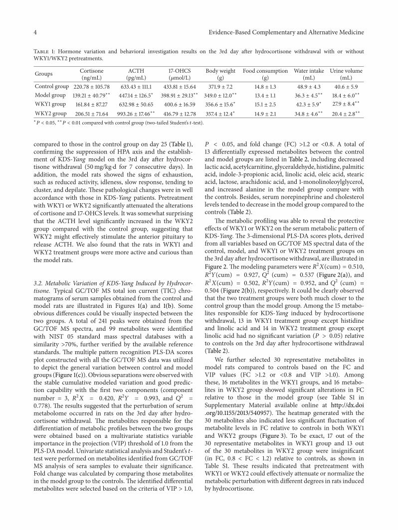
3.2. Metabolic Variation of KDS-Yang Induced by Hydrocor-tisone. Typical GC/TOF MS total ion current (TIC) chro-matograms of serum samples obtained from the control andmodel rats are illustrated in Figures 1(a) and 1(b). Someobvious differences could be visually inspected between thetwo groups. A total of 241 peaks were obtained from theGC/TOF MS spectra, and 99 metabolites were identifiedwith NIST 05 standard mass spectral databases with asimilarity >70%, further verified by the available referencestandards. The multiple pattern recognition PLS-DA scoresplot constructed with all the GC/TOF MS data was utilizedto depict the general variation between control and modelgroups (Figure 1(c)). Obvious separationswere observedwiththe stable cumulative modeled variation and good predic-tion capability with the first two components (componentnumber = 3, 𝑅2𝑋 = 0.420, 𝑅2𝑌 = 0.993, and 𝑄2 =0.778). The results suggested that the perturbation of serummetabolome occurred in rats on the 3rd day after hydro-cortisone withdrawal. The metabolites responsible for thedifferentiation of metabolic profiles between the two groupswere obtained based on a multivariate statistics variableimportance in the projection (VIP) threshold of 1.0 from thePLS-DAmodel. Univariate statistical analysis and Student’s 𝑡-test were performed on metabolites identified from GC/TOFMS analysis of sera samples to evaluate their significance.Fold change was calculated by comparing those metabolitesin the model group to the controls. The identified differentialmetabolites were selected based on the criteria of VIP > 1.0,
𝑃 < 0.05, and fold change (FC) >1.2 or <0.8. A total of13 differentially expressed metabolites between the controland model groups are listed in Table 2, including decreasedlactic acid, acetylcarnitine, glyceraldehyde, histidine, palmiticacid, indole-3-propionic acid, linolic acid, oleic acid, stearicacid, lactose, arachidonic acid, and 1-monolinoleoylglycerol,and increased alanine in the model group compare withthe controls. Besides, serum norepinephrine and cholesterollevels tended to decrease in themodel group compared to thecontrols (Table 2).
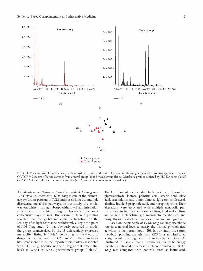
The metabolic profiling was able to reveal the protectiveeffects of WKY1 orWKY2 on the serummetabolic pattern ofKDS-Yang. The 3-dimensional PLS-DA scores plots, derivedfrom all variables based on GC/TOF MS spectral data of thecontrol, model, and WKY1 or WKY2 treatment groups onthe 3rd day after hydrocortisone withdrawal, are illustrated inFigure 2. The modeling parameters were 𝑅2𝑋(cum) = 0.510,𝑅2
𝑌(cum) = 0.927, 𝑄2 (cum) = 0.537 (Figure 2(a)), and𝑅2
𝑋(cum) = 0.502, 𝑅2𝑌(cum) = 0.952, and 𝑄2 (cum) =0.504 (Figure 2(b)), respectively. It could be clearly observedthat the two treatment groups were both much closer to thecontrol group than the model group. Among the 15 metabo-lites responsible for KDS-Yang induced by hydrocortisonewithdrawal, 13 in WKY1 treatment group except histidineand linolic acid and 14 in WKY2 treatment group exceptlinolic acid had no significant variation (𝑃 > 0.05) relativeto controls on the 3rd day after hydrocortisone withdrawal(Table 2).
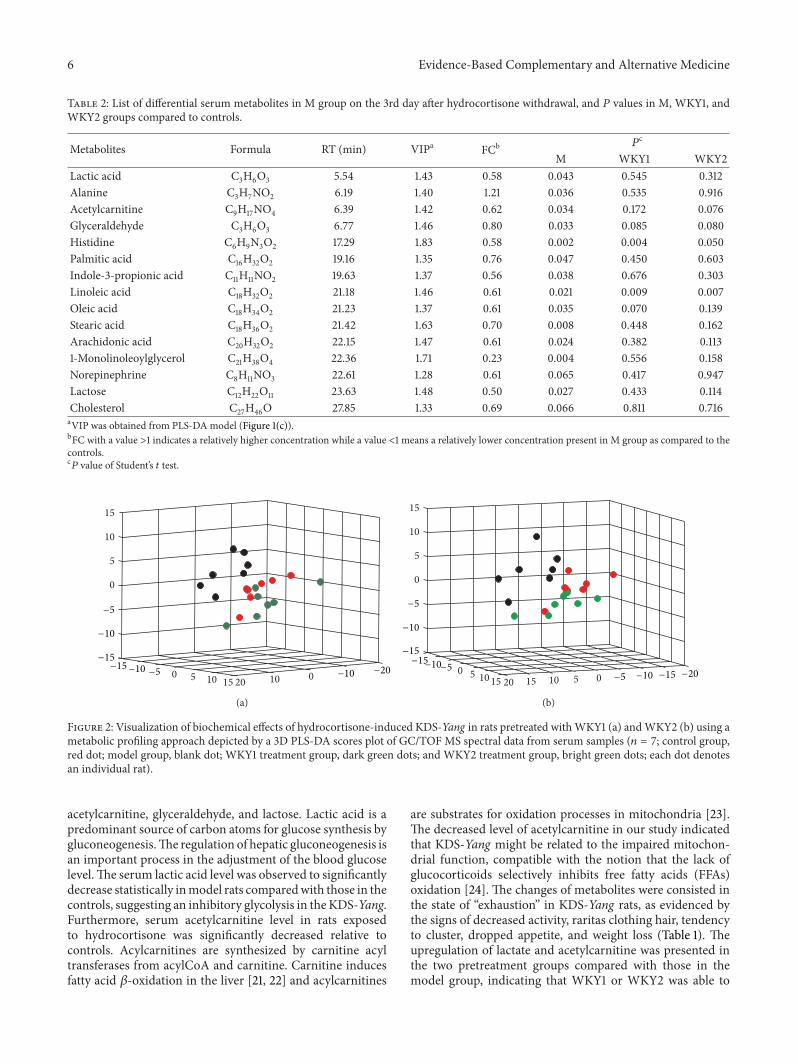
We further selected 30 representative metabolites inmodel rats compared to controls based on the FC andVIP values (FC >1.2 or <0.8 and VIP >1.0). Amongthese, 16 metabolites in the WKY1 groups, and 16 metabo-lites in WKY2 group showed significant alterations in FCrelative to those in the model group (see Table S1 inSupplementary Material available online at http://dx.doi.org/10.1155/2013/540957). The heatmap generated with the30 metabolites also indicated less significant fluctuation ofmetabolite levels in FC relative to controls in both WKY1and WKY2 groups (Figure 3). To be exact, 17 out of the30 representative metabolites in WKY1 group and 13 outof the 30 metabolites in WKY2 group were insignificant(in FC, 0.8 < FC < 1.2) relative to controls, as shown inTable S1. These results indicated that pretreatment withWKY1 or WKY2 could effectively attenuate or normalize themetabolic perturbation with different degrees in rats inducedby hydrocortisone.
Evidence-Based Complementary and Alternative Medicine 5
Control group
Time (minutes)6.66667 10 13.3333 16.6667 20 23.3333 26.6667
6e + 007
5e + 007
4e + 007
3e + 007
2e + 007
1e + 007
TIC
(a)
Model group
Time (minutes)6.66667 10 13.3333 16.6667 20 23.3333 26.6667
6e + 007
5e + 007
4e + 007
3e + 007
2e + 007
1e + 007
TIC
(b)
Control groupModel group
t[1]
t[2]0
02010
20
10
−20
−10
−20 −10
(c)
Figure 1: Visualization of biochemical effects of hydrocortisone-induced KDS-Yang in rats using a metabolic profiling approach. TypicalGC/TOF MS spectra of serum samples from control group (a) and model group (b). (c) Metabolic profiles depicted by PLS-DA sores plot ofGC/TOF MS spectral data from serum samples (𝑛 = 7, each dot denotes an individual rat).
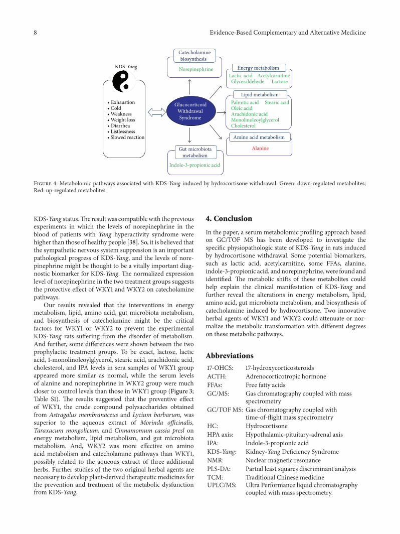
3.3. Metabolomic Pathways Associated with KDS-Yang andWKY1/WKY2 Treatments. KDS-Yang is one of the elemen-tary syndromepatterns inTCMand closely linked tomultipledisordered metabolic pathways. In our study, the modelwas established through abrupt withdrawal administrationafter exposure to a high dosage of hydrocortisone for 7consecutive days in rats. The serum metabolic profilingrevealed that the global metabolic perturbation on the3rd day after hydrocortisone withdrawal, a key time pointof KDS-Yang study [2], has obviously occurred in modelthe group characterized by the 15 differentially expressedmetabolites listing in Table 2. According to the theory ofdrugs counterevidence in TCM, most of these metabo-lites were identified as the important biomarkers associatedwith KDS-Yang, because of their insignificant differentiallevels in WKY1 or WKY2 pretreatment groups (Table 2).
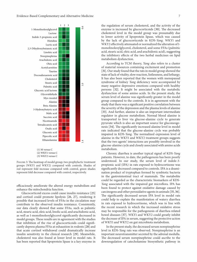
The key biomarkers included lactic acid, acetylcarnitine,glyceraldehyde, lactose, palmitic acid, stearic acid, oleicacid, arachidonic acid, 1-monolinoleoylglycerol, cholesterol,alanine, indole-3-propionic acid, and norepinephrine. Theiralterations were associated with multiple metabolic per-turbations, including energy metabolism, lipid metabolism,amino acid metabolism, gut microbiota metabolism, andbiosynthesis of catecholamine, as summarized in Figure 4.
Based on the principle of TCM, Yang can keep metabolicrate in a normal level to satisfy the normal physiologicalactivities of the human body [20]. In our study, the serummetabolic profiling analysis from KDS-Yang rats indicateda significant downregulation in metabolic activities. Asillustrated in Table 2, many metabolites related to energymetabolism showed a decreasedmetabolic tendency in KDS-Yang rats compared with controls, such as lactic acid,
6 Evidence-Based Complementary and Alternative Medicine
Table 2: List of differential serum metabolites in M group on the 3rd day after hydrocortisone withdrawal, and 𝑃 values in M, WKY1, andWKY2 groups compared to controls.
Metabolites Formula RT (min) VIPa FCb 𝑃c
M WKY1 WKY2Lactic acid C3H6O3 5.54 1.43 0.58 0.043 0.545 0.312Alanine C3H7NO2 6.19 1.40 1.21 0.036 0.535 0.916Acetylcarnitine C9H17NO4 6.39 1.42 0.62 0.034 0.172 0.076Glyceraldehyde C3H6O3 6.77 1.46 0.80 0.033 0.085 0.080Histidine C6H9N3O2 17.29 1.83 0.58 0.002 0.004 0.050Palmitic acid C16H32O2 19.16 1.35 0.76 0.047 0.450 0.603Indole-3-propionic acid C11H11NO2 19.63 1.37 0.56 0.038 0.676 0.303Linoleic acid C18H32O2 21.18 1.46 0.61 0.021 0.009 0.007Oleic acid C18H34O2 21.23 1.37 0.61 0.035 0.070 0.139Stearic acid C18H36O2 21.42 1.63 0.70 0.008 0.448 0.162Arachidonic acid C20H32O2 22.15 1.47 0.61 0.024 0.382 0.1131-Monolinoleoylglycerol C21H38O4 22.36 1.71 0.23 0.004 0.556 0.158Norepinephrine C8H11NO3 22.61 1.28 0.61 0.065 0.417 0.947Lactose C12H22O11 23.63 1.48 0.50 0.027 0.433 0.114Cholesterol C27H46O 27.85 1.33 0.69 0.066 0.811 0.716aVIP was obtained from PLS-DA model (Figure 1(c)).bFC with a value >1 indicates a relatively higher concentration while a value <1 means a relatively lower concentration present in M group as compared to thecontrols.c𝑃 value of Student’s 𝑡 test.
010200 5 10 15
0
5
10
15
−20−15−10 −10−5
−15
−10
−5
(a)
010200 5 510 15 15
0
5
10
15
−20
−15
−15−10
−10−5
−5
−15
−10
−5
(b)
Figure 2: Visualization of biochemical effects of hydrocortisone-induced KDS-Yang in rats pretreated withWKY1 (a) andWKY2 (b) using ametabolic profiling approach depicted by a 3D PLS-DA scores plot of GC/TOF MS spectral data from serum samples (𝑛 = 7; control group,red dot; model group, blank dot; WKY1 treatment group, dark green dots; and WKY2 treatment group, bright green dots; each dot denotesan individual rat).
acetylcarnitine, glyceraldehyde, and lactose. Lactic acid is apredominant source of carbon atoms for glucose synthesis bygluconeogenesis.The regulation of hepatic gluconeogenesis isan important process in the adjustment of the blood glucoselevel.The serum lactic acid level was observed to significantlydecrease statistically inmodel rats comparedwith those in thecontrols, suggesting an inhibitory glycolysis in theKDS-Yang.Furthermore, serum acetylcarnitine level in rats exposedto hydrocortisone was significantly decreased relative tocontrols. Acylcarnitines are synthesized by carnitine acyltransferases from acylCoA and carnitine. Carnitine inducesfatty acid 𝛽-oxidation in the liver [21, 22] and acylcarnitines
are substrates for oxidation processes in mitochondria [23].The decreased level of acetylcarnitine in our study indicatedthat KDS-Yang might be related to the impaired mitochon-drial function, compatible with the notion that the lack ofglucocorticoids selectively inhibits free fatty acids (FFAs)oxidation [24]. The changes of metabolites were consisted inthe state of “exhaustion” in KDS-Yang rats, as evidenced bythe signs of decreased activity, raritas clothing hair, tendencyto cluster, dropped appetite, and weight loss (Table 1). Theupregulation of lactate and acetylcarnitine was presented inthe two pretreatment groups compared with those in themodel group, indicating that WKY1 or WKY2 was able to
Evidence-Based Complementary and Alternative Medicine 7
1 2 31-Monolinoleoylglycerol
Lactose Indole-3-propionic acid
HistidineLactic acid
2,3-Dihydroxybutanoic acidLinoleic acid
NorepinephrineArachidonic acid
Oleic acidAcetylcarnitine
TaurineDocosahexaenoic acid
CholesterolStearic acid
Palmitic acidGluconic acid lactone
GlyceraldehydeMyo-inositol
AlanineBeta-Alanine
3-Hydroxybutyric acidMalic acid
Succinic acidNicotinamide
Tetradecanoic acidOxalic acid
Gluconic acidPipecolic acidEthanolamine
0.6
1.0
1.4
(1) M versus C(2) WKY1 versus C(3) WKY2 versus C
Figure 3:The heatmap of model group; two prophylactic treatmentgroups (WKY1 and WKY2) compared with controls. Shades ofred represent fold increase compared with control, green shadesrepresent fold decrease compared with control, respectively.
efficaciously ameliorate the altered energy metabolism andenhance the mitochondria function.
Glucocorticoid excess could cause insulin resistance [25]and cortisol could promote lipolysis [26, 27], rendering itpossible that increased levels of FFAs in the circulation maycontribute to the observed insulin resistance. Consistently,our data clearly showed that some FFAs, such as palmiticacid, stearic acid, oleic acid, linolic acid, and arachidonic acid,as well as 1-monolinoleoylglycerol significantly decreased inmodel groups.These results are in agreement with the studiesthat inhibition of the rise of glucocorticoids could signifi-cantly depress plasma FFAs at exhaustion in rodents [28] andthat acute cortisol withdrawal could dramatically increaseinsulin sensitivity in the clinical research [29]. Meanwhile,cholesterol was also found at lower level in model rats. Ithas been reported that lipoprotein lipase is a key enzyme in
the regulation of serum cholesterol, and the activity of theenzyme is increased by glucocorticoids [30]. The decreasedcholesterol level in the model group was presumably dueto lower activity of lipoprotein lipase, which was causedby the lack of glucocorticoids in KDS-Yang. WKY1 andWKY2 effectively attenuated or normalized the alteration of 1-monolinoleoylglycerol, cholesterol, and some FFAs (palmiticacid, stearic acid, oleic acid, and arachidonic acid), suggestingthe inhibitory effects of the two herbal medicines on lipidmetabolism dysfunction.
According to TCM theory, Yang also refers to a clusterof material resources containing excitement and promotion[31]. Our study found that the rats inmodel group showed thestate of lack of vitality, slow reaction, listlessness, and lethargy.It has also been reported that the women with menopausalsyndrome of kidney Yang deficiency were accompanied bymany negative depressive emotions compared with healthypersons [32]. It might be associated with the metabolicdysfunction of some amino acids. In the present study, theserum level of alanine was significantly greater in the modelgroup compared to the controls. It is in agreement with thestudy that there was a significant positive correlation betweenthe severity of the depression and the plasma levels of alanine[33]. And further, alanine is also an important intermediateregulator in glucose metabolism. Normal blood alanine istransported to liver via glucose-alanine cycle to generatepyruvate which is also an important source for gluconeoge-nesis [34]. The significantly increased alanine level in modelrats indicated that the glucose-alanine cycle was probablyimpaired in KDS-Yang. The normalized expression level ofalanine in the WKY1 and WKY2 treatment groups suggeststhat the two agents’ interactions are possibly involved in theglucose-alanine cycle and closely associated with amino acidsmetabolism.
Chronic diarrhea is another typical signal of KDS-Yangpatients. However, to date, the pathogenesis has been poorlyunderstood. In our study, the serum level of indole-3-propionic acid (IPA) in rats exposed to hydrocortisone wassignificantly decreased compared to controls. IPA is a deami-nation product of tryptophan formed by symbiotic bacteriain the gastrointestinal tract of mammals. The metabolitecould be regarded as the characteristic biomarkers of KDS-Yang associated with the impaired gut microflora. IPA hasbeen found to protect against oxidative damage caused bycarcinogens and other peroxidative agents in animals [35, 36].The significantly decreased serum IPA level in model ratscould help to explain the manifestation of watery diarrheain rats exposed to hydrocortisone, which was in line withthe recent research in which the increased oxidative stressmay be responsible for the pathogenesis of diarrhea-relatedbowel diseases [37]. WKY1 and WKY2 could greatly inhibitthe decrease of IPA in serum, suggesting the protective actionof WKY1 and WKY2 on gut microbiota metabolism.
In the present study, the decreased serumnorepinephrinelevel in KDS-Yang rats was observed. Norepinephrine is animportant neurotransmitter secreted by the adrenal medulla.The decreased serum norepinephrine could ascribe to thedownregulation of catecholamine biosynthetic pathway in
8 Evidence-Based Complementary and Alternative Medicine
Lactic acid AcetylcarnitineGlyceraldehyde Lactose
Energy metabolism
Lipid metabolismPalmitic acid Stearic acid Oleic acid Arachidonic acidMonolinoleoylglycerolCholesterol
Amino acid metabolism
Alanine
Indole-3-propionic acid
Gut microbiota metabolism
Norepinephrine
Catecholaminebiosynthesis
Glucocorticoid Withdrawal Syndrome
• Exhaustion• Cold• Weakness• Weight loss• Diarrhea• Listlessness• Slowed reaction
KDS-Yang
Figure 4: Metabolomic pathways associated with KDS-Yang induced by hydrocortisone withdrawal. Green: down-regulated metabolites;Red: up-regulated metabolites.
KDS-Yang status.The result was compatiblewith the previousexperiments in which the levels of norepinephrine in theblood of patients with Yang hyperactivity syndrome werehigher than those of healthy people [38]. So, it is believed thatthe sympathetic nervous system suppression is an importantpathological progress of KDS-Yang, and the levels of nore-pinephrine might be thought to be a vitally important diag-nostic biomarker for KDS-Yang. The normalized expressionlevel of norepinephrine in the two treatment groups suggeststhe protective effect of WKY1 and WKY2 on catecholaminepathways.
Our results revealed that the interventions in energymetabolism, lipid, amino acid, gut microbiota metabolism,and biosynthesis of catecholamine might be the criticalfactors for WKY1 or WKY2 to prevent the experimentalKDS-Yang rats suffering from the disorder of metabolism.And further, some differences were shown between the twoprophylactic treatment groups. To be exact, lactose, lacticacid, 1-monolinoleoylglycerol, stearic acid, arachidonic acid,cholesterol, and IPA levels in sera samples of WKY1 groupappeared more similar as normal, while the serum levelsof alanine and norepinephrine in WKY2 group were muchcloser to control levels than those in WKY1 group (Figure 3;Table S1). The results suggested that the preventive effectof WKY1, the crude compound polysaccharides obtainedfrom Astragalus membranaceus and Lycium barbarum, wassuperior to the aqueous extract of Morinda officinalis,Taraxacum mongolicum, and Cinnamomum cassia presl onenergy metabolism, lipid metabolism, and gut microbiotametabolism. And, WKY2 was more effective on aminoacid metabolism and catecholamine pathways than WKY1,possibly related to the aqueous extract of three additionalherbs. Further studies of the two original herbal agents arenecessary to develop plant-derived therapeutic medicines forthe prevention and treatment of the metabolic dysfunctionfrom KDS-Yang.
4. Conclusion
In the paper, a serum metabolomic profiling approach basedon GC/TOF MS has been developed to investigate thespecific physiopathologic state of KDS-Yang in rats inducedby hydrocortisone withdrawal. Some potential biomarkers,such as lactic acid, acetylcarnitine, some FFAs, alanine,indole-3-propionic acid, andnorepinephrine,were found andidentified. The metabolic shifts of these metabolites couldhelp explain the clinical manifestation of KDS-Yang andfurther reveal the alterations in energy metabolism, lipid,amino acid, gut microbiota metabolism, and biosynthesis ofcatecholamine induced by hydrocortisone. Two innovativeherbal agents of WKY1 and WKY2 could attenuate or nor-malize the metabolic transformation with different degreeson these metabolic pathways.
Abbreviations
17-OHCS: 17-hydroxycorticosteroidsACTH: Adrenocorticotropic hormoneFFAs: Free fatty acidsGC/MS: Gas chromatography coupled with mass
spectrometryGC/TOF MS: Gas chromatography coupled with
time-of-flight mass spectrometryHC: HydrocortisoneHPA axis: Hypothalamic-pituitary-adrenal axisIPA: Indole-3-propionic acidKDS-Yang: Kidney-Yang Deficiency SyndromeNMR: Nuclear magnetic resonancePLS-DA: Partial least squares discriminant analysisTCM: Traditional Chinese medicineUPLC/MS: Ultra Performance liquid chromatography
coupled with mass spectrometry.
Evidence-Based Complementary and Alternative Medicine 9
Authors’ Contribution
Linjing Zhao, Hongbing Wu, and Mingfeng Qiu contributedequally to this work.
Acknowledgment
This study was financially supported by Infinitus (China)Company Ltd., a member of LKK Health Products Group.
References
[1] Z. Shen, “The location of deficiency syndrome of kidney Yang,”Chinese Medical Journal, vol. 112, no. 11, pp. 973–975, 1999.
[2] Q. Chen, Experimental Methodology of PharmacologicalResearch in Traditional Chinese Medicine, People’s HealthPublishing House, Beijing, China, 1993.
[3] H. Cao, S. T. Wang, L. Y. Wu, X. T. Wang, and A. P. Jiang,“Pharmacological study on Tianxiong (tuber of Aconitumcarmichaeli Debx.), a Chinese drug for reinforcing the kidneyyang retail in Hong Kong market,” China Journal of ChineseMateria Medica, vol. 26, no. 6, pp. 369–372, 2001.
[4] J. Yang, Y. Wang, Y. Bao, and J. Guo, “The total flavones fromSemen cuscutae reverse the reduction of testosterone leveland the expression of androgen receptor gene in kidney-yangdeficientmice,” Journal of Ethnopharmacology, vol. 119, no. 1, pp.166–171, 2008.
[5] C. M. Wang, S. Y. Xu, S. Lai et al., “Curculigo orchioides (XianMao) modifies the activity and protein expression of CYP3Ain normal and Kidney-Yang Deficiency model rats,” Journal ofEthnopharmacology, vol. 144, no. 1, pp. 33–38, 2012.
[6] J. K. Nicholson and I. D. Wilson, “Understanding ‘global’systems biology: metabonomics and the continuum ofmetabolism,” Nature Reviews Drug Discovery, vol. 2, no. 8, pp.668–676, 2003.
[7] M. Gong, W. Ye, Y. Xie et al., “Metabonomic study of interven-tion effects of Morinda officinalis on ‘kidney-yang deficiencysyndrome’,” China Journal of Chinese Materia Medica, vol. 37,no. 11, pp. 1682–1685, 2012.
[8] X. Lu, Z. Xiong, J. Li, S. Zheng, T. Huo, and F. Li, “Metabonomicstudy on ‘Kidney-Yang Deficiency syndrome’ and interventioneffects of Rhizoma Drynariae extracts in rats using ultraperformance liquid chromatography coupled with mass spec-trometry,” Talanta, vol. 83, no. 3, pp. 700–708, 2011.
[9] F. Li, X. Lu, H. Liu, M. Liu, and Z. Xiong, “A pharmaco-metabonomic study on the therapeutic basis and metaboliceffects of Epimedium brevicornumMaxim. on hydrocortisone-induced rat using UPLC-MS,” Biomedical Chromatography, vol.21, no. 4, pp. 397–405, 2007.
[10] D. Huang, J. Yang, X. Lu et al., “An integrated plasma andurinary metabonomic study using UHPLC-MS: interventioneffects of Epimedium koreanum on ‘Kidney-Yang Deficiencysyndrome’ rats,” Journal of Pharmaceutical and BiomedicalAnalysis, vol. 76, pp. 200–206, 2013.
[11] M. Chen, L. Zhao, and W. Jia, “Metabonomic study on the bio-chemical profiles of a hydrocortisone-induced animal model,”Journal of Proteome Research, vol. 4, no. 6, pp. 2391–2396, 2005.
[12] Y. Qiu, M. Chen, M. Su et al., “Metabolic profiling revealstherapeutic effects of Herba Cistanches in an animal modelof hydrocortisone-induced ‘kidney-deficiency syndrome’,” Chi-nese Medicine, vol. 3, article 3, 2008.
[13] C. H. Cho, Q. B. Mei, P. Shang et al., “Study of the gastrointesti-nal protective effects of polysaccharides from Angelica sinensisin rats,” Planta Medica, vol. 66, no. 4, pp. 348–351, 2000.
[14] M. Dubois, K. A. Gilles, J. K. Hamilton, P. A. Rebers, and F.Smith, “Colorimetric method for determination of sugars andrelated substances,”Analytical Chemistry, vol. 28, no. 3, pp. 350–356, 1956.
[15] Y. Qiu, G. Cai, M. Su et al., “Serum metabolite profilingof human colorectal cancer using GC-TOFMS and UPLC-QTOFMS,” Journal of Proteome Research, vol. 8, no. 10, pp.4844–4850, 2009.
[16] P. Jonsson, J. Gullberg, A. Nordstrom et al., “A strategy foridentifying differences in large series of metabolomic samplesanalyzed by GC/MS,” Analytical Chemistry, vol. 76, no. 6, pp.1738–1745, 2004.
[17] P. Jonsson, A. I. Johansson, J. Gullberg et al., “High-throughputdata analysis for detecting and identifying differences betweensamples in GC/MS-based metabolomic analyses,” AnalyticalChemistry, vol. 77, no. 17, pp. 5635–5642, 2005.
[18] Y. Ni, M. Su, Y. Qiu et al., “Metabolic profiling using combinedGC-MS and LC-MS provides a systems understanding ofaristolochic acid-induced nephrotoxicity in rat,” FEBS Letters,vol. 581, no. 4, pp. 707–711, 2007.
[19] Z. Y. Shen, “Study on localization of kidney-yang deficiency,”Zhongguo Zhong Xi Yi Jie He Za Zhi, vol. 17, no. 1, pp. 50–52,1997.
[20] S. Lin and S. J. Feng, “Reviews of kidney-Yang deciencysyndrome,” Hebei Journal of Traditional Chinese Medicine, vol.33, no. 3, pp. 463–465, 2011.
[21] I. B. Fritz, “Action of carnitine on long chain fatty acid oxidationby liver,”The American Journal of Physiology, vol. 197, no. 2, pp.297–304, 1959.
[22] I. B. Fritz and K. T. Yue, “Effects of carnitine on acetyl-CoAoxidation by heartmusclemitochondria,”TheAmerican Journalof Physiology, vol. 206, no. 3, pp. 531–535, 1964.
[23] J. Bremer, “Carnitine in intermediary metabolism. Themetabolism of fatty acid esters of carnitine by mitochondria,”The Journal of Biological Chemistry, vol. 237, no. 12, pp.3628–3632, 1962.
[24] K. R. Short, J. Nygren, M. L. Bigelow, and K. S. Nair, “Effectof short-term prednisone use on blood flow, muscle proteinmetabolism, and function,” The Journal of Clinical Endocrinol-ogy & Metabolism, vol. 89, no. 12, pp. 6198–6207, 2004.
[25] G. Dimitriadis, B. Leighton, M. Parry-Billings et al., “Effectsof glucocorticoid excess on the sensitivity of glucose transportand metabolism to insulin in rat skeletal muscle,” BiochemicalJournal, vol. 321, part 3, pp. 707–712, 1997.
[26] C. H. Gravholt, R. Dall, J. S. Christiansen, N. Møller, andO. Schmitz, “Preferential stimulation of abdominal subcuta-neous lipolysis after prednisolone exposure in humans,”ObesityResearch, vol. 10, no. 8, pp. 774–781, 2002.
[27] C. Djurhuus, C. H. Gravholt, S. Nielsen et al., “Effects of cortisolon lipolysis and regional interstitial glycerol levels in humans,”American Journal of Physiology, vol. 283, no. 1, pp. E172–E177,2002.
[28] T. L. Sellers, A. W. Jaussi, H. T. Yang, R. W. Heninger, andW. W. Winder, “Effect of the exercise-induced increase inglucocorticoids on endurance in the rat,” Journal of AppliedPhysiology, vol. 65, no. 1, pp. 173–178, 1988.
[29] J. J. Christiansen, C. B. Djurhuus, C. H. Gravholt et al., “Effectsof cortisol on carbohydrate, lipid, and protein metabolism:
10 Evidence-Based Complementary and Alternative Medicine
studies of acute cortisol withdrawal in adrenocortical failure,”The Journal of Clinical Endocrinology &Metabolism, vol. 92, no.9, pp. 3553–3559, 2007.
[30] J. R. Mead, S. A. Irvine, and D. P. Ramji, “Lipoprotein lipase:structure, function, regulation, and role in disease,” Journal ofMolecular Medicine, vol. 80, no. 12, pp. 753–769, 2002.
[31] G. R. Sun, Basic Theory of Traditional Chinese Medicine, ChinaPress of Traditional Chinese Medicine, Beijing, China, 2006.
[32] B. J. Wu, Y. Wang, Y. X. Liu, and F. Wang, “Characteristics ofmemory functional impairment in women with menopausalsyndrome of kidney yang deficiency and yin deficiency types,”Chinese Journal of Clinical Rehabilitation, vol. 10, no. 39, pp. 18–20, 2006.
[33] H. Mitani, Y. Shirayama, T. Yamada, K. Maeda, C. R. Ashby Jr.,and R. Kawahara, “Correlation between plasma levels of glu-tamate, alanine and serine with severity of depression,” Progressin Neuro-Psychopharmacology and Biological Psychiatry, vol. 30,no. 6, pp. 1155–1158, 2006.
[34] C. A. Nichol and F. Rosen, “Changes in alanine transaminaseactivity related to corticosteroid treatment or capacity forgrowth,” Advances in Enzyme Regulation, vol. 1, pp. 341–361,1963.
[35] M. Karbownik, M. Stasiak, A. Zygmunt, K. Zasada, andA. Lewinski, “Protective effects of melatonin and indole-3-propionic acid against lipid peroxidation, caused by potassiumbromate in the rat kidney,” Cell Biochemistry and Function, vol.24, no. 6, pp. 483–489, 2006.
[36] M. Karbownik, E. Gitto, A. Lewinski, and R. J. Reiter, “Relativeefficacies of indole antioxidants in reducing autoxidation andiron-induced lipid peroxidation in hamster testes,” Journal ofCellular Biochemistry, vol. 81, no. 4, pp. 693–699, 2001.
[37] N. Sengul, S. Isık, B. Aslım et al., “The effect ofexopolysaccharide-producing probiotic strains on gut oxidativedamage in experimental colitis,”Digestive Diseases and Sciences,vol. 56, no. 3, pp. 707–714, 2011.
[38] H. Liao, D. P. Li, Q. Chen et al., “Observation on therapeuticeffect of “reducing south and reinforcing north” needlingmethod on hypertension of type of yang-hyperactivity due toyin-deficiency,” Chinese Acupuncture & Moxibustion, vol. 26,no. 2, pp. 91–93, 2006.

Submit your manuscripts athttp://www.hindawi.com
Stem CellsInternational
Hindawi Publishing Corporationhttp://www.hindawi.com Volume 2014
Hindawi Publishing Corporationhttp://www.hindawi.com Volume 2014
MEDIATORSINFLAMMATION
of
Hindawi Publishing Corporationhttp://www.hindawi.com Volume 2014
Behavioural Neurology
EndocrinologyInternational Journal of
Hindawi Publishing Corporationhttp://www.hindawi.com Volume 2014
Hindawi Publishing Corporationhttp://www.hindawi.com Volume 2014
Disease Markers
Hindawi Publishing Corporationhttp://www.hindawi.com Volume 2014
BioMed Research International
OncologyJournal of
Hindawi Publishing Corporationhttp://www.hindawi.com Volume 2014
Hindawi Publishing Corporationhttp://www.hindawi.com Volume 2014
Oxidative Medicine and Cellular Longevity
Hindawi Publishing Corporationhttp://www.hindawi.com Volume 2014
PPAR Research
The Scientific World JournalHindawi Publishing Corporation http://www.hindawi.com Volume 2014
Immunology ResearchHindawi Publishing Corporationhttp://www.hindawi.com Volume 2014
Journal of
ObesityJournal of
Hindawi Publishing Corporationhttp://www.hindawi.com Volume 2014
Hindawi Publishing Corporationhttp://www.hindawi.com Volume 2014
Computational and Mathematical Methods in Medicine
OphthalmologyJournal of
Hindawi Publishing Corporationhttp://www.hindawi.com Volume 2014
Diabetes ResearchJournal of
Hindawi Publishing Corporationhttp://www.hindawi.com Volume 2014
Hindawi Publishing Corporationhttp://www.hindawi.com Volume 2014
Research and TreatmentAIDS
Hindawi Publishing Corporationhttp://www.hindawi.com Volume 2014
Gastroenterology Research and Practice
Hindawi Publishing Corporationhttp://www.hindawi.com Volume 2014
Parkinson’s Disease
Evidence-Based Complementary and Alternative Medicine
Volume 2014Hindawi Publishing Corporationhttp://www.hindawi.com