Embed Size (px)
Citation preview
RESEARCH PAPER
Neutrophils contribute to intracerebralhaemorrhages after treatment with recombinanttissue plasminogen activator following cerebralischaemia
Sophie Gautier*, Thavarak Ouk*, Olivier Petrault1, Jacques Caron and Régis Bordet
EA1046 – Département de Pharmacologie médicale, Institut de Médecine Prédictive et de Recherche Thérapeutique, Universitéde Lille 2 – Faculté de Médecine, Centre Hospitalier Universitaire, Lille, France
Background and purpose: Polymorphonuclear neutrophils (PMNs) contribute to the vascular damage caused by transientcerebral ischaemia. Here we have evaluated the role of PMNs in intracerebral haemorrhage (ICH) induced in a model ofthrombolysis with recombinant tissue plasminogen activator (t-PA) during the acute phase of cerebral ischaemia.Experimental approach: The middle cerebral artery (MCA) of male spontaneously hypertensive rats was occluded for 1 hfollowed by reperfusion and, 5 h later, infusion of thrombolytic products (generated in vitro by t-PA on autologous clots). Effectsof pretreatment (before the MCA occlusion) with vinblastine (4 days before; 0.5 mg·kg-1), monoclonal anti-neutrophil antibody(mAbRP3; 12 h, 0.3 mg·kg-1) or saline on ICH, neutrophil infiltration, MCA vascular reactivity and brain infarct volume wereassessed, 24 h after the beginning of reperfusion.Key results: Depletion of circulating neutrophils significantly reduced t-PA-induced ICH (vinblastine, 4.6 � 1.0; mAbRP3,5.2 � 1.0 vs. saline, 10.8 � 2.7 haemorrhages; P < 0.05). This depletion was associated with a decrease in cerebral infiltrationby neutrophils and a decrease of endothelium-dependent, vascular dysfunction in isolated MCA, induced by the ischaemia/reperfusion and t-PA treatment. Brain infarct volume was significantly decreased after vinblastine treatment (159 � 13 mm3 vs.243 � 16 mm3 with saline; P < 0.01) but not after depletion with mAbRP3 (221 � 22 mm3).Conclusions and implications: Our results showed that pharmacological depletion of PMNs prevented t-PA-induced ICH, inparallel with a decrease in cerebral infiltration by PMNs and a decreased endothelial dysfunction in cerebral blood vessels.British Journal of Pharmacology (2009) 156, 673–679; doi:10.1111/j.1476-5381.2009.00068.x; published online 4February 2009
Keywords: stroke; haemorrhages; t-PA; neutrophils; vascular endothelium
Abbreviations: ICH, intracerebral haemorrhage; I/R, ischaemia/reperfusion; MCAO, middle cerebral artery occlusion; t-PA,tissue plasminogen activator; TLP, thrombolysis products
Introduction
Although clot lysis with recombinant tissue plasminogenactivator (t-PA) is effective in acute ischaemic stroke, it is alsoassociated with a risk of haemorrhage, especially deleter-ious for the patients (Hacke et al., 1995; The NINDS and Stroket-PA Study Group, 1995). Many hypotheses have been pro-posed to explain the intracerebral haemorrhages (ICH) duringthrombolysis (Tejima et al., 2001), and using adjunctive
pharmacological strategies could be one of the interesting waysto prevent these serious complications.
We previously demonstrated the role of t-PA-induced clotlysis in ICH in a mechanical thrombolysis-related brainbleeding model based on the post-ischaemic perfusion of asolution resulting from the in vitro interaction between t-PAand blood clots, leading to thrombolysis products (TLP)(Gautier et al., 2003). In this model, perfusion of TLPincreased the severity of ICH in comparison with the perfu-sion of t-PA alone, and was associated with a larger infarctvolume and an increased vascular dysfunction in compari-son with damage induced by middle cerebral artery occlu-sion (MCAO) alone. Moreover, the permeability of theblood-brain barrier (BBB) and metalloproteinase 9 (MMP-9)activity were also affected (Kahles et al., 2005). These resultsadded to those demonstrating impairment of vascular reac-tivity during t-PA treatment, in parallel to aggravation of
Correspondence: R Bordet, Laboratoire de Pharmacologie, Faculté de Méde-cine, 1 place de Verdun 59045 Lille cedex, France. E-mail: [email protected] address: Laboratoire de Physiopathologie de la Barriere Hemato-Encephalique, EA2465, Faculté de Sciences Jean Perrin, 62307 Lens cedex.*These authors contributed equally to this work.Received 1 August 2008; revised 29 September 2008; accepted 6 October 2008
British Journal of Pharmacology (2009), 156, 673–679© 2009 The AuthorsJournal compilation © 2009 The British Pharmacological Society All rights reserved 0007-1188/09www.brjpharmacol.org
brain injury (Cipolla et al., 2000; Wang and Lo, 2003; Nassaret al., 2004; Yang et al., 2007).
The link between brain injury, vascular impairment andt-PA-related ICH is not clearly defined. Reperfusion-generatedfree radicals are mediators of the extent of the infarct andof the impairment of vascular function (Petrault et al., 2004),and use of antioxidant drugs concomitant to thromboly-sis prevented ICH and decreased infarct size (Lapchak andAraujo, 2005). Neutrophils mediate inflammation and con-tribute to vascular and parenchymal damage (Del Zoppo andMabuchi, 2003). Pharmacological inhibition of neutrophilemigration from the vasculature or neutrophil depletion aftertransient MCAO reduced the post-ischaemic alterations ofvascular endothelium and in parallel reduced the infarctsize (Emerich et al., 2002; Petrault et al., 2005). Activation ofMMP-9 is responsible for matrix degradation and increasedpermeability of the BBB, associated with ICH (Kelly et al.,2006), and inhibition of MMP-9 prevented t-PA-related ICH(Pfefferkorn and Rosenberg, 2003). Interestingly, MMP-9 isup-regulated by t-PA (Ning et al., 2006), and the increasedMMP-9 levels observed in infarcted areas were related toneutrophil infiltration into brain (Rosell et al., 2008).
In the present study, we evaluated in our thrombolysis-related brain haemorrhage model, the contribution ofpolymorphonuclear neutrophils (PMNs) on t-PA-inducedhaemorrhages, and in parallel, on vascular endothelial func-tion and infarct volume, both affected during ischaemia andthrombolysis with t-PA. We used two methods to depleteneutrophils, a non-specific agent (vinblastine) and the morespecific anti-neutrophil monoclonal antibody (mAbRP3).
Methods
AnimalsAll experiments were performed in strict accordance with theguidelines of the National Institutes of Health and FrenchDepartment of Agriculture. In our model, male spontaneouslyhypertensive rats (SHR) (Elevage Janvier, France), 10 weeks oldand weighing 270 to 320 g, were used. The choice of SHR wasbased on the role of hypertension as a risk factor for haem-orrhage during thrombolysis (Gautier et al., 2003).
Experimental designThree different groups of SHR were randomly selected:vehicle-treated group (n = 9), vinblastine-treated group (n = 9)and mAbRP3-treated group (n = 9). All rats were submitted toischaemia/reperfusion (I/R) and perfused with a t-PA/clotsolution, simulating thrombolysis. The rats included in thestudy were those that completed the whole protocol and hada histologically proven ischaemia.
Neutrophil depletion was induced by i.v. injection of vin-blastine (0.5 mg·kg-1, 0.15 mL; EG Labo) 4 days before I/R orby i.p. injection of mAbRP3 (0.3 mg·kg-1, 1 mL; BD Pharmin-gen) 12 h before ischaemia and again during ischaemia(2 mL) (Sekiya et al., 1990). The vehicle group received saline(0.9%).
Twenty four hours after reperfusion, animals were killedand tissues processed to assess vasoreactivity in the MCA and
for immunohistochemical and histomorphometric analysis.Blood samples were collected before surgery for MCAO and24 h later, to count leucocytes and PMNs (Machine XE 2100,Sysmex).
Cerebral ischaemia and t-PA treatmentUnder anaesthesia (chloral hydrate 300 mg·kg-1), focal cere-bral ischaemia was induced for 60 min by an occlusion ofthe right MCA with an intraluminal filament inserted inthe external carotid artery to the internal carotid artery.Subsequently, the right jugular vein was cannulated with apolyethylene catheter (24 gauge). Physiological parameters(temperature, mean arterial pressure, arterial blood pH, PaO2
and PaCO2) were controlled throughout the experiments.After the occlusion, animals recovered from anaesthesia andwere allowed to eat and drink freely. There was no evaluationof possible neurological deficits. To mimic thrombolysisin vivo, a solution of TLP was prepared in vitro, as follows.Autologous blood (0.2 mL) was taken from the jugular veinand left for 5 h to form a thrombus. To obtain TLP for perfu-sion, the thrombus was then fragmented, and t-PA [1.5 mL oft-PA 20 mg·mL-1; this is equivalent to 10 mg·kg-1 (6 mL·kg-1)of t-PA for each rat] was applied to the pieces of thrombusfor 30 min. The resulting solution (TLP solution, contain-ing mainly plasmin; total volume 2 mL) was collected andperfused slowly (over 1 h) into the rat, 6 h after ischaemia(Gautier et al., 2003).
Assessment of ischaemia and evaluation of haemorrhagesRats were anaesthetized with pentobarbitone (1 mL of55 mg·mL-1) and the chest opened. Saline (NaCl 0.9%; 60 mL)was perfused intracardially. Then the brain was rapidlyremoved and frozen at -80°C. Coronal slices (20 mm) werecut at 12 levels according to the stereotaxic section mapof Paxinos and Watson. To locate the ischaemia, all sectionswere stained with cresyl fast violet, a marker of non-necrotizing cells, to permit differentiation between ischaemicand normal tissue. Infarct volume (in mm3) was measured byusing a numerical integration of the respective ischaemicareas for all sections per animal. The volume was corrected foroedema by using the following equation: corrected infarctvolume = infarct volume ¥ (right hemisphere volume/lefthemisphere volume). Three defined sections (+0.48, -0.92and -3.30 mm relative to Bregma) were histologically exam-ined for ICH, without knowledge of the treatment group. Theincidence of ICH was scaled according to a modified, previ-ously described, method (Niessen et al., 2003): 0 = no haem-orrhages, 1 = multiple macroscopically visible haemorrhages(seen as petechiae). Severity was estimated as the number ofpetechial haemorrhages per infarct area. In some animals, invivo magnetic resonance imaging was performed just beforedeath in a 7 tesla narrow bore small animal imaging system(Biospec 70/20 USR, Bruker Biospin, Wissembourg, Germany).We acquired two dimensional T2-weighted images, using tur-boRARE pulse sequence: TR2500 ms, TE65 ms, FOV: 4 ¥ 4 cm,matrix: 256 ¥ 256, RARE factor 8.
Myeloperoxidase immunohistochemistryNeutrophil infiltration was assessed by quantifying mye-loperoxidase (MPO), an enzyme expressed in neutrophils
Neutrophils in t-PA-associated haemorrhage674 S Gautier et al
British Journal of Pharmacology (2009) 156 673–679
(Matsuo et al., 1994). After fixation, sections were blockedand incubated with a 1:500 dilution of rabbit polyclonalanti-MPO primary antibody (DAKO) overnight at 4°C versus anegative control. Sections were then incubated with biotiny-lated goat anti-rabbit secondary antibody for 3 h at roomtemperature, followed by an ABC process (ABC kit, Vector)and finally treated with diaminobenzidine. Neutrophil infil-tration was quantified at one coronal level (+0.48 mm relativeto Bregma) counting positive cells on six adjacent fields of1 mm2 in the ischaemic zone. As controls, we used brainsections of sham rats, submitted to surgery for MCAO,without advancing the intraluminal filament into the inter-nal carotid artery and t-PA treatment.
Analysis of the vasoreactivity of isolated MCAEndothelium-dependent and independent relaxations wereassessed in a Halpern arteriograph (Living Systems Instrumen-tation, Burlington, Vermont, USA) (Petrault et al., 2005). Aproximal segment of the right MCA was mounted on twoglass cannulas perfused with oxygenated Krebs solution main-tained at 37°C and pH 7.4. Experiments were performedunder no-flow conditions. The lumen diameter was measuredby image analysis and recorded.
All mounted and pressurized arteries were stabilized at atransmural pressure of 100 mmHg for 1 h before experiments.Concentration–response curves for the relaxation to acetyl-choline (Ach) were constructed by cumulative addition of Ach(0.001 to 10 mmol·L-1). NO-mediated relaxations were deter-mined with a single concentration of sodium nitroprusside(SNP 10 mmol·L-1) on MCA pre-constricted with 5-HT. Thevasorelaxant responses observed in MCAs from the threegroups of SHR (vehicle, vinblastine and mAbRP3) were com-pared with responses measured in MCAs from a control groupof normotensive Wistar-Kyoto male rats, which served ascontrol group for physiological conditions (Dupuis et al.,2005). Relaxation responses were expressed as percentage ofincrease of the diameter of the pre-constricted artery.
Statistical analysisAll values were expressed as mean � standard error mean(SEM). Continuous variables (infarct volumes, number ofhaemorrhages and MCA relaxation) were compared with aone-way ANOVA followed by a post hoc protected least signifi-cant difference (PLSD) Fisher test, if variance analysis wassignificant. A X2 analysis was performed to compare results
expressed as frequency. A value of P < 0.05 was considered toindicate statistically significant differences between means.
Results
Physiological parameters (temperature, blood pressure andgases) remained within the normal range during the 1 hischaemia and the beginning of reperfusion in all groups.Mortality was low and comparable in the different groups:two vehicle-treated rats and two mAbRP3-treated rats diedbefore the end of the study protocol. At 24 h, group sizeswere: vehicle, n = 7; vinblastine, n = 9 and mAbRP3, n = 7.
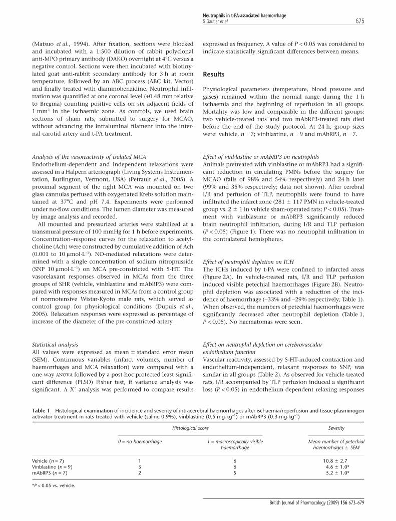
Effect of vinblastine or mAbRP3 on neutrophilsAnimals pretreated with vinblastine or mAbRP3 had a signifi-cant reduction in circulating PMNs before the surgery forMCAO (falls of 98% and 54% respectively) and 24 h later(99% and 35% respectively; data not shown). After cerebralI/R and perfusion of TLP, neutrophils were found to haveinfiltrated the infarct zone (281 � 117 PMN in vehicle-treatedgroup vs. 2 � 1 in vehicle sham-operated rats; P < 0.05). Treat-ment with vinblastine or mAbRP3 significantly reducedbrain neutrophil infiltration, during I/R and TLP perfusion(P < 0.05) (Figure 1). There was no neutrophil infiltration inthe contralateral hemispheres.
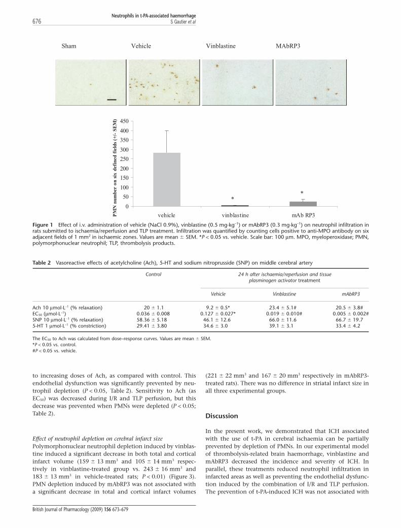
Effect of neutrophil depletion on ICHThe ICHs induced by t-PA were confined to infarcted areas(Figure 2A). In vehicle-treated rats, I/R and TLP perfusioninduced visible petechial haemorrhages (Figure 2B). Neutro-phil depletion was associated with a reduction of the inci-dence of haemorrhage (-33% and -29% respectively; Table 1).When observed, the numbers of petechial haemorrhages weresignificantly decreased after neutrophil depletion (Table 1,P < 0.05). No haematomas were seen.
Effect on neutrophil depletion on cerebrovascularendothelium functionVascular reactivity, assessed by 5-HT-induced contraction andendothelium-independent, relaxant responses to SNP, wassimilar in all groups (Table 2). As observed for vehicle-treatedrats, I/R accompanied by TLP perfusion induced a significantloss (P < 0.05) in endothelium-dependent relaxing responses
Table 1 Histological examination of incidence and severity of intracerebral haemorrhages after ischaemia/reperfusion and tissue plasminogenactivator treatment in rats treated with vehicle (saline 0.9%), vinblastine (0.5 mg·kg-1) or mAbRP3 (0.3 mg·kg-1)
Histological score Severity
0 = no haemorrhage 1 = macroscopically visiblehaemorrhage
Mean number of petechialhaemorrhages � SEM
Vehicle (n = 7) 1 6 10.8 � 2.7Vinblastine (n = 9) 3 6 4.6 � 1.0*mAbRP3 (n = 7) 2 5 5.2 � 1.0*
*P < 0.05 vs. vehicle.
Neutrophils in t-PA-associated haemorrhageS Gautier et al 675
British Journal of Pharmacology (2009) 156 673–679
to increasing doses of Ach, as compared with control. Thisendothelial dysfunction was significantly prevented by neu-trophil depletion (P < 0.05, Table 2). Sensitivity to Ach (asEC50) was decreased during I/R and TLP perfusion, but thisdecrease was prevented when PMNs were depleted (P < 0.05;Table 2).
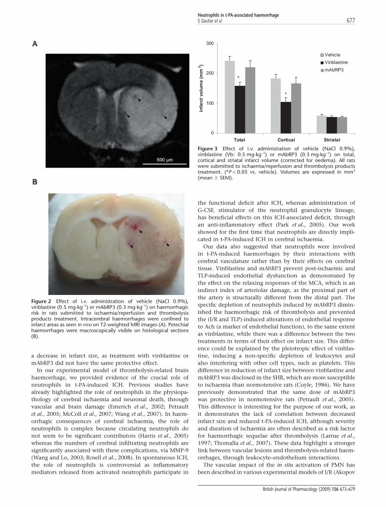
Effect of neutrophil depletion on cerebral infarct sizePolymorphonuclear neutrophil depletion induced by vinblas-tine induced a significant decrease in both total and corticalinfarct volume (159 � 13 mm3 and 105 � 14 mm3 respec-tively in vinblastine-treated group vs. 243 � 16 mm3 and183 � 13 mm3 in vehicle-treated rats; P < 0.01) (Figure 3).PMN depletion induced by mAbRP3 was not associated witha significant decrease in total and cortical infarct volumes
(221 � 22 mm3 and 167 � 20 mm3 respectively in mAbRP3-treated rats). There was no difference in striatal infarct size inall three experimental groups.
Discussion
In the present work, we demonstrated that ICH associatedwith the use of t-PA in cerebral ischaemia can be partiallyprevented by depletion of PMNs. In our experimental modelof thrombolysis-related brain haemorrhage, vinblastine andmAbRP3 decreased the incidence and severity of ICH. Inparallel, these treatments reduced neutrophil infiltration ininfarcted areas as well as preventing the endothelial dysfunc-tion induced by the combination of I/R and TLP perfusion.The prevention of t-PA-induced ICH was not associated with
Sham Vehicle Vinblastine MAbRP3
0
50
100
150
200
250
300
350
400
450
vehicle vinblastine mAb RP3PM
N n
umbe
r on
six
def
ined
fie
lds
(+/-
SE
M)
**
Figure 1 Effect of i.v. administration of vehicle (NaCl 0.9%), vinblastine (0.5 mg·kg-1) or mAbRP3 (0.3 mg·kg-1) on neutrophil infiltration inrats submitted to ischaemia/reperfusion and TLP treatment. Infiltration was quantified by counting cells positive to anti-MPO antibody on sixadjacent fields of 1 mm2 in ischaemic zones. Values are mean � SEM. *P < 0.05 vs. vehicle. Scale bar: 100 mm. MPO, myeloperoxidase; PMN,polymorphonuclear neutrophil; TLP, thrombolysis products.
Table 2 Vasoreactive effects of acetylcholine (Ach), 5-HT and sodium nitroprusside (SNP) on middle cerebral artery
Control 24 h after ischaemia/reperfusion and tissueplasminogen activator treatment
Vehicle Vinblastine mAbRP3
Ach 10 mmol·L-1 (% relaxation) 20 � 1.1 9.2 � 0.5* 23.4 � 5.1# 20.5 � 3.8#EC50 (mmol·L-1) 0.036 � 0.008 0.127 � 0.027* 0.019 � 0.010# 0.005 � 0.002#SNP 10 mmol·L-1 (% relaxation) 58.36 � 5.18 46.1 � 12.6 66.0 � 11.6 66.7 � 19.75-HT 1 mmol·L-1 (% constriction) 29.41 � 3.80 34.6 � 3.0 39.1 � 3.1 33.4 � 4.2
The EC50 to Ach was calculated from dose–response curves. Values are mean � SEM.*P < 0.05 vs. control.#P < 0.05 vs. vehicle.
Neutrophils in t-PA-associated haemorrhage676 S Gautier et al
British Journal of Pharmacology (2009) 156 673–679
a decrease in infarct size, as treatment with vinblastine ormAbRP3 did not have the same protective effect.
In our experimental model of thrombolysis-related brainhaemorrhage, we provided evidence of the crucial role ofneutrophils in t-PA-induced ICH. Previous studies havealready highlighted the role of neutrophils in the physiopa-thology of cerebral ischaemia and neuronal death, throughvascular and brain damage (Emerich et al., 2002; Petraultet al., 2005; McColl et al., 2007; Wang et al., 2007). In haem-orrhagic consequences of cerebral ischaemia, the role ofneutrophils is complex because circulating neutrophils donot seem to be significant contributors (Harris et al., 2005)whereas the numbers of cerebral infiltrating neutrophils aresignificantly associated with these complications, via MMP-9(Wang and Lo, 2003; Rosell et al., 2008). In spontaneous ICH,the role of neutrophils is controversial as inflammatorymediators released from activated neutrophils participate in
the functional deficit after ICH, whereas administration ofG-CSF, stimulator of the neutrophil granulocyte lineage,has beneficial effects on this ICH-associated deficit, throughan anti-inflammatory effect (Park et al., 2005). Our workshowed for the first time that neutrophils are directly impli-cated in t-PA-induced ICH in cerebral ischaemia.
Our data also suggested that neutrophils were involvedin t-PA-induced haemorrhages by their interactions withcerebral vasculature rather than by their effects on cerebraltissue. Vinblastine and mAbRP3 prevent post-ischaemic andTLP-induced endothelial dysfunction as demonstrated bythe effect on the relaxing responses of the MCA, which is anindirect index of arteriolar damage, as the proximal part ofthe artery is structurally different from the distal part. Thespecific depletion of neutrophils induced by mAbRP3 dimin-ished the haemorrhagic risk of thrombolysis and preventedthe (I/R and TLP)-induced alterations of endothelial responseto Ach (a marker of endothelial function), to the same extentas vinblastine, while there was a difference between the twotreatments in terms of their effect on infarct size. This differ-ence could be explained by the pleiotropic effect of vinblas-tine, inducing a non-specific depletion of leukocytes andalso interfering with other cell types, such as platelets. Thisdifference in reduction of infarct size between vinblastine andmAbRP3 was disclosed in the SHR, which are more susceptibleto ischaemia than normotensive rats (Coyle, 1986). We havepreviously demonstrated that the same dose of mAbRP3was protective in normotensive rats (Petrault et al., 2005).This difference is interesting for the purpose of our work, asit demonstrates the lack of correlation between decreasedinfarct size and reduced t-PA-induced ICH, although severityand duration of ischaemia are often described as a risk factorfor haemorrhagic sequelae after thrombolysis (Larrue et al.,1997; Thomalla et al., 2007). These data highlight a strongerlink between vascular lesions and thrombolysis-related haem-orrhages, through leukocyte–endothelium interactions.
The vascular impact of the in situ activation of PMN hasbeen described in various experimental models of I/R (Akopov
Figure 2 Effect of i.v. administration of vehicle (NaCl 0.9%),vinblastine (0.5 mg·kg-1) or mAbRP3 (0.3 mg·kg-1) on haemorrhagicrisk in rats submitted to ischaemia/reperfusion and thrombolysisproducts treatment. Intracerebral haemorrhages were confined toinfarct areas as seen in vivo on T2-weighted MRI images (A). Petechialhaemorrhages were macroscopically visible on histological sections(B).
0
100
200
300
Total Cortical Striatal
infa
rct
volu
me
(mm
3 )
Vehicle
Vinblastine
mAbRP3
*
*
Figure 3 Effect of i.v. administration of vehicle (NaCl 0.9%),vinblastine (Vb: 0.5 mg·kg-1) or mAbRP3 (0.3 mg·kg-1) on total,cortical and striatal infarct volume (corrected for oedema). All ratswere submitted to ischaemia/reperfusion and thrombolysis productstreatment. (*P < 0.05 vs. vehicle). Volumes are expressed in mm3
(mean � SEM).
Neutrophils in t-PA-associated haemorrhageS Gautier et al 677
British Journal of Pharmacology (2009) 156 673–679
et al., 1994; Ishikawa et al., 2004). It is probably linked torolling and adhesion of PMN to the endothelium, followingincreased expression of adhesion proteins (selectins and celladhesion molecules) (Okada et al., 1994). However, neutro-phil adhesion alone cannot explain all the vascular lesions,and a major role is probably also played by migration of PMNtowards the infarct zone across the endothelium and, moregenerally, through the vessel wall (Justicia et al., 2006). Giventhat neutrophils are responsible for vascular damage (loss oftight junctions and proteolysis of the extracellular matrixamong others), this phenomenon can favour the extravasa-tion of red blood cells and thus ICH (Dijkhuizen et al., 2002).Direct proof comes from the fact that PMN were observed toaccumulate inside microvessels in the infarct area in parallelto t-PA-induced ICH (Kano et al., 2000). Moreover, in humanstroke, infiltrated PMN in infarcted areas are associatedwith breakdown of the BBB, degradation of basal lamina (viaMMP-9 activation) and extravasation of blood (Rosell et al.,2008).
The mechanisms by which neutrophils, vessels and t-PAinteract remain to be determined in our model. Plasmin,resulting from clot lysis with t-PA, has already been impli-cated in the physiopathology of haemorrhagic complications,as plasmin is capable of activating leukocytes and thusincreases BBB breakdown and vascular permeability (Mon-trucchio et al., 1996; Xue and Del Bigio, 2001). In turn, PMNare responsible for the activation and release of metallopro-teases during cerebral ischaemia (Asahi et al., 2000; Justiciaet al., 2003). It is now well proven that these proteases aggra-vate the infarction and contribute to the haemorrhagic risk ofthrombolysis (Aoki et al., 2002; Sumii and Lo, 2002; Castell-anos et al., 2003; Gidday et al., 2005). Moreover, PMNs seemto be the main source of MMP-9 in haemorrhagic areas andparticularly around brain microvessels (Rosell et al., 2008).Finally, t-PA directly stimulated release of MMP-9 by degranu-lation of neutrophils (Cuadrado et al., 2008).
Our results underline the importance of the contributionof neutrophils, probably through vascular damage, in thephysiopathology of the t-PA-related ICH, independent ofthe infarct lesions. The prevention of leukocyte–endotheliuminteractions could constitute a development pathway foradjuvant treatments for thrombolysis, which would broadenthe therapeutic window of t-PA. Some pharmacological agents(such as adhesion protein inhibitors or anti-PMN antibodies)may well be useful in this respect.
Acknowledgements
The authors thank Cécile Lecointe for her skilful technicalassistance. They are grateful to the imaging plateform of theInstitut de Medecine Predictive et de Recherche Thérapeu-tique (IFR114) and to Florent Auger. This work was supportedby the Conseil Régional du Nord-Pas de Calais and by theFondation Paul Hamel.
Conflict of interest
None.
References
Akopov SE, Sercombe R, Seylaz J (1994). Leukocyte-induced acuteendothelial dysfunction in middle cerebral artery in rabbits.Response to aggregating platelets. Stroke 25: 2246–2252.
Aoki T, Sumii T, Mori T, Wang X, Lo EH (2002). Blood-brain barrierdisruption and matrix metalloproteinase-9 expression duringreperfusion injury: mechanical versus embolic focal ischemia inspontaneously hypertensive rats. Stroke 33: 271–277.
Asahi M, Asahi K, Jung JC, del Zoppo GJ, Fini ME, Lo EH (2000). Rolefor matrix metalloproteinase 9 after focal cerebral ischemia: effectsof gene knockout and enzyme inhibition with BB-94. J Cereb BloodFlow Metab 20: 1681–1689.
Castellanos M, Leira R, Serena J, Pumar JM, Lizasoain I, Castillo J et al.(2003). Plasma metalloproteinase-9 concentration predicts hemor-rhagic transformation in acute ischemic stroke. Stroke 34: 40–46.
Cipolla MJ, Lessov N, Clark WM, Haley EC, Jr (2000). Postischemicattenuation of cerebral artery reactivity is increased in the presenceof tissue plasminogen activator. Stroke 31: 940–945.
Coyle P (1986). Different susceptibilities to cerebral infarction in spon-taneously hypertensive (SHR) and normotensive Sprague-Dawleyrats. Stroke 17: 520–525.
Cuadrado E, Ortega L, Hernández-Guillamon M, Penalba A,Fernández-Cadenas I, Rosell A et al. (2008). Tissue plasminogenactivator (t-PA) promotes neutrophil degranulation and MMP-9release. J Leukoc Biol 84: 207–214.
Del Zoppo GJ, Mabuchi T (2003). Cerebral microvessel responses tofocal ischemia. J Cereb Blood Flow Metab 23: 879–894.
Dijkhuizen RM, Asahi M, Wu O, Rosen BR, Lo EH (2002). Rapidbreakdown of microvascular barriers and subsequent hemorrhagictransformation after delayed recombinant tissue plasminogenactivator treatment in a rat embolic stroke model. Stroke 33: 2100–2104.
Dupuis F, Atkinson J, Limiñana P, Chillon JM (2005). Captoprilimproves cerebrovascular structure and function in old hyperten-sive rats. Br J Pharmacol 144 (3): 349–356.
Emerich DF, Dean RL, Bartus RT (2002). The role of leukocytesfollowing cerebral ischemia: pathogenic variable or bystanderreaction to emerging infarct? Exp Neurol 173: 168–181.
Gautier S, Petrault O, Gele P, Laprais M, Bastide M, Bauters A et al.(2003). Involvement of thrombolysis in recombinant tissue plasmi-nogen activator-induced cerebral hemorrhages and effect on infarctvolume and postischemic endothelial function. Stroke 34: 2975–2979.
Gidday JM, Gasche YG, Copin JC, Shah AR, Perez RS, Shapiro SD et al.(2005). Leukocyte-derived matrix metalloproteinase-9 mediatesblood-brain barrier breakdown and is proinflammatory after tran-sient focal cerebral ischemia. Am J Physiol Heart Circ Physiol 289:H558–H568.
Hacke W, Kaste M, Fieschi C, Toni D, Lesaffre E, Von Kummer Ret al. (1995). Intravenous thrombolysis with recombinant tissueplasminogen activator for acute hemispheric stroke. The EuropeanCooperative Acute Stroke Study (ECASS). JAMA 274: 1017–1025.
Harris AK, Ergul A, Kozak A, Machado LS, Johnson MH, Fagan SC(2005). Effect of neutrophil depletion on gelatinase expression,edema formation and hemorrhagic transformation after focalischemic stroke. BMC Neurosci 6: 49.
Ishikawa M, Cooper D, Arumugam TV, Zhang JH, Nanda A, GrangerDN (2004). Platelet-leukocyte-endothelial cell interactions aftermiddle cerebral artery occlusion and reperfusion. J Cereb Blood FlowMetab 24: 907–915.
Justicia C, Panes J, Sole S, Cervera A, Deulofeu R, Chamorro A et al.(2003). Neutrophil infiltration increases matrix metalloproteinase-9in the ischemic brain after occlusion/reperfusion of the middlecerebral artery in rats. J Cereb Blood Flow Metab 23: 1430–1440.
Justicia C, Martín A, Rojas S, Gironella M, Cervera A, Panes J et al.(2006). Anti-VCAM-1 antibodies did not protect against ischemic
Neutrophils in t-PA-associated haemorrhage678 S Gautier et al
British Journal of Pharmacology (2009) 156 673–679
damage either in rats or in mice. J Cereb Blood Flow Metab 26:421–432.
Kahles T, Foerch C, Sitzer M, Schroeter M, Steinmeetz H, Rami A et al.(2005). Tissue plasminogen activator mediated blood-brain barrierdamage in transient focal cerebral ischemia in rats: relevance ofinteractions between thrombotic materiel and thrombolytic agent.Vasc Pharmacol 43: 254–259.
Kano T, Katayama Y, Tejima E, Lo EH (2000). Hemorrhagic transfor-mation after fibrinolytic therapy with tissue plasminogen activatorin a rat thromboembolic model of stroke. Brain Res 854: 245–248.
Kelly MA, Shuaib A, Todd KG (2006). Matrix metalloproteinase acti-vation and blood-brain barrier breakdown following thrombolysis.Exp Neurol 200: 38–49.
Lapchak PA, Araujo DM (2005). Reducing bleeding complications afterthrombolytic therapy for stroke: clinical potential of metallopro-teinase inhibitors and spin trap agents. CNS Drugs 15: 819–829.
Larrue V, von Kummer R, del Zoppo G, Bluhmki E (1997). Hemor-rhagic transformation in acute ischemic stroke. Potential contrib-uting factors in the European Cooperative Acute Stroke Study.Stroke 28: 957–960.
McColl BW, Rothwell NJ, Allan SM (2007). Systemic inflammatorystimulus potentiates the acute phase and CXC chemokineresponses to experimental stroke and exacerbates brain damage viainterleukin-1- and neutrophil-dependent mechanisms. J Neurosci27: 4403–4412.
Matsuo Y, Onodera H, Shiga Y, Nakamura M, Ninomiya M, Kihara Tet al. (1994). Correlation between myeloperoxidase-quantified neu-trophil accumulation and ischemic brain injury in the rat. Effects ofneutrophil depletion. Stroke 25: 1469–1475.
Montrucchio G, Lupia E, De Martino A, Silvestro L, Savu SR, Cacace Get al. (1996). Plasmin promotes an endothelium-dependant adhe-sion of neutrophils. Circulation 93: 2152–2160.
Nassar T, Akkawi S, Shina A, Haj-Vehia A, Bdeir K, Tarshis M et al.(2004). In vitro and in vivo effects of tPA and PAI-1 on blood vesseltone. Blood 103: 897–902.
Niessen F, Hilger T, Hoehn M, Hossmann KA (2003). Differences inclot preparation determine outcome of recombinant tissue plasmi-nogen activator treatment in experimental thromboembolic stroke.Stroke 34: 2019–2024.
Ning M, Furie KL, Koroshetz WJ, Lee H, Barron M, Lederer M et al.(2006). Association between tPA therapy and raised early matrixmetalloproteinase-9 in acute stroke. Neurology 66: 1550–1555.
Okada Y, Copeland BR, Mori E, Tung MM, Thomas WS, Del Zoppo GJ(1994). P-selectin and intercellular adhesion molecule-1 expressionafter focal brain ischemia and reperfusion. Stroke 25: 202–211.
Park HK, Chu K, Lee ST, Jung KH, Kim EH, Lee KB et al. (2005).Granulocyte colony-stimulating factor induces sensorimotor recov-ery in intracerebral hemorrhage. Brain Res 1041: 125–131.
Petrault O, Bastide M, Cotelle N, Gele P, Gautier S, Laprais M et al.(2004). The neuroprotective effect of the antioxidant flavonoidderivate di-tert-butylhydroxyphenyl is parallel to the preventiveeffect on post-ischemic Kir2.x impairment but not to post-ischemicendothelial dysfunction. Naunyn Schmiedebergs Arch Pharmacol 370:395–403.
Petrault O, Ouk T, Gautier S, Laprais M, Gelé P, Bastide M et al. (2005).Pharmacological neutropenia prevents endothelial dysfunction butnot smooth functions impairment induced by middle cerebralartery occlusion. Br J Pharmacol 144: 1051–1058.
Pfefferkorn T, Rosenberg GA (2003). Closure of the blood-brain barrierby matrix metalloproteinase inhibition reduces t-PA-mediatedmortality in cerebral ischemia with delayed reperfusion. Stroke 34:2025–2030.
Rosell A, Cuadrado E, Ortega-Aznar A, Hernández-Guillamon M, LoEH, Montaner J (2008). MMP-9-positive neutrophil infiltration isassociated to blood-brain barrier breakdown and basal lamina typeIV collagen degradation during hemorrhagic transformation afterhuman ischemic stroke. Stroke 39: 1121–1126.
Sekiya S, Yamashita T, Sendo F (1990). Suppression of late phaseenhanced vascular permeability in rats by selective depletionof netrophils with a monoclonal antibody. J Leukoc Biol 48: 258–265.
Sumii T, Lo EH (2002). Involvement of matrix metalloproteinase inthrombolysis-associated hemorrhagic transformation after embolicfocal ischemia in rats. Stroke 33: 831–836.
Tejima E, Katayama Y, Suzuki Y, Kano T, Lo EH (2001). Hemorrhagictransformation after fibrinolysis with tissue plasminogen activator.Stroke 32: 1336–1340.
The National Institute of Neurological Disorders and Stroke t-PA StudyGroup (1995). Tissue plasminogen activator for acute ischemicstroke. N Engl J Med 333: 1581–1587.
Thomalla G, Sobesky J, Köhrmann M, Fiebach JB, Fiehler J, Zaro WeberO et al. (2007). Two tales: hemorrhagic transformation but notparenchymal hemorrhage after thrombolysis is related to severityand duration of ischemia: MRI study of acute stroke patients treatedwith intravenous tissue plasminogen activator within 6 h. Stroke 38:313–318.
Wang Q, Tang XN, Yenari MA (2007). The inflammatory response instroke. J Neuroimmunol 184: 53–68.
Wang X, Lo EH (2003). Triggers and mediators of hemorrhagictransformation in cerebral ischemia. Mol Neurobiol 28: 229–244.
Xue SM, Del Bigio MR (2001). Acute tissue damage after injectionsof thrombin and plasmin into rat striatum. Stroke 32: 2164–2169.
Yang DY, Pan HC, Chen CJ, Cheng FC, Wang YC (2007). Effectsof tissue plasminogen activator on cerebral microvessels of ratsduring focal cerebral ischemia and reperfusion. Neurol Res 29: 274–282.
Neutrophils in t-PA-associated haemorrhageS Gautier et al 679
British Journal of Pharmacology (2009) 156 673–679


![NAOSITE: Nagasaki University's Academic Output SITEnaosite.lb.nagasaki-u.ac.jp/.../38329/1/CSB162_172.pdf · selenoproteins using the recombinant expression systems [5]. The human](https://img.pdfslide.fr/doc/110x75/5ed924766714ca7f476939f1/naosite-nagasaki-universitys-academic-output-selenoproteins-using-the-recombinant.jpg)
![Comportement sous irradiation des géopolymères · [2] Phair et al (2002) Effect of the silicate activator pH on the microstructural characteristics of waste-based geopolymer. International](https://img.pdfslide.fr/doc/110x75/601776993a62b811e91e2dd0/comportement-sous-irradiation-des-gopolym-2-phair-et-al-2002-effect-of-the.jpg)