Embed Size (px)
Citation preview
Performance Characteristics of a NewConsensus Commercial Kit for HepatitisD Virus RNA Viral Load Quantification
Frédéric Le Gal,a,b Samira Dziri,a,b Athenaïs Gerber,a,b Chakib Alloui,a,b
Zahia Ben Abdesselam,b,c Dominique Roulot,b,c Ségolène Brichler,a,b,d
Emmanuel Gordiena,b,d
Laboratoire de Bactériologie, Virologie, Hygiène des Hôpitaux Universitaires de Paris Seine-Saint-Denis,Université Sorbonne Paris Cité, Bobigny, Francea; Centre National de Référence des Virus des Hépatites B, C etDelta, Laboratoire Associé pour le Virus de l'Hépatite Delta, Bobigny, Franceb; Unité d'Hépatologie, HôpitauxUniversitaires de Paris Seine-Saint-Denis, Université Sorbonne Paris Cité, Bobigny, Francec; Unité INSERM U955,Université Paris Est, Créteil, Franced
ABSTRACT Hepatitis D virus (HDV) is responsible for fulminant hepatitis and liverfailure and accelerates evolution toward cirrhosis and hepatocellular carcinoma inhepatitis B virus (HBV)-infected patients. To date, treatment relies upon long-termadministration of pegylated alpha-interferon with a sustained virological response in30% of the patients. Very recently, new, promising anti-HDV therapies have been de-veloped and are already being used in clinical trials. HDV RNA viral load (HDVL)monitoring must be an integral part of the management of the infected patients.However, HDV genus is characterized by a high genetic variability into eight geno-types (HDV-1 to -8), and most available in-house or commercial assays are useful foronly a limited subset of genotypes. Results of a comparison of the performance of anew kit for HDVL quantification with the consensus in-house assay of the French Na-tional Reference Laboratory for HDV developed in 2005 are reported here. A total of611 clinical samples of all HDV genotypes with various HDVL values, including sev-eral consecutive samples over several years from 36 patients, were studied. A speci-ficity, sensitivity, and reproducibility evaluation was conducted using HDV-positiveclinical samples, hepatitis A, B, C and E (HAV, HBV, HCV, and HEV, respectively) andHIV mono-infected samples, and the WHO HDV RNA international standard. Overallresults were strictly comparable between the two assays (median difference, 0.07 logIU/ml), with high diagnosis precision and capacity. In summary, this new kit showedhigh performance in detection/quantification of HDVL, regardless of the genotype ofthe infecting strain used, and seems to be a suitable tool for patient management.
KEYWORDS HDV, RNA, genotypes, viral load, kit, quantification, real-time RT-PCR
The World Health Organization (WHO) estimates that 240 million people are chron-ically infected with hepatitis B virus (HBV), of which approximately 20 million are
also infected with hepatitis D virus (HDV). HDV is responsible for much more severe liverdisease, with more frequent occurrence of fulminant hepatitis and more rapid evolutionto cirrhosis and hepatocellular carcinoma (HCC) in HBV/HDV-infected patients (1–5).The diagnosis of HDV infection relies upon the detection of total anti-HDV antibodies.IgM antibodies are positive in acute infection and persist in chronic infection (6, 7).However, they are lacking in some African patients (8). The quantification of HDV RNAviral load (HDVL) is the only reliable marker of HDV replication and must be an integralpart in the management of HDV patients worldwide.
During the last decade, several commercial and in-house assays have been devel-oped. However, very recently we showed that most of them dramatically underesti-
Received 2 October 2016 Returned formodification 19 October 2016 Accepted 7November 2016
Accepted manuscript posted online 23November 2016
Citation Le Gal F, Dziri S, Gerber A, Alloui C,Ben Abdesselam Z, Roulot D, Brichler S,Gordien E. 2017. Performance characteristics ofa new consensus commercial kit for hepatitis Dvirus RNA viral load quantification. J ClinMicrobiol 55:431– 441. https://doi.org/10.1128/JCM.02027-16.
Editor Yi-Wei Tang, Memorial Sloan-KetteringCancer Center
Copyright © 2017 American Society forMicrobiology. All Rights Reserved.
Address correspondence to EmmanuelGordien, [email protected].
VIROLOGY
crossm
February 2017 Volume 55 Issue 2 jcm.asm.org 431Journal of Clinical Microbiology
on April 24, 2020 by guest
http://jcm.asm
.org/D
ownloaded from
mated or failed to detect/quantify positive HDV RNA samples, especially from patientsinfected with strains of African origin (HDV-1 and HDV-5 to -8) (9–11), highlighting thelack of efficient tools to routinely monitor HDVL for therapeutic management ofinfected patients.
In addition, several new specific anti-HDV drugs, such as entry and farnesylationinhibitors and nucleic acid polymers (12, 13), have been developed and are currently inphase 2 clinical trials, with or without the classical alpha-interferon (IFN-�) therapy(14–16). Therefore, providing standardized kits for quantification of HDVL is urgentlyneeded in order to precisely evaluate the efficiency of these new HDV therapies in largemulticenter trials. This is a crucial step toward the definition of consensus guidelines inthe management of HDV-infected patients.
The aim of the present work was to evaluate the performance of the EurobioplexHDV kit (Eurobio Company), a new commercially available assay based on real-timeone-step reverse transcription-PCR (RT-PCR) technology.
RESULTS
A total of 611 clinical samples together with dilutions of the WHO HDV internationalstandard (IS) were considered for the kit evaluation. This sampling included all of theeight known HDV genotypes with various HDVLs ranging from values for positiveunquantifiable specimens (�3 log) to 9.6 log IU/ml according to the French NationalReference Laboratory (FNRL) assay.
Global characteristics of the Eurobioplex HDV kit. A 100% efficacy (mean slope,�3.25) of the reverse transcription-quantitative PCR (RT-qPCR) was systematically observed.The values obtained for the five different points of the standard of quantification over 20different runs were highly reproducible, with a coefficient of variation (CV) of �4.4%.Similarly, good stability and reproducibility were observed with the internal control (IC),with a median threshold cycle (CT) of 29 � 1.2 with either high or low HDVL values. Noproblem of contamination was witnessed during the entire process of evaluation.Nevertheless, we observed a slight background in 6-carboxyfluorescein (FAM) signalwhile evaluating HDV-negative samples. This background allowed the determination ofthe threshold of this assay at about 1,000 relative fluorescence units (RFU) for FAM. Thisthreshold was 300 RFU for hexachlorofluorescein (HEX).
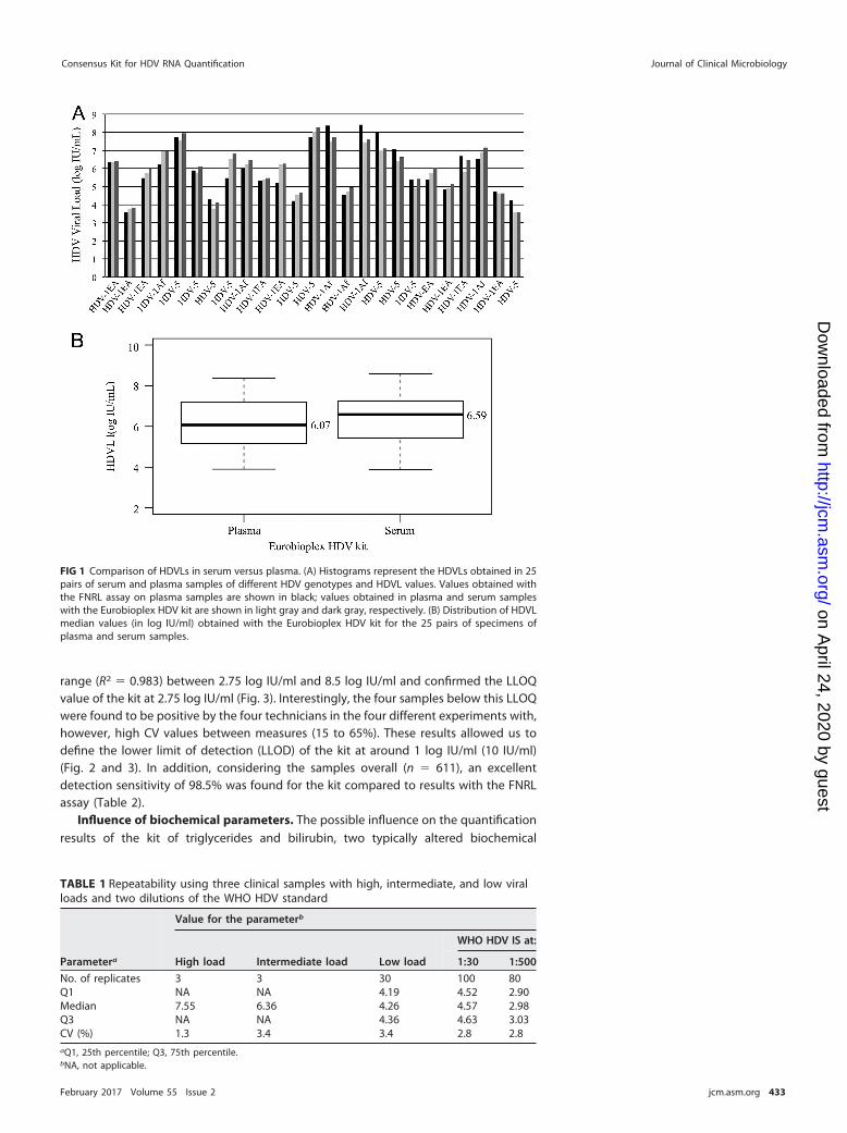
Serum versus plasma specimens. The first interesting point was that the Eurobio-plex HDV kit found comparable results using either serum or plasma specimens, asassessed by the results obtained for paired serum/plasma samples from 25 patientsinfected with HDV-1 and HDV-5 strains (Fig. 1). The median HDVL values were, respec-tively, 6.07 and 6.59 log IU/ml for plasma and serum for the Eurobioplex kit, for anexpected median value of 5.49 log IU/ml, according to HDV FNRL assay results onplasma samples (Fig. 1A and B).
Specificity. The Eurobioplex HDV kit showed an excellent specificity (100%). Indeed,105 negative anti-HDV antibody (Ab) samples positive for either for HAV IgM, HBV DNA,HCV RNA, HEV RNA, or HIV RNA were all found negative for HDV RNA.
Repeatability and reproducibility. Repeatability was investigated using threesamples, one each with a high, medium, and low HDVL value, together with onenegative control as described in the Materials and Methods section. All replicates (3) ofthe negative control were found negative. For the three positive samples, the CVbetween the replicates was �3.4%, whatever the HDVL (Table 1). In addition, whendilutions of the WHO HDV IS were used, a CV of 2.8% was obtained (Table 1).
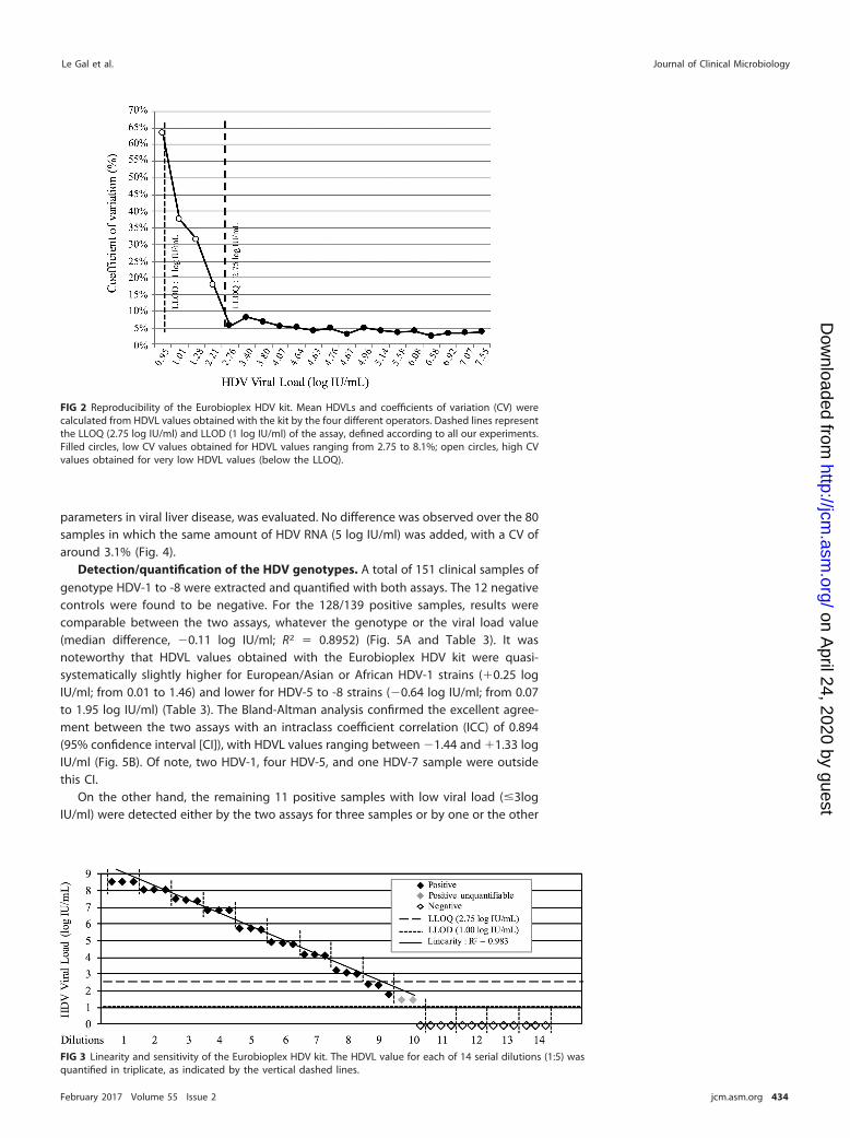
To evaluate reproducibility, four different technicians performed the experiments.The four negative samples were systematically found to be negative. When the resultsobtained with the set of positive samples (�3 to 7.55 log IU/ml) were analyzed, the CVvalues between the measures ranged from 2.5% to 8.1% for samples with an HDVLof �2.75 log IU/ml. This value defined the lower limit of quantification (LLOQ) of the kit(Fig. 2).
Linearity and sensitivity. Serial dilutions (1:5) of a clinical sample with an HDVL of8.5 log IU/ml were quantified each in triplicate. Results showed a linear quantification
Le Gal et al. Journal of Clinical Microbiology
February 2017 Volume 55 Issue 2 jcm.asm.org 432
on April 24, 2020 by guest
http://jcm.asm
.org/D
ownloaded from
range (R2 � 0.983) between 2.75 log IU/ml and 8.5 log IU/ml and confirmed the LLOQvalue of the kit at 2.75 log IU/ml (Fig. 3). Interestingly, the four samples below this LLOQwere found to be positive by the four technicians in the four different experiments with,however, high CV values between measures (15 to 65%). These results allowed us todefine the lower limit of detection (LLOD) of the kit at around 1 log IU/ml (10 IU/ml)(Fig. 2 and 3). In addition, considering the samples overall (n � 611), an excellentdetection sensitivity of 98.5% was found for the kit compared to results with the FNRLassay (Table 2).
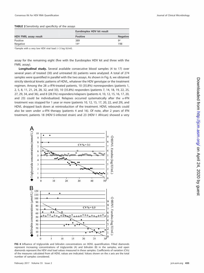
Influence of biochemical parameters. The possible influence on the quantificationresults of the kit of triglycerides and bilirubin, two typically altered biochemical
FIG 1 Comparison of HDVLs in serum versus plasma. (A) Histograms represent the HDVLs obtained in 25pairs of serum and plasma samples of different HDV genotypes and HDVL values. Values obtained withthe FNRL assay on plasma samples are shown in black; values obtained in plasma and serum sampleswith the Eurobioplex HDV kit are shown in light gray and dark gray, respectively. (B) Distribution of HDVLmedian values (in log IU/ml) obtained with the Eurobioplex HDV kit for the 25 pairs of specimens ofplasma and serum samples.
TABLE 1 Repeatability using three clinical samples with high, intermediate, and low viralloads and two dilutions of the WHO HDV standard
Parametera
Value for the parameterb
High load Intermediate load Low load
WHO HDV IS at:
1:30 1:500
No. of replicates 3 3 30 100 80Q1 NA NA 4.19 4.52 2.90Median 7.55 6.36 4.26 4.57 2.98Q3 NA NA 4.36 4.63 3.03CV (%) 1.3 3.4 3.4 2.8 2.8aQ1, 25th percentile; Q3, 75th percentile.bNA, not applicable.
Consensus Kit for HDV RNA Quantification Journal of Clinical Microbiology
February 2017 Volume 55 Issue 2 jcm.asm.org 433
on April 24, 2020 by guest
http://jcm.asm
.org/D
ownloaded from
parameters in viral liver disease, was evaluated. No difference was observed over the 80samples in which the same amount of HDV RNA (5 log IU/ml) was added, with a CV ofaround 3.1% (Fig. 4).
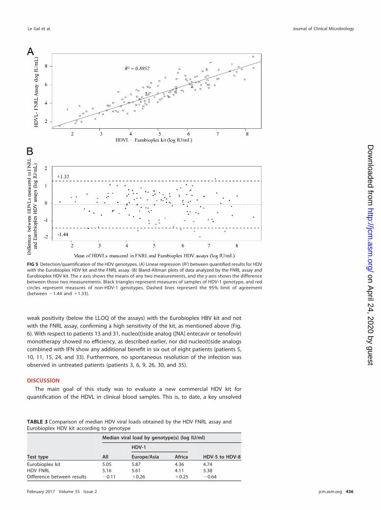
Detection/quantification of the HDV genotypes. A total of 151 clinical samples ofgenotype HDV-1 to -8 were extracted and quantified with both assays. The 12 negativecontrols were found to be negative. For the 128/139 positive samples, results werecomparable between the two assays, whatever the genotype or the viral load value(median difference, �0.11 log IU/ml; R2 � 0.8952) (Fig. 5A and Table 3). It wasnoteworthy that HDVL values obtained with the Eurobioplex HDV kit were quasi-systematically slightly higher for European/Asian or African HDV-1 strains (�0.25 logIU/ml; from 0.01 to 1.46) and lower for HDV-5 to -8 strains (�0.64 log IU/ml; from 0.07to 1.95 log IU/ml) (Table 3). The Bland-Altman analysis confirmed the excellent agree-ment between the two assays with an intraclass coefficient correlation (ICC) of 0.894(95% confidence interval [CI]), with HDVL values ranging between �1.44 and �1.33 logIU/ml (Fig. 5B). Of note, two HDV-1, four HDV-5, and one HDV-7 sample were outsidethis CI.
On the other hand, the remaining 11 positive samples with low viral load (�3logIU/ml) were detected either by the two assays for three samples or by one or the other
FIG 3 Linearity and sensitivity of the Eurobioplex HDV kit. The HDVL value for each of 14 serial dilutions (1:5) wasquantified in triplicate, as indicated by the vertical dashed lines.
FIG 2 Reproducibility of the Eurobioplex HDV kit. Mean HDVLs and coefficients of variation (CV) werecalculated from HDVL values obtained with the kit by the four different operators. Dashed lines representthe LLOQ (2.75 log IU/ml) and LLOD (1 log IU/ml) of the assay, defined according to all our experiments.Filled circles, low CV values obtained for HDVL values ranging from 2.75 to 8.1%; open circles, high CVvalues obtained for very low HDVL values (below the LLOQ).
Le Gal et al. Journal of Clinical Microbiology
February 2017 Volume 55 Issue 2 jcm.asm.org 434
on April 24, 2020 by guest
http://jcm.asm
.org/D
ownloaded from
assay for the remaining eight (five with the Eurobioplex HDV kit and three with theFNRL assay).
Longitudinal study. Several available consecutive blood samples (4 to 17) overseveral years of treated (30) and untreated (6) patients were analyzed. A total of 274samples were quantified in parallel with the two assays. As shown in Fig. 6, we obtainedstrictly identical kinetic patterns of HDVL, whatever the HDV genotype or the treatmentregimen. Among the 28 �-IFN-treated patients, 10 (35.8%) nonresponders (patients 1,2, 5, 8, 11, 21, 24, 28, 32, and 33), 10 (35.8%) responders (patients 7, 14, 18, 19, 22, 25,27, 29, 34, and 36), and 8 (28.5%) responders/relapsers (patients 4, 10, 12, 15, 16, 17, 20,and 23) could be individualized. Relapses occurred systematically after the �-IFNtreatment was stopped for 1 year or more (patients 10, 12, 15, 17, 20, 22, and 29), andHDVL dropped back down at reintroduction of the treatment. HDVL rebounds couldalso be seen under �-IFN therapy (patients 4 and 16). Of note, after 2 years of IFNtreatment, patients 18 (HDV-5-infected strain) and 23 (HDV-1 African) showed a very
TABLE 2 Sensitivity and specificity of the assays
HDV FNRL assay result
Eurobioplex HDV kit result
Positive Negative
Positive 389 9a
Negative 14a 198aSample with a very low HDV viral load (�3 log IU/ml).
FIG 4 Influence of triglyceride and bilirubin concentrations on HDVL quantification. Filled diamondsrepresent increasing concentrations of triglyceride (A) and bilirubin (B) in the samples, and opendiamonds represent the HDV viral load values measured in these samples. Coefficients of variation (CVs)of the measures calculated from all HDVL values are indicated. Values shown on the x axis are the totalnumber of samples considered.
Consensus Kit for HDV RNA Quantification Journal of Clinical Microbiology
February 2017 Volume 55 Issue 2 jcm.asm.org 435
on April 24, 2020 by guest
http://jcm.asm
.org/D
ownloaded from
weak positivity (below the LLOQ of the assays) with the Eurobioplex HBV kit and notwith the FNRL assay, confirming a high sensitivity of the kit, as mentioned above (Fig.6). With respect to patients 13 and 31, nucleo(t)side analog ([NA] entecavir or tenofovir)monotherapy showed no efficiency, as described earlier, nor did nucleo(t)side analogscombined with IFN show any additional benefit in six out of eight patients (patients 5,10, 11, 15, 24, and 33). Furthermore, no spontaneous resolution of the infection wasobserved in untreated patients (patients 3, 6, 9, 26, 30, and 35).
DISCUSSION
The main goal of this study was to evaluate a new commercial HDV kit forquantification of the HDVL in clinical blood samples. This is, to date, a key unsolved
FIG 5 Detection/quantification of the HDV genotypes. (A) Linear regression (R2) between quantified results for HDVwith the Eurobioplex HDV kit and the FNRL assay. (B) Bland-Altman plots of data analyzed by the FNRL assay andEurobioplex HDV kit. The x axis shows the means of any two measurements, and the y axis shows the differencebetween those two measurements. Black triangles represent measures of samples of HDV-1 genotype, and redcircles represent measures of non-HDV-1 genotypes. Dashed lines represent the 95% limit of agreement(between �1.44 and �1.33).
TABLE 3 Comparison of median HDV viral loads obtained by the HDV FNRL assay andEurobioplex HDV kit according to genotype
Test type
Median viral load by genotype(s) (log IU/ml)
All
HDV-1
HDV-5 to HDV-8Europe/Asia Africa
Eurobioplex kit 5.05 5.87 4.36 4.74HDV FNRL 5.16 5.61 4.11 5.38Difference between results �0.11 �0.26 �0.25 �0.64
Le Gal et al. Journal of Clinical Microbiology
February 2017 Volume 55 Issue 2 jcm.asm.org 436
on April 24, 2020 by guest
http://jcm.asm
.org/D
ownloaded from
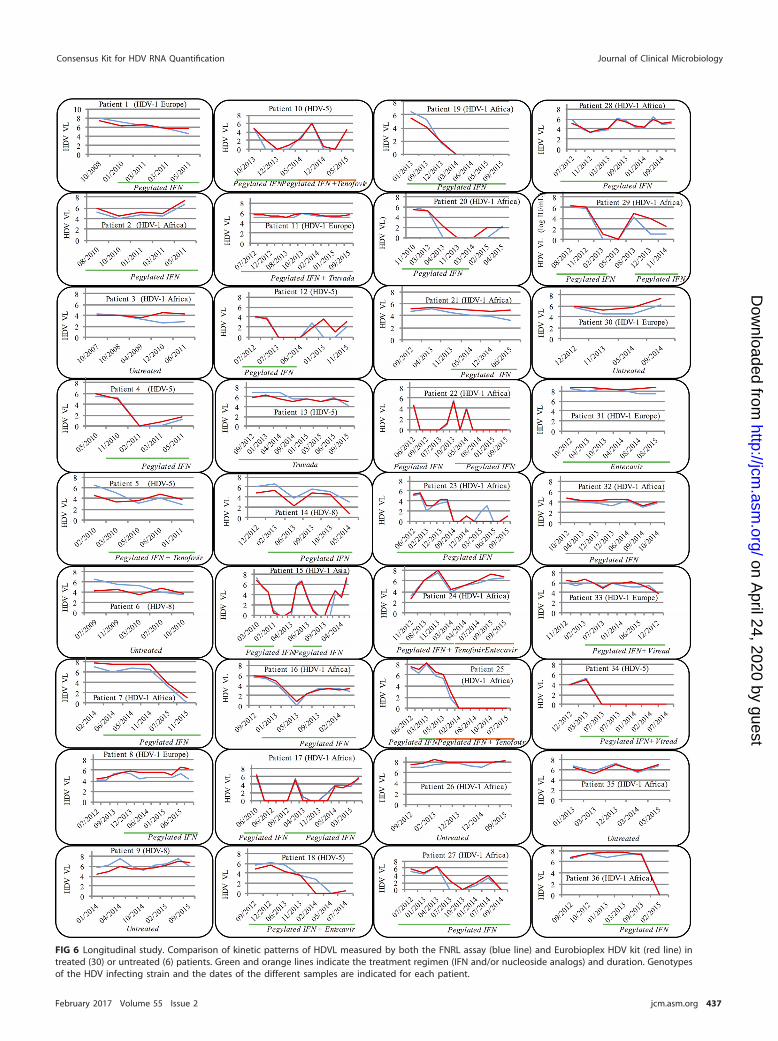
FIG 6 Longitudinal study. Comparison of kinetic patterns of HDVL measured by both the FNRL assay (blue line) and Eurobioplex HDV kit (red line) intreated (30) or untreated (6) patients. Green and orange lines indicate the treatment regimen (IFN and/or nucleoside analogs) and duration. Genotypesof the HDV infecting strain and the dates of the different samples are indicated for each patient.
Consensus Kit for HDV RNA Quantification Journal of Clinical Microbiology
February 2017 Volume 55 Issue 2 jcm.asm.org 437
on April 24, 2020 by guest
http://jcm.asm
.org/D
ownloaded from
issue in HDV diagnosis and in the management of patients, as demonstrated by severalrecent studies (9–11).
One central challenge for an HDVL assay is to detect and quantify properly alldifferent HDV genotypes. Particularly, African HDV genotypes (i.e., HDV-1 and HDV-5 to-8) are poorly quantified by almost all available assays and are either undetected ordramatically underestimated (11). These different genotypes originally had a specificgeographic distribution; however, due to ancient or recent migrations of populations,this genetic variability is now encountered in different countries worldwide (9–11,17–25). In this respect, France, where genotypes HDV-5 to -8 representing 20% ofspreading strains were characterized, is an outstanding example (26, 27). Very inter-estingly, this new Eurobioplex HDV kit showed an excellent capacity to properlyquantify HDVL in plasma and serum specimens, including strains with African geno-types (global median difference is �0.10 log IU/ml). Such capacity of the kit was alsofound in the longitudinal follow-up of infected patients (Fig. 6), where the HDVL kineticpatterns were strictly comparable. However, when we looked carefully, the mediandifference was �0.64 (from 0.07 to 1.95 log IU/ml) in samples of genotype HDV-5 to -8(n � 36), and for 13 of them (36.1%) HDVL values were �1 log IU/ml with the Eurobiokit. This suggests that African patients, mainly those with low HDVL either before,during, or after the end of the treatment, should be carefully managed. Another clinicalpoint is the sensitivity of the assay and its capacity to detect early rebounds of HDVL,as, unfortunately, relapses seem to be the rule at the end of the 12 or 18 months ofpegylated �-IFN therapy. Considering these results, the Eurobioplex HDV kit seems tobe highly suitable to monitor HDVL for patient management. Indeed, consideringpatients 18 and 23 (Fig. 6), the kit detected the rebound earlier than the FNRL assay, ata very low HDVL value (below the LLOQ of the two assays). These results confirmed thevery good sensitivity of the kit. Of note, the manufacturer proposes a range ofquantification between 3.53 and 8.53 log IU/ml regarding the concentration of the RNAstandard of quantification. However, according to the experiments conducted here, wefound an LLOQ of around 2.75 log IU/ml with very low CV, and more interestinglysamples with very low HDVLs of around 1 log IU/ml were systematically detected by alloperators. More experiments are needed with samples of African genotypes to confirmthe low level of detection of the kit for all genotypes. Nevertheless, this kit can be usedwith assurance with an LLOQ and LLOD of 2.75 and 1 log IU/ml, respectively.
From technological and technical points of view, this multiplex real-time, one-stepRT-PCR utilized primers and probe designed in the hepatitis D antigen (HDAg) codingregion and not in the highest conserved ribozyme regions of the genome, as weproposed earlier (11). While this kit was in very good agreement with the FNRL assay,we noted that six samples of African genotypes (Fig. 4B) were under the 95% CI. Thisunderestimation of �1.44 log IU/ml can be linked to the primer and probe sequencedesign, a critical point in the development of any assay (11). Unfortunately, we couldnot further address this point as primer/probe sequences are not available.
Another technical point of interest is the provision of an RNA internal control (IC) inthis kit. In a recent study comparing almost all available HDVL quantification assaysworldwide, 42% of them had no IC (11). More than 20 runs performed with this kitshowed an excellent stability of this IC, without competition for high HDVL values,providing a robust tool to easily monitor all technical steps and to detect PCR inhibitionevents. Last, this assay runs on the CFX6 thermocycler (Bio-Rad) as recommended bythe manufacturers. However, adaptation on fully automated devices might be conceiv-able and should be the next step for improvement of this kit in clinical practice.
In conclusion, the Eurobioplex HDV kit exhibited good specificity, sensitivity, preci-sion, and clinical agreement with the FNRL assay and, most importantly, detected allHDV genotypes (although with a tendency to slightly underestimate HDV-5 to -8). Thiskit, which can be easily implemented in any clinical laboratory, will be a main andefficient tool for therapeutic management of patients. This constitutes a major steptoward organization of large clinical studies, which will then issue guidelines, nonex-
Le Gal et al. Journal of Clinical Microbiology
February 2017 Volume 55 Issue 2 jcm.asm.org 438
on April 24, 2020 by guest
http://jcm.asm
.org/D
ownloaded from
istent to date, for monitoring and therapeutic management of HDV patients in the eraof development of new potent anti-HDV therapies.
MATERIALS AND METHODSSamples and study design. All serum or plasma samples stored at �80°C, were selected from the
French National Reference Laboratory for HDV (HDV FNRL) biobank. HDV genotype was determined aspreviously described by amplification and genotyping of the R0 region (26, 28). When necessary, sampledilutions were performed with hepatitis B surface antigen (HBsAg)-negative plasma kindly provided bythe French National Institute for Blood Transfusion.
Serum and plasma samples. First, we evaluated the performance of the kit on both serum andplasma samples. Quantification experiments were performed in pairs of serum and plasma specimensfrom 25 HDV-infected patients.
Specificity. For specificity evaluation, 105 clinical samples negative for anti-HDV antibody (Ab) butpositive for other hepatitis viruses and for the human immunodeficiency virus (HIV) were selected,including 5 positive IgM anti-HAV Ab, 75 positive HBV DNA, 10 positive HCV RNA, 5 positive HEV RNA,and 10 positive HIV RNA specimens.
Repeatability. Repeatability evaluation was performed using three samples with high (�7 logIU/ml), medium (�6 log IU/ml), and low (4 log IU/ml) HDVL values. High, medium, and negative sampleswere extracted one time whereas the low sample was extracted 10 times. All samples were quantifiedin triplicate. In addition, two dilutions, 3 and 4.3 log IU/ml of the recently developed WHO HDVinternational standard (IS) titrated at 5.76 log IU/ml (29), were tested 80 and 100 times, respectively.
Reproducibility. For reproducibility evaluation, four different technicians analyzed, in four differentexperiments, 4 negative samples and 20 positive samples, including 4 samples with detectable butunquantifiable HDVL (from 2 to 3 log IU/ml) and 16 samples with HDVLs ranging from 3 to 7.8 log IU/ml.
Linearity and sensitivity. To determine the sensitivity of the new kit, we performed 14 serialdilutions (1:5) from a clinical sample of genotype HDV-1 quantified with the FNRL assay at 8.5 log IU/ml.Each diluted sample was extracted in the same series and quantified in triplicate in the same run.
Influence of biochemical parameters. The influence of triglyceride and bilirubin in the bloodsamples was assessed to check for possible interference with quantification performance of the kit. Thesame amount, 5 log IU/ml (determined by the FNRL assay), of a plasma sample was added to 50plasma samples with different triglyceride concentrations (1 to �5 millimoles/liter) and to 30samples with different amounts of bilirubin (�20 to �50 micromoles/liter). All samples were thenquantified with the Eurobioplex HDV kit.
Clinical samples of various genotypes and HDVLs. The ability of the kit to detect and quantifystrains with various HDVLs and genotypes was evaluated on 151 clinical samples including the following:94 HDV-1 (33 African and 61 European/Asian patients); 1 HDV-2; 1 HDV-3; 1 HDV-4 (DNA plasmidcontaining one copy of the genome); 22 HDV-5, 7 HDV-6; 10 HDV-7; 3 HDV-8, and 12 negative controls.
Longitudinal study. The kit was also evaluated in patient follow-up, using available consecutiveserum/plasma samples (4 to 17) of treated (n � 30) or untreated (n � 6) patients. Patients were infectedwith strains of different genotypes: 26 with HDV-1 (from 19 African, 1 Asian, and 6 European patients),7 with HDV-5, and 3 with HDV-8.
Ethics statements. Evaluation of diagnosis tools, as well as prospective molecular characterizationof all new HDV replicative strains, is part of the mission attributed to the HDV FNRL, associated to theFrench National Reference Centre for Hepatitis B, C and Delta, by the French Institut de Veille Sanitaire(InVS)/Santé Publique France.
Quantification experiments. RNA extraction was performed on 500-�l patient samples, to whichthe RNA internal control (IC) of the Eurobioplex HDV kit had been previously added. The RNA extractionkit was used in strict accordance with the manufacturer’s instructions on the m2000sp device (Abbott).RNA was eluted in 110 �l of sterile nuclease-free water and stored at �80°C until use.
The Eurobioplex HDV kit is a multiplex real-time one-step RT-qPCR, comprising an IC and a titratedRNA (8.53 log IU/ml) as a standard of quantification. Serial dilutions of this standard are made to createa 5-point standard curve of quantification ranging from 8.53 to 3.53 log IU/ml. The fluorophores used areFAM for the HDV target and HEX for the IC. Primers and probes are designed in the HDV antigen-codingregion. Technical specifications indicated by the manufacturer were strictly followed. The amplificationstep was performed on a Bio-Rad CFX96 thermocycler.
The HDV FNRL consensus assay was developed in 2005 and since then has routinely been used asdescribed previously (30), including with improvements that have been validated, such as addition of anRNA internal control, automated nucleic acid extraction using an Abbott m2000sp device, and the useof an ABI 7500 fast thermocycler. In addition, according to the WHO HDV IS, the LLOQ and the LLOD ofthe assay have been defined at 103 and 102 IU/ml, respectively.
Statistical analyses. All results were expressed in international units per milliliter (IU/ml). Statisticalanalyses were performed on R software (version 3.2.1 [http://www-users.york.ac.uk/�mb55/meas/ba.htm]). The Pearson correlation coefficient (r) was calculated to determine the linear relationshipbetween the two assays. The Bland-Altman method was used to assess the agreement between theassays. The means of all differences, the standard deviations (SD), and the intraclass correlationcoefficient (ICC) were calculated.
Consensus Kit for HDV RNA Quantification Journal of Clinical Microbiology
February 2017 Volume 55 Issue 2 jcm.asm.org 439
on April 24, 2020 by guest
http://jcm.asm
.org/D
ownloaded from
ACKNOWLEDGMENTSWe express our thanks to the Eurobio Company for providing the kits and devices
for implementation of this study and especially Marie-Claude Amoureux for technicaland scientific support for result analyses. We thank Mei Chao and Chauting Yeh fromChang Gung University of Taiwan, China, who kindly provided us the HDV-4 plasmid.We thank Elhame Anouhal, Mirco Fabris, and Fernando Neri Pinto, technicians of thelaboratory, who participated in the technical experiments.
F.L.G. and E.G. received grants from Eurobio Company to attend the 50th Europeanassociation of the study of the liver (EASL) scientific meeting.
F.L.G. and E.G. were responsible for the study concept and design; F.L.G., A.G., andZ.B.A. were responsible for the technical process; F.L.G., A.G., D.R., S.D., and Z.B.A. wereresponsible for data acquisition of data; S.B., F.L.G., C.A., S.D., D.R., and E.G. wereresponsible for data analysis and interpretation; F.L.G., D.R., S.B., and E.G. drafted themanuscript; E.G. and F.L.G. supervised the study.
REFERENCES1. Fattovich G, Boscaro S, Noventa F, Pornaro E, Stenico D, Alberti A, Ruol
A, Realdi G. 1987. Influence of hepatitis delta virus infection on progres-sion to cirrhosis in chronic hepatitis type B. J Infect Dis 155:931–935.https://doi.org/10.1093/infdis/155.5.931.
2. Fattovich G, Giustina G, Christensen E, Pantalena M, Zagni I, Realdi G,Schalm SW. 2000. Influence of hepatitis delta virus infection on morbid-ity and mortality in compensated cirrhosis type B. The European Con-certed Action on Viral Hepatitis (Eurohep). Gut 46:420 – 426.
3. Manesis EK, Vourli G, Dalekos G, Vasiliadis T, Manolaki N, Hounta A,Koutsounas S, Vafiadis I, Nikolopoulou G, Giannoulis G, Germanidis G,Papatheodoridis G, Touloumi G. 2013. Prevalence and clinical course ofhepatitis delta infection in Greece: a 13-year prospective study. J Hepatol59:949 –956. https://doi.org/10.1016/j.jhep.2013.07.005.
4. Noureddin M, Gish R. 2014. Hepatitis delta: epidemiology, diagnosis andmanagement 36 years after discovery. Curr Gastroenterol Rep 16:365.https://doi.org/10.1007/s11894-013-0365-x.
5. Williams V, Brichler S, Khan E, Chami M, Deny P, Kremsdorf D, Gordien E.2012. Large hepatitis delta antigen activates STAT-3 and NF-�B viaoxidative stress. J Viral Hepat 19:744 –753. https://doi.org/10.1111/j.1365-2893.2012.01597.x.
6. Mederacke I, Yurdaydin C, Dalekos GN, Bremer B, Erhardt A, Cakaloglu Y,Yalcin K, Gurel S, Zeuzem S, Zachou K, Bozkaya H, Dienes HP, Manns MP,Wedemeyer H, Hep-Net/International Delta Hepatitis Study Group. 2012.Anti-HDV immunoglobulin M testing in hepatitis delta revisited: corre-lations with disease activity and response to pegylated interferon-�2atreatment. Antivir Ther 17:305–312. https://doi.org/10.3851/IMP1926.
7. Hughes SA, Wedemeyer H, Harrison PM. 2011. Hepatitis delta virus.Lancet 378:73– 85. https://doi.org/10.1016/S0140-6736(10)61931-9.
8. Mansour W, Malick FZ, Sidiya A, Ishagh E, Chekaraou MA, Veillon P,Ducancelle A, Brichler S, Le Gal F, Lo B, Gordien E, Lunel-Fabiani F. 2012.Prevalence, risk factors, and molecular epidemiology of hepatitis B andhepatitis delta virus in pregnant women and in patients in Mauritania. JMed Virol 84:1186 –1198. https://doi.org/10.1002/jmv.23336.
9. Brichler S, Le Gal F, Butt A, Chevret S, Gordien E. 2013. Commercialreal-time reverse transcriptase PCR assays can underestimate or fail toquantify hepatitis delta virus viremia. Clin Gastroenterol Hepatol 11:734 –740. https://doi.org/10.1016/j.cgh.2013.01.025.
10. Brichler S, Le Gal F, Neri-Pinto F, Mansour W, Roulot D, Laperche S,Gordien E. 2014. Serological and molecular diagnosis of hepatitis deltavirus infection: results of a French national quality control study. J ClinMicrobiol 52:1694 –1697. https://doi.org/10.1128/JCM.03521-13.
11. Le Gal F, Brichler S, Sahli R, Chevret S, Gordien E. 2016. First internationalexternal quality assessment for hepatitis delta virus RNA quantificationin plasma. Hepatology 64:1483–1494. https://doi.org/10.1002/hep.28772.
12. Lempp FA, Ni Y, Urban S. 2016. Hepatitis delta virus: insights into apeculiar pathogen and novel treatment options. Nat Rev GastroenterolHepatol 13:580 –589. https://doi.org/10.1038/nrgastro.2016.126.
13. Vaillant A. 2016. Nucleic acid polymers: Broad spectrum antiviral activity,antiviral mechanisms and optimization for the treatment of hepatitis B
and hepatitis D infection. Antiviral Res. 133:32– 40. https://doi.org/10.1016/j.antiviral.2016.07.004.
14. Koh C, Canini L, Dahari H, Zhao X, Uprichard SL, Haynes-Williams V, et al.2015. Oral prenylation inhibition with lonafarnib in chronic hepatitis Dinfection: a proof-of-concept randomised, double-blind, placebo-controlled phase 2A trial. Lancet Infect Dis 15:1167–1174. https://doi.org/10.1016/S1473-3099(15)00074-2.
15. Noordeen F, Scougall CA, Grosse A, Qiao Q, Ajilian BB, Reaiche-Miller G,Finnie J, Werner M, Broering R, Schlaak JF, Vaillant A, Jilbert AR. 2015.Therapeutic antiviral effect of the nucleic acid polymer REP 2055 againstpersistent duck hepatitis B virus infection. PLoS One 10:e0140909.https://doi.org/10.1371/journal.pone.0140909.
16. Bogomolov P, Alexandrov A, Voronkova N, Macievich M, Kokina K,Petrachenkova M, Lehr T, Lempp FA, Wedemeyer H, Haag M, Schwab M,Haefeli WE, Blank A, Urban S. 2016. Treatment of chronic hepatitis D withthe entry inhibitor myrcludex B: first results of a phase Ib/IIa study. JHepatol 65:490 – 498. https://doi.org/10.1016/j.jhep.2016.04.016.
17. Manesis EK, Schina M, Le Gal F, Agelopoulou O, Papaioannou C, Kallig-eros C, Arseniou V, Manolakopoulos S, Hadziyannis ES, Gault E, KoskinasJ, Papatheodoridis G, Archimandritis AJ. 2007. Quantitative analysis ofhepatitis D virus RNA and hepatitis B surface antigen serum levels inchronic delta hepatitis improves treatment monitoring. Antivir Ther12:381–388.
18. Cross TJ, Rizzi P, Horner M, Jolly A, Hussain MJ, Smith HM, Vergani D,Harrison PM. 2008. The increasing prevalence of hepatitis delta virus(HDV) infection in South London. J Med Virol 80:277–282. https://doi.org/10.1002/jmv.21078.
19. Reinheimer C, Doerr HW, Berger A. 2012. Hepatitis delta: on soft pawsacross Germany. Infection. 40:621– 625. https://doi.org/10.1007/s15010-012-0287-9.
20. De Paschale M, Ceriani C, Cerulli T, Cagnin D, Cavallari S, Ndayake J,Zaongo D, Priuli G, Vigano P, Clerici P. 2014. Prevalence of HBV, HDV,HCV, and HIV infection during pregnancy in northern Benin. J Med Virol86:1281–1287. https://doi.org/10.1002/jmv.23951.
21. Genne D, Rossi I. 2011. Hepatitis delta in Switzerland: a silent epidemic.Swiss Med Wkly 141:w13176. https://doi.org/10.4414/smw.2011.13176.
22. Niro GA, Smedile A, Ippolito AM, Ciancio A, Fontana R, Olivero A, ValvanoMR, Abate ML, Gioffreda D, Caviglia GP, Rizzetto M, Andriulli A. 2010.Outcome of chronic delta hepatitis in Italy: a long-term cohort study. JHepatol 53:834 – 840. https://doi.org/10.1016/j.jhep.2010.06.008.
23. Soriano V, Grint D, d’Arminio Monforte A, Horban A, Leen C, Poveda E,Antunes F, de Wit S, Lundgren J, Rockstroh J, Peters L. 2011. Hepatitisdelta in HIV-infected individuals in Europe. AIDS 25:1987–1992. https://doi.org/10.1097/QAD.0b013e32834babb3.
24. Heidrich B, Deterding K, Tillmann HL, Raupach R, Manns MP, WedemeyerH. 2009. Virological and clinical characteristics of delta hepatitis inCentral Europe. J Viral Hepat. 16:883– 894. https://doi.org/10.1111/j.1365-2893.2009.01144.x.
25. Santos MD, Gomes-Gouvea MS, Nunes JD, Barros LM, Carrilho FJ, FerreiraAde S, Pinho JR. 2016. The hepatitis delta genotype 8 in Northeast Brazil:
Le Gal et al. Journal of Clinical Microbiology
February 2017 Volume 55 Issue 2 jcm.asm.org 440
on April 24, 2020 by guest
http://jcm.asm
.org/D
ownloaded from
The North Atlantic slave trade as the potential route for infection. VirusRes 224:6 –11. https://doi.org/10.1016/j.virusres.2016.08.003.
26. Radjef N, Gordien E, Ivaniushina V, Gault E, Anais P, Drugan T, TrinchetJC, Roulot D, Tamby M, Milinkovitch MC, Deny P. 2004. Molecularphylogenetic analyses indicate a wide and ancient radiation of Africanhepatitis delta virus, suggesting a deltavirus genus of at least sevenmajor clades. J Virol 78:2537–2544. https://doi.org/10.1128/JVI.78.5.2537-2544.2004.
27. Le Gal F, Gault E, Ripault MP, Serpaggi J, Trinchet JC, Gordien E, Deny P.2006. Eighth major clade for hepatitis delta virus. Emerg Infect Dis12:1447–1450. https://doi.org/10.3201/eid1209.060112.
28. Ivaniushina V, Radjef N, Alexeeva M, Gault E, Semenov S, Salhi M, KiselevO, Deny P. 2001. Hepatitis delta virus genotypes I and II cocirculate in an
endemic area of Yakutia, Russia. J Gen Virol 82:2709 –2718. https://doi.org/10.1099/0022-1317-82-11-2709.
29. Chudy M, Hanschmann K-M, Bozdayi M, Kress J, Nubling CM, Collabor-ative Study Group. 2013. Collaborative study to establish a World HealthOrganization international standard for hepatitis D virus RNA for nucleicacid amplification technique (NAT)-based assays. Document WHO/BS/2013.2227. World Health Organization, Geneva, Switzerland.
30. Le Gal F, Gordien E, Affolabi D, Hanslik T, Alloui C, Deny P, Gault E.2005. Quantification of hepatitis delta virus RNA in serum by con-sensus real-time PCR indicates different patterns of virological re-sponse to interferon therapy in chronically infected patients. J ClinMicrobiol 43:2363–2369. https://doi.org/10.1128/JCM.43.5.2363-2369.2005.
Consensus Kit for HDV RNA Quantification Journal of Clinical Microbiology
February 2017 Volume 55 Issue 2 jcm.asm.org 441
on April 24, 2020 by guest
http://jcm.asm
.org/D
ownloaded from

![Comparative Genomics and Transcriptomics of ... · P. acnes strains (draft assembly) serve as reference genomes for the Human Microbiome Project [16]. The P. acnes genomes have a](https://img.pdfslide.fr/doc/110x75/5f0ac20a7e708231d42d3210/comparative-genomics-and-transcriptomics-of-p-acnes-strains-draft-assembly.jpg)