Embed Size (px)
Citation preview
Journal o f Radioanalytical Chemistry, Vol. 1 7 (1973) 55-63
SOME MICROANALYTICAL USES OF SMALL-DIAMETER CHARGED-PARTICLE BEAMS
T. B, PIERCE
Analytical Sciences Division, A.E.R.E., Harwell, Nr Didcot, Berkshire (England)
I1 est souvent n6cessaire d'obtenir des renseignements analytiques sur des 6chantillons de faible volume soit pour caract&iser qualitativement ou quantitativement des microquantit~s de mati~re, soit pour permettre P6tude des variations locales de composition. Les flux de particules charg6es produits par les aec61&ateurs it haut voltage peuvent ~tre focalis6es jusqu'fi des dia- m~tres de queklues microns permettant ainsi d'appliquer ~ l'analyse de fable volume de nombreuses m&hodes d'aetivation aux particules charg6es par r6aetions nuclgaires. Les renseignements analytiques peuvent ~tre tir6s de l'examen s61ectifs des radiations 6raises durant l'irradiation aux particules charg6es ce qui peut inclure la diffusion 61astique, les particules charg6es 6mises, l'6mission ~ et X. Les variations locales de concentrations d 'un ~l~ment peuvent ~tre suivies en balayant la surface de ]'6chantillon par le faisceau d'ions, soit par un mouvement m6canique de l'6chantillon, soit par d6flection du faisceau. Les techniques avec tm micro faisceau sont maintenant utilis6es pour obtenir des r6sultats sur des mat6riaux m6tallurgiques min&alogiques et semi-conducteurs.
Introduction
The production of analytical information from small-volumes of sample can 0e important for characterising and analysing material when only limited quantities are available, or for examining the micro-structure of larger samples to gain a better understanding of their composition and properties. Activation analysis has not normally been employed for obtaining information from relatively small volumes of larger samples, although autoradiography after irradiation with either neutrons or charged particles has provided information about the distribution of particular radioisotopes, and hence of the initial reacting species, always provided that the activity from the matrix does not swamp the radiation yield from the radioisotope under investigation. Thus the distribution of gold on silicon I has been investigated by autoradiography after neutron irradiation, while variations in carbon and oxygen contents have been followed by autoradiography of surfaces irradiated with helium-3 ions. 2 However, nuclear techniques based on the measurement of induced radio- activity have only been used to provide information about elemental distributions on a limited scale, and microanalysis of samples has more usually been achieved by means of other analytical techniques such as the
J, Radioanal. Chem. 17 (1973}
56 T.B. PIERCE: SOME M_ICROANALYTICAL USES
electron microprobe or by micro-ion sputtering methods. Measurement of prompt radiation emitted as a result of charged-particle irradiation of sample surfaces does, however, offer the basis of a technique which could satis- factorily be adapted to microprobe studies, provided that positive ion beams of suitable diameter are available; since radiation is only emitted during the irradiation process it can therefore be associated with the particular region of the sample surface under irradiation. Much of the research carried out into the application of prompt radiation measurement to analysis has employed accelerated particles with energies varying from 0.5 to 4 MeV and over this range a variety of radiations are produced which can form the basis of analytical mea,;urement; there is therefore considerable scope for the selection of an experimental technique to match the specific analytical requirements of a particular measurement. Gamma-radiation, particle groups emitted as a result of nuclear reaction, inelastically scattered particles or X-radiation may be produced at these particle energies from specific elements, so that a range of analytical methods is possible, depending upon the nature of the element to be determined and the host matrix each with their own particular charac- teristics, which can be used in conjunction with small-diameter ion beams. Production of ion beams with diameters of a few tens of tam can be produced by beam collimation, but complicated alignment problems can arise if mul- tiple stop assemblies are necessary to avoid the stop scattering; the absorption of the major part of the ion beam in the material of the beam stop also means that low beam intensities are attained when ion beams are of small diameter. Focussing of the image of a suitable beam objective on to the target provides a much more attractive method of producing small-diameter ion beams 3 since beam size is reduced without the same loss of ion beam intensity. By this means positive ion beams, with energies of a few MeV, have been produced with diameters down to about 3 pm 5 thus offering a genuine positive ion microprobe for materials science studies. Such small- diameter positive ion beams have been in regular use by the Analytical Sciences Division at Harwell for analytical work for some time, and this paper summarises some of the techniques in use and illustrates the methods employed by selective examples.
Z Radioanal. Chem. 17 (1973)
T. B. PIERCE: SOME MICROANALYTICAL USES 57
Experimental techniques
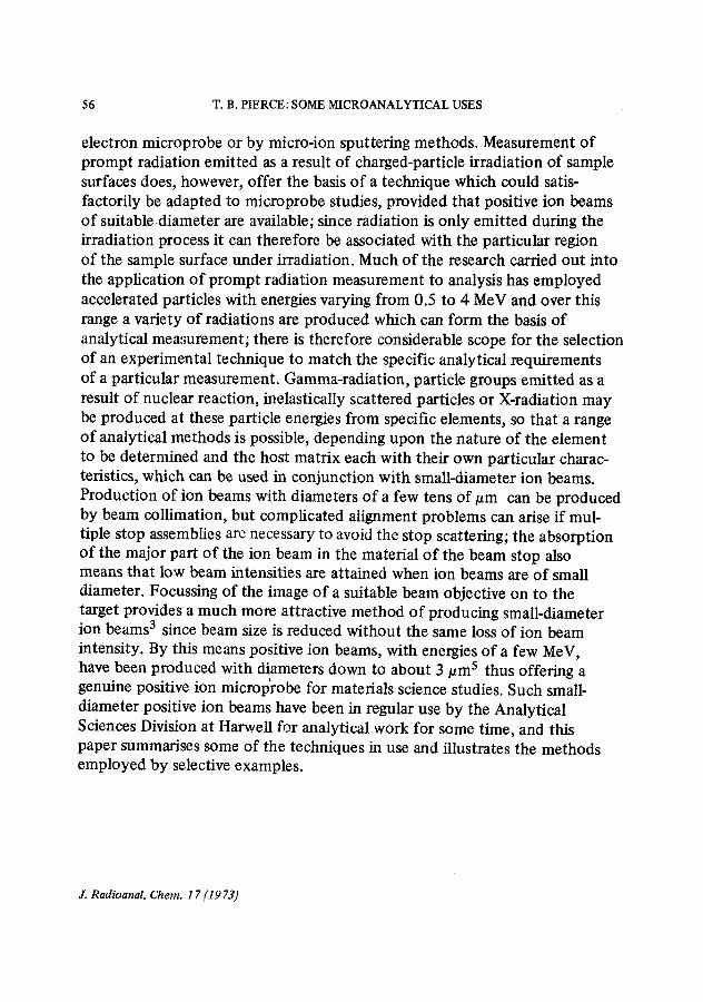
Small-diameter ion beams are produced with the 3 MeV electrostatic generator IBIS at A.E.R.E. and samples irradiated with protons, deuterons or a-particles depending upon the application. Although beam diameters of the order of 3 tam can be produced by the focussing system, somewhat larger beams, with diameters varying from 5 - 1 0 tam, were more frequently employed for sample irradiation since they are easier to produce and use. Focussing is by means of a lens system known as the Russian quadruplet consisting of four quadrupole lenses alternately focussing and defocussing in a particular place, and the lens system focusses down the objective of the beam from any one of five holes drilled in tantalum stops. The overall magnification of the system is ~0.18.
Samples are mounted on a table in the target chamber and can be moved in three dimensions by stepping motors under remote control. The table itself has space for two samples, one on either side of a quartz disc used for initial beam alignment and focussing. The region of the sample irradiated can be viewed from the front of the chamber by means of a microscope; a second microscope at the rear of the chamber permits the shape and size of the beam on the quartz disc to be examined in detail during initial focussing, and the beam movement relative to the target to be checked for scanning. Counters are available to detect charged-particles, gamma-radiation or X-
Focussing assembly Ion beam and beam deflection
~ ~ ~ raus
Microscope
Charged particles
Fig. 1. Aco~le~ator micxobeam target system
Mechanised table mount
.1=-
J. Radioanal. Chem. 17 (1973)
58 T.B. PIERCE: SOME MICROANALYTICAL USES
radiation as shown schematically in Fig. 1. Charged-particles are counted with a surface-barrier detector placed at an angle of about 150 ~ to the incoming beam and the detector housing has provision for a stop between the target and detector to restrict the area of detection when good energy resolution is required; this stop can be removed when high count rates are desirable, for example when elastic scattering is used to provide a means of preliminary survey examination of sample surfaces. An absorber can also be placed be- tween the target and the detector to facilitate the measurement o f charged- particle groups emitted as a result of nuclear reaction since elastically scattered particles present in high yield during charged particle irradiation can often be absorbed in material of suitable thickness while the more penetrating particles from nuclear reaction pass through the absorber and are registered by the detector. Gamma-radiation is detected by a 3" x 3" sodium iodide scintillator placed outside the target chamber or by a much smaller 2" x 1 1/2" scintillator with high resolution also placed outside the target chamber but in a specially shaped depression in the chamber wall to permit the crystal surface to come within 3 cm of the irradiated target face; the peak-to-valley ratio of this detector on coba l t -60 is about 18:1. No provision is made for space for a germanium counter close to the target being irradiated so that if high-resolution gamma-ray spectrometry is required, the germanium counter is positioned as near to the outside of the target chamber as is practicable under existing operating conditions. X-ray measurement is normally carried out by means of a non-dispersive X-ray detector and this is adequate for identifying the more intense lines in the X-ray spectrum, but where high selectivity is required a dispersive detector system is usually necessary to identify characteristic X-ray lines emitted from the sample.
The ion beam is initially focussed on to the quartz disc to give a spot of adequate size, and the region of sample of interest is then moved into the charged-particle beam by activating the drive to the sample table. After the sample has been correctly positioned, the table drive motors are disconnected from external controls outside the vacuum chamber to isolate the whole of the target chamber electrically and to permit beam current integration to be used as a measure o f particle dose falling on the sample. The beam is moved relative to the sample by electrostatic deflection to follow spatial variations in elemental content, the ion beam being stepped discontinuously; the radiation yield is accumulated while the beam is stationary on a particular region of the sample and all counting circuits are inhibited during beam movement. Radiation yield is normally accumulated for a pre-set particle
J. Radioanal. Chem. 17 (1973]
T. B. PIERCE: SOME MICROANALYTICAL USES 59
dose, and electronic controls connected to the beam current integrator used to inhibit and start counter circuits at appropriate times during the scanning sequence, and to initiate storage of accumulated data during beam movement. Single-channel information can be printed or punched after the completion of each irradiation without the imposition of any severe time penalties, but where more complex spectra are accumulated, a faster method of data output is required. Information can be written on to magnetic tape or fed to an analyser with separately addressable memory sectors so that the contents of one sector can be read out while data is accumulated in another. Display of more complex faster scans is achieved by means of contouring programs written for an IBM 370/165 computer or by simpler display programs written for a PDP-8 data processor. The use of computer aids to data presentation has been found to ease, substantially, the load on laboratory staff demanded by graphical interpretation and substantially to simplify the problems of data interpretation.
Results and discussion
The potential applications of microbeam techniques are dictated by the particular characteristics of the several analytical methods available based upon measurement of prompt-radiation. In general, the techniques have so far been directed primarily to the determination of elements present at major or minor rather than at very low levels in the volume of material examined, although the small size of the sample ncessarily demands sensitive analytical techniques since the total mass of element analysed may be very small. Choice o f method will depend not only upon the elements to be determined but also upon matrix composition and the nature of analytical information required, so that gamma-radiation, charged-particles and X-rays resulting from sample irradiation with small-diamter positive-ion beams have all been used to provide information about limited areas o f sample surfaces. Application of positive ion methods to micro-analysis are given below for a number o f practical examples, chosen to illustrate the use of the different measurement techniques.
Gamma-lines of characteristic energy can be produced from most of the fight elements by nuclear reaction with particles with energies of the order o f a few MeV, and established techniques of "r-ray spectroscopy can sometimes identify contributions from a number of components in theaccumulated y-ray spectrum, thus permitting multi-elemental analysis. As an example of
J. Radioanal. Chem. 17(1973)
60 T.B. PIERCE: SOME MICROANALYTICAL USES
. , ,% / / ' , ; , - \ o/ ,
, / No I \ _ _ , . ~ J ~ / X 56 Fe (p,p,) % Fe
�9 ~ J . , ~ " l r ' " *~ -~. , . ,~ _ ~
um
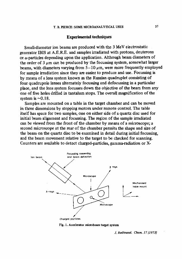
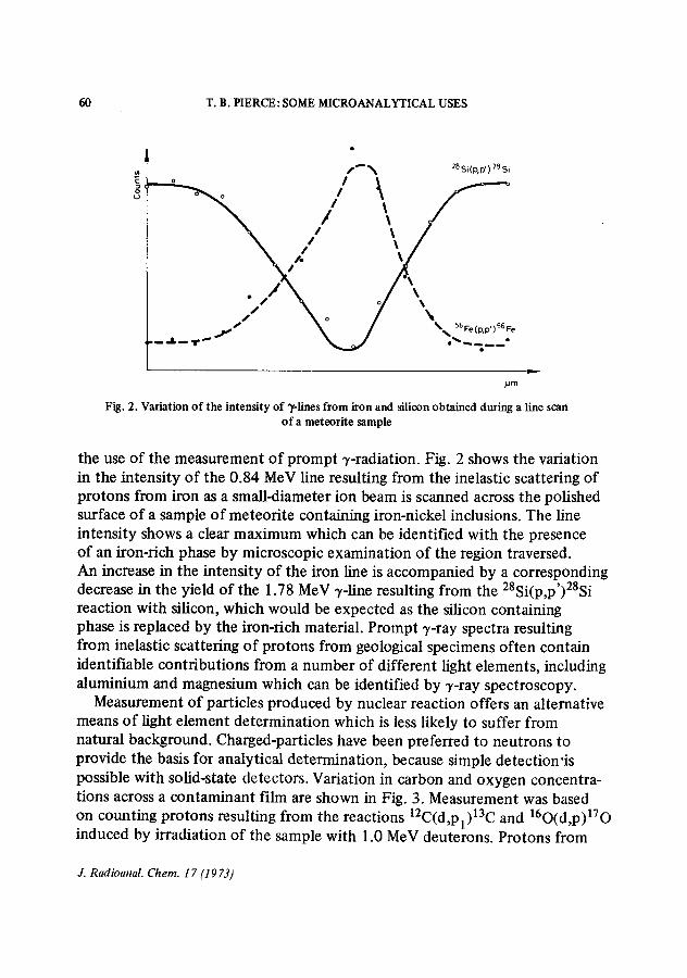
Fig. 2. Variation o f the intensity of Tlines f rom iron and silieon obtained during a line scan o f a meteorite sample
the use of the measurement of prompt -/-radiation. Fig. 2 shows the variation in the intensity of the 0.84 MeV line resulting from the inelastic scattering of protons from iron as a small-diameter ion beam is scanned across the polished surface of a sample of meteorite containing iron-nickel inclusions. The line intensity shows a clear maximum which can be identified with the presence of an iron-rich phase by microscopic examination of the region traversed. An increase in the intensity of the iron line is accompanied by a corresponding decrease in the yield of the 1.78 MeV y-line resulting from the 28Si(p,p')28Si reaction with silicon, which would be expected as the silicon containing phase is replaced by the iron-rich material. Prompt 7-ray spectra resulting from inelastic scattering of protons from geological specimens often contain identifiable contributions from a number of different light elements, including aluminium and magnesium which can be identified by -/-ray spectroscopy.
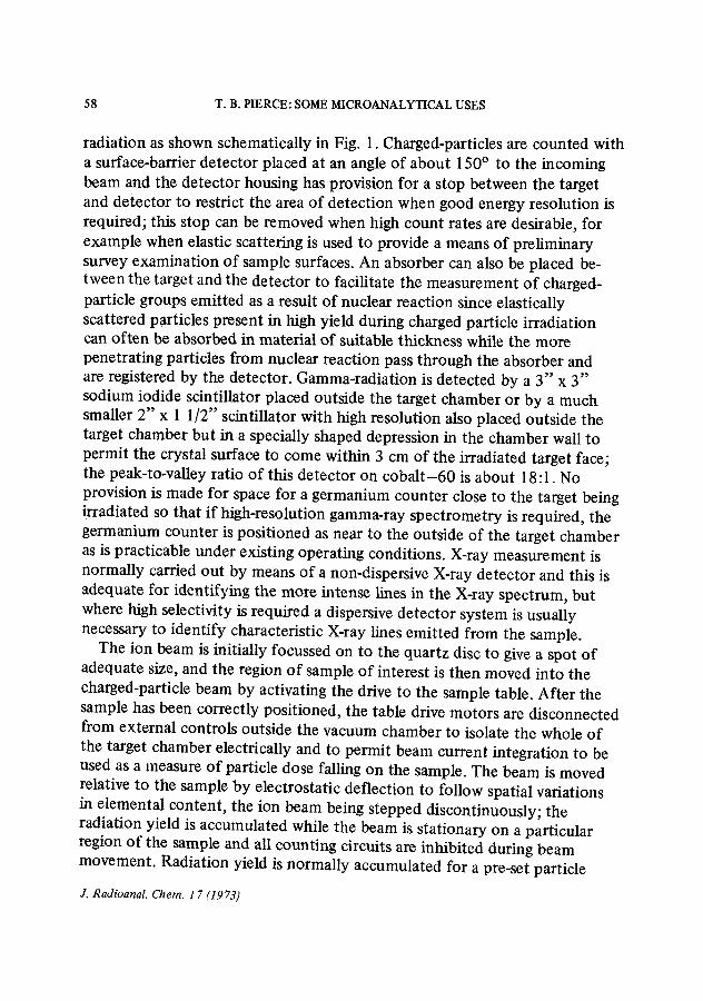
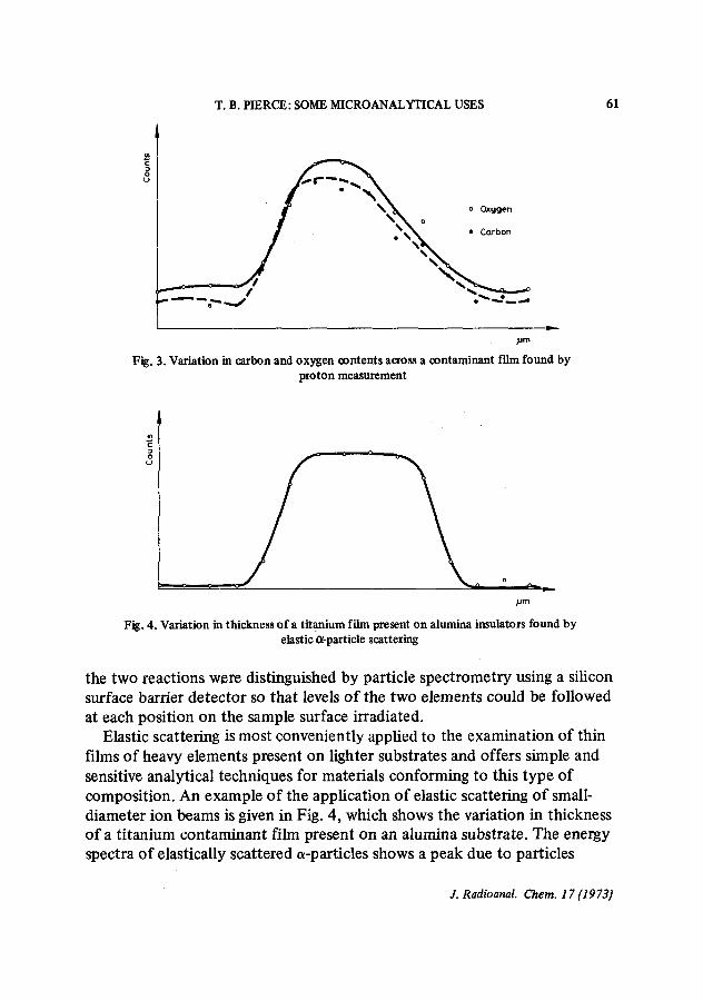
Measurement of particles produced by nuclear reaction offers an alternative means of light element determination which is less likely to suffer from natural background. Charged-particles have been preferred to neutrons to provide the basis for analytical determination, because simple detection'is possible with solid-state detectors. Variation in carbon and oxygen concentra- tions across a contaminant film are shown in Fig. 3. Measurement was based on counting protons resulting from the reactions 12C(d,Pl)13C and 160(d,p)170 induced by irradiation of the sample with 1.0 MeV deuterons. Protons from
J. Radioanal. Chem. 17 (1973)
T. B. PIERCE: SOME MICROANALYTICAL USES 61
~m
Fig. 3. Variation in carbon and oxygen contents across a contaminant film found by proton measurement
pm
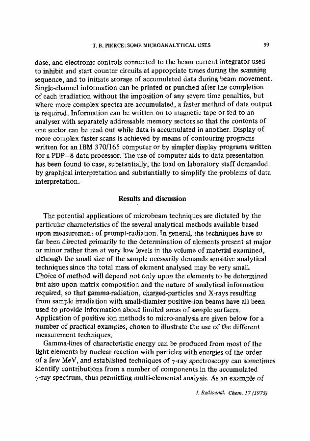
Fig. 4. Variation in thickness of a titanium film present on alumina insulators found by elastic a-particle scattering
the two reactions were distinguished by particle spectrometry using a silicon surface barrier detector so that levels of the two elements could be followed at each position on the sample surface irradiated.
Elastic scattering is most conveniently applied to the examination of thin films of heavy elements present on lighter substrates and offers simple and sensitive analytical techniques for materials conforming to this type of composition. An example of the application of elastic scattering of small- diameter ion beams is given in Fig. 4, which shows the variation in thickness of a titanium contaminant film present on an alumina substrate. The energy spectra of elastically scattered a-particles shows a peak due to particles
3". Radioanal. Chem. 17 (1973)
62 T.B. PIERCE: SOME MICROANALYTICAL USES
scattered from the titanium, clear of the Rutherford plateaux due to those particles scattered from the aluminium and oxygen present in the substrate. 4 Monitoring of the counts in the peak for different incident ion beam positions on the sample permits the variation in film thickness with position to be followed.
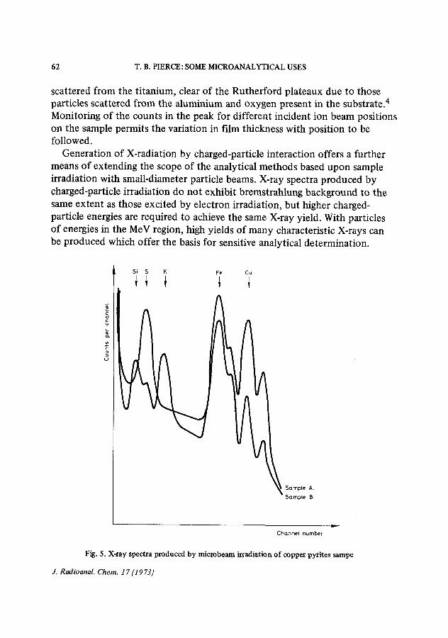
Generation of X-radiation by charged-particle interaction offers a further means of extending the scope of the analytical methods based upon sample irradiation with small-diameter particle beams. X-ray spectra produced by charged-particle irradiation do not exhibit bremstrahlung background to the same extent as those excited by electron irradiation, but higher charged- particle energies are required to achieve the same X-ray yield. With particles of energies in the MeV region, high yields of many characteristic X-rays can be produced which offer the basis for sensitive analytical determination.
Si S K Fe Cu I I I I I
! ! Y I
.c u
o
Sample A.
Sample B.
Channel number
Fig. 5. X-ray spectra produced by microbeam irradiation of copper pyrites sarape
J. Radioanal. Chem. 17 (19 73)
T. B. PIERCE: SOME MICROANALYTICAL USES 63
Fig. 5 shows two X-ray spectra obtained with a non-dispersive X-ray detector from the irradiation of a sample of copper pyrites with a beam of 3.0 MeV protons of approximately 5 lam diameter; Spectrum A shows major X-ray lines corresponding to the primary constituents of copper pyrites i.e. S, Fe and Cu. Spectrum B, obtained with the incident ion beam on an inclusion only slightly larger than the beam diameter shows an entirely different X-ray composition which can be interpreted to provide analytical information about the irradiated region.
References
1. T. B. PIERCE, P. F. PECK in L. S. BARK fEd.) Selected Annual Reviews of the Analytical Sciences Vol 1, The Society for Analytical Chemistry, London 1971, p. 147.
2. D. M. HOLM, W. M. SANDERS, W. L. BRISCE, J. L. PARKER in P. POLISHUK (Ed.) Nucleonics in Aerospace, Plenum Press, 1968, p. 306.
3. J. A. COOKSON, F. D. PILLING, Report A.E.R.E. R 6300, 1970. 4. T. B. PIERCE, P. F. PECK, D. R. A. CUFF, J. Inorg. Nucl. Chem., 33 (1971) 1963.
J. Radioanal. Chem. 1 7 (1973)

![Présentation Orange foncé avec photo · Euler beams : POU_D_E Timoshenko beams : POU_D_T If you need help to choose the best formulation : cf. [U2.02.01] Aster Génie Civil | 24/05/2018](https://img.pdfslide.fr/doc/110x75/6065480e44550913080a76e2/prsentation-orange-fonc-avec-euler-beams-poude-timoshenko-beams-poudt.jpg)


![chap1PL.ppt [Mode de compatibilité]fuuu.be/polytech/MECAH201/cours/MECAH201_chap1PL.pdf · Moulage à moules permanents (permanent mold) BEAMS. 13 Mise en forme Moulage à moule](https://img.pdfslide.fr/doc/110x75/5b9d7aab09d3f275078c72bc/mode-de-compatibilitefuuubepolytechmecah201coursmecah201chap1plpdf.jpg)

![Configuration Manual Polarized Proton Collider at RHIC · colliding nuclei. RHIC will also collide intense beams of polarized protons[2], reaching transverse energies where the protons](https://img.pdfslide.fr/doc/110x75/5e6bfa7f4a9ff14e3c4630d1/configuration-manual-polarized-proton-collider-at-rhic-colliding-nuclei-rhic-will.jpg)