Embed Size (px)
Citation preview

Année 2017/2018 N°
Thèse Pour le
DOCTORAT EN MEDECINE Diplôme d’État
par
Lise COURTOT Née le 03/08/1988 à Nantes (44)
TITRE Facteurs de risque d’iléus post-opératoire après colectomies droites
coelioscopiques réglées. Etude multicentrique rétrospective. Présentée et soutenue publiquement le 24 Avril 2018 devant un jury composé de :
Président du Jury : Professeur Ephrem SALAME, Chirurgie Digestive, Faculté de Médecine – Tours Membres du Jury : Professeur Guillaume MEURETTE, Chirurgie Digestive, Faculté de Médecine – Nantes Docteur Raphael DENHAUT, Anesthésiologie - Réanimation, PH, CHU – Tours Directeur de thèse : Professeur Mehdi OUAISSI, Chirurgie Digestive, Faculté de Médecine – Tours
2
Année 2017/2018 N°
Thèse Pour le
DOCTORAT EN MEDECINE Diplôme d’État
par
Lise COURTOT Née le 03/08/1988 à Nantes (44)
TITRE Facteurs de risque d’iléus post-opératoire après colectomies droites
coelioscopiques réglées. Etude multicentrique rétrospective. Présentée et soutenue publiquement le 24 Avril 2018 devant un jury composé de :
Président du Jury : Professeur Ephrem SALAME, Chirurgie Digestive, Faculté de Médecine – Tours Membres du Jury : Professeur Guillaume MEURETTE, Chirurgie Digestive, Faculté de Médecine – Nantes Docteur Raphael DENHAUT, Anesthésiologie - Réanimation, PH, CHU – Tours Directeur de thèse : Professeur Mehdi OUAISSI, Chirurgie Digestive, Faculté de Médecine – Tours
3 Faculté de Médecine – 10, boulevard Tonnellé – CS 73223 – 37032 TOURS Cedex 1 – Tél : 02.47.36.66.00 – www.med.univ-tours.fr 1
28/09/2017
UNIVERSITE FRANCOIS RABELAIS FFAACCUULLTTEE DDEE MMEEDDEECCIINNEE DDEE TTOOUURRSS
DOYEN Pr. Patrice DIOT
VICE-DOYEN
Pr. Henri MARRET
ASSESSEURS Pr. Denis ANGOULVANT, Pédagogie
Pr. Mathias BUCHLER, Relations internationales Pr. Hubert LARDY, Moyens – relations avec l’Université Pr. Anne-Marie LEHR-DRYLEWICZ, Médecine générale Pr. François MAILLOT, Formation Médicale Continue
Pr. Patrick VOURC’H, Recherche
SECRETAIRE GENERALE Mme Fanny BOBLETER
********
DOYENS HONORAIRES
Pr. Emile ARON (†) – 1962-1966 Directeur de l’Ecole de Médecine - 1947-1962
Pr. Georges DESBUQUOIS (†) - 1966-1972 Pr. André GOUAZE - 1972-1994
Pr. Jean-Claude ROLLAND – 1994-2004 Pr. Dominique PERROTIN – 2004-2014
PROFESSEURS EMERITES Pr. Daniel ALISON
Pr. Catherine BARTHELEMY Pr. Philippe BOUGNOUX
Pr. Pierre COSNAY Pr. Etienne DANQUECHIN-DORVAL Pr. Loïc DE LA LANDE DE CALAN
Pr. Noël HUTEN Pr. Olivier LE FLOCH Pr. Yvon LEBRANCHU
Pr. Elisabeth LECA Pr. Gérard LORETTE Pr. Roland QUENTIN
Pr. Alain ROBIER Pr. Elie SALIBA
PROFESSEURS HONORAIRES P. ANTHONIOZ – A. AUDURIER – A. AUTRET – P. BAGROS – G. BALLON – P.BARDOS – J.L. BAULIEU – C. BERGER – JC. BESNARD – P. BEUTTER – P. BONNET – M. BROCHIER – P. BURDIN – L. CASTELLANI – B. CHARBONNIER – P. CHOUTET – T. CONSTANS – C. COUET - J.P. FAUCHIER – F. FETISSOF – J. FUSCIARDI – P. GAILLARD – G. GINIES – A. GOUAZE – J.L. GUILMOT – M. JAN – J.P. LAMAGNERE – F. LAMISSE – Y. LANSON – J. LAUGIER – P. LECOMTE – G. LELORD – E. LEMARIE – G. LEROY – Y. LHUINTRE – M. MARCHAND – C. MAURAGE – C. MERCIER – J. MOLINE – C. MORAINE – J.P. MUH – J. MURAT – H. NIVET – L. POURCELOT – P. RAYNAUD – D. RICHARD-LENOBLE – M. ROBERT – J.C. ROLLAND – D. ROYERE - A. SAINDELLE – J.J. SANTINI – D. SAUVAGE – B. TOUMIEUX – J. WEILL
4
5
6
7
Résumé :
L’iléus post-opératoire (IPO) est source de complications médico-chirurgicales et représente un
coût hospitalier important. L’objectif est d’identifier les facteurs favorisant l’IPO après
colectomie droite coelioscopique.
Entre 2004 et 2016, 637 colectomies ont été réalisées et étudiées de façon rétrospective à partir
de la base de données CLIMHET. Les facteurs favorisants potentiels ont été analysés par
régression logistique.
Les patients avec IPO (n=113, 17,7%) étaient comparés à ceux sans iléus post-opératoire
(SIPO) (n=524, 82.3%). Dans le groupe IPO, il y avait plus d’hommes (62%vs49% p=0.012),
plus d’anesthésies péridurales (19%vs9% p=0.004), de transfusions peropératoires (7%vs3%
p=0.020) et un remplissage vasculaire plus important (2000mL vs 1750mL, p<0.001). La
section vasculaire extracorporelle et l’extraction de la pièce par une incision transverse étaient
plus fréquentes dans le groupe IPO (20%vs12%, p=0.049 et 34%vs23% p=0.044). Les
complications chirurgicales étaient plus fréquentes dans le groupe IPO (31.9%vs12.0%
p<0.0001). En analyse multivariée les facteurs de risques indépendant d’IPO étaient : sexe
masculin (HR=2.316, 1.102–4.866), anesthésie péridurale (HR=2.958, 1.250–6.988) et
transfusion peropératoire (HR=6.994, 1.550–31.560).
Cette étude est l’une des premières à exploiter la base données CLIMHET et la première à
s’intéresser aux facteurs de risque d’IPO. Les facteurs de risque d’IPO modifiables sont :
anesthésie péridurale et transfusion peropératoire ; celles-ci doivent être utilisées avec
précaution afin de diminuer le taux d’IPO.
Mots clés : Iléus post-opératoire – facteurs de risque – colectomie droite – cœlioscopie
8
Abstract:
Postoperative ileus (POI) is associated with an elevated risk of other complications and
increases the economic impact on healthcare services. The aim of this study was to identify pre-
, intra- and post-operative risk factors associated with the development of POI following
elective laparoscopic right colectomy.
Between 2004 and 2016, 637 laparoscopic right colectomies were performed. Data were
analysed retrospectively thanks to the CLIHMET database. Potential contributing factors were
analysed by logistic regression.
Patients with POI (n=113, 17.7%) were compared to those without postoperative ileus (WPOI)
(n=524, 82.3%). In the POI group, there were more men (62% vs 49%; p=0.012), more use of
epidural anaesthesia (19% vs. 9%; p=0.004), more intraoperative blood transfusion
requirements (7% vs. 3%; p=0.018) and greater perioperative intravenous fluid administration
(2000mL vs. 1750mL; p<0.001). POIs were more frequent when extracorporeal vascular
section (20% vs 12%; p=0.049) and transversal incision for extraction site (34% vs 23%;
p=0.044) were performed. Overall surgical complications in the POI group were significantly
greater than in the control group WPOI (31.9% vs 12.0%; p<0.0001). Multivariate analysis
found the following independent POI risk factors: male gender (HR=2.316, 1.102 – 4.866),
epidural anaesthesia (HR=2.958, 1.250 – 6.988) and postoperative blood transfusion
requirement (HR=6.994, 1.550 – 31.560).
This study is one of the first to explore the CLIHMET database and the first to use it for
investigating risk factors for POI development. Modifiable risk factors such as epidural
anaesthesia and intraoperative blood transfusion should be used with caution in order to
decrease POI rates.
Key Words: postoperative ileus - risk factor - right colectomy – laparoscopy
9
Remerciements :
Merci au Professeur Salamé d’avoir accepté de présider ce jury de thèse. Merci pour votre soutien tout au long de ma formation, merci pour votre enseignement et votre détermination exemplaire. Un grand merci au Professeur Ouaïssi d’avoir dirigé ce travail. Merci pour votre engagement inconditionnel et votre disponibilité. Vos connaissances et votre exigence m’ont beaucoup apporté. Merci au Professeur Meurette, d’avoir accepté d’être membre du jury. Merci pour ce semestre passé à vos côtés qui m’a permis de redécouvrir avec plaisir la CCDE de Nantes. Votre savoir et votre envie de le transmettre m’ont été très bénéfique. Merci au Docteur Raphaël Denhaut, d’avoir accepté d’être membre du jury et d’apporter votre expertise à ce travail.
10
Un grand merci à toute l’équipe de chirurgie digestive du CHU de Tours : - aux PH qui ont eu l’envie de nous former, Docteur Bourlier, Louise, Céline et Pétru. - à mes différents chefs de Clinique Baudouin, Zeynal, Giovanni, David, Nico, Fred et Fabien,
pour la patience dont ils ont fait preuve et leur pédagogie. Désolé pour vos surrénales et vos coronaires.
- aux infirmières, aides-soignantes et secrétaires qui nous ont aidés et supportés au quotidien, avec une attention particulière à Pauline, Karine , Faustine, Laure, Valérie, Sarah.
- aux coordinatrices pour leurs doux appels nocturnes même si elles ont toujours refusé ma demande d’hélico
- aux cadres qui ont dû faire face à nos besoins impérieux. Avec un merci particulier à Dodo pour nos longues conversations.
- aux anesthésistes, car même si un champ nous sépare, nous travaillons ensemble avec plaisir. Merci notamment à Jean-Louis et au Dr Lepage.
Merci au service de la CCDE de Nantes, pour ces 6 premiers mois d’exil :
- au professeur Mirallié pour son talent et son piquant éternel. - à tous les PH et chefs qui nous ont accueillis, formés et fait confiance rapidement. - à une grande partie des infirmières pour leur accueil.
Merci au service d’urologie, au professeur Bruyère et aux PH pour leur accueil et leur enseignement. A Colas, Mélanie, FX, et Alex pour leur confiance et leur pédagogie. Avec une pensée particulière pour M. Boutin. Merci à l’équipe de la chirurgie digestive du CHRO, au Dr Piquard pour son enseignement et son humanité, Dr Saint-Marc pour son talent et sa bonne parole, au Dr AbouMrad grâce à qui je sais filmer les mariages ®boobies et au Dr Bellouard franc carabin qui m’a appris le ski nautique. Aux infirmières, AS, panseuses (Sarah, Sophie, Fred, Bruno, Phillipe) et à Denis (qui a succédé à ma chère Gueguette) Merci à ma famille, mes parents pour leur soutien lors de ses longues années d’études, notamment à ma mère pour les petits plats et le repassage à Nantes. Bien consciente que tout cela ne soit pas toujours facile à appréhender d’un œil extérieur (je rappelle juste qu’une garde dure 24h). Merci à mes ami(e)s de l’externat grâce à qui ses années ont été plus douces. Gillian, Lucile, Hoël, Hélène, Marie, Camille Claire et Amandine Merci au VHO, refuge pour internes en détresse où j’ai rencontré des copines devenue des amies. Margaux (un rayon de soleil), Camille (ton côté maternel) et lolo, Alice (mon acolyte), Axelle (je suis ravie d’être du bon côté et merci pour tes défilés quotidiens), Marion (pour notre humour connecté et tes photos pas spontanées) et Julie (une personne sur qui on peut compter) Merci à mes différents co-internes rencontrés lors de ses stages : Perrine, Nico (merci du fond du cœur pour ton investissement dans ce travail), Julien, Théo, Remi Aurélie, Pierre, Greg, Lucas, Louise, Marina, Luc. Un remerciement particulier à Benjam’ pour son compagnonnage en uro et bon entendeur. A mon mari qui me soutient au quotidien, qui a dû écouter pendant des heures mes divagations sur les urgentistes, participer à mes astreintes téléphoniques et qui m’a aidé pour ce travail. Merci pour ta bienveillance. Merci aux pilotes d’avions Merci à la région Centre, plus qu’une région, une chance. Je ne remercierai pas : Vinci, ceux qui n’allument pas la lumière de la piste d’atterrissage et l’interdiction du port de vernis à ongles.
11
Table des matières
I) Lettre de soumission ...................................................................................................... 12
II) Article .......................................................................................................................... 13
a. Introduction ................................................................................................................ 14
b. Matériels et méthodes ................................................................................................ 15
c. Résultats ...................................................................................................................... 19
d. Discussion ................................................................................................................... 22
e. Conclusion .................................................................................................................. 22
f. Références ................................................................................................................... 22
III) Annexes ....................................................................................................................... 29
a. Tableau 1 .................................................................................................................... 29
b. Tableau 2 .................................................................................................................... 30
c. Tableau 3 .................................................................................................................... 31
d. Tableau 4 .................................................................................................................... 32
e. Figure 1 ....................................................................................................................... 33
f. Figure 2 ....................................................................................................................... 34
12
I) Lettre de soumission
28/03/2018 17:27Gmail - International Journal of Colorectal Disease - Submission Notification to co-author
Page 1 sur 1file:///Volumes/lisecourtot/Desktop/colon%20droit/Dépôt%20de%20…20Disease%20-%20Submission%20Notification%20to%20co-author.htm
Lise Courtot <[email protected]>
International Journal of Colorectal Disease - Submission Notification to co-author1 message
Editorial Office <[email protected]> 9 mars 2018 à 14:46Répondre à : Editorial Office <[email protected]>À : Lise Courtot <[email protected]>
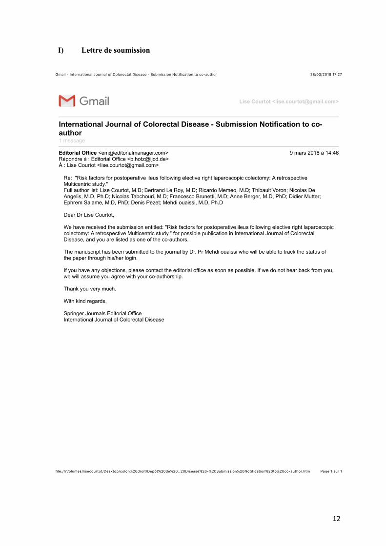
Re: "Risk factors for postoperative ileus following elective right laparoscopic colectomy: A retrospectiveMulticentric study."Full author list: Lise Courtot, M.D; Bertrand Le Roy, M.D; Ricardo Memeo, M.D; Thibault Voron; Nicolas DeAngelis, M.D, Ph.D; Nicolas Tabchouri, M.D; Francesco Brunetti, M.D; Anne Berger, M.D, PhD; Didier Mutter;Ephrem Salame, M.D, PhD; Denis Pezet; Mehdi ouaissi, M.D, Ph.D
Dear Dr Lise Courtot,
We have received the submission entitled: "Risk factors for postoperative ileus following elective right laparoscopiccolectomy: A retrospective Multicentric study." for possible publication in International Journal of ColorectalDisease, and you are listed as one of the co-authors.
The manuscript has been submitted to the journal by Dr. Pr Mehdi ouaissi who will be able to track the status ofthe paper through his/her login.
If you have any objections, please contact the editorial office as soon as possible. If we do not hear back from you,we will assume you agree with your co-authorship.
Thank you very much.
With kind regards,
Springer Journals Editorial OfficeInternational Journal of Colorectal Disease
13
II) Article
Risk factors for postoperative ileus following elective laparoscopic right colectomy:
A retrospective multicentric study.
Lise Courtot, M.D.1, Bertrand Leroy M.D.2, Ricardo Memeo M.D.3, Thibault Voron M.D.4, Nicolas de Angelis M.D.5, Nicolas Tabchouri M.D.1, Francesco Brunetti M.D.5, Anne Berger M.D., Ph.D. 4, Didier Mutter M.D., Ph.D. 3, Johan Gagniere, MD, PhD2, Ephrem Salamé, M.D., Ph.D. 1, Denis Pezet M.D., Ph.D. 2, Mehdi Ouaïssi M.D., Ph.D.1
1Department of Digestive, Oncological, Endocrine, and Hepatic Surgery, and Hepatic Transplantation. Trousseau Hospital, Tours, France
2Department of Digestive Surgery, Estaing University Hospital, Clermont-Ferrand, France 3Hepato-Biliary and Pancreatic Surgical Unit, IRCAD-IHU, University of Strasbourg, Strasbourg, France 4Department of Digestive Surgery. George Pompidou European Hospital, Paris, France 5Department of Digestive Surgery, Hepato-Pancreato-Biliary Surgery, and Liver Transplantation, Henri-Mondor Hospital, AP-HP, Créteil, France Running title: Predictive factors for ileus following elective laparoscopic right colectomy Corresponding author Mehdi Ouaïssi, M.D., Ph.D. Department of Digestive, Oncological, Endocrine, and Hepatic Surgery, and Hepatic Transplantation. Trousseau Hospital. Colorectal Unit. Centre Hospitalier Universitaire (CHU) Tours, avenue de la République, 37170 Chambray-les-Tours, France. Fax: Tel.: E-mail: [email protected]
Source of financial support: None
14
a. Introduction
Postoperative ileus (POI) following open or laparoscopic colorectal surgery is one of
the most common complications, with an incidence of 10 to 17%[1]. POI contributes to
prolonged hospital stays, is associated with an increased risk of other complications,
nosocomial infections and higher postoperative mortality. Consequently, POI has a major
economic impact on healthcare services [2, 3]. A recent study with a large database (N=29,201)
highlighted that POI occurred more frequently following right colectomy compared with left
colectomy [4]. Because it reduces overall postoperative morbidity (mainly ileus, length of
hospital stay and time to oral intake), the laparoscopic approach is considered to be the gold
standard in colorectal surgery[5–7]. Despite significantly reduced rate of POI with the
laparoscopic approach, recent studies have shown that the rate of POI after laparoscopic right
colectomy is 10.6% [8] and explain that POI could raise public health issues. A better
understanding of the factors associated with POI could lead to better prevention at each stage
of care, avoiding the operative factors implicated and monitoring, and providing early care to
high-risk patients. No large study has analysed POI risk factors following elective laparoscopic
right colectomy, based on a homogenous definition of POI. The purpose of our study was to
characterise preoperative, intraoperative and postoperative POI risk factors in patients
undergoing elective laparoscopic right colectomy.
15
b. Materials and methods
Patients
A retrospective study was conducted using the CLIHMET Study Group Database. 637
consecutive elective laparoscopic right colectomies were reviewed from January 2005 to
December 2015, at five University Hospital centres in France (the CHU in Clermont-Ferrand,
Hôpital Civil in Strasbourg – IRCAD, Hôpital Henri-Mondor in Créteil, Hôpital Européen
Georges Pompidou in Paris and the CHU in Tours). The CLIHMET database included adult
patients (over 18 years of age) scheduled for elective laparoscopic right colectomy for
malignant or benign colonic diseases. All patients were treated with a curative intent for their
right colonic disease. Patients with metastatic disease, locally advanced cancer requiring multi-
visceral resection or undergoing emergency surgery were excluded.
Data collection
According to Chapuis [9], POI was defined as the presence of abdominal distension
with a lack of bowel sounds in a patient who has experienced nausea or vomiting and has failed
to pass flatus or stool for more than 3 days postoperatively, in the absence of mechanical bowel
obstruction. The study population was therefore divided into two groups: 113 patients in the
POI group and 524 in the control group without postoperative ileus (WPOI). Patients’
characteristics and operative data were retrospectively collected to identify risk factors
associated with POI. Patient background factors were age, gender, BMI score, American
Society of Anesthesiologists (ASA) score, comorbidities (smoking, diabetes, cardio vascular
disease and previous open or laparoscopic surgery), type and site of the colonic disease, tumour
stage and neoadjuvant chemotherapy.
16
Preoperative and intraoperative workup
Patients with colorectal cancer and colonic polyps or adenoma underwent the following:
preoperative colonoscopy, tumour biopsy and an abdominal computed tomography (CT) scan.
In patients with inflammatory bowel disease (IBD), preoperative colonoscopy and magnetic
resonance imaging (MRI) were performed. Bowel preparation was not conducted before
surgery. A single dose of prophylactic antibiotics was routinely given (750 mg of cefuroxime)
at induction of general anaesthesia and was repeated intraoperatively if surgery lasted for >2
hours. Prophylaxis for deep-vein thrombosis was given, i.e. low molecular-weight heparin (50
IU/kg per day) was given to all patients and was continued postoperatively for 30 days in
patients with colon cancer and 7 days in patients with benign disease. Operative features
recorded for all patients were epidural analgesia, perioperative blood transfusion, perioperative
intravenous fluids, duration of operation, conversion rate, nasogastric tube insertion and drain
insertion.
Surgery
All surgical teams were experts in both laparoscopic and open colorectal surgery
(around 400 colorectal surgical procedures were performed each year in all five departments).
Laparoscopic right colectomies were performed as previously reported using a medial-to-lateral
approach for radical operations [10, 11] for cancer and lateral to medial for benign diseases.
The type of ileocolic anastomosis performed was left to the surgeon’s discretion:
intracorporeally or extracorporeally [12], mechanical or manual, peristaltic or anti-peristaltic.
Anastomosis techniques could be performed as follows: side-to-side, end-to-end, side-to-end
or end-to-side. Surgeons performed middle, transverse or suprapubic incision to extract the
specimen. Conversion was defined as the completion of the right colectomy procedure through
either an enlarged incision or an abdominal incision measuring ≥6cm.
17
Postoperative outcomes
Postoperative morbidity and mortality were defined as events occurring during hospital
stay or within 30 and 90 postoperative days. Postoperative complications were classified
according to Dindo-Clavien, their management (medical, radiological, surgical) and their
severity [13]. Postoperative complications included POI, anastomotic leakage, anastomotic
haemorrhage, wound infection, intra-abdominal abscess, bleeding and evisceration. Non-
surgical complications were cardiac, vascular and pulmonary. Postoperative outcomes also
included the following: reoperation rate, time to resumption of a regular diet, time before
ambulation, time to flatus, time to first stool and time before perfusion removal, length of in-
hospital stay and mortality.
Postoperative follow-up
Patients were systematically clinically examined at 4 to 6 weeks after discharge from
hospital. The length of hospitalisation was measured from the time of surgery to the date of
discharge from hospital. Regarding colorectal cancer, postoperative follow-up visits included
clinical, biochemical and radiological assessments every 3 months during the first three
postoperative years and then every 6 months up to 5 postoperative years [14]. Surviving patients
were assessed for disease recurrence and, if so, the site of recurrence. Follow-up information
was obtained from medical records, direct consultation with patients and/or telephone
interview. At the end of the follow-up, the statuses of all patients were assessed, i.e. mortality,
recurrence and lost to follow-up. The endpoint of data collection was April 2017. Patient
follow-up was carried out from the time of surgery to this endpoint, until death if occurring
prior to this date, or until the date of last contact. Loss to follow-up was defined as a follow-up
inferior to 3 months, in the absence of death. Overall, 165 patients (25.9%) were lost to follow-
up. Median follow-up was 27 months.
18
Statistical Analysis
Statistical analyses were performed using IBM SPSS Statistics version 20 (IBM SPSS
Inc., Chicago, IL, USA). Continuous variables are expressed as their means ± standard
deviations (SD), or as their medians and ranges (min, max). Categorical variables are reported
as numbers and percentages. Mean values between the two groups were compared using
Student's t-test or the Mann–Whitney U test, when necessary. Comparisons between
percentages were made using the χ2 test or Fisher’s exact test, as appropriate, for the qualitative
variables. Factors included in the multivariate analyses were significant in the univariate
analyses at a p-value of <0.10. Univariate and multivariate Cox's proportional hazard regression
models were used to estimate the hazard ratio (HR). The HRs were expressed with their 95%
confidence intervals. All tests were two-sided. Overall survival (OS) and disease-free survival
(DFS) rates were computed using the Kaplan-Meier method and compared between groups
using the log rank (Mantel–Cox) test. Statistical significance was defined as a p-value of <0.05
19
c. Results
Patients were allocated into 2 groups: 113 patients with POI and 524 patients WPOI. As
the mortality rate before the fourth postoperative day was null, all 637 patients were able to be
included for statistical analyses.
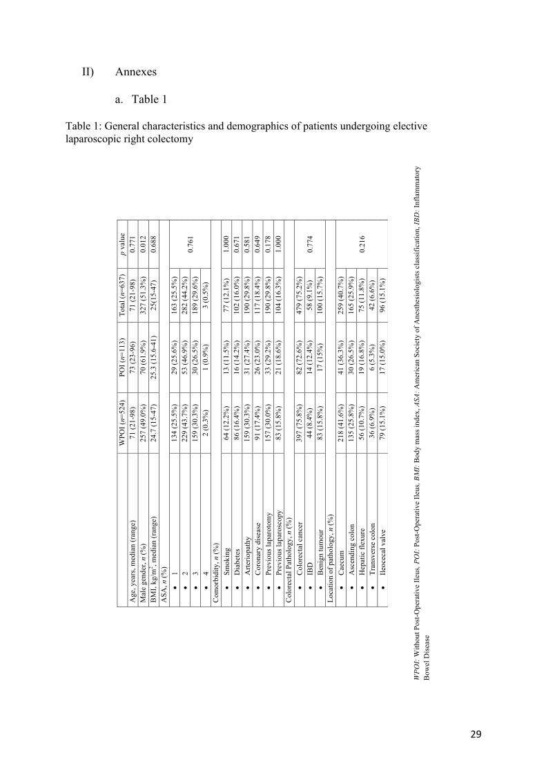
General characteristics and demographics (table 1)
Median age was 71 years (range 21-98). Median body mass index (BMI) was 25.0 kg/m2
(range 15-47). The male/female gender ratio was 1.05 for the whole series. Thirty percent of
patients were considered at high risk (ASA 3 and 4). There were significantly more male
individuals in the POI group compared to the control group (61.9% vs 49.0%; p=0.012). There
was no significant association between ileus and age, comorbidities (smoking, diabetes,
vascular, coronary disease or previous surgery) and ASA scores. Colorectal disease (colorectal
cancer, IBD, benign tumour) and location did not differ between the two groups.
Operative procedure (table 2)
Median preoperative fasting was 11 hours (range 2-96). Anastomosis was performed
extracorporeally and mechanically in 76.3% and 56.2% of cases respectively. Overall
conversion rate was 6.3%. Median operative time was 180 minutes (range 60-695). There were
statistically significant associations between prolonged ileus and operative features.
Concerning perioperative anaesthetic procedures, POI was more frequent in patients
undergoing epidural anaesthesia (18.6% and 9.0%, respectively, p=0.004), in patients requiring
blood transfusion (7.1% and 2.7%, respectively, p=0.018) and in patients receiving more
perioperative intravenous fluid (2000 mL and 1500mL p <0.0001). Concerning surgical
techniques, extracorporeal vascular section (19.5% and 12.2%, respectively, p=0.049) and
transverse periumbilical extraction incision (33.6% and 23.1%, respectively, p=0.044) were
performed more in the POI group than in the control group.
20
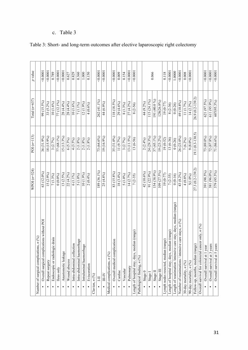
Short- and long-term outcomes (table 3)
Overall 30- and 90-day mortality rates were 1.7% and 2.2%. Thirty and 90-day mortality
rates were significantly higher in the POI group than in the WPOI group (6.2% vs 0.8%; 8.0%
vs 1.0%; p<0.0001, respectively).
According to Clavien-Dindo’s classification [13], the postoperative complication (stage III and
IV) rate was higher in the POI group than in the WPOI group (16.9% vs 4.8%, respectively, p
<0.0001). POI excluded, overall surgical complications in the POI group were significantly
higher than in the control group WPOI (31.9% vs 12.0%; p<0.0001), respectively. Concerning
surgical complications, surgical revision and anastomotic leakage in the POI group were
significantly greater than in the control group WPOI (15.9% vs 2.9%; 13.3% and 2.5%;
p<0.0001), respectively. However, there were no statistical differences regarding other surgical
complications (endoscopic or radiologic drain, wound abscess, intra-abdominal haemorrhage,
intraluminal haemorrhage and evisceration) between the two groups. Cardiac and pulmonary
complication rates in the POI group were significantly higher than in the control group WPOI
(9.7% vs 3.4% and 11.5% vs 2.7%, respectively, p=0.009, p<0.0001). Median of length of
hospital stay was significantly higher in the POI group than in the control group WPOI (13 vs
7 days, respectively, p <0.0001). More patients were admitted to the intensive care unit or
reanimation unit in the POI group than in the control group (23% vs 8.2%, respectively,
p<0.0001).
Pathological findings
According to the TNM classification [15] for colorectal cancer, the tumour stage did not
differ significantly between the POI group and the control group WPOI. The median number
of lymph nodes resected did not differ between the two groups, at 19.
21
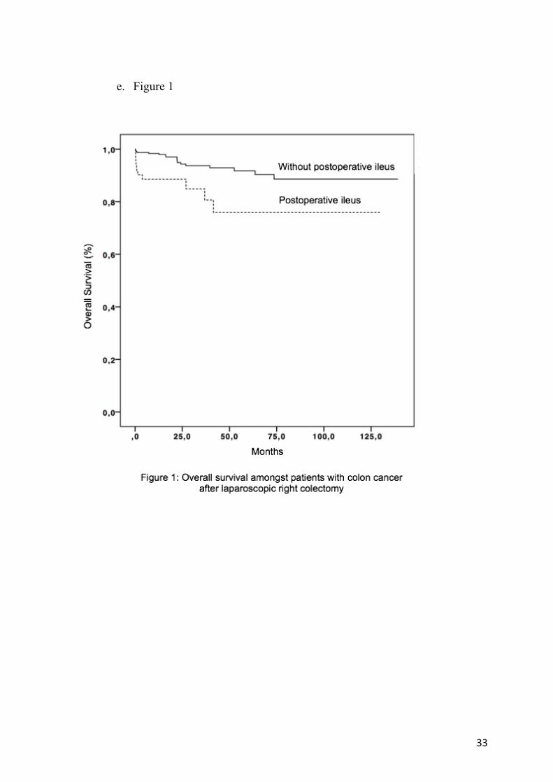
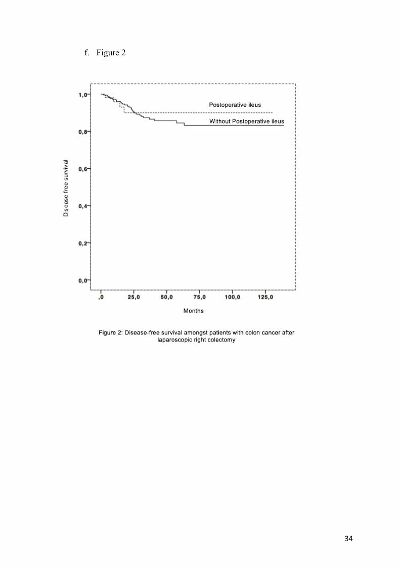
Overall and disease-free survival (figure 1 and 2)
Overall Survival (OS) and Desease-Free Survival (DFS) are displayed in Figures 1 and
2. Only 479 patients with colorectal cancer were analysed, 82 in the POI group and 397 in the
control group WPOI. OS of the POI group was significantly lower than for the control group
WPOI (89.0% vs 98.5% at 1 year, 87.8% vs 96% at 3 years, 86.6% vs 95.5% at 5 years,
respectively, p <0.0001). DFS rates were comparable in the 2 groups, with 97.6% at 1 year,
95.1% at 3 years, 95.1% at 5 years in the POI group vs. 97.2% at 1 year, 93.7% at 3 years,
92.9% at 5 years in the control group WPOI, respectively (p=0.671).
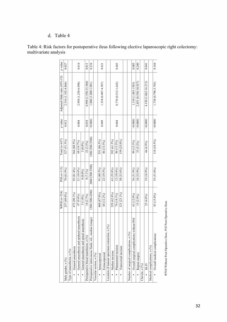
Multivariate analysis (table 4)
Factors that were independently associated with an increased risk of POI were male
gender (HR=2.316, 95% CI, 1.102 – 4.866), epidural anaesthesia (HR=2.958, 1.250 – 6.988)
and perioperative blood transfusion requirement (HR=6.994, 1.550 – 31.560). Extracorporeal
vascular section and extraction sites were not risk factors for POI.
22
d. Discussion
This large series of 637 patients focused on risk factors associated with POI specifically
following laparoscopic right colectomy. A 17.7% POI rate was observed, which was associated
with non-modifiable risk factors such as male gender, but also with modifiable factors such as
the type of anaesthesia, perioperative intravenous fluid volume, blood transfusion requirements
and technical surgical points, such as extra-vascular section of the vessels and extraction
modalities. Furthermore, POI was found to be correlated with other operative complications,
both surgical and medical, and was associated with increased postoperative mortality.
This study contrasts with previous reports through patient homogeneity (only elective
laparoscopic right colectomies were included) and its large sample size. Patients had
standardised follow-up by expert teams. Thanks to previous studies, clinical and biological POI
risk factors have been identified, but few studies have shown any interest as to the precise
surgical technique performed. The fact that this study was multicentric, with surgical habits
specific to each department, made it possible to analyse the different surgical techniques used.
Indeed, as described in Venara et al. [16], the clinical criteria and the number of days defining
ileus vary widely between studies (3 to 7 days) [9, 17]. This explains the great heterogeneity in
the onset of ileus. Livingston described small intestine motility recovery as occurring within
12-24 hours and colonic motility within 3-5 days [18]. Nowadays, our understanding of POI
management has reduced median recuperation of transit to 24-48 hours [19]. POI was therefore
defined as the absence of transit resumption for more than 3 days postoperatively in the absence
of a mechanical obstacle [9].
In the current series, male gender was a POI risk factor, which is consistent with the
large series focusing on colorectal surgery previously published by Chapuis, Murphy and
Vather [9, 20, 21]. Contrary to previous studies [17, 20], however, we did not demonstrate that
23
age and ASA scores greater than 3 were associated with POI occurrence.
Concerning perioperative management, our study suggests that excess fluid
administration during surgery is associated with increased POI. This is corroborated by the
recommendations of learned societies of anaesthesiology [22], which do not recommend excess
fluid administration. Indeed, meta-analyses found that restricting fluid administration decreases
the rate of postoperative complications [23]. Fluid administration induces tissue oedema, which
hinders the recovery of intestinal motility. Intraoperative blood transfusion requirement was
identified as a risk factor for POI, which is consistent with previous studies [9, 21, 24]. Analysis
of our data suggests that epidural anaesthesia was significantly associated with POI. A recent
study identified epidural anaesthesia as a risk factor for the reinsertion of a nasogastric tube
[25]. These results differ, however, from previous studies that showed that in colorectal surgery,
epidural anaesthesia decreased the number of POI [26, 27]. Nevertheless, according to the
French guidelines for enhanced recovery after elective laparoscopic colorectal surgery, thoracic
epidural analgesia should probably not be recommended by means of multimodal analgesia. It
is difficult to isolate the role of the analgesic technique in terms of hospital stay or re-admission
rate [22].
Regarding surgical techniques, extracorporeal vessel section was a POI risk factor in
the univariate analysis. This could be ascribed to the reduced manipulation of the abdominal
organs and also to the related traction on the transverse colon and mesocolon (sometimes
necessary to exteriorize a large, heavy specimen through a small laparotomy incision). We
could not, however, pinpoint any difference between the two groups as to performing extra-
corporal anastomosis. This is consistent with the meta-analysis by Wu et al., which did not find
any difference between the intra and extra-corporal anastomosis groups regarding the
occurrence of POI [28]. Concerning the specimen extraction site, the transverse periumbilical
24
incision seemed to be more frequently associated with POI occurrence, without its being
significant. This trend is interesting and will need to be confirmed by further studies. Previous
studies have shown that intracorporeal anastomosis was associated with a transverse supra-
pubic incision (Pfannenstiel), while extracorporeal anastomosis involved a periumbilical
incision (mainly transverse incisions) [12].
Patients with POI had an increased overall postoperative complication rate compared
with those without POI, and an increased reoperation rate. Anastomotic leakage was more
frequent in the POI group, in agreement with the results of Moghadamyeghaneh et al., who
identified anastomotic leakage as a risk factor for POI [17]. In the same way, the severe
postoperative complication rate was higher in the POI group, suggesting that POI should be
considered as a warning signal. When POI occurs, it is necessary to look for other postoperative
complications.
According to our results, patients treated for right colon cancer with POI had a lower
overall survival rate than patients without POI. It is important to emphasise this result because
few recent studies have shown that POI onset following laparoscopic right colectomy for cancer
has a negative impact on the overall prognosis. This can probably be explained by the fact that,
as we said earlier, the occurrence of POI is correlated with that of other postoperative
complications, in particular anastomotic leakage. Previous studies have shown that patients
with anastomosis leakage had worse overall survival [29, 30]. POI is therefore a harbinger of
other postoperative complications that deserve attention, especially in patients who have had
cancer, due to poorer overall survival.
The present study’s limitations are inherent to its retrospective nature. It can hardly
produce cause and effect links and cannot dismiss all confounding factors. Data in this study
were extracted from the discharge data and coding errors could potentially have occurred.
25
CLIHMET did not collect some important information such as opioid dosage administration,
use of prokinetic agents, chronic preoperative use of narcotics and colic preparation, which may
impact the risk of prolonged ileus [1, 2]. Despite these limitations, this study provides a large
sample size reporting POI risk factors following laparoscopic right colectomy.
26
e. Conclusion
This study is one of the first to explore the CLIHMET database and the first to use it for
investigating risk factors for developing POI. We used univariate and multivariate analyses to
identify independent risk factors for POI (gender, epidural anaesthesia and perioperative blood
transfusion). A better understanding of these risk factors may lead to targeted preoperative
teaching, heightened postoperative surveillance and more rapid treatment of POI. Lower POI
rates would improve patient comfort and reduce hospital stays and costs.
Conflict of Interest:
The authors declare that they have no conflicts of interest.
27
f. References
1. Kronberg U, Kiran RP, Soliman MSM, et al (2011) A characterization of factors determining postoperative ileus after laparoscopic colectomy enables the generation of a novel predictive score. Ann Surg 253:78–81. doi: 10.1097/SLA.0b013e3181fcb83e
2. Kehlet H, Holte K (2001) Review of postoperative ileus. Am J Surg 182:3S–10S
3. Iyer S, Saunders WB, Stemkowski S (2009) Economic burden of postoperative ileus associated with colectomy in the United States. J Manag Care Pharm JMCP 15:485–494. doi: 10.18553/jmcp.2009.15.6.485
4. Asgeirsson T, El-Badawi KI, Mahmood A, et al (2010) Postoperative ileus: it costs more than you expect. J Am Coll Surg 210:228–231. doi: 10.1016/j.jamcollsurg.2009.09.028
5. COLOR Study Group (2000) COLOR: a randomized clinical trial comparing laparoscopic and open resection for colon cancer. Dig Surg 17:617–622. doi: 10.1159/000051971
6. Colon Cancer Laparoscopic or Open Resection Study Group, Buunen M, Veldkamp R, et al (2009) Survival after laparoscopic surgery versus open surgery for colon cancer: long-term outcome of a randomised clinical trial. Lancet Oncol 10:44–52. doi: 10.1016/S1470-2045(08)70310-3
7. Braga M, Vignali A, Gianotti L, et al (2002) Laparoscopic versus open colorectal surgery: a randomized trial on short-term outcome. Ann Surg 236:759–766; disscussion 767. doi: 10.1097/01.SLA.0000036269.60340.AE
8. Campana JP, Pellegrini PA, Rossi GL, et al (2017) Right versus left laparoscopic colectomy for colon cancer: does side make any difference? Int J Colorectal Dis 32:907–912. doi: 10.1007/s00384-017-2776-x
9. Chapuis PH, Bokey L, Keshava A, et al (2013) Risk factors for prolonged ileus after resection of colorectal cancer: an observational study of 2400 consecutive patients. Ann Surg 257:909–915. doi: 10.1097/SLA.0b013e318268a693
10. Clinical Outcomes of Surgical Therapy Study Group, Nelson H, Sargent DJ, et al (2004) A comparison of laparoscopically assisted and open colectomy for colon cancer. N Engl J Med 350:2050–2059. doi: 10.1056/NEJMoa032651
11. Deo SV, Puntambekar SP (2012) Laparoscopic right radical hemicolectomy. J Minimal Access Surg 8:21–24. doi: 10.4103/0972-9941.91779
12. Shapiro R, Keler U, Segev L, et al (2016) Laparoscopic right hemicolectomy with intracorporeal anastomosis: short- and long-term benefits in comparison with extracorporeal anastomosis. Surg Endosc 30:3823–3829. doi: 10.1007/s00464-015-4684-x
13. Dindo D, Demartines N, Clavien P-A (2004) Classification of surgical complications: a new proposal with evaluation in a cohort of 6336 patients and results of a survey. Ann Surg 240:205–213
14. T Lecomte, T André, Y Panis, P Laurent-Puig, F Bibeau, J Taieb. «Cancer du côlon». Thésaurus National de Cancérologie Digestive, 11-2016
15. Edge SB, Compton CC (2010) The American Joint Committee on Cancer: the 7th edition of the AJCC cancer staging manual and the future of TNM. Ann Surg Oncol 17:1471–1474. doi: 10.1245/s10434-010-0985-4
16. Venara A, Neunlist M, Slim K, et al (2016) Postoperative ileus: Pathophysiology, incidence, and prevention. J Visc Surg 153:439–446. doi: 10.1016/j.jviscsurg.2016.08.010
17. Moghadamyeghaneh Z, Hwang GS, Hanna MH, et al (2016) Risk factors for prolonged ileus following colon surgery. Surg Endosc 30:603–609. doi: 10.1007/s00464-015-4247-1
28
18. Livingston EH, Passaro EP (1990) Postoperative ileus. Dig Dis Sci 35:121–132
19. van Bree SHW, Bemelman WA, Hollmann MW, et al (2014) Identification of clinical outcome measures for recovery of gastrointestinal motility in postoperative ileus. Ann Surg 259:708–714. doi: 10.1097/SLA.0b013e318293ee55
20. Murphy MM, Tevis SE, Kennedy GD (2016) Independent risk factors for prolonged postoperative ileus development. J Surg Res 201:279–285. doi: 10.1016/j.jss.2015.10.047
21. Vather R, Josephson R, Jaung R, et al (2015) Development of a risk stratification system for the occurrence of prolonged postoperative ileus after colorectal surgery: a prospective risk factor analysis. Surgery 157:764–773. doi: 10.1016/j.surg.2014.12.005
22. Alfonsi P, Slim K, Chauvin M, et al (2014) French guidelines for enhanced recovery after elective colorectal surgery. J Visc Surg 151:65–79. doi: 10.1016/j.jviscsurg.2013.10.006
23. Rahbari NN, Zimmermann JB, Schmidt T, et al (2009) Meta-analysis of standard, restrictive and supplemental fluid administration in colorectal surgery. Br J Surg 96:331–341. doi: 10.1002/bjs.6552
24. Artinyan A, Nunoo-Mensah JW, Balasubramaniam S, et al (2008) Prolonged postoperative ileus-definition, risk factors, and predictors after surgery. World J Surg 32:1495–1500. doi: 10.1007/s00268-008-9491-2
25. Kummer A, Slieker J, Grass F, et al (2016) Enhanced Recovery Pathway for Right and Left Colectomy: Comparison of Functional Recovery. World J Surg 40:2519–2527. doi: 10.1007/s00268-016-3563-5
26. Halabi WJ, Kang CY, Nguyen VQ, et al (2014) Epidural analgesia in laparoscopic colorectal surgery: a nationwide analysis of use and outcomes. JAMA Surg 149:130–136. doi: 10.1001/jamasurg.2013.3186
27. Gendall KA, Kennedy RR, Watson AJM, Frizelle FA (2007) The effect of epidural analgesia on postoperative outcome after colorectal surgery. Colorectal Dis Off J Assoc Coloproctology G B Irel 9:584-598; discussion 598-600. doi: 10.1111/j.1463-1318.2007.1274.x
28. Wu Q, Jin C, Hu T, et al (2017) Intracorporeal Versus Extracorporeal Anastomosis in Laparoscopic Right Colectomy: A Systematic Review and Meta-Analysis. J Laparoendosc Adv Surg Tech A 27:348–357. doi: 10.1089/lap.2016.0485
29. Wang S, Liu J, Wang S, et al (2017) Adverse Effects of Anastomotic Leakage on Local Recurrence and Survival After Curative Anterior Resection for Rectal Cancer: A Systematic Review and Meta-analysis. World J Surg 41:277–284. doi: 10.1007/s00268-016-3761-1
30. Bakker IS, Grossmann I, Henneman D, et al (2014) Risk factors for anastomotic leakage and leak-related mortality after colonic cancer surgery in a nationwide audit. Br J Surg 101:424–432; discussion 432. doi: 10.1002/bjs.9395
29
II) Annexes
a. Table 1 Table 1: General characteristics and demographics of patients undergoing elective laparoscopic right colectomy
Tabl
e 1:
Gen
eral
cha
ract
eris
tics a
nd d
emog
raph
ics o
f pat
ient
s und
ergo
ing
elec
tive
lapa
rosc
opic
righ
t col
ecto
my
WPO
I: W
ithou
t Pos
t-Ope
rativ
e Ile
us, P
OI:
Pos
t-Ope
rativ
e Ile
us, B
MI:
Bod
y m
ass i
ndex
, ASA
: Am
eric
an S
ocie
ty o
f Ane
sthe
siol
ogis
ts c
lass
ifica
tion,
IBD
: Inf
lam
mat
ory
Bow
el D
isea
se
W
POI (
n=52
4)
POI (
n=11
3)
Tota
l (n=
637)
p
valu
e A
ge, y
ears
, med
ian
(ran
ge)
71 (2
1-98
) 73
(23-
96)
71 (2
1-98
) 0.
771
Mal
e ge
nder
, n (%
) 25
7 (4
9.0%
) 70
(61.
9%)
327
(51.
3%)
0.01
2 B
MI,
kg/m
2 , med
ian
(ran
ge)
24.7
(15-
47)
25.3
(15.
6-41
) 25
(15-
47)
0.68
8 A
SA, n
(%)
• 1
134
(25.
5%)
29 (2
5.6%
) 16
3 (2
5.5%
)
0.76
1 •
2 22
9 (4
3.7%
) 53
(46.
9%)
282
(44.
2%)
• 3
159
(30.
3%)
30 (2
6.5%
) 18
9 (2
9.6%
) •
4 2
(0.3
%)
1 (0
.9%
) 3
(0.5
%)
Com
orbi
dity
, n (%
) •
Smok
ing
64 (1
2.2%
) 13
(11.
5%)
77 (1
2.1%
) 1.
000
• D
iabe
tes
86 (1
6.4%
) 16
(14.
2%)
102
(16.
0%)
0.67
1 •
Arte
riopa
thy
159
(30.
3%)
31 (2
7.4%
) 19
0 (2
9.8%
) 0.
581
• C
oron
ary
dise
ase
91 (1
7.4%
) 26
(23.
0%)
117
(18.
4%)
0.64
9 •
Prev
ious
lapa
roto
my
157
(30.
0%)
33 (2
9.2%
) 19
0 (2
9.8%
) 0.
178
• Pr
evio
us la
paro
scop
y 83
(15.
8%)
21 (1
8.6%
) 10
4 (1
6.3%
) 1.
000
Col
orec
tal P
atho
logy
, n (%
) •
Col
orec
tal c
ance
r 39
7 (7
5.8%
) 82
(72.
6%)
479
(75.
2%)
0.77
4 •
IBD
44
(8.4
%)
14 (1
2.4%
) 58
(9.1
%)
• B
enig
n tu
mou
r 83
(15.
8%)
17 (1
5%)
100
(15.
7%)
Loca
tion
of p
atho
logy
, n (%
) •
Cae
cum
21
8 (4
1.6%
) 41
(36.
3%)
259
(40.
7%)
0.21
6 •
Asc
endi
ng c
olon
13
5 (2
5.8%
) 30
(26.
5%)
165
(25.
9%)
• H
epat
ic fl
exur
e 56
(10.
7%)
19 (1
6.8%
) 75
(11.
8%)
• Tr
ansv
erse
col
on
36 (6
.9%
) 6
(5.3
%)
42 (6
.6%
) •
Ileoc
ecal
val
ve
79 (1
5.1%
) 17
(15.
0%)
96 (1
5.1%
)

30
b. Table 2 Table 2: Operative procedure for elective laparoscopic right colectomy
Tabl
e 2:
Ope
rativ
e pr
oced
ure
for e
lect
ive
lapa
rosc
opic
righ
t col
ecto
my
WPO
I: W
ithou
t Pos
t-Ope
rativ
e Ile
us, P
OI:
Pos
t-Ope
rativ
e Ile
us
W
POI (
n=52
4)
POI (
n=11
3)
Tota
l (n=
637)
p
valu
e Pr
ior f
astin
g, h
ours
, med
ian
(ran
ge)
12 (2
-50)
10
(2-9
6)
11(2
-96)
0.
444
Type
of a
naes
thes
ia, n
(%)
• G
ener
al a
naes
thes
ia
472
(90.
1%)
92 (8
1.4%
) 56
4 (8
8.5%
) 0.
004
• G
ener
al a
naes
thes
ia a
nd e
pidu
ral a
naes
thes
ia
47 (9
.0%
) 21
(18.
6%)
68 (1
0.7%
) •
Gen
eral
ana
esth
esia
and
spin
al a
naes
thes
ia
5 (1
.0%
) 0
(0.0
%)
5 (0
.8%
) Pe
riope
rativ
e bl
ood
trans
fusi
on, n
(%)
14 (2
.7%
) 8
(7.1
%)
22 (3
.5%
) 0.
018
Perio
pera
tive
intra
veno
us fl
uids
, mL,
med
ian
(ran
ge)
1500
(500
-450
0)
2000
(500
-550
0)
1500
(500
-550
0)
<0.0
001
Vas
cula
r sec
tion,
n (%
)
•
Intra
corp
orea
l 46
0 (8
7.8%
) 91
(80.
5%)
551
(86.
5%)
0.04
9 •
Extra
corp
orea
l 64
(12.
2%)
22 (1
9.5%
) 86
(13.
5%)
Lapa
roto
my
conv
ersi
on, n
(%)
32 (6
.1%
) 8
(7.1
%)
40 (6
.3%
) 0.
641
Loca
tion
of tu
mou
r spe
cim
en e
xtra
ctio
n, n
(%)
• M
edia
n in
cisi
on
329
(62.
8%)
63 (5
5.8%
) 39
2 (6
1.5%
) 0.
044
• Pf
anne
nstie
l inc
isio
n 74
(14.
1%)
12 (1
0.6%
) 86
(13.
5%)
• Tr
ansv
ersa
l inc
isio
n 12
1 (2
3.1%
) 38
(33.
6%)
159
(25.
0%)
Type
of a
nast
omos
is, n
(%)
• Si
de-to
-sid
e 47
5 (9
0.6%
) 10
1 (8
9.4%
) 57
6 (9
0.4%
)
0.72
3 •
End-
to-e
nd
9 (1
.7%
) 3
(2.7
%)
12 (1
.9%
) •
Side
-to-e
nd
2 (0
.4%
) 0
(0.0
%)
2 (0
.3%
) •
End-
to-s
ide
38 (7
.3%
) 9
(8.0
%)
47 (7
.4%
) A
nast
omot
ic p
roce
dure
, n (%
)
•
Mec
hani
cal
303
(57.
8%)
55 (4
8.7%
) 35
8 (5
6.2%
) 0.
094
• M
anua
l 22
1 (4
2.2%
) 58
(51.
3%)
279
(43.
8%)
Ana
stom
otic
site
, n (%
)
•
Intra
corp
orea
l 12
5 (2
3.9%
) 26
(23.
0%)
151
(23.
7%)
0.90
3 •
Extra
corp
orea
l 39
9 (7
6.1%
) 87
(77.
0%)
486
(76.
3%)
Dra
in, n
(%)
48 (9
.2%
) 12
(10.
6%)
60 (9
.4%
) 0.
588
Ope
rativ
e tim
e, m
inut
es, m
edia
n (r
ange
) 18
5 (7
5-69
5)
161
(60-
380)
18
0 (6
0-69
5)
0.26
4
31
c. Table 3 Table 3: Short- and long-term outcomes after elective laparoscopic right colectomy
W
POI (
n=52
4)
POI (
n=11
3)
Tota
l (n=
637)
p
valu
e N
umbe
r of s
urgi
cal c
ompl
icat
ions
, n (%
)
•!
Ove
rall
surg
ical
com
plic
atio
ns w
ithou
t PO
I 63
(12.
0%)
36 (3
1.9%
) 99
(15.
5%)
<0.0
001
•!R
epea
t sur
gery
15
(2.9
%)
18 (1
5.9%
) 33
(5.2
%)
<0.0
001
•!En
dosc
opic
or r
adio
logi
c dr
ain
7 (1
.3%
) 3
(2.7
%)
10 (1
.6%
) 0.
789
•!Ile
us o
nly
0 (0
.0%
) 77
(68.
1%)
77 (1
2.1%
) <0
.000
1 •!
Ana
stom
otic
leak
age
13 (2
.5%
) 15
(13.
3%)
28 (4
.4%
) <0
.000
1 •!
Wou
nd a
bsce
ss
22 (4
.2%
) 6
(5.3
%)
28 (4
.4%
) 0.
627
•!In
tra-a
bdom
inal
col
lect
ion
6 (1
.1%
) 4
(3.5
%)
10 (1
.6%
) 0.
829
•!In
tra-a
bdom
inal
hae
mor
rhag
e 5
(1.0
%)
2 (1
.8%
) 7
(1.1
%)
0.36
0 •!
Intra
lum
inal
hae
mor
rhag
e 10
(1.9
%)
2 (1
.8%
) 12
(1.9
%)
1.00
0 •!
Evis
cera
tion
2 (0
.4%
) 2
(1.8
%)
4 (0
.6%
) 0.
150
Cla
vien
, n (%
)
•!
I-II
18
9 (3
6.1%
) 73
(64.
6%)
262
(41.
1%)
<0.0
001
•!II
I-IV
25
(4.8
%)
19 (1
6.9%
) 44
(6.9
%)
<0.0
001
Med
ical
com
plic
atio
ns, n
(%)
•!O
vera
ll m
edic
al c
ompl
icat
ion
83 (1
5.8%
) 35
(31.
0%)
118
(18.
5%)
<0.0
001
•!C
ardi
ac
18 (3
.4%
) 11
(9.7
%)
29 (4
.6%
) 0.
009
•!V
ascu
lar
5 (1
.0%
) 3
(2.7
%)
8 (1
.3%
) 0.
154
•!Pu
lmon
ary
14 (2
.7%
) 13
(11.
5%)
27 (4
.2%
) <0
.000
1 Le
ngth
of h
ospi
tal s
tay,
day
s, m
edia
n (r
ange
) 7
(2-3
5)
13 (6
-56)
8
(2-5
6)
<0.0
001
Path
olog
ical
find
ing,
n (%
)
•!
Stag
e 0
42 (1
0.6%
) 2
(2.4
%)
44 (9
.2%
)
0.06
6 •!
Stag
e I
91 (2
2.9%
) 24
(29.
3%)
115
(24.
1%)
•!St
age
II
154
(38.
9%)
37 (4
5.1%
) 19
1 (4
0.0
%)
•!St
age
III
109
(27.
5%)
19 (2
3.2%
) 12
8(26
.8%
) Ly
mph
nod
es re
sect
ed, m
edia
n (r
ange
) 19
(0-7
7)
19 (0
-52)
19
(0-7
7)
0.11
8 Le
ngth
of h
ospi
tal s
tay,
day
s, m
edia
n (r
ange
) 7
(2-3
5)
13 (6
-36)
8
(2-3
6)
<0.0
001
Leng
th o
f rea
nim
atio
n /in
tens
ive
care
stay
, day
s, m
edia
n (r
ange
) 0
(0-1
6)
0 (0
-26)
0
(0-2
6)
1.00
00
Num
ber o
f rea
nim
atio
ns /
inte
nsiv
e ca
re st
ays,
n (%
) 43
(8.2
%)
26 (2
3.0%
) 69
(10.
8%)
<0.0
001
30-d
ay m
orta
lity,
n (%
) 4
(0.8
%)
7 (6
.2%
) 11
(1.7
%)
0.00
8 90
-day
mor
talit
y, n
(%)
5(1.
0%)
9 (8
.0%
) 14
(2.2
%)
<0.0
001
Follo
w-u
p, m
onth
s, m
edia
n (r
ange
) 27
.3 (0
.2-1
39.2
) 19
.1 (0
.3-1
30.5
) 26
.9 (0
.2-1
39.2
)
Ove
rall
surv
ival
for c
olor
ecta
l can
cer o
nly,
n (%
)
•!
Ove
rall
surv
ival
at 1
yea
r 39
1 (9
8.5%
) 73
(89.
0%)
621
(97.
5%)
<0.0
001
•!O
vera
ll su
rviv
al a
t 3 y
ears
38
1 (9
6.0%
) 72
(87.
8%)
611
(95.
9%)
<0.0
001
•!O
vera
ll su
rviv
al a
t 5 y
ears
37
9 (9
5.5%
) 71
(86.
6%)
607(
95.3
%)
<0.0
001
32
d. Table 4
Table 4: Risk factors for postoperative ileus following elective laparoscopic right colectomy: multivariate analysis
Tabl
e 4:
Ris
k fa
ctor
s for
pos
tope
rativ
e ile
us fo
llow
ing
elec
tive
lapa
rosc
opic
righ
t col
ecto
my:
mul
tivar
iate
ana
lysi
s ! W
POI W
ithou
t Pos
t-Ope
rativ
e Ile
us, P
OI P
ost-O
pera
tive
Ileus
!
W
POI (
n=52
4)
POI (
n=11
3)
Tota
l (n=
637)
p
valu
e A
djus
ted
Odd
s rat
io (9
5% C
I)
p va
lue
Mal
e ge
nder
, n (%
) 25
7 (4
9.0%
) 70
(61.
9%)
327
(51.
3%)
0.01
2 2.
316
(1.1
02-4
.866
) 0.
027
Type
of a
naes
thes
ia, n
(%)
•!G
ener
al a
naes
thes
ia
472
(90.
1%)
92 (8
1.4%
) 56
4 (8
8.5%
) 0.
004
2.95
8 (1
.250
-6.9
98)
0.01
4 •!
Gen
eral
ana
esth
esia
and
epi
dura
l ana
esth
esia
47
(9.0
%)
21 (1
8.6%
) 68
(10.
7%)
•!G
ener
al a
naes
thes
ia a
nd sp
inal
ana
esth
esia
5
(1.0
%)
0 (0
.0%
) 5
(0.8
%)
Perio
pera
tive
bloo
d tra
nsfu
sion
, n (%
) 14
(2.7
%)
8 (7
.1%
) 22
(3.5
%)
0.01
8 6.
994
(1.5
50-3
1.56
0)
0.01
1 Pe
riope
rativ
e in
trave
nous
flui
ds, m
L, m
edia
n (r
ange
) 15
00 (5
00-4
500)
20
00 (5
00-5
500)
15
00 (5
00-5
500)
<0
.000
1 1.
000
(1.0
00-1
.001
) 0.
234
Vas
cula
r sec
tion,
n (%
)
•!
Intra
corp
orea
l 46
0 (8
7.8%
) 91
(80.
5%)
551
(86.
5%)
0.04
9 1.
354
(0.4
07-4
.507
) 0.
621
•!Ex
traco
rpor
eal
64 (1
2.2%
) 22
(19.
5%)
86 (1
3.5%
) Lo
catio
n of
tum
our s
peci
men
ext
ract
ion,
n (%
)
•!
Med
ian
inci
sion
32
9 (6
2.8%
) 63
(55.
8%)
392
(61.
5%)
0.04
4 0.
770
(0.5
12-1
.642
) 0.
683
•!Pf
anne
nstie
l inc
isio
n 74
(14.
1%)
12 (1
0.6%
) 86
(13.
5%)
•!Tr
ansv
ersa
l inc
isio
n 12
1 (2
3.1%
) 38
(33.
6%)
159
(25.
0%)
N
umbe
r of s
urgi
cal c
ompl
icat
ions
, n (%
)
•!
Ove
rall
surg
ical
com
plic
atio
ns w
ithou
t PO
I 63
(12.
0%)
36 (3
1.9%
) 99
(15.
5%)
<0.0
001
1.31
0 (1
.083
-158
3)
0.00
5 •!
Rep
eat s
urge
ry
15 (2
.9%
) 18
(15.
9%)
33 (5
.2%
) <0
.000
1 2.
451
(0.5
50-1
0.92
7)
0.24
0 C
lavi
en, n
(%)
•!II
I-IV
25
(4.8
%)
19 (1
6.9%
) 44
(6.9
%)
<0.0
001
4.15
0 (1
.062
-16.
213)
0.
041
Med
ical
com
plic
atio
ns, n
(%)
•!O
vera
ll m
edic
al c
ompl
icat
ions
83
(15.
8%)
35 (3
1.0%
) 11
8 (1
8.5%
) <0
.000
1 1.
738
(0.7
98-3
.783
) 0.
164
33
e. Figure 1
34
f. Figure 2
35
SERMENT D’HIPPOCRATE En présence des Maîtres de cette Faculté,
de mes chers condisciples et selon la tradition d’Hippocrate,
je promets et je jure d’être fidèle aux lois de l’honneur et de la probité dans l’exercice de la Médecine.
Je donnerai mes soins gratuits à l’indigent,
et n’exigerai jamais un salaire au-dessus de mon travail.
Admis dans l’intérieur des maisons, mes yeux ne verront pas ce qui s’y passe, ma langue taira
les secrets qui me seront confiés et mon état ne servira pas à corrompre les mœurs ni à favoriser le crime.
Respectueux et reconnaissant envers mes Maîtres,
je rendrai à leurs enfants l’instruction que j’ai reçue de leurs pères.
Que les hommes m’accordent leur estime
si je suis fidèle à mes promesses. Que je sois couvert d’opprobre
et méprisé de mes confrères si j’y manque.
36
37
38
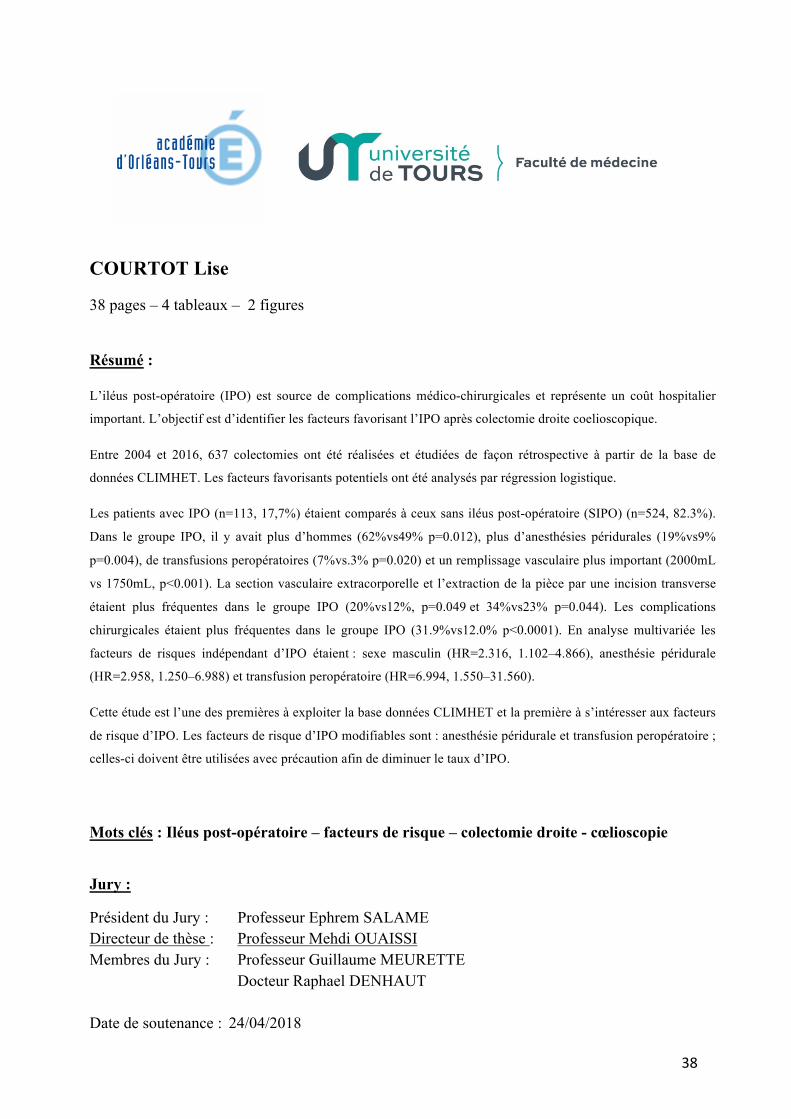
COURTOT Lise 38 pages – 4 tableaux – 2 figures Résumé : L’iléus post-opératoire (IPO) est source de complications médico-chirurgicales et représente un coût hospitalier
important. L’objectif est d’identifier les facteurs favorisant l’IPO après colectomie droite coelioscopique.
Entre 2004 et 2016, 637 colectomies ont été réalisées et étudiées de façon rétrospective à partir de la base de
données CLIMHET. Les facteurs favorisants potentiels ont été analysés par régression logistique.
Les patients avec IPO (n=113, 17,7%) étaient comparés à ceux sans iléus post-opératoire (SIPO) (n=524, 82.3%).
Dans le groupe IPO, il y avait plus d’hommes (62%vs49% p=0.012), plus d’anesthésies péridurales (19%vs9%
p=0.004), de transfusions peropératoires (7%vs.3% p=0.020) et un remplissage vasculaire plus important (2000mL
vs 1750mL, p<0.001). La section vasculaire extracorporelle et l’extraction de la pièce par une incision transverse
étaient plus fréquentes dans le groupe IPO (20%vs12%, p=0.049 et 34%vs23% p=0.044). Les complications
chirurgicales étaient plus fréquentes dans le groupe IPO (31.9%vs12.0% p<0.0001). En analyse multivariée les
facteurs de risques indépendant d’IPO étaient : sexe masculin (HR=2.316, 1.102–4.866), anesthésie péridurale
(HR=2.958, 1.250–6.988) et transfusion peropératoire (HR=6.994, 1.550–31.560).
Cette étude est l’une des premières à exploiter la base données CLIMHET et la première à s’intéresser aux facteurs
de risque d’IPO. Les facteurs de risque d’IPO modifiables sont : anesthésie péridurale et transfusion peropératoire ;
celles-ci doivent être utilisées avec précaution afin de diminuer le taux d’IPO.
Mots clés : Iléus post-opératoire – facteurs de risque – colectomie droite - cœlioscopie
Jury :
Président du Jury : Professeur Ephrem SALAME Directeur de thèse : Professeur Mehdi OUAISSI Membres du Jury : Professeur Guillaume MEURETTE Docteur Raphael DENHAUT Date de soutenance : 24/04/2018