Embed Size (px)
Citation preview
Processing and Application of Ceramics 14 [4] (2020) 355–361
https://doi.org/10.2298/PAC2004355N
Crystal structure, optical and magnetic properties of PrFeO3nanoparticles prepared by modified co-precipitation method
Anh Tien Nguyen1, Ngoc Tram Nguyen1, Irina Yakovlevna Mittova2,Nikolai Sergeevich Perov3, Valentina Olegovna Mittova4, Thi Cam Chuong Hoang1,Van My Nguyen1, Van Hung Nguyen5, Vinh Pham6, Xuan Vuong Bui7,8,∗
1Ho Chi Minh City University of Education, Ho Chi Minh City, 700000, Vietnam2Voronezh State University, Universitetskaya pl.1, Voronezh, 394018, Russia3Magnetism Department, Faculty of Physics, Lomonosov Moscow State University, Moscow, Russia4Burdenko Voronezh State Medical University, Voronezh, 394036, Russia5Dong Thap University, Cao Lanh City, 81000, Vietnam6Institute of Research and Development, Duy Tan University, Da Nang, 550000, Vietnam7Department for Management of Science and Technology Development, Ton Duc Thang University, Ho Chi
Minh City, 700000, Vietnam8Faculty of Applied Sciences, Ton Duc Thang University, Ho Chi Minh City, 700000, Vietnam
Received 13 March 2020; Received in revised form 7 July 2020; Received in revised form 22 September 2020;Accepted 26 October 2020
Abstract
In this work, PrFeO3 nanoparticles were synthesized by modified co-precipitation method and annealed atdifferent temperatures up to 850 °C. The annealed PrFeO3 nanoparticles have single phase orthorhombicstructure and the average particle size of 25–30 nm. Due to the very small particle size the prepared PrFeO3
nanoparticles are capable of being used as photocatalyst materials thanks to their strong adsorption bands at230–400 nm and 400–800 nm observed from the UV-Vis spectra. Additionally, the PrFeO3 nanoparticles areparamagnetic materials with Hc ∼ 10 Oe and Mr ∼ 0. These findings demonstrate their potential use not onlyas photocatalysts, but also as magnetic materials.
Keywords: PrFeO3, nanoparticles, improved co-precipitation method, optical and magnetic properties
I. Introduction
Complex oxides with ReMO3 perovskite structure,where Re and M are rare-earth and transition met-als, respectively, represent an important class of func-tional materials [1–3]. Especially, the materials basedon orthoferrite rare-earth elements (Re = La, Pr, Nd,Ho, etc.) possess good electrical and magnetic proper-ties which are decreased from microscale to nanoscale[4–9]. Among the rare-earth orthoferrites, PrFeO3 hasfound applications in magnetic field materials [10–13],photocatalysis [14–16], dyes and inorganic pigments[17,18].
∗Corresponding author: tel: +84 28 37755037,e-mail: [email protected]
Properties of the orthoferrite ReFeO3 perovskitenanomaterials, such as optical and magnetic proper-ties, depend on the chemical composition, distributionof cations, particle size and synthesis method. Typi-cally, the PrFeO3 nanoparticles with size of 80 nm showa band gap value of 2.4 eV [15], which decreases to2.08 eV when the particle size is 50 nm [14]. The de-crease of the band gap leads to the increase of photo-catalytic yield in visible light and offers new areas ofapplication.
Magnetic behaviour of PrFeO3 has also attracted sig-nificant attention. Thus, the magnetic characteristics(antiferromagnetic Néel temperature TN , curves of mag-netic susceptibility reciprocal 1/χ against temperatureand the temperature dependence of magnetization ZFCand FC) of orthoferrite PrFeO3 materials with a parti-
355
A.T. Nguyen et al. / Processing and Application of Ceramics 14 [4] (2020) 355–361
cle size of 20–30µm have been investigated under theapplied magnetic field of 1000 Oe [10]. In addition,the temperature dependence of magnetization (ZFC andFC) with a magnetic field of 500 Oe has been investi-gated for the orthoferrite PrFeO3 thin film (having thick-ness ∼200 nm) [11,12] prepared by a ceramic reactiontechnique.
Orthoferrite PrFeO3 nanoparticles have been synthe-sized via various methods including high-temperatureceramic fabrication [9,13,18], hydrothermal methods[10,16], and sol-gel complex methods [14,15,17,19].However, orthoferrite PrFeO3 particles with a sizeof 25–30 nm (prepared by simple co-precipitationmethod), a small band gap and the features of mag-netism under a high magnetic field (∼15000 Oe) havenot been reported. Thus, the aim of this work was to pre-pare PrFeO3 nanoparticles (20–25 nm) and study theirstructure, optical, and magnetic properties.
II. Experimental procedure
Pr(NO3)3 · 6 H2O (99.8% purity, Merck),Fe(NO3)3 · 9 H2O (99.6% purity, Sigma-Aldrich)and NH3 solution (Xilong, 85% purity), were employedas the starting materials. The synthetic process was sim-ilar to that described in our previous reports [20–22]. Asolution including an equal amounts of Pr(NO3)3 andFe(NO3)3 salts was gradually introduced to hot water(> 90 °C). Then, the mixture was continuously stirredfor 5 min and cooled to room temperature (25–30 °C).In the next step a 5% NH3 solution was slowly addedand continuously stirred until the solution colourchanged into light pink indicated by phenolphthalein.The precipitate was filtered, washed by deionized wateruntil pH = 7 and dried in air at room temperature(25–30 °C). Finally, the products were ground andthermally treated at different temperatures.
Furthermore, the pure oxide materials, i.e. Fe2O3 andPr2O3, were also prepared by a similar procedure tocompare their structure with the structure of the synthe-sized PrFeO3 perovskite materials.
TGA-DSC analyser (Labbsys Evo, TG-DSC1600 °C, SETERAM Instrumentation, Caluire, France)was used to determine the appropriate temperature forobtaining the single-phase PrFeO3 perovskite structure.The sample was placed in a platinum cylindricalcrucible and heated from 25 to 1000 °C at 10 °C/min indried air.
X-ray powder diffraction (PXRD) patterns were ob-tained on a D8-ADVANCE (Brucker, Bremen, Ger-many) with CuKα radiation (λ = 1.5418 Å) using stepsize of 0.02° in the range of 10–80°. The crystallinesizes of the annealed PrFeO3 powders were determinedbased on Scherrer’s equation [23]:
D =0.89λβ · cos θ
(1)
where λ is X-ray wavelength of CuKα = 1.5418 Å, θ isthe diffraction angle of the maximum reflection and βis the full-width at half maximum (FWHM). The latticeparameters (a, b, c) and cell volume (V) were calculatedby Eqs. 2 and 3, respectively [23]:
1d2=
h2
a2+
k2
b2+
l2
c2(2)
V = a · b · c (3)
where d is the distance between crystalline planes withMiller indices hkl.
Particle size and morphology of the annealed PrFeO3crystals were studied by transmission electron mi-croscopy (TEM, Joel JEM-1400, Joel Ltd., Tokyo,Japan) and field emission scanning electron microscopy(FESEM, S-4800, Hitachi, Japan). The content and sur-face distribution of Pr, Fe and O elements were deter-mined by energy-dispersive X-ray spectroscopy (EDXand EDX-mapping, Horiba H-7593, Horiba, Northamp-ton, UK).
The UV-Vis absorption spectra of the PrFeO3nanocrystals were recorded on a UV-Visible spec-trophotometer (UV-Vis, JASCO V-550, Hachioji,Tokyo, Japan). Magnetic properties of the samples (sat-uration magnetization Ms, coercivity Hc and remanentmagnetization Mr) were measured at room tempera-ture with a vibrating sample magnetometer (VSM, Mi-croSense EV11, Japan).
III. Results and discussion
3.1. Thermal analysis
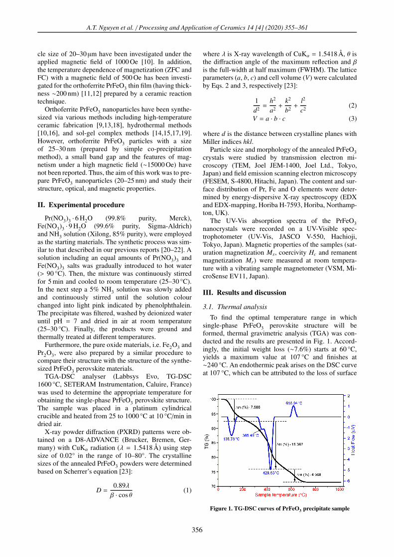
To find the optimal temperature range in whichsingle-phase PrFeO3 perovskite structure will beformed, thermal gravimetric analysis (TGA) was con-ducted and the results are presented in Fig. 1. Accord-ingly, the initial weight loss (∼7.6%) starts at 60 °C,yields a maximum value at 107 °C and finishes at∼240 °C. An endothermic peak arises on the DSC curveat 107 °C, which can be attributed to the loss of surface
Figure 1. TG-DSC curves of PrFeO3 precipitate sample
356
A.T. Nguyen et al. / Processing and Application of Ceramics 14 [4] (2020) 355–361
water. From 240 to ∼550 °C, the weight loss (∼16.4%)is due to water removal through crystallization, the de-hydration of Fe2O3 · xH2O (where x = 1–5) [24] and thepyrolysis of PrO(OH) · yH2O [25]. In this stage, two en-dothermic peaks appeared at 356 and 430 °C, respec-tively. The small weight loss (∼4.6%) between 550 and730 °C may account for the release of adsorbed CO2from the Pr2O(CO3)2 · 1.4H2O or Pr2(CO3)3 · 8 H2Ostructures [25,26]. In addition, in the temperature inter-val from 600 to 650 °C there is an exothermic peak withmaximum at 619 °C indicating formation of perovskitephase. Hence, the temperatures of 650, 750 and 850 °Cwere chosen for investigating the structure and morphol-ogy of the synthesized PrFeO3 nanoparticles.
3.2. Structure and morphology
PXRD patterns of the samples PrFeO3, Fe2O3 andPr6O11 after annealing for 1 h at 750 °C are presentedin Fig. 2. For the pure Pr-based sample, the stablePr6O11 phase was generated after annealing the hydrox-ide praseodymium precipitate instead of the unstablePr2O3 phase. This is clearly explained by the followingequation [25]:
6 Pr(OH)3 +O2 −−−→ Pr6O11 + 9 H2O (4)
Interestingly, the observed reflections of the PrFeO3sample are in good agreement with the standard JCPDS047-0065 of the orthorhombic PrFeO3 phase withoutany XRD peaks of Fe2O3 and Pr6O11 phases. This isclear confirmation of the TGA-DSC results and indica-tion that the reaction between precursors is completedafter annealing.
Figure 2. PXRD patterns of PrFeO3, Fe2O3 and Pr6O11
powders annealed at 750 °C for 1 h
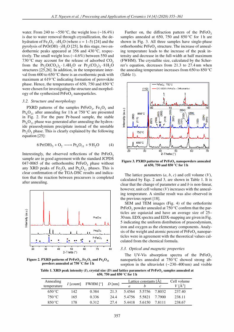
Further on, the diffraction pattern of the PrFeO3samples annealed at 650, 750 and 850 °C for 1 h areshown in Fig. 3. All three samples have single-phaseorthorhombic PrFeO3 structure. The increase of anneal-ing temperature leads to the increase of the peak in-tensity and decrease in the full-width at half maximum(FWHM). The crystallite size, calculated by the Scher-rer’s equation, decreases from 21.3 to 27.4 nm whenthe annealing temperature increases from 650 to 850 °C(Table 1).
Figure 3. PXRD patterns of PrFeO3 nanopowders annealedat 650, 750 and 850 °C for 1 h
The lattice parameters (a, b, c) and cell volume (V),calculated by Eqs. 2 and 3, are shown in Table 1. It isclear that the change of parameter a and b is non-linear,however, unit cell volume (V) increases with the anneal-ing temperature. A similar result was also observed inthe previous report [18].
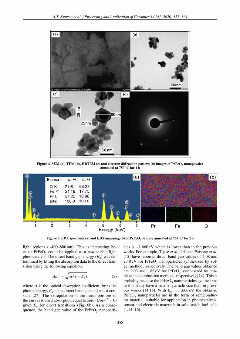
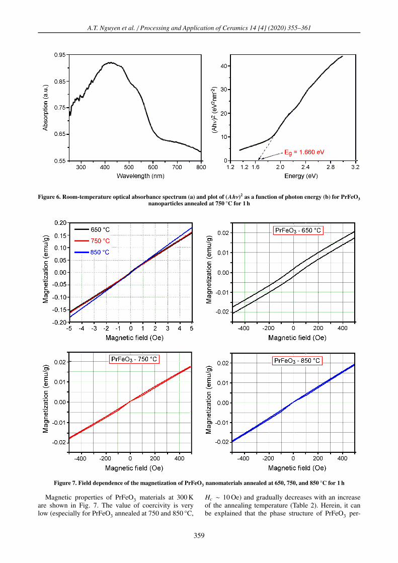
SEM and TEM images (Fig. 4) of the orthoferritePrFeO3 powder annealed at 750 °C confirm that the par-ticles are equiaxial and have an average size of 25–30 nm. EDX spectra and EDX-mapping are given in Fig.5 indicating the uniform distribution of praseodymium,iron and oxygen as the elementary components. Analy-sis of the weight and atomic percent of PrFeO3 nanopar-ticles were in agreement with the theoretical values cal-culated from the chemical formula.
3.3. Optical and magnetic properties
The UV-Vis absorption spectra of the PrFeO3nanoparticles annealed at 750 °C showed strong ab-sorption in the ultraviolet (∼230–400 nm) and visible
Table 1. XRD peak intensity (I), crystal size (D) and lattice parameters of PrFeO3 samples annealed at650, 750 and 850 °C for 1 h
AnnealingI [count] FWHM [°] D [nm]
Lattice constants [Å] Cell volumetemperature a b c V [Å3]
650 °C 142 0.384 21.3 5.4564 5.5756 7.8032 237.40750 °C 165 0.336 24.4 5.4756 5.5821 7.7900 238.11850 °C 178 0.312 27.4 5.4418 5.6150 7.8111 238.67
357
A.T. Nguyen et al. / Processing and Application of Ceramics 14 [4] (2020) 355–361
Figure 4. SEM (a), TEM (b), HRTEM (c) and electron diffraction pattern (d) images of PrFeO3 nanopowderannealed at 750 °C for 1 h
Figure 5. EDX spectrum (a) and EDX-mapping (b) of PrFeO3 sample annealed at 750 °C for 1 h
light regions (∼400–800 nm). This is interesting be-cause PrFeO3 could be applied as a new visible-lightphotocatalyst. The direct band gap energy (Eg) was de-termined by fitting the absorption data to the direct tran-sition using the following equation:
Ahν =
√
α(hν − Eg) (5)
where A is the optical absorption coefficient, hν is thephoton energy, Eg is the direct band gap and α is a con-stant [27]. The extrapolation of the linear portions ofthe curves toward absorption equal to zero ((Ahν)2 = 0)gives Eg for direct transitions (Fig. 6b). As a conse-quence, the band gap value of the PrFeO3 nanoparti-
cles is ∼1.660 eV which is lower than in the previousworks. For example, Tijare et al. [14] and Peisong et al.
[15] have reported direct band gap values of 2.08 and2.40 eV for PrFeO3 nanoparticles synthesized by sol-gel method, respectively. The band gap values obtainedare 2.03 and 1.88 eV for PrFeO3 synthesized by tem-plate and combustion methods, respectively [14]. This isprobably because the PrFeO3 nanoparticles synthesizedin this study have a smaller particle size than in previ-ous works [14,15]. With Eg = 1.660 eV, the obtainedPrFeO3 nanoparticles are in the form of semiconduc-tor material, suitable for application in photocatalysis,sensor and electrode materials in solid oxide fuel cells[1,14–16].
358
A.T. Nguyen et al. / Processing and Application of Ceramics 14 [4] (2020) 355–361
Figure 6. Room-temperature optical absorbance spectrum (a) and plot of (Ahν)2 as a function of photon energy (b) for PrFeO3
nanoparticles annealed at 750 °C for 1 h
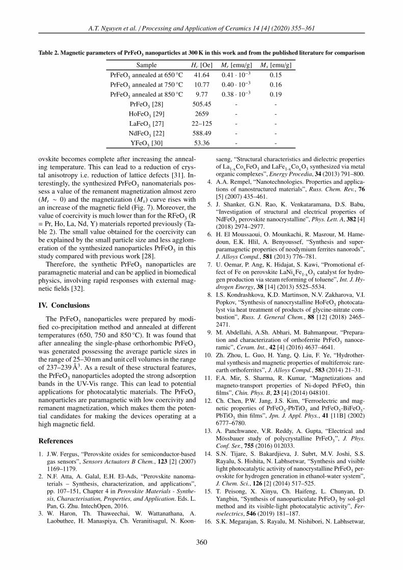
Figure 7. Field dependence of the magnetization of PrFeO3 nanomaterials annealed at 650, 750, and 850 °C for 1 h
Magnetic properties of PrFeO3 materials at 300 Kare shown in Fig. 7. The value of coercivity is verylow (especially for PrFeO3 annealed at 750 and 850 °C,
Hc ∼ 10 Oe) and gradually decreases with an increaseof the annealing temperature (Table 2). Herein, it canbe explained that the phase structure of PrFeO3 per-
359
A.T. Nguyen et al. / Processing and Application of Ceramics 14 [4] (2020) 355–361
Table 2. Magnetic parameters of PrFeO3 nanoparticles at 300 K in this work and from the published literature for comparison
Sample Hc [Oe] Mr [emu/g] Ms [emu/g]
PrFeO3 annealed at 650 °C 41.64 0.41 · 10−3 0.15
PrFeO3 annealed at 750 °C 10.77 0.40 · 10−3 0.16
PrFeO3 annealed at 850 °C 9.77 0.38 · 10−3 0.19
PrFeO3 [28] 505.45 - -
HoFeO3 [29] 2659 - -
LaFeO3 [27] 22–125 - -
NdFeO3 [22] 588.49 - -
YFeO3 [30] 53.36 - -
ovskite becomes complete after increasing the anneal-ing temperature. This can lead to a reduction of crys-tal anisotropy i.e. reduction of lattice defects [31]. In-terestingly, the synthesized PrFeO3 nanomaterials pos-sess a value of the remanent magnetization almost zero(Mr ∼ 0) and the magnetization (Ms) curve rises withan increase of the magnetic field (Fig. 7). Moreover, thevalue of coercivity is much lower than for the RFeO3 (R= Pr, Ho, La, Nd, Y) materials reported previously (Ta-ble 2). The small value obtained for the coercivity canbe explained by the small particle size and less agglom-eration of the synthesized nanoparticles PrFeO3 in thisstudy compared with previous work [28].
Therefore, the synthetic PrFeO3 nanoparticles areparamagnetic material and can be applied in biomedicalphysics, involving rapid responses with external mag-netic fields [32].
IV. Conclusions
The PrFeO3 nanoparticles were prepared by modi-fied co-precipitation method and annealed at differenttemperatures (650, 750 and 850 °C). It was found thatafter annealing the single-phase orthorhombic PrFeO3was generated possessing the average particle sizes inthe range of 25–30 nm and unit cell volumes in the rangeof 237–239 Å3. As a result of these structural features,the PrFeO3 nanoparticles adopted the strong adsorptionbands in the UV-Vis range. This can lead to potentialapplications for photocatalytic materials. The PrFeO3nanoparticles are paramagnetic with low coercivity andremanent magnetization, which makes them the poten-tial candidates for making the devices operating at ahigh magnetic field.
References
1. J.W. Fergus, “Perovskite oxides for semiconductor-basedgas sensors”, Sensors Actuators B Chem., 123 [2] (2007)1169–1179.
2. N.F. Atta, A. Galal, E.H. El-Ads, “Perovskite nanoma-terials – Synthesis, characterization, and applications”,pp. 107–151, Chapter 4 in Perovskite Materials - Synthe-
sis, Characterisation, Properties, and Application. Eds. L.Pan, G. Zhu. IntechOpen, 2016.
3. W. Haron, Th. Thaweechai, W. Wattanathana, A.Laobuthee, H. Manaspiya, Ch. Veranitisagul, N. Koon-
saeng, “Structural characteristics and dielectric propertiesof La1-xCoxFeO3 and LaFe1-xCoxO3 synthesized via metalorganic complexes”, Energy Procedia, 34 (2013) 791–800.
4. A.A. Rempel, “Nanotechnologies. Properties and applica-tions of nanostructured materials”, Russ. Chem. Rev., 76
[5] (2007) 435–461.5. J. Shanker, G.N. Rao, K. Venkataramana, D.S. Babu,
“Investigation of structural and electrical properties ofNdFeO3 perovskite nanocrystalline”, Phys. Lett. A, 382 [4](2018) 2974–2977.
6. H. El Moussaoui, O. Mounkachi, R. Masrour, M. Hame-doun, E.K. Hlil, A. Benyoussef, “Synthesis and super-paramagnetic properties of neodymium ferrites nanorods”,J. Alloys Compd., 581 (2013) 776–781.
7. U. Oemar, P. Ang, K. Hidajat, S. Kawi, “Promotional ef-fect of Fe on perovskite LaNixFe1-xO3 catalyst for hydro-gen production via steam reforming of toluene”, Int. J. Hy-
drogen Energy, 38 [14] (2013) 5525–5534.8. I.S. Kondrashkova, K.D. Martinson, N.V. Zakharova, V.I.
Popkov, “Synthesis of nanocrystalline HoFeO3 photocata-lyst via heat treatment of products of glycine-nitrate com-bustion”, Russ. J. General Chem., 88 [12] (2018) 2465–2471.
9. M. Abdellahi, A.Sh. Abhari, M. Bahmanpour, “Prepara-tion and characterization of orthoferrite PrFeO3 nanoce-ramic”, Ceram. Int., 42 [4] (2016) 4637–4641.
10. Zh. Zhou, L. Guo, H. Yang, Q. Liu, F. Ye, “Hydrother-mal synthesis and magnetic properties of multiferroic rare-earth orthoferrites”, J. Alloys Compd., 583 (2014) 21–31.
11. F.A. Mir, S. Sharma, R. Kumar, “Magnetizations andmagneto-transport properties of Ni-doped PrFeO3 thinfilms”, Chin. Phys. B, 23 [4] (2014) 048101.
12. Ch. Chen, P.W. Jang, J.S. Kim, “Ferroelectric and mag-netic properties of PrFeO3-PbTiO3 and PrFeO3-BiFeO3-PbTiO3 thin films”, Jpn. J. Appl. Phys., 41 [11B] (2002)6777–6780.
13. A. Panchwanee, V.R. Reddy, A. Gupta, “Electrical andMössbauer study of polycrystalline PrFeO3”, J. Phys.
Conf. Ser., 755 (2016) 012033.14. S.N. Tijare, S. Bakardjieva, J. Subrt, M.V. Joshi, S.S.
Rayalu, S. Hishita, N. Labhsetwar, “Synthesis and visiblelight photocatalytic activity of nanocrystalline PrFeO3 per-ovskite for hydrogen generation in ethanol-water system”,J. Chem. Sci., 126 [2] (2014) 517–525.
15. T. Peisong, X. Xinyu, Ch. Haifeng, L. Chunyan, D.Yangbin, “Synthesis of nanoparticulate PrFeO3 by sol-gelmethod and its visible-light photocatalytic activity”, Fer-
roelectrics, 546 (2019) 181–187.16. S.K. Megarajan, S. Rayalu, M. Nishibori, N. Labhsetwar,
360
A.T. Nguyen et al. / Processing and Application of Ceramics 14 [4] (2020) 355–361
“Improved catalytic activity of PrMO3 (M = Co and Fe)perovskite: synthesis of thermally stable nanoparticles bya novel hydrothermal method”, New J. Chem., 39 (2105)2342–2348.
17. O. Opuchovic, G. Kreiza, J. Senvaitiene, K. Kazlauskas,A. Beganskiene, A. Kareiva, “Sol-gel synthesis, charac-terization and application of selected sub-microsized lan-thanide (Ce, Pr, Nd, Tb) ferrites”, Dyes Pigments, 118
(2015) 176–182.18. J. Luxova, P. Sulcova, M. Trojan, “Influence of firing
temperature on the color properties orthoferrite PrFeO3”,Thermochim. Acta, 579 (2014) 80–85.
19. O. Pekinchak, L. Vasylechko, I. Lutsyuk, Ya. Vakhula,Yu. Prots, W. Carrillo-Cabrela, “Sol-gel-preparednanoparticles of mixed praseodymium cobaltites-ferrites”,Nanoscale Res. Lett., 11 (2016) 75.
20. T.A. Nguyen, V.N.T. Pham, H.T. Le, D.H. Chau, V.O.Mittova, L.T.Tr. Nguyen, D.A. Dinh, T.V. Nhan Hao,I.Ya. Mittova, “Crystal structure and magnetic proper-ties of LaFe1-xNixO3 nanomaterials prepared via a simpleco-precipitation method”, Ceram. Int., 45 (2019) 21768–21772.
21. A.T. Nguyen, V.N.T. Pham, T.Tr.L. Nguyen, V.O. Mit-tova, Q.M. Vo, M.V. Berezhnaya, I.Ya. Mittova, Tr.H. Do,H.D. Chau, “Crystal structure and magnetic properties ofperovskite YFe1-xMnxO3 nanopowders synthesized by co-precipitation method”, Solid State Sci., 96 (2019) 105922.
22. T.A. Nguyen, M.V. Berezhnaya, T.L. Pham, V.O. Mit-tova, M.Q. Vo, L.T.Tr. Nguyen, H.Tr. Do, I.Ya. Mittova,E.L. Viryutina, “Synthesis and magnetic characteristicsof neodymium ferrite powders with perovskite structure”,Russ. J. Appl. Chem., 92 [4] (2019) 498–504.
23. C. Sasikala, N. Durairaj, I. Baskaran, B. Sathyaseelan,M. Henini, “Transition metal titanium (Ti) doped LaFeO3nanoparticles for enhanced optical structural and magneticproperties”, J. Alloys Compd., 712 (2017) 870–877.
24. A.G. Belous, E.V. Pashkova, V.A. Elshanskii, V.P. Ivanit-
skii, “Effect of precipitation conditions on the phase com-position, particle morphology and properties of iron (III,II) hydroxide precipitates”, Inorg. Mater., 36 [4] (2000)343–351.
25. N. Imanaka, Physical and Chemical Properties of Rare
Earth Oxides, Binary Rare Earth Oxides, Kluwer Aca-demic Publishers, 2004, pp. 111–113.
26. N. Kozo, H. Wakita, A. Mochizuki, “The synthesis of crys-talline rare earth carbonates”, Bull. Chem. Soc. Jpn., 46 [1](1973) 152–156.
27. S. Phokha, S. Pinitsoontorn, S. Maensiri, S. Rujirawat,“Structure, optical and magnetic properties of LaFeO3nanoparticles prepared by polymerized complex method”,J. Sol-Gel Sci. Technol., 71 (2014) 333–341.
28. A.T.S. Sudandararaj, G.S. Kumar, M. Dhivya, R.D. Ei-thiraj, I.B.Sh. Banu, “Spin reorientation transition innanoscale multiferroic PrFeO3 and its band structure cal-culation”, J. Alloys Compd., 817 (2020) 152747.
29. Z. Habib, K. Majid, M. Ikram, Kh. Sultan, “Influence of Nisubstitution at B-site for Fe3+ ions on morphological, op-tical, and magnetic properties of HoFeO3 ceramics”, Appl.
Phy. A. Mater. Sci. Process., 122 (2016) 550.30. T.A. Nguyen, V. Pham, D.H. Chau, V.O. Mittova, I.Ya.
Mittova, E.I. Kopeychenko, L.T.Tr. Nguyen, V.X. Bui,A.T.P. Nguyen, “Effect of Ni substitution on phase tran-sition, crystal structure and magnetic properties of nanos-tructured YFeO3 perovskite”, J. Mol. Structure, 1215
(2020) 128293.31. T.A. Nguyen, V. Pham, T.L. Pham, L.T.Tr. Nguyen, I.Ya.
Mittova, V.O. Mittova, L.N. Vo, B.T.T. Nguyen, V.X. Bui,E.L. Viryutina, “Simple synthesis of NdFeO3 by the so-precipitation method based on a study of thermal behaviorsof Fe (III) and Nd (III) hydroxides”, Crystals, 10 (2020)219.
32. B.D. Cullity, C.D. Graham, Introduction to Magnetic Ma-
terials, 2nd ed., John Wiley & Sons, Inc., Publication,Canada, 2009.
361


![Optical Fiber Communicationfiber.hardfree.net/2011/open_data/fiber_edu.pdf · 2011-01-20 · 2 1. 광통신의개요 광통신[ Optical Fiber Communication ]이란? ☞기존의금속심선을이용한유선통신이나주파수를이용한무선통신과는달리광섬유케이블[](https://img.pdfslide.fr/doc/110x75/5f0333cf7e708231d4080b25/optical-fiber-2011-01-20-2-1-eeoe-e-optical-fiber-communication.jpg)