Embed Size (px)
Citation preview
FACULDADE DE MEDICINA DA UNIVERSIDADE DE COIMBRA
TRABALHO FINAL DO 6º ANO MÉDICO COM VISTA A ATRIBUIÇÃO DO GRAU DE
MESTRE NO ÂMBITO DO CICLO DE ESTUDOS DE MESTRADO INTEGRADO EM
MEDICINA
JOÃO PAULO FERNANDES ABRANTES
MITOCHONDRIAL DNA VARIANTS IN COMPLEX V
CODING GENES CONTRIBUTING TO
FRONTOTEMPORAL LOBAR DEGENERATION
ARTIGO CIENTÍFICO
ÁREA CIENTÍFICA DE GENÉTICA MOLECULAR
TRABALHO REALIZADO SOB A ORIENTAÇÃO DE:
PROFESSORA DOUTORAMARIA MANUELA MONTEIRO GRAZINA
PROFESSORA DOUTORA MARIA ISABEL JACINTO SANTANA
MAIO /2013
Página 2 de 50
Mitochondrial DNA variants in Complex V coding genes contributing to frontotemporal
lobar degeneration
João PF Abrantes1, Isabel Santana
1,2, Maria João Santos
3, Diana Duro
2, Daniela Luís
3,
Manuela Grazina1,3,*
1Faculty of Medicine, University of Coimbra, Portugal; 2
Centro Hospitalar e Universitário de
Coimbra (CHUC), Coimbra, Portugal; 3CNC - Center for Neuroscience and Cell Biology – Laboratory
of Biochemical Genetics, University of Coimbra, Portugal
*Author for correspondence:
Professor Manuela Grazina:
Faculty of Medicine, University of Coimbra, Pólo III – Subunit I
Azinhaga de Sta. Comba Celas, 3000-548 Coimbra
Tel. +351-239480040
Fax. +351-239480048
Email: [email protected]
Página 3 de 50
Abstract
FTLD is the second most common early-onset type of dementia, it has a broad
spectrum of clinic and histopathologic manifestations and its pathophysiological mechanisms
are not yet fully understood. Over the last decades a growing number of evidence has
supported the involvement of the mitochondria in the aetiology of several neurodegenerative
diseases. One suggested mechanism is the existence of mitochondrial DNA mutations which
render impairment of the mitochondrial respiratory chain. The aim of our study is to add
further knowledge to this area. Accordingly, thetwo genes of the mitochondrial DNA coding
for complex V subunits, MT-ATP8 and MT-ATP6, were sequenced and analysed, in 70 FTDL
patients. The results reveal that29 patients (41.4%) present at least one sequence variation in
one of the studied genes, being 38.9% of all the alterations found, synonymous, whereas non-
synonymous variations account for 61.1% of total alterations. The latter were submitted to in
silico analysis. This study disclosed three probably damaging mutations
(m.8393C>G,m.8519G>A and m.8945T>C), and others, such asm.8573G>A, m.8839G>A
and m.8842A>C, which may cause functional impairment. The majority of the alterations
identified in the present study have not been described before in association with any
neurodegenerative disease and are barely documented. Even if additional studies are needed,
the present study represents a significant contribution to a better understanding of a complex
neurodegenerative disease, such as frontotemporal lobar degeneration.
Keywords
Frontotemporal lobar degeneration, mitochondrial DNA, sequence variations, complex V,
dementia, mutations.
Página 4 de 50
Abbreviations
AD Alzheimer‟s disease
ALS amyotrophic lateral sclerosis
ATP adenosine triphosphate
bvFTD behaviour frontotemporal dementia
FTLD frontotemporal lobar degeneration
FUS fused in sarcoma
HD Huntington‟s disease
KSS Kearns–Sayre syndrome
LHON Leber‟s hereditary optic neuropathy
LS Leigh Syndrome
LPA logopenic or phonological variant
MELAS mitochondrial encephalomyopathy, lactic acidosis, and stroke-like
episodes
MERRF myoclonus epilepsy ragged-red fibers
MRC mitochondrial respiratory chain
mtDNA mitochondrial DNA
mtDB Human Mitochondrial Genome Database
NARP neuropathy ataxia and retinitis pigmentosa
nDNA nuclear DNA
PD Parkinson‟s Disease
PEO progressive external ophthalmoplegia
PNFA progressive non-fluent aphasia
SD semantic dementia
TDP TAR DNA-bindingprotein
UPS ubiquitin proteasome system
Página 5 de 50
Introduction
FTLD is the second most common early-onset type of dementia (Grazina et al., 2004;
Rollinson et al., 2011; Seelaar et al., 2011), typically manifesting between 45 and 65 years
(Neary et al., 2005; Shi et al., 2005; Sleegers et al., 2010), and being diagnosed at youngest
ages as 21 or oldest as 85 years old (Neary et al., 2005; Pickering-Brown SM, 2007). No
difference has been found between the familial and sporadic forms of the disease concerning
the age of onset (Neary et al., 2005).The incidence appears to be equal between both genders
(McKhann et al., 2001; Neary et al., 2005; Pickering-Brown SM, 2007; Seelaar et al., 2011).
It has a median duration of illness of 6 to 8 years, ranging from 2 to 20 (Neary et al., 2005;
Seelaar et al., 2011).
The term FTLD includes a heterogeneous group of clinical presentations which can be
divided in behaviour and language variants (McKhann et al., 2001; Seelaar et al., 2011). The
first, also called behaviour FTD form, is the most common (McKhann et al., 2001; Pickering-
Brown, 2007; Seelaar et al., 2011). Patients who suffer from this variant, show predominantly
personality and behaviour changes, becoming either uninhibited, apathetic or displaying
stereotypical actions (Neary et al., 2005; Pickering-Brown, 2007; Rollinson et al., 2011). The
language component is classically divided in two major syndromes, SD and PNFA
(Pickering-Brown, 2007). The first (SD) is characterized by loss of semantic knowledge
which impairs word comprehension with a relatively conserved fluency of speech (Sleegerset
al., 2010; Rollinson et al., 2011), whereas PNFA is roughly the opposite, with affected speech
and preserved semantic recognition (Sleegerset al., 2010; Rollinson et al., 2011).
Recentevidences support the existence of a third group, the LPA, with slow rate of speech,
word finding difficulties and occasional phonemic errors (Seelaar et al., 2011).
As the pathologic process progresses the boundaries of these classifications become
blurry, and patients often show a mixed clinical picture (McKhann et al., 2001; Neary et al.,
Página 6 de 50
2005; Sleegers et al., 2010; Seelaar et al., 2011). Furthermore, some other conditions can
overlap, such as Parkinson syndromes, progressive supra-nuclear palsy, cortico-basal
syndromes or motor-neuron disease (Sleegers et al., 2010; Seelaar et al., 2011). The latter,
frequently related with the behaviour FTD, specially with the stereotypic variant, is associated
with a worst prognosis with a mean survival of 3 years (Neary et al., 2005; Seelaar et al.,
2011).
As for the clinical picture, the alterations found in the brain of these patients also
present distinct and heterogeneous characteristics. The common finding is the frontal and
temporal lobe atrophy (McKhann et al., 2001; Mackenzie et al., 2009; Seelaar et al., 2011),
that can be also seen in imaging studies. The clinical presentation usually reflects the affected
part of the brain by the disease, whereas the opposite relation is not necessarily valid (Neary
et al., 2005; Shi et al., 2005). The histopathological classification allows to distinguishfive
variants, according to Mackenzie et al. (2010): FTDL-tau, characterized by insoluble forms of
tau proteins in form of neurofibrillary tangle-like structures or in Prick body type;FTDL-
TDP,in which the accumulation of TDP-43 protein is detected; FTDL-FUS, as consequence
of the accumulation of FUS protein; FTDL-UPS for tau, TDP-43 and FUS negative histology
test that are positive for the immunohistochemistry of the UPS proteins, and FTDL-ni for
cases in which inclusions are not detected by the current tests. FTDL-tau and FTDL-TDP are
the most common forms (Mackenzie et al., 2010).
The aetiology of FTDL has not yet been fully understood. To date, there are some
known nDNA mutations which lead to the histopathologic changes described. According to
Seelaar et al. (2011), the most frequents are mutations in MAPT gene (locus 17q21.1) that
lead to the accumulation of tau protein (Mackenzie et al., 2009, 2010; Seelaar et al., 2011),
and in GRN gene (locus 17q21.32) leading to the formation of TDP-43 positive inclusions
(Mackenzie et al., 2009, 2010; Rollinson et al., 2011; Seelaar et al., 2011).
Página 7 de 50
Additionally,although less frequently, mutations in TARDBP(locus 1p36.22),VCP (locus
9p13.3) andC9orf72 (locus 9p21.2)genes have also been described, all associated with TDP-
43 inclusions. Other mutations have been assigned to FUS gene (locus 16p11.2) with FUS
protein accumulation, andtoCHMP2B gene (locus 3p11.2), which is one known cause of
FTDL-UPS (Mackenzie et al., 2010).
Despite all the causative mutations identified, most of FTDL cases, either sporadic or
familial, cannot be explained by these alterations. Therefore, other pathogenic mechanisms
must be implicated in the genesis of the disease (Rollinson et al., 2011; Seelaar et al.,
2011).Some patients present an overlapbetween Alzheimer‟s disease (AD) and FTD both in
neuropathologicaland clinical aspects. This may suggest a similar overlap in
physiopathology,namely an involvement of mitochondrial DNA (mtDNA) inFTD, as it has
been associated to AD (Grazina et al., 2004). For review on genes involved in FTDL see
Cruts et al. (2012).
Mitochondria have been implicated in the pathophysiology of several
neurodegenerative diseases such as PD (Autere et al., 2004; Mawrin et al., 2004; DiMauro
and Schon, 2008; Lee et al., 2009; Federico et al., 2012), AD (Emerit et al., 2004;Grazina et
al., 2005; Grazina et al., 2006; Onyango et al., 2006; DiMauro and Schon, 2008), HD
(DiMauro and Schon, 2008; Lee et al., 2009) and ALS (Mawrin et al., 2004; DiMauro and
Schon, 2008; Lee et al., 2009). In fact, the role that the dysfunction of this organelle plays in
the human pathology has begun to be better understood over the last decades, being one of the
major contributors not only to neurodegeneration, but also to several encephalomyopathic
diseases like LS, LHON, PEO, KSS, MELAS or MERRF (Leonard and Schapira, 2000;
Emerit et al., 2004;DiMauro and Schon, 2008; Lee, 2009; Federico et al., 2012; Schapira,
2012).
Página 8 de 50
The pathologies that arise from mitochondrial dysfunction affect essentially high
energy demanding tissues, such as liver, skeletal or cardiac muscle and nervous system. In
fact, these tissues withelevated metabolic needs, relying mainly on the ATP produced via
aerobic metabolism in the mitochondria (Pieczenik and Neustadt , 2007; Lee et al., 2009),
compared to other tissues (Emerit et al., 2004; Lee et al., 2009; Greaves et al., 2012). In spite
of being the powerhouse of the eukaryotic cells, mitochondria is also promoter of other key
functions of the cell like apoptosis-signalling pathway (Pieczenik and Neustadt, 2007;
Greaves et al., 2012), cytosolic calcium concentration and iron-sulfur cluster biogenesis
(Greaves et al., 2012), moreover its involvement in ageing and carcinogenesis processes is
only beginning to be unveiled (Greaves et al., 2012; Schapira, 2012).
The process by which mitochondrial dysfunction leads to neurodegeneration is
relatively well established: it is a consequence of the impairment of the MRC activity, which
by its turn decreases ATP production, increasing the ROS generation and the concentration of
intracellular calcium (Emerit et al., 2004; Grazina et al., 2005; Federico et al., 2012; Greaves
et al., 2012; Schapira AHV, 2012). This process ultimately ends in the activation of the
intrinsic mitochondrial pathway resulting in apoptosis and consequent neuronal cell loss
(Emerit et al., 2004; Grazina et al., 2006; DiMauro and Schon, 2008; Lee et al., 2009).
There are several mechanisms that can trigger this events, among them are the
presence of environmental stressors such as diet deficits or toxics (Pieczenik and Neustadt,
2007) that inhibit the MRC enzymes, inflammatory mediators such as TNF-α (Pieczenik and
Neustadt, 2007), proteasome malfunctions and protein misfoldings, or DNA mutations. Such
mutations can occur in the nDNA, affecting the MRC proteins, the mtDNA maintenance and
expression, the salvage nucleotide synthesis and transport, or the mitochondrial dynamics.
They can also occur directly over the mtDNA in which case they affect the MRC proteins, the
tRNA‟s or the rRNA‟s. To date almost 600 mtDNA mutations have been reported associated
Página 9 de 50
to disease (http://www.mitomap.org). Some of those mtDNA polymorphisms have been
associated with several neurodegenerative disorders like PD (Federico et al., 2012), AD
(Grazina et al., 2005, 2006) or FTDL (Grazina et al., 2004). This relation with
neurodegenerative diseases is not always easy to establish as these mutations also can occur
with normal ageing resulting from the ROS aggression (Mawrin et al., 2004; Grazina et al.,
2006). Currently, two theories seek to explain this relation. The first one proposes a vicious
cycle occurring between ROS production and mtDNA mutations in which one leads to
another (Pieczenik and Neustadt, 2007; Greaves et al., 2012), however a more likely
explanation is the occurrence of mtDNA mutations throughout life, either resulting from ROS
damage, errors in mtDNA replication or repair mechanisms, that clonally expand as the
mitochondria divides independently from the cell (Greaves et al., 2012).
The aim of this study is therefore to search and access the implication of mtDNA
variations in patients suffering from FTLD. The screening included the analysis
ofMTATP6and MTATP8genes (coding for ATP synthase F0 subunits 6 and 8, respectively),
the only two genes of mtDNA that encode Complex V subunits, as part of a larger project
whose objective is to analyse the whole mtDNA in FTDL patients. ATP synthase is the last
complex of the MRC, essential for the oxidative phosphorylation process, where the majority
of the ATP molecules are produced. Therefore, its dysfunction could lead to a major
impairment in the ATP generation with consequent neuronal degeneration. Currently, two
diseases are associated with point mutations in the MTATP6 gene, NARP and LS (Greaves et
al., 2012).Mutations in Complex V nDNA coding genes ATPAF2 and ATP5E had been also
related with encephalopathy (Schapira, 2012). So far, there is no evidence of association
between mutations in MTATP8 gene and disease.
To our knowledge, this is the first study investigating the genetic sequence of MTATP6and
MTATP8 genesin FTLD patients.
Página 10 de 50
Patients and Methods
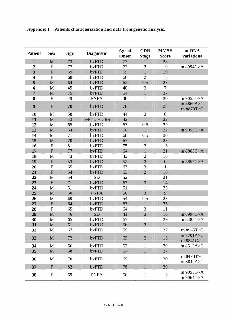
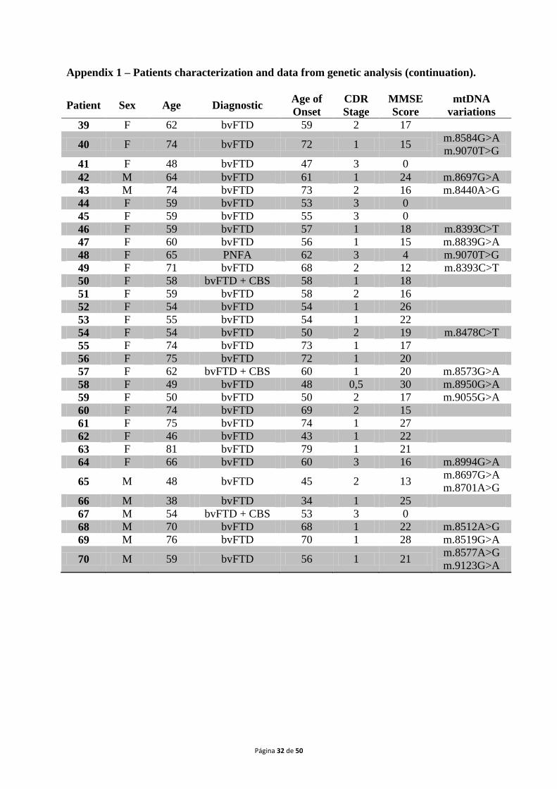
The samples included on this study belong to70 patients (31 males and 39 females;
mean age:63 years, range: 38 to 82 years)with a probable diagnosis of FTLD according to the
standard criteria of DSM-IV (Brun et al., 1994; McKhann et al., 2001), recruited at the
Neurological Unit of the “Centro Hospitalar e Universitário de Coimbra”. The mean age of
onset was60 years, ranging from 34 to 79 years. Concerning clinical forms,60 patients
(85.7%) presented bvFTD, while 4 (5.7%) had bvFTD with CBS, other 4 (5.7%) PNFA and 2
(2.9%) showed SD. All patients gave written informed consent in order to participate in the
study, which has been approved by the localEthical Committee. A list of all patients‟ data can
be found in appendix 1.
Total cellular DNA was extracted from peripheral blood leukocytes after erythrocytes
lysis, following standard phenol-chloroform method. Automated sequencing analysis was
performed using 3130 ABI Prism sequencing system and BigDye®
Terminator Ready
Reaction Mix 3.1 (Applied Biosystems) with specific primers for target genes in order to
study both MTATP6 and MTATP8coding genes. The sequences obtained were compared with
the human mithochondrial DNA revised Cambridge reference sequence (Andrews et al.,
1999), obtained from GenBank, using Sequencing Analysis®
v.5.4 and SeqScape®
v.2.5
software (Applied Biosystems)to search for mutations, polymorphisms or novel sequence
variations. All sequence variations were classified using the MITOMAP database
(http:\\www.mitomap.org) and frequency was obtained in the Human Mitochondrial Genome
Database (Ingman et al., 2006)toestimate its frequency in general population.
For non-synonymous sequence variations an in silicoanalysis was performed using a
set of softwares: PolyPhen v. 2.2.2, which takes into account sequence, phylogenetic and
structural information, predicting protein functional outcomes of sequence variations by two
models, HumVar and HumDiv; Mutation Assessor v. 2, that predicts the impact of a variation

Página 11 de 50
by aligning multiple sequences, clustering them into subfamilies and scoring a variation by
global and sub-family specific conservation patterns; Provean, that allows a similar prediction
approachoffered by Mutation Assessor, but calculates the mutation score by the average of
each sub-family average score; and SIFT, which predicts the mutation score by aligning user
defined sequences. For this purpose,we have selected 10 different species (Homo Sapiens,
Pan troglodytes, Gorilla gorilla, Pongo abelii, Macaca mulatta, Mus musculus, Rattus
norvegicus, Bos taurus, Sus scrofa and Canis familiaris). Clustal Omega softwarewas used in
order to visualize the alignment of the referred sequences as shown in figure 3 and appendix
2. The sequences were obtained through the UniProt sequence databases, being the majority
of them from the European Nucleotide Archive database.
Statistic analysis was performed to infer if variations in the studied locus correlates
with different variables of the patients. Accordingly,t-test or Mann-Whitney test (if data did
not follow a Gaussian distribution), were performed to compareage, age of onset, CDR
staging or the MMSE score of the patients, according to the presence of genetic variations;
contingency tables, with Fisher‟s exact test, were used to investigate gender, age of onset,
MMSE class and clinical outcome, in association to genetic variants.
Results
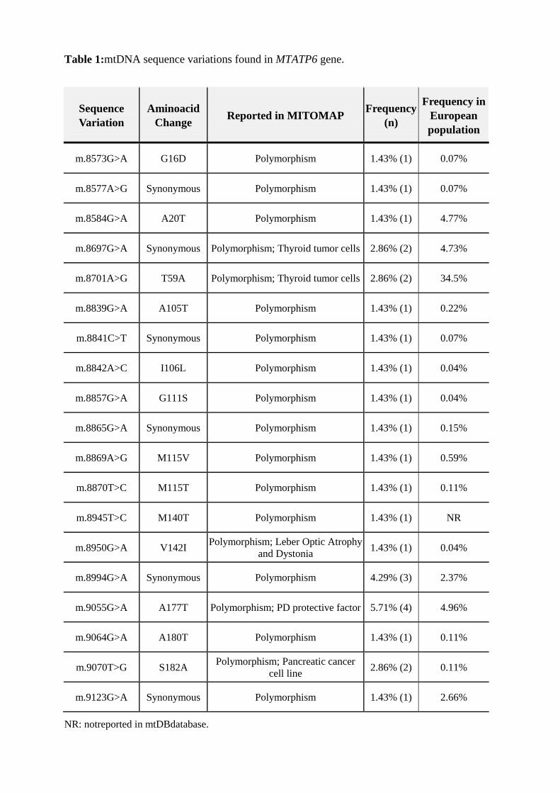
Sequence variants inthe mtDNA genes under study were found in 29 patients (41.4%).
From these,8 (11.4%) presented one MTATP8 variation, 14 (20.0%) one MTATP6alteration, 1
(1.4%) one variation in both genes, and 6 (8.6%) presented two MTATP6alterations. The
sequence variations found in MTATP6and MTATP8 genes are listed in Table 1 and Table 2,
respectively.
Página 12 de 50
Table 1:mtDNA sequence variations found in MTATP6 gene.
Sequence
Variation
Aminoacid
Change Reported in MITOMAP
Frequency
(n)
Frequency in
European
population
m.8573G>A G16D Polymorphism 1.43% (1) 0.07%
m.8577A>G Synonymous Polymorphism 1.43% (1) 0.07%
m.8584G>A A20T Polymorphism 1.43% (1) 4.77%
m.8697G>A Synonymous Polymorphism; Thyroid tumor cells 2.86% (2) 4.73%
m.8701A>G T59A Polymorphism; Thyroid tumor cells 2.86% (2) 34.5%
m.8839G>A A105T Polymorphism 1.43% (1) 0.22%
m.8841C>T Synonymous Polymorphism 1.43% (1) 0.07%
m.8842A>C I106L Polymorphism 1.43% (1) 0.04%
m.8857G>A G111S Polymorphism 1.43% (1) 0.04%
m.8865G>A Synonymous Polymorphism 1.43% (1) 0.15%
m.8869A>G M115V Polymorphism 1.43% (1) 0.59%
m.8870T>C M115T Polymorphism 1.43% (1) 0.11%
m.8945T>C M140T Polymorphism 1.43% (1) NR
m.8950G>A V142I Polymorphism; Leber Optic
Atrophy and Dystonia 1.43% (1) 0.04%
m.8994G>A Synonymous Polymorphism 4.29% (3) 2.37%
m.9055G>A A177T Polymorphism; PD protective factor 5.71% (4) 4.96%
m.9064G>A A180T Polymorphism 1.43% (1) 0.11%
m.9070T>G S182A Polymorphism; Pancreatic cancer
cell line 2.86% (2) 0.11%
Table 1:mtDNA sequence variations found in MTATP6 gene.
Sequence
Variation
Aminoacid
Change Reported in MITOMAP
Frequency
(n)
Frequency in
European
population
m.8573G>A G16D Polymorphism 1.43% (1) 0.07%
m.8577A>G Synonymous Polymorphism 1.43% (1) 0.07%
m.8584G>A A20T Polymorphism 1.43% (1) 4.77%
m.8697G>A Synonymous Polymorphism; Thyroid tumor cells 2.86% (2) 4.73%
m.8701A>G T59A Polymorphism; Thyroid tumor cells 2.86% (2) 34.5%
m.8839G>A A105T Polymorphism 1.43% (1) 0.22%
m.8841C>T Synonymous Polymorphism 1.43% (1) 0.07%
m.8842A>C I106L Polymorphism 1.43% (1) 0.04%
m.8857G>A G111S Polymorphism 1.43% (1) 0.04%
m.8865G>A Synonymous Polymorphism 1.43% (1) 0.15%
m.8869A>G M115V Polymorphism 1.43% (1) 0.59%
m.8870T>C M115T Polymorphism 1.43% (1) 0.11%
m.8945T>C M140T Polymorphism 1.43% (1) NR
m.8950G>A V142I Polymorphism; Leber Optic Atrophy
and Dystonia 1.43% (1) 0.04%
m.8994G>A Synonymous Polymorphism 4.29% (3) 2.37%
m.9055G>A A177T Polymorphism; PD protective factor 5.71% (4) 4.96%
m.9064G>A A180T Polymorphism 1.43% (1) 0.11%
m.9070T>G S182A Polymorphism; Pancreatic cancer
cell line 2.86% (2) 0.11%
m.9123G>A Synonymous Polymorphism 1.43% (1) 2.66%
NR: notreported in mtDBdatabase.
Página 13 de 50
In MTATP6 gene, a total of 19 different sequence variations were found: 6
synonymous and 13 non-synonymous. All variations were reported in MITOMAP database as
polymorphisms and 4 were also reported in different diseases, and 1 as a protective factor of
PD. These alterations were detected predominantly in only one patient each, except for
variations m.8697G>A, m.8701A>G and m.9070T>G, detected in two patients each,
m.8994G>A detected in three, and for m.9055G>A, that was detected in four different
patients.
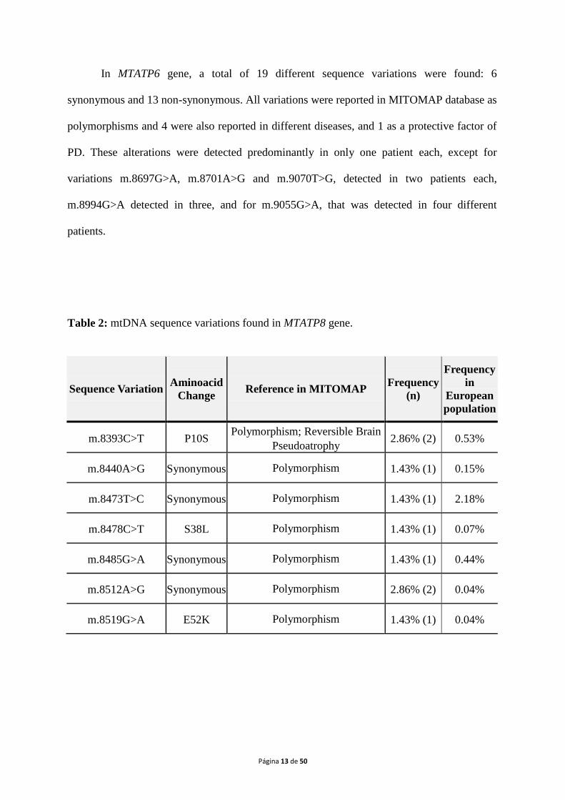
Table 2: mtDNA sequence variations found in MTATP8 gene.
Sequence Variation Aminoacid
Change Reference in MITOMAP
Frequency
(n)
Frequency
in
European
population
m.8393C>T P10S Polymorphism; Reversible Brain
Pseudoatrophy 2.86% (2) 0.53%
m.8440A>G Synonymous Polymorphism 1.43% (1) 0.15%
m.8473T>C Synonymous Polymorphism 1.43% (1) 2.18%
m.8478C>T S38L Polymorphism 1.43% (1) 0.07%
m.8485G>A Synonymous Polymorphism 1.43% (1) 0.44%
m.8512A>G Synonymous Polymorphism 2.86% (2) 0.04%
m.8519G>A E52K Polymorphism 1.43% (1) 0.04%
Página 14 de 50
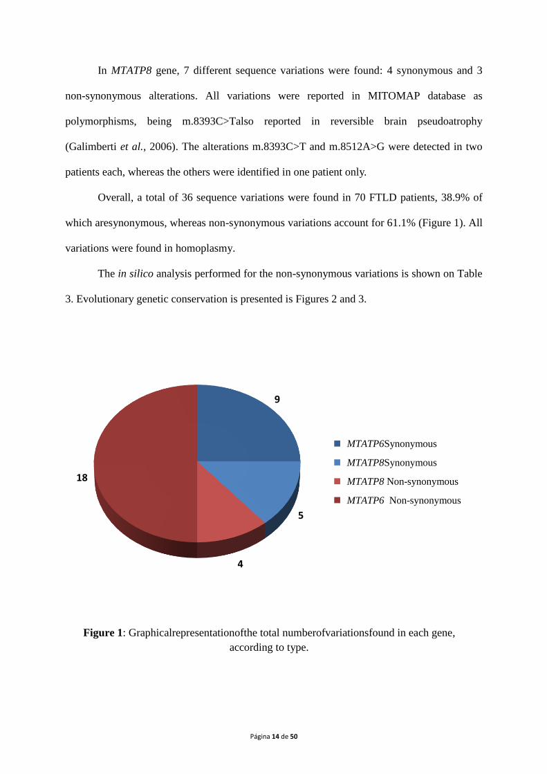
In MTATP8 gene, 7 different sequence variations were found: 4 synonymous and 3
non-synonymous alterations. All variations were reported in MITOMAP database as
polymorphisms, being m.8393C>Talso reported in reversible brain pseudoatrophy
(Galimberti et al., 2006). The alterations m.8393C>T and m.8512A>G were detected in two
patients each, whereas the others were identified in one patient only.
Overall, a total of 36 sequence variations were found in 70 FTLD patients, 38.9% of
which aresynonymous, whereas non-synonymous variations account for 61.1% (Figure 1). All
variations were found in homoplasmy.
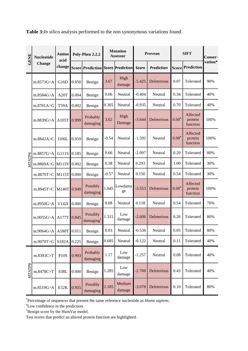
The in silico analysis performed for the non-synonymous variations is shown on Table
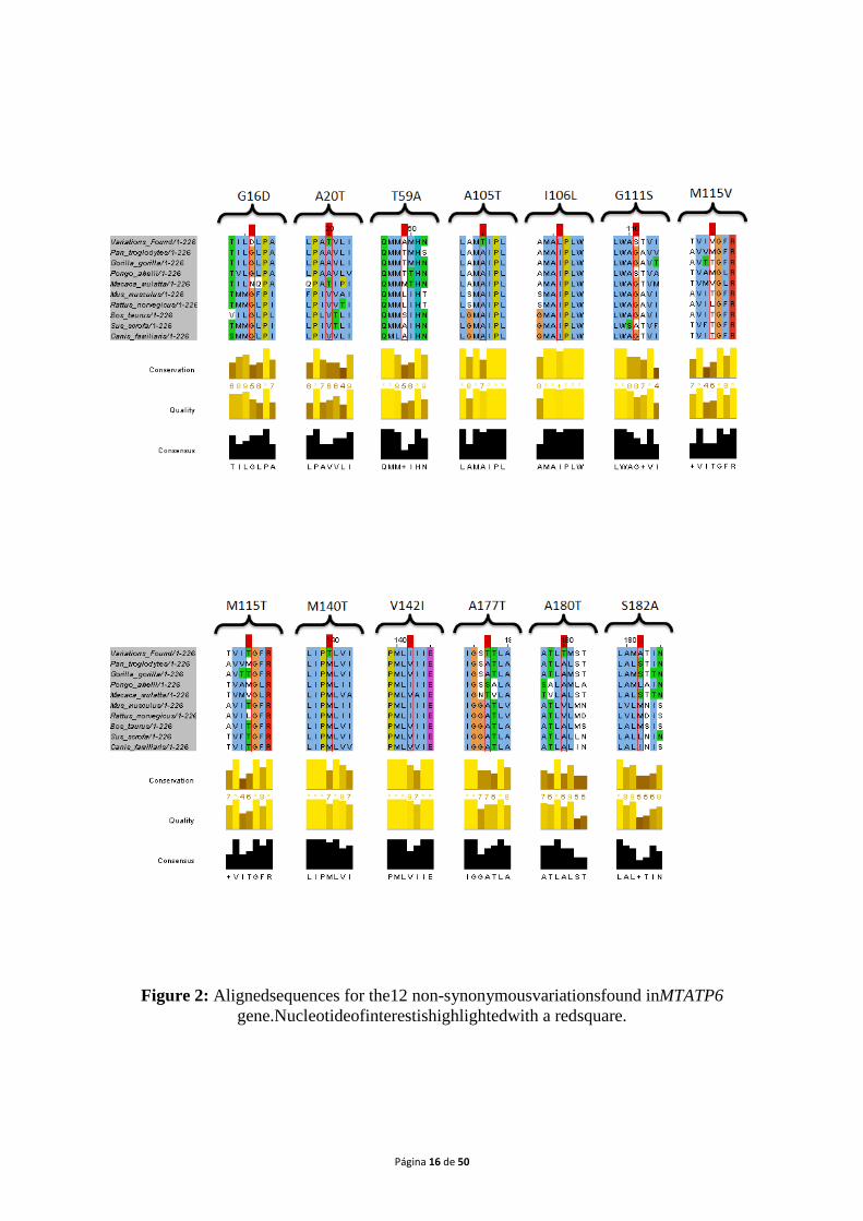
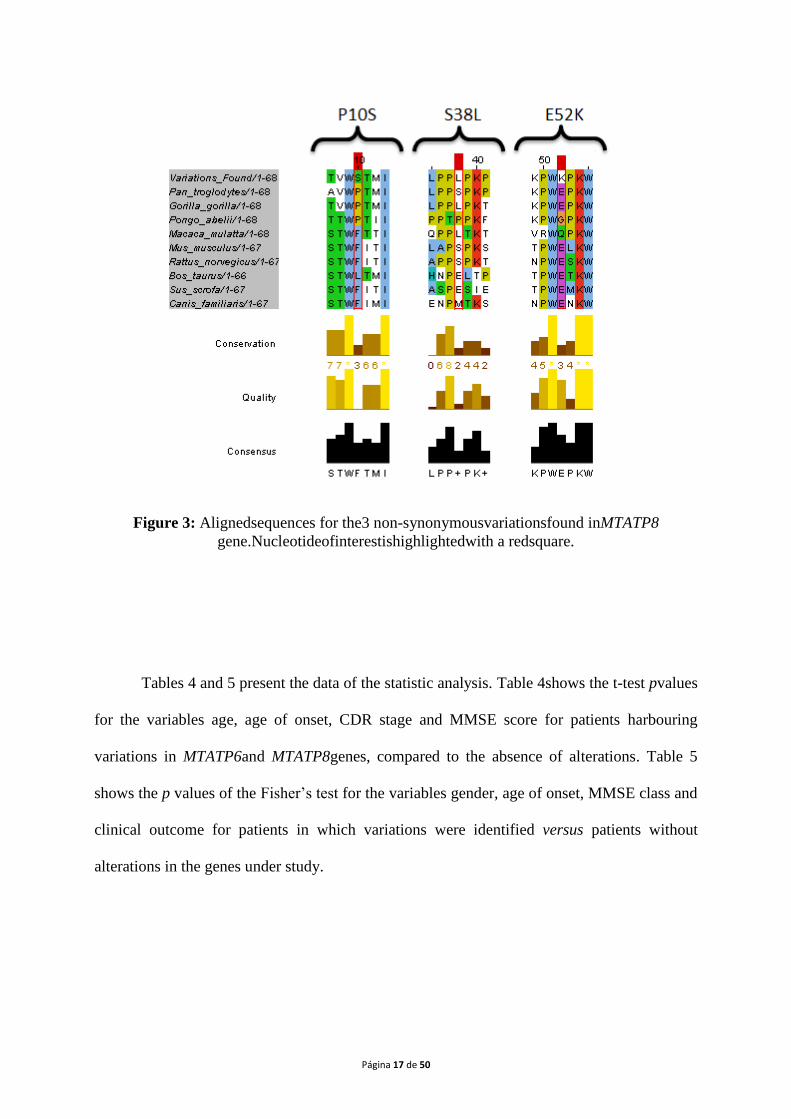
3. Evolutionary genetic conservation is presented is Figures 2 and 3.
9
5
4
18
MTATP6 Synonymous
MTATP8 Synonymous
MTATP8 Non-synonymous
MTATP6 Non-synonymous
Figure 1: Graphicalrepresentationofthe total numberofvariationsfound in each gene,
according to type.
MTATP6Synonymous
MTATP8Synonymous
MTATP8 Non-synonymous
MTATP6 Non-synonymous
Página 15 de 50
Table 3:In silico analysis performed to the non synonymous variations found.
GE
NE
Nucleotide
Change
Amino
acid
change
Poly-Phen 2.2.2 Mutation
Assessor Provean SIFT
Conser-
vation*
Score Prediction Score Prediction Score Prediction Score Prediction
MT
AT
P6
m.8573G>A G16D 0.050 Benign 3.67 High
damage -5.425 Deleterious 0.07 Tolerated 90%
m.8584G>A A20T 0.004 Benign 0.06 Neutral -0.404 Neutral 0.34 Tolerated 40%
m.8701A>G T59A 0.002 Benign 0.365 Neutral -0.935 Neutral 0.70 Tolerated 40%
m.8839G>A A105T 0.999 Probably
damaging 3.62
High
Damage -3.644 Deleterious 0.00
#
Affected
protein
function
100%
m.8842A>C I106L 0.059 Benign -0.54 Neutral -1.592 Neutral 0.00#
Affected
protein
function
100%
m.8857G>A G111S 0.185 Benign 0.66 Neutral -2.097 Neutral 0.20 Tolerated 80%
m.8869A>G M115V 0.002 Benign 0.38 Neutral 0.293 Neutral 1.00 Tolerated 30%
m.8870T>C M115T 0.000 Benign -0.57 Neutral 0.150 Neutral 0.54 Tolerated 30%
m.8945T>C M140T 0.949 Possibly
damaging 1.845
Lowdama
ge -3.553 Deleterious 0.00
#
Affected
protein
function
100%
m.8950G>A V142I 0.000 Benign 0.08 Neutral 0.118 Neutral 0.54 Tolerated 70%
m.9055G>A A177T 0.845 Possibly
damaging†
1.315 Low
damage -2.606 Deleterious 0.26 Tolerated 80%
m.9064G>A A180T 0.011 Benign 0.03 Neutral -0.536 Neutral 0.05 Tolerated 80%
m.9070T>G S182A 0.225 Benign 0.685 Neutral -0.122 Neutral 0.11 Tolerated 40%
MT
AT
P8
m.8393C>T P10S 0.993 Probably
damaging 1.17
Low
damage -1.257 Neutral 0.08 Tolerated 40%
m.8478C>T S38L 0.000 Benign 1.285 Low
damage -2.708 Deleterious 0.43 Tolerated 40%
m.8519G>A E52K 0.955 Possibly
damaging 2.185
Medium
damage -3.078 Deleterious 0.10 Tolerated 80%
*Percentage of sequences that present the same reference nucleotide as Homo sapiens.
#Low confidence in the prediction.
†Benign score by the HumVar model.
Test scores that predict an altered protein function are highlighted.
Página 16 de 50
Figure 2: Alignedsequences for the12 non-synonymousvariationsfound inMTATP6
gene.Nucleotideofinterestishighlightedwith a redsquare.
Página 17 de 50
Figure 3: Alignedsequences for the3 non-synonymousvariationsfound inMTATP8
gene.Nucleotideofinterestishighlightedwith a redsquare.
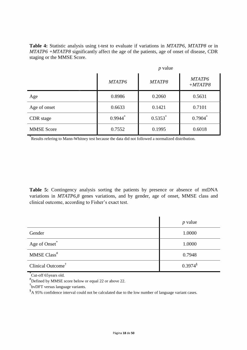
Tables 4 and 5 present the data of the statistic analysis. Table 4shows the t-test pvalues
for the variables age, age of onset, CDR stage and MMSE score for patients harbouring
variations in MTATP6and MTATP8genes, compared to the absence of alterations. Table 5
shows the p values of the Fisher‟s test for the variables gender, age of onset, MMSE class and
clinical outcome for patients in which variations were identified versus patients without
alterations in the genes under study.
Página 18 de 50
Table 4: Statistic analysis using t-test to evaluate if variations in MTATP6, MTATP8 or in
MTATP6 +MTATP8 significantly affect the age of the patients, age of onset of disease, CDR
staging or the MMSE Score.
p value
MTATP6 MTATP8 MTATP6
+MTATP8
Age 0.8986 0.2060 0.5631
Age of onset 0.6633 0.1421 0.7101
CDR stage 0.9944* 0.5353
* 0.7904
*
MMSE Score 0.7552 0.1995 0.6018
*Results refering to Mann-Whitney test because the data did not followed a normalized distribution.
Table 5: Contingency analysis sorting the patients by presence or absence of mtDNA
variations in MTATP6,8 genes variations, and by gender, age of onset, MMSE class and
clinical outcome, according to Fisher‟s exact test.
p value
Gender 1.0000
Age of Onset* 1.0000
MMSE Class# 0.7948
Clinical Outcome† 0.3974
§
*Cut-off 65years old.
#Defined by MMSE score below or equal 22 or above 22.
†bvDFT versus language variants.
§A 95% confidence interval could not be calculated due to the low number of language variant cases.

Página 19 de 50
Discussion
The total number of sequence variations found washigher forMTATP6gene (n=27)
than forMTATP8 (n=9). This fact may be due to the length of each gene, being the first one
larger (681 bp) than MTATP8 (207 bp). Apparently, the alterations are evenly
distributed.Concerning the alteration type, a different scenario was observed: the non-
synonymous to synonymous ratio in MTATP8gene is 1:0.8; however in the MTATP6locus that
ratio drops to 1:2. This couldindicate that non-synonymous variations in the MTATP8 locus
are less tolerated than in the MTATP6, possibly leading to cell apoptosis and thus being less
prevalent. In fact, the scarcity of described deleterious mutations in the final MRC complexes,
specially in the MTATP8 locus,suggests that either those are rare or incompatible with life
(DiMauro and Schon, 2008). However,Canis lupus familiaris (dog), sus scrofa (pig), mus
musculus (mouse) and rattus norvegicus (rat) ATP8 protein lacked 1 aminoacid compared to
the other species, and thus their mtDNA was 3 nucleotides shorter. Bos taurus (bovine) lacked
even one more aminoacid (and 6 nucleotides). It could mean that this protein structure is not
highly conserved among species, and thus allowing some alterations without loss of function.
Despite that, all amino acid variations found in MTATP8 were represented in the 10 species
analysed.
All the variations found in the present work are already reported in MITOMAP
database (http://www.mitomap.org), although only a few are reported in association with
pathologies.The majority of the mtDNA variations found (60%) in the patients studied are
non-synonymous variations. Concerning the other 40% synonymous variations, it is not
expected that these represent a major cause of protein malfunction. Interestingly, some of the
synonymous variations here reported are fairly rare, such asm.8512A>G,identified in two
FTLD patients, but only in one of the 2704 sequences in the mtDB database.
Moreover,m.8577A>G and m.8841C>Tboth reported in two different patients of this study,
Página 20 de 50
wereonlyeach one reported in 2 of the 2704 mtDB sequences.Although synonymous
alterations do not change protein structure, they can still be involved in pathologic process
(Rollinset al., 2009). It is possible that different arrays of polymorphisms coding for MRC
coding components may result in different MRC functional outcomes (Grazina et al., 2005;
DiMauro and Schon, 2008), and so, albeit mtDNA polymorphisms are not pathological by
themselves, they can create susceptibility, or protection, for certain diseases (Autere, 2004;
DiMauro and Schon, 2008), as result of DNA expression modulation.
The m.8573G>A changes the non polar glycine at position 16 of the ATP6
protein,which is preserved in 9 of the 10 sequences analysed (Figure 2). The
sorroundingaminoacids are also preserved among higher primates. As a result, Mutation
Assessor and Provean predicts that this variation is harmful, being the SIFT score close
enough to the cut-off point so that it can be considered that this variation is possibly
pathogenic, although the change to an acidic polar aspartic acid is not enough to raise the
PolyPhen score.
The m.8697G>A, a synonymous variation,and m.8701A>G wereidentifiedby Máximo
et al., (2002) in the thyroid tumor cells of three different patients. Thesecond variation is also
a haplogroup marker for the macro-haplogroup N, that includes the European-specific
haplogroups.Therefore,it could be involved in population adaptation or neoplastic
transformation (Brandon et al., 2006). In the present study,this variation was found in a
notably less proportion, when compared to the one found in mtDB datatbase. Therefore, it
could be postulated that this variation would represent a protective factor regarding FTDL
development.However, this could also be a consequence of the prevalence of the haplogroups
in the populations, being diminished in the group of patients analysed. Studies in groups of
patients from different world regions should be carried out to clarify this point. As for the in
silico analysis, a non functional impairment was predicted by all the softwares used. The same
Página 21 de 50
resultsmay apply tom.8584G>A, m.8857G>A, m.8869A>G, m.8870T>C and m.9064G>A.
Nevertheless, as discussed earlier for synonymous variations, this does not mean that these
variations could not have a role in the modulation of pathophysiological process. Being so, it
is difficult to demonstrate given the present scientific knowledge.
For the m.8839G>A variation, the in silico analysis gave similar results for each of the
4 softwares, predicting thatchanges of a nonpolar alanine to a polar threonine at position 105,
significantly affectsthe ATP6 proteinfunction. Additionally, this amino acidand the the others
surrounding this position, are conserved in all of the sequences aligned (Figure 2). Thus, this
variation is probably harmful.
A change of a non polar isoleucine to a nonpolar leucine conditioned by the
m.8842A>C alteration should not have a major impact in the functional outcome of the ATP6
protein, as predicted by PolyPhen, Mutation Assessor and Provean. Regardless of that, this
variation occurs in the vicinity of the previously mentioned one, in a highly conserved region
of the protein among species.Adding to that, its frequency in mtDB database is 0.04%, so
although it might not have a direct impact on the protein function, could have a pathologic
relevance.
The m.8945T>C is predicted to alter protein function in every software, except
forMutation assessor. Additionally, this variation is not reported in mtDB database, meaning
that it has a frequency of less than 0.04% in the population. One possible explanation is that
this variation, which changes a nonpolar methionine to a polar threonine on a highly
conserved site of the protein (position 140), renders a severe dysfunction in the MRC and the
cell.
Despite thefact that m.8950G>A variation has been pointed out to be involved in
LHONand Dystonia, according to MITOMAP database, the references do not seem to support
Página 22 de 50
such hypothesis. It is apparently a tolerated mutation that occurs in a less conserved site of the
protein.
The most frequent variation, found,m.9055G>A(identified in 4 patients) is reported to
decrease PD risk in women and it is also a haplogroup K marker (van der Walt et al., 2003).
Could it be a protective factor for PD and a risk factor for FTDL? Apparently no, due to the
fact that its frequency in FTLD patientsof the present study, is similar to the one reported in
mtDB. This variation promotes a switch from a nonpolar alanine to a polar threonine in the
amino acid 177, a conserved position in 8 of the 10 sequences (Figure 2), which is enough to
have a deleterious score in Provean and in one model of the PolyPhen analysis. It appears that
this variation could have some functional effect in the ATP6 protein that is notclear yet.
The variation m.9070T>Ghas been reported in associationto a pancreatic cancer cell
line byJones et al.(2001).However, according to the present analysis, it is not expected that it
could lead to functional disruption of the ATP6 protein.
The m.8393C>G alteration was reported by Galimbertiet al. (2006) as being
associated with brain pseudoatrophy and mental regression. This causes a change of a
nonpolar proline to a polar serine at position 10 of the ATP8 protein; therefore, the PolyPhen
score is fairly high, although all the other in silicotests predict that it would be a tolerated
change. This could be explained by the analysis of the sequences alignment (Figure 3), where
the amino acid 10 is not globally well conserved. Even if the surrounding amino acids and the
closest phylogenetic species are taken into account, it is clear that this change occur in a
highly conserved region of the protein among higher primates. In fact, the SIFT score
approaches the cut-off value of the test. And so it is possible that the m.8393C>G variation is
not only associated with the brain pseudoatrophy and mental regression, but also with FTDL.
The m.8478C>T variation is classified as deleterious by Provean software. However,
the change of a polar serine to a non polar leucine in a poorly conserved aminoacid is not
Página 23 de 50
enough to alter the protein function outcome in any other software used. Accordingly, itseems
unlikely that this alteration could be pathogenic. A fact that could make us think otherwise is
the low frequency in which is reported in mtDB database and the absence of literature
concerning this variation.
The last non-synonymous mutation found in the MTATP8 gene, m.8519G>A, seems to
be pathogenic. The shift of an acidic polar glutamic acid for a basic polar lysine at the amino
acid position 52, conserved in 8 of the 10 sequences, takes place in the middle of a highly
conserved region of the protein (Figure 3). Theseresults reveal a deleterious effect prediction
by PolyPhen, Mutation Assessor and Provean softwares. This result, in addition to the fact
that the frequency of the m.8519G>A variation in 2704 reported mtDB sequences is only
0.07%, suggest that it as a probably deleterious mutation.
Although the in silico analysis presented showed interesting and important results, it is
important to complement this study with functional studies, such as determination of the
MRC activity, particularly complex V, in the patients presenting such alterations. Additionally,
future functional genomics analyses will allow clarifying the pathogenic role of the mtDNA
sequence variations identified in the present study.
Concerning the statistical analysis, no significant positive correlation was found
between patients with variations inMTATP6,MTATP8, or both genesand their age, age of
onset, CDR staging or the MMSE score. The same observation was achieved relating to
clinical outcome. However, it is worth to mention that the number of patients with the
language variants is insufficient to calculate a 95% confidence interval.
The existence of an association between the variations found and one of these
variables would give more strength to the assumption that such variations are a contributing
factor to the development of a particular clinical form of FTDL. However the absence of
statistically significant association means that the variations found are not involved in the
Página 24 de 50
pathophysiology of a particular form of the disease, but their effect is related to the
neurodegeneration instead. On the other hand, even the majority of patients present alteration
on the 2 genes investigated, MTATP6 and MTATP8, they are not present in every patient and
not all the patients harbour the same alterations, meaning that different alterations could lead
to the same effect of impairing, even slightly, MRC function, and that there are other factors
which contribute to the modulation of this complex disease. Extending the analysis to anage
matched healthy control group, could add some clarification, but the comparison with the
frequency of the variations from mtDB database was very helpful to address important points
about the alterations found.
On the other hand, the present findings report to peripheral blood mtDNA variations.
The fact that all these variations are found in homoplasmy is a good indicator that they may
be present in other tissues, including the brain. Nonetheless, somatic mtDNA mutations may
occur in brain cells, and thus not being present in peripheral blood (Grazinaet al., 2003).
Testing other tissues, such as muscle or skin fibroblasts, or post-mortem brain,wouldadd more
evidence to this association.
Although we are aware that it is impossible for the majority of cases, the study of
maternal lineage would also be a significant contribution to the present study.
On the other hand, further studies are needed in order to replicate these findings in
patients‟ cohorts from other world populations.
Página 25 de 50
Conclusion
Our results report new data regarding the presence of mtDNA variations in patients
with FTLD. None of the variations described had been associated to any neurodegenerative
disorder in the references available in literature.
It is reported here that alterations m.8393C>G and m.8519G>A, both located to ATP8
locus, a gene rarely found to be involved in human pathology, areprobably damaging
mutations contributing to the development of FTLD. We also describe two variations in
MTATP6gene, m.8839G>A and m.8945T>C, which are expected to have deleterious
effectson the MRC function. In the same gene, we also reportm.8573G>A, m.8839G>A and
m.8842A>C that have potential to be functionally impairing. Other variations here reported
either are synonymous or the in silico analysis classified them as benign. However, they can
also play a role in the pathophysiological mechanisms leading to complex pathologies such
like FTLD. Data from literature point toboth directions: one stating that mtDNA mutations
aremore likely to be a by-product, secondary to other pathologicalfeatures during the
development of the disease; the otherbeing consistent with the „mitochondrial cascade
hypothesis‟,claiming that mtDNA modifications (both pathogenicand/or polymorphic) are a
cause of the disease (Grazina et al., 2006). In particular, it is possiblethat distinct
combinations of nonmutated electrontransport chain components do not function
identically;being so, nonpathogenic mtDNA variations could contribute to FTLD risk,
similarly to what has been suggested for AD (Grazina et al., 2005; Onyango et al., 2006).
The mtDNA mutations might modify age ofonset, contributing to the
neurodegenerative process,probably due to an impairment of MRC and/or
translationmechanisms (Grazina et al., 2006). Additionally, mtDNA mutations may have a
Página 26 de 50
cumulative effect, increasingthe probability to develop an energy failure (Grazina et al.,
2004).
The report and identification of mtDNA mutations is not only a valuable contribution
towards the better understanding of the FTDL etiology, but also a way to improve diagnosis
protocols of this pathology in a near future, and certainly a way to develop new treatment
strategies in the long term (Grazina et al., 2006).
The present work is a valuable and original contribution to the knowledge of genetic
factors involved in FTLD.
The more we understand about FTDL, the more we unveil its complexity, walking
towards better ways to diagnose, manage and treat this neurodegenerative and fatal disease.
Acknowledgments
This study was financed by Portuguese Foundation for Science and Technology
(FCT), with the Project PTDC/SAL-EPI/121811/2010 (FCT) and partiallysupported by FCT
project PEst-C/SAU/LA0001/2011.
Página 27 de 50
References
Andrews RM, Kubacka I, Chinnery PF, Lightowlers RN, Turnbull DM, Howell N. 1999.
Reanalysis and revision of the Cambridge reference sequence for human mitochondrial
DNA. Nature Genetics 23:147.
Autere J, Moilanen JS, Finnilä S, Soininen H, Mannermaa A, Hartikainen P, Hallikainen M,
Majamaa K. 2004. Mitochondrial DNA polymorphisms as risk factors for Parkinson‟s
disease and Parkinson‟s disease dementia. Hum Genet 115:29-35.
Brandon M, Baldi P, Wallace DC. 2006. Mitochondrial mutations in cancer. Oncogene
25:4647-4662.
Brun A, Englund B, Gustafson L, Passant U, Mann DMA, Neary D, Snowden JS. 1994.
Clinical and neuropathological criteria for frontotemporal dementia: the Lund and
Manchester Groups. J Neurol Neurosurg Psychiatry 57:416-418.
Cruts M, Theuns J, Broeckhoven CV. 2012. Locus-Specific Mutation Databases for
Neurodegenerative Brain Diseases. Human Mutation 1-5
DiMauro S, Schon EA. 2008. Mitochondrial Disorders in the Nervous System. Annu Rev
Neurosci 31:91-123.
Emerit J, Edeas M, Bricaire F. 2004. Neurodegenerative diseases and oxidative stress.
Biomedicine & Pharmacotherapy 58:39-46.
Federico A, Cardaioli E, Pozzo PD, Formichi P, Gallus GN, Radi E. 2012. Mitochondria,
oxidative stress and neurodegeneration. Journal of Neurological Sciences 322:254-262.
Galimberti CA, Diegoli M, Sartori I, Uggetti C, Brega A, Tartara A, Arbustini E. 2006. Brain
pseudoatrophy and mental regression on valproate and a mitochondrial DNA mutation
Neurology. 67(9):1715-1717.
Página 28 de 50
Grazina M, Silva F, Januário C, Oliveira M, Cunha L, Oliveira C. 2003. Parkinson‟s disease
and mitochondrial DNA NADH dehydrogenase subunit 1 nucleotides 3337-3340: study in
a population from central region of Portugal (Coimbra). Eur Neurol 50:60-61.
Grazina M, Silva F, Santana I, Santiago B, Mendes C, Simões M, Oliveira M, Cunha L,
Oliveira C. 2004. Frontotemporal dementia and mitochondrial DNA transitions.
Neurobiology of Disease 15:306-311.
Grazina M, Silva F, Santana I, Pratas J, Santiago B, Oliveira M, Carreira I, Cunha L, Oliveira
C. 2005. Mitochondrial DNA variants in a portuguese population of patients with
Alzheimer‟s disease. Eur Neurol 53:121-124.
Grazina M, Pratas J, Silva F, Oliveira S, Santana I, Oliveira C. 2006. Genetic basis of
Alzheimer‟s dementia: role of mtDNA mutations. Genes, Brain and Behavior 5(Suppl.
2):92-107.
Greaves LC, Reeve AK, Taylor RW, Turnbull DM. 2012. Mitochondrial DNA and disease. J
Pathol 226(2):274-86
Ingman M, Gyllensten U. 2006. mtDB: Human Mitochondrial Genome Database, a resource
for population genetics and medical sciences. Nucleic Acids Res 34, D749-D751.
Jones JB, Song JJ, Hempen PM, Parmigiani G, Hruban RH, Kern SE. 2001. Detection of
mitochondrial DNA mutations in pancreatic cancer offers a "mass"-ive advantage over
detection of nuclear DNA mutations Cancer Research. 61(4):1299-1304.
Lee J, Boo JH, Ryu H. 2009 The failure of mitochondria leads to neurodegeneration: Do
mitochondria need a jump start? Advanced Drug Delivery Reviews 61:1316-1323.
Leonard JV, Schapira AHV. 2000. Mitochondrial respiratory chain disorders I: mitochondrial
DNA defects. The Lancet 355:299-304.
Página 29 de 50
Mawrin C, Kirches E, Krause G, Schneider-Stock R, Bogerts B, Vorwerk CK, Dietzmann K.
2004. Region-specific analysis of mitochondrial DNA deletions in neurodegenerative
disorders in humans. Neuroscience Letters 357:111-114
Mackenzie IR, Neumann M, Bigio EH, Cairns NJ, Alafuzoff I, Kril J, Kovacs GG, Ghetti B,
Halliday G, Holm IE, Ince PG, Kamphorst W, et al.. 2009. Nomenclature for
neuropathologic subtypes of frontotemporal lobar degeneration: consensus
recommendations. Acta Neuropathol 117(1):15-18.
Mackenzie IR, Neumann M, Bigio EH, Cairns NJ, Alafuzoff I, Kril J, Kovacs GG, Ghetti B,
Halliday G, Holm IE, Ince PG, Kamphorst W, et al.. 2010. Nomenclature for
neuropathologic subtypes of frontotemporal lobar degeneration: an update. Acta
Neuropathol 119(1):1-4.
Máximo V, Soares P, Lima J, Cameselle-Teijeiro J, Sobrinho-Simões M. 2002. Mitochondrial
DNA somatic mutations (point mutations and large deletions) and mitochondrial DNA
variants in human thyroid pathology. Americn Journal of Pathology 160(5):1857-1865.
McKhann GM, Albert MS, Grossman M, Miller B, Dickson D, Trojanowski JW. 2001.
Clinical and pathological diagnosis of frontotemporal dementia: report of the workgroup
on frontotemporal dementia and Prick‟s disease. Arch Neurol 58:1803-1809.
MITOMAP: A Human Mitochondrial Genome Database. http://www.mitomap.org, 2013
(consultada no dia 18 de Abril de 2013).
Neary D, Snowden J, Mann D. Frontotemporal Dementia. 2005. Lancet Neurol 4(11):771-80.
Onyango I, Khan S, Miller B, Swerdlow R, Trimmer P, Bennett P Jr. Mitochondrial genomic
contribution to mitochondrial dysfunction in Alzheimer's disease.J Alzheimers Dis.
2006;9(2):183-93.
Pickering-Brown SM. 2007. The complex aetiology of frontotemporal lobar degeneration.
Experimental Neurology 206:1-10.
Página 30 de 50
Pieczenik SR, Neustadt J. 2007. Mitochondrial dysfunction and molecular pathways of
disease. Experimental and Molecular Pathology 83:84-92.
Rollins B, Martin MV, Sequeira PS, Moon EA, Morgan LZ, Watson SJ, Schatzberg A, Akil
H, Myers RM, Jones EG, Wallace DC, Bunney WE, Vawter MP. 2009. Mitochondrial
variants in schizophrenia, bipolar disorder, and major depressive disorder. PloS One
4(3):e4913
Rollinson S, Mead S, Snowden J, Richardson A, Rohrer J, Halliwell N, Usher S, Neary D,
Mann D, Hardy J, Pickering-Brown S. 2011. Frontotemporal lobar degeneration genome
wide association study replication confirms a risk locus shared with amyotrophic lateral
sclerosis. Neurobiology of Aging 32:758.e1-758.e7
Schapira AHV. 2012. Mitochondrial disease. Lancet 368(9529):70-82.
Seelaar H, Rohrer JD, Pijnenburg YA, Fox NC, van Swieten JC. 2011. Clinical, genetic and
pathological heterogeneity of frontotemporal dementia: a review. J Neurol Neurosurg
Psychiatry 82(5):476-86.
Sleegers K, Cruts M, Van Broeckhoven C. 2010. Molecular pathways of frontotemporal lobar
degeneration. Annu Rev Neurosci 33:71-88.
Shi J, Shaw CL, Plessis DD, Richardson AMT, Bailey KL, Julien C, Stopford C, Thompson J,
Varma A, Craufurd D, Tian J, Pickering-Brown S, et al.. 2005. Histopathological changes
underlying frontotemporal lobar degeneration with clinicopathological correlation. Acta
Neuropathol 110:501-512.
van der Walt JM, Nicodemus KK, Martin ER, Scott WK, Nance MA, Watts RL, Hubble JP,
Haines JL, Koller WC, Lyons K, Pahwa R, Stern MB, et al.. 2003. Mitochondrial
polymorphisms significantly reduce the risk of Parkinson disease. Am J Hum Genet
72:804-811.
Página 31 de 50
Appendix 1 – Patients characterization and data from genetic analysis.
Patient Sex Age Diagnostic Age of
Onset
CDR
Stage
MMSE
Score
mtDNA
variations
1 M 73 bvFTD 73 1 28
2 F 77 bvFTD 73 3 10 m.8994G>A
3 F 69 bvFTD 69 1 19
4 F 68 bvFTD 66 2 15
5 M 64 bvFTD 62 0,5 28
6 M 45 bvFTD 40 3 7
7 M 75 bvFTD 64 1 27
8 F 49 PNFA 48 1 30 m.9055G>A
9 F 78 bvFTD 78 1 28 m.8869A>G
m.8870T>C
10 M 58 bvFTD 44 3 6
11 M 43 bvFTD + CBS 42 1 22
12 M 65 bvFTD 63 0.5 29
13 M 64 bvFTD 60 1 22 m.9055G>A
14 M 71 bvFTD 68 0.5 30
15 M 70 bvFTD 67 1 25
16 F 81 bvFTD 75 2 13
17 F 77 bvFTD 64 1 21 m.8865G>A
18 M 43 bvFTD 43 2 16
19 F 53 bvFTD 52 3 0 m.8857G>A
20 F 65 bvFTD 63 3 1
21 F 54 bvFTD 53 2 18
22 M 54 SD 52 1 21
23 F 71 bvFTD 67 3 14
24 M 51 bvFTD 51 1 25
25 M 60 PNFA 58 3 9
26 M 69 bvFTD 54 0.5 28
27 F 64 bvFTD 63 1 25
28 F 65 bvFTD 64 3 11
29 M 46 SD 41 3 10 m.8994G>A
30 M 65 bvFTD 63 1 29 m.8485G>A
31 M 61 bvFTD 56 2 12
32 M 67 bvFTD 59 1 27 m.8945T>C
33 M 72 bvFTD 69 2 13 m.8701A>G
m.8841C>T
34 M 66 bvFTD 63 1 29 m.8512A>G
35 M 68 bvFTD 67 1 27
36 M 70 bvFTD 69 1 20 m.8473T>C
m.8842A>C
37 F 82 bvFTD 78 1 20
38 F 69 PNFA 56 1 13 m.9055G>A
m.9064G>A
Página 32 de 50
Appendix 1 – Patients characterization and data from genetic analysis (continuation).
Patient Sex Age Diagnostic Age of
Onset
CDR
Stage
MMSE
Score
mtDNA
variations
39 F 62 bvFTD 59 2 17
40 F 74 bvFTD 72 1 15 m.8584G>A
m.9070T>G
41 F 48 bvFTD 47 3 0
42 M 64 bvFTD 61 1 24 m.8697G>A
43 M 74 bvFTD 73 2 16 m.8440A>G
44 F 59 bvFTD 53 3 0
45 F 59 bvFTD 55 3 0
46 F 59 bvFTD 57 1 18 m.8393C>T
47 F 60 bvFTD 56 1 15 m.8839G>A
48 F 65 PNFA 62 3 4 m.9070T>G
49 F 71 bvFTD 68 2 12 m.8393C>T
50 F 58 bvFTD + CBS 58 1 18
51 F 59 bvFTD 58 2 16
52 F 54 bvFTD 54 1 26
53 F 55 bvFTD 54 1 22
54 F 54 bvFTD 50 2 19 m.8478C>T
55 F 74 bvFTD 73 1 17
56 F 75 bvFTD 72 1 20
57 F 62 bvFTD + CBS 60 1 20 m.8573G>A
58 F 49 bvFTD 48 0,5 30 m.8950G>A
59 F 50 bvFTD 50 2 17 m.9055G>A
60 F 74 bvFTD 69 2 15
61 F 75 bvFTD 74 1 27
62 F 46 bvFTD 43 1 22
63 F 81 bvFTD 79 1 21
64 F 66 bvFTD 60 3 16 m.8994G>A
65 M 48 bvFTD 45 2 13 m.8697G>A
m.8701A>G
66 M 38 bvFTD 34 1 25
67 M 54 bvFTD + CBS 53 3 0
68 M 70 bvFTD 68 1 22 m.8512A>G
69 M 76 bvFTD 70 1 28 m.8519G>A
70 M 59 bvFTD 56 1 21 m.8577A>G
m.9123G>A
Página 33 de 50
Appendix 2 – Guide for Authors from “Human Mutation”
Author Guidelines
Revised July 2012
CONTACT INFORMATION: Human Mutation Editorial Office, John Wiley & Sons, Inc.,
111 River Street, Hoboken, NJ 07030-5774, USA, Phone: (201) 748-6404; Fax: (201) 748-
6398; E-mail: [email protected]; http://www.wiley.com/humanmutation
ONLINE SUBMISSION AND PEER REVIEW
Prepare your manuscript and illustrations in appropriate format, according to the instructions
given at ScholarOne Manuscripts (formerly known as Manuscript Central)
http://mc.manuscriptcentral.com/humu/. Be sure that your paper conforms to the scientific and
style instructions of the journal, given below.
Create an account for yourself in the system at the submission site,
http://mc.manuscriptcentral.com/humu/ or enter your e-mail address if you think you might
already have an account.
Please be sure to study the Instructions and Forms given at the site carefully, and then let the
system guide you through the submission process. Online help is available to you at all times
during the process. You are also able to exit/re-enter the process at any stage before finally
"submitting" your work. All submissions are kept strictly confidential. If you have any
questions, do not hesitate to contact us at [email protected].
Página 34 de 50
EDITORIAL AIMS AND SCOPE
Human Mutation is a peer-reviewed journal that publishes original Research Articles, Brief
Reports, Rapid Communications, Methods, Informatics, Databases, Reviews, and Mutation
Updates on broad aspects of human genetic variation and inherited disease research. Reports
of novel DNA variations and their functional and phenotypic consequences, new disease
genes/phenotypes, studies of SNVs demonstrated as valuable for genomic analysis,
descriptions of new molecular detection methods, and cutting-edge bioinformatic applications
to medical genetics are welcomed. Novel reports of gene organization at the genomic level,
reported in the context of inherited or common disease, may be considered. The journal
provides a unique forum for the exchange of ideas, methods, and applications of interest to
molecular, human, and medical geneticists in academic, industrial, and clinical research
settings worldwide.
GENERAL
All manuscripts submitted to Human Mutation must be submitted solely to this journal and
may not have been published in another publication of any type. No portion of a manuscript
may be under consideration for publication elsewhere, or published elsewhere in a manner
that could be construed as a prior or duplicate publication of the same, or very similar work.
No published material may be reproduced or published elsewhere without the written
permission of the publisher and the author. The journal will not be responsible for the loss of
manuscripts at any time. All statements in, or omissions from, published manuscripts are the
responsibility of the authors, who will assist the Publisher by reviewing page proofs before
publication. Reprints: Reprints may be purchased at
https://caesar.sheridan.com/reprints/redir.php?pub=10089&acro=humu. Additional data
Página 35 de 50
accompanying some articles may be published as online Supporting Information, at the
Editors‟ discretion.
COPYRIGHT/LICENSING
If your paper is accepted, the author identified as the formal corresponding author for the
paper will receive an email prompting them to login into Author Services; where via the
Wiley Author Licensing Service (WALS) they will be able to complete the license agreement
on behalf of all authors on the paper.
For authors signing the copyright transfer agreement:
If the OnlineOpen option is not selected the corresponding author will be presented with the
copyright transfer agreement (CTA) to sign. The terms and conditions of the CTA can be
previewed in the samples associated with the Copyright FAQs below:
CTA Terms and Conditions http://authorservices.wiley.com/bauthor/faqs_copyright.asp
For authors choosing OnlineOpen:
If the OnlineOpen option is selected the corresponding author will have a choice of the
following Creative Commons License Open Access Agreements (OAA):
Creative Commons Attribution License OAA
Creative Commons Attribution Non-Commercial License OAA
Creative Commons Attribution Non-Commercial -NoDerivs License OAA
To preview the terms and conditions of these open access agreements please visit the
Copyright FAQs hosted on Wiley Author Services
http://authorservices.wiley.com/bauthor/faqs_copyright.asp and visit
http://www.wileyopenaccess.com/details/content/12f25db4c87/Copyright--License.html

Página 36 de 50
If you select the OnlineOpen option and your research is funded by The Wellcome Trust and
members of the Research Councils UK (RCUK) you will be given the opportunity to publish
your article under a CC-BY license supporting you in complying with Wellcome Trust and
Research Councils UK requirements. For more information on this policy and the Journal‟s
compliant self-archiving policy please visit: http://www.wiley.com/go/funderstatement.
COPE
Human Mutation is a member of, and subscribes to the principles of, the Committee on
Publication Ethics (COPE) (www.publicationethics.org).
Authors in Japan please note: Wiley-Japan can provide authors in Japan with a list of
recommended services so they may copyedit and correct English grammar in their papers
BEFORE submission. Please visit the website
http://www.wiley.co.jp/journals/editcontribute.html.
Authors in China please note: Authors in China can visit the Author Resources site on
WileyChina.com for additional resources, including tips for English writing skills, and
bilingual, short presentations on different topics of interest.
CONFLICT OF INTEREST DISCLOSURE
Wiley-Blackwell requires that all authors disclose any potential sources of conflict of interest.
Any interest or relationship, financial or otherwise, that might be perceived as influencing an
author‟s objectivity is considered a potential source of conflict of interest. These must be
disclosed when directly relevant or indirectly related to the work that the authors describe in
their manuscript. Potential sources of conflict of interest include but are not limited to patent
Página 37 de 50
or stock ownership, membership of a company board of directors, membership of an advisory
board or committee for a company, and consultancy for or receipt of speaker‟s fees from a
company. The existence of a conflict of interest does not preclude publication in this journal.
If the authors have no conflict of interest to declare, they must also state this at submission.It
is the responsibility of the corresponding author to review this policy with all authors and to
collectively list in the cover letter to the Editor-in-Chief, in the manuscript (under the
Acknowledgments section), and in the online submission system ALL pertinent commercial
and other relationships.
EDITORIAL FEATURES
Research Articles
Describe functional/structural consequences of mutations in proteins.
Examine important genotype/phenotype relationships, especially in context of multiple
mutations, SNPs, and/or novel gene(s) in single-gene disorders or complex diseases.
Apply large-scale mutation and polymorphism detection or screening techniques for
clinical diagnosis and counseling.
Highlight identification and use of mutations in forensics, disease diagnosis, tissue typing,
and cancer.
Describe a significant number of related novel mutations in expressed genes.
Describe in detail mutations in other species that have relevance to human disease.
Describe in-depth functional studies of novel single mutations in newly cloned genes or
unique cases, when of demonstrated significance.
Página 38 de 50
Brief Reports
Are concise (~2-4 page), high-impact observations relevant to medical and molecular
genetics. Evaluation is highly selective and only a few will be published per issue. Brief
Reports describe:
Original discoveries of novel disease-causing genes of broad interest, with phenotypic
data but with little functional analysis;
Descriptions of one or a few novel disease mutations with basic functional studies that
the editors feel provide key insights into the biological basis of inherited Mendelian or
common disease or phenotype;
Other observations relevant to medical and molecular genetics, at the editors‟ discretion.
The editors welcome author inquiries regarding potential content. Please contact the
Managing Editor with questions ([email protected]).
Rapid Communications
Report exceptionally timely new research results. Contact the Managing Editor at
[email protected] before submission for advice or appropriateness.
Methods Articles
Detail novel methods for mutation detection, analysis, and use in screening.
Report in-depth comparative analysis of different detection methods; should include
cost/benefit analysis and/or discuss quality control.
Página 39 de 50
Describe progress useful in mutation collection, documentation, databasing and
distribution.
Mutation Updates
Briefly review and summarize all mutations and polymorphisms in specific genes,
including functional and clinical significance.
Synthesize the spectrum of mutations in genes with particular research or medical interests.
Highlight relevant animal models.
Include sections discussing genotype-phenotype correlation, diagnostic relevance, clinical
implications, and future prospects.
In cases where a significant number of novel mutations/polymorphisms accompanied with
detailed functional analysis are to be reported, the Editors may consider co-review and co-
publication of a separate Research Article presenting such variants.
Please write the Reviews Coordinator, Rania Horaitis ([email protected]), to
suggest a topic and to receive the journal's Mutation Update author guidelines (updated 2009).
Review Articles
Summarize information on gene families or regions and their function and clinical
significance.
Analyze strategies in the clinical investigation of mutations.
Survey methodologies or diagnostic strategies relevant to the field of mutation/SNP
detection, clinical diagnosis, etc.
Survey mutation databases or programs used for databases.
Página 40 de 50
Please write the Reviews Coordinator, Rania Horaitis ([email protected]), to
suggest a topic.
Informatics
Describe software tools, platforms, etc., for bioinformatic analysis.
Database Articles
Describe individual locus-specific (LSDB) or central mutation databases and related
software or bioinformatic technologies.
Include data analysis that provides novel biologic information gained from the database.
Discuss current issues of importance regarding genome variation databases.
At the editors' discretion, Database articles that are notable, concise, descriptive, but that do
not provide significant information or insight regarding the biology of mutations, may be
published as a Database in Brief (DIB), an online-only article type.
Simple descriptions of LSDBs or other databases that in the editors‟ view do not add
sufficiently to the published literature will not be considered.
Special Articles
Cover a wide range of topics pertinent to the field, including nomenclature, genetic testing,
bioinformatics, and ethical issues.
Usually invited. Suggestions are welcome. Please contact the Managing Editor at
Página 41 de 50
Letters to the Editors
Peer-reviewed general correspondence. Letters commenting on and/or calling into question
research published in Human Mutation will be sent to the original author for rebuttal if they
pass peer and editorial review.
CRITERIA FOR ACCEPTANCE
Ethical Compliance
Authors are responsible for confirming that research performed on human subjects complies
with standards established by an appropriate ethics review committee (IRB in the United
States) and the granting agency. If the manuscript includes data or description of patients, the
authors should provide (1) a statement in the manuscript that the research was prospectively
reviewed and approved by a duly constituted ethics committee OR (2) a statement in a cover
letter to the editors that the manuscript is a retrospective case report that does not require
ethics committee approval at that institution. For Database articles: it should be stated that
patient clinical data have been obtained in a manner conforming with IRB and/or granting
agency ethical guidelines. Any other situations not covered here should be discussed with the
editorial staff.
Patient Anonymity and Pedigrees
Página 42 de 50
To protect patient confidentiality, authors must not use actual patient names or initials in the
manuscript text, tables, or pedigrees. Please use a coded designation instead. Patient pedigrees
should only be used when absolutely necessary, and the editors may request their omission.
Pedigree nomenclature guidelines should be as stipulated in Bennett et al. AJHG 56:745–52,
1995. At the editor's discretion, patient photos can be included if proper consent is provided.
Animal Research Data
Animal experiments should be in keeping with ARRIVE guidelines (published in PLoS
Biology http://dx.doi.org/10.1371/journal.pbio.1000412; for more information see
http://www.nc3rs.org.uk/ARRIVE). These guidelines describe the minimum information
necessary to ensure that the reporting of animal experiments is transparent and
comprehensive.
Validation of Mutations (approved by the HUGO Mutation Database Initiative/Human
Genome Variation Society)
Identified mutations resulting in disease should be confirmed on a second sample. Mutations
detected by PCR methods must be confirmed on a second PCR product.
The complete coding sequence should have been scanned or sequenced to eliminate the
possibility of the presence of other mutations.
For disease-causing mutations, it must be established by at least two methods (in absence of
expression studies, and with the exceptions of generated stop codons and deletions) that the
mutation likely causes disease. Possible methods include linkage to disease in a family,
Página 43 de 50
concurrent appearance of the phenotype with a de novo mutation, or determination that the
mutation is absent among at least 50 normal individuals (100 alleles) or involves a highly
conserved amino acid.
Submission of Data to Genetic Databases
Human Mutation supports the recommendations of the Human Variome Project (Cotton RGH
et al. 2007. Nat Genet. 39: 433 http://www.nature.com/ng/journal/v39/n4/full/ng2024.html).
Consequently, authors are required to submit all variants included in an article to the
respective Locus Specific Database (LSDB) prior to acceptance. (Submission to LSDB is a
requirement for Mutation Updates if such an LSDB is available.) In the case of dbSNP, the
identification numbers should be used to describe the SNPs in the manuscript. Authors must
confirm the status of database submission in their cover letter. In addition, authors should note
in the manuscript (e.g., in the methods section) the LSDB(s) to which they have submitted
their variants and provide the URL. The Editors also encourage the use of widely accessible
genetics databases as repositories for human gene mapping information, including loci (genes,
fragile sites, DNA segments), and probes. Further information and updates on this policy,
including links to Locus-Specific Databases, can be obtained from the Human Genome
Variation Society (HGVS) web site http://www.hgvs.org/dblist/dblist.html.
Microarray data should be MIAME compliant (for guidelines see
http://www.mged.org/Workgroups/MIAME/miame.html).
Conventions and Nomenclature
MUTATIONS AND POLYMORPHISMS
Página 44 de 50
Because of the importance of the issue and the overall consensus on the rules, Human
Mutation is adopting an editorial policy that requires absolute compliance with the rules to
describe sequence variants before manuscripts will be accepted and published.
The most current guidelines are summarized on the Mutation Nomenclature Homepage at the
HGVS website (http://www.hgvs.org/mutnomen/). Examples of acceptable nomenclature are
also provided. Important considerations include:
Variants should be described in the text and tables using both DNA and protein
designations whenever appropriate.
If alternative nomenclature schemes are commonly found in the literature, they may also be
used in addition to approved nomenclature, but they must be defined clearly.
Variants may be described using dbSNP identifiers (e.g., rs123456:A>G).
Authors should always include the GenBank Accession Number of the relevant wild-type
gene sequence(s), with version number (e.g.: RefSeq NM_123456.3 or GenBank U654321.1),
in the Materials and Methods section and as a footnote in tables listing mutations.
Acceptance and/or publication may be delayed if authors are unable to follow the guidelines
properly. Authors are advised to check sequence variant descriptions using the Mutalyzer
program (http://www.LOVD.nl/mutalyzer/). Using batch mode, all variants can be analyzed at
once. Recently, an update to the mutation nomenclature was published regarding complex
rearrangements (Taschner and den Dunnen, Hum Mutat 32:507-511, 2011). In addition,
please see the article Wildeman et al., Hum Mutat 29:6-13, 2008. Authors should also refer to
Página 45 de 50
den Dunnen and Antonarakis (Hum Mutat 15:7–12, 2000) and den Dunnen and Paalman
(Hum Mutat 22:181-182, 2003) for additional information. Visit www.hgvs.org/mutnomen/
for the latest nomenclature updates or if you have further questions.
GENE SYMBOLS
All manuscripts must include (when available) HGNC-approved gene symbols and OMIM
database reference numbers (visit their new website at http://www.omim.org) for genes and/or
disorders. Approved human gene symbols should be obtained prior to submission from the
HUGO Gene Nomenclature Committee (HGNC), at www.genenames.org or by email to
[email protected]. In addition, commonly used alternative gene and disease symbols may
also be used in the abstract (180–200 words) and key words. Note: OMIM entries now clearly
indicate the most current HGNC-approved gene symbol, but it may not be listed in the main
title.
CONSIDERATIONS FOR REVIEW AND ACCEPTANCE
Priority scores given by reviewers and the Editorial Board, and available space, will be used
to determine acceptance or rejection. In the interest of space, page limits will be strictly
enforced at the discretion of the Editors. Online use of supporting information may be
requested.
MANUSCRIPT SUBMISSION

Página 46 de 50
Authors should submit their manuscript directly to the journal via the online submission
system, available at http://mc.manuscriptcentral.com/humu/. A cover letter should include a
list of 2-3 Communicating Editors whom the author would consider appropriate to administer
the paper. (The most up-to-date list of Communicating Editors can be found on the journal
home page.) The Editorial Office will make every effort to accommodate these requests. If the
manuscript is considered by the Editorial Board as a candidate for peer review, the Managing
Editor will work with the Communicating Editors and/or Co-Editors to coordinate peer
review and will receive recommendations regarding publication or rejection. Please note that
Communicating Editors cannot communicate work from their own laboratories and are
discouraged from communicating work from colleagues at their own institutions.
IMPORTANT: If the manuscript was invited, please indicate so, and by whom, in a cover
letter.
Redundant material such as a repeat of often-described background or previously defined
methodology should be avoided by referring to reviews or recent papers.
Authors who have questions about the appropriateness of their work for Human Mutation or
questions about submissions should contact the Managing Editor, Dr. Mark H. Paalman, at the
Editorial Office ([email protected]).
Delayed revisions policy: Following review, any manuscript for which revision has been
requested must be returned within three months or it will be considered withdrawn. A revised
manuscript sent after three months may be treated as a new submission, subject to any new
editorial policies.
Página 47 de 50
All Manuscripts
Formatting of manuscripts: Manuscripts must be formatted as double-spaced, 8½ x 11 inch or
A4 documents with 1" margins all around. A 12-point font (preferably Times New Roman)
should be used, but common Greek letters, symbols, or special characters are allowed.
Color figures policy: Color figures are welcome but they are published in the print journal at
cost to the author; quotes will be provided from the publisher upon acceptance. All color
figures will be reproduced in full color in the online edition of the journal at no cost to
authors. Authors are encouraged to submit color illustrations that convey essential scientific
information. For best reproduction, bright, clear colors should be used. Dark colors against a
dark background do not reproduce well; please place your color images against a white
background wherever possible. There are no page charges for publication, other than for color
reproduction. Please contact Production Editor Tom O'Brien at [email protected] for
further information.
Article types:
Research Articles, Methods, and Rapid Communications must contain sections in the
following order: Title Page, Abstract (200 words max), Key Words, Introduction, Materials
and Methods, Results, Discussion, Acknowledgments, References, and Figure Legends.
Tables and figures must be submitted as separate files. Long tables and excessive figures may
be published as online Supporting Information. Use generic names of drugs and give
manufacturer of all trademarked equipment mentioned in the text. Research Article page
limits are normally 20–30 double-spaced manuscript pages, including references. Methods
Página 48 de 50
articles should not exceed 25 manuscript pages with references. Rapid Communications
should not exceed 20 manuscript pages with references and may include 1–2 tables and 2
figures as needed. To facilitate rapid review of Rapid Communications, kindly contact the
Managing Editor prior to submission of the manuscript for advice on appropriateness of
subject.
Brief Reports are limited to 12 double-spaced manuscript pages max with references and a
maximum of 2 figures/short tables. Abbreviated methods and minimal references are allowed.
A title page and an abstract of 150 words are required. The main text should then be
continuous, not subdivided with headings. Authors are encouraged to supplement Brief
Reports with online supporting information as necessary, in order to maintain the brief format.
Informatics and Database Articles must include an Abstract (200 words max), but otherwise
can contain headings appropriate to the subject discussed. Figures and tables are encouraged.
Length should be approximately 18–25 double-spaced manuscript pages with references.
Extra data can be published online. If the editors suggest instead a Database in Brief (DIB)
format, further instructions will be provided by the Editorial Office.
Mutation Updates must include an Abstract (180–200 words), Key Words, and specific
headings such as Mutations and Polymorphisms Defined, Genotype-Phenotype Correlation,
Biological Significance, Clinical Significance, Diagnostic Strategies, and Future Prospects.
They normally range from 15–20 double-spaced pages of text without references. Clear tables
with the correct mutation nomenclature are required and figures are encouraged. Mutation
Updates are usually invited but the Editors welcome proposals and suggestions for topics.
Authors may use Mutation Updates to publish some novel mutations/polymorphisms for the
Página 49 de 50
first time only if accompanied by verification of variant sequence and proof of causation (see
Validation of Mutations, above). Very long tables and/or extra data can be published online as
Supporting Information.
Important: Please write Rania Horaitis ([email protected]), to suggest a topic and
to receive the latest author guidelines (new as of April 2008).
Review Articles cover a specific topic through an appropriate literature survey. An Abstract
(180-200 words) and Key Words are required. Although there are no restrictions on the
number of pages or figures, the Review should be as concise as possible. Review Articles are
usually invited. The Editors welcome proposals and suggestions for topics.
REFERENCE STYLE
The accuracy of references is the responsibility of the authors. Only published papers and
those in press may be included in the reference list. Unpublished data and submitted
manuscripts must be cited parenthetically within the text. Personal communications should
also be cited within the text; permission in writing from the communicator is required.
References cited only in Supporting Information should still be added to the main Reference
list (confirm if this is the case in a cover letter).
Citations in the text should be made by author name followed by year of publication, arranged
chronologically and then alphabetically (e.g., Hershkovitz and Leipe, 1998; Jones, 2000). For
three or more authors, use the first author's name and "et al." in citation. When references are
made to more than one paper by the same author, published in the same year, they are to be
Página 50 de 50
designated as a, b, etc. in alphabetical order of the second author (e.g., Smith et al., 1998,
2000a, 2000b).
In the reference list, references are to be arranged alphabetically by author and then by year of
publication. List only the first 12 authors, followed by "et al.". Journal titles should be
abbreviated according to Index Medicus; the titles of unindexed journals should be spelled out
in full. Examples:
Journal article
Pollin TI, Dobyns WB, Crowe CA, Ledbetter DH, Bailey-Wilson JE, Smith ACM. 1999. Risk
of abnormal pregnancy outcome in carriers of balanced reciprocal translocations involving the
Miller-Dieker Syndrome (MDS) Critical Region in Chromosome 17p13.3. Am J Med Genet
85:369–375.
Book
Reece RJ. 2004. Analysis of Genes and Genomes. New York: Wiley-Liss. 469 p.
Chapter in a Book
Hunter AGW. 2005. Down syndrome. In: Cassidy SB, Allanson JE, editors. Management of
Genetic Syndromes, 2e. New York: Wiley-Liss, p 191–210.
References to government publications should include the department, bureau or office, title,
location of the publisher, year, pages cited, and the publication series, report, or monograph.
![What can sodium MRI reveal about sodium accumulation in ... · to Wallerian degeneration [17]. ... be combined with sodium MRI, which is more prone to detect the occurrence and to](https://img.pdfslide.fr/doc/110x75/5e74deecf0f7f2057b5043aa/what-can-sodium-mri-reveal-about-sodium-accumulation-in-to-wallerian-degeneration.jpg)