Embed Size (px)
Citation preview
Université de Franche-Comté – CHU de Besançon
Bruno Hoen
La prise en charge diagnostique d'un patient suspect de méningite :
apport des scores d'aide à la décision

Les étapes du diagnostic des méningites communautaires
Diagnostic positif de méningitePlace de l'imagerie dans la stratégie diagnostique et thérapeutique initialeDiagnostic étiologique (bactérie vs. virus)
Les étapes du diagnostic des méningites communautaires
Diagnostic positif de méningitePlace de l'imagerie dans la stratégie diagnostique et thérapeutique initialeDiagnostic étiologique (bactérie vs. virus)
17ème Conférence de Consensus SPILF, 19 Novembre 2008
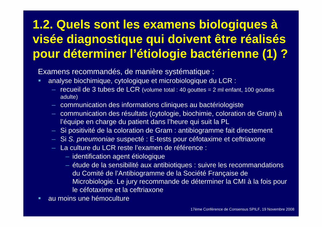
1.2. Quels sont les examens biologiques à visée diagnostique qui doivent être réalisés pour déterminer l’étiologie bactérienne (1) ?
Examens recommandés, de manière systématique :analyse biochimique, cytologique et microbiologique du LCR : – recueil de 3 tubes de LCR (volume total : 40 gouttes = 2 ml enfant, 100 gouttes
adulte)– communication des informations cliniques au bactériologiste– communication des résultats (cytologie, biochimie, coloration de Gram) à
l’équipe en charge du patient dans l’heure qui suit la PL – Si positivité de la coloration de Gram : antibiogramme fait directement – Si S. pneumoniae suspecté : E-tests pour céfotaxime et ceftriaxone – La culture du LCR reste l’examen de référence :
– identification agent étiologique – étude de la sensibilité aux antibiotiques : suivre les recommandations
du Comité de l’Antibiogramme de la Société Française de Microbiologie. Le jury recommande de déterminer la CMI à la fois pour le céfotaxime et la ceftriaxone
au moins une hémoculture
17ème Conférence de Consensus SPILF, 19 Novembre 2008
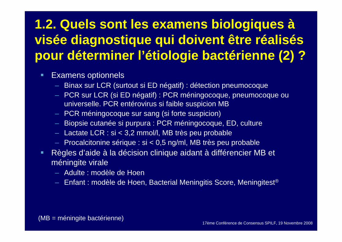
1.2. Quels sont les examens biologiques à visée diagnostique qui doivent être réalisés pour déterminer l’étiologie bactérienne (2) ?
Examens optionnels– Binax sur LCR (surtout si ED négatif) : détection pneumocoque– PCR sur LCR (si ED négatif) : PCR méningocoque, pneumocoque ou
universelle. PCR entérovirus si faible suspicion MB– PCR méningocoque sur sang (si forte suspicion)– Biopsie cutanée si purpura : PCR méningocoque, ED, culture– Lactate LCR : si < 3,2 mmol/l, MB très peu probable– Procalcitonine sérique : si < 0,5 ng/ml, MB très peu probable
Règles d’aide à la décision clinique aidant à différencier MB et méningite virale– Adulte : modèle de Hoen– Enfant : modèle de Hoen, Bacterial Meningitis Score, Meningitest®
(MB = méningite bactérienne)
Les étapes du diagnostic des méningites communautaires
Diagnostic positif de méningitePlace de l'imagerie dans la démarche diagnostique initialeDiagnostic étiologique (bactérie vs. virus)
17ème Conférence de Consensus SPILF, 19 Novembre 2008
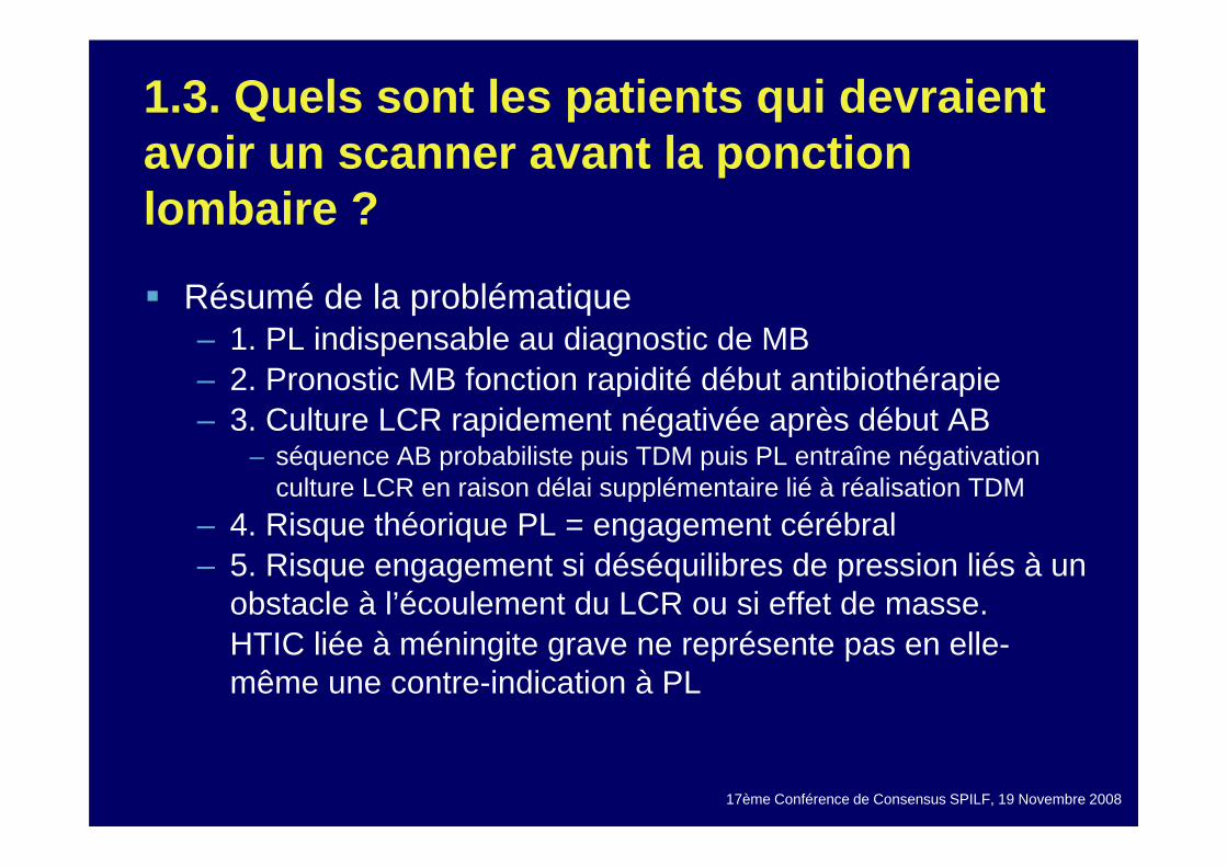
1.3. Quels sont les patients qui devraient avoir un scanner avant la ponction lombaire ?
Résumé de la problématique– 1. PL indispensable au diagnostic de MB– 2. Pronostic MB fonction rapidité début antibiothérapie– 3. Culture LCR rapidement négativée après début AB
– séquence AB probabiliste puis TDM puis PL entraîne négativation culture LCR en raison délai supplémentaire lié à réalisation TDM
– 4. Risque théorique PL = engagement cérébral– 5. Risque engagement si déséquilibres de pression liés à un
obstacle à l’écoulement du LCR ou si effet de masse. HTIC liée à méningite grave ne représente pas en elle-même une contre-indication à PL
17ème Conférence de Consensus SPILF, 19 Novembre 2008
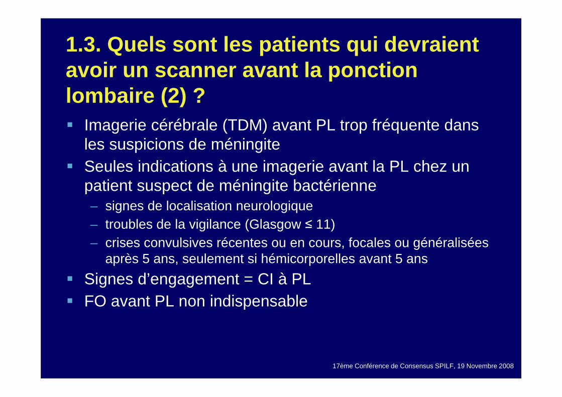
1.3. Quels sont les patients qui devraient avoir un scanner avant la ponction lombaire (2) ?
Imagerie cérébrale (TDM) avant PL trop fréquente dans les suspicions de méningiteSeules indications à une imagerie avant la PL chez un patient suspect de méningite bactérienne– signes de localisation neurologique– troubles de la vigilance (Glasgow ≤ 11)– crises convulsives récentes ou en cours, focales ou généralisées
après 5 ans, seulement si hémicorporelles avant 5 ansSignes d’engagement = CI à PLFO avant PL non indispensable
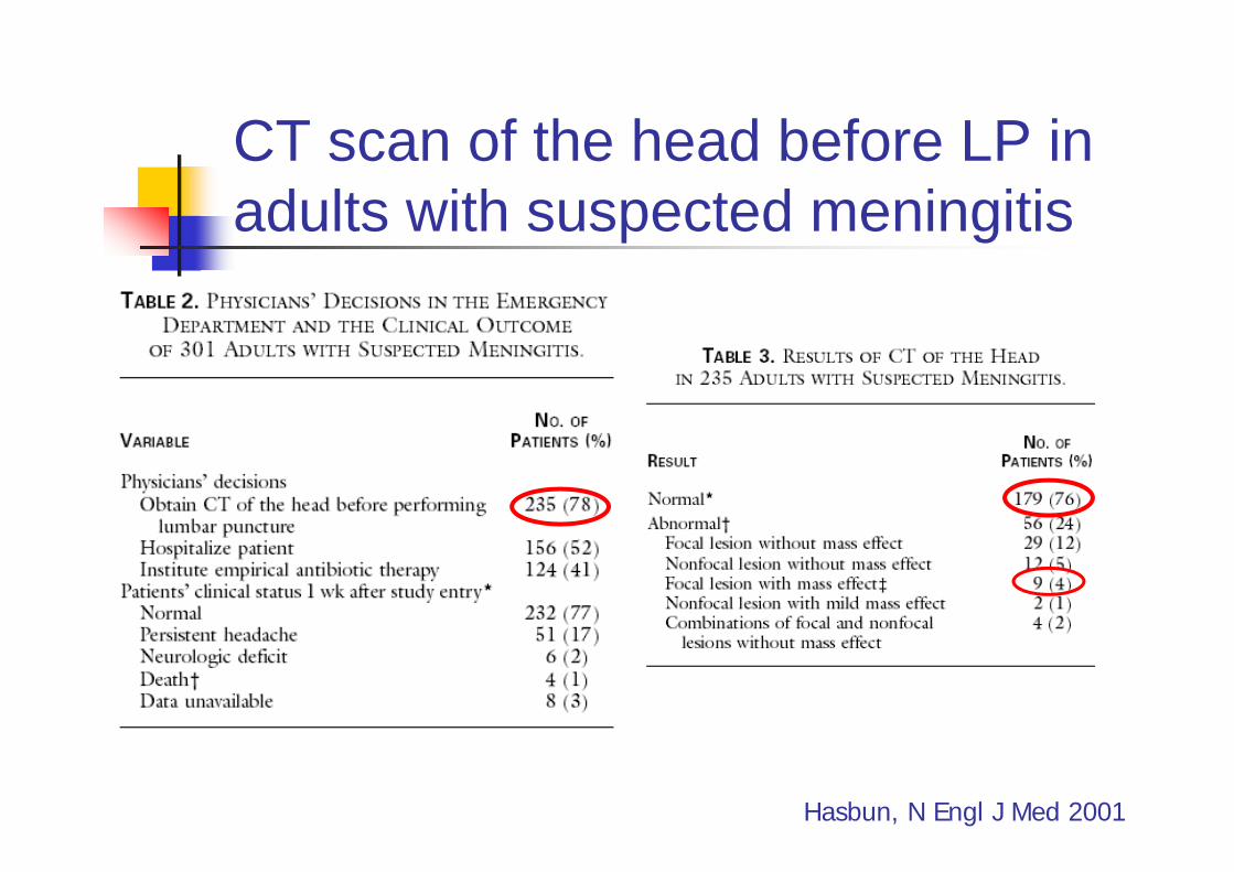
CT scan of the head before LP in adults with suspected meningitis
Hasbun, N Engl J Med 2001
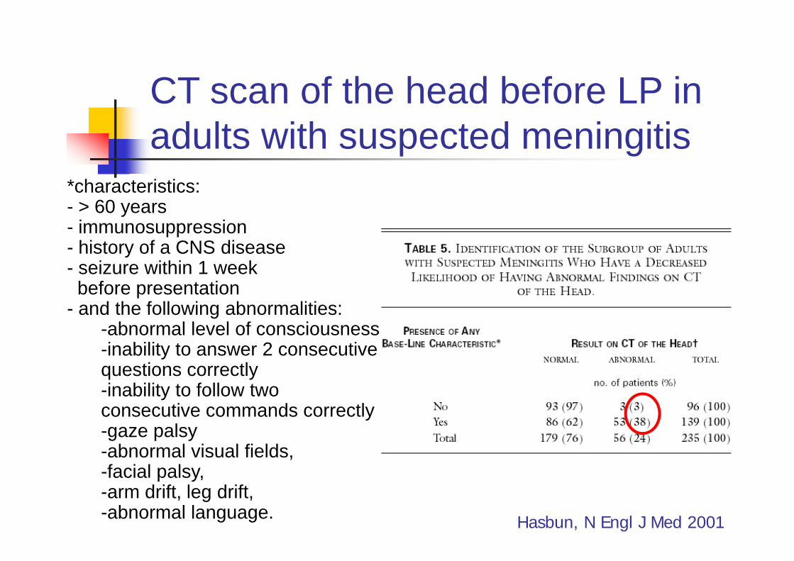
CT scan of the head before LP in adults with suspected meningitis
*characteristics:- > 60 years- immunosuppression- history of a CNS disease- seizure within 1 weekbefore presentation
- and the following abnormalities:-abnormal level of consciousness-inability to answer 2 consecutive questions correctly-inability to follow two consecutive commands correctly-gaze palsy-abnormal visual fields,-facial palsy, -arm drift, leg drift, -abnormal language. Hasbun, N Engl J Med 2001
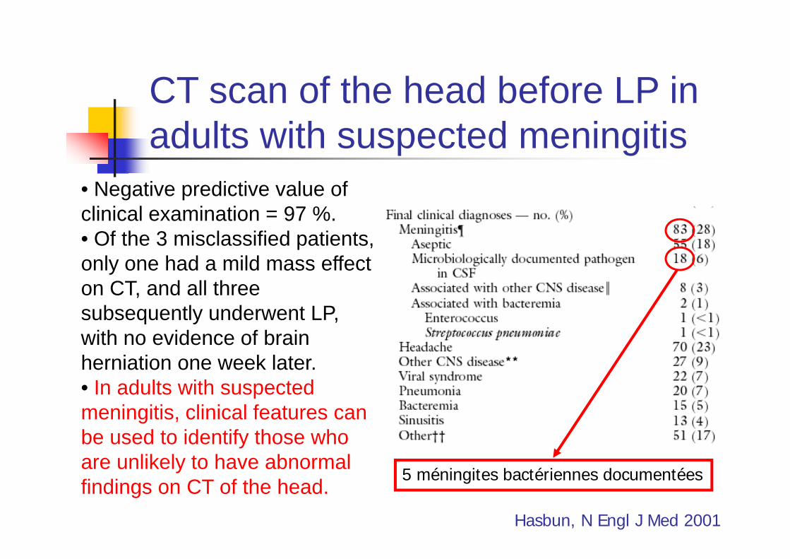
CT scan of the head before LP in adults with suspected meningitis
Hasbun, N Engl J Med 2001
• Negative predictive value of clinical examination = 97 %.• Of the 3 misclassified patients, only one had a mild mass effect on CT, and all three subsequently underwent LP, with no evidence of brain herniation one week later.• In adults with suspected meningitis, clinical features can be used to identify those who are unlikely to have abnormal findings on CT of the head. 5 méningites bactériennes documentées
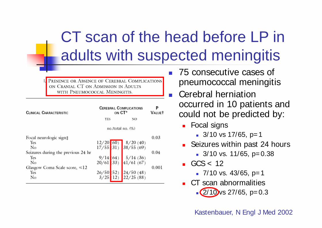
CT scan of the head before LP in adults with suspected meningitis
Kastenbauer, N Engl J Med 2002
75 consecutive cases of pneumococcal meningitisCerebral herniation occurred in 10 patients and could not be predicted by:
Focal signs3/10 vs 17/65, p=1
Seizures within past 24 hours3/10 vs. 11/65, p=0.38
GCS < 127/10 vs. 43/65, p=1
CT scan abnormalities2/10 vs 27/65, p=0.3
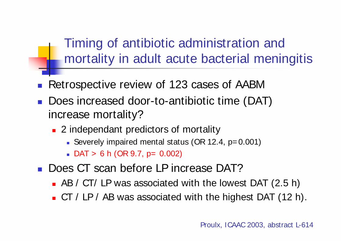
Timing of antibiotic administration and mortality in adult acute bacterial meningitis
Retrospective review of 123 cases of AABMDoes increased door-to-antibiotic time (DAT) increase mortality?
2 independant predictors of mortalitySeverely impaired mental status (OR 12.4, p=0.001)DAT > 6 h (OR 9.7, p= 0.002)
Does CT scan before LP increase DAT?AB / CT/ LP was associated with the lowest DAT (2.5 h)CT / LP / AB was associated with the highest DAT (12 h).
Proulx, ICAAC 2003, abstract L-614
17ème Conférence de Consensus SPILF, 19 Novembre 2008
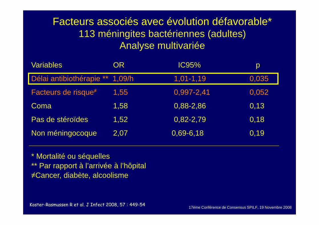
Facteurs associés avec évolution défavorable*113 méningites bactériennes (adultes)
Analyse multivariée
Variables OR IC95% p
Délai antibiothérapie ** 1,09/h 1,01-1,19 0,035
Facteurs de risque≠ 1,55 0,997-2,41 0,052
Coma 1,58 0,88-2,86 0,13
Pas de stéroïdes 1,52 0,82-2,79 0,18
Non méningocoque 2,07 0,69-6,18 0,19
* Mortalité ou séquelles** Par rapport à l’arrivée à l’hôpital≠Cancer, diabète, alcoolisme
Koster-Rasmussen R et al. J Infect 2008, 57 : 449-54
Les étapes du diagnostic des méningites communautaires
Diagnostic positif de méningitePlace de l'imagerie dans la démarche diagnostique initialeDiagnostic étiologique (bactérie vs. virus)
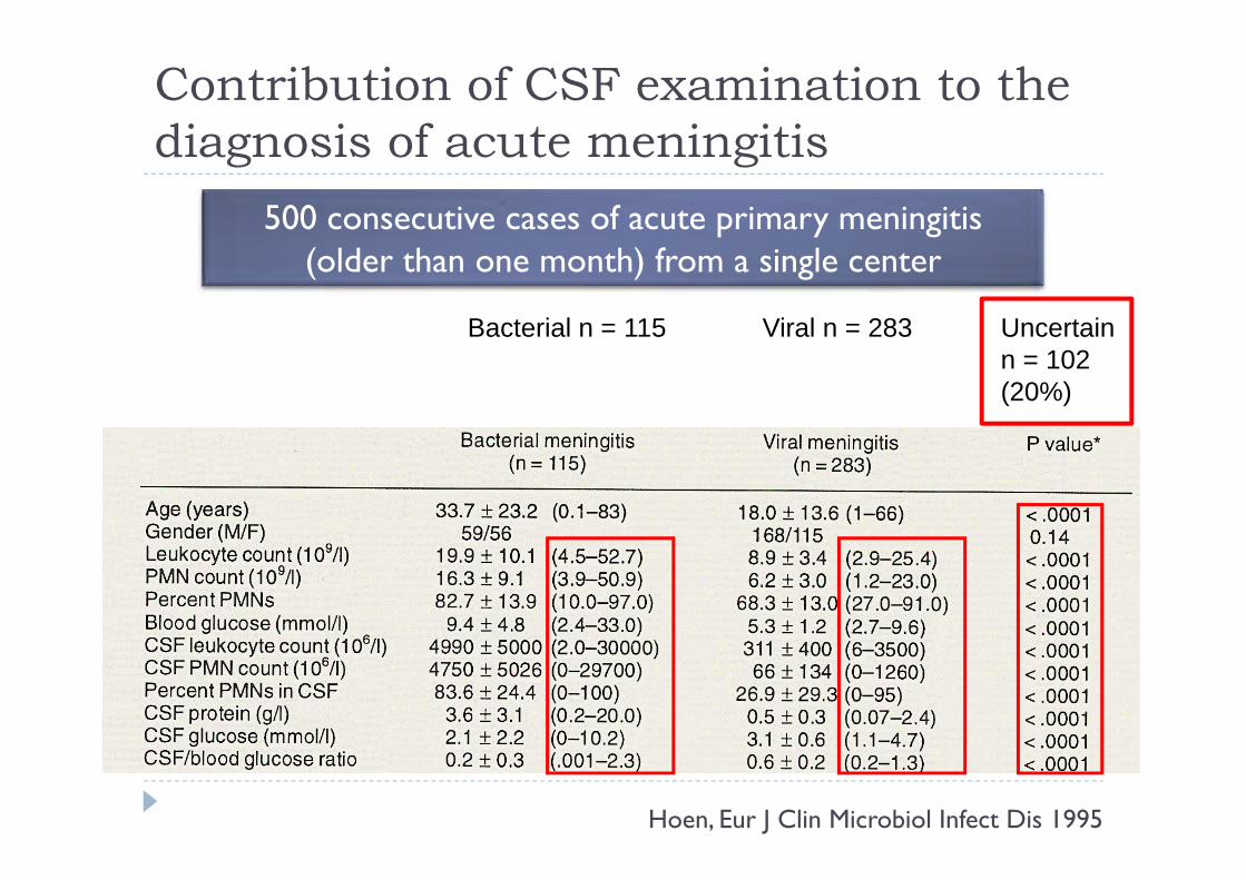
Contribution of CSF examination to the diagnosis of acute meningitis
Hoen, Eur J Clin Microbiol Infect Dis 1995
500 consecutive cases of acute primary meningitis(older than one month) from a single center
Bacterial n = 115 Viral n = 283 Uncertain n = 102 (20%)
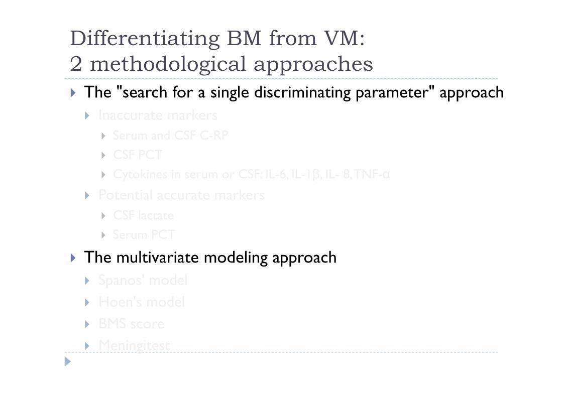
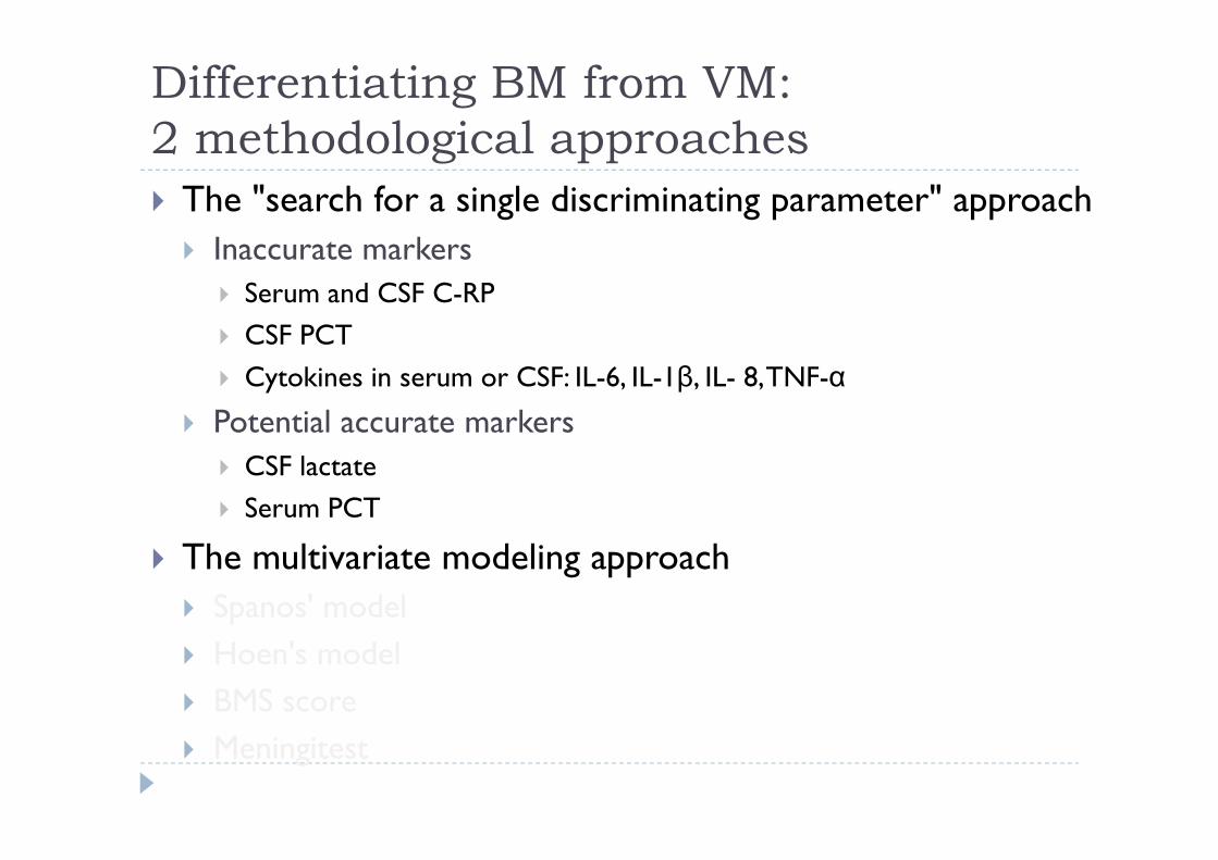
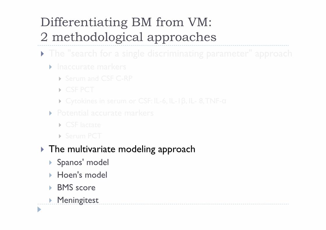
Differentiating BM from VM:2 methodological approaches
The "search for a single discriminating parameter" approachInaccurate markers
Serum and CSF C-RPCSF PCTCytokines in serum or CSF: IL-6, IL-1β, IL- 8, TNF-α
Potential accurate markersCSF lactateSerum PCT
The multivariate modeling approachSpanos' modelHoen's modelBMS scoreMeningitest
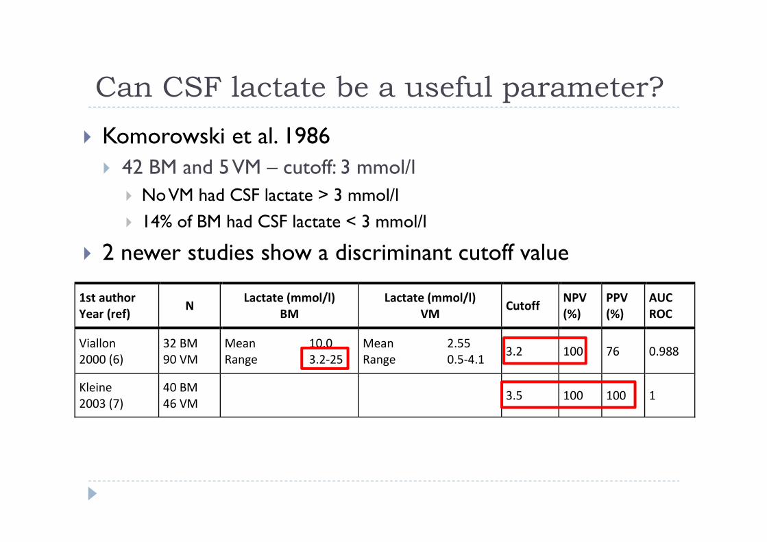
Can CSF lactate be a useful parameter?Komorowski et al. 1986
42 BM and 5 VM – cutoff: 3 mmol/lNo VM had CSF lactate > 3 mmol/l14% of BM had CSF lactate < 3 mmol/l
2 newer studies show a discriminant cutoff value
1st author Year (ref)
N Lactate (mmol/l)
BM Lactate (mmol/l)
VM Cutoff
NPV (%)
PPV (%)
AUC ROC
Viallon 2000 (6)
32 BM 90 VM
Mean 10.0 Range 3.2‐25
Mean 2.55 Range 0.5‐4.1
3.2 100 76 0.988
Kleine 2003 (7)
40 BM 46 VM
3.5 100 100 1
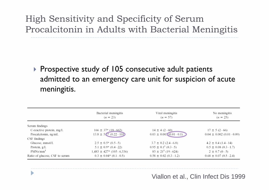
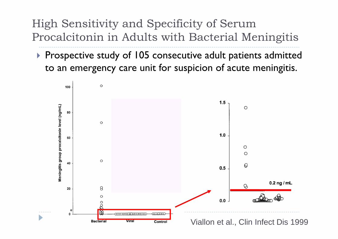
High Sensitivity and Specificity of Serum Procalcitonin in Adults with Bacterial Meningitis
Prospective study of 105 consecutive adult patients admitted to an emergency care unit for suspicion of acute meningitis.
Viallon et al., Clin Infect Dis 1999
Viallon et al., Clin Infect Dis 1999
High Sensitivity and Specificity of Serum Procalcitonin in Adults with Bacterial Meningitis
Prospective study of 105 consecutive adult patients admitted to an emergency care unit for suspicion of acute meningitis.
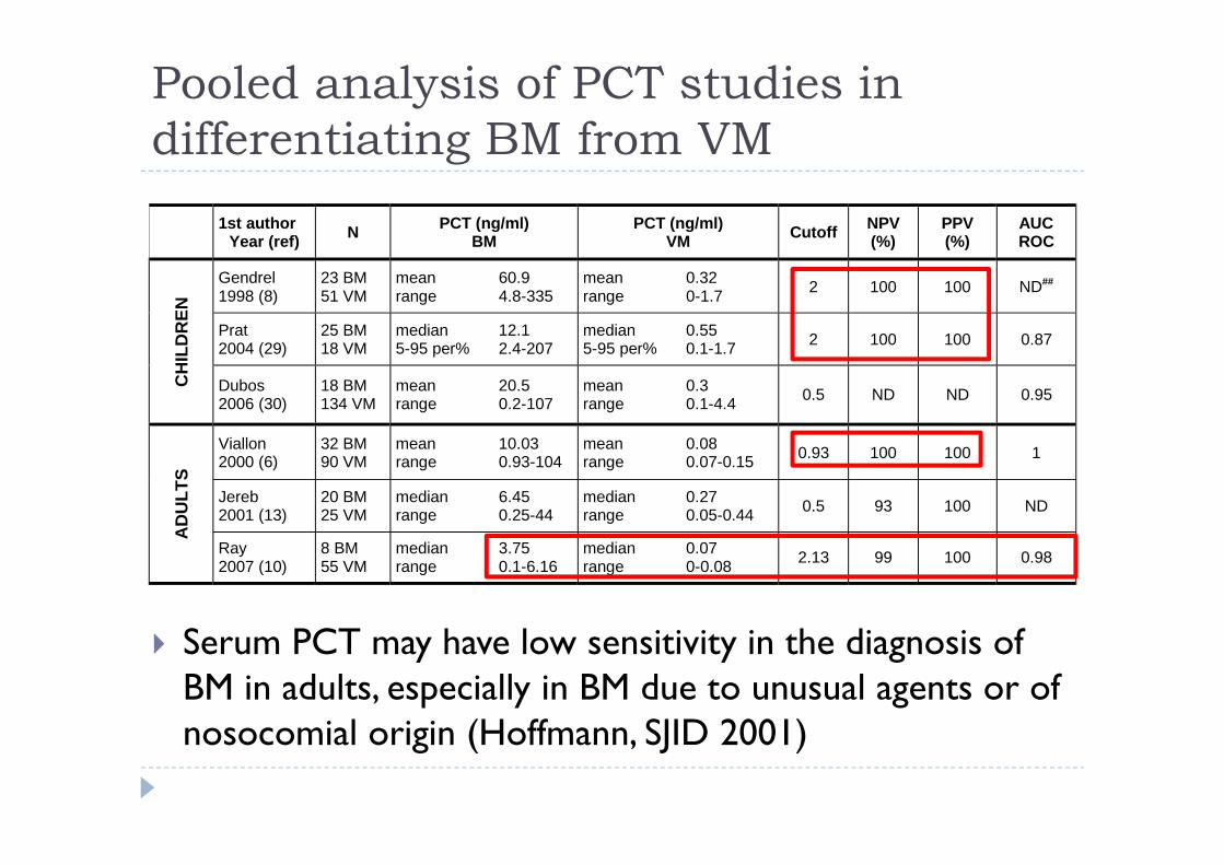
Pooled analysis of PCT studies in differentiating BM from VM
Serum PCT may have low sensitivity in the diagnosis of BM in adults, especially in BM due to unusual agents or of nosocomial origin (Hoffmann, SJID 2001)
1st author Year (ref) N PCT (ng/ml)
BM PCT (ng/ml)
VM Cutoff NPV (%)
PPV (%)
AUC ROC
CH
ILD
REN
Gendrel 1998 (8)
23 BM 51 VM
mean 60.9 range 4.8-335
mean 0.32 range 0-1.7 2 100 100 ND##
Prat 2004 (29)
25 BM 18 VM
median 12.1 5-95 per% 2.4-207
median 0.55 5-95 per% 0.1-1.7 2 100 100 0.87
Dubos 2006 (30)
18 BM 134 VM
mean 20.5 range 0.2-107
mean 0.3 range 0.1-4.4 0.5 ND ND 0.95
AD
ULT
S
Viallon 2000 (6)
32 BM 90 VM
mean 10.03 range 0.93-104
mean 0.08 range 0.07-0.15 0.93 100 100 1
Jereb 2001 (13)
20 BM 25 VM
median 6.45 range 0.25-44
median 0.27 range 0.05-0.44 0.5 93 100 ND
Ray 2007 (10)
8 BM 55 VM
median 3.75 range 0.1-6.16
median 0.07 range 0-0.08 2.13 99 100 0.98

Serum-PCT and CSF-lactate, an update
Serum parameters
CSFparameters
Viallon A., Crit Care Med, 2011, in press
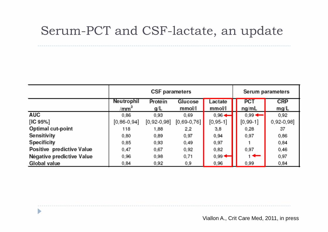
Serum-PCT and CSF-lactate, an update
Viallon A., Crit Care Med, 2011, in press
Differentiating BM from VM:2 methodological approaches
The "search for a single discriminating parameter" approachInaccurate markers
Serum and CSF C-RPCSF PCTCytokines in serum or CSF: IL-6, IL-1β, IL- 8, TNF-α
Potential accurate markersCSF lactateSerum PCT
The multivariate modeling approachSpanos' modelHoen's modelBMS scoreMeningitest
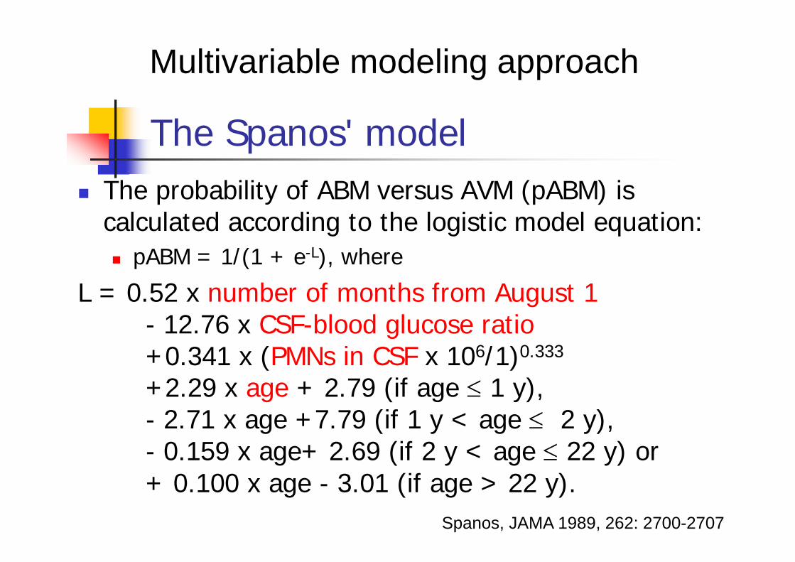
The Spanos' modelThe probability of ABM versus AVM (pABM) is calculated according to the logistic model equation:
pABM = 1/(1 + e-L), where
L = 0.52 x number of months from August 1- 12.76 x CSF-blood glucose ratio+0.341 x (PMNs in CSF x 106/1)0.333
+2.29 x age + 2.79 (if age ≤ 1 y),- 2.71 x age +7.79 (if 1 y < age ≤ 2 y),- 0.159 x age+ 2.69 (if 2 y < age ≤ 22 y) or+ 0.100 x age - 3.01 (if age > 22 y).
Spanos, JAMA 1989, 262: 2700-2707
Multivariable modeling approach
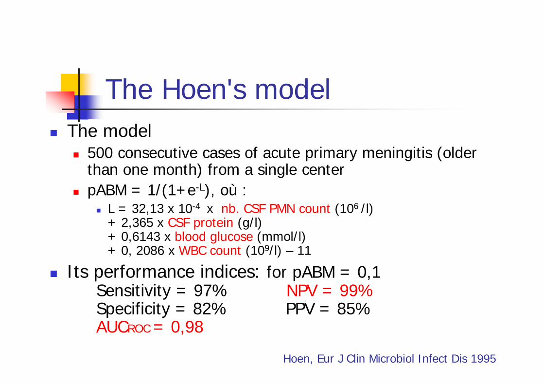
The Hoen's modelThe model
500 consecutive cases of acute primary meningitis (older than one month) from a single centerpABM = 1/(1+e-L), où :
L = 32,13 x 10⁻⁴ x nb. CSF PMN count (106 /l)+ 2,365 x CSF protein (g/l)+ 0,6143 x blood glucose (mmol/l)+ 0, 2086 x WBC count (109/l) – 11
Its performance indices: for pABM = 0,1Sensitivity = 97% NPV = 99%Specificity = 82% PPV = 85%AUCROC = 0,98
Hoen, Eur J Clin Microbiol Infect Dis 1995
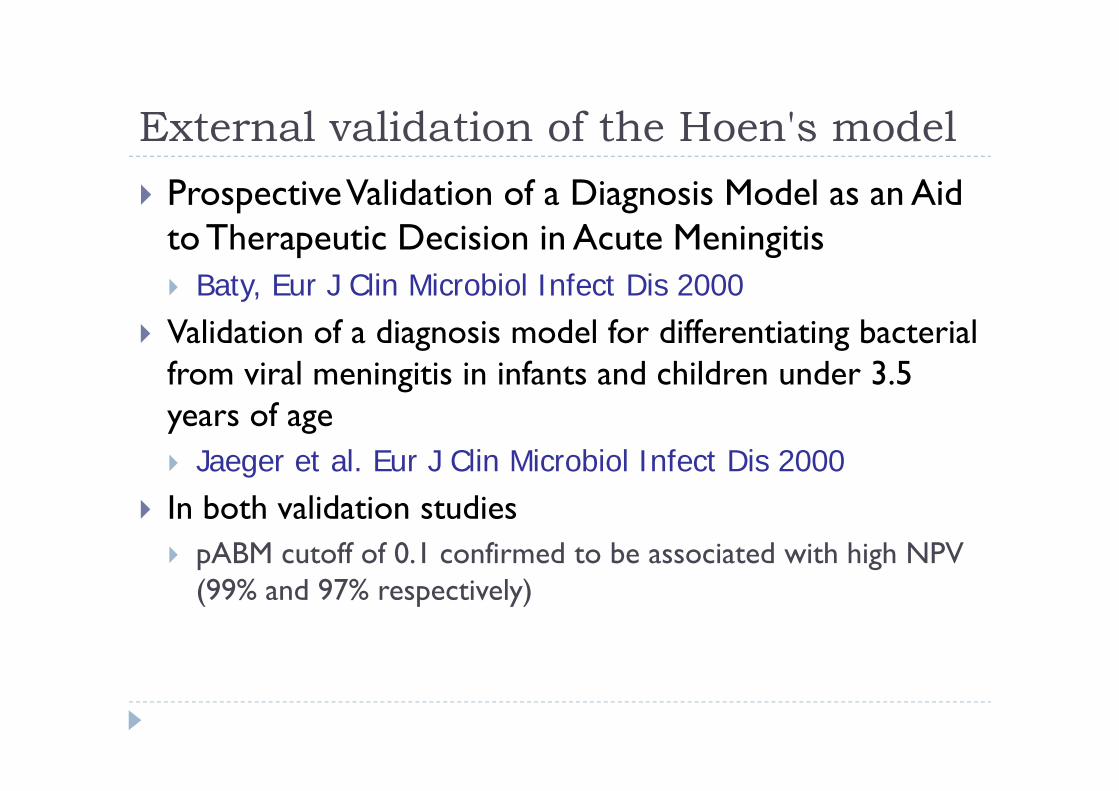
External validation of the Hoen's modelProspective Validation of a Diagnosis Model as an Aid to Therapeutic Decision in Acute Meningitis
Baty, Eur J Clin Microbiol Infect Dis 2000Validation of a diagnosis model for differentiating bacterial from viral meningitis in infants and children under 3.5 years of age
Jaeger et al. Eur J Clin Microbiol Infect Dis 2000In both validation studies
pABM cutoff of 0.1 confirmed to be associated with high NPV (99% and 97% respectively)
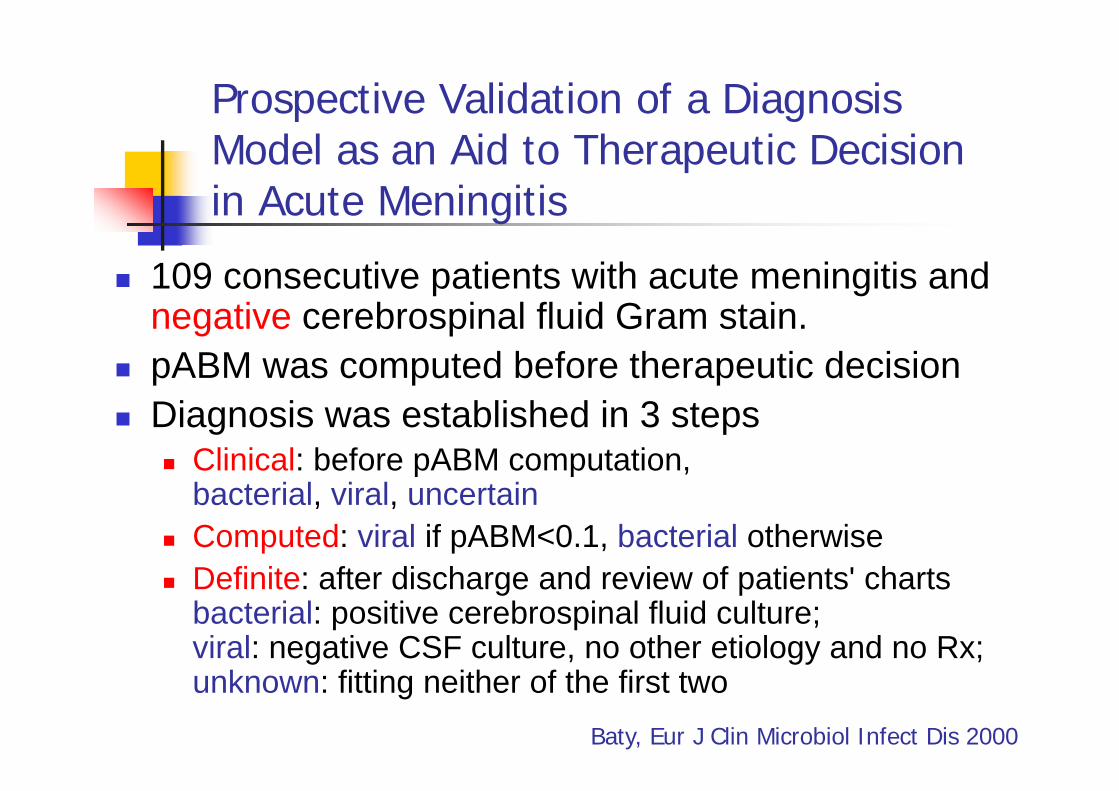
Prospective Validation of a Diagnosis Model as an Aid to Therapeutic Decision in Acute Meningitis
109 consecutive patients with acute meningitis and negative cerebrospinal fluid Gram stain. pABM was computed before therapeutic decisionDiagnosis was established in 3 steps
Clinical: before pABM computation,bacterial, viral, uncertainComputed: viral if pABM<0.1, bacterial otherwiseDefinite: after discharge and review of patients' chartsbacterial: positive cerebrospinal fluid culture; viral: negative CSF culture, no other etiology and no Rx;unknown: fitting neither of the first two
Baty, Eur J Clin Microbiol Infect Dis 2000
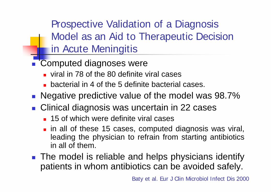
Computed diagnoses wereviral in 78 of the 80 definite viral casesbacterial in 4 of the 5 definite bacterial cases.
Negative predictive value of the model was 98.7%Clinical diagnosis was uncertain in 22 cases
15 of which were definite viral casesin all of these 15 cases, computed diagnosis was viral,leading the physician to refrain from starting antibioticsin all of them.
The model is reliable and helps physicians identifypatients in whom antibiotics can be avoided safely.
Prospective Validation of a Diagnosis Model as an Aid to Therapeutic Decision in Acute Meningitis
Baty et al. Eur J Clin Microbiol Infect Dis 2000
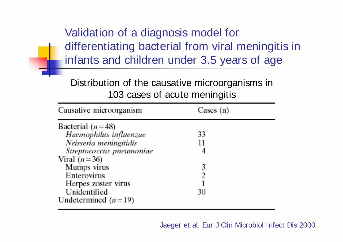
Validation of a diagnosis model for differentiating bacterial from viral meningitis in infants and children under 3.5 years of age
Distribution of the causative microorganisms in 103 cases of acute meningitis
Jaeger et al. Eur J Clin Microbiol Infect Dis 2000
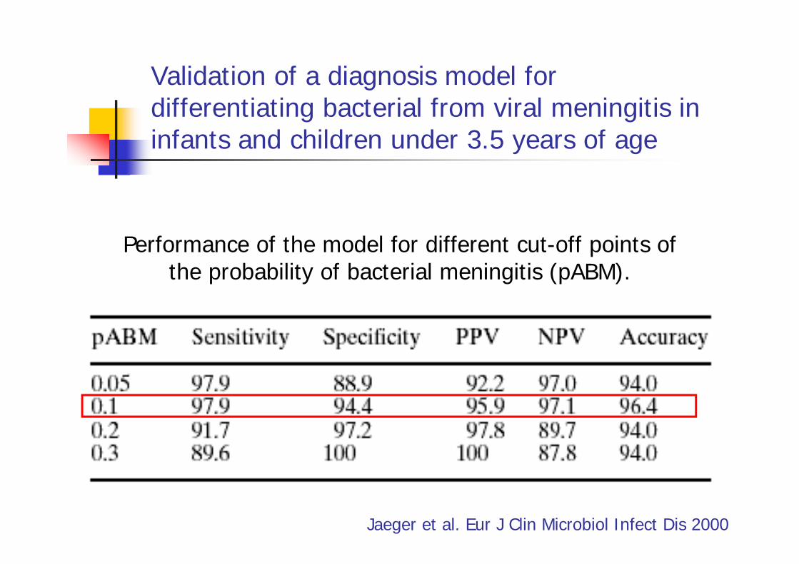
Validation of a diagnosis model for differentiating bacterial from viral meningitis in infants and children under 3.5 years of age
Jaeger et al. Eur J Clin Microbiol Infect Dis 2000
Performance of the model for different cut-off points ofthe probability of bacterial meningitis (pABM).
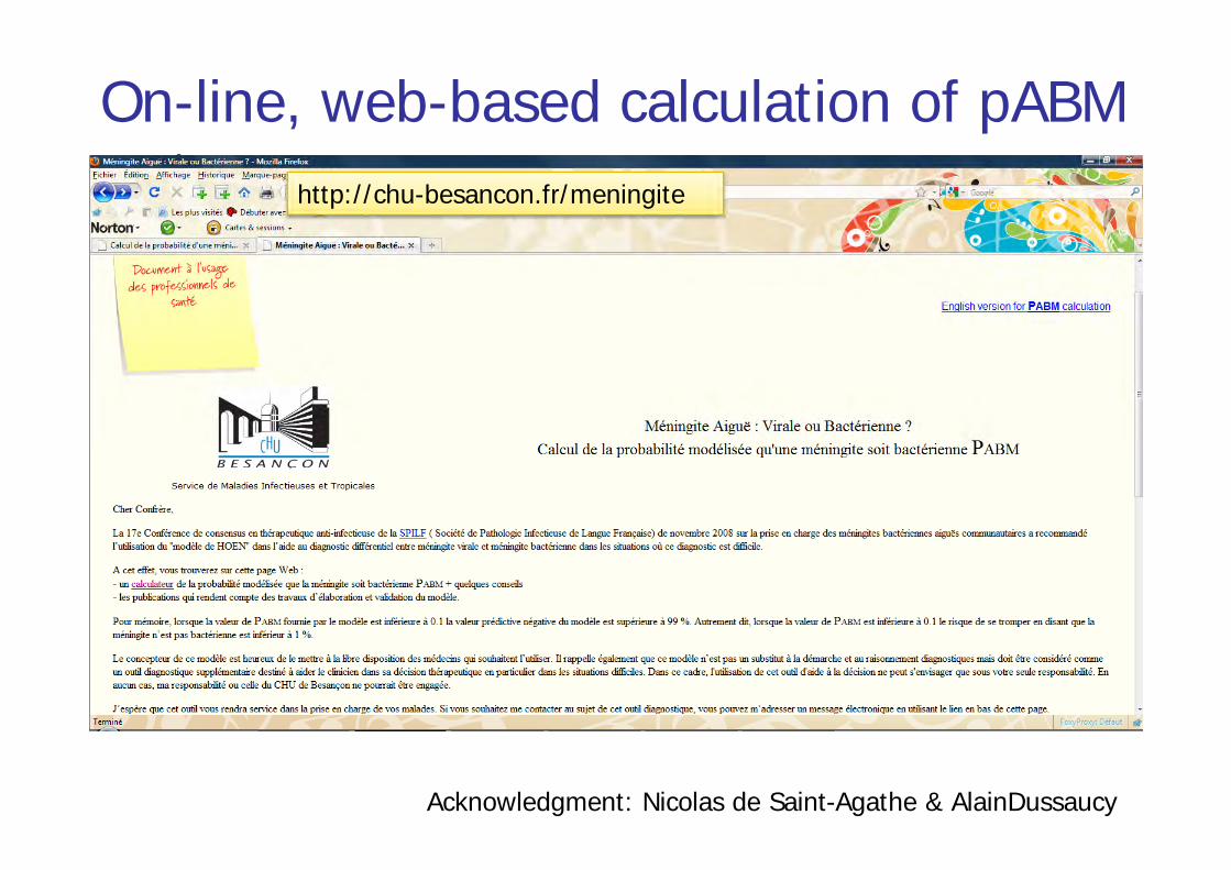
On-line, web-based calculation of pABMhttp://chu-besancon.fr/meningite
Acknowledgment: Nicolas de Saint-Agathe & AlainDussaucy
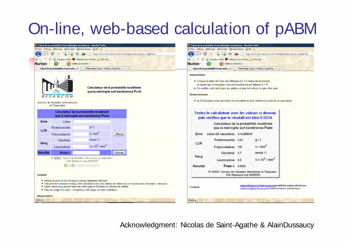
On-line, web-based calculation of pABM
Acknowledgment: Nicolas de Saint-Agathe & AlainDussaucy
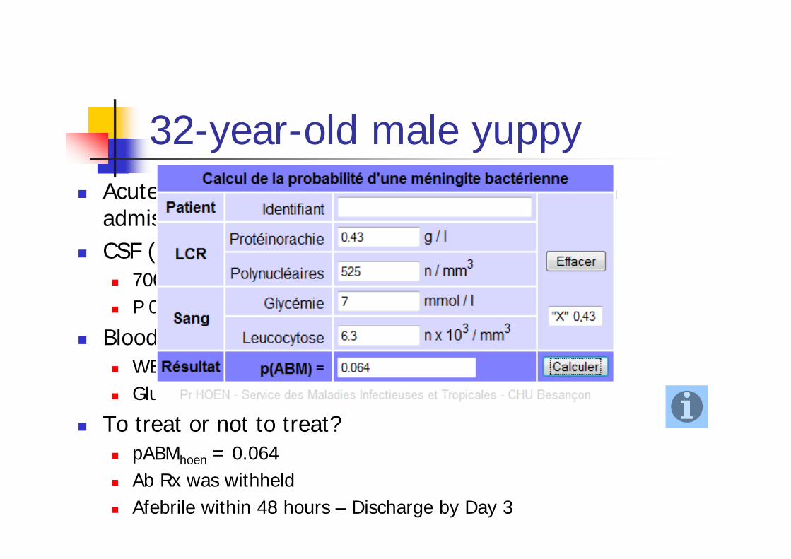
32-year-old male yuppyAcute fever + signs of meningitis for 12 hours upon admission to ERCSF (slightly cloudy):
700 WBC/mm3 (525 PMN) – Gram stain negativeP 0.43 g/l, G 3.5 mmol/l
Blood hematology and chemistryWBC count 6300/mm3
Glucose: 7 mmol/l – Serum C-RP: 25 mg/l
To treat or not to treat?pABMhoen = 0.064Ab Rx was withheldAfebrile within 48 hours – Discharge by Day 3
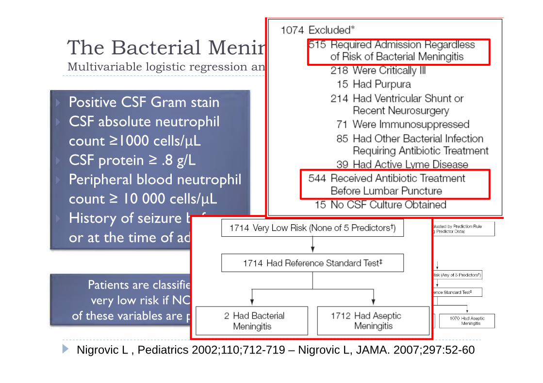
The Bacterial Meningitis Score (BMS)Multivariable logistic regression and recursive partitioning analyses
Patients are classified as very low risk if NONE
of these variables are present.
Positive CSF Gram stainCSF absolute neutrophilcount ≥1000 cells/μLCSF protein ≥ .8 g/LPeripheral blood neutrophilcount ≥ 10 000 cells/μLHistory of seizure before or at the time of admission
Nigrovic L , Pediatrics 2002;110;712-719 – Nigrovic L, JAMA. 2007;297:52-60
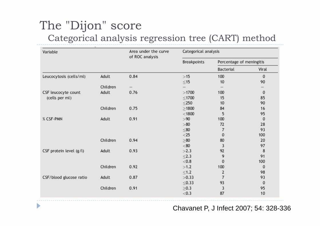
The "Dijon" scoreCategorical analysis regression tree (CART) method
Chavanet P, J Infect 2007; 54: 328-336
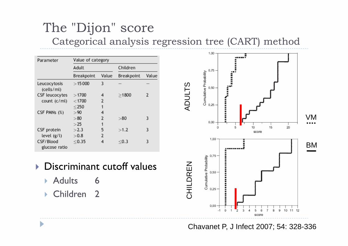
Discriminant cutoff valuesAdults 6Children 2
The "Dijon" scoreCategorical analysis regression tree (CART) method
Chavanet P, J Infect 2007; 54: 328-336
AD
ULT
SC
HIL
DR
EN
BM
VM
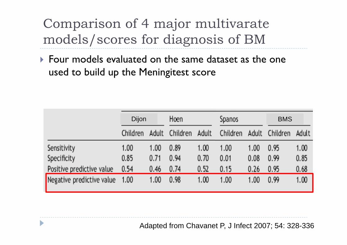
Four models evaluated on the same dataset as the one used to build up the Meningitest score
Comparison of 4 major multivarate models/scores for diagnosis of BM
Adapted from Chavanet P, J Infect 2007; 54: 328-336
Dijon BMS
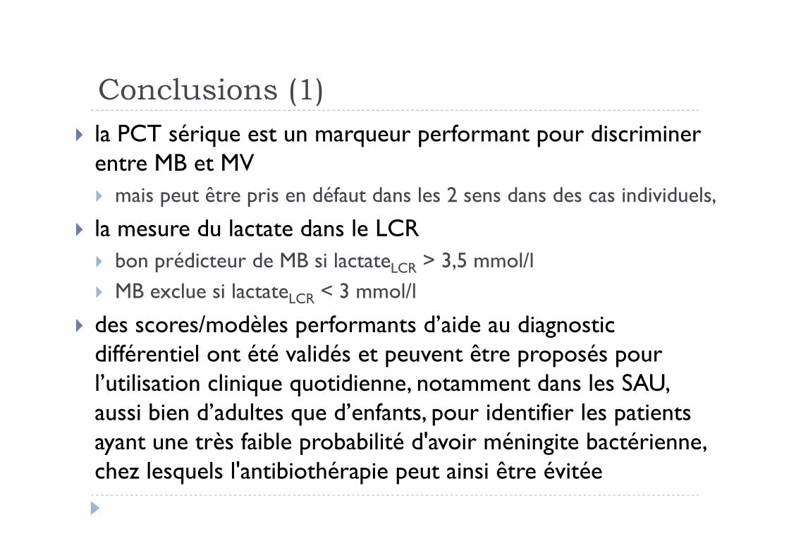
Conclusions (1)la PCT sérique est un marqueur performant pour discriminer entre MB et MV
mais peut être pris en défaut dans les 2 sens dans des cas individuels,
la mesure du lactate dans le LCRbon prédicteur de MB si lactateLCR > 3,5 mmol/lMB exclue si lactateLCR < 3 mmol/l
des scores/modèles performants d’aide au diagnostic différentiel ont été validés et peuvent être proposés pour l’utilisation clinique quotidienne, notamment dans les SAU, aussi bien d’adultes que d’enfants, pour identifier les patients ayant une très faible probabilité d'avoir méningite bactérienne, chez lesquels l'antibiothérapie peut ainsi être évitée
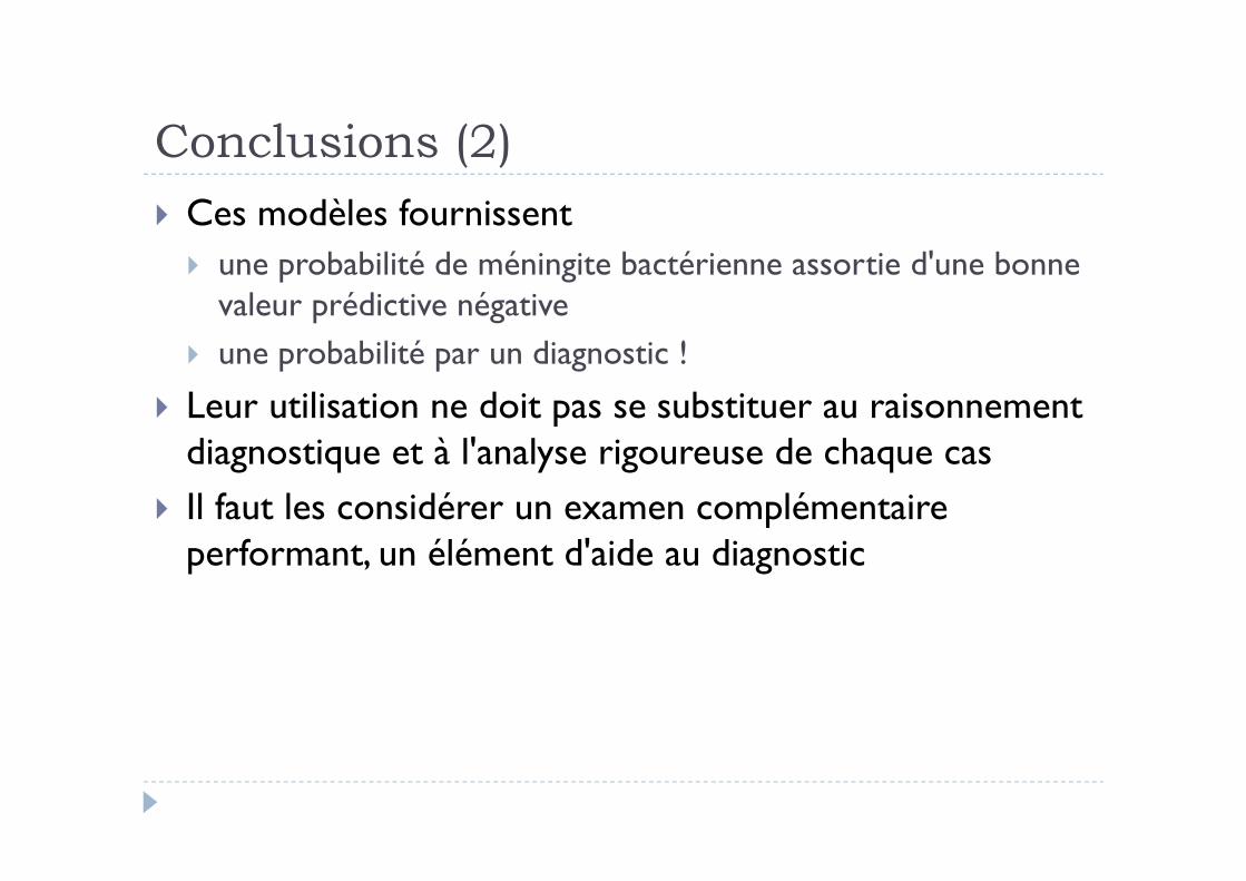
Ces modèles fournissentune probabilité de méningite bactérienne assortie d'une bonne valeur prédictive négativeune probabilité par un diagnostic !
Leur utilisation ne doit pas se substituer au raisonnement diagnostique et à l'analyse rigoureuse de chaque casIl faut les considérer un examen complémentaire performant, un élément d'aide au diagnostic
Conclusions (2)
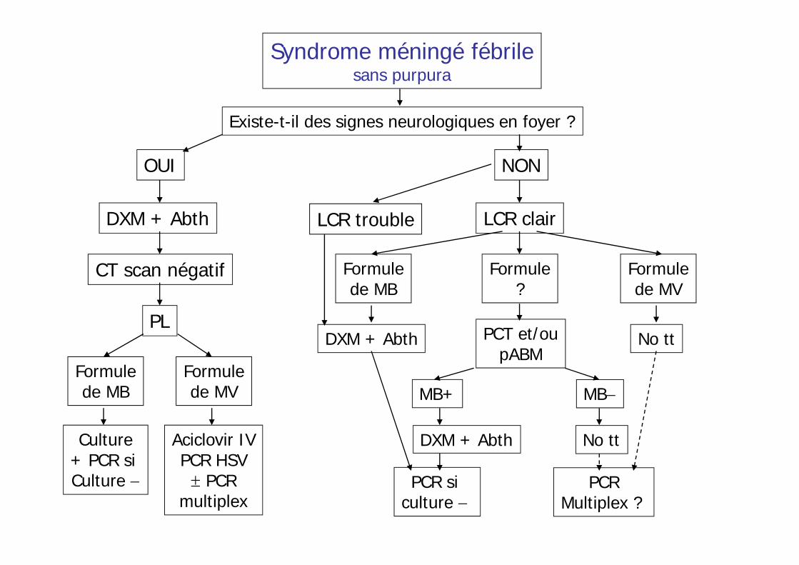
Syndrome méningé fébrilesans purpura
Existe-t-il des signes neurologiques en foyer ?
OUI
DXM + Abth
CT scan négatif
PL
Formulede MB
Culture+ PCR si Culture −
Formulede MV
Aciclovir IVPCR HSV± PCR
multiplex
Formulede MV
No tt
LCR clair
Formulede MB
MB−
No tt
MB+
DXM + Abth
Formule?
PCT et/oupABM
PCRMultiplex ?
PCR si culture −
NON
DXM + Abth
LCR trouble

Back-up slide
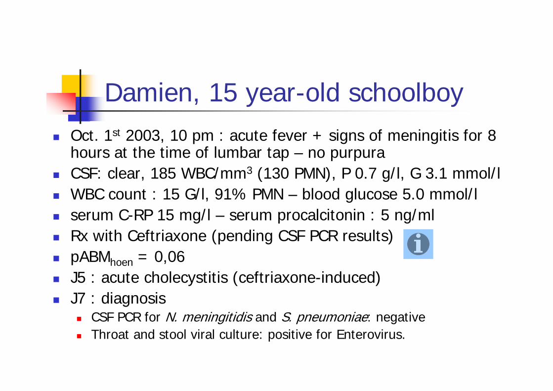
Damien, 15 year-old schoolboyOct. 1st 2003, 10 pm : acute fever + signs of meningitis for 8 hours at the time of lumbar tap – no purpuraCSF: clear, 185 WBC/mm3 (130 PMN), P 0.7 g/l, G 3.1 mmol/lWBC count : 15 G/l, 91% PMN – blood glucose 5.0 mmol/lserum C-RP 15 mg/l – serum procalcitonin : 5 ng/mlRx with Ceftriaxone (pending CSF PCR results)pABMhoen = 0,06J5 : acute cholecystitis (ceftriaxone-induced) J7 : diagnosis
CSF PCR for N. meningitidis and S. pneumoniae: negativeThroat and stool viral culture: positive for Enterovirus.


![Obésité infantile...2018/11/21 · Définitions Category Adults (18 years and older)[1] Youth (2 to 18 yrs) CDC, AAP, IOM, ES, IOTF[2,3] Underweight BMI](https://img.pdfslide.fr/doc/110x75/5f880e8654ab6162e068d9df/obsit-infantile-20181121-dfinitions-category-adults-18-years-and.jpg)