Embed Size (px)
Citation preview

Dear Participants,
It is our great pleasure to welcome you to Nancy and to the INRS Nano2011 Occupational Health Research Conference.
This conference is the first in the new series of INRS Occupational Health Research Conferences. For this year, the conference is addressing the occupational risks associated with nanoparticles and nanomaterials. It has been organised in association with the Partnership for European Research in Occupational Safety and Health (PEROSH).
Nanoparticles and nanomaterials hold promise for improving existing technologies, and new applications including materials engineering, industrial, pharmaceutical, and biomedical applications are being worked on in hundreds of laboratories and industries around the globe.
Many questions arise regarding the risks associated with the development and use of nanoparticles and nanomaterials since some of the physical and chemical properties specific to the nanoscale that make them beneficial for applications may also cause danger to human health. Since research, production and use of nanoparticles and nanomaterials will continue to increase in the coming years, human occupational exposure throughout their manufacture, use and disposal is likely to occur and increase in the future.
The lack of knowledge of hazard characterization and health effects, how to assess exposure, exposure information at the workplace, how to control emission, performance of protective equipment, limits today the ability to adequately assess the risks. It further hinders the management of risks related to these materials. Simultaneous advances in these different topics are necessary to develop the knowledge base and deploy appropriate prevention measures for workers.
Over the 2 ½ days of the INRS Nano2011 conference, nearly all of the above‐mentioned topics are covered by the 6 keynote lectures, 24 oral presentations and 80 poster presentations, given by young and renowned researchers representing the full spectrum of necessary disciplines from epidemiology, medicine, chemistry, physics, industrial hygiene to sociology.
By bringing together more than 400 researchers, experts, industrial hygienists, engineers and physicians from 20 countries and different backgrounds with the common aim of sharing latest knowledge and discussing research needs, we hope that this conference will be stimulating and beneficial to you.
We would like to sincerely acknowledge the efforts of the different Organisation and Advisory Committees, keynote speakers and the chairs of the sessions for the valuable scientific and organisational input for this conference.
We do hope you will enjoy the conference and have a wonderful stay in Nancy!
Kind regards, Conference Chairmen
Olivier Witschger Didier Baptiste Senior Scientist Science Director of INRS President of PEROSH

2
SUMMARY
COMMITTEES 3
PROGRAM 4
EXHIBITORS 8
KEYNOTE SPEAKERS 9
TABLE OF CONTENTS 13
SESSION I: HEALTH EFFECT ASSESSMENT 21
SESSION II: INSTRUMENTATION, CHARACTERIZATION & EXPOSURE EVALUATION 59
SESSION III: EMISSION CONTROL & PROTECTIVE EQUIPMENT 101
SESSION IV: RISK ASSESSMENT AND RISK MANAGEMENT 113
INDEX AUTHORS 127
3
COMMITTEES
Conference Chairmen
Didier Baptiste, Science Director Olivier Witschger, Senior Scientist
Local organizing committee
Claudine Cericola Dominique Mur Stéphane Vaxelaire
Technical program committee
Denis Bemer Stéphane Malard Stéphane Binet Anca Radauceanu Eric Drais Martine Reynier Jean‐Raymond Fontaine Myriam Ricaud
International advisory committee
Christof Asbach Institut für Energie und Umwelttechnik, Duisburg, DE Jorge Boczkowski INSERM, U955, Créteil, FR Odile Boutou‐Kempf InVS, Lyon, FR Patrick Brochard ISPED, Bordeaux, FR Derk Brouwer TNO, Quality of Life Research & Development, Zeist, NL Emeric Fréjafon INERIS, Verneuil‐en‐Halatte, FR François Gensdarmes IRSN, Gif‐sur‐Yvette, FR Eileen D. Kuempel NIOSH, Cincinnati, US Stéphanie Lacour CNRS‐CECOJI, Poitiers, FR Francelyne Marano Université Paris 7, Paris, FR Andreas Mayer Technik Thermische Maschinen, Niederrohrdorf, CH Rémi Maximillien CEA/DSV, Fontenay‐aux‐Roses, FR Claude Ostiguy IRSST, Montréal, QC, CA Didier Rouxel Institut Jean‐Lamour, UMR 7198 CNRS, Vandoeuvre‐lès‐Nancy, FR Martin Seipenbush Karlsruhe Institut of Technology, DE Lang Tran IOM, Edinburgh, UK Su‐Jung (Candace) Tsai U. Massachussets Lowell North, Lowell, US Dominique Vinck Université Pierre Mendès‐France, Grenoble, FR

4
PROGRAM
Tuesday, April 5
9:00 Registration & Coffee
10:15 Welcoming Stéphane Pimbert, General Director Didier Baptiste, Science Director, President of PEROSH Olivier Witschger, Senior Scientist
10:45 Keynote I: Risk assessment and risk management of nanomaterials in the workplace: what we know and what we still need to know Eileen D. Kuempel; NIOSH, USA
11:30 Keynote II: Toxicokinetics of insoluble nanoparticles in rodents after different routes of administration Wolfgang G. Kreyling, Helmholtz Center Munich, Germany
12:15 Lunch
Session I ‐ Health Effect Assessment
Chairs: Eileen Kuempel (NIOSH, USA) & Emmanuel Flahaut (CIRIMAT CNRS, France)
14:00 Keynote III: Nanofibres and asbestos: new materials with an old hazard Ken Donaldson, University of Edinburgh, UK
14:45 Nano‐Silicon Dioxides toxicological characterization on human colonic epithelial cell line HT‐29 V. Paget; J.A. Sergent and S. Chevillard
I‐01
15:00 Comparative study of cytotoxic and genotoxic effects of nano‐ and submicron‐sized metal oxide Y. Guichard; J. Schmitt; M. Goutet; O. Rastoix; D. Rousset; A. Boivin; R. Wrobel; L. Gaté; C. Darne and S. Binet
I‐02
15:15 SiO2 nanoparticles activate immune dendritic cells S. Barillet; C. Nhim; S. Kerdine‐Römer and M. Pallardy
I‐03
15:30 Coffee break & Posters & Exhibition viewing
16:15 Pro‐inflammatory Response of Manganese Oxide Nanoparticles is altered upon Exposure to Endotoxin‐Injured Alveolar Epithelial Cells A. Schlicker; M. Urner; R. Frick; L. K. Limbach; W. J. Stark and B. Beck‐Schimmer
I‐04
16:30 In vivo genotoxicity of inhaled nanosized TiO2 in mice H. Norppa; H. K. Lindberg; G.C.‐M. Falck; J. Koivisto1, E. Rossi; L. Pylkkänen; H. Nykäsenoja; H. Järventaus; S. Suhonen; M. Vippola; J. Catalán and K. Savolainen
I‐05
16:45 Pulmonary toxicity comparison of raw and super‐purified single‐wall carbon nanotubes after intra‐tracheal instillation in rats D. Elgrabi; B. Trouiller; F. Rogerieux and G. Lacroix
I‐06
17:00 Panel on Session I
17:30 Poster Session A
19:00 Welcome Cocktail ‐ Palais des Congrès
5
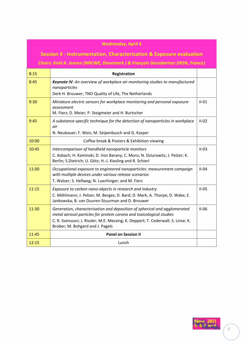
Wednesday, April 6
Session II ‐ Instrumentation, Characterization & Exposure evaluation
Chairs: Keld A. Jensen (NRCWE, Danemark ) & François Gensdarmes (IRSN, France)
8:15 Registration
8:45 Keynote IV: An overview of workplace air monitoring studies to manufactured nanoparticles
Derk H. Brouwer, TNO Quality of Life, The Netherlands
9:30 Miniature electric sensors for workplace monitoring and personal exposure assessment M. Fierz; D. Meier; P. Steigmeier and H. Burtscher
II‐01
9:45 A substance‐specific technique for the detection of nanoparticles in workplace air
N. Neubauer; F. Weis; M. Seipenbusch and G. Kasper
II‐02
10:00 Coffee break & Posters & Exhibition viewing
10:45 Intercomparison of handheld nanoparticle monitors
C. Asbach; H. Kaminski; D. Von Barany; C. Monz; N. Dziurowitz; J. Pelzer; K. Berlin; S.Dietrich; U. Götz; H.‐J. Kiesling and R. Schierl
II‐03
11:00 Occupational exposure to engineered nanoparticles: measurement campaign with multiple devices under various release scenarios
T. Walser; S. Hellweg; N. Luechinger; and M. Fierz
II‐04
11:15 Exposure to carbon nano‐objects in research and industry
C. Möhlmann; J. Pelzer; M. Berges; D. Bard; D. Mark; A. Thorpe; D. Wake; E. Jankowska; B. van Duuren‐Stuurman and D. Brouwer
II‐05
11:30 Generation, characterisation and deposition of spherical and agglomerated metal aerosol particles for protein corona and toxicological studies
C. R. Svensson; J. Rissler; M.E. Messing; K. Deppert; T. Cederwall; S. Linse; K. Brober; M. Bohgard and J. Pagels
II‐06
11:45 Panel on Session II
12:15 Lunch

6
Wednesday, April 6
Session III ‐ Emission Control & Protective Equipment
Chairs: Martin A. Seipenbush (KIT, Germany) & Andreas Mayer (TTM, Switzerland)
14:00 Keynote V: Airborne nanoparticles in the workplace ‐ Sources, transport, evolution and the consequences for exposure and filtration
Pr. Gerhard Kasper, Karlsruhe Institute of Technology, Germany
14:45 Assessment of spray products containing engineered nanoparticles
A. Ulrich; H. Hagendorfer; C. Lorenz; N. V. Götz and K. Hungerbühler
III‐01
15:00 Nanosize Metal Oxide Emissions from CI and SI Vehicle Engines
A. Mayer; J. Czerwinski; M. Kasper; A. Ulrich and J. Mooney
III‐02
15:15 Nanopowders explosions: A few nanometres less that change everything
O. Dufaud; A. Vignes; F. Henry; J. Bouillard; L. Perrin and D. Fleury
III‐03
15:30 Coffee break & Posters & Exhibition viewing
16:15 Preliminary evaluation of nanoparticle transfer across the dynamical air barrier of a microbiological safety cabinet
V. Cesard; E. Belut and C. Prevost
III‐04
16:30 Engineering Control Technology of Filtration Performance for Engineered Nanoparticles
Su‐Jung (Candace) Tsai; M. E. Echevarría‐Vega; G. Sotiriou; C. Huang; P. Demokritou and M. Ellenbecker
III‐05
16:45 Particle emission source characterization and modeling of the fate of emitted particles
A.J. Koivisto; M. Yu; K. Hämeri and M. Seipenbusch
III‐06
17:00 Panel on Session III
17:30 Poster Session B
19:00 Adjourn
20:00 Conference Banquet
Hôtel de Ville, Place Stanislas

7
Thursday, April 7
Session IV ‐ Risk assessment and risk management
Chairs: Sonia Desmoulin (CNRS, France ) & Claude Ostiguy (IRSST, Québec)
8:00 Registration
8:45 Keynote VI: The object of ‘nano‐risks’ regulation: a legal and sociological view
Stéphanie Lacour, CNRS‐CECOJI Poitiers, France
Dominique Vinck, U. Pierre Mendès‐France, Grenoble, France
9:30 Time to shift paradigms? How to practice Nanotechnology risk governance
A.J. Dijkman; J. Terwoert and A.L. Hollander
IV‐01
9:45 Nanotechnology Occupational Safety and Health: Global Standards Development
V. Murashov and J. Howard
IV‐02
10:00 Coffee break & posters & exhibition viewing
10:45 The role of the employer in prevention and compensation of risks associated to nanoparticles and nanomaterials
M. Bary; N. Dedessus‐Le‐Moustier and A. Moriceau
IV‐03
11:00 Development of a control banding tool adapted to nanomaterials
M. Riediker; C. Ostiguy ; J. Triolet ; P. Troisfontaines ; D. Vernez ; G. Bourdel ; N. Thieriet ; A. Cadène and I. Daguet
IV04
11:15 How to manage nanomaterials safety in research environment? A. Groso; A. Petri‐Fink; A. Magrez; M. Riediker and T. Meyer
IV‐05
11:30 NANOKEM ‐Risk assessment of nanoparticles in the paint and lacquer industry
F. Fotel; A. Permin; K.H. Cohr; H.R. Lam; A.T. Saber; K.A. Jensen; K.S. Hougaard; I. Koponen; S.T. Larsen; N.R. Jacobsen; R. Birkedal; M. Roursgaard; L. Mikkelsen; P. Møller; S. Loft; H. Wallin and U. Vogel
IV‐06
11:45 Panel on Session IV
12:15 Conference Closure
Didier Baptiste, Science Director, President of PEROSH
12:30 Adjourn
8
EXHIBITORS The following companies have exhibited at the conference:
9
KEYNOTE SPEAKERS Six keynote presentations were given by international experts to provide participants with a broader perspective on the different topic areas of the conference.
Keynote I:
Risk assessment and risk management of nanomaterials in the workplace: what we know and what we still need to know.
Eileen D. Kuempel; NIOSH, USA
Since its inception in 2004, the NIOSH Nanotechnology Research Center (NTRC) has conducted research and developed guidance on working safely with nanomaterials. While progress has been made on understanding the hazardous properties of specific nanomaterials and their biological modes of action, challenges remain in translating that knowledge to developing exposure limits and other risk management guidance.
Rather than continuing to evaluate one type of nanoparticle at a time, NIOSH is exploring hazard‐ and risk‐based grouping strategies and corresponding exposure control options. Nanoparticles with similar physical‐chemical properties and hypothesized mode of action could be evaluated more efficiently in short‐term in vivo or in vitro assays to compare their potency with well‐studied “benchmark” particles.
Based on nanomaterials research to date, what we currently know (to some degree) includes: (1) nanomaterials can be measured using standard measurement methods (respirable mass or number concentration); (2) workplace exposures to nanomaterials can be reduced using conventional engineering controls and personal protective equipment; and (3) standard risk assessment methods are applicable to nanomaterials.
What we still need to know includes: (1) worker exposure data are very limited and remains a critical need for nanomaterials; (2) measurement methods are needed with greater sensitivity and specificity for the particle characteristics most associated with the hazard; (3) engineering controls and respirators need further evaluation of their effectiveness against nanoparticle exposure; (4) hazard information is lacking for a vast majority of nanomaterials, especially chronic exposure and effects data; and (5) more efficient methods for risk assessment are needed to address the gaps in occupational health guidance. New areas of research at NIOSH and elsewhere are beginning to focus on monitoring workers’ health for early responses (biomonitoring, medical surveillance, epidemiology).
10
Keynote II:
Toxicokinetics of insoluble nanoparticles in rodents after different routes of administration
Wolfgang G. Kreyling; Helmholtz Center Munich, Germany
Nanoparticles (NP) are increasingly used in a wide range of applications in science, technology and medicine. Since they are produced for specific purposes which cannot be met by larger particles and bulk material they are likely to be highly reactive, in particular, with biological systems. Direct routes of intake into the organism are (1) inhalation and deposition of NP in the respiratory tract and (2) oral intake of NP and ingestion. Recently there is evidence that nanoparticles can cross body membranes ‐ such as the air‐blood‐barrier in lungs and the intestinal epithelium – reaching blood circulation and accumulating in secondary target organs. Therefore, direct intravenous administration of NP into circulation provides a powerful tool to shed light on the various interactions of crossing body membranes.
To quantitatively determine accumulated NP fractions in such organs the ultimate aim is to balance the NP fractions in all interesting organs and tissues including the remaining body and total excretion. Since these gross determinations of NP contents in organs and tissues do not provide microscopic information on the anatomical and cellular location of nanoparticles such studies are to be complemented by electron microscopy analysis as demonstrated for inhaled titanium dioxide nanoparticles.
Based on quantitative biokinetics after all three routes of administration in a rat model (lungs, blood, gastro‐intestinal tract) we found small NP fractions (iridium, carbon, titanium dioxide, gold,) in all secondary organs studied including brain, heart and even in foetuses. Fractions per secondary organ were usually below 0.1 % of the administered dose but depended strongly on particle size, material and surface modifications as well as on the route of intake.
The current knowledge on systemic translocation of NP and their accumulation in secondary target organs and tissues of man and animal models does not suggest to cause acute effects of translocated NP but chronic exposure may lead to elevated NP accumulations resulting eventually in adverse health effects.
In fact, there is growing evidence that ambient ultrafine particles and some of the engineered NP can induce acute adverse health effects in humans and in animal models not only in the respiratory tract but also in the cardio‐vascular‐system. Since NP translocation is so low these effects are likely to be triggered by mediators released in the organ of intake.
11
Keynote III:
Nanofibres and asbestos: new materials with an old hazard.
Ken Donaldson; University of Edinburgh, UK
Our recent research has focused on the pathogenicity of long and short multi‐wall carbon nanotubes (MWCNT) and especially the unique hazard posed to the pleural mesothelium by asbestos e.g. mesothelioma. A small fraction of all deposited particles, including fibres and nanotubes, reach the pleura. Evolution has provided a mechanism of particle clearance from the pleura, through stomata in the parietal pleura. We have therefore instilled particles and fibres into the pleural space as an mimicking the true translocation of a fraction of inhaled particles and fibres to the pleural space. We injected a panel of long and short fibres (long and short amosite asbestos samples, two long nanotubes samples and two short/tangled nanotubes samples and nanoparticulate carbon black as a graphene control) into the pleural space Following injection, the pleural cavity was lavaged to determine the inflammatory response and the chest wall examined for evidence of fibre retention and its consequences i.e. inflammation and fibrosis. We found clear evidence of length‐related retention and inflammation in the pleural space, with both the long amosite and the two long nanotubes samples causing inflammation while all the short samples and the graphene control failed to elicit significant inflammation or fibrosis in the longer term. We also examined the behaviour of short and long silver nanowires and nickel oxide nanowires as examples of other High Aspect Ratio Nanoparticles (HARN) to determine if there was general applicability of length‐dependent inflammogenicity across different HARN. All HARN so far examined show length dependent pathogenicity. We are examining the mechanism underlying length dependent inflammogenicity of HARN with the goal of designing safer nanotubes and nanofibres of various types.
Keynote IV:
An overview of workplace air monitoring studies to manufactured nanoparticles.
Derk H. Brouwer, TNO Quality of Life, The Netherlands
Workplace air monitor studies can be conducted for various reasons, e.g. exposure exploration/ analysis, risk assessment, epidemiology, (effectiveness of) exposure control measures, or compliance with any occupational exposure limit. For studies focused on (manufactured) nanoparticles exposure the majority of the studies rather has an explorative character, partly due the lack of methods to assess exposure to manufactured nanoparticles in an accurate quantitative way. In potential, the synthesis, down‐stream use, and formulation of manufactured nanomaterials, as well as application of nano‐endproducts will result in a tremendous amount of various workplace exposure scenarios. It is well understood that both the type and the numbers of (manufactured) nanoaerosols emitted during activities will differ substantially between these scenarios. In general, the likelihood of exposure to (manufactured) nano particles tend to decrease during the market value chain of nanomaterials, however, some typical scenarios related to application of ready‐to‐use nano‐endproducts, e.g. sprays, may form an exception. Faced with the impossibilities to address all various workplace scenarios, some interesting initiatives have been taken to overcome the lack of measurement data, including harmonization of measurement strategy, exposure modeling and risk‐ or control banding approaches.
12
Keynote V:
Airborne nanoparticles in the workplace ‐ Sources, transport, evolution and the consequences for exposure and filtration
Gerhard Kasper, Karlsruhe Institute of Technology, Germany
Airborne NP are highly dynamic systems which undergo rapid changes in size distribution and concentration between source and human receptor. These changes and their consequences for workplace measurements, for human exposure, as well as for the design of protective devices such as face masks will be analyzed on the basis of likely exposure scenarios in a typical work place environment as well as typical source characteristics. The analysis of aerosol evolution between source an receptor is based on well known aerosol dynamic concepts of collisional growth kinetics and dilution supported by models and actual measurements in a test chamber. The cases discussed will include (1) a relatively strong source of “true” NP (10 nm) at concentrations well above the general background aerosol; (2) the fate of NP emitted at concentrations on the order of the concentration of the normal background in a workplace. Binary coagulation between NP and background aerosol particles is generally faster than self‐coagulation among emitted primary NP, except in the unlikely case of extremely high source strengths, and then only locally. The general background aerosol is thus an effective scavenger for airborne NP on a relatively short time scale of about 10 minutes. Based on experimental evidence of the fragmentation of such loosely bonded agglomerates, one may assume that ‐ from a toxicological or chemical perspective ‐ such attached NP have not “disappeared”, even though they will no longer be detectable in the size range of the original source, nor will their original size be relevant for estimates of lung deposition. Given the typical size range of common aerosol backgrounds (a few hundred nm) (the typical ‘most penetrating particle size’ of the human lung, one has to assume that attached NP penetrate deeply into the human airways and may also be re‐exhaled to a significant degree. Attached NP may therefore also use background aerosol particles in the 100 nm size range as “Trojan Horses” to penetrate filters in the range of the MPPS. Resulting implications for the performance criteria of personal protective equipment need to be discussed.
Keynote VI:
The object of ‘nano‐risks’ regulation: a legal and sociological view
Stéphanie Lacour, CNRS‐CECOJI Poitiers, France
Dominique Vinck, U. Pierre Mendès‐France, Grenoble, France
Nanoparticles, nanomaterials, nanotechnologies, substances in the nanoparticulate state… The choice of the terms used to designate the subject of the regulation is carefully weighed up, depending on whether we are talking about development of research, assessment of potential risks, or indeed management of any such risks. The diversity of the players and their positioning in the controversies are, in this respect, as crucial as the objectives that they set themselves and that vary over time. Using critical analysis to enlighten understanding of the issues behind the choices of a definition, such will be the subject of this talk, which is the fruit of an exchange of perspectives involving a sociologist and a legal specialist.
13
TABLE OF CONTENTS
Session I – HEALTH EFFECT ASSESSMENT
Page
I‐01 Nano‐Silicon Dioxides toxicological characterization on human colonic
epithelial cell line HT‐29
V. Paget, J.A. Sergent, S. Chevillard
22
I‐02 Comparative study of cytotoxic and genotoxic effects of nano‐ and submicron‐sized metal oxide
Y. Guichard, J. Schmitt, M. Goutet, O. Rastoix, D. Rousset, A. Boivin, R. Wrobel, L. Gaté, C. Darne and S. Binet
23
I‐03 SiO2 nanoparticles activate immune dendritic cells
S. Barillet, C. Nhim, S. Kerdine‐Römer and M. Pallardy
24
I‐04 Pro‐inflammatory Response of Manganese Oxide Nanoparticles is altered upon Exposure to Endotoxin‐Injured Alveolar Epithelial Cells
A. Schlicker, M. Urner, R. Frick, L. K. Limbach, W. J. Stark and B. Beck‐Schimmer
25
I‐05 In vivo genotoxicity of inhaled nanosized TiO2 in mice
H. Norppa, H. K. Lindberg, G.C.‐M. Falck, J. Koivisto, E. Rossi, L. Pylkkänen, H. Nykäsenoja, H. Järventaus, S. Suhonen, M. Vippola, J. Catalán and K. Savolainen
26
I‐06 Pulmonary toxicity comparison of raw and super‐purified single‐wall carbon nanotubes after intra‐tracheal instillation in rats
D. Elgrabi, B. Trouiller, F. Rogerieux and G. Lacroix
27
I‐07 Nanosized particles systemic transport by phagocytes and vaccine adjuvant safety
R. K. Gherardi, Z. Khan, V. Itier, F‐J. Authier, O. Tillement, and J. Cadusseau
28
I‐08 Effects of Carbon Black nanoparticles on the biotransformation of carcinogen aromatic amines by the human arylamine N‐acetyltranferase 1
E. Sanfins, J. Dairou, S. Hussain, F. Busi, A. Chaffotte, F. Rodrigues‐Lima,
J.M. Dupret
29
I‐09 Cell uptake and cytotoxicity studies of metal oxide nanoparticles
E. Rojas, I. Estrela Lopis, E. Donath, Ch. Gao, S. E. Moya
30
I‐10 Polycyclic aromatic hydrocarbon components contribute to the mitochondria‐antiapoptotic effect of fine particles on human bronchial epithelial cells via the aryl hydrocarbon receptor
I. Ferecatu, M. C. Borot, C. Bossard, M. Leroux, N. Boggetto, F. Marano, A. Baeza‐Squiban, K. Andreau
31
I‐11 Nanoparticles for photodynamic therapy applications
M. Barberi‐Heyob, D. Bechet, P. Couleaud, H. Benachour, A. Seve, M. Pernot, R. Vanderesse, T. Bastogne, F. Lux, O. Tillement, S. Mordon, F. Guillemin, C. Frochot
32
14
I‐12 Inflammatory and genotoxic effects of nanoparticles and dust generated from nanoparticle‐containing paints and lacquers
U. Vogel, A.T. Saber, K.A. Jensen, I.K. Koponen, N.R. Jacobsen, R. Birkedal, L. Mikkelsen, P. Møller, S. Loft, and H. Wallin
33
I‐13 Toxic Potential of Two‐ and Four‐Stroke Scooter and Diesel Car Exhaust Emissions in Lung Cells In Vitro
L. Müller, S. Steiner, P. Comte, J. Czerwinski, M. Kasper, A.C.R. Mayer, P.Gehr and B. Rothen‐Rutishauser
34
I‐14 Comparison of Manganese(II,III) Oxide Nanoparticles and Soluble Manganese Sulfate with Regard to Oxidative Stress, Apoptosis and Uptake in Alveolar Epithelial Cells
R. Frick, B. Müller‐Edenborn, A. Schlicker, B. Rothen‐Rutishauser, D. Raemy, B. Hattendorf, D. Günther, W. Stark and B. Beck‐Schimmer
35
I‐15 Pulmonary toxicity of Fe3O4 nano‐ and sub‐micrometric particles following intratracIl instillation in Sprague Dawley rats
L. Gaté, Y. Guichard, S. Sébillaud, C. Langlais, J‐M. Micillino, C. Darne, O. Rastoix, D. Rousset and S. Binet
36
I‐16 Evaluation of the toxicity of fluorescent nanoparticles used in the detection of the sentinel lymph node in breast cancer
M. Helle, E. Pic, T. Pons, E. Rampazzo, L. Bezdetnaya, F. Guillemin, L. Prodi, B. Dubertret and F. Marchal
37
I‐17 Investigation on cytotoxicity and genotoxic/oxidative effects induced by functionalized multiwalled carbon nanotubes on human lung A549 cells
C.L. Ursini, D. Cavallo, A. Ciervo, A.M. Fresegna, R. Maiello, G. Buresti, S. Casciardi, F. Tombolini, S. Iavicoli
38
I‐18 Long‐term effects of repeated exposure to Paris ambient nanoparticles on the pro‐inflammatory response and differentiation of human bronchial epithelial cells in vitro
L. Boublil, E. Assémat, M. C. Borot, L. Martinon, F. Marano, A. Baeza‐Squiban
39
I‐19 Synthesis and toxicity assessments of manganese‐dopped zinc sulfide quantum dots
R. Schneider, M. Geszke‐Moritz, M. Murias, J. Lulek
40
I‐20 Modification of the Ames test reveals Mutagenicity of Manufactured Titanium Dioxide Nanoparticles
S. Jomini, C. Pagnout and P. Bauda
41
I‐21 NANOTRANS : Evaluation of the human tissue distribution of fine and
ultrafine particles
M. Rinaldo, A. Lacourt, M.A. Billon‐Galland, P. Dumortier, S. Gromb, J.C. Pairon, E. Sellier, A.Vital, P. Brochard
42
I‐22 The calibrated thrombin generation test (cTGT) is the gold standard assay to assess the procoagulant activity of nanomaterials
J. Laloy, S. Robert, C. Marbehant, F. Mullier, J. Mejia, J.P. Piret, S. Lucas, B. Chatelain, J.M. Dogné, O. Toussaint, B. Masereel and S. Rolin
43
I‐23 Interaction of nanoparticules used in medical applications with lung epithelial cells: uptake, cytotoxicity, oxidant stress and proinflammatory response
R. Guadagnini, S. Boland, S. Vranic, S. Hussain, K. Moreau, F. Marano
44
15
I‐24 Genotoxicity of nanocellulose whiskers in human bronchial epithelial cells in
vitro
K.S. Hannukainen, J. Catalán, H. Järventaus, E. Kontturi, E. Vanhala, K. Savolainen, H. Norppa
45
I‐25 Internalisation and transcytosis of SiO2 and TiO2 nanoparticles by lung epithelial cells
S. Vranic, R. Guadagnini, A. Baeza, M. C. , S. Boland
46
I‐26 Genotoxic, mutagenic and clastogenic effects of Quantum dots (CdSe/ZnS)
M. Aye, C. Di Giorgio, I. Berque‐Bestel, Y. Jammes, P. Barthélémy and M. De Méo
47
I‐27 Toxicology of iron oxyde nanoparticles : impact of the size and surface modifications
P. Hugounenq, R. Bazzi, S. Boland, A. Baeza, V. Cabuil
48
I‐28 The comparison between cytotoxic effects induced by Multi‐Wall Carbon Nanotubes (MWCNTs) on two different human cell lines
C. Fanizza, C.L. Ursini, S. Casciardi, E. Paba, A.M. Marcelloni , A. M. Fresegna, A. Ciervo, R. Maiello, F. Tombolini, S. Iavicoli, D. Cavallo
49
I‐29 Nanosized ZnO induces micronuclei by both aneugenic and clastogenic mechanisms in human bronchial epithelial cells in vitro K. Siivola, H. Järventaus, S. Suhonen, K. Savolainen and H. Norppa
50
I‐30 Predominant effect of finest size‐segregated particles of the ambient air on the induction of mucus expression in airway epithelial cells
S. Val, I. George, L. Martinon, H. Cachier, A. Baeza‐Squiban
51
I‐31 Epidemiological surveillance of workers producing or handling engineered nanomaterials on the workplace: the French proposal
O. Boutou‐Kempf, J.L. Marchand, A. Radauceanu, O. Witschger, E. Imbernon and the group “Health risks of nanotechnologies”
52
I‐32 Multifunctional nanoparticles based on silicon for cancer therapy
A. Bragaru, I. Kleps, M. Miu, M. Simion, M. Danila, F. Craciunoiu
53
I‐33 Nanoparticle – cell interaction M. Garvas, S. Pajk, P. Umek, J. Štrancar
54
I‐34 Potential impact of carbon nanotubes on health and the environment
E. Flahaut, E. Meunier, B. Pipy, L. De Gabory, L. Bordenave, P. Puech, D. Crouzier, J.C. Debouzy, F. Bourdiol, F. Mouchet and L. Gauthier
55
I‐35 Genotoxic and cytotoxic potential of Aluminium oxide (Al2O3) nanoparticles on periferal blood cells of fish
O. Mutlu, T. Akan M. Adiloglu and N. Gulsoy
56
I‐36 Long, fibrous carbon nanotubes activate the NLRP3 inflammasome in human macrophages in vitro
J. Palomäki, E. Välimäki, K. Savolainen, S. Matikainen, H. Alenius
57
I‐37 In Vitro Evidences of Dysregulation of the Blood‐Brain Barrier Function after Acute and Repeated/long term Nano‐TiO2 Exposure
E. Brun, A‐C. Guyot, M. Carrière and A. Mabondzo
58
16
Session II – INSTRUMENTATION, CHARACTERIZATION & EXPOSURE EVALUATION
Page
II‐01 Miniature electric sensors for workplace monitoring and personal exposure assessment
M. Fierz, D. Meier, P. Steigmeier and H. Burtscher
60
II‐02 A substance‐specific technique for the detection of nanoparticles in workplace air
N. Neubauer, F. Weis, M. Seipenbusch and G. Kasper
61
II‐03 Intercomparison of handheld nanoparticle monitors
C. Asbach, H. Kaminski, D. Von Barany, C. Monz, N. Dziurowitz, J. Pelzer, K. Berlin, S.Dietrich5, U. Götz, H.‐J. Kiesling, R. Schierl
62
II‐04 Occupational exposure to engineered nanoparticles: measurement campaign with multiple devices under various release scenarios
T. Walser, S. Hellweg, N. Luechinger, and M. Fierz
63
II‐05 Exposure to carbon nano‐objects in research and industry
C. Möhlmann, J. Pelzer, M. Berges, D. Bard, D. Mark, A. Thorpe, D. Wake, E. Jankowska, B. van Duuren‐Stuurman, D. Brouwer
64
II‐06 Generation, characterisation and deposition of spherical and agglomerated
CR. Svensson, J. Rissler, M.E. Messing, K. Deppert, T. Cederwall, S. Linse, K. Broberg, M. Bohgard, J. Pagels
65
II‐07 The NanoDevice project – a general overview
M. Keller, Sari Sirviö, Kai Savolainen
66
II‐08 Monitoring method for nanofibers: Personal sampler and corresponding reading device
M. Keller, N. Neubauer, M. Seipenbusch
67
II‐09 Quality Control in the NanoDevice project: The Nano Test Facility of IGF
D. Dahmann and C. Monz
68
II‐10 Size fractionated analysis of engineered nanoparticles in liquids using field flow fractionation coupled to plasma mass spectrometry
A. Ulrich, H. Hagendorfer, Kaegi, Ch. Ludwig
69
II‐11 Mass spectrometry as a means for analyzing II‐VI quantum dots
J.J. Gaumet, M. Fregnaux, S. Dalmasso and J.P. Laurenti
70
II‐12 ELPINVERSE: a tool for inverting data from the ELPI
S. Bau, O. Witschger
71
II‐13 Comparison of two methods of fractal analysis based on TEM‐images analysis
S. Bau, F.X. Ouf, O. Rastoix and O. Witschger
72
II‐14 Prestandardization study on the characterization of airborne nanoparticles size: qualification of a generation protocol for nanometer aerosols of SiO2
C. Motzkus, T. Macé, S. Vaslin‐Reimann, N. Michielsen, F. Gensdarmes, P. Ausset and M. Maillé
73
17
II‐15 Design and Calibration of a TEM‐Sampler with automatic determination of
sampling time
H. Burtscher, M. Fierz and P. Steigmeier
74
II‐16 Monitoring the accumulation of quantum dots in bacterial biofilm using fluorescence correlation spectroscopy and confocal microscopy
F. Aldeek, M.‐P. Fontaine‐Aupart, C. Mustin, R. Schneider
75
II‐17 Characterization of model oxide nanoparticle dispersions for toxicological studies
V. S. Nguyen, D. Rouxel, R. Hadji, B. Vincent, Y. Fort, O. Witschger
76
II‐18 Characterization of occupational exposure to nanometric particles: construction of a job‐ exposure matrix (MatPUF)
S. Audignon Durand, Y. Isidore, A. Lacourt, M. Rinaldo, S. Ducamp, P. Brochard
77
II‐19 Qualitative characterization of airborne nanoparticles at workplace: advantages and limits of the SEM‐EDS technique
S. Derrough, X. Ravanel, C. Durand
78
II‐20 Towards a harmonized assessment of the exposure to manufactured nanoobjects, Common approaches in measurement strategy and obstacles ‐ Report of a workshop
M. Berges, D. Brouwer, W. Fransman, L. Hodson, C. Asbach, D. Bard, U. Backman, I. Lynch and M. Riediker
79
II‐21 Detection of airborne micrometric‐sized CNT (Carbon NanoTubes) bundles using TEM (Transmission Electron Microscopy) samplers and LIBS (Laser‐Induced Breakdown Spectroscopy
B. R’Mili, C. Dutouquet, J.B. Sirven, O. Aguerre‐Chariol and E. Fréjafon
80
II‐22 Study of nanoparticle collection efficiency of an aspiration‐based TEM (Transmission Electron Microscopy) sampler
B. R’Mili, O. Le Bihan, O. Aguerre‐Chariol, C. Dutouquet and E. Fréjafon
81
II‐23 Manufactured nanoparticles detection using LIBS
T. Amodeo, C. Dutouquet, E. Fréjafon, P. Lecerf, J.P. Du
82
II‐24 Identification of the Main Exposure Scenarios for Producing Nanocomposite polymers by Melt‐Moulding Process
D. Fleury, J. A. S. Bomfim, C. Girard, A. Vignes, J. X. Bouillard
83
II‐25 On the Release of Airborne Carbon Nanotubes when Burning Nanocomposite Polymers
D. Fleury, B. R’Mili, A. Janes, A. Vignes, J. A. S. Bomfim, J. X. Bouillard
84
II‐26 Methodology for Prospective Exposure Assessment of Engineered Nanoparticles Based on Life Cycle Scenarios
H. Wigger, A. von Gleich
85
II‐27 Spatial and temporal influences on the concentration (manufactured) nano‐objects released by commercial available spray products C. Bekker, I. Tuinman, R. Schimmel, R. Engel, P. Tromp, and D.H. Brouwer
86
II‐28 Development of modular preseparators for aerosol fractions deposited in the respiratory tract – the gas‐exchange region and the head airways
G. Lidén, J. Waher and A. Gudmundsson
87
18
II‐29 Continuous measurements of particle size distribution in outdoor and indoor
air
M. Smerajec, J. Vaupotič
88
II‐30 Performance of a Personal Sampler for Nanoparticles
Y.S. Cheng, Y. Zhou and C.J. Tsai
89
II‐31 Assessment of exposure risk during sanding of nanoparticle‐doped paints
I.K. Koponen and K.A. Jensen
90
II‐32 Dustiness testing of Nanopowders
D. Bard, G. Burdett and A. Kelly
91
II‐33 Emission of nanosized particles during weighing and mixing silica nanosphers with chemicals in the process of nanocomposites production
E. Jankowska and M. Zielecka
92
II‐34 Approach towards an Exposure Assessment Strategy for Aerosols Released from Engineered Nanomaterials from Workplace Operations
M. Reuter, N. Schröter, D. Eichstädt, A. Rommert, R. Fischer, S. Engel, J. Ragot, M. Voetz, K. Kund, S. Klages‐Büchner, K. Swain, S. Knobl, M. Reisinger, R. Weinand, U. Billerbeck, M. Heinemann
93
II‐35 Experimental and theoretically investigation on the particle density effect in the ELPI
O. Witschger, F. Gensdarmes, S. Bau, B. Bianchi
94
II‐36 Production and usage of nano‐objects in France
B. Honnert , M. Grzebyk
95
II‐37 Morphological analysis of nanoparticle aggregates by transmission electron microscopy: from sampling to micrographs analysis
F.X. Ouf, J. Yon and S. Pontreau
96
II‐38 Simulation facility for workplace aerosol samplers
G.C. Dragan, E. Karg, H. Nordsieck , J Maguhn, J. Schnelle‐Kreis and R. Zimmermann
97
II‐39 Granulometry re‐invented ? Characterization of as‐produced, released and as‐tested nanomaterials
W. Wohlleben, K. Wiench, R. Landsiedel
98
II‐40 Physicochemical characterization of manufactured nanomaterials (TiO2, SiO2) used for genotoxicity testing
C. Guiot and O. Spalla
99
II‐41 A novel type of silver nanoparticles and their advantages in toxicity testing in cell culture systems
A. Mantion, A. Haase, P. Graf, J. Plendl, A.F. Thuenemann, A. Mašić, W. Meier, A. Luch, A. Taubert
100
19
Session III – EMISSION CONTROL & PROTECTIVE EQUIPMENT
Page
III‐01 Assessment of spray products containing engineered nanoparticles
A. Ulrich, H. Hagendorfer, C. Lorenz, N. V. Götz, K. Hungerbühler
102
III‐02 Nanosize Metal Oxide Emissions from CI and SI Vehicle Engines
A. Mayer, J. Czerwinski, M. Kasper, A. Ulrich, J. Mooney
103
III‐03 Nanopowders explosions: A few nanometres less that change everything
O. Dufaud, A. Vignes, F. Henry, J. Bouillard, L. Perrin and D. Fleury
104
III‐04 Preliminary evaluation of nanoparticle transfer across the dynamical air barrier of a microbiological safety cabinet
V. Cesard, E. Belut and C. Prevost
105
III‐05 Engineering Control Technology of Filtration Performance for Engineered Nanoparticles
Su‐Jung (Candace) Tsai, M. E. Echevarría‐Vega, G. Sotiriou, C. Huang, P. Demokritou, M. Ellenbecker
106
III‐06 Particle emission source characterization and modeling of the fate of emitted particles
A.J. Koivisto, M. Yu, K. Hämeri, M. Seipenbusch
107
III‐07 Compilation of nanomaterial exposure mitigation guidelines relating to laboratories
M. Baron, N. Dziurowitz, S. Plitzko and T. Wolf
108
III‐08 Nanoparticles release from agglomerates. A rheological approach
F. Henry, Ph. Marchal, J. X. Bouillard, A. Vignes, O. Dufaud and L. Perrin
109
III‐09 Development of a test bench for the measurement of protection factor of Respiratory Protective Devices towards nanoparticles
C. Brochot, N. Michielsen, S. Chazelet and D. Thomas
110
III‐10 Characterization and filtration of ultrafine particles generated by thermal spraying processes
S. Chazelet, F. Grippari, D. Bemer, D. Thomas, J.C. Appert‐Collin
111
III‐11 Effect of the turbulent dispersion model on nanoparticle deposition in the presence of thermophoresis
A. Mehel, B. Sagot, A. Tanière and B. Oesterlé
112
20
Session IV – RISK ASSESSMENT & RISK MANAGEMENT
Page
IV‐01 Time to shift paradigms? How to practice Nanotechnology risk governance
A.J. Dijkman, J. Terwoert, A.L. Hollander
114
IV‐02 Nanotechnology Occupational Safety and Health: Global Standards Development
V. Murashov, J. Howard
115
IV‐03 The role of the employer in prevention and compensation of risks associated to nanoparticles and nanomaterials
M. Bary, N. Dedessus‐Le‐Moustier et A. Moriceau
116
IV‐04 Development of a control banding tool adapted to nanomaterials
M. Riediker, C. Ostiguy, J. Triolet, P. Troisfontaines, D. Vernez, G. Bourdel, N. Thieriet, A. Cadène and I. Daguet
117
IV‐05 How to manage nanomaterials safety in research environment?
A. Groso, A. Petri‐Fink, A. Magrez, M. Riediker, T. Meyer
118
IV‐06 NANOKEM ‐Risk assessment of nanoparticles in the paint and lacquer industry
F. Fotel, A. Permin, K.H. Cohr, H.R. Lam, A.T. Saber, K.A. Jensen, K.S. Hougaard, I. Koponen, S.T. Larsen, N.R. Jacobsen, R. Birkedal, M. Roursgaard, L. Mikkelsen, P. Møller, S. Loft, H. Wallin and U. Vogel
119
IV‐07 From public to occupational health: Towards an inverse push‐pull paradigm in nanotechnologies innovation?
P. Couleaud, M. Verhille, C. Frochot, R. Vanderesse, D. Bechet, M. Barberi‐Heyob, C. Hervé, J. Bockzowski, P. Chaskiel and J.C. André
120
IV‐08 NanoTrust – Contributing to an informed public risk debate on nanotechnologies
A. Gazsó, R. Fries and R. Piringer
121
IV‐09 Technics and social life: case of nanoparticles
A. Mohamed, Y. Schwartz, D. Rouxel
122
IV‐10 Risk Perception of Nanotechnology among Occupational Health and Safety managers in Taiwan
Y.P. Lin, T.T. Wu and T.J. Cheng
123
IV‐11 The equipment in researchers work process as a privileged entrance point for nanoparticles risk management
H. Skaiky, S. Caroly, D.Vinck and E.Drais
124
IV‐12 Outlining the regulatory context: National initiatives and the role of standards on nanotechnology
A. Ponce Del Castillo
125
IV‐13 Contribution of ergotoxicology to the analyse of workers exposition to NTC
A. Garrigou, P. Pasquereau, P. Gaillard, S. Bordère, C. Blanchard, S. Pierrettes, F. Leroyer
126
21
SESSION I
HHEEAALLTTHH EEFFFFEECCTT AASSSSEESSSSMMEENNTT
Chairs:
Eileen D. Kuempel (NIOSH, USA) Emmanuel Flahaut (CIRIMAT CNRS, France)
22
Nano-Silicon Dioxides toxicological characterization on human colonic epithelial cell line HT-29
V. Paget1, J.A. Sergent1, S. Chevillard1
1 CEA -, DSV/IRCM/LCE, 18 Route du Panorama 92265 Fontenay-aux-Roses, France
Keywords: nano-SiO2, confocal microscopy, flow cytometry, toxicology.
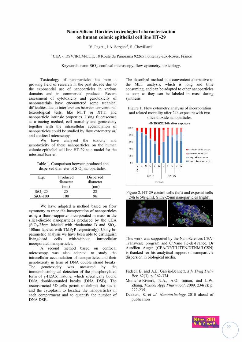
Toxicology of nanoparticles has been a growing field of research in the past decade due to the exponential use of nanoparticles in various domains and in commercial products. Recent assessment of cytotoxicity and genotoxicity of nanomaterials have encountered some technical difficulties due to interferences between conventional toxicological tests, like MTT or XTT, and nanoparticle intrinsic properties. Using fluorescence as a tracing method, cell mortality and gentoxicity together with the intracellular accumulation of nanoparticles could be studied by flow cytometry or/ and confocal microscopy. We have analysed the toxicity and genotoxicity of these nanoparticles on the human colonic epithelial cell line HT-29 as a model for the intestinal barrier.
Table 1. Comparison between produced and dispersed diameter of SiO2 nanoparticles.
We have adapted a method based on flow cytometry to trace the incorporation of nanoparticles using a fluoro-rapporter incorporated in mass in the silica-dioxide nanoparticles produced by the CEA (SiO2-25nm labeled with rhodamine B and SiO2-100nm labeled with TMPyP respectively). Using bi-parametric analysis we have been able to distinguish living/dead cells with/without intracellular incorporated nanoparticles.
A second method based on confocal microscopy was also adapted to study the intracellular accumulation of nanoparticles and their genotoxicity in term of DNA double strand breaks. The genotoxicity was measured by the immunohistological detection of the phosphorylated form of γ-H2AX histone, which specifically bound DNA double-stranded breaks (DNA DSB). The reconstructed 3D cells permit to delimit the nuclei and the cytoplasm to localize the nanoparticles in each compartment and to quantify the number of DNA DSB.
The described method is a convenient alternative to the MET analysis, which is long and time consuming, and can be adapted to other nanoparticles as soon as they can be labeled in mass during synthesis.
Figure 1. Flow cytometry analysis of incorporation and related mortality after 24h-exposure with two
silica dioxide nanoparticles.
Figure 2. HT-29 control cells (left) and exposed cells 24h to 50μg/mL SiO2-25nm nanoparticles (right).
This work was supported by the NanoSciences CEA-Transverse program and C’Nano Ile-de-France. Dr Aurélien Auger (CEA/DRT/LITEN/DTNM/LCSN) is thanked for his analytical support of nanoparticle dispersion in biological media. Fadeel, B. and A.E. Garcia-Bennett, Adv Drug Deliv
Rev. 62(3): p. 362-374. Monteiro-Riviere, N.A., A.O. Inman, and L.W.
Zhang, Toxicol Appl Pharmacol, 2009. 234(2): p. 222-235.
Dekkers, S. et al. Nanotoxicology 2010 ahead of publication
Exp. Produced diameter
(nm)
Dispersed diameter
(nm) SiO2-25 25 28
SiO2-100 100 96
23
Comparative study of cytotoxic and genotoxic effects of nano- and submicron-sized metal oxide
Y. Guichard1, J. Schmitt1, M. Goutet1, O. Rastoix2, D. Rousset2, A. Boivin2, R. Wrobel2, L. Gaté1,
C. Darne1 and S. Binet
1Department of pollutants and Health; 2Department of metrology of pollutants Institut National de Recherche et de Sécurité, rue du Morvan, CS 60027, 54519 Vandoeuvre Cedex, France
Keywords: titanium dioxides, iron oxides, cytotoxicity, genotoxicity
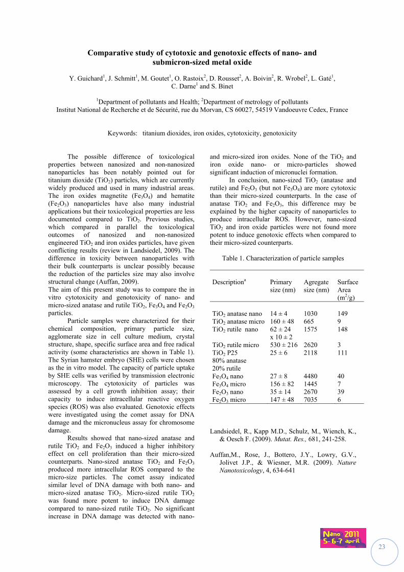
The possible difference of toxicological properties between nanosized and non-nanosized nanoparticles has been notably pointed out for titanium dioxide (TiO2) particles, which are currently widely produced and used in many industrial areas. The iron oxides magnetite (Fe3O4) and hematite (Fe2O3) nanoparticles have also many industrial applications but their toxicological properties are less documented compared to TiO2. Previous studies, which compared in parallel the toxicological outcomes of nanosized and non-nanosized engineered TiO2 and iron oxides particles, have given conflicting results (review in Landsiedel, 2009). The difference in toxicity between nanoparticles with their bulk counterparts is unclear possibly because the reduction of the particles size may also involve structural change (Auffan, 2009). The aim of this present study was to compare the in vitro cytotoxicity and genotoxicity of nano- and micro-sized anatase and rutile TiO2, Fe3O4 and Fe2O3 particles. Particle samples were characterized for their chemical composition, primary particle size, agglomerate size in cell culture medium, crystal structure, shape, specific surface area and free radical activity (some characteristics are shown in Table 1). The Syrian hamster embryo (SHE) cells were chosen as the in vitro model. The capacity of particle uptake by SHE cells was verified by transmission electronic microscopy. The cytotoxicity of particles was assessed by a cell growth inhibition assay; their capacity to induce intracellular reactive oxygen species (ROS) was also evaluated. Genotoxic effects were investigated using the comet assay for DNA damage and the micronucleus assay for chromosome damage. Results showed that nano-sized anatase and rutile TiO2 and Fe2O3 induced a higher inhibitory effect on cell proliferation than their micro-sized counterparts. Nano-sized anatase TiO2 and Fe2O3 produced more intracellular ROS compared to the micro-size particles. The comet assay indicated similar level of DNA damage with both nano- and micro-sized anatase TiO2. Micro-sized rutile TiO2 was found more potent to induce DNA damage compared to nano-sized rutile TiO2. No significant increase in DNA damage was detected with nano-
and micro-sized iron oxides. None of the TiO2 and iron oxide nano- or micro-particles showed significant induction of micronuclei formation. In conclusion, nano-sized TiO2 (anatase and rutile) and Fe2O3 (but not Fe3O4) are more cytotoxic than their micro-sized counterparts. In the case of anatase TiO2 and Fe2O3, this difference may be explained by the higher capacity of nanoparticles to produce intracellular ROS. However, nano-sized TiO2 and iron oxide particles were not found more potent to induce genotoxic effects when compared to their micro-sized counterparts.
Table 1. Characterization of particle samples
Descriptiona
Primary size (nm)
Agregate size (nm)
Surface Area (m2/g)
TiO2 anatase nano 14 ± 4 1030 149 TiO2 anatase micro 160 ± 48 665 9 TiO2 rutile nano 62 ± 24
x 10 ± 2 1575 148
TiO2 rutile micro 530 ± 216 2620 3 TiO2 P25 80% anatase 20% rutile
25 ± 6 2118 111
Fe3O4 nano 27 ± 8 4480 40 Fe3O4 micro 156 ± 82 1445 7 Fe2O3 nano 35 ± 14 2670 39 Fe2O3 micro 147 ± 48 7035 6
Landsiedel, R., Kapp M.D., Schulz, M., Wiench, K.,
& Oesch F. (2009). Mutat. Res., 681, 241-258. Auffan,M., Rose, J., Bottero, J.Y., Lowry, G.V.,
Jolivet J.P., & Wiesner, M.R. (2009). Nature Nanotoxicology, 4, 634-641
24
SiO2 nanoparticles activate immune dendritic cells
S. Barillet1, C. Nhim1, S. Kerdine-Römer1 and M. Pallardy1
1INSERM UMR 996, Faculty of Pharmacy, University of Paris-Sud XI, 92290, Chatenay-Malabry, France
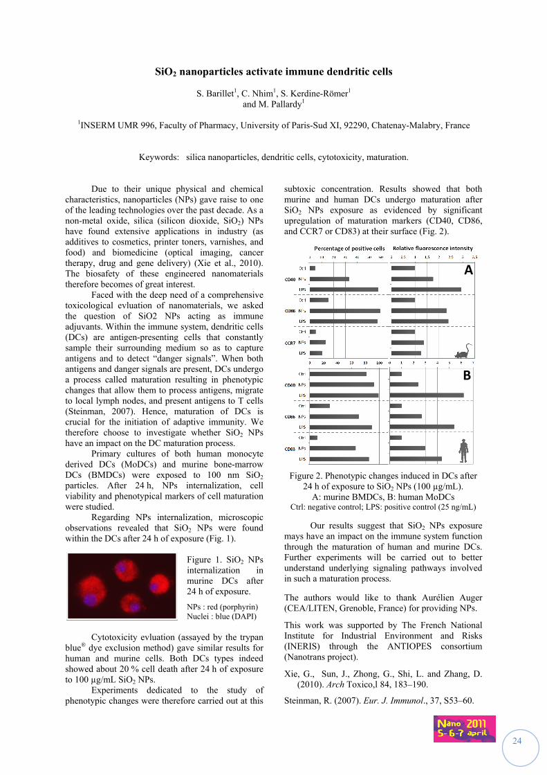
Keywords: silica nanoparticles, dendritic cells, cytotoxicity, maturation. Due to their unique physical and chemical characteristics, nanoparticles (NPs) gave raise to one of the leading technologies over the past decade. As a non-metal oxide, silica (silicon dioxide, SiO2) NPs have found extensive applications in industry (as additives to cosmetics, printer toners, varnishes, and food) and biomedicine (optical imaging, cancer therapy, drug and gene delivery) (Xie et al., 2010). The biosafety of these engineered nanomaterials therefore becomes of great interest. Faced with the deep need of a comprehensive toxicological evluation of nanomaterials, we asked the question of SiO2 NPs acting as immune adjuvants. Within the immune system, dendritic cells (DCs) are antigen-presenting cells that constantly sample their surrounding medium so as to capture antigens and to detect “danger signals”. When both antigens and danger signals are present, DCs undergo a process called maturation resulting in phenotypic changes that allow them to process antigens, migrate to local lymph nodes, and present antigens to T cells (Steinman, 2007). Hence, maturation of DCs is crucial for the initiation of adaptive immunity. We therefore choose to investigate whether SiO2 NPs have an impact on the DC maturation process. Primary cultures of both human monocyte derived DCs (MoDCs) and murine bone-marrow DCs (BMDCs) were exposed to 100 nm SiO2 particles. After 24 h, NPs internalization, cell viability and phenotypical markers of cell maturation were studied. Regarding NPs internalization, microscopic observations revealed that SiO2 NPs were found within the DCs after 24 h of exposure (Fig. 1).
Figure 1. SiO2 NPs internalization in murine DCs after 24 h of exposure.
NPs : red (porphyrin) Nuclei : blue (DAPI)
Cytotoxicity evluation (assayed by the trypan blue® dye exclusion method) gave similar results for human and murine cells. Both DCs types indeed showed about 20 % cell death after 24 h of exposure to 100 µg/mL SiO2 NPs. Experiments dedicated to the study of phenotypic changes were therefore carried out at this
subtoxic concentration. Results showed that both murine and human DCs undergo maturation after SiO2 NPs exposure as evidenced by significant upregulation of maturation markers (CD40, CD86, and CCR7 or CD83) at their surface (Fig. 2).
Figure 2. Phenotypic changes induced in DCs after 24 h of exposure to SiO2 NPs (100 µg/mL).
A: murine BMDCs, B: human MoDCs Ctrl: negative control; LPS: positive control (25 ng/mL)
Our results suggest that SiO2 NPs exposure mays have an impact on the immune system function through the maturation of human and murine DCs. Further experiments will be carried out to better understand underlying signaling pathways involved in such a maturation process.
The authors would like to thank Aurélien Auger (CEA/LITEN, Grenoble, France) for providing NPs.
This work was supported by The French National Institute for Industrial Environment and Risks (INERIS) through the ANTIOPES consortium (Nanotrans project).
Xie, G., Sun, J., Zhong, G., Shi, L. and Zhang, D. (2010). Arch Toxico,l 84, 183–190.
Steinman, R. (2007). Eur. J. Immunol., 37, S53–60.
A
B
25
Pro-inflammatory Response of Manganese Oxide Nanoparticles is altered upon Exposure to Endotoxin-Injured Alveolar Epithelial Cells
A. Schlicker1, M. Urner1, R. Frick1, L. K. Limbach2, W. J. Stark2 and B. Beck-Schimmer1
1Institute of Anesthesiology, University Hospital Zurich, Switzerland 2Institute for Chemical and Bioengineering, ETH Zurich, Switzerland
Keywords: nanotoxicology, inflammation, alveolar epithelial cells, metal oxide nanoparticles
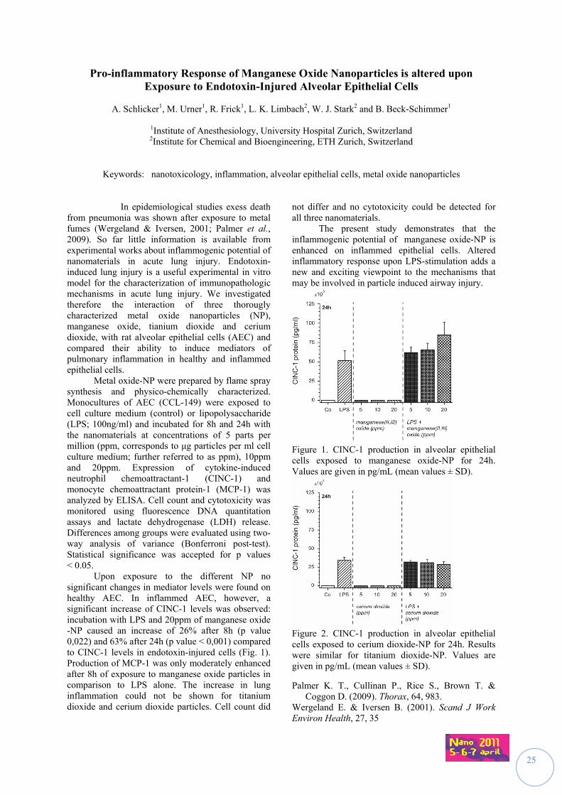
In epidemiological studies exess death from pneumonia was shown after exposure to metal fumes (Wergeland & Iversen, 2001; Palmer et al., 2009). So far little information is available from experimental works about inflammogenic potential of nanomaterials in acute lung injury. Endotoxin-induced lung injury is a useful experimental in vitro model for the characterization of immunopathologic mechanisms in acute lung injury. We investigated therefore the interaction of three thorougly characterized metal oxide nanoparticles (NP), manganese oxide, tianium dioxide and cerium dioxide, with rat alveolar epithelial cells (AEC) and compared their ability to induce mediators of pulmonary inflammation in healthy and inflammed epithelial cells.
Metal oxide-NP were prepared by flame spray synthesis and physico-chemically characterized. Monocultures of AEC (CCL-149) were exposed to cell culture medium (control) or lipopolysaccharide (LPS; 100ng/ml) and incubated for 8h and 24h with the nanomaterials at concentrations of 5 parts per million (ppm, corresponds to μg particles per ml cell culture medium; further referred to as ppm), 10ppm and 20ppm. Expression of cytokine-induced neutrophil chemoattractant-1 (CINC-1) and monocyte chemoattractant protein-1 (MCP-1) was analyzed by ELISA. Cell count and cytotoxicity was monitored using fluorescence DNA quantitation assays and lactate dehydrogenase (LDH) release. Differences among groups were evaluated using two-way analysis of variance (Bonferroni post-test). Statistical significance was accepted for p values < 0.05.
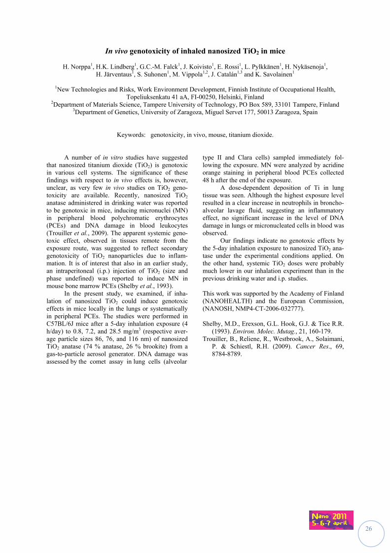
Upon exposure to the different NP no significant changes in mediator levels were found on healthy AEC. In inflammed AEC, however, a significant increase of CINC-1 levels was observed: incubation with LPS and 20ppm of manganese oxide -NP caused an increase of 26% after 8h (p value 0,022) and 63% after 24h (p value < 0,001) compared to CINC-1 levels in endotoxin-injured cells (Fig. 1). Production of MCP-1 was only moderately enhanced after 8h of exposure to manganese oxide particles in comparison to LPS alone. The increase in lung inflammation could not be shown for titanium dioxide and cerium dioxide particles. Cell count did
not differ and no cytotoxicity could be detected for all three nanomaterials.
The present study demonstrates that the inflammogenic potential of manganese oxide-NP is enhanced on inflammed epithelial cells. Altered inflammatory response upon LPS-stimulation adds a new and exciting viewpoint to the mechanisms that may be involved in particle induced airway injury.
Figure 1. CINC-1 production in alveolar epithelial cells exposed to manganese oxide-NP for 24h. Values are given in pg/mL (mean values ± SD).
Figure 2. CINC-1 production in alveolar epithelial cells exposed to cerium dioxide-NP for 24h. Results were similar for titanium dioxide-NP. Values are given in pg/mL (mean values ± SD).
Palmer K. T., Cullinan P., Rice S., Brown T. & Coggon D. (2009). Thorax, 64, 983.
Wergeland E. & Iversen B. (2001). Scand J Work Environ Health, 27, 35
26
In vivo genotoxicity of inhaled nanosized TiO2 in mice
H. Norppa1, H.K. Lindberg1, G.C.-M. Falck1, J. Koivisto1, E. Rossi1, L. Pylkkänen1, H. Nykäsenoja1, H. Järventaus1, S. Suhonen1, M. Vippola1,2, J. Catalán1,3 and K. Savolainen1
1New Technologies and Risks, Work Environment Development, Finnish Institute of Occupational Health,
Topeliuksenkatu 41 aA, FI-00250, Helsinki, Finland 2Department of Materials Science, Tampere University of Technology, PO Box 589, 33101 Tampere, Finland
3Department of Genetics, University of Zaragoza, Miguel Servet 177, 50013 Zaragoza, Spain
Keywords: genotoxicity, in vivo, mouse, titanium dioxide. A number of in vitro studies have suggested that nanosized titanium dioxide (TiO2) is genotoxic in various cell systems. The significance of these findings with respect to in vivo effects is, however, unclear, as very few in vivo studies on TiO2 geno-toxicity are available. Recently, nanosized TiO2 anatase administered in drinking water was reported to be genotoxic in mice, inducing micronuclei (MN) in peripheral blood polychromatic erythrocytes (PCEs) and DNA damage in blood leukocytes (Trouiller et al., 2009). The apparent systemic geno-toxic effect, observed in tissues remote from the exposure route, was suggested to reflect secondary genotoxicity of TiO2 nanoparticles due to inflam-mation. It is of interest that also in an earlier study, an intraperitoneal (i.p.) injection of TiO2 (size and phase undefined) was reported to induce MN in mouse bone marrow PCEs (Shelby et al., 1993). In the present study, we examined, if inha-lation of nanosized TiO2 could induce genotoxic effects in mice locally in the lungs or systematically in peripheral PCEs. The studies were performed in C57BL/6J mice after a 5-day inhalation exposure (4 h/day) to 0.8, 7.2, and 28.5 mg/m3 (respective aver-age particle sizes 86, 76, and 116 nm) of nanosized TiO2 anatase (74 % anatase, 26 % brookite) from a gas-to-particle aerosol generator. DNA damage was assessed by the comet assay in lung cells (alveolar
type II and Clara cells) sampled immediately fol-lowing the exposure. MN were analyzed by acridine orange staining in peripheral blood PCEs collected 48 h after the end of the exposure. A dose-dependent deposition of Ti in lung tissue was seen. Although the highest exposure level resulted in a clear increase in neutrophils in broncho-alveolar lavage fluid, suggesting an inflammatory effect, no significant increase in the level of DNA damage in lungs or micronucleated cells in blood was observed. Our findings indicate no genotoxic effects by the 5-day inhalation exposure to nanosized TiO2 ana-tase under the experimental conditions applied. On the other hand, systemic TiO2 doses were probably much lower in our inhalation experiment than in the previous drinking water and i.p. studies. This work was supported by the Academy of Finland (NANOHEALTH) and the European Commission, (NANOSH, NMP4-CT-2006-032777). Shelby, M.D., Erexson, G.L. Hook, G.J. & Tice R.R.
(1993). Environ. Molec. Mutag., 21, 160-179. Trouiller, B., Reliene, R., Westbrook, A., Solaimani,
P. & Schiestl, R.H. (2009). Cancer Res., 69, 8784-8789.
27
Pulmonary toxicity comparison of raw and super-purified single-wall carbon nanotubes after intra-tracheal instillation in rats
D. Elgrabi1, B. Trouiller1, F. Rogerieux1 and G. Lacroix1
1Institut National de l’Environnement Industriel et des Risque (INERIS), Parc Technologique ALATA, 60550
VERNEUIL-EN-HALATTE, France
Keywords: single-wall carbon nanotubes, toxicity, oxidative stress, inflammation. Nanomaterials are part of an industrial revolution to develop lightweight but strong materials for a variety of purposes. Single-wall carbon nanotubes (SWCNTs) are an important member of this class of materials. Carbon nanotubes possess unique electrical, mechanical, and thermal properties and have many potential applications in the electronics, computer, and aerospace industries. Unprocessed nanotubes are very light and could become airborne and potentially reach the lungs. Because the toxicity of nanotubes in the lung is not known, their pulmonary toxicity was investigated. In order to evaluate the relation between the respiratory toxicological effects of carbon nanotubes and their physicochemical characteristics, we compared raw and super-purified SWCNTs toxicity. Oxidative stress and inflammation involvements as possible mechanisms of the toxicological effects of these nanotubes were especially examined. Rats were intratracheally instilled with 0 or 200 µg of raw or super-purified SWCNTs, and euthanized 1, 7, 30, 90 or 180 days after the single treatment for histo-pathological study of the lungs, broncho-alveolar fluids and mRNA expression measurements. SWCNTs were suspended at 1.4 mg/ml in 3% ethanol and 1.4 mg/ml of bovine serum albumin solution. The suspension was sonicated for 10 min in an ultrasonic bath. In this SWCNTs solution, about less than 80% were agglomerates smaller than 10 µm and about 20 % were between 10 and 30 µm.
Results showed that these two types of SWCNTs can induce granulomas. The quantity and size of granulomas appear to be greater after treatment with raw SWCNTs than super-purified SWCNTs. Granulomas formation was noted after 24 hours exposure to raw SWCNTs and after 7 days exposure to super purified SWCNTs. The presence of these pathological formations lasted less than 6 months for raw SWCNTs and more than 6 months for super-purified SWCNTs. However, after 6 months, there were only few smaller granulomas compared to days 7, 30 and 90.
Regarding inflammation, the amount of total cells, total protein and the cellular composition did not change in broncho-alveolar fluids after exposure to either type of SWCNTs. However, interleukin-1 (IL-1) mRNA expression increased 24 hours after instillation, 9.2 and 2.7 fold for super-purified and raw SWCNTs respectively. This induction was followed by an inhibition of IL-1expression, 7 days after treatment with raw SWCNTs. A return to baseline was observed for the 2 SWCNTs after 30 days. After SWCNTs super-purified exposure, interleukin 6 (IL-6) expression was induced 24 hours after instillation, 80 and 41 fold for super-purified and raw SWCNTs respectively. A return to the baseline for these SWCNTs is observed at 7 days.
Regarding oxidative stress, expression analysis of biomarkers, heme oxygenase 1 and iNOS was performed. Results showed induction of these enzymes expression, with a maximum reached at 7 days in presence of raw SWCNTs and 1 month in the presence of super purified SWCNTs. This induction was followed by a gradual return to baseline at 180 days. These results are similar to those observed in the apoptosis study (variation in the expression of extracellular phosphatidylserine and caspase 3) and phagocytosis (quantification of cells with endocytosed SWCNTs)
Overall, the biological response after treatment with raw SWCNTs containing a high amount of iron (20%) is similar to treatment with super-purified SWCNTs containing a small amount of iron (2%). In both cases, the formation of granulomas and the presence of inflammatory markers, oxidative stress, apoptosis and phagocytosis were noted. These effects occurred earlier with raw SWCNTs and these SWCNTs seem eliminated more rapidly than super-purified SWCNTs. So purity of SWCNT is an important factor in pulmonary toxicity.
28
Nanosized particles systemic transport by phagocytes and vaccine adjuvant safety
R.K. Gherardi,1 Z. Khan,1 V. Itier,1 F-J. Authier,1 O. Tillement,2 and J. Cadusseau1
1Institut Mondor de Recherche Biomédicale, INSERM U955-E10, Univeristé Paris-Est, Faculté de Médecine, F-94010 Créteil, France
2Laboratoire de Physico-Chimie des Matériaux Luminescents, UMR 5620, UCBL, 69622 Villeurbanne, France
Keywords: aluminium, vaccine adjuvant, nanoparticle, biodistribution
Nanosized particles (NSPs) have various innovative medical applications in fields such as, imaging contrast fluids, topic antimicrobials, surgery tools, and drug, gene or vaccine delivery. In balance with these promising applications, safety issues need to be very carefully assessed. Due to the rapidly growing number of novel compounds and formulations, questions relative to biodistribution, persistence and toxicity of most nanomaterials have not been thoroughly explored, and long-term data are lacking. Therefore, the understanding of general mechanisms that may underlie beneficial/adverse effects of NSPs, especially those interacting with immune cells, is mandatory.
The use of NSPs in man is not as contemporary as it seems to be since aluminium hydroxide [Al(OH)3], a paradigmatic nano-crystaline compound also kwown as Alum, has been introduced in vaccine for its immunologic adjuvant effect in 1927. Alum remains the most commonly used vaccine adjuvant although mechanisms by which it stimulates immune responses remain incompletely understood. Although generally well tolerated, Alum has been suspected to occasionally cause chronic disabling health problems (Gherardi et al, 2001). For example, a subset of susceptible individuals has been found to combine delayed onset of diffuse myalgia, chronic fatigue and a stereotyped cognitive dysfunction with long-term (up to 12 years) persistence of Alum-loaded macrophages at site of previous intra-muscular (i.m.) immunization. Significance of these observations remains uncertain. Alum is used at concentrations viewed as an acceptable compromise between its adjuvanticity and aluminium neurotoxicity by industry and regulatory agencies. However, cognitive dysfunction in affected patients is suggestive of organic cortico-subcortical damage, and reminiscent of cognitive deficits described in foundry workers exposed to inhaled Al fumes or powder (Couette et al, 2009).
Though Alum safety crucially depends on whether the compound will remain localized at site of injection or diffuse and accumulate in distant organs, the biodistribution of NSPs injected into muscle has not been investigated. Since muscle injury elicits huge monocyte/macrophage (MO/MP) infiltration and migration to lymphoid organs, we wondered if a proportion of NSPs injected into muscle could translocate to distant organs as part of a general mechanism linked to phagocytosis
After i.m. injection of 36µL Alum-adjuvanted vaccine, #50% of Al was cleared from mouse muscle within d4, and Al deposits were detected by particle induced X-ray emission (PIXE) in spleen and brain, at d21, m6 and m12.
To examine if and how particles translocate to distant sites, we injected 20µL suspension of two types of NSPs: exploratory polychromatic fluorescent latex beads (FLBs), and a confirmatory Alum-relevant nanohybrid (Al-rho) in which Al(OH)3 is coupled with rhodamine. After i.m. injection, both NSPs massively reached draining lymph nodes (dLNs), peaking at d4 and decreasing at d21; dLN emptying was associated with increase of particle-loaded phagocytes in blood and spleen peaking at d21; slow but relentless accumulation occurred in brain from d21 to the d180 endpoint.
Neurodelivery increased by 2-fold in mice with chronically altered blood-brain barrier. However, compared to the i.m. route, intravenous injection resulted in virtually no neurodelivery. In contrast dLN ablation prior to i.m. injection reduced particle-loaded cells by 60-80% in blood, spleen and brain. Intracerebral particle injection showed lack of recirculation unique to brain, likely contributing to progressive cerebral NSP accumulation.
In mice deficient in CCL2/MCP-1 (the master chemoattractant of inflammatory monocytes), the amount of particle-loaded cells after i.m. injection markedly decreased at d21, in blood (-85%) and brain (-82%). Conversely, i.m. co-injection of Al-rho and CCL2 was associated with dramatic increase of the number of particle-loaded cells in blood (+274%) and brain (+414%).
Thus a MCP-1-driven Trojan horse mechanism is likely involved in NSP biodistribution and neurodelivery. Such a mechanism was previously documented for viral particle neurodelivery (HIV, HCV). It could underly Alum adverse neurologic effects in a small subset of susceptible individuals with constitutionally high MCP-1 tissue levels. Gherardi, R.K., et al. (2001) Macrophagic
myofasciitis lesions assess long-term persistence of vaccine-derived aluminium hydroxide in muscle. Brain 124:1821-31.
Couette M. et al. (2009). Long-term persistence of vaccine-derived aluminum hydroxide is associated with chronic cognitive dysfunction. J Inorg Biochem 103: 1571-8.
29
Effects of Carbon Black nanoparticles on the biotransformation of carcinogen aromatic amines by the human arylamine N-acetyltranferase 1
E. Sanfins1, J. Dairou1, S. Hussain1, F. Busi1, A. Chaffotte2, F. Rodrigues-Lima1, J.M. Dupret1
1Université Paris Diderot-Paris 7, Unité de Biologie Fonctionnelle et Adaptative (BFA), CNRS EAC 4413,
Équipe Réponses Moléculaires et Cellulaires aux Xénobiotiques, 75013 Paris, France. (email : [email protected])
2Unité de RMN des Biomolécules, Institut Pasteur, 75015, Paris France.
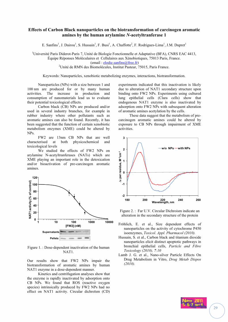
Keywords: Nanoparticles, xenobiotic metabolizing enzymes, interactions, biotransformation. Nanoparticles (NPs) with a size between 1 and 100 nm are produced for or by many human activities. The increase in production and consumption of nanomaterials lead us to evaluate their potential toxicological effects. Carbon black (CB) NPs are produced and/or used in several industry branches, for example in rubber industry where other pollutants such as aromatic amines can also be found. Recently, it has been suggested that the function of certain xenobiotic metabolism enzymes (XME) could be altered by NPs. FW2 are 13nm CB NPs that are well characterised at both physicochemical and toxicological levels We studied the effects of FW2 NPs on arylamine N-acetyltranferases (NATs) which are XME playing an important role in the detoxication and/or bioactivation of pre-carcinogen aromatic amines.
Supernatants
Pellets
1 10 100 1000 100000
20
40
60
80
100
120
[FW2] (nM)
NA
T1
acti
vity
(% o
f co
ntr
ol)
Figure 1. : Dose-dependent inactivation of the human NAT1.
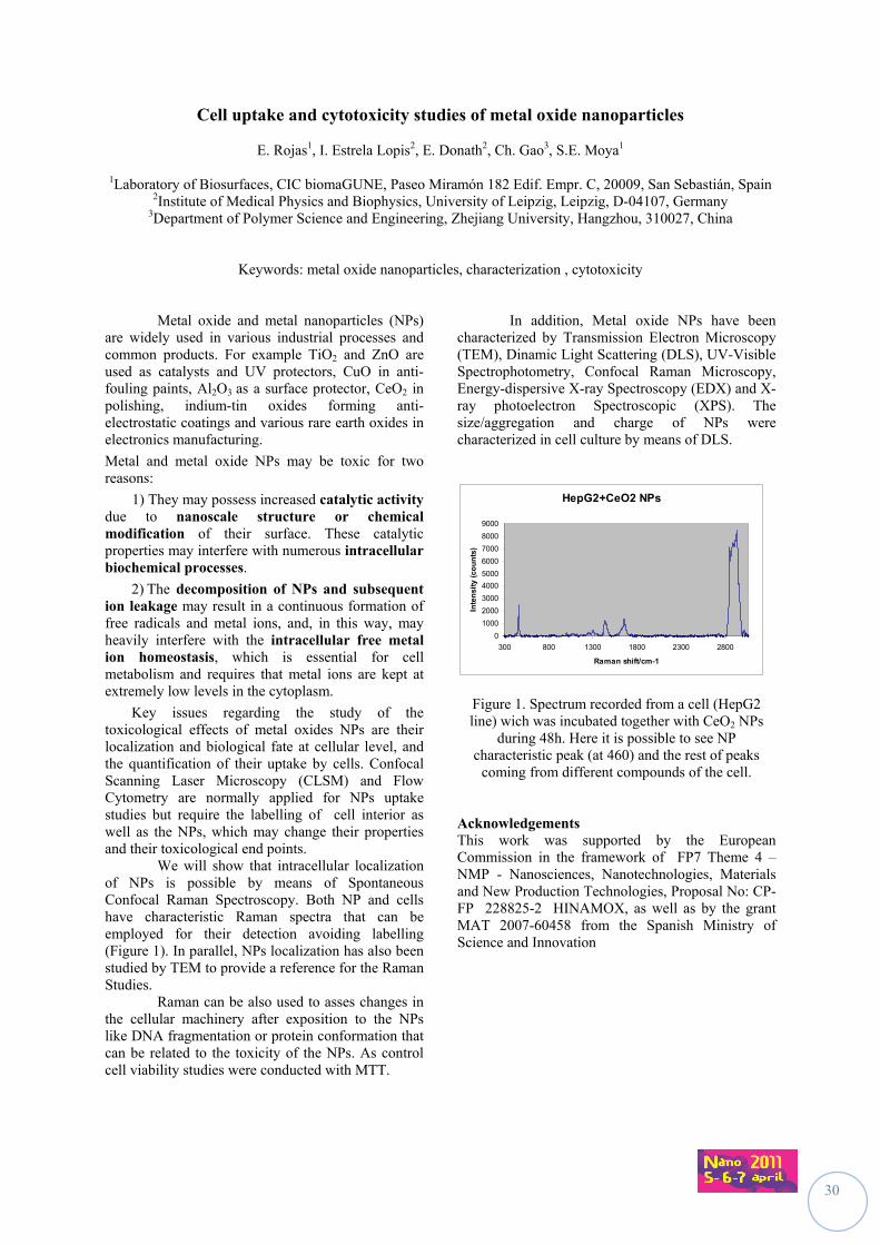
Our results show that FW2 NPs impair the biotransformation of aromatic amines by human NAT1 enzyme in a dose-dependent manner. Kinetics and centrifugation analyses show that the enzyme is rapidly inactivated by adsorption onto CB NPs. We found that ROS (reactive oxygen species) intrinsically produced by FW2 NPs had no effect on NAT1 activity. Circular dichroïsm (CD)
experiments indicated that this inactivation is likely due to alteration of NAT1 secondary structure upon binding onto FW2 NPs. Experiments using cultured lung epithelial cells (Clara cells) show that endogenous NAT1 enzyme is also inactivated by adsorption onto FW2 NPs with subsequent alteration of aromatic amines acetylation by the cells. These data suggest that the metabolism of pre-carcinogen aromatic amines could be altered by exposure to CB NPs through impairment of XME activities.
-2
-1
0
1
2
3
180 200 220 240 260
∆
per
res
idu
e, M
-1cm
-1
Wavelength, nm
w/o NPs with NPs
Figure 2. : Far U.V. Circular Dichroism indicate an alteration in the secondary structure of the protein
Fröhlich, E. et al., Size dependent effects of
nanoparticles on the activity of cytochrome P450 isoenzymes, Toxicol. Appl. Pharmacol (2010).
Hussain, S. et al., Carbon black and titanium dioxide nanoparticles elicit distinct apoptotic pathways in bronchial epithelial cells, Particle and Fibre Toxicology (2010), 7:10
Lamb J. G. et al., Nano-silver Particle Effects On Drug Metabolism in Vitro, Drug Metab Dispos (2010).
30
Cell uptake and cytotoxicity studies of metal oxide nanoparticles
E. Rojas1, I. Estrela Lopis2, E. Donath2, Ch. Gao3, S.E. Moya1
1Laboratory of Biosurfaces, CIC biomaGUNE, Paseo Miramón 182 Edif. Empr. C, 20009, San Sebastián, Spain 2Institute of Medical Physics and Biophysics, University of Leipzig, Leipzig, D-04107, Germany
3Department of Polymer Science and Engineering, Zhejiang University, Hangzhou, 310027, China
Keywords: metal oxide nanoparticles, characterization , cytotoxicity
Metal oxide and metal nanoparticles (NPs)
are widely used in various industrial processes and common products. For example TiO2 and ZnO are used as catalysts and UV protectors, CuO in anti-fouling paints, Al2O3 as a surface protector, CeO2 in polishing, indium-tin oxides forming anti-electrostatic coatings and various rare earth oxides in electronics manufacturing.
Metal and metal oxide NPs may be toxic for two reasons:
1) They may possess increased catalytic activity due to nanoscale structure or chemical modification of their surface. These catalytic properties may interfere with numerous intracellular biochemical processes.
2) The decomposition of NPs and subsequent ion leakage may result in a continuous formation of free radicals and metal ions, and, in this way, may heavily interfere with the intracellular free metal ion homeostasis, which is essential for cell metabolism and requires that metal ions are kept at extremely low levels in the cytoplasm.
Key issues regarding the study of the toxicological effects of metal oxides NPs are their localization and biological fate at cellular level, and the quantification of their uptake by cells. Confocal Scanning Laser Microscopy (CLSM) and Flow Cytometry are normally applied for NPs uptake studies but require the labelling of cell interior as well as the NPs, which may change their properties and their toxicological end points.
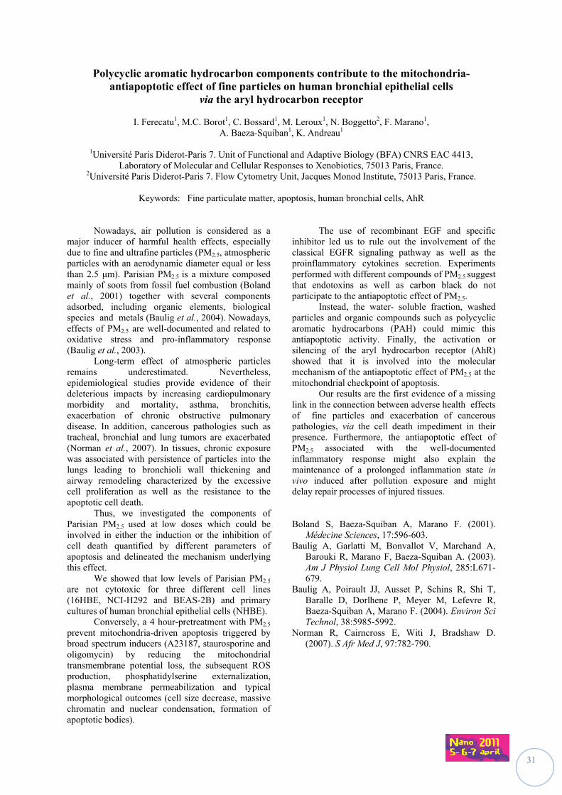
We will show that intracellular localization of NPs is possible by means of Spontaneous Confocal Raman Spectroscopy. Both NP and cells have characteristic Raman spectra that can be employed for their detection avoiding labelling (Figure 1). In parallel, NPs localization has also been studied by TEM to provide a reference for the Raman Studies.
Raman can be also used to asses changes in the cellular machinery after exposition to the NPs like DNA fragmentation or protein conformation that can be related to the toxicity of the NPs. As control cell viability studies were conducted with MTT.
In addition, Metal oxide NPs have been characterized by Transmission Electron Microscopy (TEM), Dinamic Light Scattering (DLS), UV-Visible Spectrophotometry, Confocal Raman Microscopy, Energy-dispersive X-ray Spectroscopy (EDX) and X-ray photoelectron Spectroscopic (XPS). The size/aggregation and charge of NPs were characterized in cell culture by means of DLS.
HepG2+CeO2 NPs
0
1000
2000
3000
4000
5000
6000
7000
8000
9000
300 800 1300 1800 2300 2800
Raman shift/cm-1
Inte
nsi
ty (
cou
nts
)
Figure 1. Spectrum recorded from a cell (HepG2 line) wich was incubated together with CeO2 NPs
during 48h. Here it is possible to see NP characteristic peak (at 460) and the rest of peaks
coming from different compounds of the cell.
Acknowledgements This work was supported by the European Commission in the framework of FP7 Theme 4 – NMP - Nanosciences, Nanotechnologies, Materials and New Production Technologies, Proposal No: CP-FP 228825-2 HINAMOX, as well as by the grant MAT 2007-60458 from the Spanish Ministry of Science and Innovation
31
Polycyclic aromatic hydrocarbon components contribute to the mitochondria-antiapoptotic effect of fine particles on human bronchial epithelial cells
via the aryl hydrocarbon receptor
I. Ferecatu1, M.C. Borot1, C. Bossard1, M. Leroux1, N. Boggetto2, F. Marano1, A. Baeza-Squiban1, K. Andreau1
1Université Paris Diderot-Paris 7. Unit of Functional and Adaptive Biology (BFA) CNRS EAC 4413,
Laboratory of Molecular and Cellular Responses to Xenobiotics, 75013 Paris, France. 2Université Paris Diderot-Paris 7. Flow Cytometry Unit, Jacques Monod Institute, 75013 Paris, France.
Keywords: Fine particulate matter, apoptosis, human bronchial cells, AhR
Nowadays, air pollution is considered as a major inducer of harmful health effects, especially due to fine and ultrafine particles (PM2.5, atmospheric particles with an aerodynamic diameter equal or less than 2.5 μm). Parisian PM2.5 is a mixture composed mainly of soots from fossil fuel combustion (Boland et al., 2001) together with several components adsorbed, including organic elements, biological species and metals (Baulig et al., 2004). Nowadays, effects of PM2.5 are well-documented and related to oxidative stress and pro-inflammatory response (Baulig et al., 2003). Long-term effect of atmospheric particles remains underestimated. Nevertheless, epidemiological studies provide evidence of their deleterious impacts by increasing cardiopulmonary morbidity and mortality, asthma, bronchitis, exacerbation of chronic obstructive pulmonary disease. In addition, cancerous pathologies such as tracheal, bronchial and lung tumors are exacerbated (Norman et al., 2007). In tissues, chronic exposure was associated with persistence of particles into the lungs leading to bronchioli wall thickening and airway remodeling characterized by the excessive cell proliferation as well as the resistance to the apoptotic cell death. Thus, we investigated the components of Parisian PM2.5 used at low doses which could be involved in either the induction or the inhibition of cell death quantified by different parameters of apoptosis and delineated the mechanism underlying this effect. We showed that low levels of Parisian PM2.5 are not cytotoxic for three different cell lines (16HBE, NCI-H292 and BEAS-2B) and primary cultures of human bronchial epithelial cells (NHBE).
Conversely, a 4 hour-pretreatment with PM2.5 prevent mitochondria-driven apoptosis triggered by broad spectrum inducers (A23187, staurosporine and oligomycin) by reducing the mitochondrial transmembrane potential loss, the subsequent ROS production, phosphatidylserine externalization, plasma membrane permeabilization and typical morphological outcomes (cell size decrease, massive chromatin and nuclear condensation, formation of apoptotic bodies).
The use of recombinant EGF and specific inhibitor led us to rule out the involvement of the classical EGFR signaling pathway as well as the proinflammatory cytokines secretion. Experiments performed with different compounds of PM2.5 suggest that endotoxins as well as carbon black do not participate to the antiapoptotic effect of PM2.5.
Instead, the water- soluble fraction, washed particles and organic compounds such as polycyclic aromatic hydrocarbons (PAH) could mimic this antiapoptotic activity. Finally, the activation or silencing of the aryl hydrocarbon receptor (AhR) showed that it is involved into the molecular mechanism of the antiapoptotic effect of PM2.5 at the mitochondrial checkpoint of apoptosis. Our results are the first evidence of a missing link in the connection between adverse health effects of fine particles and exacerbation of cancerous pathologies, via the cell death impediment in their presence. Furthermore, the antiapoptotic effect of PM2.5 associated with the well-documented inflammatory response might also explain the maintenance of a prolonged inflammation state in vivo induced after pollution exposure and might delay repair processes of injured tissues. Boland S, Baeza-Squiban A, Marano F. (2001).
Médecine Sciences, 17:596-603. Baulig A, Garlatti M, Bonvallot V, Marchand A,
Barouki R, Marano F, Baeza-Squiban A. (2003). Am J Physiol Lung Cell Mol Physiol, 285:L671-679.
Baulig A, Poirault JJ, Ausset P, Schins R, Shi T, Baralle D, Dorlhene P, Meyer M, Lefevre R, Baeza-Squiban A, Marano F. (2004). Environ Sci Technol, 38:5985-5992.
Norman R, Cairncross E, Witi J, Bradshaw D. (2007). S Afr Med J, 97:782-790.
32
Nanoparticles for photodynamic therapy applications
M. Barberi-Heyob1, D. Bechet1, P. Couleaud2, H. Benachour1, A. Seve2, M. Pernot1, R. Vanderesse3, T. Bastogne1, F. Lux4, O. Tillement4, S. Mordon5, F. Guillemin1, C. Frochot2
1CRAN, Nancy-University, CNRS, Centre Alexis Vautrin, Vandœuvre-lès-Nancy, F54511, France 2LRGP, Nancy-University, CNRS, INPL, Nancy, F54000, France 3LCPM, Nancy-University, CNRS, INPL, Nancy, F54000, France 4LCPML, Lyon-University, CNRS,Villeurbanne, F69622, France
5INSERM U703, Université de Lille Nord de France, CHRU, F59000, France
Keywords: photodynamic therapy, nanoparticle, MRI, vascular effect, peptide targeting
Photodynamic Therapy (PDT) involves the uptake of a photosensitizer by cancer or neo-vessel endothelial cells followed by photoirradiation. The selectivity of PDT as an anti-cancer treatment relies on the local generation of cytotoxic reactive oxygen species (ROS) in the tumor, mainly singlet oxygen 1O2, due to both preferential uptake of the photosensitizer by malignant tissue and subsequent localized light irradiation. Development of new therapeutic drug delivery systems is an area of significant research interest. The ability to directly target a therapeutic agent to a tumor site would minimize systemic drug exposure, thus providing the potential for increasing the therapeutic index. The tumor selectivity of photosensitizers used in the clinic is limited, as they tend to accumulate in normal tissue. This can be improved by using third generation photosensitizers, which consist of photosensitizers to which a tumor-targeting moiety is covalently attached (Tirand et al., 2006). Moreover, there are some limitations for the clinical application of existing photosensitizers: most of them are hydrophobic and can aggregate easily in aqueous media resulting in the decrease of their efficiency. Selective accumulation of the photosensitizers in cancer cells or neo-vasculature is required to avoid collateral damage. In bio-nanotechnology, development of nanoparticles as photosensitizer carriers can overcome most of the shortcomings of classic photosensitizers and it is reasonable to think that nanoparticles will become a real progress for the future development of PDT. In a first part, the different types of nanoparticles that are under development in the field of PDT will be presented especially in terms of advantages and drawbacks for each kind of nanoparticles (polymeric, ceramic, gold…). In a second part, the results concerning a new targeted multifunctional hybrid Gd2O3/silica nanoparticle will be presented. Indeed, the strategy developed by our team for several years aims at favouring the vascular effect of PDT by targeting tumor-associated vascularisation using peptide as ligand. We previously described the conjugation of a photosensitizer (a chlorin) to an heptapeptide (ATWLPPR), specific for the vascular endothelial growth factor receptor, neuropilin-1 (NRP-1). This targeted photosensitizer proved to be
very efficient in vitro in human umbilical vein endothelial cells compared to its non-conjugated counterpart. In vivo, using human malignant glioma-bearing nude mice we also demonstrated the interest of using this active-targeting strategy by the induction of tissue factor protein expression immediately post-treatment (Bechet et al., 2010), and a specific localization in endothelial cells lining tumor vessels of the conjugated photosensitizer. Nevertheless, we could observe a peptide degradation in plasma from 2 h post-intravenous injection and biodistribution results suggested that it could be due to the degradation of the peptidic moiety into organs of the reticulo-endothelial system (Thomas et al., 2009). As we previously described, non-biodegradable nanoparticles seem to be very promising careers satisfying all the requirements for an ideal targeted PDT (Bechet, Couleaud et al., 2008). We have designed and photophysically characterized a multifunctional nanoparticle consisting of ATWLPPR and encapsulated photodynamic therapy and imaging agents. Nanoparticles functionalized with ~4 peptides specifically bound to neuropilin-1 recombinant protein. Nanoparticles conferred photosensitivity to cells over-expressing NRP-1, providing evidence that the chlorin grafted within the nanoparticle matrix can be photo activated to yield photocytotoxic effects in vitro. Moreover, in vivo using an orthotopic stereotactic implantation of U87 glioblastoma tumor cells, MRI analysis evidenced a positive contrast enhancement.
This work was supported by the Ligue Nationale contre le Cancer, ANOCEF, CPER and the ANR “project no. ANR-08-PCVI-0021 Nano-VTP”. Bechet D., Couleaud P., Frochot C., Viriot M.L., Barberi-Heyob
M. (2008) Trends in Biotechnology, 26, 612-621. Bechet D., Tirand L., Faivre B., Plénat F., Bonnet C., Bastogne T.,
Frochot C., Guillemin F., Barberi-Heyob M., (2010) Pharm. Res. , 27(3), 468-479.
Thomas N., Bechet D., Becuwe P., Tirand L., Vanderesse R., Frochot C., Guillemin F., Barberi-Heyob M., (2009), J. Photochem. Photobiol. B, 96, 101-108.
Tirand L., Frochot C., Vanderesse R., Thomas N., Trinquet E., Pinel S., Viriot M.L., Guillemin F., & Barberi-Heyob M. (2006) J. Control. Release, 111, 153-164.
33
Inflammatory and genotoxic effects of nanoparticles and dust generated from nanoparticle-containing paints and lacquers
U. Vogel1,2, A.T. Saber1, K.A. Jensen1, I.K. Koponen1, N.R. Jacobsen1, R. Birkedal1, L. Mikkelsen3, P. Møller3, S. Loft3, and H. Wallin1,3
1National Research Centre for the Working Environment, Copenhagen, Denmark
2Institute for Science, Systems, and Models, University of Roskilde, Roskilde, Denmark 3Department of Environmental and Occupational Health, University of Copenhagen, Copenhagen, Denmark
Keywords: inflammation, DNA damage, nanoparticles, paint, lacquer Nanotechnology has potential applications in many processes and products. Therefore there is a growing need to establish knowledge about the health risks in relation to exposure to nanoparticles. The paint- and lacquer industry already use nanoparticles in substantial amounts in their products and the number of applications will be growing in the near future. The aim of NanoKem project is to investigate if the pure technical nanoparticles and sanding dust generated from nanoparticle-doped paint cause inflammation or DNA damage, associated with carcinogenicity.Nanoparticles (8 materials), dust containing nanoparticles and dust from reference paints and lacquers without nanoparticles (14 materials), has been tested in vitro and in vivo. Initially, an in vitro screening of the cytotoxicity of the materials was performed by incubating FE1-MutaTMMouse lung epithelial cells, at concentrations between 50 and 800µg/ml. The in vitro screening showed no sign of severe toxicity and made it reasonably to perform an in vivo screening of the materials. Twenty four h after a single intratracheal instillation of 54 µg particles in mice, the level of inflammation was assessed by measuring mRNA expression of cytokines in lung tissue and bronchoalveolar lavage cell composition. DNA damage was detected by the Comet assay in bronchoalveolar cells.
Based on this in vivo screening we chose to evaluate the dose-time-response of one of the materials from the screening and the corresponding paint with and without nanosized titanium dioxide. The mice were exposed by a single intratracheal instillation. The doses were 18, 54 and 162 µg, and 54, 162 and 486 for the nanoparticles and the paint dusts, respectively. DNA damage and inflammation was evaluated 1, 3 and 28 days after intratracheal instillation. We will present data on inflammation and DNA damage from the in vivo screening and the in vivo dose-response study. This work was supported by the Danish Working Environment Research Fund.
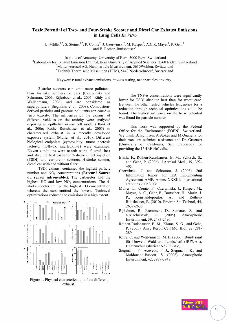
34
Toxic Potential of Two- and Four-Stroke Scooter and Diesel Car Exhaust Emissions in Lung Cells In Vitro
L. Müller1,2, S. Steiner1,2, P. Comte2, J. Czerwinski2, M. Kasper3, A.C.R. Mayer4, P. Gehr1
and B. Rothen-Rutishauser1
1Institute of Anatomy, University of Bern, 3000 Bern, Switzerland 2Laboratory for Exhaust Emission Control, Bern University of Applied Sciences, 2560 Nidau, Switzerland
3Matter Aerosol AG, Nanoparticle Measurement, 5610Wohlen, Switzerland 4Technik Thermische Maschinen (TTM), 5443 Niederrohrdorf, Switzerland
Keywords: total exhaust emissions, in vitro testing, nanoparticles, toxicity. 2-stroke scooters can emit more pollutants than 4-stroke scooters or cars (Czerwinski and Schramm, 2006; Rijkeboer et al., 2005; Rüdy and Weilenmann, 2006) and are considered as superpolluters (Siegmann et al., 2008). Combustion-derived particles and gaseous pollutants can cause in vitro toxicity. The influences of the exhaust of different vehicles on the toxicity were analyzed exposing an epithelial airway cell model (Blank et al., 2006; Rothen-Rutishauser et al., 2005) to characterized exhaust in a recently developed exposure system (Muller et al., 2010). Different biological endpoints (cytotoxicity, tumor necrosis factor-α (TNF-α), interleukin-8) were examined. Eleven conditions were tested: worst, filtered, best and absolute best cases for 2-stroke direct injection (TSDI) and carburetor scooters, 4-stroke scooter, diesel car with and without filter. TSDI exhaust contained the highest particle number and NOx concentrations (Erreur ! Source du renvoi introuvable.). The carburetor had the highest HC and low NOx concentrations. The 4-stroke scooter emitted the highest CO concentration whereas the cars emitted the lowest. Technical optimizations reduced the emissions in a high extent.
Figure 1. Physical characterization of the different exhaust.
The TNF-α concentrations were significantly lower for TSDI absolute best than for worst case. Between the other tested vehicles tendencies for a reduction through technical optimizations could be found. The highest influence on the toxic potential was found for particle number. This work was supported by the Federal Office for the Environment (FOEN), Switzerland. We thank B.Tschirren, A.Stokes and M.Ouanella for their excellent technical assistance and Dr. Gruenert (University of California, San Francisco) for providing the 16HBE14o- cells. Blank, F., Rothen-Rutishauser, B. M., Schurch, S.,
and Gehr, P. (2006). J.Aerosol Med., 19, 392-405.
Czerwinski, J. and Schramm, J. (2006). 2nd Information Report for IEA Implementing Agreement AMF, Annex XXXIII, international activities 2005/2006,
Muller, L., Comte, P., Czerwinski, J., Kasper, M., Mayer, A. C., Gehr, P., Burtscher, H., Morin, J. P., Konstandopoulos, A., and Rothen-Rutishauser, B. (2010). Environ Sci Technol, 44, 2632-2638.
Rijkeboer, R., Bremmers, D., Samaras, Z., and Ntziachristosb, L. (2005). Atmospheric Environment, 39, 2483-2490.
Rothen-Rutishauser, B. M., Kiama, S. G., and Gehr, P. (2005). Am J Respir Cell Mol Biol, 32, 281-289.
Rüdy, C. and Weilenmann, M. F. (2006). Bundesamt für Umwelt, Wald und Landschaft (BUWAL), Untersuchungsbericht Nr.203270e,
Siegmann, P., Acevedo, F. J., Siegmann, K., and Maldonado-Bascon, S. (2008). Atmospheric Environment, 42, 3937-3948.
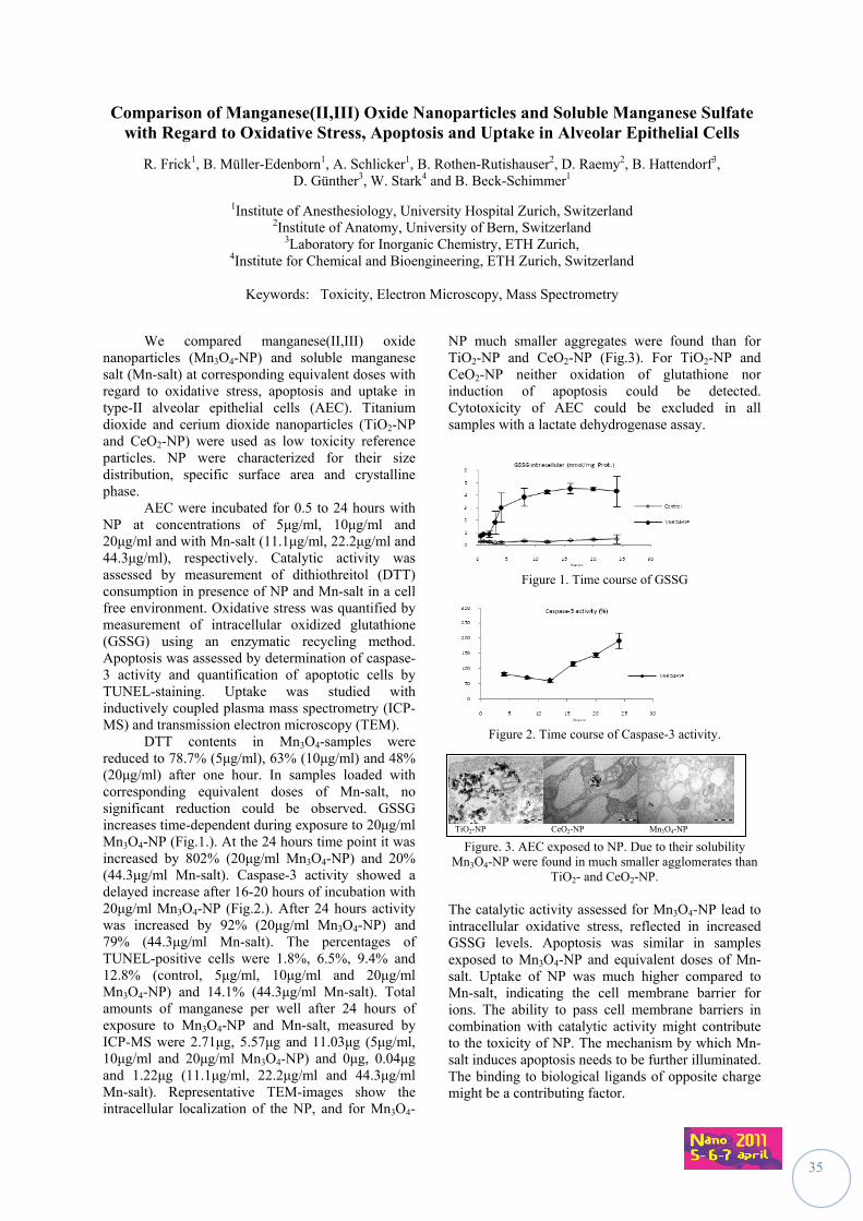
35
Comparison of Manganese(II,III) Oxide Nanoparticles and Soluble Manganese Sulfate with Regard to Oxidative Stress, Apoptosis and Uptake in Alveolar Epithelial Cells
R. Frick1, B. Müller-Edenborn1, A. Schlicker1, B. Rothen-Rutishauser2, D. Raemy2, B. Hattendorf3, D. Günther3, W. Stark4 and B. Beck-Schimmer1
1Institute of Anesthesiology, University Hospital Zurich, Switzerland
2Institute of Anatomy, University of Bern, Switzerland 3Laboratory for Inorganic Chemistry, ETH Zurich,
4Institute for Chemical and Bioengineering, ETH Zurich, Switzerland
Keywords: Toxicity, Electron Microscopy, Mass Spectrometry
We compared manganese(II,III) oxide nanoparticles (Mn3O4-NP) and soluble manganese salt (Mn-salt) at corresponding equivalent doses with regard to oxidative stress, apoptosis and uptake in type-II alveolar epithelial cells (AEC). Titanium dioxide and cerium dioxide nanoparticles (TiO2-NP and CeO2-NP) were used as low toxicity reference particles. NP were characterized for their size distribution, specific surface area and crystalline phase. AEC were incubated for 0.5 to 24 hours with NP at concentrations of 5μg/ml, 10μg/ml and 20μg/ml and with Mn-salt (11.1μg/ml, 22.2μg/ml and 44.3μg/ml), respectively. Catalytic activity was assessed by measurement of dithiothreitol (DTT) consumption in presence of NP and Mn-salt in a cell free environment. Oxidative stress was quantified by measurement of intracellular oxidized glutathione (GSSG) using an enzymatic recycling method. Apoptosis was assessed by determination of caspase-3 activity and quantification of apoptotic cells by TUNEL-staining. Uptake was studied with inductively coupled plasma mass spectrometry (ICP-MS) and transmission electron microscopy (TEM). DTT contents in Mn3O4-samples were reduced to 78.7% (5μg/ml), 63% (10μg/ml) and 48% (20μg/ml) after one hour. In samples loaded with corresponding equivalent doses of Mn-salt, no significant reduction could be observed. GSSG increases time-dependent during exposure to 20μg/ml Mn3O4-NP (Fig.1.). At the 24 hours time point it was increased by 802% (20μg/ml Mn3O4-NP) and 20% (44.3μg/ml Mn-salt). Caspase-3 activity showed a delayed increase after 16-20 hours of incubation with 20μg/ml Mn3O4-NP (Fig.2.). After 24 hours activity was increased by 92% (20μg/ml Mn3O4-NP) and 79% (44.3μg/ml Mn-salt). The percentages of TUNEL-positive cells were 1.8%, 6.5%, 9.4% and 12.8% (control, 5μg/ml, 10μg/ml and 20μg/ml Mn3O4-NP) and 14.1% (44.3μg/ml Mn-salt). Total amounts of manganese per well after 24 hours of exposure to Mn3O4-NP and Mn-salt, measured by ICP-MS were 2.71μg, 5.57μg and 11.03μg (5μg/ml, 10μg/ml and 20μg/ml Mn3O4-NP) and 0μg, 0.04μg and 1.22μg (11.1μg/ml, 22.2μg/ml and 44.3μg/ml Mn-salt). Representative TEM-images show the intracellular localization of the NP, and for Mn3O4-
NP much smaller aggregates were found than for TiO2-NP and CeO2-NP (Fig.3). For TiO2-NP and CeO2-NP neither oxidation of glutathione nor induction of apoptosis could be detected. Cytotoxicity of AEC could be excluded in all samples with a lactate dehydrogenase assay.
Figure 1. Time course of GSSG
Figure 2. Time course of Caspase-3 activity.
TiO2-NP CeO2-NP Mn3O4-NP
Figure. 3. AEC exposed to NP. Due to their solubility Mn3O4-NP were found in much smaller agglomerates than
TiO2- and CeO2-NP. The catalytic activity assessed for Mn3O4-NP lead to intracellular oxidative stress, reflected in increased GSSG levels. Apoptosis was similar in samples exposed to Mn3O4-NP and equivalent doses of Mn-salt. Uptake of NP was much higher compared to Mn-salt, indicating the cell membrane barrier for ions. The ability to pass cell membrane barriers in combination with catalytic activity might contribute to the toxicity of NP. The mechanism by which Mn-salt induces apoptosis needs to be further illuminated. The binding to biological ligands of opposite charge might be a contributing factor.
36
Pulmonary toxicity of Fe3O4 nano- and sub-micrometric particles following intratracheal instillation in Sprague Dawley rats
L. Gaté1, Y. Guichard1, S. Sébillaud1, C. Langlais1, J-M. Micillino1, C. Darne1, O. Rastoix2, D. Rousset2
and S. Binet1
1Département Polluants et Santé, 2Département Métrologie des Aérosols,
Institut National de Recherche et de Sécurité, Rue du Morvan, CS 60027, 54519 Vandoeuvre lès Nancy, France
Keywords: intratracheal instillation, lung toxicity, inflammation, magnetite
The growing use of nanomaterials is leading to a possible increasing exposure of workers to nanoaerosols, the toxic properties of which are not always known. The experimental results currently available suggest that the pulmonary toxicity of nanoparticles can be influenced by the structure of the particle, its size, its specific surface area, and its surface reactivity (Sayes and Warheit, 2009) . Due to their magnetic properties, magnetite (Fe3O4) nanoparticles are currently used in medical and industrial applications, and occupational exposure may occur wherever they are used. However, their toxicological properties have not been thoroughly evaluated. In order to gain understandings of their pulmonary toxicity, we administered by a single intratracheal instillation nano and sub-micrometric Fe3O4 particles (Table 1) (2.5, 5 and 10 mg/kg) to rats and compared their pulmonary effects to those obtained with a crystalline silica (Min U Sil 5, 10 mg/kg)) known to induce persistent pulmonary inflammation and lung fibrosis. Table 1. Physical characteristics of the tested Fe3O4
particles
The size was determined by transmission electron microscopy. The specific surface area (SSA) was obtained by using the method of Brunauer, Emmett and Teller (1938). The toxicological effects of instilled particles were assessed 1, 7 and 42 days after the exposure. While, as compared to the control group, Min U Sil 5 induced an increase of lung neutrophilic granulocytes, in the bronchoalveolar lavage fluid (BALF), up to 42 days after treatment, Fe3O4 particles caused only a slight increase of such cells 24 hours following the instillation which disappeared at 7 days. The cytological analysis of the BALF and the histological evaluation of the lung tissue also
showed that particles were phagocytised by alveolar macrophages. However, no significant change in the activities of the lactate deshydrogenase (a marker of general cytoxictity) and the alkaline phosphatase (a marker of epithelial cell toxicity) was observed in the BALF of exposed animals to Fe3O4 particles. From the results obtained so far, in our experimental conditions, no chronic inflammatory response, a good indicator of pulmonary toxicity for particles (Schins and Knapper, 2007) was detected following the intratracheal instillation of both nano- and submicronic Fe3O4 particles. However, additional experiments including the evaluation of alveolar epithelial cell proliferation and the analysis of the genotoxicty of such particles in BALF and alveolar epithelial cells using the comet assay will be performed. Sayes CM and Warheit DB. (2009). Wiley Interdiscip
Rev Nanomed Nanobiotechnol., 1, 660-670. Brunauer S, Emmett PH and Teller E (1938), J. Am.
Chem. Soc., 60, 309-319. Schins RP and Knaapen AM. (2007), Inhal
Toxicol.,19 Suppl 1:189-98. Fe3O4
nano Fe3O4
sub-micro Size (nm) 29 ± 8 156 ± 82
SSA (m2/g) 40,3 7,3
37
Evaluation of the toxicity of fluorescent nanoparticles used in the detection of the sentinel lymph node in breast cancer
M. Helle1, E. Pic1, T. Pons2, E. Rampazzo3, L. Bezdetnaya1, F. Guillemin1, L. Prodi3,
B. Dubertret2 and F. Marchal1
1CRAN, Nancy-University, CNRS, Centre Alexis Vautrin, Avenue de Bourgogne, 54511 Vandoeuvre-lès-Nancy Cedex, France
2Laboratoire Physique et Etude des Matériaux, CNRS UPR0005, ESPCI, 10, rue Vauquelin, 75005 Paris, France 3Dipartimento di Chimica “G. Ciamician”, Latemar Unit, Università di Bologna, Bologna, Italy
Keywords: sentinel lymph node, fluorescent nanoparticles, inflammation, hemolysis
Breast cancer is the first cause of cancer death among women and its incidence doubled in the last two decades. Several approaches for the treatment of these cancers have been developed. The axillary lymph node dissection (ALND) leads to numerous morbidity complications and is now advantageously replaced by the dissection and the biopsy of the sentinel lymph node. Although this approach has strong advantages, it has its own limitations which are manipulation of radioactive products and possible anaphylactic reactions to the dye. As recently proposed, these limitations could in principle be by-passed if nanoparticles (quantum dots – QDs or silica nanoparticles doped with cyanine 7) were used as fluorescent contrast agents for the in vivo imaging of sentinel lymph nodes. QDs are fluorescent nanoparticles with unique optical properties like high extinction coefficient, brightness and photostability, which make them promising fluorescent probes for biological imaging.
The first QDs tested in our lab were composed of CdTeSe/CdZnS core/shell. The right axillary lymph node (RALN) of healthy Balb/c mice injected subcutaneously in the right anterior paw with 20 pmol of CdTeSe/CdZnS QDs was rapidly visualised. The RALN of injected mice were resected and weighted 7 days post injection and were fixed in 10 % formaldehyde. Five µm sections of LNs were prepared and hematoxylin and eosin (H&E) staining was performed and examined to visualize inflammatory changes. We observed that injection of 20 pmol of CdTeSe/CdZnS QDs in the right anterior paw induced an immune response revealed by an inflammation of the two regional LNs (Pons et al., 2010). This inflammation is a reflection of the acute local toxicity of CdTeSe/CdZnS QDs and represents a serious obstacle to potential clinical applications, even though no signs of abnormal behavior were noticed in injected mice.
Moreover, in vitro studies have been made to test the nanoparticles interaction with blood components and to determine the hemolytic properties of QDs. Mice blood stabilized with EDTA was obtained by intracardiac punction on healthy mice. The QDs were incubated in purified red blood cells, the blood was centrifuged and the percentage of
hemolysis was determined by colorimetric detection of hemoglobin in the supernatant. The hemolysis rate increased whith increasing QDs doses.
We attributed this toxicity to the degradation of the QDs in vivo and the release of toxic heavy metal ions such as Cd and Te.
In order to reduce the QD’s toxicity, our works turned to QDs without cadmium (CuInS2/ZnS core/shell) and to silica nanoparticles doped with cyanine 7 (Rampazzo et al, in press). The RALN could be visualised as soon as 5 minutes post-injection and, contrary to CdTeSe/CdZnS QDs, no inflammation was shown as well as absence of in vitro hemolysis.
In conclusion, CuInS2/ZnS QDs and silica nanoparticles doped with cyanine 7 allow the effective and rapid detection of ALN with a reduced acute local toxicity than with CdTeSe/CdZnS QDs. Although they are less toxic than the CdTeSe/CdZnS QD, an improvement of their surface chemistry is required to allow their excretion, and therefore, to minimize toxicity risks.
This work was supported by the Institut
National du Cancer (INCA), Comités départementaux (54, 55) de la Ligue Nationale de Lutte contre le Cancer and the Ligue Nationale de Lutte contre le Cancer.
Pons T., Pic E., Lequeux N., Cassette E., Bezdetnaya L., Guillemin F., Marchal F., Dubertret B. (2010) ACS Nano May 25;4(5):2531-8
Rampazzo, E.; Bonacchi, S.; Juris, R.; Montalti, M.; Genovese, D.; Zaccheroni, N.; Prodi, L.; Rambaldi, D. C.; Zattoni, A.; Reschiglian, P. (article ASAP) J. Phys. Chem. B
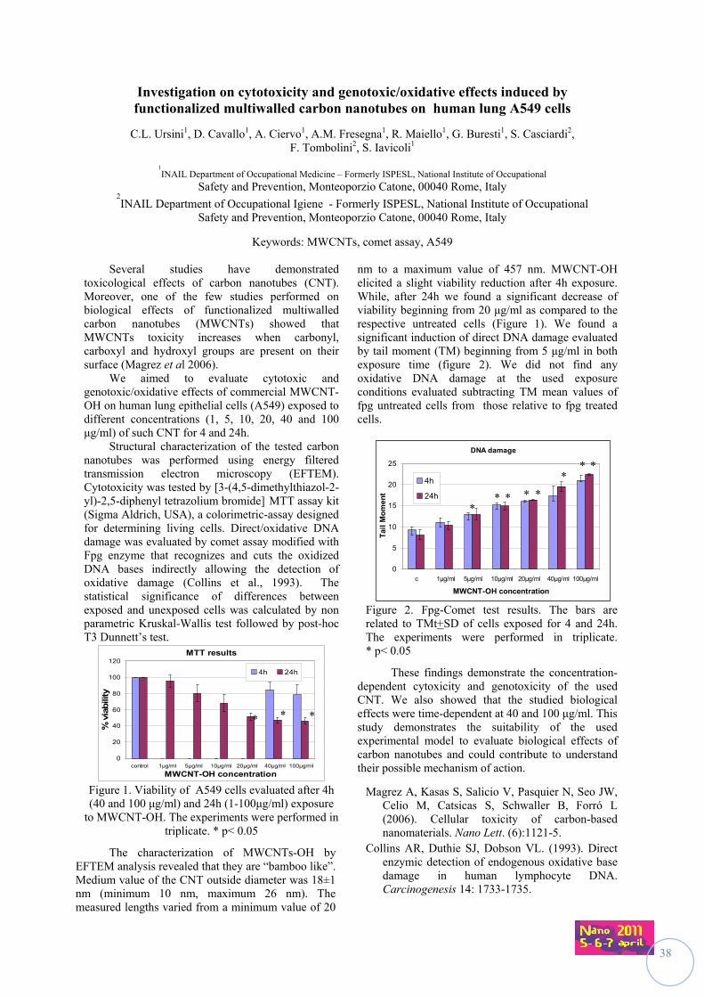
38
Investigation on cytotoxicity and genotoxic/oxidative effects induced by functionalized multiwalled carbon nanotubes on human lung A549 cells
C.L. Ursini1, D. Cavallo1, A. Ciervo1, A.M. Fresegna1, R. Maiello1, G. Buresti1, S. Casciardi2, F. Tombolini2, S. Iavicoli1
1INAIL Department of Occupational Medicine – Formerly ISPESL, National Institute of Occupational
Safety and Prevention, Monteoporzio Catone, 00040 Rome, Italy 2INAIL Department of Occupational Igiene - Formerly ISPESL, National Institute of Occupational
Safety and Prevention, Monteoporzio Catone, 00040 Rome, Italy
Keywords: MWCNTs, comet assay, A549
Several studies have demonstrated toxicological effects of carbon nanotubes (CNT). Moreover, one of the few studies performed on biological effects of functionalized multiwalled carbon nanotubes (MWCNTs) showed that MWCNTs toxicity increases when carbonyl, carboxyl and hydroxyl groups are present on their surface (Magrez et al 2006).
We aimed to evaluate cytotoxic and genotoxic/oxidative effects of commercial MWCNT-OH on human lung epithelial cells (A549) exposed to different concentrations (1, 5, 10, 20, 40 and 100 μg/ml) of such CNT for 4 and 24h.
Structural characterization of the tested carbon nanotubes was performed using energy filtered transmission electron microscopy (EFTEM). Cytotoxicity was tested by [3-(4,5-dimethylthiazol-2-yl)-2,5-diphenyl tetrazolium bromide] MTT assay kit (Sigma Aldrich, USA), a colorimetric-assay designed for determining living cells. Direct/oxidative DNA damage was evaluated by comet assay modified with Fpg enzyme that recognizes and cuts the oxidized DNA bases indirectly allowing the detection of oxidative damage (Collins et al., 1993). The statistical significance of differences between exposed and unexposed cells was calculated by non parametric Kruskal-Wallis test followed by post-hoc T3 Dunnett’s test.
MTT results
0
20
40
60
80
100
120
control 1μg/ml 5μg/ml 10μg/ml 20μg/ml 40μg/ml 100μg/ml
MWCNT-OH concentration
% v
iabili
ty
4h 24h
Figure 1. Viability of A549 cells evaluated after 4h (40 and 100 μg/ml) and 24h (1-100μg/ml) exposure
to MWCNT-OH. The experiments were performed in triplicate. * p< 0.05
The characterization of MWCNTs-OH by EFTEM analysis revealed that they are “bamboo like”. Medium value of the CNT outside diameter was 18±1 nm (minimum 10 nm, maximum 26 nm). The measured lengths varied from a minimum value of 20
nm to a maximum value of 457 nm. MWCNT-OH elicited a slight viability reduction after 4h exposure. While, after 24h we found a significant decrease of viability beginning from 20 μg/ml as compared to the respective untreated cells (Figure 1). We found a significant induction of direct DNA damage evaluated by tail moment (TM) beginning from 5 μg/ml in both exposure time (figure 2). We did not find any oxidative DNA damage at the used exposure conditions evaluated subtracting TM mean values of fpg untreated cells from those relative to fpg treated cells.
DNA damage
0
5
10
15
20
25
c 1μg/ml 5μg/ml 10μg/ml 20μg/ml 40μg/ml 100μg/ml
MWCNT-OH concentration
Tai
l M
om
ent
4h
24h
Figure 2. Fpg-Comet test results. The bars are related to TMt+SD of cells exposed for 4 and 24h. The experiments were performed in triplicate. * p< 0.05
These findings demonstrate the concentration-dependent cytoxicity and genotoxicity of the used CNT. We also showed that the studied biological effects were time-dependent at 40 and 100 μg/ml. This study demonstrates the suitability of the used experimental model to evaluate biological effects of carbon nanotubes and could contribute to understand their possible mechanism of action.
Magrez A, Kasas S, Salicio V, Pasquier N, Seo JW, Celio M, Catsicas S, Schwaller B, Forró L (2006). Cellular toxicity of carbon-based nanomaterials. Nano Lett. (6):1121-5.
Collins AR, Duthie SJ, Dobson VL. (1993). Direct enzymic detection of endogenous oxidative base damage in human lymphocyte DNA. Carcinogenesis 14: 1733-1735.
* * *
** *
*
* *
* *
39
Long-term effects of repeated exposure to Paris ambient nanoparticles on the pro-inflammatory response and differentiation of human bronchial epithelial cells in vitro
L. Boublil1, E. Assémat1, M.C. Borot1, L. Martinon2, F. Marano1, A. Baeza-Squiban1
1Unit of Fonctional and Adaptive Biology, University Paris Diderot – Paris 7, Laboratory of Molecular and Cellular Responses to Xenobiotics, EAC CNRS 4413, 5 rue Thomas Mann, 75205 Paris Cedex 13, France.
2Laboratoire des particules inhalées (LEPI), 11, rue George Eastman 75013 PARIS, France.
Our aim was to investigate the long term effects of ambient urban nanoparticles exposure on bronchial epithelium in order to determine whether nanoparticles could contribute to airway remodelling by inducing sustained inflammation and mucus secretion. For this purpose nanoparticles were sampled in Paris (site of background pollution) with a Dekati impactor in order to collect particles between 30 nm and 100 nm. Their effect was compared to the one of standard diesel exhaust particles (DEP) on primary cultures of normal human bronchial epithelial cells (NHBE) grown at an air-liquid interface (ALI) up to 2 months. After four 48 hrs-spaced treatments of 6 hrs (at 5µg/cm² for ambient NP or from 1 to 10µg/cm² for DEP) during the first week of ALI, the evolution of the pro-inflammatory response, the epithelium differentiation and the fate of particles were studied during the 5 following weeks.
Observations by transmission electron microscopy (TEM) revealed that nanoparticles are still present in the bronchial epithelium 5 weeks after the last treatment and are generally enclosed in vesicles. The expression of cytochrome P450 1A1, a xenobiotic metabolizing enzyme known to be specifically induced by polyaromatic hydrocarbons and involved in their metabolism, is highly dose-dependently overexpressed during NP treatment and return nearly to basal expression at the end of NP treatment. GM-CSF and IL-6, two biomarkers of a pro-inflammatory response, are slightly released after each treatment but increased and maintained up to 6 weeks. Amphiregulin (an EGFR ligand involved in the pro-inflammatory response) is released after each treatment but its release is resolved after the end of the treatments. MUC5AC (mucin) and DNALI1 (cilia dynein) were measured in order to characterize the epithelium differentiation. After 6 weeks, MUC5AC gene is strongly induced in cells exposed to NP compared to the control conditions. A weaker induction was observed for DNALI1.
Our results suggest that airway epithelial cells repeatedly exposed to ambient urban nanoparticles exhibit a sustained pro-inflammatory response and evolve towards a mucous phenotype. Funding by ANR Megatox CESA 009 02
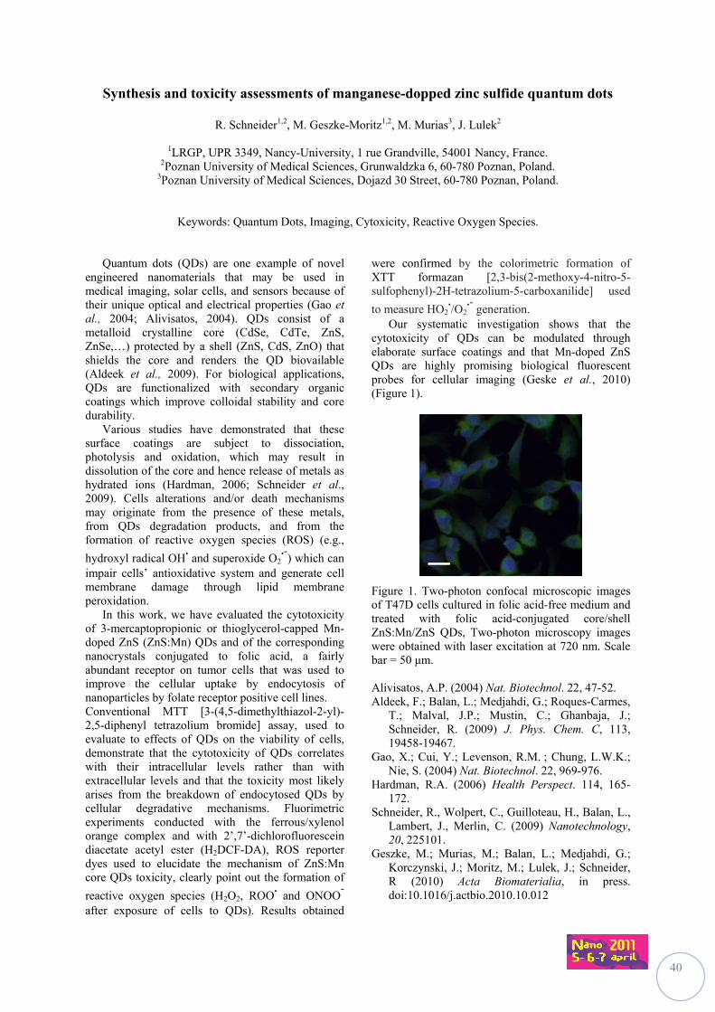
40
Synthesis and toxicity assessments of manganese-dopped zinc sulfide quantum dots
R. Schneider1,2, M. Geszke-Moritz1,2, M. Murias3, J. Lulek2
1LRGP, UPR 3349, Nancy-University, 1 rue Grandville, 54001 Nancy, France. 2Poznan University of Medical Sciences, Grunwaldzka 6, 60-780 Poznan, Poland.
3Poznan University of Medical Sciences, Dojazd 30 Street, 60-780 Poznan, Poland.
Keywords: Quantum Dots, Imaging, Cytoxicity, Reactive Oxygen Species.
Quantum dots (QDs) are one example of novel engineered nanomaterials that may be used in medical imaging, solar cells, and sensors because of their unique optical and electrical properties (Gao et al., 2004; Alivisatos, 2004). QDs consist of a metalloid crystalline core (CdSe, CdTe, ZnS, ZnSe,…) protected by a shell (ZnS, CdS, ZnO) that shields the core and renders the QD biovailable (Aldeek et al., 2009). For biological applications, QDs are functionalized with secondary organic coatings which improve colloidal stability and core durability.
Various studies have demonstrated that these surface coatings are subject to dissociation, photolysis and oxidation, which may result in dissolution of the core and hence release of metals as hydrated ions (Hardman, 2006; Schneider et al., 2009). Cells alterations and/or death mechanisms may originate from the presence of these metals, from QDs degradation products, and from the formation of reactive oxygen species (ROS) (e.g.,
hydroxyl radical OH. and superoxide O2
.-) which can
impair cells’ antioxidative system and generate cell membrane damage through lipid membrane peroxidation.
In this work, we have evaluated the cytotoxicity of 3-mercaptopropionic or thioglycerol-capped Mn-doped ZnS (ZnS:Mn) QDs and of the corresponding nanocrystals conjugated to folic acid, a fairly abundant receptor on tumor cells that was used to improve the cellular uptake by endocytosis of nanoparticles by folate receptor positive cell lines. Conventional MTT [3-(4,5-dimethylthiazol-2-yl)-2,5-diphenyl tetrazolium bromide] assay, used to evaluate to effects of QDs on the viability of cells, demonstrate that the cytotoxicity of QDs correlates with their intracellular levels rather than with extracellular levels and that the toxicity most likely arises from the breakdown of endocytosed QDs by cellular degradative mechanisms. Fluorimetric experiments conducted with the ferrous/xylenol orange complex and with 2’,7’-dichlorofluorescein diacetate acetyl ester (H2DCF-DA), ROS reporter dyes used to elucidate the mechanism of ZnS:Mn core QDs toxicity, clearly point out the formation of
reactive oxygen species (H2O2, ROO. and ONOO
-
after exposure of cells to QDs). Results obtained
were confirmed by the colorimetric formation of XTT formazan [2,3-bis(2-methoxy-4-nitro-5-sulfophenyl)-2H-tetrazolium-5-carboxanilide] used
to measure HO2./O2
.- generation.
Our systematic investigation shows that the cytotoxicity of QDs can be modulated through elaborate surface coatings and that Mn-doped ZnS QDs are highly promising biological fluorescent probes for cellular imaging (Geske et al., 2010) (Figure 1).
Figure 1. Two-photon confocal microscopic images of T47D cells cultured in folic acid-free medium and treated with folic acid-conjugated core/shell ZnS:Mn/ZnS QDs, Two-photon microscopy images were obtained with laser excitation at 720 nm. Scale bar = 50 μm. Alivisatos, A.P. (2004) Nat. Biotechnol. 22, 47-52. Aldeek, F.; Balan, L.; Medjahdi, G.; Roques-Carmes,
T.; Malval, J.P.; Mustin, C.; Ghanbaja, J.; Schneider, R. (2009) J. Phys. Chem. C, 113, 19458-19467.
Gao, X.; Cui, Y.; Levenson, R.M. ; Chung, L.W.K.; Nie, S. (2004) Nat. Biotechnol. 22, 969-976.
Hardman, R.A. (2006) Health Perspect. 114, 165-172.
Schneider, R., Wolpert, C., Guilloteau, H., Balan, L., Lambert, J., Merlin, C. (2009) Nanotechnology, 20, 225101.
Geszke, M.; Murias, M.; Balan, L.; Medjahdi, G.; Korczynski, J.; Moritz, M.; Lulek, J.; Schneider, R (2010) Acta Biomaterialia, in press. doi:10.1016/j.actbio.2010.10.012
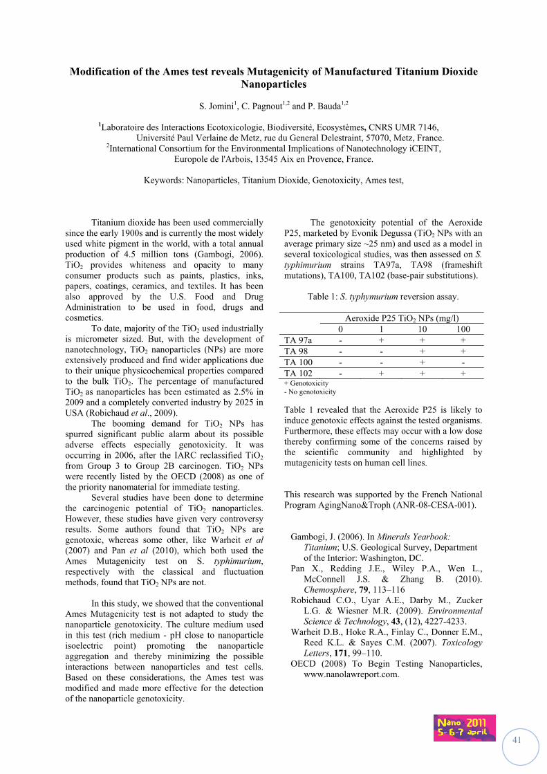
41
Modification of the Ames test reveals Mutagenicity of Manufactured Titanium Dioxide Nanoparticles
S. Jomini1, C. Pagnout1,2 and P. Bauda1,2
1Laboratoire des Interactions Ecotoxicologie, Biodiversité, Ecosystèmes, CNRS UMR 7146,
Université Paul Verlaine de Metz, rue du General Delestraint, 57070, Metz, France. 2International Consortium for the Environmental Implications of Nanotechnology iCEINT,
Europole de l'Arbois, 13545 Aix en Provence, France.
Keywords: Nanoparticles, Titanium Dioxide, Genotoxicity, Ames test,
Titanium dioxide has been used commercially since the early 1900s and is currently the most widely used white pigment in the world, with a total annual production of 4.5 million tons (Gambogi, 2006). TiO2 provides whiteness and opacity to many consumer products such as paints, plastics, inks, papers, coatings, ceramics, and textiles. It has been also approved by the U.S. Food and Drug Administration to be used in food, drugs and cosmetics.
To date, majority of the TiO2 used industrially is micrometer sized. But, with the development of nanotechnology, TiO2 nanoparticles (NPs) are more extensively produced and find wider applications due to their unique physicochemical properties compared to the bulk TiO2. The percentage of manufactured TiO2 as nanoparticles has been estimated as 2.5% in 2009 and a completely converted industry by 2025 in USA (Robichaud et al., 2009).
The booming demand for TiO2 NPs has spurred significant public alarm about its possible adverse effects especially genotoxicity. It was occurring in 2006, after the IARC reclassified TiO2 from Group 3 to Group 2B carcinogen. TiO2 NPs were recently listed by the OECD (2008) as one of the priority nanomaterial for immediate testing.
Several studies have been done to determine the carcinogenic potential of TiO2 nanoparticles. However, these studies have given very controversy results. Some authors found that TiO2 NPs are genotoxic, whereas some other, like Warheit et al (2007) and Pan et al (2010), which both used the Ames Mutagenicity test on S. typhimurium, respectively with the classical and fluctuation methods, found that TiO2 NPs are not.
In this study, we showed that the conventional
Ames Mutagenicity test is not adapted to study the nanoparticle genotoxicity. The culture medium used in this test (rich medium - pH close to nanoparticle isoelectric point) promoting the nanoparticle aggregation and thereby minimizing the possible interactions between nanoparticles and test cells. Based on these considerations, the Ames test was modified and made more effective for the detection of the nanoparticle genotoxicity.
The genotoxicity potential of the Aeroxide P25, marketed by Evonik Degussa (TiO2 NPs with an average primary size ~25 nm) and used as a model in several toxicological studies, was then assessed on S. typhimurium strains TA97a, TA98 (frameshift mutations), TA100, TA102 (base-pair substitutions).
Table 1: S. typhymurium reversion assay.
Aeroxide P25 TiO2 NPs (mg/l)
0 1 10 100 TA 97a - + + + TA 98 - - + + TA 100 - - + - TA 102 - + + + + Genotoxicity - No genotoxicity
Table 1 revealed that the Aeroxide P25 is likely to induce genotoxic effects against the tested organisms. Furthermore, these effects may occur with a low dose thereby confirming some of the concerns raised by the scientific community and highlighted by mutagenicity tests on human cell lines. This research was supported by the French National Program AgingNano&Troph (ANR-08-CESA-001).
Gambogi, J. (2006). In Minerals Yearbook: Titanium; U.S. Geological Survey, Department of the Interior: Washington, DC.
Pan X., Redding J.E., Wiley P.A., Wen L., McConnell J.S. & Zhang B. (2010). Chemosphere, 79, 113–116
Robichaud C.O., Uyar A.E., Darby M., Zucker L.G. & Wiesner M.R. (2009). Environmental Science & Technology, 43, (12), 4227-4233.
Warheit D.B., Hoke R.A., Finlay C., Donner E.M., Reed K.L. & Sayes C.M. (2007). Toxicology Letters, 171, 99–110.
OECD (2008) To Begin Testing Nanoparticles, www.nanolawreport.com.
42
NANOTRANS : Evaluation of the human tissue distribution of fine and ultrafine particles
M. Rinaldo1,2,7, A. Lacourt1, M.A. Billon-Galland3, P. Dumortier4, S. Gromb1, J.C. Pairon5, E. Sellier6,
A.Vital1, P. Brochard1,2
1CHU de Bordeaux, Bordeaux 33076 France, 2Université de Bordeaux 2Institut de santé publique, d’épidémiologie et de développement, Laboratoire santé travail environnement,
Bordeaux 33076 France 3 LEPI, 75013 Paris France
4 Hôpital ERASME, Bruxelles 1070 Belgique 5 INSERM, Créteil 94010 France
6 CREMEM Université Bordeaux 1,Talence 33405 France 7 mail : [email protected]
Keywords : nanoparticle , translocation, transmission electron microscopy, biometrology.
Context and objectives Toxicological studies led in the context of emerging nanotechnologies highlighted dangerousness of nanoparticles and atmospheric ultrafine particles with pulmonary and extrapulmonary possible effects (central nervous and cardiovascular systems). (Ostiguy 2010) No reliable datum excludes that extrapulmonary effects can result of systemic translocation across the alveolo-capillary barrier. There is also lack of datum on human translocation of these particles across other biological barriers (placenta, kidney, lymph node). Méthods : NANOTRANS study consists in analyzing tissues from four 4 groups of samples: - Group 1: 90 lung samples from 90 patients surgically treated for bronchopulmonary cancer classified into three exposure groups (nanoparticles occupational exposure, exposure to tobacco smoke particles and no exposure) in order to analyze relationships between exposure and lung retention; - Group 2: 90 lung and pleural samples of adult patients in order to analyze pulmonary regional translocation of nanoparticles; - Group 3: 50 samples from 10 patients (lung, myocardium, kidney, brain and lymph node) in order to evaluate extrapulmonary translocation of nanoparticles; - Group 4 (FOETOPUF): 20 fetal lung samples associated to maternal exposure data in order to evaluate placental transfer. After sodium hypochloric digestion of samples, particles are collected on a polycarbonate carbon coated filter and transferred on cupper electron microcopy grid. Particular retention and chemical characterization are performed using transmission electron microscopy and energy X-ray dispersive spectroscopy.
First results: First analyses were performed on 9 samples of FOETOPUF study in order to adapt the method used for micronic particles. Average lung retention was found to be 17.2 millions of particles per gram of dry tissue for individual or aggregated particles of less than 100 nm in diameter. It varies from 0 to 71 millions depending of the samples. 38 % of these particles were metallic (iron, titanium and others metals), 23 % were silica or silicates particles and 18 % were carbon particles. These data are compatible with results obtained for non exposed adult. (Brauer 2000) Conclusion : NANOTRANS study will allow us to obtain first data on nanoparticles human lung retention and on the possible systemic translocation of these compounds. Theses results are important for development of nanoparticles biométrology. Ostiguy C, Soucy B, Lapointe G, Woods C, Ménard
L, Trottier M,. Les effets sur la santé reliés aux nanoparticules - 2e édition. Série Études et recherches, Rapport IRSST R-558 2008
Brauer M, Avila-casado C, Fortoul T, Vedal S, Bonnie S , Churg A Air pollution and retained particles in the lung Env Health Persp vol 2000 109 (10) 1039-43-

43
The calibrated thrombin generation test (cTGT) is the gold standard assay to assess the procoagulant activity of nanomaterials
J. Laloy*1, S. Robert* 1, C. Marbehant1, F. Mullier1,2,3, J. Mejia4,5, J.P. Piret6, S. Lucas5, B. Chatelain2,3,
J.M. Dogné1,3, O. Toussaint6, B. Masereel1 and S. Rolin1
1Department of Pharmacy – Drug Design & Discovery Center, NARILIS, FUNDP-Namur, Namur, Belgium
2Hematology Laboratory, NARILIS, UCL Mont-Godinne, Yvoir, Belgium 3Thrombosis and Hemostasis Center, Namur, Belgium
4Laboratory of Chemistry and Electrochemistry of Surfaces, FUNDP-Namur, Namur, Belgium 5Research Centre in Physics of Matter and Radiation, FUNDP-Namur, Namur, Belgium
6Laboratory of Cellular Biochemistry and Biology, FUNDP-Namur, Namur, Belgium
*Both authors have equally participated to the study reported here
Keywords: Nanoparticles, thrombin generation, coagulation cascade Objectives We aimed to validate an universal, fast, accurate, reliable and relevant toxicological preclinical screening test to measure the potential impact on coagulation of nanoparticles (NP) whatever their physicochemical properties. Materials and methods We evaluated several methods of clotting times and thrombin generation assays for their suitability to assess the impact of NP on coagulation in human normal pool plasma. Five NP (21 nm CB, 15 nm SiO2, 20 nm TiC, 60 nm SiC and 12 nm CuO) were studied and chosen for their medical interest together with their various physicochemical properties which were extensively characterised. Results The optical clotting time, TAT complex and chromogenic thrombin generation assays were unsuited for studying the NP as these latter interfere with the light absorbance detection method. With the mechanical clotting time assays, the NP did not modify the PT nor the aPTT until 500 µg.mL-1 while the recalcification clotting time (RCT) was concentration-dependently decreased. Within calibrated thrombin generation test (cTGT), the NP concentration-dependently decreased the lag time and increased the Cmax, and especially in contact pathway-induced cTGT. Both RCT and cTGT showed that the studied NP exhibited procoagulant activity (SiO2 ≥ SiC ≥ TiC > CuO > CB) but cTGT was more specific, sensitive, relevant and rapid than RCT. Moreover, the addition of the Pluronic F-108 surfactant and/or the sonication for the preparation of the NP suspensions affected their procoagulant activity by impacting on their physicochemical parameters.
Conclusion The cTGT appears as the gold standard assay to assess the NP procoagulant activity in human plasma. Potential application and key benefits The cTGT would be used as the reference preclinical toxicological screening tool for evaluating at an early stage of the manufacturing process the potential impact of nanomaterials on coagulation in order to design and develop safe and nontoxic products with optimum hemocompatibility. international advisory committee. Abstracts which do not fulfil the standards will be rejected. Please, avoid to submit abstracts containing only future results.
44
Interaction of nanoparticules used in medical applications with lung epithelial cells: uptake, cytotoxicity, oxidant stress and proinflammatory response
R. Guadagnini1, S. Boland1, S. Vranic1, S. Hussain1, K. Moreau1, F. Marano1
1Laboratory of Functional and Adaptative Biology, unit of Réponses Moléculaires et Cellulaires aux
Xénobiotiques (RMCX), CNRS EAC, University Paris Diderot, CC 7073, 75205 Paris cedex 13, France
Keywords: nanomedicine, metal oxides, oxidative stress, inflammation. Nanoscience and Nanotechnology are developing very rapidly. Strategy for Nanotechnology of the European Commission (COM (2004) 338) has highlighted the need to invest in knowledge, technology and experience in this field in Europe. There are many applications of nanomaterials and it is thus important to assess their potential risks to human health. While a lot of attention has been paid to the development of new engineered nanoparticles and many new applications of nanotechnologies, comparatively less research has been performed to assess the potential danger of these new materials.
Our goal was to determine the effects of nanoparticles (NPs) on the lung as first target during inhalation of NPs. We investigated the effects of different NPs [titanium dioxide (TiO2), poly (lactic-co-glycolic acid) (PLGA), non coated Fe3O4 (N Fe3O4), Fe3O4 coated with oleic acid (C Fe3O4), fluorescent silica dioxide 25nm (SiO2 25) and fluorescent silica dioxide 50nm (SiO2 50)], on human bronchial (16HBE line) and human alveolar type II cells (A549 line). We evaluated the cytotoxicity of these NPs by WST-1 assay and propidium iodide uptake showing that toxicity depends on particle type, size and coating.
We determined the ability of NPs to enter cells measuring by flow cytometry the right angle scattering of the laser and we notice that they can be internalized by cells. We measured also the induction of oxidative stress in 16HBE and A549 cells after 24 and 48h of treatment with NPs by dihydroethidium oxidation assay (flow cytometry) seeing that they have different ability to induce oxidant stress. Finally we investigated wethether NPs have capacity to induce inflammatory response evaluating the thiol content of A549 and 16HBE cells after treatment with N-ethyl-maleimide, buthionine sulfoximine, NPs by monoBromoBimane (mBBr) assay (flow cytometry). We determined the production of cytokines (GM-CSF, IL-8, IL-6) by A549 and 16HBE cells after 24 and 48 hours of treatment with NPs by ELISA test and by RT- qPCR.
Results show that at non toxic concentration NPs can induce inflammation in cells. In conclusion, NPs could be taken up by lung epithelial cells and could lead to oxidative stress and cell death at high concentrations but also to inflammation at lower concentrations. These effects depend on particle type and coating. This work was supported by FP7 program NanoTEST.
45
Genotoxicity of nanocellulose whiskers in human bronchial epithelial cells in vitro
K.S. Hannukainen1, J. Catalán1,2, H. Järventaus1, E. Kontturi3, E. Vanhala1, K. Savolainen1, H. Norppa1
1New Technologies and Risks, Work Environment Development; Finnish Institute of Occupational Health; Topeliuksenkatu 41aA; 00250 Helsinki; Finland
2Department of Genetics; University of Zaragoza; Miguel Servet 177; 50013; Zaragoza; Spain 3Department of Forest Products; Aalto University School of Science and Technology, P.O. Box 16300; 00076
Aalto, Finland
Nanocellulose, whisker, genotoxicity, nanomaterials.
Nanocellulosics are among the most promising innovations for wide-variety applications in materials science. Although nanocellulose is presently prepared and applied only in laboratory scale, its possible impacts on public health and the environment should be investigated at an early stage. The aim of the present study was to examine in vitro the potential genotoxicity of two celluloses: Avicel, a commercially available microcrystalline cellulose (Fluka) used as a model of a non-nanoscale material (particle size ~50 µm), and nanocellulose whiskers produced by the Aalto University School of Science and Technology (mean length 152.2 nm, mean diameter 15.7 nm).
Cytotoxicity was analyzed at three different exposure times (4, 24 and 48 h) by the propidium iodide exclusion technique and luminometric detection of ATP in human bronchial epithelial BEAS 2B cells.
Cytotoxicity reached the 50% level at about the 100 µg/ml dose for both celluloses. Genotoxicity was assessed by the analysis of micronuclei (MN) in BEAS 2B cells using various doses (2.5-100 µg/ml) of Avicel and nanocellulose whiskers. The induction of MN was examined by the cytokinesis-block method after a 48-h treatment with the materials. Our results indicated no induction of micronucleated binucleate or mononucleate cells by Avicel or nanocellulose whiskers. No linear dose-dependent response could neither be found for the materials. In conclusion, our results from the MN assay show that nanocellulose whiskers are not genotoxic under the conditions tested [Supported by SUNPAP, NMP-2008-1.2-1, The Finnish Centre for Nanocellulosic Technologies and the Spanish Ministry of Science and Innovation]
46
Internalisation and transcytosis of SiO2 and TiO2 nanoparticles by lung epithelial cells
S. Vranic1, R. Guadagnini1, A. Baeza1, M.C. Borot1, S. Boland1
1 Laboratoire de Biologie Fonctionnelle et Adaptative, équipe Réponses Moléculaires et Cellulaires aux Xénobiotiques (RMCX), CNRS EAC 7059, Université Paris Diderot,
CC 7073, 75205 Paris cedex 13, France.
Email : [email protected]
Keywords: nanoparticles, endocytosis, transcytosis, cytotoxicity.
In view of the considerable development of nanotechnologies it is important to evaluate their potential risk for human health. Our goal was to determine the cytotoxic effects of nanoparticles (NPs) in the lung, which is the first target after inhalation of NPs. We further studied the endocytosis of NPs by respiratory epithelial cells and their capacity to cross the epithelial barrier. We investigated the effects of different NP [fluorescently labelled or non fluorescent titanium dioxide (TiO2) and silicium dioxide (SiO2)] on human bronchial epithelial cells (16HBE14O-), bronchial glandular adenocarcinoma cells (Calu-3) and human mucoepidermoid carcinoma cells (NCI-H292).
First we evaluated the cytotoxicity of NPs by
WST-1 assay. TiO2 NPs are cytotoxic for 16HBE and NCI-H292 cell lines at high concentrations inducing apoptosis. SiO2 NPs are cytotoxic in a size-dependent manner.
We also evaluated quantitatively and
qualitatively the endocytosis of NPs by epithelial cells. We determined the ability of NPs to enter 16HBE and NCI-H292 cells by measuring with a flow cytometer the right angle scattering of the laser or intensity of fluorescence of the cells treated with fluorescent NPs. We further studied the endocytosis of NPs by confocal microscopy to determine which of the three major endocytotic pathways (clathrin dependent, caveolin dependent or macropinocytosis) is involved in the internalisation of TiO2 NPs. For this study we used specific inhibitors for each pathway after evaluating the specificity of each inhibitor using positive controls for each endocytotic pathway. TiO2 and SiO2 NPs are internalized by respiratory epithelial cells using predominantly clathrin dependent cellular machinery, but we have shown a poor specificity of the inhibitors used.
Regarding the transcytosis of NPs we
examined the possibility of NPs to pass through pulmonary epithelial barriers. First we compared the capacity of different cell lines to develop a tight epithelial layer by measuring the transepithelial electric resistance (TEER), passage of fluorescent
molecule Lucifer Yellow, marker of paracellular passage and regarding by confocal microscopy the expression of proteins specific for tight junctions. After establishing a model by comparing different cell lines and culture conditions (Transwells with pore size of 0.4µm and 3µm) we evaluated the possibility of transcytosis of NPs. TiO2 and SiO2 NPs are able to cross the epithelial barrier but the percentage of particles crossing the epithelium is very low.
In conclusion, NPs are cytotoxic at high
concentrations, depending on the cell line used and on their size. However, at non cytotoxic concentrations these NPs are taken up by respiratory epithelial cells but have poor capacity to cross the epithelial barrier by transcytosis.
This work was supported by grant from EC FP7 201335 (Nanotest) and EC FP7 228789 (ENPRA), National Grant Nanotrans.
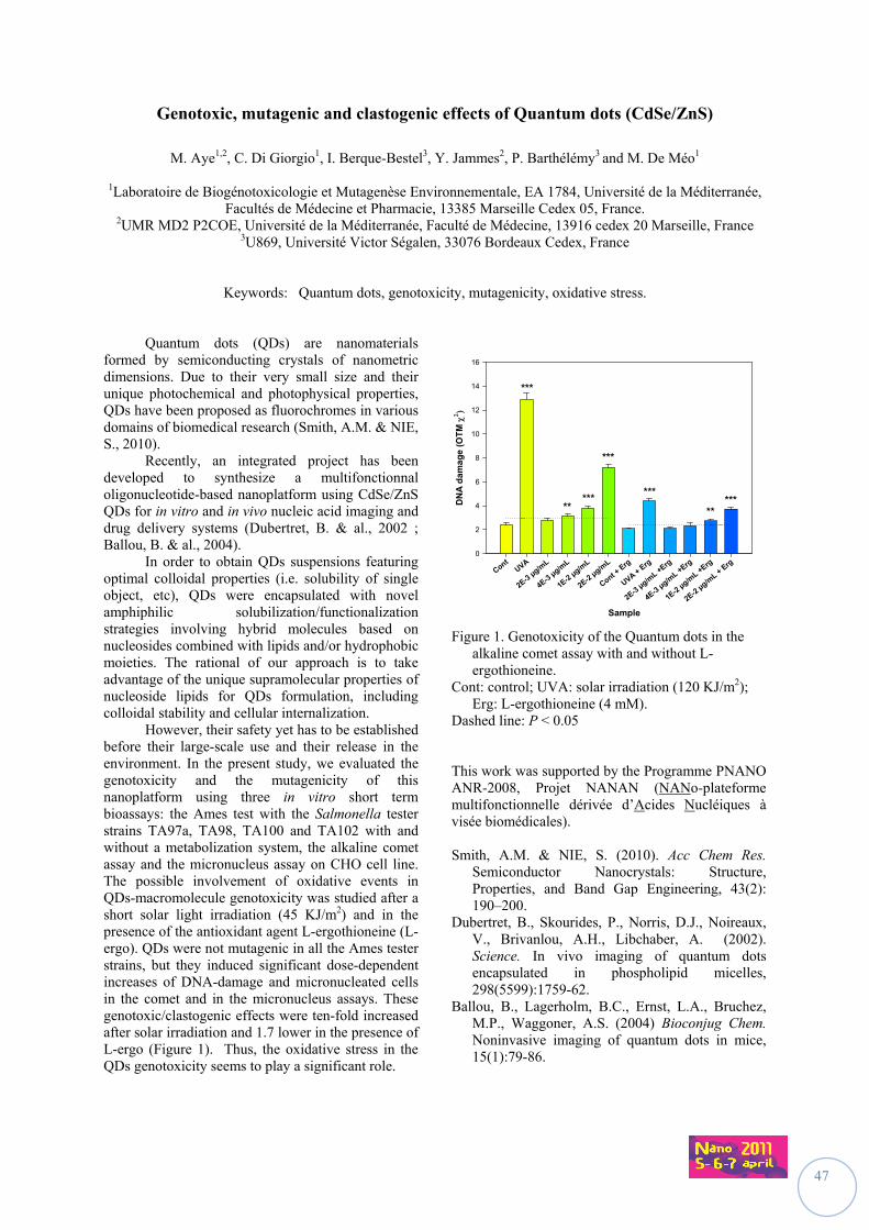
47
Genotoxic, mutagenic and clastogenic effects of Quantum dots (CdSe/ZnS)
M. Aye1,2, C. Di Giorgio1, I. Berque-Bestel3, Y. Jammes2, P. Barthélémy3 and M. De Méo1
1Laboratoire de Biogénotoxicologie et Mutagenèse Environnementale, EA 1784, Université de la Méditerranée,
Facultés de Médecine et Pharmacie, 13385 Marseille Cedex 05, France. 2UMR MD2 P2COE, Université de la Méditerranée, Faculté de Médecine, 13916 cedex 20 Marseille, France
3U869, Université Victor Ségalen, 33076 Bordeaux Cedex, France
Keywords: Quantum dots, genotoxicity, mutagenicity, oxidative stress.
Quantum dots (QDs) are nanomaterials formed by semiconducting crystals of nanometric dimensions. Due to their very small size and their unique photochemical and photophysical properties, QDs have been proposed as fluorochromes in various domains of biomedical research (Smith, A.M. & NIE, S., 2010). Recently, an integrated project has been developed to synthesize a multifonctionnal oligonucleotide-based nanoplatform using CdSe/ZnS QDs for in vitro and in vivo nucleic acid imaging and drug delivery systems (Dubertret, B. & al., 2002 ; Ballou, B. & al., 2004). In order to obtain QDs suspensions featuring optimal colloidal properties (i.e. solubility of single object, etc), QDs were encapsulated with novel amphiphilic solubilization/functionalization strategies involving hybrid molecules based on nucleosides combined with lipids and/or hydrophobic moieties. The rational of our approach is to take advantage of the unique supramolecular properties of nucleoside lipids for QDs formulation, including colloidal stability and cellular internalization. However, their safety yet has to be established before their large-scale use and their release in the environment. In the present study, we evaluated the genotoxicity and the mutagenicity of this nanoplatform using three in vitro short term bioassays: the Ames test with the Salmonella tester strains TA97a, TA98, TA100 and TA102 with and without a metabolization system, the alkaline comet assay and the micronucleus assay on CHO cell line. The possible involvement of oxidative events in QDs-macromolecule genotoxicity was studied after a short solar light irradiation (45 KJ/m2) and in the presence of the antioxidant agent L-ergothioneine (L-ergo). QDs were not mutagenic in all the Ames tester strains, but they induced significant dose-dependent increases of DNA-damage and micronucleated cells in the comet and in the micronucleus assays. These genotoxic/clastogenic effects were ten-fold increased after solar irradiation and 1.7 lower in the presence of L-ergo (Figure 1). Thus, the oxidative stress in the QDs genotoxicity seems to play a significant role.
Sample
ContUVA
2E-3 µg/m
L
4E-3 µg/m
L
1E-2 µg/m
L
2E-2 µg/m
L
Cont + E
rg
UVA + Erg
2E-3 µg/m
L +Erg
4E-3 µg/m
L +Erg
1E-2 µg/m
L +Erg
2E-2 µg/m
L + Erg
DN
A d
ama
ge
(O
TM
0
2
4
6
8
10
12
14
16
***
***
***
***
***
** **
Figure 1. Genotoxicity of the Quantum dots in the alkaline comet assay with and without L-ergothioneine.
Cont: control; UVA: solar irradiation (120 KJ/m2); Erg: L-ergothioneine (4 mM).
Dashed line: P < 0.05 This work was supported by the Programme PNANO ANR-2008, Projet NANAN (NANo-plateforme multifonctionnelle dérivée d’Acides Nucléiques à visée biomédicales). Smith, A.M. & NIE, S. (2010). Acc Chem Res.
Semiconductor Nanocrystals: Structure, Properties, and Band Gap Engineering, 43(2): 190–200.
Dubertret, B., Skourides, P., Norris, D.J., Noireaux, V., Brivanlou, A.H., Libchaber, A. (2002). Science. In vivo imaging of quantum dots encapsulated in phospholipid micelles, 298(5599):1759-62.
Ballou, B., Lagerholm, B.C., Ernst, L.A., Bruchez, M.P., Waggoner, A.S. (2004) Bioconjug Chem. Noninvasive imaging of quantum dots in mice, 15(1):79-86.
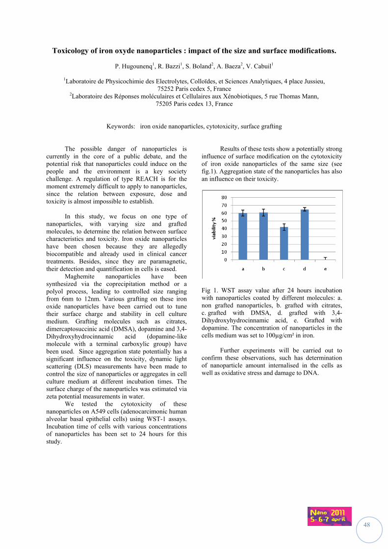
48
Toxicology of iron oxyde nanoparticles : impact of the size and surface modifications.
P. Hugounenq1, R. Bazzi1, S. Boland2, A. Baeza2, V. Cabuil1
1Laboratoire de Physicochimie des Electrolytes, Colloïdes, et Sciences Analytiques, 4 place Jussieu, 75252 Paris cedex 5, France
2Laboratoire des Réponses moléculaires et Cellulaires aux Xénobiotiques, 5 rue Thomas Mann, 75205 Paris cedex 13, France
Keywords: iron oxide nanoparticles, cytotoxicity, surface grafting The possible danger of nanoparticles is currently in the core of a public debate, and the potential risk that nanoparticles could induce on the people and the environment is a key society challenge. A regulation of type REACH is for the moment extremely difficult to apply to nanoparticles, since the relation between exposure, dose and toxicity is almost impossible to establish.
In this study, we focus on one type of nanoparticles, with varying size and grafted molecules, to determine the relation between surface characteristics and toxicity. Iron oxide nanoparticles have been chosen because they are allegedly biocompatible and already used in clinical cancer treatments. Besides, since they are paramagnetic, their detection and quantification in cells is eased.
Maghemite nanoparticles have been synthesized via the coprecipitation method or a polyol process, leading to controlled size ranging from 6nm to 12nm. Various grafting on these iron oxide nanoparticles have been carried out to tune their surface charge and stability in cell culture medium. Grafting molecules such as citrates, dimercaptosuccinic acid (DMSA), dopamine and 3,4-Dihydroxyhydrocinnamic acid (dopamine-like molecule with a terminal carboxylic group) have been used. Since aggregation state potentially has a significant influence on the toxicity, dynamic light scattering (DLS) measurements have been made to control the size of nanoparticles or aggregates in cell culture medium at different incubation times. The surface charge of the nanoparticles was estimated via zeta potential measurements in water.
We tested the cytotoxicity of these nanoparticles on A549 cells (adenocarcimonic human alveolar basal epithelial cells) using WST-1 assays. Incubation time of cells with various concentrations of nanoparticles has been set to 24 hours for this study.
Results of these tests show a potentially strong influence of surface modification on the cytotoxicity of iron oxide nanoparticles of the same size (see fig.1). Aggregation state of the nanoparticles has also an influence on their toxicity.
Fig 1. WST assay value after 24 hours incubation with nanoparticles coated by different molecules: a. non grafted nanoparticles, b. grafted with citrates, c. grafted with DMSA, d. grafted with 3,4-Dihydroxyhydrocinnamic acid, e. Grafted with dopamine. The concentration of nanoparticles in the cells medium was set to 100µg/cm² in iron.
Further experiments will be carried out to
confirm these observations, such has determination of nanoparticle amount internalised in the cells as well as oxidative stress and damage to DNA.
49
The comparison between cytotoxic effects induced by Multi-Wall Carbon Nanotubes (MWCNTs) on two different human cell lines
C. Fanizza1, C.L. Ursini2, S. Casciardi3, E. Paba3, A.M. Marcelloni 3, A.M. Fresegna2, A. Ciervo2, R. Maiello2,
F. Tombolini3, S. Iavicoli2, D. Cavallo2
1INAIL, DIPIA, ex-ISPESL, National Institute for Occupational Safety and Prevention, Via Urbana 167, 00184 Rome, Italy
2INAIL, Department of Occupational Medicine, ex-ISPESL, National Institute for Occupational Safety and Prevention, Via Fontana Candida 1, 00040 Monteporzio Catone, Rome, Italy
3INAIL, Department of Occupational Hygiene, ex-ISPESL, National Institute for Occupational Safety and Prevention, Via Fontana Candida 1, 00040 Monteporzio Catone, Rome, Italy
Keywords: cellular responses, cytotoxic effects, MWCNTs, SEM observations. Carbon nanotubes are widely used in multiple engineering disciplines. However, little is known about the toxicity or interaction of these particles with cells. In vitro studies demonstrated that CNT induced cytotoxic effects in different cell types (Ding et al., 2005; Monteiro-Riviere et al., 2005). The aim of this study is the comparison between cytotoxic effects induced by Multi-Walled Carbon Nanotubes (MWCNTs) on two different human cell line.
MWCNTs were characterized using energy filtered transmission electron microscopy (FEI TECNAI 12 G2 Twin). A commercially available human bronchial normal cells (BEAS-2B) and human lung epithelial cell line (A549) were exposed for 24h to 10, 40 and 100 μg/mL of MWCNTs. Cytotoxic effects were studied by viability analysis with MTT test (Sigma-Aldrich, USA), LDH release (Roche Diagnostics, Italy), and Scanning Electron Microscope (SEM) analysis evaluating modifications in the cell surface morphology.
The characterization of MWCNT structural parameters by TEM analysis revealed that they are “bamboo like”. MWCNTs outside diameter resulted ranging from a minimum value of 3 nm to a maximum value of 66 nm, with a mean value of (32 ± 2) nm. Lengths varied from a minimum value of 70 nm to a maximum value of 7.8m. A statistically significant decrease of viability evaluated by MTT assay was found at all concentrations in A549 cells and at the highest concentrations (40 and 100 μg/ml) in BEAS-2B cells. Moreover, we found membrane damage evaluated by LDH assay in both cell lines reaching similar percentages of cytotoxicity at 100 μg/ml that were 25% (A549) and 26% (BEAS-2B). SEM observations showed that the surface of BEAS-2B and A549 control cells is covered by regular microvilli. BEAS-2B cells showed the bleb development and a reduction in the number of microvilli after exposure to 10 μg/mL of MWCNTs. Cells treated with 40 μg/mL showed reduction in the number of microvilli, presence of microvilli squashed on cell membrane and bleb development. Microvilli structure modifications, a depletion in microvilli
density and, in lower amount, bleb development were present in cells exposed to 100 μg/mL of MWCNTs. Moreover, changes in the cell surface morphology such as a reduction in the number of microvilli and areas with a lower number of microvilli associated with areas with microvilli structure modifications were observed in A549 cells exposed to 10 μg/mL of MWCNTs. A depletion in microvilli density and the presence of holes in the plasma membrane were detected in cells treated with 40 and 100 μg/mL of MWCNTs. In addition, MWCNT aggregate uptake was observed at all concentrations in A549 cells and only in a few cells at the highest concentration in BEAS-2B cells.
Regarding cell surface modifications the reduction in the number of microvilli was observed in both cell lines at all concentrations tested. Cells affected by this change increase with increasing concentration, in fact the depletion of microvilli density is the only modification observed in A549 cells at the highest concentration and it is accompanied by microvilli structure modification in BEAS-2B. This study evaluates simultaneously cytotoxicity of MWCNT exposure on two human respiratory cell lines by complementary methods and demonstrates that the tested nanomaterials induce cytotoxicity in both cell lines. However, MTT assay and SEM analysis showed a slightly higher cytotoxic response of A549 cells probably due to carbon nanotubes internalization that occurs already at the lower concentrations. Ding L, Stilwell J, Zhang T, Elboudwarej O, Jiang H,
Selegue JP, Cooke PA, Gray JW, Chen FF.(2005). Nano Letters, 5, 2448-2464.
Monteiro-Riviere, N.A., Nemanich, R.J., Inman, A.O., Wang, Y.Y., and Riviere, J.E. (2005) Toxicol Lett. 155(3), 377-384.
50
Nanosized ZnO induces micronuclei by both aneugenic and clastogenic mechanisms in human bronchial epithelial cells in vitro
K. Siivola, H. Järventaus, S. Suhonen, K. Savolainen and H. Norppa
New Technologies and Risks, Work Environment Development, Finnish Institute of Occupational Health,
Topeliuksenkatu 41 aA, 00250, Helsinki, Finland
Keywords: Genotoxicity, ZnO ZnO nanoparticles, used e.g. in cosmetics, are genotoxic and strongly cytotoxic in various in vitro systems, but the underlying mechanisms are not well understood. ZnO is partly soluble, and its solubility is increased in acidic conditions and in the presence of chelators. Our previous results show that the geno-toxic effects of ZnO are not simply explained by Zn ions dissolved in the culture medium. Here, we applied the cytokinesis-block micro-nucleus (CBMN) assay and fluorescence in situ hybridization (FISH) to examine the mechanism of the in vitro genotoxicity of ZnO nanoparticles (zincite; primary particle size 30-35 nm) in human bronchial epithelial cells (BEAS 2B). The CBMN assay was performed with six doses of ZnO showing low cytotoxicity (<50%). Negative control cultures received only medium, and a known clastogen, mito-mycin C, was used as a positive control. Each experimental point was represented by replicate cultures. The cells were exposed to ZnO for 48 h, and cytochalasin B was added at 6 h to avoid its influence on the early uptake of particles by BEAS 2B cells. The frequency of micronuclei was scored from binucleated cells using a fluorescence microscope, and staining with acridine orange and 4',6-diamidino-2-phenylindole (DAPI). The contents of micronuclei were analyzed utilising centromeric and telomeric FISH. In each culture, micronuclei were categorized by the type and number of FISH signals.
ZnO caused a clear dose-dependent induction of MN in BEAS 2B cells. In the FISH analysis, we found a significant dose-dependent increase in the frequency of centromere- and telomere-positive micronuclei. A significant increase was also seen in telomere-positive micronuclei at the highest noncyto-toxic dose. Centromere- and telomere-positive micro-nuclei were interpreted to contain whole chromo-somes and, therefore, represented aneugenic events, whereas telomere-positive micronuclei were con-sidered chromosome fragments caused by a clasto-genic effect. Our findings suggest that ZnO induces micronuclei by both clastogenic and aneugenic mechanisms. This work was supported by the Academy of Finland (NANOHEALTH).
51
Predominant effect of finest size-segregated particles of the ambient air on the induction of mucus expression in airway epithelial cells
S. Val1, I. George1, L. Martinon2, H. Cachier3, A. Baeza-Squiban1
1Unit of Functional and Adaptative Biology, University Paris Diderot - Paris 7, Laboratory of Molecular and
Cellular Responses to Xenobiotics, EAC CNRS 4413, 5 rue Thomas Mann, 75205 Paris Cedex 13, France
2Laboratory of study of inhaled particles, Mairie de Paris, 11, rue George-Eastman, 75013 Paris, France 3Laboratory of climate and environment sciences, CEA-CNRS, Orme des merisiers, Centre de Saclay,
91190 Gif sur Yvette cedex, France
Keywords: particles, mucus, airway remodelling, oxidative stress Particulate aerosols and particularly the fine particles are suspected to aggravate various human pathologies such as cardiopulmonary diseases and among them obstructive pulmonary diseases involving chronic inflammation. Mechanistic studies have highlighted the critical role of the Epidermal Growth Factor Receptor pathway (EGFR) in such pathologies, by contributing to chronic inflammation, airway remodelling and overexpression of mucus proteins leading to the obstruction of the airway tract. Until now few studies have investigated the ability of particles to modulate mucus secretion. Previous studies of our group have shown that particles induce an overexpression of EGFR ligands (Rumelhard et al., 2007) suggesting that they could induce mucus release by an autocrine effect. In this context we studied the effect of size segregated Particulate Matter (PM) on human bronchial epithelial cells to determine (i) their ability to induce mucus expression in parallel to EGFR ligands and (ii) the mechanisms involved in this induction. Size-segregated particles (coarse, fine and ultrafine) sampled with Dekati impactors were tested on human bronchial epithelial cells (16HBE NCI-H292 cell lines and primary cultures NHBE) at non cytotoxic concentrations (max 10µg/cm²) for 24h and different biomarkers were evaluated. mRNA were studied by RT-qPCR and protein release in cell medium was assayed by the use of ELISA. We have shown that the finest PM (fine and ultrafine) were able to dose-dependently induce amphiregulin (AR, an EGFR ligand) expression and release after 24h exposure in 16HBE or NHBE cells. It was associated to the (i) overexpression of heme oxygenase-1 (HO-1), an antioxidant enzyme, revealing an unbalance in cell redox homeostasis and (ii) the increased expression and release of two cytokines, GM-CSF and IL-6, revealing the induction of a pro-inflammatory response. Such responses are size-dependent as the finest particles are always more reactive than coarse ones. These finest particles also exhibit a specific chemical composition characterized by a high amount of carbonaceous compounds. Among them are polyaromatic hydrocarbon (PAH) that become bioavailable as we observed an
increased expression of cytochrome P450 1A1 (CYP1A1) that is known to be specifically induced by PAH. In order to investigate the effect of particles on mucin expression, we studied the expression of MUC5AC, a mucin which is one of the most expressed mucins in the airway tract, and increased in patients with airway obstructive pathologies (Vestbo et al., 2002). The NCI-H292 cells were chosen for this purpose as it is a cell line that efficiently produces mucus in vitro. The finest PM were the most efficient to induce MUC5AC expression. The particle-induced MUC5AC expression is EGFR dependent as it decreased (i) in presence of an inhibitor of EGFR activation (AG1478) as well as (ii) in presence of a neutralizing antibody of EGFR. Moreover we hypothesize that MUC5AC expression could be relieved by an autocrine effect as using an inhibitor of TACE, a metalloprotease implicated in EGFR ligand shedding, PM-induced MUC5AC expression is reduced. In conclusion, particles induce mucus expression by an EGFR dependent pathway. This expression associated to the increased release of EGFR ligands suggests an autocrine effect. The finest PM characterized by a pronounced organic content are the most reactive towards bronchial epithelial cells. This work finally underlines that in addition to their well known pro-inflammatory effect, PM also promote mucus hypersecretion suggesting that in chronic exposures, they could contribute to bronchial remodelling. This work was supported by AFSSET (EST-2007-65), the French ANR (Agence Nationale pour la Recherche) and the PhD of Stéphanie VAL is supported by AFSSET (now ANSES) and ADEME. Rumelhard, M., Ramgolam, K., Augier, F., Dazy,
A.C., Blanchet, S., Marano, F., Baeza-Squiban, A. (2007). Toxicol. Letters 158; 155-164.
Vestbo, J. (2002). Novartis Found Symp. 248:3-12; discussion 12-9, 277-82.
52
Epidemiological surveillance of workers producing or handling engineered nanomaterials on the workplace: the French proposal
O. Boutou-Kempf1, J.L. Marchand1, A. Radauceanu2, O. Witschger2, E. Imbernon1
and the group “Health risks of nanotechnologies” 3
1French Institute for Public Health Surveillance (InVS), F-94415, St Maurice, France 2Institut National de Recherche et de Sécurité (INRS), F-54519, Vandoeuvre, France
3French Institute for Public Health Research (IReSP), F-75654, Paris, France
Keywords: epidemiological surveillance, occupational exposure, exposure registry, cohort
Alerted by the possible impact of nanomaterials exposure on human health, the French Ministries of Health and of Labour have given the French Institute for Public Health Surveillance responsibility for designing the protocol of an epidemiological surveillance system of workers likely to be exposed to engineered nanomaterials. The project was carried out in close connection with a multi-disciplinary working group held by the French Institute for Public Health Research.
Developing an epidemiological surveillance system of workers in the field of occupational exposure to nanomaterials needs to face numerous issues related to the wide range of nanomaterials, the registration and collaboration of companies and workers producing or handling nanomaterials, the identification of health outcomes that should be followed-up and the assessment of exposure.
A double epidemiological surveillance design is about to be proposed to the French ministries, consisting in a prospective cohort study and repeated cross-sectional studies.
The objectives of the prospective cohort study will be 1) to monitor long-term possible health effects of nanomaterials exposure and 2) to allow for further research studies.
The cohort study will first be restricted to workers likely to be exposed while producing or handling powder of nano-objects including aggregates and agglomerates. Priority will be given to carbon nanotubes, carbon black, titanium dioxide and amorphous silica.
A step by step protocol is suggested. The first step will be to set up an exposure registry, including identification of target companies and laboratories, obtaining participation of management and workers, collection of demographic and exposure data, and addressing issues of personal confidentiality (Schulte et al., 2009). The exposure registry is thought to be the inclusion step of the prospective cohort study for epidemiological studies and surveillance. In a first step, exposure will be assessed in a qualitative or semi-quantitative way. Data available in the registry will make it possible to design a mortality follow-up thus providing a first basic surveillance system.
In a second step, a non specific passive health follow-up could be implemented based on medical
records collected for administrative purposes by health insurance organisations and from hospitals. Medical data recorded on a regular basis by occupational health physicians could be collected as well. An active health follow-up could be based on annual self-questionnaire. Standardised clinical exams, specific diagnostic testing and biobank could be implemented later on.
A quantitative exposure assessment strategy is currently under development in cooperation with the Institut National de Recherche et de Sécurité. It will combine epidemiological tools such as job-exposure matrix and measurement campaign of the ambient aerosol on the workplace. In order to evaluate the feasibility of the cohort study, an exploratory study has been conducted among ten companies and research laboratories producing or incorporating powder of nano-objects. On each site, the number of workers likely to be exposed is small. Collaboration issues can be anticipated especially with companies incorporating nano-objects.
All nanomaterials produced or handled in France will be in the scope of the repeated cross-sectional studies. The goal will be to document circumstances of exposure and to raise hypotheses on possible health effects. The protocol is still under development.
In the emergent field of nanomaterials and despite the numerous uncertainties relating to health and exposure issues, it is time to anticipate epidemiological studies by beginning the registration of workers producing and handling nano-objects. In France, the general protocol of the health surveillance system is about to be submitted for approval to the Ministries of Health and of Labour. If accepted, the exposure registry could be implemented within the next three years while finalizing the protocol of the health follow-up and the quantitative exposure assessment. Schulte, P.A., Schubauer-Berigan, M.K.,
Mayweather, C., Geraci, C.L., Zumwalde, R., McKernan, J.L. (2009). J. Occup. Environ. Med. 323-335
53
Multifunctional nanoparticles based on silicon for cancer therapy
A. Bragaru, I. Kleps, M. Miu, M. Simion, M. Danila, F. Craciunoiu
Laboratory of Nanotechnology, National Institute for Research and Development in Microtechnologies IMT Bucharest, Romania
077190
Keywords: SPION, cancer therapy, The main idea of this work is to develop new nanostructurated systems based on silicon for vectorisation and controlled release of the biological active substances of therapeutic interest. These systems contains superparamagnetic microparticles of nanostructurated silicon which are carrying iron oxides (Super Paramagnetic Iron Oxide Nanosized Particles -SPION) and biological active substances, drugs- dacarbazine, integrated in an organic polymeric membrane- dextran, in order to facilitate the controlled delivery process direct to specific sites (normal or pathologic) from the human body or animals. Also, this is used as carrier agent and also, to prevent drug diffusion during the vectorisation process In the biological medium, due to the iron oxides deposited in the porous structure of the nanostructured support, the Si microparticles will suffer controlled biodegradation, which leads to a monitorised delivery of included therapeutic substances. The microparticles of nanostructurated silicon were prepared by chemical etching of the p type silicon [100] oriented using an AMMT etching system (Germany). It is a multisteps process (150 cycles) by applying 0.554 A for 10 sec and 2.825 A for 4 sec., using concentrated HF as etching solution.
Figure 1. SEM images of the porous silicon nanoparticles
Iron oxide nanoparticles were prepared by precipitation of the Fe+3/ Fe+2 in the 2:1 molar ratio, using NH4OH 1 M. The experiment was made using inert atmosphere and continuous stirring. In order to obtain the proposed system, an amount of nanostructurated silicon particles and superparamagnetic iron oxide nanoparticles were put together with ethanol in a Berzelius glass, under intensive stirring for 1 hour
The X-ray Diffraction analyses shown in figure 2, using SmartLab system (Rigaku Corporation, Japan) reveals the presence of the both phase components, Fe3O4 (magnetite) and porous silicon nanoparticles.
Figure 2. X-Ray diffraction analyses of the SPION system
The next step was to ingrain the therapeutic agent - dacarbazine. Finally, the SPION system with this agent was covered by a polymeric matrix, dextran, in order to facilitate the controlled delivery process. The characterization methods used were SEM, XRD and FTIR. This obtained system will be injected close to the damaged organ of the animal, using SPION like vectorisation technique, avoiding the standard surgical procedures or chemotherapy, which are traumatic. Acknowledgement This work was supported by the National Research Programme- PN II- NANOSIC Project, contract no. 71-120/2007. Jin Pan, Qingwei Yang, (2007), Anal. Bioanal.
Chem. 388, 279- 286. J. K. Vasir, M. K. Reddy, V. D. Labhasetwar, (2005)
Current Nanoscience, vol. 1, no. 1, pp. 47–64.
20 40 60 80
14000
21000
28000
Fe3O
4(400)
Fe3O
4(311)
Fe3O
4(220)
Fe3O
4(511)
Fe3O
4(440)
Fe 3
O4(1
11)
Si(311)
Si(220)
I [c
ps]
2 [o]
Magnetite SiO2/Magnetite
Glass holder
SiO2, low
Crystobalite,
Si(111)
54
Nanoparticle – cell interaction
M. Garvas1, S. Pajk2, P. Umek1, J. Štrancar1
1Department of Solid State Physics, Jožef Štefan Institute, Jamova 39, SI-1000 Ljubljana, Slovenia 2Faculty of pharmacy, University of Ljubljana, Aškerčeva 7, SI-1000 Ljubljana, Slovenia
Keywords: nanoparticle-cell interaction, fluorescence microspectroscopy, cytotoxicity Different nanoparticles’ characteristics, such as size, surface charge, and functionalization, play crucial role in particle-cell interaction. Despite nanoparticles’ tendency to aggregate in solution, many of them have actually been identified inside the cell, proving they are able to penetrate the cell membrane. The question is how particle pass the membrane or attach to it, do they interact with nucleus and other cell organelles. If so, what kind of damage is caused, what are toxicity key factors and mechanisms? If they enter the cell interior they can act like nanocatalists thus changing significantly physicochemical processes. While exploring the uptaking of the nanomaterials, we have to enable detection of individual nanoparticles taking into account their size which is below optical resolution of the microscopy. This problem is addressed within new method, denoted as “fluorescence microspectroscopy”. Method enables us to detect objects smaller than optical resolution in live cells via detecting the whole emission spectral response in every voxel of the image. Since two different fluorophores are employed to label cells and nanomaterial at the same time, the spectral-enabled detection localizes both types of fluorophores and at the same time identifies them via spectral characteristics, even if they are located in the same volume element and even if their spectral maximum position differs less than the width of normal broad-band fluorescence emission filters.
We succeed to overcome the fuctionalization problem of titanate nanoparticles with fluorescence molecule. We covalently label them, characterize them and now we can finally observe their interactions inside live cell in real time. Cellular response will be observed, dependent of concentration and time (cytotoxicity). This work was supported by the Slovenian Research Agency.
55
Potential impact of carbon nanotubes on health and the environment
E. Flahaut1, 2, E. Meunier1, 3, B. Pipy3, L. De Gabory4, L. Bordenave4, P. Puech5, D. Crouzier6, J.C. Debouzy6, F. Bourdiol1, 2, 7, F. Mouchet2, 7 and L. Gauthier2, 7
1CIRIMAT, UMR 5084, University of Toulouse, 31062 Toulouse Cedex 9, France
2Nautile, Laboratoire commun Arkema France – ECOLAB – CIRIMAT, France 3UMR MD3 EA2405, Université Paul Sabatier – Toulouse 3, 31432 Toulouse cedex 4
4INSERM U577, Université Victor Segalen – Bordeaux 2, 33076 Bordeaux cedex 5CEMES, 31055 Toulouse Cedex 4
6CRSSA, 24, avenue des maquis du Grésivaudan, BP 87-38 702 La Tronche Cedex, France 7EcoLab UMR 5245, Campus INP-ENSAT, 31326 Auzeville-Tolosane
Keywords: Double-walled carbon nanotubes, toxicity, ecotoxicity, characterisation. Carbon nanotubes (CNT), with an annual world production reaching several hundreds of tons, represent a special category of nanomaterials with exceptional characteristics and numerous potential applications. Although the toxicity and the environmental impact of CNT have now both been investigated by different groups (although ecotoxicity never focused much attention until very recently), there is yet a controversy about the results and still no answer to the simple question: "are CNT toxic?"
The fact is that the large range of kinds of
CNT and methods to produce them and then (in most cases) to process them, make any comparison almost impossible. Indeed, most CNT prepared on a large scale are synthesised by Catalytic Chemical Vapour Desposition (CCVD) but there are many variations for this single generic name (carbon source, nature of the catalyst, synthesis conditions, etc.). There are also many concerns about the purity of CNT samples because depending on the way they are prepared the content of residual catalyst can vary quite a lot (from a few hundreds of ppm to tens of weight percents in some cases), thus potentially playing a very important role in terms of potential toxicity of the CNT samples. It must also be noted that CNT samples can also be contaminated by other forms of carbon (amorphous carbon deposit, or even graphitic carbon in case of arc-discharged-produced CNT), and not only by metals or catalyst supports (typical residual supports are alumina or silica).
The results presented here have been obtained with the same batch of CCVD-produced DWNT (Flahaut et al., 2003) and concern the investigation of both their potential impact on human health (Flahaut et al., (2006), Salvador Morales et al., Crouzier et al.) and the environment (in vivo models) (Mouchet et al., Kwok et al.). They lead to the conclusion that all the experimental parameters (dealing both with CNT preparation and processing, biological models and toxicity assays used) play a very important role and can easily explain the large
differences between the results published by the different research groups worldwide. The main conclusions of our studies will be presented. They indicate that acid-treated (oxidised) DWNT exhibit a higher toxicity than their non-oxidised counterparts, probably due to their higher dispersibility in the aqueous environment (which can be enhanced by the use of non-toxic surfactants (Datsyuk et al., 2009) Part of this work was supported by the French ANR under grant SEST-2006. Flahaut, E., Basca, R., Peigney, A., Laurent, Ch.
(2003) Chem. Commun., 12, 1442-1443. Flahaut, E., Durrieu, M.C., Remy-Zholgadri, M.,
Bareille, R., Baquey, Ch. (2006) Carbon, 44, (6), 1093-1099.
Salvador-Morales, C., Flahaut, E., Sim, E., Sloan, J., Green, M.L.H., Sim, R.B. (2006) Molec. Immun., 43, (3), 193-201.
Salvador-Morales, C., Townsend, P., Flahaut, E., Vénien, C., Vlandas, A., Green, M.L.H., Sim, R.B. (2007) Carbon, 45, 607-617.
Crouzier, D., Follot, S., Gentilhomme, E., Flahaut, E., Arnaud, R., Dabouis, V., Castellarin, C., Debouzy, J.C. (2010) Toxicology, 272, (1-3), 39-45.
Mouchet, F., Landois, P., Sarremejean, E., Bernard, G., Puech, P., Pinelli, E., Flahaut, E., Gauthier, L. (2008) Aquatic Toxicology, 87, (2), 127-137.
Kwok, K. W., Leung, K. M., Flahaut, E., Cheng, J., Cheng S. H. (2010) Nanomedicine, 5, (6), 951-961.
Mouchet, F., Landois, P., Puech, P., Pinelli, E., Flahaut, E., Gauthier L. (2010) Nanomedicine, 5, (6), 963-974.
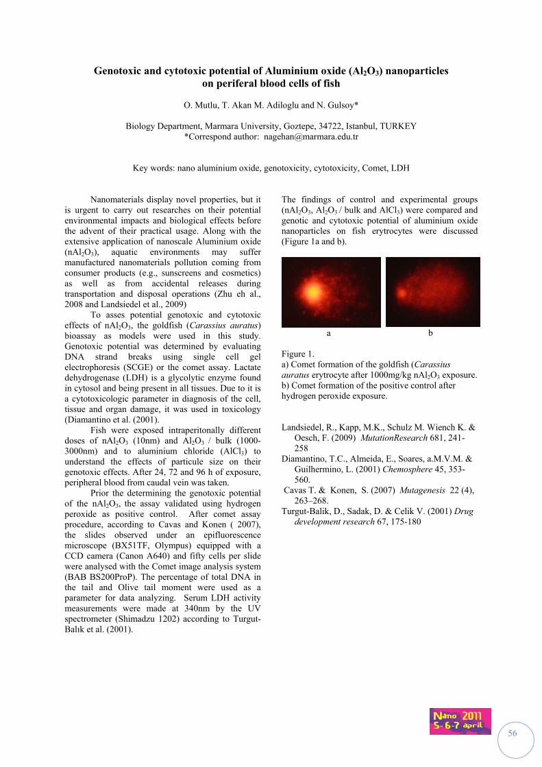
56
Genotoxic and cytotoxic potential of Aluminium oxide (Al2O3) nanoparticles on periferal blood cells of fish
O. Mutlu, T. Akan M. Adiloglu and N. Gulsoy*
Biology Department, Marmara University, Goztepe, 34722, Istanbul, TURKEY
*Correspond author: [email protected]
Key words: nano aluminium oxide, genotoxicity, cytotoxicity, Comet, LDH
Nanomaterials display novel properties, but it is urgent to carry out researches on their potential environmental impacts and biological effects before the advent of their practical usage. Along with the extensive application of nanoscale Aluminium oxide (nAl2O3), aquatic environments may suffer manufactured nanomaterials pollution coming from consumer products (e.g., sunscreens and cosmetics) as well as from accidental releases during transportation and disposal operations (Zhu eh al., 2008 and Landsiedel et al., 2009) To asses potential genotoxic and cytotoxic effects of nAl2O3, the goldfish (Carassius auratus) bioassay as models were used in this study. Genotoxic potential was determined by evaluating DNA strand breaks using single cell gel electrophoresis (SCGE) or the comet assay. Lactate dehydrogenase (LDH) is a glycolytic enzyme found in cytosol and being present in all tissues. Due to it is a cytotoxicologic parameter in diagnosis of the cell, tissue and organ damage, it was used in toxicology (Diamantino et al. (2001). Fish were exposed intraperitonally different doses of nAl2O3 (10nm) and Al2O3 / bulk (1000-3000nm) and to aluminium chloride (AlCl3) to understand the effects of particule size on their genotoxic effects. After 24, 72 and 96 h of exposure, peripheral blood from caudal vein was taken. Prior the determining the genotoxic potential of the nAl2O3, the assay validated using hydrogen peroxide as positive control. After comet assay procedure, according to Cavas and Konen ( 2007), the slides observed under an epifluorescence microscope (BX51TF, Olympus) equipped with a CCD camera (Canon A640) and fifty cells per slide were analysed with the Comet image analysis system (BAB BS200ProP). The percentage of total DNA in the tail and Olive tail moment were used as a parameter for data analyzing. Serum LDH activity measurements were made at 340nm by the UV spectrometer (Shimadzu 1202) according to Turgut-Balık et al. (2001).
The findings of control and experimental groups (nAl2O3, Al2O3 / bulk and AlCl3) were compared and genotic and cytotoxic potential of aluminium oxide nanoparticles on fish erytrocytes were discussed (Figure 1a and b).
a b
Figure 1. a) Comet formation of the goldfish (Carassius auratus erytrocyte after 1000mg/kg nAl2O3 exposure. b) Comet formation of the positive control after hydrogen peroxide exposure. Landsiedel, R., Kapp, M.K., Schulz M. Wiench K. &
Oesch, F. (2009) MutationResearch 681, 241-258
Diamantino, T.C., Almeida, E., Soares, a.M.V.M. & Guilhermino, L. (2001) Chemosphere 45, 353-560.
Cavas T. & Konen, S. (2007) Mutagenesis 22 (4), 263–268.
Turgut-Balik, D., Sadak, D. & Celik V. (2001) Drug development research 67, 175-180
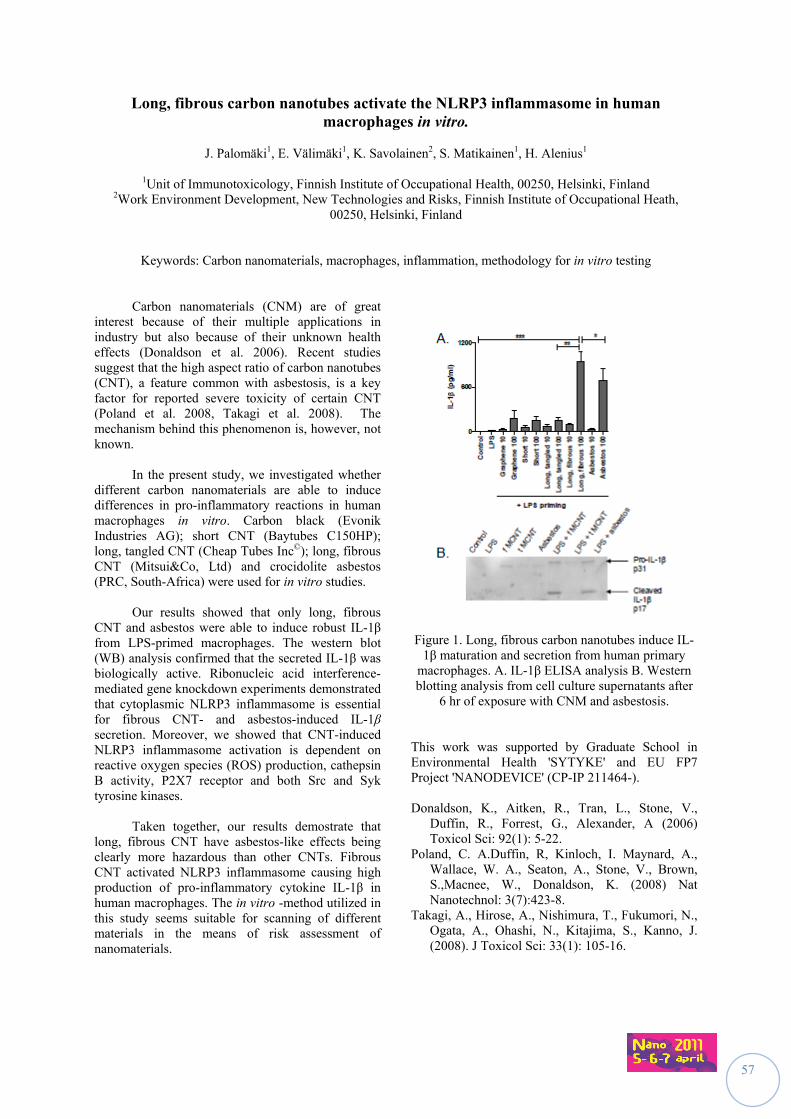
57
Long, fibrous carbon nanotubes activate the NLRP3 inflammasome in human macrophages in vitro.
J. Palomäki1, E. Välimäki1, K. Savolainen2, S. Matikainen1, H. Alenius1
1Unit of Immunotoxicology, Finnish Institute of Occupational Health, 00250, Helsinki, Finland
2Work Environment Development, New Technologies and Risks, Finnish Institute of Occupational Heath, 00250, Helsinki, Finland
Keywords: Carbon nanomaterials, macrophages, inflammation, methodology for in vitro testing
Carbon nanomaterials (CNM) are of great
interest because of their multiple applications in industry but also because of their unknown health effects (Donaldson et al. 2006). Recent studies suggest that the high aspect ratio of carbon nanotubes (CNT), a feature common with asbestosis, is a key factor for reported severe toxicity of certain CNT (Poland et al. 2008, Takagi et al. 2008). The mechanism behind this phenomenon is, however, not known.
In the present study, we investigated whether different carbon nanomaterials are able to induce differences in pro-inflammatory reactions in human macrophages in vitro. Carbon black (Evonik Industries AG); short CNT (Baytubes C150HP); long, tangled CNT (Cheap Tubes Inc©); long, fibrous CNT (Mitsui&Co, Ltd) and crocidolite asbestos (PRC, South-Africa) were used for in vitro studies.
Our results showed that only long, fibrous CNT and asbestos were able to induce robust IL-1β from LPS-primed macrophages. The western blot (WB) analysis confirmed that the secreted IL-1β was biologically active. Ribonucleic acid interference-mediated gene knockdown experiments demonstrated that cytoplasmic NLRP3 inflammasome is essential for fibrous CNT- and asbestos-induced IL-1β secretion. Moreover, we showed that CNT-induced NLRP3 inflammasome activation is dependent on reactive oxygen species (ROS) production, cathepsin B activity, P2X7 receptor and both Src and Syk tyrosine kinases.
Taken together, our results demostrate that
long, fibrous CNT have asbestos-like effects being clearly more hazardous than other CNTs. Fibrous CNT activated NLRP3 inflammasome causing high production of pro-inflammatory cytokine IL-1β in human macrophages. The in vitro -method utilized in this study seems suitable for scanning of different materials in the means of risk assessment of nanomaterials.
Figure 1. Long, fibrous carbon nanotubes induce IL-
1β maturation and secretion from human primary macrophages. A. IL-1β ELISA analysis B. Western blotting analysis from cell culture supernatants after
6 hr of exposure with CNM and asbestosis. This work was supported by Graduate School in Environmental Health 'SYTYKE' and EU FP7 Project 'NANODEVICE' (CP-IP 211464-). Donaldson, K., Aitken, R., Tran, L., Stone, V.,
Duffin, R., Forrest, G., Alexander, A (2006) Toxicol Sci: 92(1): 5-22.
Poland, C. A.Duffin, R, Kinloch, I. Maynard, A., Wallace, W. A., Seaton, A., Stone, V., Brown, S.,Macnee, W., Donaldson, K. (2008) Nat Nanotechnol: 3(7):423-8.
Takagi, A., Hirose, A., Nishimura, T., Fukumori, N., Ogata, A., Ohashi, N., Kitajima, S., Kanno, J. (2008). J Toxicol Sci: 33(1): 105-16.
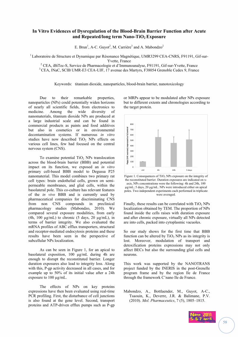
58
In Vitro Evidences of Dysregulation of the Blood-Brain Barrier Function after Acute and Repeated/long term Nano-TiO2 Exposure
E. Brun1, A-C. Guyot2, M. Carrière3 and A. Mabondzo2
1 Laboratoire de Structure et Dynamique par Résonance Magnétique, UMR3299 CEA-CNRS, F91191, Gif-sur-
Yvette, France 2 CEA, iBiTec-S, Service de Pharmacologie et d’Immunoanalyse, F91191, Gif-sur-Yvette, France
3 CEA, INaC, SCIB UMR-E3 CEA-UJF, 17 avenue des Martyrs, F38054 Grenoble Cedex 9, France
Keywords: titanium dioxide, nanoparticles, blood-brain barrier, nanotoxicology
Due to their remarkable properties, nanoparticles (NPs) could potentially widen horizons of nearly all scientific fields, from electronics to medicine. Among the wide diversity of nanomaterials, titanium dioxide NPs are produced at a large industrial scale and can be found in commercial products as paints and food additives but also in cosmetics or in environmental decontamination systems. If numerous in vitro studies have now described TiO2 NPs effects on various cell lines, few had focused on the central nervous system (CNS). To examine potential TiO2 NPs translocation across the blood-brain barrier (BBB) and potential impact on its function, we exposed an in vitro primary cell-based BBB model to Degussa P25 nanomaterial. This model combines two primary rat cell types: brain endothelial cells, grown on semi-permeable membranes, and glial cells, within the basolateral pole. This co-culture has relevant features of the in vivo BBB and is currently used by pharmaceutical companies for discriminating CNS from non CNS compounds in preclinical pharmacology studies (Mabondzo, 2010). We compared several exposure modalities, from early (4h, 100 µg/mL) to chronic (5 days, 20 µg/mL), in terms of barrier integrity. We also evaluated the mRNA profiles of ABC efflux transporters, structural and receptor-mediated endocytosis proteins and these results have been seen in the perspective of subcellular NPs localization.
As can be seen in Figure 1, for an apical to basolateral exposition, 100 µg/mL during 4h are enough to disrupt the reconstituted barrier. Longer duration exposures also lead to integrity loss. Along with this, P-gp activity decreased in all cases, and for example up to 50% of its initial value after a 24h exposure to 100 µg/mL.
The effects of NPs on key proteins
expressions have then been evaluated using real-time PCR profiling. First, the disturbance of cell junctions is also found at the gene level. Second, transport proteins and ATP-driven efflux pumps such as P-gp
or MRPs appear to be modulated after NPs exposure but to different extents and chronologies according to the target protein.
Figure 1. Consequences of TiO2 NPs exposure on the integrity of the reconstituted barrier. Duration exposures are indicated on x-axis, NPs concentrations were the following: 4h and 24h, 100
µg/mL; 5 days, 20 µg/mL. NPs were introduced either on apical poles. Two independent experiments each performed in triplicate
were averaged.
Finally, these results can be correlated with TiO2 NPs localization obtained by TEM. The proportion of NPs found inside the cells raises with duration exposure and after chronic exposure, virtually all NPs detected are into cells, packed into cytoplasmic vacuoles. So our study shows for the first time that BBB function can be altered by TiO2 NPs as its integrity is lost. Moreover, modulation of transport and detoxification proteins expressions may not only affect BECs but also the surrounding glial cells and neurons. This work was supported by the NANOTRANS project funded by the INERIS in the post-Grenelle program frame and by the region Ile de France through the framework C’nano Ile de France. Mabondzo, A., Bottlaender, M., Guyot, A-C.,
Tsaouin, K., Deverre, J.R. & Balimane, P.V. (2010). Mol. Pharmaceutics, 7 (5), 1805–1815.
0
100
200
300
400
500
600
700
800
ctrl 4h 24h 5 days
Papp (14C‐sucrose) expressed as %/control
59
SESSION II
IINNSSTTRRUUMMEENNTTAATTIIOONN,, CCHHAARRAACCTTEERRIIZZAATTIIOONN
&& EEXXPPOOSSUURREE EEVVAALLUUAATTIIOONN
Chairs:
Keld A. Jensen (NRCWE, Danemark) François Gensdarmes (IRSN, France)
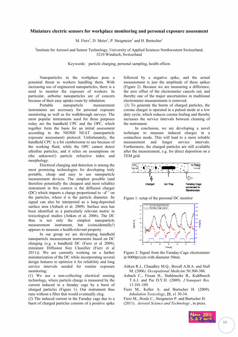
60
Miniature electric sensors for workplace monitoring and personal exposure assessment
M. Fierz1, D. Meier1, P. Steigmeier1 and H. Burtscher1
1Institute for Aerosol and Sensor Technology, University of Applied Sciences Northwestern Switzerland, 5210 Windisch, Switzerland
Keywords: particle charging, personal sampling, health effects
Nanoparticles in the workplace pose a potential threat to workers handling them. With increasing use of engineered nanoparticles, there is a need to monitor the exposure of workers. In particular, airborne nanoparticles are of concern because of their easy uptake route by inhalation. Portable nanoparticle measurement instruments are necessary for personal exposure monitoring as well as for walkthrough surveys. The most popular instruments used for these purposes today are the handheld CPC and the OPC, which together form the basis for an initial assessment according to the NIOSH NEAT (nanoparticle exposure assessment) protocol. Unfortunately, the handheld CPC is a bit cumbersome to use because of the working fluid, while the OPC cannot detect ultrafine particles, and it relies on assumptions on (the unknown!) particle refractive index and morphology. Electrical charging and detection is among the most promising technologies for developing truly portable, cheap and easy to use nanoparticle measurement devices. The simplest possible (and therefore potentially the cheapest and most reliable) instrument in this context is the diffusion charger (DC) which imparts a charge proportional to ~d1.1 on the particles, where d is the particle diameter. Its signal can also be interpreted as a lung-deposited surface area (Asbach et al. 2009). Surface area has been identified as a particularly relevant metric in toxicological studies (Aitken et al. 2006). The DC thus is not only the simplest nanoparticle measurement instrument, but (coincidentally!) appears to measure a health-relevant property. In our group we are developing handheld nanoparticle measurement instruments based on DC charging (e.g. a handheld DC (Fierz et al 2009), miniature Diffusion Size Classifier (Fierz et al 2011)). We are currently working on a further miniaturization of the DC while incorporating several design features to optimize it for reliability and long service intervals needed for routine exposure monitoring: (1) We use a non-collecting electrical sensing technology, where particle charge is measured by the current induced in a faraday cage by a burst of charged particles (Figure 1). Our instrument thus runs without a filter that would eventually clog. (2) The induced current in the Faraday cage due to a burst of charged particles consists of a positive spike
followed by a negative spike, and the actual measurement is just the amplitude of these spikes (Figure 2). Because we are measuring a difference, the zero offset of the electrometer cancels out, and thereby one of the major uncertainties in traditional electrometer measurements is removed. (3) To generate the bursts of charged particles, the corona charger is operated in a pulsed mode at a low duty cycle, which reduces corona fouling and thereby increases the service intervals between cleaning of the instrument. In conclusion, we are developing a novel technique to measure induced charges in a contactless mode. This will lead to a more reliable measurement and longer service intervals. Furthermore, the charged particles are still available after the measurement, e.g. for direct deposition on a TEM grid.
Figure 1: setup of the personal DC monitor
Figure 2: Signal from the Faraday-Cage electrometer at 6000pt/ccm with diameter 50nm. Aitken R.J., Chaudhry M.Q., Boxall A.B.A. and Hull
M. (2006). Occupational Medicine 56:300-306. Asbach C., Fissan H., Stahlmecke B., Kuhlbusch
T.A.J. and Pui D.Y.H. (2009). J.Nanopart Res. 11:101-109.
Fierz M., Keller A. and Burtscher H. (2009). Inhalation Toxicology, 21, s1 30-34.
Fierz M., Houle C., Steigmeier P. and Burtscher H. (2011). Aerosol Science and Technology , in press.
61
A substance-specific technique for the detection of nanoparticles in workplace air
N. Neubauer1, F. Weis1, M. Seipenbusch1 and G. Kasper1
1Institute for Mechanical Process Engineering and Mechanics, Karlsruhe Institute of Technology (KIT), Straße am Forum 8, 76131 Karlsruhe, Germany
Keywords: specific detection, engineered nanoparticles, catalysis, workplace air
Introduction
Engineered nanoparticles (ENP) in the workplace air are normally mixed with background particles. However, available measurement devices are not capable of distinguishing between a target ENP and a background aerosol (Kuhlbusch et al., 2009; Murashov et al., 2009). Furthermore, the chemical composition of ENP can only be analyzed by off-line techniques. Therefore, new, substance-specific on-line analytical methods are required to identify target ENP from background particles. Catalysis offers a good potential for the material-specific detection of catalytically active nanoparticles as well as for the discrimination of trace amounts of airborne ENP against an inactive background aerosol. We therefore investigated the applicability of catalysis for the specific measurement of ENP, both in their airborne state and after deposition from the aerosol onto a substrate. The experiments were conducted with catalytically active nanoparticles generated by spark discharge. These nanoaerosols were exposed for a defined time to gaseous educts. The reaction products were detected by infrared spectroscopy. Based on the experimental results detection limits for several ENP were calculated.
Detection techniques Catalysis on airborne nanoparticles
The method of catalysis on gasborne nanoparticles offers the possibility to determine the catalytic activity of nanoparticles on very short time scales in the order of seconds to minutes. It is therefore a true on-line technique for measurements of the catalytic activity of ENP. In addition, the method requires only small catalyst mass concentrations in the order of ng/cm3. Another advantage is the avoidance of contaminations like in off-line methods.
Catalysis on deposited nanoparticles
By sampling the particles on a filter prior to the catalytic reaction, it is possible to accumulate small amounts of catalyst material in the order of micrograms. This approach is therefore better suited for ENP aerosols at low concentrations, for slow catalytic reactions and/or less active nanoparticles. The catalytic activity is nevertheless determined on-line, as educt gases are added directly to the filter. Thereby filter handling and potential contaminations of the sample by exposure to ambient air are avoided.
Results Direct measurements on airborne particles
showed that this on-line approach is suitable for the detection of very active ENP aerosols in high concentrations, such as platinum or nickel. The detection of platinum is even possible with a room temperature reaction, where as the other catalysts require temperatures between about 200ºC and 450ºC. The filter based technique shows a good potential for the detection of less concentrated or less active ENP aerosols like iron oxide (Fe2O3) based on their catalytic activity. In addition, the filter method can be considered a true real-time method for the detection of palladium and a near-real-time technique for catalysts with similar activities as platinum or nickel. Calculated detection limits indicate that concentrations in the range of 1 ng/cm3 can be detected in a few seconds for palladium, in a few minutes for platinum or nickel, and in about 30 min for iron oxide. Conclusion
A new, substance-specific technique for the detection of airborne nanoparticles via their catalytic activity was presented. In our experiments we could demonstrate that very small amounts of catalytically active nanoparticles like palladium, platinum, nickel or iron oxide (Fe2O3) can be very rapidly detected on the basis of their catalytic activity. Acknowledgement
The research leading to these results has received funding from the European Community's Seventh Framework Programme (FP7/2007-2013) under grant agreement n° 211464-2 (Nanodevice). Literature Kuhlbusch, T.A.J., Fissan, H. & Asbach, C. (2009)
Nanomaterials: Risks and Benefits, Springer, 233-243
Murashov, V., Engel, S., Savolainen, K., Fullam, B., Lee, M. & Kearns, P. (2009) J. Nanopart. Res. 11, 1587-1591
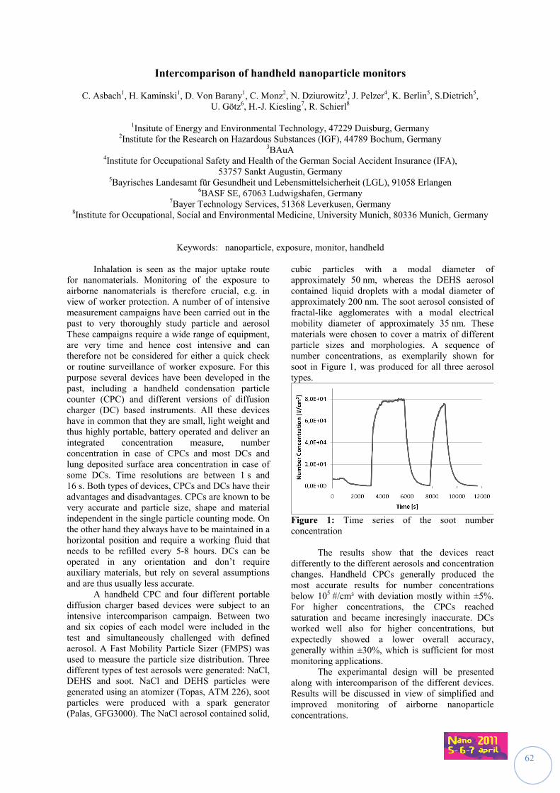
62
Intercomparison of handheld nanoparticle monitors
C. Asbach1, H. Kaminski1, D. Von Barany1, C. Monz2, N. Dziurowitz3, J. Pelzer4, K. Berlin5, S.Dietrich5, U. Götz6, H.-J. Kiesling7, R. Schierl8
1Insitute of Energy and Environmental Technology, 47229 Duisburg, Germany
2Institute for the Research on Hazardous Substances (IGF), 44789 Bochum, Germany 3BAuA
4Institute for Occupational Safety and Health of the German Social Accident Insurance (IFA), 53757 Sankt Augustin, Germany
5Bayrisches Landesamt für Gesundheit und Lebensmittelsicherheit (LGL), 91058 Erlangen 6BASF SE, 67063 Ludwigshafen, Germany
7Bayer Technology Services, 51368 Leverkusen, Germany 8Institute for Occupational, Social and Environmental Medicine, University Munich, 80336 Munich, Germany
Keywords: nanoparticle, exposure, monitor, handheld Inhalation is seen as the major uptake route for nanomaterials. Monitoring of the exposure to airborne nanomaterials is therefore crucial, e.g. in view of worker protection. A number of of intensive measurement campaigns have been carried out in the past to very thoroughly study particle and aerosol These campaigns require a wide range of equipment, are very time and hence cost intensive and can therefore not be considered for either a quick check or routine surveillance of worker exposure. For this purpose several devices have been developed in the past, including a handheld condensation particle counter (CPC) and different versions of diffusion charger (DC) based instruments. All these devices have in common that they are small, light weight and thus highly portable, battery operated and deliver an integrated concentration measure, number concentration in case of CPCs and most DCs and lung deposited surface area concentration in case of some DCs. Time resolutions are between 1 s and 16 s. Both types of devices, CPCs and DCs have their advantages and disadvantages. CPCs are known to be very accurate and particle size, shape and material independent in the single particle counting mode. On the other hand they always have to be maintained in a horizontal position and require a working fluid that needs to be refilled every 5-8 hours. DCs can be operated in any orientation and don’t require auxiliary materials, but rely on several assumptions and are thus usually less accurate. A handheld CPC and four different portable diffusion charger based devices were subject to an intensive intercomparison campaign. Between two and six copies of each model were included in the test and simultaneously challenged with defined aerosol. A Fast Mobility Particle Sizer (FMPS) was used to measure the particle size distribution. Three different types of test aerosols were generated: NaCl, DEHS and soot. NaCl and DEHS particles were generated using an atomizer (Topas, ATM 226), soot particles were produced with a spark generator (Palas, GFG3000). The NaCl aerosol contained solid,
cubic particles with a modal diameter of approximately 50 nm, whereas the DEHS aerosol contained liquid droplets with a modal diameter of approximately 200 nm. The soot aerosol consisted of fractal-like agglomerates with a modal electrical mobility diameter of approximately 35 nm. These materials were chosen to cover a matrix of different particle sizes and morphologies. A sequence of number concentrations, as exemplarily shown for soot in Figure 1, was produced for all three aerosol types.
Figure 1: Time series of the soot number concentration The results show that the devices react differently to the different aerosols and concentration changes. Handheld CPCs generally produced the most accurate results for number concentrations below 105 #/cm³ with deviation mostly within ±5%. For higher concentrations, the CPCs reached saturation and became incresingly inaccurate. DCs worked well also for higher concentrations, but expectedly showed a lower overall accuracy, generally within ±30%, which is sufficient for most monitoring applications. The experimantal design will be presented along with intercomparison of the different devices. Results will be discussed in view of simplified and improved monitoring of airborne nanoparticle concentrations.
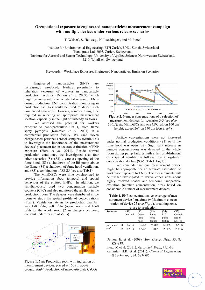
63
Occupational exposure to engineered nanoparticles: measurement campaign with multiple devices under various release scenarios
T. Walser1, S. Hellweg1, N. Luechinger2, and M. Fierz3
1Institute for Environmental Engineering, ETH Zurich, 8093, Zurich, Switzerland
2Nanograde Ltd, 8093, Zurich, Switzerland 3Institute for Aerosol and Sensor Technology, University of Applied Sciences Northwestern Switzerland,
5210, Windisch, Switzerland
Keywords: Workplace Exposure, Engineered Nanoparticles, Emission Scenarios
Engineered nanoparticles (ENP) are increasingly produced, leading potentially to inhalation exposure of workers in nanoparticle production facilities (Demou et al. 2009), which might be increased in an accidental release of ENPs during production. ENP concentration monitoring in production facilities could be used to detect such unintended emissions. However, some care might be required in selecting an appropriate measurement location, especially in the light of unsteady air flows. We assessed the potential for workers exposure to nano-particulate CaCO3 from flame spray pyrolysis (Kammler et al. 2001) in a commercial production facility. We used eleven charge-based personal aerosol samplers (MiniDiSC) to investigate the importance of the measurement devices’ placement for an accurate estimation of ENP exposure (Fierz et al. 2011). Beside normal production conditions, we investigated also four other scenarios (S): (S2) a careless opening of the fume hood, (S3) a shutdown of the lift pump above the flame, (S4) a shutdown of fume hood ventilation, and (S5) a combination of S3-S5 (see also Tab.1). The MiniDiSCs were time synchronized to provide information about temporal and spatial behaviour of the emitted ENPs. In addition, we simultaneously used two condensation particle counters (CPC) and also monitored the air flow in the production room. The devices were distributed in the room to study the spatial profile of concentrations (Fig.1). Ventilation rate in the production chamber was 150 m3/hr, 860 m3/hr (open hood), and 1660 m3/h for the whole room (2 air changes per hour, constant underpressure of -5 Pa).
Particle concentrations were not increased
under normal production conditions (S1) or if the fume hood was open (S2). Significant increase in number concentrations was detected in the whole room during pump failures with a fast establishment of a spatial equilibrium followed by a log-linear concentration decline (S3-5, Tab.1, Fig.2).
We conclude that one measurement device might be appropriate for an accurate estimation of workplace exposure to ENPs. The measurements will be further investigated to derive conclusions about highly resolved spatial and temporal nanoparticle evolution (number concentration, size) based on considerable number of measurement devices.
Table 1. ENP concentrations. a: Average of mea-surement devices’ maxima; b: Maximum concen-tration of device 25 (see Fig. 1), breathing zone,
close to production. Scenario
(S1) Normal
(S2) Open fume hood
(S3) Fume hood failure
(S4) Lift pump failure
(S5) Combi-nation (2,3,4)
particles/ cm3
a 3.2E3 3.3E3 9.6E4 5.0E5 2.8E6 b 3.5E3 4.5E3 1.8E5 2.8E5 2.4E6
Demou, E. et al. (2009). Ann. Occup. Hyg., 53, 8,
829-838. Fierz, M et al. (2011). Aeros. Sci. Tech., 45,1-10. Kammler, H.K. et al. (2011). Chemical Engineering
& Technology, 24, 583-596.
Nano-CaCO3
production
ENP collectionon filter
Lift pump
Figure 2. Number concentrations of a selection of measurement devices for scenarios 3-5 (see also
Tab.1): six MiniDiSCs and one CPC, all on 160 cm height, except 26* on 140 cm (Fig.1, left).
100
1'000
10'000
100'000
1'000'000
10'000'000
42000 44000 46000 48000 50000 520000 2000 4000 6000 8000 10000 seconds
Above detection limit of the CPC
particles/cm3
Background concentration
S3
S4S5
Figure 1. Left: Production room with indication of measurement devices, placed at 160 cm above ground. Right: Production of nanoparticulate CaCO3
64
Exposure to carbon nano-objects in research and industry
C. Möhlmann1, J. Pelzer1, M. Berges1, D. Bard2, D. Mark2, A. Thorpe2, D. Wake2, E. Jankowska3,
B. van Duuren-Stuurman4, D. Brouwer4
1Institut für Arbeitsschutz - IFA, Alte Heerstraße 111, 53757 Sankt Augustin, Germany 2Health and Safety Laboratory - HSL, Harpur Hill, Buxton, SK17 9JN, UK
3Central Institute for Labour Protection - National Research Institute – CIOP-PIB, Warsaw, Poland 4TNO Quality of Life, Food & Chemical Risk Analysis, P.O. Box 360, 3700 AJ Zeist, The Netherlands
Keywords: carbon nanotubes, carbon black, nanoparticles, exposure
The NANOSH project focused on
occupational exposure to nanoparticles and their health effects. The overall goal of the project was to investigate exposure and health effects of selected nano-sized particles relevant to the occupational environment. Reliable and simple methods and strategies were to be developed for discriminating between manufactured nanoparticles and non-intentionally produced nano-sized particles present in the ambient atmosphere.
During the NANOSH project 4 partners performed measurements at workplaces in nanotechnology in 19 companies resulting in 150 single measurement sets. Workplaces varied from research settings, nanomaterial production sites and downstream use. Potential exposures from the production and/or handling of carbon nanotubes (CNTs) were measured in 26 measurement sets and 109 measurements were made (italic numbers in table). In the field of production 12 sets were achieved, in downstream use 14, both including mainly research activities. The table lists the number of measurements according to the material used.
Substance Number of measurement sets (N)
Number of measurem
ents (N)
High Aspect Ratio Nanoparticles
Carbon nanotubes 12 51
MWCNT 9 35
SWCNT 1 5
Carbon fibres 8 18 Carbon fibres and carbon nanotubes
4 12
Carbon black
Carbon black 3 14 Carbon black and various powders
1 5
A measurement strategy was applied to
distinguish between non-engineered and engineered nanoparticles (Brouwer 2009). Two approaches had been used: sequential measurement periods of similar
length for nano-activities and non nano-activities or the use of a closed cabin to avoid influence of external sources. Several direct-reading instruments were used to determine the number concentrations, particle size distributions, surface area concentrations and additional mass concentrations.
The results were collected in a database and the differences between nano-activity and non nano-activity periods assessed to derive a significant change in concentration. Additional aerosol sampling using TEM grids was performed to get an additional assessment criterion for the occurrence of nanoparticles.
Considering the specific case of CNTs and taking all these criteria into consideration, exposure was found to be “likely” for 4 of the 18 measurement sets obtained. For 7 measurement sets, exposure was found to be “not likely”, and for the remaining 7 measurement sets exposure was found to be “possible/not excluded”. The ratios for the number and surface area concentrations between nano- and non nano-activity ranged between 0.65 and 30, the highest values coming from an extrusion process. A change in particle size could only be observed in 4 measurement sets: dry sawing of composites with CNTs, extrusion and cutting of plastics containing multiwalled carbon nanotubes (MWCNTs), production of MWCNTs, and weighing and pouring MWCNTs. When handling agglomerates of CNTs a change in the size range of 1 to 10 µm was detected. Processes with composite materials are also able to emit non-engineered ultrafine particles and superpose the concentration measurements. For good exposure estimation it is very important to have aerosol samples analysed. In 5 cases the TEM analysis revealed the occurrence of CNTs. These findings gave the clearest evidence for exposure to CNTs.
Brouwer D, van Duuren-Stuurman B, Berges M,
Jankowska E, Bard D and Mark D (2009) From workplace air measurement results toward estimates of exposure? Development of a strategy to assess exposure to manufactured nano-objects. J Nanopart. Res., 11, 1867-1881
The NANOSH project was supported by EU-contract NMP4-CT-2006-032777.
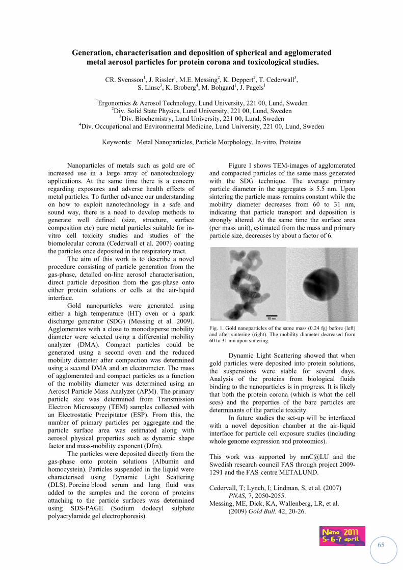
65
Generation, characterisation and deposition of spherical and agglomerated metal aerosol particles for protein corona and toxicological studies.
CR. Svensson1, J. Rissler1, M.E. Messing2, K. Deppert2, T. Cederwall3,
S. Linse3, K. Broberg4, M. Bohgard1, J. Pagels1
1Ergonomics & Aerosol Technology, Lund University, 221 00, Lund, Sweden
2Div. Solid State Physics, Lund University, 221 00, Lund, Sweden
3Div. Biochemistry, Lund University, 221 00, Lund, Sweden 4Div. Occupational and Environmental Medicine, Lund University, 221 00, Lund, Sweden
Keywords: Metal Nanoparticles, Particle Morphology, In-vitro, Proteins
Nanoparticles of metals such as gold are of increased use in a large array of nanotechnology applications. At the same time there is a concern regarding exposures and adverse health effects of metal particles. To further advance our understanding on how to exploit nanotechnology in a safe and sound way, there is a need to develop methods to generate well defined (size, structure, surface composition etc) pure metal particles suitable for in-vitro cell toxicity studies and studies of the biomolecular corona (Cederwall et al. 2007) coating the particles once deposited in the respiratory tract. The aim of this work is to describe a novel procedure consisting of particle generation from the gas-phase, detailed on-line aerosol characterisation, direct particle deposition from the gas-phase onto either protein solutions or cells at the air-liquid interface. Gold nanoparticles were generated using either a high temperature (HT) oven or a spark discharge generator (SDG) (Messing et al. 2009). Agglomerates with a close to monodisperse mobility diameter were selected using a differential mobility analyzer (DMA). Compact particles could be generated using a second oven and the reduced mobility diameter after compaction was determined using a second DMA and an electrometer. The mass of agglomerated and compact particles as a function of the mobility diameter was determined using an Aerosol Particle Mass Analyzer (APM). The primary particle size was determined from Transmission Electron Microscopy (TEM) samples collected with an Electrostatic Precipitator (ESP). From this, the number of primary particles per aggregate and the particle surface area was estimated along with aerosol physical properties such as dynamic shape factor and mass-mobility exponent (Dfm). The particles were deposited directly from the gas-phase onto protein solutions (Albumin and homocystein). Particles suspended in the liquid were characterised using Dynamic Light Scattering (DLS). Porcine blood serum and lung fluid was added to the samples and the corona of proteins attaching to the particle surfaces was determined using SDS-PAGE (Sodium dodecyl sulphate polyacrylamide gel electrophoresis).
Figure 1 shows TEM-images of agglomerated and compacted particles of the same mass generated with the SDG technique. The average primary particle diameter in the aggregates is 5.5 nm. Upon sintering the particle mass remains constant while the mobility diameter decreases from 60 to 31 nm, indicating that particle transport and deposition is strongly altered. At the same time the surface area (per mass unit), estimated from the mass and primary particle size, decreases by about a factor of 6. Fig. 1. Gold nanoparticles of the same mass (0.24 fg) before (left) and after sintering (right). The mobility diameter decreased from 60 to 31 nm upon sintering.
Dynamic Light Scattering showed that when gold particles were deposited into protein solutions, the suspensions were stable for several days. Analysis of the proteins from biological fluids binding to the nanoparticles is in progress. It is likely that both the protein corona (which is what the cell sees) and the properties of the bare particles are determinants of the particle toxicity. In future studies the set-up will be interfaced with a novel deposition chamber at the air-liquid interface for particle cell exposure studies (including whole genome expression and proteomics). This work was supported by nmC@LU and the Swedish research council FAS through project 2009-1291 and the FAS-centre METALUND. Cedervall, T; Lynch, I; Lindman, S, et al. (2007)
PNAS, 7, 2050-2055. Messing, ME, Dick, KA, Wallenberg, LR, et al.
(2009) Gold Bull. 42, 20-26.
66
NANODEVICE Novel Concepts, Methods, and Technologies for the Production of Portable, Easy-to-use
Devices for the Measurement and Analysis of Airborne Engineered Nanoparticles in Workplace Air
M. Keller1, S. Sirviö2, K. Savolainen3
1Fraunhofer Institute for Manufacturing Engineering and Automation IPA, 70569 Stuttgart, Germany
2Finnish Institute of Occupational Health, Helsinki, Finland Email: [email protected]
Keywords: NANODEVICE, portable devices, ENP-measurement, nanoparticles, workplace safety
NANODEVICE is a research project funded by the European Commission in the context of the 7th Framework Program. The duration is 48 months starting 1st of April 2009.
Due to their unique properties, engineered nanoparticles (ENP) are now used for a myriad of novel applications with great economic and technological importance. However, some of these properties, especially their surface reactivity, have raised health concerns, which have prompted scientists, regulators, and industry to seek consensus protocols for the safe production and use of the different forms of ENP.
There is currently a shortage of field-worthy, cost-effective ways - especially in real time - for reliable assessment of exposure levels to ENP in workplace air. In addition to the problems with the size distribution, a major uncertainty in the safety assessment of airborne ENP arises from the lack of knowledge of their physical and chemical properties, and the levels of exposure. A special challenge of ENP monitoring is to separate ubiquitous background nanoparticles from different sources from the ENP.
NANODEVICE will provide new information on the physico-chemical properties of engineered nanoparticles (ENP) and information about their toxicology. Also a novel measuring device will be developed to assess the exposure to ENP´s from workplace air. The purpose of the project is also to promote the safe use of ENP through guidance, standards and education, implementing of safety objectives in ENP production and handling, and promotion of safety related collaborations through an international nanosafety forum.
The main project goal is to develop innovative concepts and reliable methods for characterizing ENP in workplace air with novel, portable and easy-to-use devices suitable for workplaces. Additional research objectives are:
1) Identification of relevant physico-chemical properties and metrics of airborne ENP; establishment of reference materials.
2) Exploring the association between physico-chemical and toxicological properties of ENP
3) Analyzing industrial processes as a source of ENP in workplace air;
4) Developing methods for calibration and testing of the novel devices in real and simulated exposure situations.
5) Dissemination of the research results to promote the safe use of ENP through guidance, standards and education, implementing of safety objectives in ENP production and handling, and promotion of safety related collaborations through an international nanosafety forum.
The research leading to these results has received funding from the European Commission under grant agreement FP7-211464-2 (NANODEVICE)
67
Monitoring method for nanofibers: Personal sampler and corresponding reading device
M. Keller1; N. Neubauer2, M. Seipenbusch2
1 Fraunhofer Institute for Manufacturing Engineering and Automation IPA, 70569 Stuttgart, Germany
2 Karlsruhe Institut für Technologie KIT, 76131 Karlsruhe, Germany
Keywords: nanofibres, workplace safety, personal sampler At present, CNT and other nanofibre materials in air can only be detected by deposition on a substrate and the use of off-line imaging analysis such as SEM. The majority of techniques suitable for the quasi-real-time ENP measurement such as ELPI, SMPS, and CPC can not distinguish between nanoparticles in general and airborne nanofibres in particular. In common workplace settings the considerable background of fine and ultrafine particles thus poses a challenge for these instruments. Furthermore, a true portability of the present devices is not given due to their size and power consumption. However, there is an urgent need due to the expected toxicity of nanofibres to control workplace environments with a robust and mobile device to ensure the safety of the working personnel as soon as possible. To meet these challenges, a suitable personal sampler together with a corresponding reading device is under development in the NANODEVICE-project, funded by the European Commission under grant agreement FP7-211464-2. The measurement principle, a pre-prototype personal sampler together with first results will be shown. The research leading to these results has received funding from the European Commission under grant agreement FP7-211464-2 (NANODEVICE).
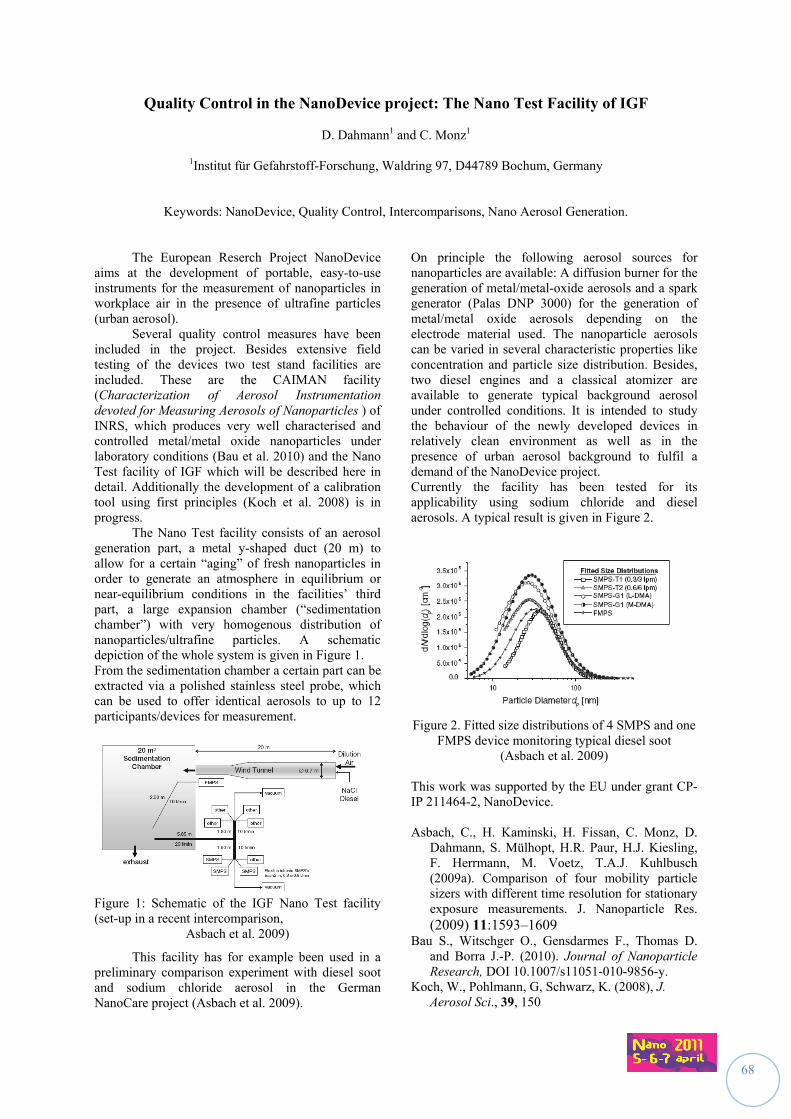
68
Quality Control in the NanoDevice project: The Nano Test Facility of IGF
D. Dahmann1 and C. Monz1
1Institut für Gefahrstoff-Forschung, Waldring 97, D44789 Bochum, Germany
Keywords: NanoDevice, Quality Control, Intercomparisons, Nano Aerosol Generation.
The European Reserch Project NanoDevice aims at the development of portable, easy-to-use instruments for the measurement of nanoparticles in workplace air in the presence of ultrafine particles (urban aerosol). Several quality control measures have been included in the project. Besides extensive field testing of the devices two test stand facilities are included. These are the CAIMAN facility (Characterization of Aerosol Instrumentation devoted for Measuring Aerosols of Nanoparticles ) of INRS, which produces very well characterised and controlled metal/metal oxide nanoparticles under laboratory conditions (Bau et al. 2010) and the Nano Test facility of IGF which will be described here in detail. Additionally the development of a calibration tool using first principles (Koch et al. 2008) is in progress. The Nano Test facility consists of an aerosol generation part, a metal y-shaped duct (20 m) to allow for a certain “aging” of fresh nanoparticles in order to generate an atmosphere in equilibrium or near-equilibrium conditions in the facilities’ third part, a large expansion chamber (“sedimentation chamber”) with very homogenous distribution of nanoparticles/ultrafine particles. A schematic depiction of the whole system is given in Figure 1. From the sedimentation chamber a certain part can be extracted via a polished stainless steel probe, which can be used to offer identical aerosols to up to 12 participants/devices for measurement.
Figure 1: Schematic of the IGF Nano Test facility (set-up in a recent intercomparison,
Asbach et al. 2009)
This facility has for example been used in a preliminary comparison experiment with diesel soot and sodium chloride aerosol in the German NanoCare project (Asbach et al. 2009).
On principle the following aerosol sources for nanoparticles are available: A diffusion burner for the generation of metal/metal-oxide aerosols and a spark generator (Palas DNP 3000) for the generation of metal/metal oxide aerosols depending on the electrode material used. The nanoparticle aerosols can be varied in several characteristic properties like concentration and particle size distribution. Besides, two diesel engines and a classical atomizer are available to generate typical background aerosol under controlled conditions. It is intended to study the behaviour of the newly developed devices in relatively clean environment as well as in the presence of urban aerosol background to fulfil a demand of the NanoDevice project. Currently the facility has been tested for its applicability using sodium chloride and diesel aerosols. A typical result is given in Figure 2.
Figure 2. Fitted size distributions of 4 SMPS and one
FMPS device monitoring typical diesel soot (Asbach et al. 2009)
This work was supported by the EU under grant CP-IP 211464-2, NanoDevice. Asbach, C., H. Kaminski, H. Fissan, C. Monz, D.
Dahmann, S. Mülhopt, H.R. Paur, H.J. Kiesling, F. Herrmann, M. Voetz, T.A.J. Kuhlbusch (2009a). Comparison of four mobility particle sizers with different time resolution for stationary exposure measurements. J. Nanoparticle Res. (2009) 11:1593–1609
Bau S., Witschger O., Gensdarmes F., Thomas D. and Borra J.-P. (2010). Journal of Nanoparticle Research, DOI 10.1007/s11051-010-9856-y.
Koch, W., Pohlmann, G, Schwarz, K. (2008), J. Aerosol Sci., 39, 150
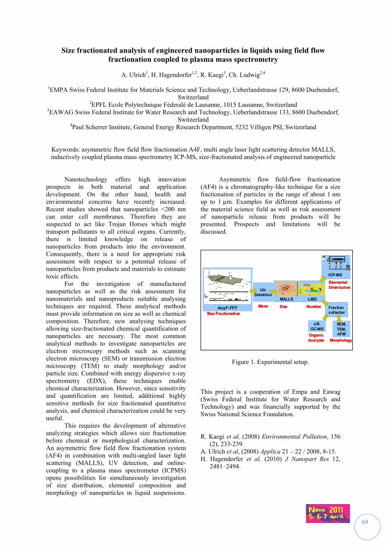
69
Size fractionated analysis of engineered nanoparticles in liquids using field flow fractionation coupled to plasma mass spectrometry
A. Ulrich1, H. Hagendorfer1,2, R. Kaegi3, Ch. Ludwig2,4
1EMPA Swiss Federal Institute for Materials Science and Technology, Ueberlandstrasse 129, 8600 Duebendorf,
Switzerland 2EPFL Ecole Polytechnique Féderalé de Lausanne, 1015 Lausanne, Switzerland
3EAWAG Swiss Federal Institute for Water Research and Technology, Ueberlandstrasse 133, 8600 Duebendorf, Switzerland
4Paul Scherrer Institute, General Energy Research Department, 5232 Villigen PSI, Switzerland
Keywords: asymmetric flow field flow fractionation A4F, multi angle laser light scattering detector MALLS, inductively coupled plasma mass spectrometry ICP-MS, size-fractionated analysis of engineered nanoparticle
Nanotechnology offers high innovation prospects in both material and application development. On the other hand, health and environmental concerns have recently increased. Recent studies showed that nanoparticles <200 nm can enter cell membranes. Therefore they are suspected to act like Trojan Horses which might transport pollutants to all critical organs. Currently, there is limited knowledge on release of nanoparticles from products into the environment. Consequently, there is a need for appropriate risk assessment with respect to a potential release of nanoparticles from products and materials to estimate toxic effects. For the investigation of manufactured nanoparticles as well as the risk assessment for nanomaterials and nanoproducts suitable analysing techniques are required. These analytical methods must provide information on size as well as chemical composition. Therefore, new analysing techniques allowing size-fractionated chemical quantification of nanoparticles are necessary. The most common analytical methods to investigate nanoparticles are electron microscopy methods such as scanning electron microscopy (SEM) or transmission electron microscopy (TEM) to study morphology and/or particle size. Combined with energy dispersive x-ray spectrometry (EDX), these techniques enable chemical characterization. However, since sensitivity and quantification are limited, additional highly sensitive methods for size fractionated quantitative analysis, and chemical characterization could be very useful. This requires the development of alternative analyzing strategies which allows size fractionation before chemical or morphological characterization. An asymmetric flow field flow fractionation system (AF4) in combination with multi-angled laser light scattering (MALLS), UV detection, and online-coupling to a plasma mass spectrometer (ICPMS) opens possibilities for simultaneously investigation of size distribution, elemental composition and morphology of nanoparticles in liquid suspensions.
Asymmetric flow field-flow fractionation (AF4) is a chromatography-like technique for a size fractionation of particles in the range of about 1 nm up to 1 µm. Examples for different applications of the material science field as well as risk assessment of nanoparticle release from products will be presented. Prospects and limitations will be discussed.
Figure 1. Experimental setup.
This project is a cooperation of Empa and Eawag (Swiss Federal Institute for Water Research and Technology) and was financially supported by the Swiss National Science Foundation. R. Kaegi et al, (2008) Environmental Pollution, 156
(2), 233-239. A. Ulrich et al, (2008) Applica 21 – 22 / 2008, 8-15. H. Hagendorfer et al, (2010) J Nanopart Res 12,
2481–2494.
SEM, TEM, AFM
Fractioncollector
MALLS
AsyF-FFFSize Fractionation
UV-Detektion
Mass Size
LIBD
Number
ICP-MS
V V V
ElementalDistribution
Morphology
z.B. GC-MS
OrganicAnalysis
SEM, TEM, AFM
Fractioncollector
MALLS
AsyF-FFFSize Fractionation
UV-Detektion
Mass Size
LIBD
Number
ICP-MS
V V VV V V
ElementalDistribution
Morphology
z.B. GC-MS
OrganicAnalysis
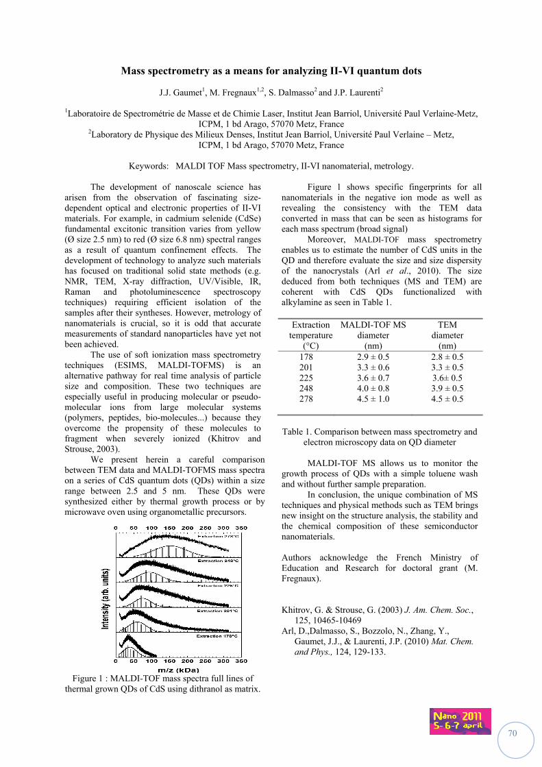
70
Mass spectrometry as a means for analyzing II-VI quantum dots
J.J. Gaumet1, M. Fregnaux1,2, S. Dalmasso2 and J.P. Laurenti2
1Laboratoire de Spectrométrie de Masse et de Chimie Laser, Institut Jean Barriol, Université Paul Verlaine-Metz, ICPM, 1 bd Arago, 57070 Metz, France
2Laboratory de Physique des Milieux Denses, Institut Jean Barriol, Université Paul Verlaine – Metz, ICPM, 1 bd Arago, 57070 Metz, France
Keywords: MALDI TOF Mass spectrometry, II-VI nanomaterial, metrology.
The development of nanoscale science has arisen from the observation of fascinating size- dependent optical and electronic properties of II-VI materials. For example, in cadmium selenide (CdSe) fundamental excitonic transition varies from yellow (Ø size 2.5 nm) to red (Ø size 6.8 nm) spectral ranges as a result of quantum confinement effects.
The development of technology to analyze such materials has focused on traditional solid state methods (e.g. NMR, TEM, X-ray diffraction, UV/Visible, IR, Raman and photoluminescence spectroscopy techniques) requiring efficient isolation of the samples after their syntheses. However, metrology of nanomaterials is crucial, so it is odd that accurate measurements of standard nanoparticles have yet not been achieved. The use of soft ionization mass spectrometry techniques (ESIMS, MALDI-TOFMS) is an alternative pathway for real time analysis of particle size and composition. These two techniques are especially useful in producing molecular or pseudo-molecular ions from large molecular systems (polymers, peptides, bio-molecules...) because they overcome the propensity of these molecules to fragment when severely ionized (Khitrov and Strouse, 2003). We present herein a careful comparison between TEM data and MALDI-TOFMS mass spectra on a series of CdS quantum dots (QDs) within a size range between 2.5 and 5 nm. These QDs were synthesized either by thermal growth process or by microwave oven using organometallic precursors.
Figure 1 : MALDI-TOF mass spectra full lines of
thermal grown QDs of CdS using dithranol as matrix.
Figure 1 shows specific fingerprints for all nanomaterials in the negative ion mode as well as revealing the consistency with the TEM data converted in mass that can be seen as histograms for each mass spectrum (broad signal)
Moreover, MALDI-TOF mass spectrometry enables us to estimate the number of CdS units in the QD and therefore evaluate the size and size dispersity of the nanocrystals (Arl et al., 2010). The size deduced from both techniques (MS and TEM) are coherent with CdS QDs functionalized with alkylamine as seen in Table 1.
Table 1. Comparison between mass spectrometry and
electron microscopy data on QD diameter MALDI-TOF MS allows us to monitor the growth process of QDs with a simple toluene wash and without further sample preparation. In conclusion, the unique combination of MS techniques and physical methods such as TEM brings new insight on the structure analysis, the stability and the chemical composition of these semiconductor nanomaterials. Authors acknowledge the French Ministry of Education and Research for doctoral grant (M. Fregnaux). Khitrov, G. & Strouse, G. (2003) J. Am. Chem. Soc.,
125, 10465-10469 Arl, D.,Dalmasso, S., Bozzolo, N., Zhang, Y.,
Gaumet, J.J., & Laurenti, J.P. (2010) Mat. Chem. and Phys., 124, 129-133.
Extraction temperature
(°C)
MALDI-TOF MS diameter
(nm)
TEM diameter
(nm) 178 2.9 ± 0.5 2.8 ± 0.5 201 225 248 278
3.3 ± 0.6 3.6 ± 0.7 4.0 ± 0.8 4.5 ± 1.0
3.3 ± 0.5 3.6± 0.5 3.9 ± 0.5 4.5 ± 0.5
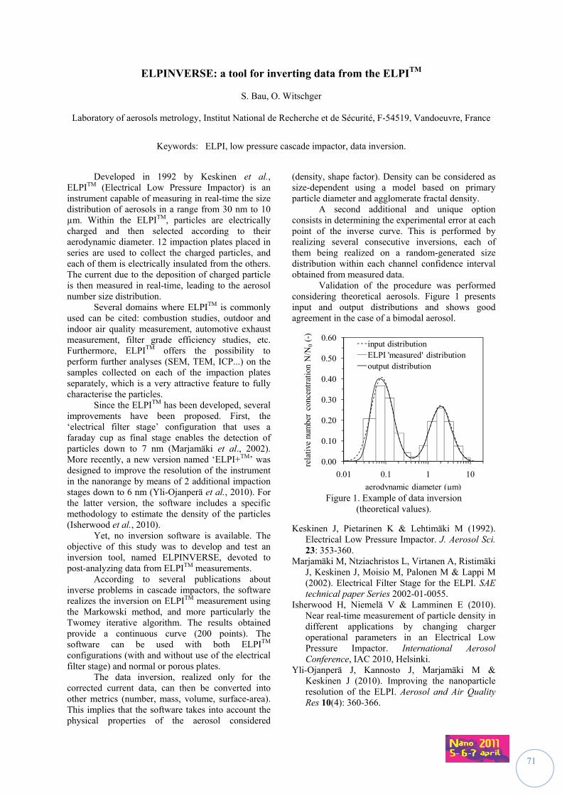
71
ELPINVERSE: a tool for inverting data from the ELPITM
S. Bau, O. Witschger
Laboratory of aerosols metrology, Institut National de Recherche et de Sécurité, F-54519, Vandoeuvre, France
Keywords: ELPI, low pressure cascade impactor, data inversion.
Developed in 1992 by Keskinen et al., ELPITM (Electrical Low Pressure Impactor) is an instrument capable of measuring in real-time the size distribution of aerosols in a range from 30 nm to 10 µm. Within the ELPITM, particles are electrically charged and then selected according to their aerodynamic diameter. 12 impaction plates placed in series are used to collect the charged particles, and each of them is electrically insulated from the others. The current due to the deposition of charged particle is then measured in real-time, leading to the aerosol number size distribution. Several domains where ELPITM is commonly used can be cited: combustion studies, outdoor and indoor air quality measurement, automotive exhaust measurement, filter grade efficiency studies, etc. Furthermore, ELPITM offers the possibility to perform further analyses (SEM, TEM, ICP...) on the samples collected on each of the impaction plates separately, which is a very attractive feature to fully characterise the particles. Since the ELPITM has been developed, several improvements have been proposed. First, the ‘electrical filter stage’ configuration that uses a faraday cup as final stage enables the detection of particles down to 7 nm (Marjamäki et al., 2002). More recently, a new version named ‘ELPI+TM’ was designed to improve the resolution of the instrument in the nanorange by means of 2 additional impaction stages down to 6 nm (Yli-Ojanperä et al., 2010). For the latter version, the software includes a specific methodology to estimate the density of the particles (Isherwood et al., 2010). Yet, no inversion software is available. The objective of this study was to develop and test an inversion tool, named ELPINVERSE, devoted to post-analyzing data from ELPITM measurements. According to several publications about inverse problems in cascade impactors, the software realizes the inversion on ELPITM measurement using the Markowski method, and more particularly the Twomey iterative algorithm. The results obtained provide a continuous curve (200 points). The software can be used with both ELPITM configurations (with and without use of the electrical filter stage) and normal or porous plates. The data inversion, realized only for the corrected current data, can then be converted into other metrics (number, mass, volume, surface-area). This implies that the software takes into account the physical properties of the aerosol considered
(density, shape factor). Density can be considered as size-dependent using a model based on primary particle diameter and agglomerate fractal density. A second additional and unique option consists in determining the experimental error at each point of the inverse curve. This is performed by realizing several consecutive inversions, each of them being realized on a random-generated size distribution within each channel confidence interval obtained from measured data. Validation of the procedure was performed considering theoretical aerosols. Figure 1 presents input and output distributions and shows good agreement in the case of a bimodal aerosol.
0.00
0.10
0.20
0.30
0.40
0.50
0.60
0.01 0.1 1 10
rela
tive
num
ber
conc
entr
atio
n N
/N0
(-)
aerodynamic diameter (µm)
input distributionELPI 'measured' distributionoutput distribution
Figure 1. Example of data inversion
(theoretical values).
Keskinen J, Pietarinen K & Lehtimäki M (1992). Electrical Low Pressure Impactor. J. Aerosol Sci. 23: 353-360.
Marjamäki M, Ntziachristos L, Virtanen A, Ristimäki J, Keskinen J, Moisio M, Palonen M & Lappi M (2002). Electrical Filter Stage for the ELPI. SAE technical paper Series 2002-01-0055.
Isherwood H, Niemelä V & Lamminen E (2010). Near real-time measurement of particle density in different applications by changing charger operational parameters in an Electrical Low Pressure Impactor. International Aerosol Conference, IAC 2010, Helsinki.
Yli-Ojanperä J, Kannosto J, Marjamäki M & Keskinen J (2010). Improving the nanoparticle resolution of the ELPI. Aerosol and Air Quality Res 10(4): 360-366.
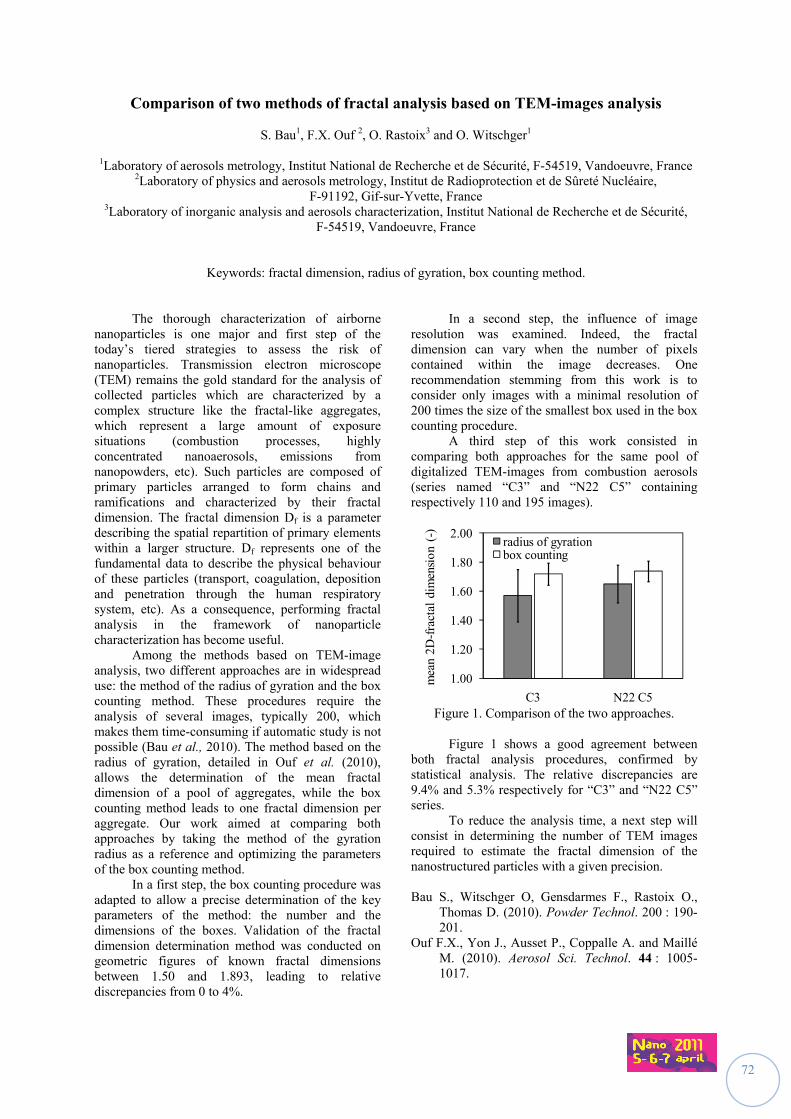
72
Comparison of two methods of fractal analysis based on TEM-images analysis
S. Bau1, F.X. Ouf 2, O. Rastoix3 and O. Witschger1
1Laboratory of aerosols metrology, Institut National de Recherche et de Sécurité, F-54519, Vandoeuvre, France 2Laboratory of physics and aerosols metrology, Institut de Radioprotection et de Sûreté Nucléaire,
F-91192, Gif-sur-Yvette, France 3Laboratory of inorganic analysis and aerosols characterization, Institut National de Recherche et de Sécurité,
F-54519, Vandoeuvre, France
Keywords: fractal dimension, radius of gyration, box counting method.
The thorough characterization of airborne nanoparticles is one major and first step of the today’s tiered strategies to assess the risk of nanoparticles. Transmission electron microscope (TEM) remains the gold standard for the analysis of collected particles which are characterized by a complex structure like the fractal-like aggregates, which represent a large amount of exposure situations (combustion processes, highly concentrated nanoaerosols, emissions from nanopowders, etc). Such particles are composed of primary particles arranged to form chains and ramifications and characterized by their fractal dimension. The fractal dimension Df is a parameter describing the spatial repartition of primary elements within a larger structure. Df represents one of the fundamental data to describe the physical behaviour of these particles (transport, coagulation, deposition and penetration through the human respiratory system, etc). As a consequence, performing fractal analysis in the framework of nanoparticle characterization has become useful. Among the methods based on TEM-image analysis, two different approaches are in widespread use: the method of the radius of gyration and the box counting method. These procedures require the analysis of several images, typically 200, which makes them time-consuming if automatic study is not possible (Bau et al., 2010). The method based on the radius of gyration, detailed in Ouf et al. (2010), allows the determination of the mean fractal dimension of a pool of aggregates, while the box counting method leads to one fractal dimension per aggregate. Our work aimed at comparing both approaches by taking the method of the gyration radius as a reference and optimizing the parameters of the box counting method. In a first step, the box counting procedure was adapted to allow a precise determination of the key parameters of the method: the number and the dimensions of the boxes. Validation of the fractal dimension determination method was conducted on geometric figures of known fractal dimensions between 1.50 and 1.893, leading to relative discrepancies from 0 to 4%.
In a second step, the influence of image resolution was examined. Indeed, the fractal dimension can vary when the number of pixels contained within the image decreases. One recommendation stemming from this work is to consider only images with a minimal resolution of 200 times the size of the smallest box used in the box counting procedure. A third step of this work consisted in comparing both approaches for the same pool of digitalized TEM-images from combustion aerosols (series named “C3” and “N22 C5” containing respectively 110 and 195 images).
1.00
1.20
1.40
1.60
1.80
2.00
C3 N22 C5
mea
n 2D
-fra
ctal
dim
ensi
on (
-)
radius of gyrationbox counting
Figure 1. Comparison of the two approaches.
Figure 1 shows a good agreement between both fractal analysis procedures, confirmed by statistical analysis. The relative discrepancies are 9.4% and 5.3% respectively for “C3” and “N22 C5” series. To reduce the analysis time, a next step will consist in determining the number of TEM images required to estimate the fractal dimension of the nanostructured particles with a given precision. Bau S., Witschger O, Gensdarmes F., Rastoix O.,
Thomas D. (2010). Powder Technol. 200 : 190-201.
Ouf F.X., Yon J., Ausset P., Coppalle A. and Maillé M. (2010). Aerosol Sci. Technol. 44 : 1005-1017.
73
Prestandardization study on the characterization of airborne nanoparticles size: qualification of a generation protocol for nanometer aerosols of SiO2
C. Motzkus1, T. Macé1, S. Vaslin-Reimann1, N. Michielsen2, F. Gensdarmes2, P. Ausset3 and M. Maillé3
1Département Qualité de l'air et débitmétrie gazeuse, Laboratoire National de Métrologie et d’Essais,
1 rue Gaston Boissier, 75724 Paris Cedex 15, France 2Aerosol Physics and Metrology Laboratory, Institut de Radioprotection et de Sûreté Nucléaire, B.P 68,
91192 Gif-sur-Yvette Cedex, France 3Laboratoire Interuniversitaire des Systèmes Atmosphériques, Université Paris-Est Créteil (UPEC),
CNRS UMR 7583, 61 Avenue du Général de Gaulle, 94010 Créteil, France
Keywords: nanoparticles, microscopy, Scanning Mobility Particle Sizer, atomization
Day after day, new applications using manufactured nanoparticles appear in industry. To evaluate the occupational risk associated to nanoparticles, it is important to have reliable, accurate and standardized measurement methods. It is therefore necessary to work on pre-normalization projects to develop reference methods to characterize the number and the size distribution of airborne nanoparticles. This study is led in the framework of Technical Working Area 34 - Properties of Nanoparticle Populations of the Versailles Project on Advanced Materials and Standards (VAMAS). The VAMAS is an international collaborative organization on prestandardization research projects and specifications for advanced materials. The working group is composed of National Metrology Institutes such as BAM (Germany), CENAM (Mexico), DFM (Denmark), NMIA (Australia), NMISA (South Africa), INPL (Israel), KRISS (South Korea), LNE (France), NIST (USA), NMIJ-AIST (Japan), NPL (UK), NPLI (India) and other laboratories involved in nanoparticle metrology such as the Aerosol Physics and Metrology Laboratory of IRSN (The French Institute for Radiological Protection and Nuclear Safety), the Laboratoire InterUniversitaire des Systèmes Atmosphériques (LISA) of University Paris-Est Créteil and the department of chemistry and industrial chemistry of the University of Genoa. The aim of this project is to develop characterization methods for airborne nanoparticles that include all the measurement chain (sampling, instrumentation, analysis, treatments of the data…) which will be traceable, adequate and accurate: this project includes the estimation of the uncertainties on the measurements (Song et al., 2009).
This study describes the generation and the characterization of SiO2 airborne nanoparticles from a liquid suspension by atomization. First, the particles sizes of three colloidal suspensions have been characterized by several laboratories using different techniques: Dynamic Light Scattering (DLS), Scanning Electron Microscopy (SEM),
Transmission Electron Microscopy (TEM) and Atomic Force Microscopy (AFM).
Secondly, experiments have been performed in order to study the stability, the repeatability and the reproducibility of the generation protocol of SiO2 airborne nanoparticles by measuring their count size distributions with a Scanning Mobility Particle Sizer (SMPS, ISO 15900). Preliminary tests have been made to optimize the generation protocol to obtain count size distributions in the size range between 20 and 80 nm: with this protocol the results are stable in term of time, are repeatable and reproducible. One of the great benefit of this protocol and the used material is the ability to generate a nanometer aerosol composed of spherical particles: these particles are clearly distinct from parasites particles which appear, for example, during the atomization of solutions containing calibrated polystyrene latex beads and surfactant (Billard et al. 1970, Fuchs 1973). The next step will be to conduct a round robin test for characterizing SiO2 airborne nanoparticles on the one hand, by using on line techniques like SMPS, and, on the other hand, by using indirect techniques such as TEM, SEM and AFM which imply to collect the particles on adapted supports like grid or wafer. This work will provide international harmonized methodologies for organizations in charge of standardization (ISO TC229 “Nanotechnology” and CEN TC352) in order to disseminate consensual and applicable standards for the characterization of airborne nanoparticles.
Billard F., Madelaine G. and Delhaye J. (1970). J. Aerosol Science, 1, 357-367.
Fuchs N.A. (1973). J. Aerosol Science, 4, 405-410. ISO 15900 (2009) Determination of particle size
distribution-Differential electrical mobility analysis for aerosol particles, 57p.
Song N. W., Park K.M., Lee I.-H. and Huh H. (2009). Metrologia, 46, 480–488.
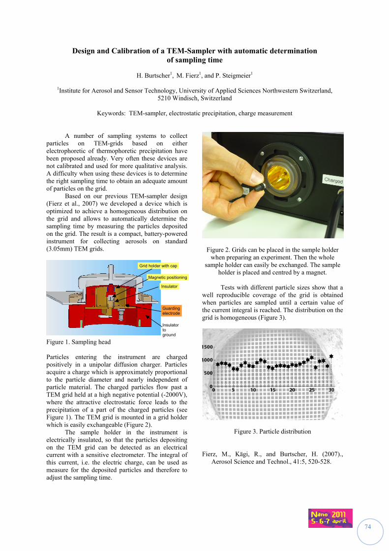
74
Design and Calibration of a TEM-Sampler with automatic determination of sampling time
H. Burtscher1, M. Fierz1, and P. Steigmeier1
1Institute for Aerosol and Sensor Technology, University of Applied Sciences Northwestern Switzerland,
5210 Windisch, Switzerland
Keywords: TEM-sampler, electrostatic precipitation, charge measurement A number of sampling systems to collect particles on TEM-grids based on either electrophoretic of thermophoretic precipitation have been proposed already. Very often these devices are not calibrated and used for more qualitative analysis. A difficulty when using these devices is to determine the right sampling time to obtain an adequate amount of particles on the grid. Based on our previous TEM-sampler design (Fierz et al., 2007) we developed a device which is optimized to achieve a homogeneous distribution on the grid and allows to automatically determine the sampling time by measuring the particles deposited on the grid. The result is a compact, battery-powered instrument for collecting aerosols on standard (3.05mm) TEM grids.
Figure 1. Sampling head Particles entering the instrument are charged positively in a unipolar diffusion charger. Particles acquire a charge which is approximately proportional to the particle diameter and nearly independent of particle material. The charged particles flow past a TEM grid held at a high negative potential (-2000V), where the attractive electrostatic force leads to the precipitation of a part of the charged particles (see Figure 1). The TEM grid is mounted in a grid holder which is easily exchangeable (Figure 2). The sample holder in the instrument is electrically insulated, so that the particles depositing on the TEM grid can be detected as an electrical current with a sensitive electrometer. The integral of this current, i.e. the electric charge, can be used as measure for the deposited particles and therefore to adjust the sampling time.
Figure 2. Grids can be placed in the sample holder when preparing an experiment. Then the whole
sample holder can easily be exchanged. The sample holder is placed and centred by a magnet.
Tests with different particle sizes show that a well reproducible coverage of the grid is obtained when particles are sampled until a certain value of the current integral is reached. The distribution on the grid is homogeneous (Figure 3).
Figure 3. Particle distribution Fierz, M., Kägi, R., and Burtscher, H. (2007).,
Aerosol Science and Technol., 41:5, 520-528.
Grid holder with cap
Insulator
Magnetic positioning
Guarding electrode
Insulator to ground
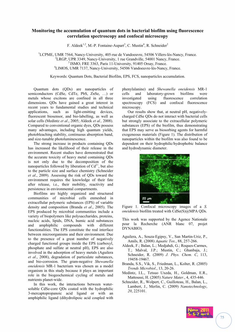
75
Monitoring the accumulation of quantum dots in bacterial biofilm using fluorescence correlation spectroscopy and confocal microscopy
F. Aldeek1,2, M.-P. Fontaine-Aupart3, C. Mustin4, R. Schneider2
1LCPME, UMR 7564, Nancy-University, 405-rue de Vandoeuvre, 54506 Villers-lès-Nancy, France.
2LRGP, UPR 3349, Nancy-University, 1 rue Grandville, 54001 Nancy, France. 3 ISMO, FRE 3363, Paris 11-University, 91405 Orsay, France.
4LIMOS, UMR 7137, Nancy-University, 54506 Vandoeuvre-lès-Nancy, France.
Keywords: Quantum Dots, Bacterial Biofilm, EPS, FCS, nanoparticles accumulation.
Quantum dots (QDs) are nanoparticles of
semiconductors (CdSe, CdTe, PbS, ZnSe, …) or metals whose excitons are confined in all three dimensions. QDs have gained a great interest in recent years to fundamental studies and technical applications, such as light-emitting devices, fluorescent biosensor, and bio-labelling, as well as solar cells (Medintz et al., 2005; Aldeek et al., 2008). Compared to conventional organic dyes, QDs possess many advantages, including high quantum yields, photobleaching stability, continuous absorption band, and size-tunable photoluminescence.
The strong increase in products containing QDs has increased the likelihood of their release in the environment. Recent studies have demonstrated that the accurate toxicity of heavy metal containing QDs is not only due to the decomposition of the nanoparticles followed by liberation of Cd2+, but also to the particle size and surface chemistry (Schneider et al., 2009). Assessing the risk of QDs toward the environment requires the knowledge of their fate after release, i.e., their mobility, reactivity and persistence in environmental compartments.
Biofilms are highly organized and structured communities of microbial cells enmeshed in extracellular polymeric substances (EPS) of variable density and composition (Branda et al., 2005). The EPS produced by microbial communities include a variety of biopolymers like polysaccharides, proteins, nucleic acids, lipids, DNA, humic acid substances and amphiphilic compounds with different functionalities. The EPS constitute the real interface between microorganisms and their environment. Due to the presence of a great number of negatively charged functional groups inside the EPS (carboxyl, phosphate and sulfate at neutral pH), EPS are also involved in the adsorption of heavy metals (Aguilera et al., 2008), degradation of particulate substances, and bio-corrosion. The gram-negative Shewanella oneidensis MR-1 bacterium was chosen as a model organism in this study because it plays an important role in the biogeochemical cycling of metals and nutrients planet-wide.
In this work, the interactions between water-soluble CdSe-core QDs coated with the hydrophilic 3-mercaptopropanoic acid ligand or with an amphiphilic ligand (dihydrolipoic acid coupled with
phenylalanine) and Shewanella oneidensis MR-1 cells and laboratory-grown biofilms were investigated using fluorescence correlation spectroscopy (FCS) and confocal fluorescence microscopy.
Our results show that, at neutral pH, negatively-charged CdSe QDs do not interact with bacterial cells but strongly associate to the extracellular polymeric substances (EPS) of the biofilm, thus demonstrating that EPS may serve as biosorbing agents for harmful exogeneous materials (Figure 1). The distribution of nanoparticles within the biofilm was also found to be dependent on their hydrophilic/hydrophobic balance and hydrodynamic diameter.
20 µm20 µm
Figure 1. Confocal microscopy images of a S. oneidensis biofilm treated with CdSe(S)@MPA QDs.
This work was supported by the Agence Nationale pour la Recherche (ANR blanc 07, projet DYNABIO).
Aguilera, A., Souza-Egipsy, V., San Martin-Uriz, P., Amils, R. (2008) Aquatic Tox., 88, 257-266.
Aldeek, F.; Balan, L.; Medjahdi, G.; Roques-Carmes, T.; Malval, J.P.; Mustin, C.; Ghanbaja, J.; Schneider, R. (2009) J. Phys. Chem. C, 113, 19458-19467.
Branda, S.S., Vik, S., Friedman, L., Kolter, R. (2005) Trends Microbiol., 13, 20-26.
Medintz, I.L., Tetsuo Uxeda, H., Goldman, E.R., Mattoussi, H. (2005) Nature Mater., 4, 435-446.
Schneider, R., Wolpert, C., Guilloteau, H., Balan, L., Lambert, J., Merlin, C. (2009) Nanotechnology, 20, 225101.
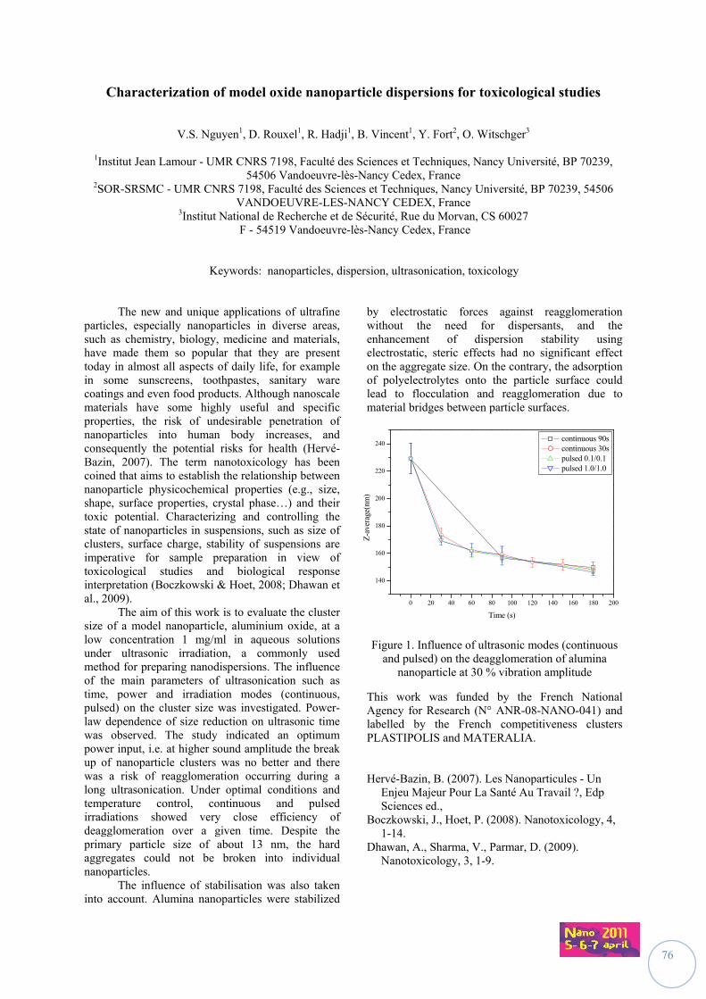
76
Characterization of model oxide nanoparticle dispersions for toxicological studies
V.S. Nguyen1, D. Rouxel1, R. Hadji1, B. Vincent1, Y. Fort2, O. Witschger3
1Institut Jean Lamour - UMR CNRS 7198, Faculté des Sciences et Techniques, Nancy Université, BP 70239, 54506 Vandoeuvre-lès-Nancy Cedex, France
2SOR-SRSMC - UMR CNRS 7198, Faculté des Sciences et Techniques, Nancy Université, BP 70239, 54506 VANDOEUVRE-LES-NANCY CEDEX, France
3Institut National de Recherche et de Sécurité, Rue du Morvan, CS 60027 F - 54519 Vandoeuvre-lès-Nancy Cedex, France
Keywords: nanoparticles, dispersion, ultrasonication, toxicology
The new and unique applications of ultrafine particles, especially nanoparticles in diverse areas, such as chemistry, biology, medicine and materials, have made them so popular that they are present today in almost all aspects of daily life, for example in some sunscreens, toothpastes, sanitary ware coatings and even food products. Although nanoscale materials have some highly useful and specific properties, the risk of undesirable penetration of nanoparticles into human body increases, and consequently the potential risks for health (Hervé-Bazin, 2007). The term nanotoxicology has been coined that aims to establish the relationship between nanoparticle physicochemical properties (e.g., size, shape, surface properties, crystal phase…) and their toxic potential. Characterizing and controlling the state of nanoparticles in suspensions, such as size of clusters, surface charge, stability of suspensions are imperative for sample preparation in view of toxicological studies and biological response interpretation (Boczkowski & Hoet, 2008; Dhawan et al., 2009).
The aim of this work is to evaluate the cluster size of a model nanoparticle, aluminium oxide, at a low concentration 1 mg/ml in aqueous solutions under ultrasonic irradiation, a commonly used method for preparing nanodispersions. The influence of the main parameters of ultrasonication such as time, power and irradiation modes (continuous, pulsed) on the cluster size was investigated. Power-law dependence of size reduction on ultrasonic time was observed. The study indicated an optimum power input, i.e. at higher sound amplitude the break up of nanoparticle clusters was no better and there was a risk of reagglomeration occurring during a long ultrasonication. Under optimal conditions and temperature control, continuous and pulsed irradiations showed very close efficiency of deagglomeration over a given time. Despite the primary particle size of about 13 nm, the hard aggregates could not be broken into individual nanoparticles.
The influence of stabilisation was also taken into account. Alumina nanoparticles were stabilized
by electrostatic forces against reagglomeration without the need for dispersants, and the enhancement of dispersion stability using electrostatic, steric effects had no significant effect on the aggregate size. On the contrary, the adsorption of polyelectrolytes onto the particle surface could lead to flocculation and reagglomeration due to material bridges between particle surfaces.
0 20 40 60 80 100 120 140 160 180 200
140
160
180
200
220
240
Z-a
vera
ge(n
m)
Time (s)
continuous 90s continuous 30s pulsed 0.1/0.1 pulsed 1.0/1.0
Figure 1. Influence of ultrasonic modes (continuous and pulsed) on the deagglomeration of alumina
nanoparticle at 30 % vibration amplitude
This work was funded by the French National Agency for Research (N° ANR-08-NANO-041) and labelled by the French competitiveness clusters PLASTIPOLIS and MATERALIA.
Hervé-Bazin, B. (2007). Les Nanoparticules - Un Enjeu Majeur Pour La Santé Au Travail ?, Edp Sciences ed.,
Boczkowski, J., Hoet, P. (2008). Nanotoxicology, 4, 1-14.
Dhawan, A., Sharma, V., Parmar, D. (2009). Nanotoxicology, 3, 1-9.
77
Characterization of occupational exposure to nanometric particles: construction of a job- exposure matrix (MatPUF)
S. Audignon Durand1, Y. Isidore1, A. Lacourt1, M. Rinaldo1, S. Ducamp2, P. Brochard1
1University of Bordeaux 2, the Bordeaux School of Public Health, Occupational and Environmental health laboratory, Associated team in Occupational health, Bordeaux
2National Institute for Public Health Surveillance, Department of Occupational Health, Associated team in Occupational health, Bordeaux
Keywords : nanometric particles, job-exposure matrix, health, occupational
Background and Purpose - The health
effects of aerosols and alveolar respirable fraction have been widely studied and both regarding environmental pollution and the workplace. However, many questions remain concerning the submicron fraction, especially about nano particles which size involves a deposition in the deep airways. Until today, determinants of their toxicity are well positioned, and short term health effects have been highlighted. Moreover, there is yet no epidemiological evidence about relationship between long-term effects and nanometric particles exposure, particularly in the context of an occupational activity. In this context, the project MatPUF aims to identify and evaluate occupational exposure to manufactured nanometric particles and those generated unintentionally through the construction of a job-exposure matrix in order to study the potential long term effects.
Materials and methods - A job-exposure
matrix consists in two axes: one of them represents jobs (combination of an occupation and an industry sector) and the other one represents exposure indices. At the intersection of each work with indices, exposure values are assigned. The development of the matrix is divided into two stages: 1) Qualitative assessment of exposure of industries and occupations through the identification of work processes that generate nanometric particles and the physicochemical profile of these particles, 2) Semi-quantitative assessment of exposure for identified industries and occupations by expertise Values of exposure indices (probability, intensity, frequency) according to historical periods were determined in this step.
To validate each step of development of the matrix, a panel of experts from different disciplines has been consulted. Thus, different organizations are participating to this project: AHI33, Anses, Carsat Aquitaine, Ineris, INRS, Inserm, InVS, IRSST (Quebec), Lépi, Universities of Bordeaux 1, Montreal, Paris Sud and Paris 7.
Expected Results - The matrix MatPUF introduces all jobs potentially exposed to nanometric particles. Therefore, data from occupational exposures in the French population, such as the prevalence of exposure can be estimated by applying the matrix to a representative jobs histories sample. Moreover, using this matrix to individual exposure assessment in case-control studies, will allow to study the relationship between different cancers and occupational nanometric particle exposure.
Conclusion and perspectives – Today occupational exposure to manufactured and unmanufactured nanometric particles raises many questions from public health and prevention professionals. By providing the first comprehensive assessment of occupational exposures in the general population and by being available freely on Internet (InVS and Evalutil websites), the matrix MatPUF should help to identify risk situations, and to set ad'hoc preventive measures and improvement of epidemiological knowledge in monitoring studies of workers potentially exposed.
78
Qualitative characterization of airborne nanoparticles at workplace: advantages and limits of the SEM-EDS technique
S. Derrough1, X. Ravanel1, C. Durand1
1CEA-Grenoble, DSP/SMR, Nanoparticles Expertise Center, 17 avenue des Martyrs,
38054, Cedex 9, Grenoble, France
Keywords: airborne nanoparticles, qualitative characterization, electron microscopy, chemical analysis
This work is conducted in the frame of the French national program dedicated to nanotechnologies: NanoINNOV. One topic of this project, covering nanoparticles and safety, includes the measure and the characterization of airborne nanoparticles emissions in different workplaces in, industries, research laboratories and universities. For 2010, more than 100 workstations were examined over three integration centers based in Paris, Toulouse and Grenoble. When emission of nanoparticles is suspected during an operation at a workplace, several characterization steps are conducted. The first one is a real-time quantitative characterization, as close as possible to the operation taking place, to have access to the number of emitted particles per unit of volume, the size range and the associated specific surface. This initial phase also includes sampling on dedicated membranes. The following stage of the methodology is the qualification of the emitted nanoparticles, which is done offline on the collected samples. This second set of characterization gives access to new and very important data such as the shape and the composition of the nanoparticles. Are we in front of isolated species or agglomerates? What is the chemical composition? Are the emitted nanoparticles linked to the operation taking place? Those fundamental questions are answered through this qualification step by using different analysis instruments. Most of the time the analysis starts with the use of the SEM (Scanning Electron Microscopy) –EDS (Energy Dispersive X-Ray Spectroscopy) technique. On one side, the high resolution of the SEM permits a comfortable visualization of the nanoparticles down to sizes close to the nanometer. On the other side, the fact that the chemical analysis by EDS could be run simultaneously by aiming the nanoparticles on the SEM screen, makes the combination SEM/EDS ideal for a majority of analysis. This will be illustrated through different scenarios involving various conditions, type of nano-objects, amounts, chemical species…Nevertheless, the use of complementary techniques to SEM/EDS is often needed to go further in the understanding of the
nanoparticles release during operation. Results obtained by Total Reflection X-ray Fluorescence (TXRF) will be presented. This technique gives additional interesting information as the entire collected sample is analyzed instead of a limited number of nanoparticles by SEM/EDS. Auger Electron Spectroscopy (AES) will also be presented as this characterization technique gives access to the chemical composition of the nanoparticle’s surface and as such is an interesting option to extend the qualification study. Finally the possibility of using other techniques for nanoparticles identification such as Secondary Ion Mass Spectrometry (SIMS) or Atomic force microscopy (AFM) will be discussed.
79
Towards a harmonized assessment of the exposure to manufactured nanoobjects, Common approaches in measurement strategy and obstacles - Report of a workshop
M. Berges1, D. Brouwer2, W. Fransman2, L. Hodson3, C. Asbach4, D. Bard5, U. Backman6, I. Lynch7
and M. Riediker8
1DGUV-IFA, 53757, Sankt Augustin, Germany 2TNO Quality of Life, 3700 AJ, Zeist, The Netherlands
3NIOSH, Cincinnati, OH 45226, USA 4IUTA, 47229, Duisburg, Germany
5HSL, Buxton, UK 6VTT, Finland
7UCD, Belfield Dublin 4, Ireland 8IST, 1011, Lausanne, Switzerland
Keywords: Strategies to assess exposure, Measurement strategy to differentiate from background,
Particle collection for electron microscopy. The number of workplace air measurement studies focused on the assessment of exposure to manufactured nano objects has increased substantially, the last few years. However, due to the large variation of exposure situations with respect to the life cycle of nanomaterials and nanoproducts, actual exposure data will remain scarce in the near future. Therefore, it is acknowledged that data that will be generated should enable future use for either exposure scenario building, exposure modelling, or meta-analysis in view of risk assessment or epidemiology. A crucial step in harmonizing data collection is the application of internationally agreed measurement strategies and sampling protocols. We here report on a workshop under the umbrella of the EU project NanoImpactNet where key players in Europe and the US did discuss the current knowledge and tried to set a path for future progress. In the first part of the workshop, Brouwer (2009) discussed the literature available through early 2009 (14 studies) with an emphasis on possible ways to cope with the problem of background distinction. One major finding of most of the studies is that during the production and handling of nanoparticles, the workplace particle number concentration of particles below 100 nm is close to the background concentration in companies. The common finding is that aggregates or agglomerates above 100 nm in size were quite often detected at the workplace and correlated with the operations. This is in line with theoretical calculations indicating that most of the particles emitted from processes are agglomerated when reaching the exposed person (Seipenbusch et al [2008]). In summing up the available studies, Brouwer et al. (2009) concluded that the studies are more explorative in character and focused on the potential for emission of manufactured nanoparticles. No shift averages were presented based on particle number concentration or surface area concentration or fibre concentration.
Brower et al. (2009) provided a decision logic, developed in the EU project NANOSH, to cope with the problem of background distinction and explore the likelihood of potential exposure to nanoobjects. NIOSH developed a somewhat different approach to explore potential exposure (emission releases) by relying more on easy-to-use and portable devices like the CPC and OPC in connection with sampling on filters for subsequent elemental and microscopic analysis of the particles. Hodson (Methner et al., 2010a, 2010b) described the Nanoparticle Emission Assessment Technique (NEAT) used by NIOSH and described that in-depth sampling is also applied at some of the workplaces they have evaluated. A participant from industry proposed a tiered approach with a decision logic and criteria taking account of the needs and constraints imposed on a globally acting company. Workshop participants discussed the way to a harmonized framework of measurement strategies serving the different reasons why measurements of nanoobjects are performed. There are obstacles and uncertainties associated with this approach. Consequently, joint and coordinated efforts and round robin testing were identified as a solution to close this critical gap in exposure assessment. This work was supported by the EU FP7 Grant CSA-CA218539 (NanoImpactNet). Brouwer D., van Duuren-Stuurman B., Berges M,
Jankowska E., Bard D. & Mark D. (2009). J. Nanopart Res 11:1867-1881, DOI 10.1007s/11051-009-9772-1
Methner M., Hodson L. & Geraci C.(2010) JOEH, 7:3, 127-132 (2010a)
Methner M., Hodson L. & Geraci C. (2010). JOEH, 7:3, 163-176 (2010b)
Seipenbusch M, Binder A & Kasper G (2008). Ann. Occup. Hyg., 52(8), 707-716.
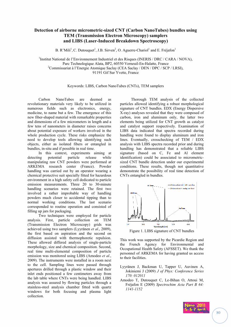
80
Detection of airborne micrometric-sized CNT (Carbon NanoTubes) bundles using TEM (Transmission Electron Microscopy) samplers and LIBS (Laser-Induced Breakdown Spectroscopy)
B. R’Mili1, C. Dutouquet1, J.B. Sirven2, O. Aguerre-Chariol1 and E. Fréjafon1
1Institut National de l’Environnement Industriel et des Risques (INERIS / DRC / CARA / NOVA),
Parc Technologique Alata, BP2, 60550 Verneuil-En-Halatte, France 2Commissariat à l’Energie Atomique Saclay (CEA Saclay / DEN / DPC / SCP / LRSI),
91191 Gif Sur Yvette, France
Keywords: LIBS, Carbon NanoTubes (CNTs), TEM samplers
Carbon NanoTubes are deemed as revolutionary materials very likely to be utilized in numerous fields such as electronics, energy, medicine, to name but a few. The emergence of this new fiber-shaped material with remarkable properties and dimensions of a few micrometers in length and a few tens of nanometers in diameter raises concerns about potential exposure of workers involved in the whole production cycle. These risks emphasize the need to develop tools allowing identifying such objects, either as isolated fibers or entangled in bundles, in-situ and if possible in real time. In this context, experiments aiming at detecting potential particle release while manipulating raw CNT powders were performed at ARKEMA research center (France). Powder handling was carried out by an operator wearing a chemical protective suit specially fitted for hazardous environment in a high safety cell dedicated to particle emission measurements. Three 20 to 30-minute handling scenarios were retained. The first two involved a rather improbable way of handling powders much closer to accidental tipping than to normal working conditions. The last scenario corresponded to routine operation and consisted of filling up jars for packaging. Two techniques were employed for particle analysis. First, particle collection on TEM (Transmission Electron Microscopy) grids was achieved using two samplers (Lyyränen et al., 2009), the first based on aspiration and the second on diffusion assisted with thermophoretic repulsion. These allowed differed analysis of single-particle morphology, size and chemical composition. Second, real time multi-elemental composition of particle emission was monitored using LIBS (Amodeo et al., 2009). The instruments were installed in a room next to the cell. Sampling lines were passed through apertures drilled through a plastic window and their inlet ends positioned a few centimeters away from the lab table where CNTs were being handled. LIBS analysis was assured by flowing particles through a stainless-steel analysis chamber fitted with quartz windows for both focusing and plasma light collection.
Thorough TEM analysis of the collected particles allowed identifying a robust morphological signature of CNT bundles. EDX (Energy Dispersive X-ray) analyses revealed that they were composed of carbon, iron and aluminum only, the latter two elements being utilized for CNT growth as catalyst and catalyst support respectively. Examination of LIBS data indicated that spectra recorded during handling were found to display aluminum and iron lines. Eventually, crosschecking of TEM / EDX analysis with LIBS spectra recorded prior and during handling has demonstrated that a reliable LIBS signature (based on C, Fe and Al element identification) could be associated to micrometric-sized CNT bundle detection under our experimental conditions. These results, though not quantitative demonstrate the possibility of real time detection of CNTs entangled in bundles.
Figure 1. LIBS signature of CNT bundles This work was supported by the Picardie Region and the French Agency for Environmental and Occupational Health Safety (AFSSET). We thank the personnel of ARKEMA for having granted us access to their facilities. Lyyränen J, Backman U, Tapper U, Auvinen A,
Jokiniemi J (2009) J of Phys: Conference Series 170: 012011
Amodeo T, Dutouquet C, Le-Bihan O, Attoui M, Fréjafon E (2009) Spectrochim Acta Part B 64: 1141-1152
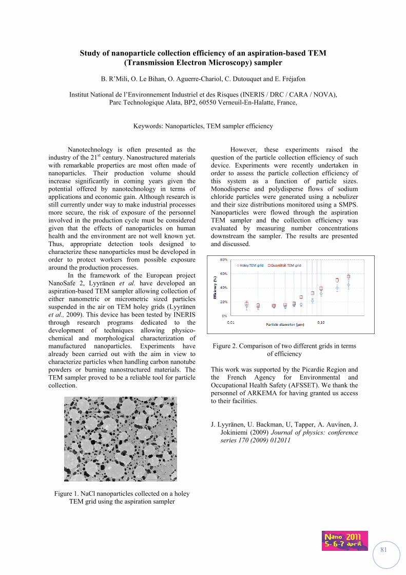
81
Study of nanoparticle collection efficiency of an aspiration-based TEM (Transmission Electron Microscopy) sampler
B. R’Mili, O. Le Bihan, O. Aguerre-Chariol, C. Dutouquet and E. Fréjafon
Institut National de l’Environnement Industriel et des Risques (INERIS / DRC / CARA / NOVA),
Parc Technologique Alata, BP2, 60550 Verneuil-En-Halatte, France,
Keywords: Nanoparticles, TEM sampler efficiency
Nanotechnology is often presented as the industry of the 21st century. Nanostructured materials with remarkable properties are most often made of nanoparticles. Their production volume should increase significantly in coming years given the potential offered by nanotechnology in terms of applications and economic gain. Although research is still currently under way to make industrial processes more secure, the risk of exposure of the personnel involved in the production cycle must be considered given that the effects of nanoparticles on human health and the environment are not well known yet. Thus, appropriate detection tools designed to characterize these nanoparticles must be developed in order to protect workers from possible exposure around the production processes. In the framework of the European project NanoSafe 2, Lyyränen et al. have developed an aspiration-based TEM sampler allowing collection of either nanometric or micrometric sized particles suspended in the air on TEM holey grids (Lyyränen et al., 2009). This device has been tested by INERIS through research programs dedicated to the development of techniques allowing physico-chemical and morphological characterization of manufactured nanoparticles. Experiments have already been carried out with the aim in view to characterize particles when handling carbon nanotube powders or burning nanostructured materials. The TEM sampler proved to be a reliable tool for particle collection.
Figure 1. NaCl nanoparticles collected on a holey
TEM grid using the aspiration sampler
However, these experiments raised the question of the particle collection efficiency of such device. Experiments were recently undertaken in order to assess the particle collection efficiency of this system as a function of particle sizes. Monodisperse and polydisperse flows of sodium chloride particles were generated using a nebulizer and their size distributions monitored using a SMPS. Nanoparticles were flowed through the aspiration TEM sampler and the collection efficiency was evaluated by measuring number concentrations downstream the sampler. The results are presented and discussed.
Figure 2. Comparison of two different grids in terms
of efficiency This work was supported by the Picardie Region and the French Agency for Environmental and Occupational Health Safety (AFSSET). We thank the personnel of ARKEMA for having granted us access to their facilities. J. Lyyränen, U. Backman, U, Tapper, A. Auvinen, J.
Jokiniemi (2009) Journal of physics: conference series 170 (2009) 012011
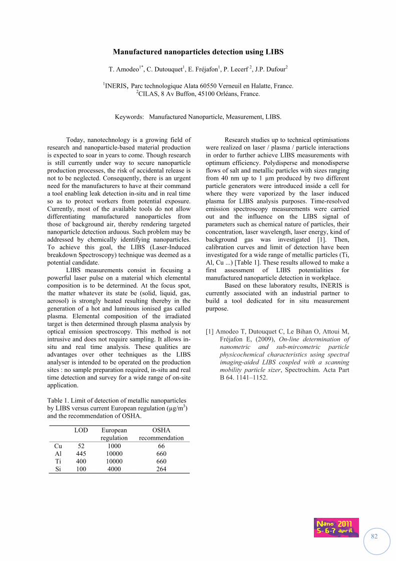
82
Manufactured nanoparticles detection using LIBS
T. Amodeo1*, C. Dutouquet1, E. Fréjafon1, P. Lecerf 2, J.P. Dufour2
1INERIS, Parc technologique Alata 60550 Verneuil en Halatte, France. 2CILAS, 8 Av Buffon, 45100 Orléans, France.
Keywords: Manufactured Nanoparticle, Measurement, LIBS.
Today, nanotechnology is a growing field of research and nanoparticle-based material production is expected to soar in years to come. Though research is still currently under way to secure nanoparticle production processes, the risk of accidental release is not to be neglected. Consequently, there is an urgent need for the manufacturers to have at their command a tool enabling leak detection in-situ and in real time so as to protect workers from potential exposure. Currently, most of the available tools do not allow differentiating manufactured nanoparticles from those of background air, thereby rendering targeted nanoparticle detection arduous. Such problem may be addressed by chemically identifying nanoparticles. To achieve this goal, the LIBS (Laser-Induced breakdown Spectroscopy) technique was deemed as a potential candidate.
LIBS measurements consist in focusing a powerful laser pulse on a material which elemental composition is to be determined. At the focus spot, the matter whatever its state be (solid, liquid, gas, aerosol) is strongly heated resulting thereby in the generation of a hot and luminous ionised gas called plasma. Elemental composition of the irradiated target is then determined through plasma analysis by optical emission spectroscopy. This method is not intrusive and does not require sampling. It allows in-situ and real time analysis. These qualities are advantages over other techniques as the LIBS analyser is intended to be operated on the production sites : no sample preparation required, in-situ and real time detection and survey for a wide range of on-site application. Table 1. Limit of detection of metallic nanoparticles by LIBS versus current European regulation (µg/m3) and the recommendation of OSHA.
Research studies up to technical optimisations were realized on laser / plasma / particle interactions in order to further achieve LIBS measurements with optimum efficiency. Polydisperse and monodisperse flows of salt and metallic particles with sizes ranging from 40 nm up to 1 µm produced by two different particle generators were introduced inside a cell for where they were vaporized by the laser induced plasma for LIBS analysis purposes. Time-resolved emission spectroscopy measurements were carried out and the influence on the LIBS signal of parameters such as chemical nature of particles, their concentration, laser wavelength, laser energy, kind of background gas was investigated [1]. Then, calibration curves and limit of detection have been investigated for a wide range of metallic particles (Ti, Al, Cu ...) [Table 1]. These results allowed to make a first assessment of LIBS potentialities for manufactured nanoparticle detection in workplace.
Based on these laboratory results, INERIS is currently associated with an industrial partner to build a tool dedicated for in situ measurement purpose. [1] Amodeo T, Dutouquet C, Le Bihan O, Attoui M,
Fréjafon E, (2009), On-line determination of nanometric and sub-mircometric particle physicochemical characteristics using spectral imaging-aided LIBS coupled with a scanning mobility particle sizer, Spectrochim. Acta Part B 64. 1141–1152.
LOD European regulation
OSHA recommendation
Cu 52 1000 66 Al 445 10000 660 Ti 400 10000 660 Si 100 4000 264

83
Identification of the Main Exposure Scenarios for Producing Nanocomposite polymers by Melt-Moulding Process
D. Fleury1, J.A.S. Bomfim2,3, C. Girard4, A. Vignes1, J.X. Bouillard1
1INERIS, Parc Technologique ALATA, 60550 Verneuil-en-Halatte, France
2Centro Ricerche Plast-Optica (CRP), Via Jacopo Linussio 1, 33020 Amaro (UD), Italy 3Public Research Center Henri Tudor, AMS, Technoport Schlassgoart, L-4002 Esch-sur-Alzette, Luxembourg 4Polytech’Grenoble (Joseph Fourier University), 26 av. Benoit Frachon, 38400 Saint-Martin-d’Hères, France
Keywords: nanocomposite polymers, exposure scenarios, carbon nanotubes, process
Nanocomposite plastics can have a variety of useful applications in a wide range of industries (automotive, energy storage, avionics…). Especially, preparing CNT-polymer nanocomposites using melt-mixing routes are particularly desirable because of the speed, simplicity and availability of the processes in the plastic industry. They can lead to an easier process scale-up and reduce the time-to-market of final products. This study investigates the risk associated with the production reinforced nanocomposites materials. The results are based on a safety analysis previously led in the pilot line of the plastics processing plant located in Centro Ricerche Plast-optica (CRP , Italy), that aims to produce CNT-reinforced polymer nanocomposites (enhanced performance plastic components) for the purpose of the automotive industry. Our approach of risks is based on the use of cause tree and event tree diagrams. Through the analysis of hazards and hazardous situations, and the critical events they lead to, the main event chains have been identified. Through a cascade of other events (indirect and direct causes), these chains go from an initial cause, to the critical events that result in consequences, and possibly dangerous phenomena in regards to the nanocomposite process. The multiple chains of events that result in the top one (critical event) are called the fault tree, while the different consequences of a given top event are grouped in an “event tree” (Figure 1). Considering the CRP process and its main steps, four scenarios have been studied (i) mixture preparation, (ii) mixing/melting process, and (iii) grinding of nanocomposite plastics. The “release of nano-objects at workplace” is the critical event for each of these scenarios, as it gathers both the process emission and passive exposure. The succession of faults that may lead to the critical events and the consequence that may result from it has been modelled, according to the risk analysis previously performed. Then, existing protective barriers in place have been identified, and when necessary, new barriers have been proposed. Like this, the full “bow-tie” diagrams (with existing and proposed barriers) have been built for each scenario. The most critical scenario listed at CRP is the mixing. This is probably due to the thermal
depolymerization process occurring when the polymer is heated to be homogeneously mixed with CNT. On the contrary, the lower risk of exposure is observed during the handling of CNT into exhaust hood. This is partially or totally thanks to the good efficiency of the prevention barriers already in place. The list of basic events, combined with their existing or preconized barriers, gives a real view of actions engaged and to be engaged. It makes easy to detect the weak points (in term of safety), and propose adapted recommendations to cure them. However, the lack of international consensus about the hazards associated to nano-objects imposes the application of the precautionary principle. Therefore the recommendations we have formulated for this process are written in the respect of reasoned/proportional precautionary principle application. With more or less adaptation, they can be applied to similar process operations and provide general view of how the critical event can be avoided. It is also clear that research must be pursuing for a better understanding of the toxicological, fire, and explosion properties of nano-objects.
Figure 1. “bow-tie” diagram used to identify the main exposure scenarios in the CRP process
The present work was developed inside the European Project “SAPHIR, supported by the EC through the Sixth Framework Program for Research and Technological Development (Contract No. 026666-2).
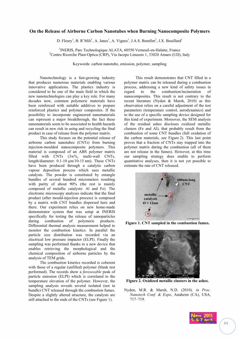
84
On the Release of Airborne Carbon Nanotubes when Burning Nanocomposite Polymers
D. Fleury1, B. R’Mili1, A. Janes1, A. Vignes1, J.A.S. Bomfim2, J.X. Bouillard1
1INERIS, Parc Technologique ALATA, 60550 Verneuil-en-Halatte, France 2Centro Ricerche Plast-Optica (CRP), Via Jacopo Linussio 1, 33020 Amaro (UD), Italy
Keywords: carbon nanotube, emission, polymer, sampling
Nanotechnology is a fast-growing industry that produces numerous materials enabling various innovative applications. The plastics industry is considered to be one of the main field in which the new nanotechnologies can play a key role. For many decades now, common polymeric materials have been reinforced with suitable additives to prepare reinforced plastics and polymer composites. If the possibility to incorporate engineered nanomaterials can represent a major breakthrough, the fact these nanomaterials seem to be associated to health hazards can result in new risk in using and recycling the final product in case of release from the polymer matrix. This study focuses on the potential release of airborne carbon nanotubes (CNTs) from burning injection-moulded nanocomposite polymers. This material is composed of an ABS polymer matrix filled with CNTs (3w%, multi-wall CNTs, length/diameter: 0.1-10 µm/10-15 nm). These CNTs have been produced through a catalytic carbon vapour deposition process which uses metallic catalysts. The powder is constituted by entangle bundles of several hundred micrometers resulting with purity of about 90% (the rest is mainly composed of metallic catalysts: Al and Fe). The electronic microscopy analyses indicate that the final product (after mould-injection process) is composed by a matrix with CNT bundles dispersed here and there. Our experiment relies on new home-made demonstrator system that was setup at INERIS specifically for testing the release of nanoparticles during combustion of polymeric products. Differential thermal analysis measurement helped to monitor the combustion kinetics. In parallel the particle size distribution was recorded via an electrical low pressure impactor (ELPI). Finally the sampling was performed thanks to a new device that enables retrieving the morphological and the chemical composition of airborne particles by the analysis of TEM grids. The combustion kinetics recorded is coherent with those of a regular (unfilled) polymer (blank test performed). The records show a foreseeable peak of particle emission (ELPI) which is correlated to the temperature elevation of the polymer. However, the sampling analysis reveals several isolated (not in bundle) CNT released through the combustion fumes. Despite a slightly altered structure, the catalysts are still attached to the ends of the CNTs (see Figure 1).
This result demonstrates that CNT filled in a polymer matrix can be released during a combustion process, addressing a new kind of safety issues in regard to the combustion/incineration of nanocomposites. This result is not contrary to the recent literature (Nyden & Marsh, 2010) as this observation relies on a careful adjustment of the test parameters (temperature control, aerodynamics) and to the use of a specific sampling device designed for this kind of experiment. Moreover, the SEM analysis of the residual ashes discloses oxidized metallic clusters (Fe and Al), that probably result from the combustion of some CNT bundles (full oxidation of the carbon materials, see Figure 2). This last point proves that a fraction of CNTs stay trapped into the polymer matrix during the combustion (all of them are not release in the fumes). However, at this time our sampling strategy does enable to perform quantitative analyses, then it is not yet possible to estimate the rate of CNT released.
Figure 1. CNT sampled in the combustion fumes.
Figure 2. Oxidized metallic clusters in the ashes.
Nyden, M.R. & Marsh, N.D. (2010). in Proc.
Nanotech Conf. & Expo, Anaheim (CA), USA, 717–719.
metallic catalysts
Ø ≈ 12nm
600nm-long CNT
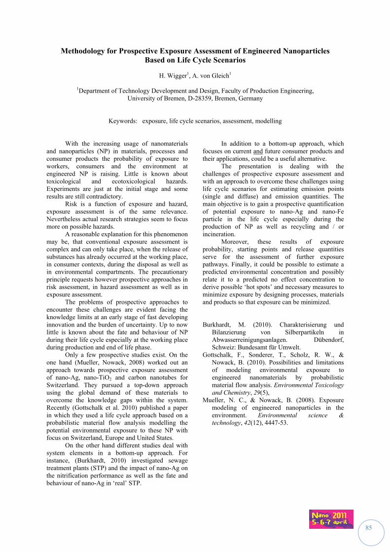
85
Methodology for Prospective Exposure Assessment of Engineered Nanoparticles Based on Life Cycle Scenarios
H. Wigger1, A. von Gleich1
1Department of Technology Development and Design, Faculty of Production Engineering,
University of Bremen, D-28359, Bremen, Germany
Keywords: exposure, life cycle scenarios, assessment, modelling With the increasing usage of nanomaterials and nanoparticles (NP) in materials, processes and consumer products the probability of exposure to workers, consumers and the environment at engineered NP is raising. Little is known about toxicological and ecotoxicological hazards. Experiments are just at the initial stage and some results are still contradictory. Risk is a function of exposure and hazard, exposure assessment is of the same relevance. Nevertheless actual research strategies seem to focus more on possible hazards. A reasonable explanation for this phenomenon may be, that conventional exposure assessment is complex and can only take place, when the release of substances has already occurred at the working place, in consumer contexts, during the disposal as well as in environmental compartments. The precautionary principle requests however prospective approaches in risk assessment, in hazard assessment as well as in exposure assessment. The problems of prospective approaches to encounter these challenges are evident facing the knowledge limits at an early stage of fast developing innovation and the burden of uncertainty. Up to now little is known about the fate and behaviour of NP during their life cycle especially at the working place during production and end of life phase. Only a few prospective studies exist. On the one hand (Mueller, Nowack, 2008) worked out an approach towards prospective exposure assessment of nano-Ag, nano-TiO2 and carbon nanotubes for Switzerland. They pursued a top-down approach using the global demand of these materials to overcome the knowledge gaps within the system. Recently (Gottschalk et al. 2010) published a paper in which they used a life cycle approach based on a probabilistic material flow analysis modelling the potential environmental exposure to these NP with focus on Switzerland, Europe and United States. On the other hand different studies deal with system elements in a bottom-up approach. For instance, (Burkhardt, 2010) investigated sewage treatment plants (STP) and the impact of nano-Ag on the nitrification performance as well as the fate and behaviour of nano-Ag in ‘real’ STP.
In addition to a bottom-up approach, which focuses on current and future consumer products and their applications, could be a useful alternative. The presentation is dealing with the challenges of prospective exposure assessment and with an approach to overcome these challenges using life cycle scenarios for estimating emission points (single and diffuse) and emission quantities. The main objective is to gain a prospective quantification of potential exposure to nano-Ag and nano-Fe particle in the life cycle especially during the production of NP as well as recycling and / or incineration. Moreover, these results of exposure probability, starting points and release quantities serve for the assessment of further exposure pathways. Finally, it could be possible to estimate a predicted environmental concentration and possibly relate it to a predicted no effect concentration to derive possible ‘hot spots’ and necessary measures to minimize exposure by designing processes, materials and products so that exposure can be minimized. Burkhardt, M. (2010). Charakterisierung und
Bilanzierung von Silberpartikeln in Abwasserreinigungsanlagen. Dübendorf, Schweiz: Bundesamt für Umwelt.
Gottschalk, F., Sonderer, T., Scholz, R. W., & Nowack, B. (2010). Possibilities and limitations of modeling environmental exposure to engineered nanomaterials by probabilistic material flow analysis. Environmental Toxicology and Chemistry, 29(5),
Mueller, N. C., & Nowack, B. (2008). Exposure modeling of engineered nanoparticles in the environment. Environmental science & technology, 42(12), 4447-53.
86
Spatial and temporal influences on the concentration (manufactured) nano-objects released by commercial available spray products
C. Bekker, I. Tuinman, R. Schimmel, R. Engel, P. Tromp, and D.H. Brouwer
TNO Quality of Life, Research and Development, 3700 AJ, Zeist, The Netherlands
Keywords: Manufactured nano-objects, spray can, experimental study, nano products Both industrial and consumer products containing manufactured nano-objects (MNOs) show a remarkable increase during the past few years The increasing production and application result in an emerging number of workers and consumers exposed to MNOs. Although the number of published studies on workplace air measurements on MNOs has increased, the number of published studies investigating the release of MNOs from spray products is very scarce. In this study, the release of MNOs during the use of two commercially available nanotechnology- based spray products and two similar ‘blank’ products was investigated. The spray experiments were conducted in a closed gas-free chamber to allow a controlled situation. The release of MNOs was investigated by measuring number concentration and size distribution with a SMPS (5.6-560 nm), APS (0.5-20 µm), ELPI (0.007-10 µm), and two NanoTracers (10-300 nm). In addition, active surface area concentration was measured with a LQ1 diffusion charger (0.004-10 µm) and mass concentration with a DustTrak (PM10). The release of MNOs was measured at two distances from the source,, approximately at 0.3 m (near field) and at 3.6 m (far field), in order to get more insight into the shift of concentration between the two compartments. The inlets of the instruments were placed at breathing zone height (1.5 m above floor level). For characterization, air samples for TEM-analysis were collected on TEM grids using a NAS. In addition, specially developed 25-mm nickel-coated filter/TEM grid combinations were used to collect air samples for SEM- and TEM analysis. The nickel-coated filters had a pore size of 0.4 μm, respectively. The filter/TEM grid combinations were placed in an IOM sampler at t a flow rate of 1 L/min.
The filters were analyzed using a FEG-SEM combined with an EDX in order to characterize particles based on their morphology and elemental composition. TEM analysis included X-ray mapping and analysis of transmitted or scanned images for size and agglomeration of collected particles. It must be noted that all instruments, including the personal sampling pumps, were used for activity-based and static sampling. Preliminary real-time monitoring results combined with off-line EM results indicate that MNO aerosols were released, however, detailed data analysis of the data is in progress, A summary of the detailed results and conclusions will be presented.
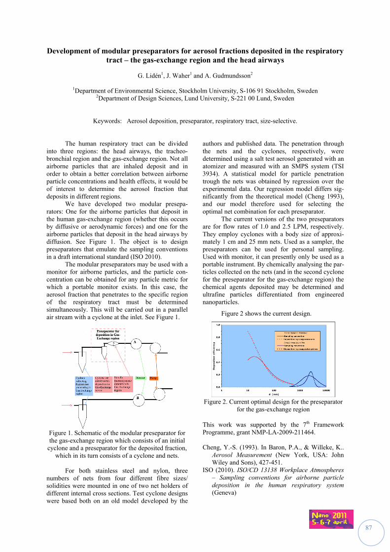
87
Development of modular preseparators for aerosol fractions deposited in the respiratory tract – the gas-exchange region and the head airways
G. Lidén1, J. Waher1 and A. Gudmundsson2
1Department of Environmental Science, Stockholm University, S-106 91 Stockholm, Sweden
2Department of Design Sciences, Lund University, S-221 00 Lund, Sweden
Keywords: Aerosol deposition, preseparator, respiratory tract, size-selective.
The human respiratory tract can be divided into three regions: the head airways, the tracheo-bronchial region and the gas-exchange region. Not all airborne particles that are inhaled deposit and in order to obtain a better correlation between airborne particle concentrations and health effects, it would be of interest to determine the aerosol fraction that deposits in different regions. We have developed two modular presepa-rators: One for the airborne particles that deposit in the human gas-exchange region (whether this occurs by diffusive or aerodynamic forces) and one for the airborne particles that deposit in the head airways by diffusion. See Figure 1. The object is to design preseparators that emulate the sampling conventions in a draft international standard (ISO 2010). The modular preseparators may be used with a monitor for airborne particles, and the particle con-centration can be obtained for any particle metric for which a portable monitor exists. In this case, the aerosol fraction that penetrates to the specific region of the respiratory tract must be determined simultaneously. This will be carried out in a parallel air stream with a cyclone at the inlet. See Figure 1.
Figure 1. Schematic of the modular preseparator for the gas-exchange region which consists of an initial
cyclone and a preseparator for the deposited fraction, which in its turn consists of a cyclone and nets.
For both stainless steel and nylon, three numbers of nets from four different fibre sizes/ solidities were mounted in one of two net holders of different internal cross sections. Test cyclone designs were based both on an old model developed by the
authors and published data. The penetration through the nets and the cyclones, respectively, were determined using a salt test aerosol generated with an atomizer and measured with an SMPS system (TSI 3934). A statistical model for particle penetration trough the nets was obtained by regression over the experimental data. Our regression model differs sig-nificantly from the theoretical model (Cheng 1993), and our model therefore used for selecting the optimal net combination for each preseparator. The current versions of the two preseparators are for flow rates of 1.0 and 2.5 LPM, respectively. They employ cyclones with a body size of approxi-mately 1 cm and 25 mm nets. Used as a sampler, the preseparators can be used for personal sampling. Used with monitor, it can presently only be used as a portable instrument. By chemically analysing the par-ticles collected on the nets (and in the second cyclone for the preseparator for the gas-exchange region) the chemical agents deposited may be determined and ultrafine particles differentiated from engineered nanoparticles.
Figure 2 shows the current design.
Figure 2. Current optimal design for the preseparator
for the gas-exchange region
This work was supported by the 7th Framework Programme, grant NMP-LA-2009-211464. Cheng, Y.-S. (1993). In Baron, P.A., & Willeke, K..
Aerosol Measurement (New York, USA: John Wiley and Sons), 427-451.
ISO (2010). ISO/CD 13138 Workplace Atmospheres – Sampling conventions for airborne particle deposition in the human respiratory system (Geneva)
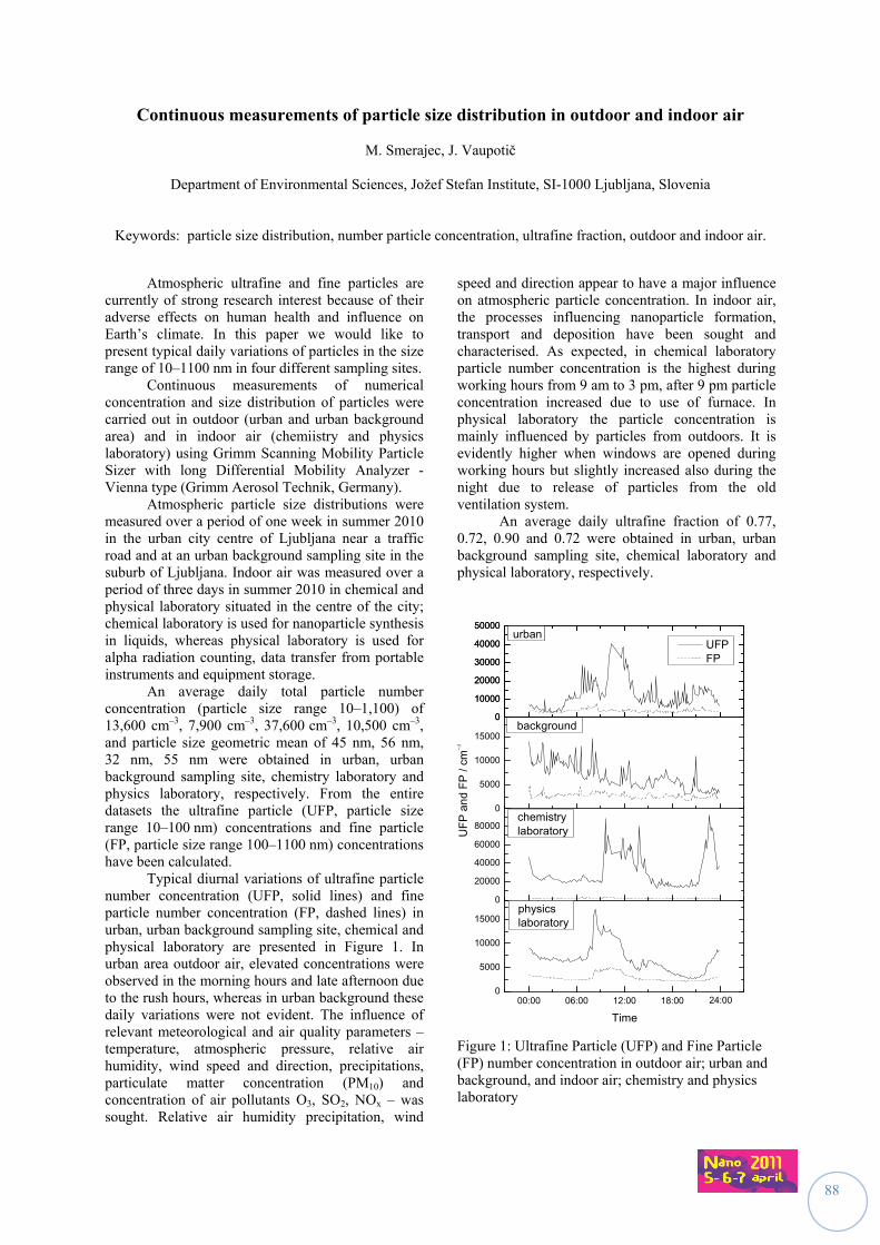
88
Continuous measurements of particle size distribution in outdoor and indoor air
M. Smerajec, J. Vaupotič
Department of Environmental Sciences, Jožef Stefan Institute, SI-1000 Ljubljana, Slovenia
Keywords: particle size distribution, number particle concentration, ultrafine fraction, outdoor and indoor air.
Atmospheric ultrafine and fine particles are
currently of strong research interest because of their adverse effects on human health and influence on Earth’s climate. In this paper we would like to present typical daily variations of particles in the size range of 10–1100 nm in four different sampling sites.
Continuous measurements of numerical concentration and size distribution of particles were carried out in outdoor (urban and urban background area) and in indoor air (chemiistry and physics laboratory) using Grimm Scanning Mobility Particle Sizer with long Differential Mobility Analyzer - Vienna type (Grimm Aerosol Technik, Germany).
Atmospheric particle size distributions were measured over a period of one week in summer 2010 in the urban city centre of Ljubljana near a traffic road and at an urban background sampling site in the suburb of Ljubljana. Indoor air was measured over a period of three days in summer 2010 in chemical and physical laboratory situated in the centre of the city; chemical laboratory is used for nanoparticle synthesis in liquids, whereas physical laboratory is used for alpha radiation counting, data transfer from portable instruments and equipment storage.
An average daily total particle number concentration (particle size range 10–1,100) of 13,600 cm–3, 7,900 cm–3, 37,600 cm–3, 10,500 cm–3, and particle size geometric mean of 45 nm, 56 nm, 32 nm, 55 nm were obtained in urban, urban background sampling site, chemistry laboratory and physics laboratory, respectively. From the entire datasets the ultrafine particle (UFP, particle size range 10–100 nm) concentrations and fine particle (FP, particle size range 100–1100 nm) concentrations have been calculated.
Typical diurnal variations of ultrafine particle number concentration (UFP, solid lines) and fine particle number concentration (FP, dashed lines) in urban, urban background sampling site, chemical and physical laboratory are presented in Figure 1. In urban area outdoor air, elevated concentrations were observed in the morning hours and late afternoon due to the rush hours, whereas in urban background these daily variations were not evident. The influence of relevant meteorological and air quality parameters – temperature, atmospheric pressure, relative air humidity, wind speed and direction, precipitations, particulate matter concentration (PM10) and concentration of air pollutants O3, SO2, NOx – was sought. Relative air humidity precipitation, wind
speed and direction appear to have a major influence on atmospheric particle concentration. In indoor air, the processes influencing nanoparticle formation, transport and deposition have been sought and characterised. As expected, in chemical laboratory particle number concentration is the highest during working hours from 9 am to 3 pm, after 9 pm particle concentration increased due to use of furnace. In physical laboratory the particle concentration is mainly influenced by particles from outdoors. It is evidently higher when windows are opened during working hours but slightly increased also during the night due to release of particles from the old ventilation system.
An average daily ultrafine fraction of 0.77, 0.72, 0.90 and 0.72 were obtained in urban, urban background sampling site, chemical laboratory and physical laboratory, respectively.
00:00 06:00 12:00 18:00 00:000
5000
10000
15000
0
20000
40000
60000
80000
0
5000
10000
15000
0
10000
20000
30000
40000
50000
0
10000
20000
30000
40000
50000
physicslaboratory
Time
chemistrylaboratory
UFP FP
UF
P a
nd
FP
/ cm
background
urban
24:00
urban
Figure 1: Ultrafine Particle (UFP) and Fine Particle (FP) number concentration in outdoor air; urban and background, and indoor air; chemistry and physics laboratory
89
Performance of a Personal Sampler for Nanoparticles
Y.S. Cheng1, Y. Zhou1 and C.J. Tsai2
1Lovelace Respiratory Research Institute, 2425 Ridgecrest, 87108, Albuquerque, USA 2National Chiao Tung University, 1001 University Road, 30010, Hsinchu, Taiwan
Keywords: nanoparticles, personal sampler
Production of nanomaterials has increased continuously because of the unique physicochemical characteristics and extensive applications of these materials. There are great concerns for the potential health effects of exposure to these nanoparticles. Because nanomaterials are small in size with large surface areas, many studies have shown that the biological effects of nanomaterials are greater than bulk material of the same chemical composition. In 2005, the National Institute for Occupational Safety and Health (NIOSH) recommended exposure limits of 1.5 mg/m3 for fine TiO2 and 0.1 mg/m3 for ultrafine TiO2, as time-weighted average (TWA) concentrations for up to 10 hr/day during a 40-hr work week. However, there are no suitable personal samplers capable of assessing the exposure level of ultrafine particles or nanoparticles.
The overall objective of this study is to develop a personal sampler capable of collecting the ultrafine particles (nanoparticles) in the occupational environment. This sampler consists of a cyclone for respirable particle classification (Tsai et al., 2001), micro-orifice impactor stage with an acceleration nozzle to achieve nanoparticle classification and a backup filter to collect nanoparticles. By applying high and localized velocity in the nozzle, diffusion deposition of nanoparticles can be avoided in the classifying process, and nanoparticles can be collected in the downstream backup filter. The cut off diameter for the cyclone is 4 m. The cutoff-diameter for the impactor stage is 100 nm. The backup filter collect nanoparticles <100 nm.
Performance test of the sampler includes efficiency testing using monodisperse NaCl particles using a dry evaporation and condensation method (Cheng et al., 1990). The dry evaporation and condensation method produces mainly particles < 800 nm. The aerosol is then passed through a single-stage impactor to remove coarse particles > 1000 nm before entering the DMA for classification into monodisperse particles in the range of 10 to about 800 nm. The particle size of the classified aerosol is calculated from the operating parameters of the DMA, including aerosol and sheath flow rates and applied voltage (Cheng et al., 1990), and is checked using an SMPS (TSI, Inc.). The classified aerosol is brought into charge equilibrium in a charge
neutralizer and enters the test sampler. The concentrations of the inlet (Cin) and outlet (Cout) of the sampler are monitored using a CPC (Model 3022A, TSI, Inc.). From the inlet and outlet concentration, the overall sampler collection efficiency is estimated. Our results show that the 50% efficiency of the impactor stage is around 100 nm at a flow rate of 2 L/min.
Aspiration efficiency of the sampler is also measured in an aerosol wind tunnel. The sampler is placed inside the test section of the wind tunnel with the sampling inlet facing the wind direction. The test sampler is operated side-by-side with two isokinetic probes for a fixed sampling time (Cheng et al., 2004). These probes are used as reference samplers. The sampler is tested at wind speeds of 0.3, 0.75, and 2.0 m/s that cover a wide range of air velocity in occupational environment (Aizenberg et al., 2000). The flow rate to the personal sampler is maintained at 2 L/min. Monodisperse fluorescent polymer microspheres in the size range of 20 to 800 nm (Thermo Scientific, Fremont, CA) are used as the test particles. The test aerosol is generated using a Hospitak nebulizer. The concentration of fluorescent particles collected on the personal sampler and isokinetic samplers will then be used to calculate the aspiration efficiency. Also particle losses inside the sampler can be determined. Our preliminary results shows that the personal sampler are able to collect nanoparticles <100 nm for gravimetric analysis. This work was supported by the NIOSH under grant R01OH009801 and IOSH under the grant 99-A323. Aizenberg, V., Grinshpun, S.A., Willeke, K., Smith,
J., & Baron, P.A. (2000). J. Aerosol. Sci. 3,169-179.
Cheng, Y.S., Yamada, Y., Yeh, H.C., & Su, Y.F. (1990). J. Aerosol Res., 5,44-51.
Cheng, Y.S., Irshad, H., McFarland, A.R., Su, W.C., Zhou, Y., & Barringer, D. (2004). Aerosol Sci. Technol., 38,1099-1107.
Tsai, C.J., Huang, C.H., Wang, S.H., & Shih, T.S. (2001). Aerosol Sci Technol, 35,611-616.
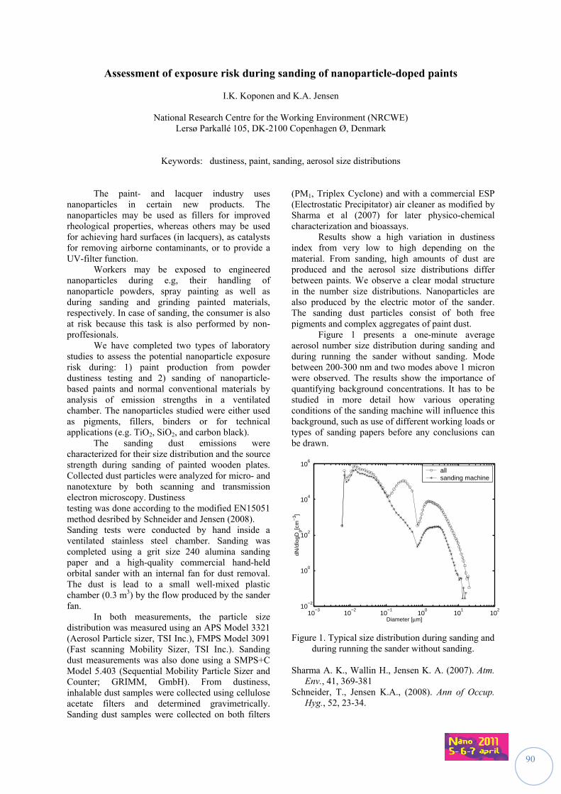
90
Assessment of exposure risk during sanding of nanoparticle-doped paints
I.K. Koponen and K.A. Jensen
National Research Centre for the Working Environment (NRCWE) Lersø Parkallé 105, DK-2100 Copenhagen Ø, Denmark
Keywords: dustiness, paint, sanding, aerosol size distributions The paint- and lacquer industry uses nanoparticles in certain new products. The nanoparticles may be used as fillers for improved rheological properties, whereas others may be used for achieving hard surfaces (in lacquers), as catalysts for removing airborne contaminants, or to provide a UV-filter function. Workers may be exposed to engineered nanoparticles during e.g, their handling of nanoparticle powders, spray painting as well as during sanding and grinding painted materials, respectively. In case of sanding, the consumer is also at risk because this task is also performed by non-proffesionals. We have completed two types of laboratory studies to assess the potential nanoparticle exposure risk during: 1) paint production from powder dustiness testing and 2) sanding of nanoparticle-based paints and normal conventional materials by analysis of emission strengths in a ventilated chamber. The nanoparticles studied were either used as pigments, fillers, binders or for technical applications (e.g. TiO2, SiO2, and carbon black). The sanding dust emissions were characterized for their size distribution and the source strength during sanding of painted wooden plates. Collected dust particles were analyzed for micro- and nanotexture by both scanning and transmission electron microscopy. Dustiness testing was done according to the modified EN15051 method desribed by Schneider and Jensen (2008). Sanding tests were conducted by hand inside a ventilated stainless steel chamber. Sanding was completed using a grit size 240 alumina sanding paper and a high-quality commercial hand-held orbital sander with an internal fan for dust removal. The dust is lead to a small well-mixed plastic chamber (0.3 m3) by the flow produced by the sander fan. In both measurements, the particle size distribution was measured using an APS Model 3321 (Aerosol Particle sizer, TSI Inc.), FMPS Model 3091 (Fast scanning Mobility Sizer, TSI Inc.). Sanding dust measurements was also done using a SMPS+C Model 5.403 (Sequential Mobility Particle Sizer and Counter; GRIMM, GmbH). From dustiness, inhalable dust samples were collected using cellulose acetate filters and determined gravimetrically. Sanding dust samples were collected on both filters
(PM1, Triplex Cyclone) and with a commercial ESP (Electrostatic Precipitator) air cleaner as modified by Sharma et al (2007) for later physico-chemical characterization and bioassays. Results show a high variation in dustiness index from very low to high depending on the material. From sanding, high amounts of dust are produced and the aerosol size distributions differ between paints. We observe a clear modal structure in the number size distributions. Nanoparticles are also produced by the electric motor of the sander. The sanding dust particles consist of both free pigments and complex aggregates of paint dust. Figure 1 presents a one-minute average aerosol number size distribution during sanding and during running the sander without sanding. Mode between 200-300 nm and two modes above 1 micron were observed. The results show the importance of quantifying background concentrations. It has to be studied in more detail how various operating conditions of the sanding machine will influence this background, such as use of different working loads or types of sanding papers before any conclusions can be drawn.
10−3
10−2
10−1
100
101
102
10−2
100
102
104
106
dN/d
logD
p[cm
−3 ]
Diameter [μm]
allsanding machine
Figure 1. Typical size distribution during sanding and during running the sander without sanding.
Sharma A. K., Wallin H., Jensen K. A. (2007). Atm.
Env., 41, 369-381 Schneider, T., Jensen K.A., (2008). Ann of Occup.
Hyg., 52, 23-34.
91
Dustiness testing of Nanopowders
D. Bard, G. Burdett* and A. Kelly
Health and Safety Laboratory, Harpur Hill, Buxton SK17 9JN, UK
Keywords: Dustiness testing, gravimetric, nanopowders, electron microscopy.
The European standard for “dustiness” EN15051 has been used for several years to assess and classify the dustiness of conventional powders and bulk materials by the gravimetric determination of the three biologically relevant fractions: inhalable, thoracic and respirable. Manufactured nanopowders whose primary particles (nano-objects) are below 100 nm are thought to have additional biological potential due to their small size and large surface areas, which may not be adequately described by the gravimetric standard alone. This paper investigates a number of additional measurements and descriptors of the particles released to air during the standard rotating drum dustiness test with a range of nanopowders.
The aim was to first carry out a standard gravimetric dustiness test as described in EN15051, then to replace the final filter to allow the particles to enter a number of instruments to further describe the airborne particle number and size distribution.
The range of instruments used included: TSI Inc. P-TRAK condensation nucleus counters to give particle number count over the range of 10- 1000nm, a Grimm scanning mobility particle sizer (SMPS) to give a number count and size distribution over the range of 10 – 875 nm, a TSI Inc. 3321 TSI Inc. 3091 fast mobility particle sizer (FMPS) to give a number count and size distribution over the range of 5.6 - 560 nm, a TSI Inc. 3321 aerosol particle sizer (APS) to look at particle count and size distribution in the range of 500 – 20,000 nm and an electrostatic precipitator for the collection of particles onto a carbon film on a electron microscopy grid for off-line sizing by transmission electron microscopy.
When testing the dustiness of any powder, the size of the particles released to air is initially dependent on the degree of dis-agglomeration of the particles from the bulk powder sample, i.e. the cohesive / adhesive forces joining the particles together. Once airborne, particles may rapidly re-agglomerate and settle out during the dust generation and transport to the measurement instruments. These losses are due to a combination of diffusion, gravitational settling and inertial impaction depending on the size of the particles and agglomerates formed. The particle concentration of the dust released and the time it takes to reach the measurement instrument are key determinants of the particle number and size distribution that will be measured. Therefore standardisation on instrumentation and run conditions are essential so that useful comparisons between dusts can be made. The current standardised set-up used by HSL and the results from a number of nanopowders are described. The relevance of the additional parameters measured and how this information can be usefully incorporated into a standardised nanopowder dustiness test is discussed. EN15051:1996 Workplace atmospheres.
Measurement of the dustiness of bulk materials. Requirements and reference test methods. ISBN 0 580 48345 2
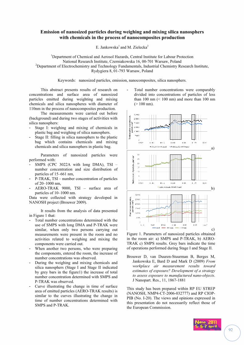
92
Emission of nanosized particles during weighing and mixing silica nanosphers with chemicals in the process of nanocomposites production
E. Jankowska1 and M. Zielecka2
1Department of Chemical and Aerosol Hazards, Central Institute for Labour Protection
National Research Institute, Czerniakowska 16, 00-701 Warsaw, Poland 2Department of Electrochemistry and Technology Fundamentals, Industrial Chemistry Research Institute,
Rydygiera 8, 01-793 Warsaw, Poland
Keywords: nanosized particles, emission, nanocomposites, silica nanosphers. This abstract presents results of research on concentrations and surface area of nanosized particles emitted during weighting and mixing chemicals and silica nanospheres with diameter of 110nm in the process of nanocomposites production. The measurements were carried out before (background) and during two stages of activities with silica nanosphers: - Stage I: weighing and mixing of chemicals in
plastic bag and weighing of silica nanosphers. - Stage II: filling in silica nanosphers to the plastic
bag which contains chemicals and mixing chemicals and silica nanosphers in plastic bag.
Parameters of nanosized particles were performed with: - SMPS (CPC 3022A with long DMA), TSI –
number concentration and size distribution of particles of 15–661 nm,
- P-TRAK, TSI – number concentration of particles of 20–1000 nm,
- AERO-TRAK 9000, TSI – surface area of particles of 10–1000 nm.
Data were collected with strategy developed in NANOSH project (Brouwer 2009).
It results from the analysis of data presented in Figure 1 that: - Total number concentrations determined with the
use of SMPS with long DMA and P-TRAK were similar, when only two persons carrying out measurements were present in the room and no activities related to weighing and mixing the components were carried out.
- When another two persons, who were preparing the components, entered the room, the increase of number concentrations was observed.
- During the weighing and mixing chemicals and silica nanosphers (Stage I and Stage II indicated by grey bars in the figure1) the increase of total number concentration determined with SMPS and P-TRAK was observed.
- Curve illustrating the change in time of surface area of emitted particles (AERO-TRAK results) is similar to the curves illustrating the change in time of number concentrations determined with SMPS and P-TRAK.
- Total number concentrations were comparably divided into concentrations of particles of less than 100 nm (< 100 nm) and more than 100 nm (> 100 nm).
a)
b)
c) Figure 1. Parameters of nanosized particles obtained in the room air: a) SMPS and P-TRAK, b) AERO-TRAK c) SMPS results. Grey bars indicate the time of operations performed during Stage I and Stage II.
Brouwer D, van Duuren-Stuurman B, Berges M, Jankowska E, Bard D and Mark D (2009) From workplace air measurement results toward estimates of exposure? Development of a strategy to assess exposure to manufactured nano-objects. J Nanopart. Res., 11, 1867-1881
This study has been prepared within RP EU STREP (NANOSH, NMP4-CT-2006-032777) and RP CIOP-PIB (No. I-20). The views and opinions expressed in this presentation do not necessarily reflect those of the European Commission.
93
Approach towards an Exposure Assessment Strategy for Aerosols Released from Engineered Nanomaterials from Workplace Operations
M. Reuter1, N. Schröter2, D. Eichstädt3, A. Rommert3, R. Fischer4, S. Engel5, J. Ragot6, M. Voetz7, K. Kund8, S. Klages-Büchner9, K. Swain10, S. Knobl11, M. Reisinger12, R. Weinand12, U. Billerbeck13, M. Heinemann14
1German Chemical Industry Association (VCI)
2German Industry Association for Construction Chemicals 3German Paint and Printing Ink Association (VdL) 4Verband der Mineralfarbenindustrie e. V. (VdMi)
5BASF SE, 69121, Ludwigshafen, Germany 6Bayer MaterialScience AG 51368 Leverkusen, Germany,
7Bayer Technology Services GmbH, 51368 Leverkusen, Germany 8Clariant Deutschland GmbH, 65926 Frankfurt, Germany
9DuPont Deutschland Holding GmbH & Co. KG, 10117 Berlin, Germany 10DuPont, Wilmington, Delaware 19880-0322, USA
11Eckart GmbH, 91235 Hartenstein, Germany 12Evonik Degussa GmbH, 63457 Hanau, Germany
13Merck KGaA, 64271 Darmstadt, Germany 14Wacker Chemie AG, 84480 Burghausen, Germany
Keywords: Engineered Nanomaterials, Aerosols, Exposure Assessment in the Workplace
Engineered nanomaterials are fascinating, new materials with significantly improved or completely novel properties. They are being handled more and more in the workplaces both in research and in production.
The German Chemical Industry is committed to a safe, responsible and sustainable development of this highly promising technology.
Efficient, reliable and also pragmatic exposure assessment is a crucial element to manage potential risks potentially posed by hazardous chemicals in the workplace.
VCI has thus established a working group dealing with the challenges of exposure to nanomaterials in the workplace. The working group is aiming at developing a harmonized approach to exposure assessment of aerosols released from engineered nanomaterials in the workplace. A pragmatic, tiered-type approach, which can be widely used in both small and medium enterprises as well as in large chemical companies with global business and operations, is in the focus of the working group.
The approach refers to the usage of both, easy-to-use equipment (in tier 2 exposure assessment) as well as on a higher tier sophisticated equipment (in tier 3 exposure assessment) for an extended exposure assessment if required. For example tier 3 exposure assessment requires condensation particle counters (CPC) or comparable portable equipment, scanning mobility particle sizers (SMPS), optical particle counters (OPC), dust monitors and methodologies for chemical analysis as well as electron microscope (EM). In addition these methodologies should be supplemented by climatic data collection equipment for the characterization of air flow, temperature and humidity in the environment of the potential emission source.
First, basic information about the product characteristics (e.g., chemical identity, dustiness, etc.), the workplace and the process steps, which may lead to aerosol formation should be collected. This information will be supplemented by data characterizing the ventilation situation and potential confounding variables, like exhaust fumes, thermal effects, etc. Subsequently, the aerosol background concentration will be characterized, either with a near- or a far-field approach. In the near-field approach comparative measurements before and after operations are carried out with a single methodology, whereas the aerosol background concentration is monitored in parallel to the exposure assessment at the source utilizing a second piece of equipment in the far-field procedure. Finally, the concentration of aerosols released from engineered nanomaterials will be assessed during regular operations.
The usage of easy-to-use equipment is recommended in lower tier exposure assessment. If these results will indicate potential exposure to aerosols released from engineered nanomaterials a more extended exposure assessment (i.e., tier 3 exposure asssessment) is recommended. Higher tier exposure assessment will require the usage of additional sophisticated equipment and in addition the collection of dust samples for subsequent chemical or EM analysis.
The data will be evaluated by comparison of the aersosol background data with the concentration during regular operations. If an increase of the particle concentration above an interference value is observed, appropriate risk management measures have to be taken and their efficiency has to be evaluated.
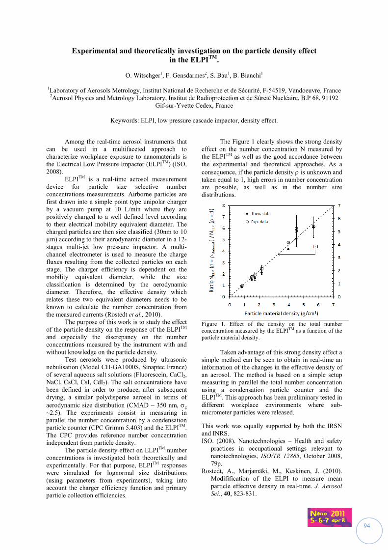
94
Experimental and theoretically investigation on the particle density effect in the ELPITM.
O. Witschger1, F. Gensdarmes2, S. Bau1, B. Bianchi1
1Laboratory of Aerosols Metrology, Institut National de Recherche et de Sécurité, F-54519, Vandoeuvre, France
2Aerosol Physics and Metrology Laboratory, Institut de Radioprotection et de Sûreté Nucléaire, B.P 68, 91192 Gif-sur-Yvette Cedex, France
Keywords: ELPI, low pressure cascade impactor, density effect.
Among the real-time aerosol instruments that can be used in a multifaceted approach to characterize workplace exposure to nanomaterials is the Electrical Low Pressure Impactor (ELPITM) (ISO, 2008). ELPITM is a real-time aerosol measurement device for particle size selective number concentrations measurements. Airborne particles are first drawn into a simple point type unipolar charger by a vacuum pump at 10 L/min where they are positively charged to a well defined level according to their electrical mobility equivalent diameter. The charged particles are then size classified (30nm to 10 µm) according to their aerodynamic diameter in a 12-stages multi-jet low pressure impactor. A multi-channel electrometer is used to measure the charge fluxes resulting from the collected particles on each stage. The charger efficiency is dependent on the mobility equivalent diameter, while the size classification is determined by the aerodynamic diameter. Therefore, the effective density which relates these two equivalent diameters needs to be known to calculate the number concentration from the measured currents (Rostedt et al., 2010). The purpose of this work is to study the effect of the particle density on the response of the ELPITM and especially the discrepancy on the number concentrations measured by the instrument with and without knowledge on the particle density.
Test aerosols were produced by ultrasonic nebulisation (Model CH-GA1000S, Sinaptec France) of several aqueous salt solutions (Fluorescein, CaCl2, NaCl, CsCl, CsI, CdI2). The salt concentrations have been defined in order to produce, after subsequent drying, a similar polydisperse aerosol in terms of aerodynamic size distribution (CMAD ~ 350 nm, g ~2.5). The experiments consist in measuring in parallel the number concentration by a condensation particle counter (CPC Grimm 5.403) and the ELPITM. The CPC provides reference number concentration independent from particle density.
The particle density effect on ELPITM number concentrations is investigated both theoretically and experimentally. For that purpose, ELPITM responses were simulated for lognormal size distributions (using parameters from experiments), taking into account the charger efficiency function and primary particle collection efficiencies.
The Figure 1 clearly shows the strong density effect on the number concentration N measured by the ELPITM as well as the good accordance between the experimental and theoretical approaches. As a consequence, if the particle density is unknown and taken equal to 1, high errors in number concentration are possible, as well as in the number size distributions.
Figure 1. Effect of the density on the total number concentration measured by the ELPITM as a function of the particle material density.
Taken advantage of this strong density effect a simple method can be seen to obtain in real-time an information of the changes in the effective density of an aerosol. The method is based on a simple setup measuring in parallel the total number concentration using a condensation particle counter and the ELPITM. This approach has been preliminary tested in different workplace environments where sub-micrometer particles were released.
This work was equally supported by both the IRSN and INRS. ISO. (2008). Nanotechnologies – Health and safety
practices in occupational settings relevant to nanotechnologies, ISO/TR 12885, October 2008, 79p.
Rostedt, A., Marjamäki, M., Keskinen, J. (2010). Modifification of the ELPI to measure mean particle effective density in real-time. J. Aerosol Sci., 40, 823-831.
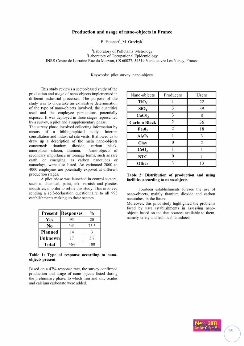
95
Production and usage of nano-objects in France
B. Honnert1 , M. Grzebyk2
1Laboratory of Pollutants Metrology 2Laboratory of Occupational Epidemiology
INRS Centre de Lorraine Rue du Morvan, CS 60027, 54519 Vandoeuvre Les Nancy, France.
Keywords: pilot survey, nano-objects This study reviews a sector-based study of the production and usage of nano-objects implemented in different industrial processes. The purpose of the study was to undertake an exhaustive determination of the type of nano-objects involved, the quantities used and the employee populations potentially exposed. It was deployed in three stages represented by a survey, a pilot and a supplementary phase. The survey phase involved collecting information by means of a bibliographical study, Internet consultation and industrial site visits. It allowed us to draw up a description of the main nano-objects concerned: titanium dioxide, carbon black, amorphous silicon, alumina. Nano-objects of secondary importance in tonnage terms, such as rare earth, or emerging, as carbon nanotubes or nanoclays, were also listed. An estimated 2000 to 4000 employees are potentially exposed at different production stages. A pilot phase was launched in control sectors, such as chemical, paint, ink, varnish and plastics industries, in order to refine this study. This involved sending a self-declaration questionnaire to all 993 establishments making up these sectors.
Present Responses % Yes 93 20
No 341 73.5
Planned 14 3
Unknown 17 3.7
Total 464 100 Table 1: Type of response according to nano-objects present Based on a 47% response rate, the survey confirmed production and usage of nano-objects listed during the preliminary phase, to which iron and zinc oxides and calcium carbonate were added.
Nano-objects Producers Users
TiO2 1 22
SiO2 3 39
CaC03 3 8
Carbon Black 2 36
Fe203 2 18
Al2O3 1 3
Clay 0 2
CeO2 1 1
NTC 0 1
Other 3 13
Table 2: Distribution of production and using facilities according to nano-objects Fourteen establishments foresee the use of nano-objects, mainly titanium dioxide and carbon nanotubes, in the future. Moreover, this pilot study highlighted the problems faced by user establishments in assessing nano-objects based on the data sources available to them, namely safety and technical datasheets.
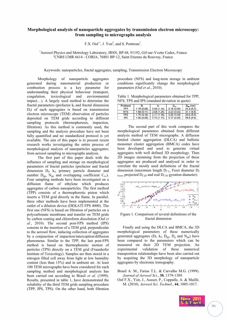
96
Morphological analysis of nanoparticle aggregates by transmission electron microscopy: from sampling to micrographs analysis
F.X. Ouf 1, J. Yon2, and S. Pontreau1.
1Aerosol Physics and Metrology Laboratory, IRSN, BP 68, 91192, Gif-sur-Yvette Cedex, France
2CNRS UMR 6614 - CORIA, 76801 BP-12, Saint Etienne du Rouvray, France
Keywords: nanoparticles, fractal aggregates, sampling, Transmission Electron Microscopy
Morphology of nanoparticle aggregates generated during nanomaterial production or combustion process is a key parameter for understanding their physical behaviour (transport, coagulation, toxicological and environmental impact…). A largely used method to determine the fractal parameters (prefactor kf and fractal dimension Df) of such aggregates is based on transmission electron microscope (TEM) observation of particles deposited on TEM grids according to different sampling protocols (thermophoresis, impaction, filtration). As this method is commonly used, the sampling and the analysis procedure have not been fully quantified and no standardized protocol is yet available. The aim of this paper is to present recent research works investigating the entire process of morphological analysis of nanoparticles aggregates; from aerosol sampling to micrographs analysis. The first part of this paper deals with the influence of sampling and storage on morphological parameters of fractal particles (prefactor and fractal dimension Df, kf, primary particle diameter and number Dpp, Npp and overlapping coefficient Cov). Four sampling methods have been investigated on a diffusion flame of ethylene which produces aggregates of carbon nanoparticles. The first method (TPP) consists of a thermophoretic piston which inserts a TEM grid directly in the flame. In parallel, three other methods have been implemented at the outlet of a dilution device (DEKATI FPS 4000). The first one (NFS) is based on filtration of particles on a polycarbonate membrane and transfer on TEM grids by carbon coating and chloroform dissolution (Ouf et al., 2010). The second post-FPS method (IPS) consists in the insertion of a TEM grid, perpendicular to the aerosol flow, inducing collection of aggregates by a conjunction of impaction/interception/diffusion phenomena. Similar to the TPP, the last post-FPS method is based on thermophoretic motion of particles (TPS) directly on a TEM grid (Fraunhofer Institute of Toxicology). Samples are then stored in a nitrogen filled cell away from light at low humidity content (less than 15%) and in ambient air. At least 100 TEM micrographs have been considered for each sampling method and morphological analysis has been carried out according to Brasil et al. (1999). Results, presented in table 1, have demonstrated the reliability of the third TEM grids sampling procedure (TPP, IPS, TPS). On the other hand, both filtration
procedure (NFS) and long-term storage in ambient conditions significantly change the morphological parameters (Ouf et al., 2010). Table 1. Morphological parameters obtained for TPP, NFS, TPS and IPS (standard deviation in quote)
The second part of this work compares the morphological parameters obtained from different analysis method of TEM micrographs. A diffusion limited cluster aggregation (DLCA) and ballistic monomer cluster aggregation (BMCA) codes have been developed and used to generate virtual aggregates with well defined 3D morphology. Then 2D images stemming from the projection of these aggregates are produced and analysed in order to correlate the mostly used definitions of the fractal dimension (maximum length Df L, Feret diameter Df
Feret, projected Df 2D and real Df 3D gyration diameter).
Figure 1. Comparison of several definitions of the
fractal dimension
Finally and using the DLCA and BMCA, the 3D morphological parameters of these numerically generated aggregates (Df, kf, Dpp, Dg and Npp) have been compared to the parameters which can be measured on their 2D TEM projection. An experimental validation of these numerical transposition relationships have been also carried out by acquiring the 3D morphology of nanoparticle aggregates by electronic tomography. Brasil A. M., Farias T.L. & Carvalho M.G. (1999).
Journal of Aerosol Sci., 30, 1379-1389. Ouf F.X., Yon, J., Ausset, P., Coppalle, A. & Maillé,
M. (2010). Aerosol Sci. Technol., 44, 1005-1017.
97
A Simulation Facility to Test Workplace Aerosol Samplers
G.C. Dragan1, E. Karg1, H. Nordsieck1,2 , J. Maguhn1, J. Schnelle-Kreis1 and R. Zimmermann1,3
1Institute of Ecological Chemistry, Helmholtz Zentrum Muenchen, D-85764, Munich, Germany 2bifa Environmental Institute, Am Mittleren Moos 46, D-86167, Augsburg, Germany
3Department of Technical and Analytical Chemistry, University Rostock, Dr.-Lorenz-Weg 1, D-18051 Rostock, Germany
Keywords: mineral oil mist, mixed phases measurement, sampling errors, sampling strategy.
Airborne semi-volatile hazardous substances are often partitioned between gas and particle phase; the mass concentration in each phase mainly depends on ambient temperature and partial vapour pressure. Due to their separation, artefacts are likely to occur in workspace aerosol samplers (filter blow-on, blow-off, evaporation, condensation, particle penetration) and may lead to errors in risk assessment as a consequence. Mineral oil, frequently used as a coolant, lubricant or cutting fluid during the machining of metal components in many industrial processes is known to form oil mist. Mineral oil mainly contains a mixture of straight-chain, aliphatic hydrocarbons from C12H26 to C23H48 (Yue, 1998; Raynor, 2008). In the case of oil mist, the particulate phase is considered to be of greater toxicological concern than the vapour, even though the vapour is often present in much higher concentrations (Simpson, 2008). Since cutting fluids are complex in composition, they may be more toxic than their constituents taken individually and may be irritant or allergenic even if the raw materials are safe (Bienkowski, 1993). Biological response and toxicity of hazardous workplace aerosols depend to a great extent on the state of aggregation of the substances applied (Kreyling, 2007). Health hazards are mainly caused by the exposure to oil mists. The measurement of semi-volatile hydrocarbons in workplace air is complicated by their readiness to condense and form particles or adsorb on to surfaces. On the other hand, traditional filter sampling of the mist loses oil by evaporation. (Simpson, 2008). Metalworking fluid mists containing mineral oil are typical examples of non-equilibrium aerosols, because long residence times are needed in order to approach thermodynamic equilibrium between droplets and vapour. (Nagel & Schaber, 2006) Our research objective is to evaluate the impact of sampling and analysis on the real phase distribution of dynamic gas-particle-mixtures. In order to achieve this purpose, an experimental system was designed for in-situ gas-particle analysis inside of a twin set of environmental chambers. They are operated independently from each other at temperatures ranging from 10 to 40 °C. The setup
allows simulating the three phases in an aerosol life cycle: (1) generation, (2) transport and (3) deposition/analysis. The experimental system consists of: a Sinclair-La Mer type aerosol generator (Topas SLG 270) to produce particles from the test substances (decane…octadecane) by vapour deposition on condensation nuclei. a mixing head designed to replicate atmospheric dilution while obtaining a homogenous aerosol - dilution gas mixing. a flow-tube reactor to study the dynamic processes during and directly after the aerosol production. a white light particle sizer (Palas WELAS) and a FTIR gas analyzer (Gasmet DX 4000) for in-situ characterization of particle- and gas phase. a 600 l Tedlar ageing bag that allows 10…60 minutes of residence time to simulate aerosol ageing. The aged aerosol is recirculated and analysed with the identical sampling equipment, therefore aerosol transformations during ageing are assessed. Errors in risk assessment can be studied by comparing the results obtained from the traditional filter/sampling equipment with the in-situ experimental system. Our first results show that condensation aerosols from all test substances can be produced with sufficient stability and that the aerosol is very sensitive to variations in temperature, vapour pressure and residence time. This work is supported by the German Statutory Accident Insurance (DGUV) under Research Contract FP 299. Bienkowski, K. (1993). Manufacturing Engineering,
March 1993, 90-96. Kreyling, W.G., Möller, W., Semmler-Behnke, M., &
Oberdörster, G. (2007). in Particle Toxicology,Boca Raton, CRC Press, Taylor & Francis Group, 47-74.
Nagel, D., & Schaber, K. (2006). in Characterization and separation of oil mist - Research Report FZKA-BWPLUS (Karlsruhe, Germany).
Raynor, P.C., & Leith, D. (1999). Ann. Occup. Hyg., 43(3), 181-192.
Simpson, A.T., & Wright, M.D. (2008). Ann. Occup. Hyg., 52(4), 249-257.
Yue, Y., Zheng, Y., & Sutherland, J.W. (1998). in Proc. 1998 Japan-USA Symp. on Flexible Automation.
98
Granulometry re-invented ? Characterization of as-produced, released and as-tested nanomaterials
W. Wohlleben1, K. Wiench2, R. Landsiedel3
1Polymer Physics Research, BASF SE, 67056 Ludwigshafen, Germany
2Product Safety, BASF SE 3Experimental Toxicology, BASF SE
Keywords: Characterisation for toxicology purposes, Size selective quantification,
Nanoparticle release from products. The existing regulatory framework for the registration, evaluation, authorisation and restriction of chemicals (REACH) does not contain specific provisions for nanomaterials. The commission’s scientific committees (SCENIHR, SCCP), EFSA and Competent Authority Working Groups such as CASG Nano have confirmed that the established principles and approaches to risk assessment of substances are, in general, applicable to nanomaterials. However, OECD judged only a minority of physico-chemical methods as applicable to nanomaterials, and updates to the guidance documents will be needed.. Granulometry is the outstanding property where the differences between traditional materials and nanomaterials culminate. In the present version of Guidance R.7.1.14, many nano-specific properties are discussed already for the micron scale: size, shape, specific surface, and dustiness, too. Even the issue of crystallomorphology, currently hidden under Naming, is closely connected to primary particle size via the X-ray diffraction broadening. Clearly, the methods and metrics for nanomaterials require a re-definition of granulometry guidance from scratch. The focus of the presentation is on pro's and con's for granulometry characterisation, aiming at the nano-guidance that will have to be developed for REACH, currently developed inter alia by the RIPoNs. In general, it would be naïve to define a single method for all nanomaterials. The methods actually measure different sizes: number/mass/ intensity distributions, with/without a solvent-swollen functionalization, with/without respect to agglomeration. Specific test cases from actual industrial products shows that results from 3 methods with complementary working principles should be reported: an ensemble method (best: volume-specific surface area VSSA), a microscopy (best: TEM) and a fractionating method (best: FFF or centrifuge). Further test cases of deliberately mixed nano-objects and micron-particles explores the detection limits in weight-% and substantiate why the above methods are preferred over popular 'simple' methods such as DLS and nanoparticle tracking. Beyond the characterization of as-produced nanomaterials, scientific evidence has accumulated that some properties such as their state of agglomeration, their solubility and also their surface
properties, are not constant, but change with the test medium.(Landsiedel 2010) We present novel metal-oxide examples of enhanced structure-activity correlation with biophysical characterization of the as-tested granulometry. With specific relevance for ecotoxicology, the paradigm of solubility as key physico-chemical indicator for transport and bioavailability may need revision. The property that determines transport and fate of nanomaterials is: dispersability (vs. agglomeration). Contrary to molecularly dissolved substances, solubility of nanomaterials depends on granulometry. The same applies to surface tension and partition coefficient, reducing their value for risk assessment. But what about the changes to nanomaterials over their life cycle? Typical consumer products are nanocomposite materials, where a matrix contains some wt-% of nanoparticles or –fibers. In what (hybrid) state are they released, if at all? First evidence emerged on nanocomposites coatings and paints. As a practical example of advanced granulometry, we characterized (with SEM, AUC, XPS, SIMS, diffraction) the degradation products from thermoplastic and cementitious nanocomposite materials after weathering vs. sanding vs. normal use, both as-released and as-tested-in-vivo-after-release. Results set upper limits on release of free nanofillers and reveal aerosol artifacts for specific (nano)materials. R. Landsiedel, L. Ma-Hock, A. Kroll, D. Hahn,
J. Schnekenburger, K. Wiench, and W. Wohlleben, 'Testing metal-oxide nanomaterials for human safety', Adv. Mater. 22, (2010), 2601-2627.
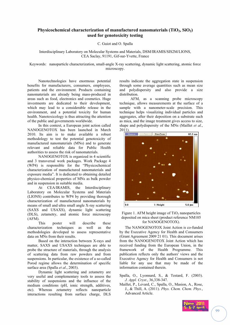
99
Physicochemical characterization of manufactured nanomaterials (TiO2, SiO2) used for genotoxicity testing
C. Guiot and O. Spalla
Interdisciplinary Laboratory on Molecular Systems and Materials, DSM/IRAMIS/SIS2M/LIONS, CEA Saclay, 91191, Gif-sur-Yvette, France
Keywords: nanoparticle characterization, small-angle X-ray scattering, dynamic light scattering, atomic force microscopy.
Nanotechnologies have enormous potential
benefits for manufacturers, consumers, employees, patients and the environment. Products containing nanomaterials are already being mass-produced in areas such as food, electronics and cosmetics. Huge investments are dedicated to their development, which may lead to a considerable release in the environment, and a potential toxicity for human health. Nanotoxicology is thus attracting the attention of the public and governments worldwide.
In this context, a European joint action called NANOGENOTOX has been launched in March 2010. Its aim is to make available a robust methodology to test the potential genotoxicity of manufactured nanomaterials (MNs) and to generate relevant and reliable data for Public Health authorities to assess the risk of nanomaterials.
NANOGENOTOX is organized in 4 scientific and 3 transversal work packages. Work Package 4 (WP4) is responsible for the “Physicochemical characterization of manufactured nanomaterials and exposure media”. It is dedicated to obtaining detailed physico-chemical properties of MNs as bulk powder and in suspension in suitable media.
At CEA/IRAMIS, the Interdisciplinary Laboratory on Molecular Systems and Materials (LIONS) contributes to WP4 by providing thorough characterization of manufactured nanomaterials by means of small and ultra small angle X-ray scattering (SAXS and USAXS), dynamic light scattering (DLS), zetametry, and atomic force microscopy (AFM).
This poster will describe these characterization techniques as well as the methodologies developed to assess representative data on MNs from their results.
Based on the interaction between X-rays and matter, SAXS and USAXS techniques are able to probe the structure of materials, through the analysis of scattering data from raw powders and from suspensions. In particular, the existence of a so-called Porod regime allows the determination of specific surface area (Spalla et al., 2003).
Dynamic light scattering and zetametry are very useful and complementary tools to assess the stability of suspensions and the influence of the medium conditions (pH, ionic strength, additives, etc). Whereas zetametry reflects nanoparticle interactions resulting from surface charge, DLS
results indicate the aggregation state in suspension through some average quantities such as mean size and polydispersity and also provide a size distribution.
AFM, as a scanning probe microscopy technique, allows measurements at the surface of a sample with a nanometer-scale precision. This technique helps visualizing individual particles and aggregates, after their deposition on a substrate such as mica, and the image treatment gives access to size, shape and polydispersity of the MNs (Maillet et al., 2011).
Figure 1. AFM height image of TiO2 nanoparticles deposited on mica sheet (product reference NM105
for NANOGENOTOX).
The NANOGENOTOX Joint Action is co-funded by the Executive Agency for Health and Consumers (Grant Agreement 2009 21 01). This document arises from the NANOGENOTOX Joint Action which has received funding from the European Union, in the framework of the Health Programme. This publication reflects only the authors' views and the Executive Agency for Health and Consumers is not liable for any use that may be made of the information contained therein.
Spalla, O., Lyonnard, S., & Testard, F. (2003). J. Appl. Cryst., 36,338-347.
Maillet, P., Levard, C., Spalla, O., Masion, A., Rose, J., & Thill, A. (2011). Phys. Chem. Chem. Phys., Advanced Article.
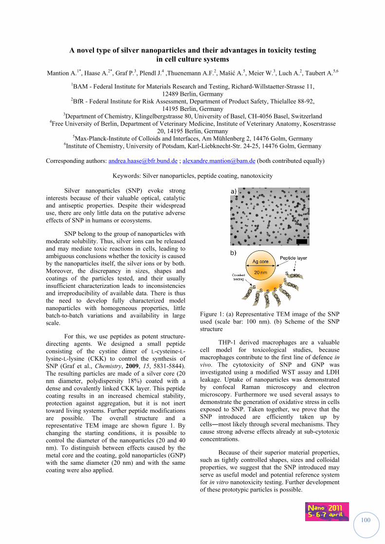
100
A novel type of silver nanoparticles and their advantages in toxicity testing in cell culture systems
Mantion A.1*, Haase A.2*, Graf P.3, Plendl J.4 ,Thuenemann A.F.2, Mašić A.5, Meier W.3, Luch A.2, Taubert A.5,6
1BAM - Federal Institute for Materials Research and Testing, Richard-Willstaetter-Strasse 11, 12489 Berlin, Germany
2BfR - Federal Institute for Risk Assessment, Department of Product Safety, Thielallee 88-92, 14195 Berlin, Germany
3Department of Chemistry, Klingelbergstrasse 80, University of Basel, CH-4056 Basel, Switzerland 4Free University of Berlin, Department of Veterinary Medicine, Institute of Veterinary Anatomy, Koserstrasse
20, 14195 Berlin, Germany 5Max-Planck-Institute of Colloids and Interfaces, Am Mühlenberg 2, 14476 Golm, Germany
6Institute of Chemistry, University of Potsdam, Karl-Liebknecht-Str. 24-25, 14476 Golm, Germany Corresponding authors: [email protected] ; [email protected] (both contributed equally)
Keywords: Silver nanoparticles, peptide coating, nanotoxicity
Silver nanoparticles (SNP) evoke strong interests because of their valuable optical, catalytic and antiseptic properties. Despite their widespread use, there are only little data on the putative adverse effects of SNP in humans or ecosystems.
SNP belong to the group of nanoparticles with moderate solubility. Thus, silver ions can be released and may mediate toxic reactions in cells, leading to ambiguous conclusions whether the toxicity is caused by the nanoparticles itself, the silver ions or by both. Moreover, the discrepancy in sizes, shapes and coatings of the particles tested, and their usually insufficient characterization leads to inconsistencies and irreproducibility of available data. There is thus the need to develop fully characterized model nanoparticles with homogeneous properties, little batch-to-batch variations and availability in large scale.
For this, we use peptides as potent structure-directing agents. We designed a small peptide consisting of the cystine dimer of L-cysteine-L-lysine-L-lysine (CKK) to control the synthesis of SNP (Graf et al., Chemistry, 2009, 15, 5831-5844). The resulting particles are made of a silver core (20 nm diameter, polydispersity 18%) coated with a dense and covalently linked CKK layer. This peptide coating results in an increased chemical stability, protection against aggregation, but it is not inert toward living systems. Further peptide modifications are possible. The overall structure and a representative TEM image are shown figure 1. By changing the starting conditions, it is possible to control the diameter of the nanoparticles (20 and 40 nm). To distinguish between effects caused by the metal core and the coating, gold nanoparticles (GNP) with the same diameter (20 nm) and with the same coating were also applied.
Figure 1: (a) Representative TEM image of the SNP used (scale bar: 100 nm). (b) Scheme of the SNP structure
THP-1 derived macrophages are a valuable cell model for toxicological studies, because macrophages contribute to the first line of defence in vivo. The cytotoxicity of SNP and GNP was investigated using a modified WST assay and LDH leakage. Uptake of nanoparticles was demonstrated by confocal Raman microscopy and electron microscopy. Furthermore we used several assays to demonstrate the generation of oxidative stress in cells exposed to SNP. Taken together, we prove that the SNP introduced are efficiently taken up by cells―most likely through several mechanisms. They cause strong adverse effects already at sub-cytotoxic concentrations.
Because of their superior material properties, such as tightly controlled shapes, sizes and colloidal properties, we suggest that the SNP introduced may serve as useful model and potential reference system for in vitro nanotoxicity testing. Further development of these prototypic particles is possible.
101
SESSION III
EEMMIISSSSIIOONN CCOONNTTRROOLL &&
PPRROOTTEECCTTIIVVEE EEQQUUIIPPMMEENNTT
Chairs: Martin A. Seipenbush (KIT, Germany) Andreas Mayer (TTM, Switzerland)
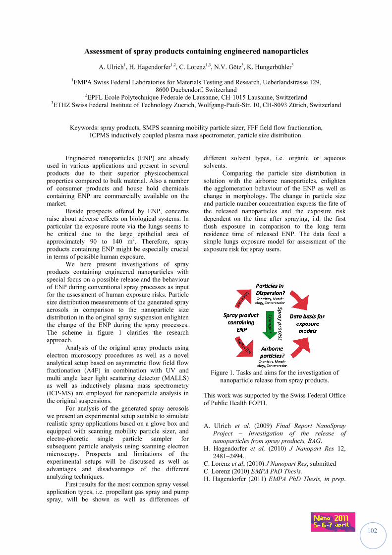
102
Assessment of spray products containing engineered nanoparticles
A. Ulrich1, H. Hagendorfer1,2, C. Lorenz1,3, N.V. Götz3, K. Hungerbühler3
1EMPA Swiss Federal Laboratories for Materials Testing and Research, Ueberlandstrasse 129,
8600 Duebendorf, Switzerland 2EPFL Ecole Polytechnique Federale de Lausanne, CH-1015 Lausanne, Switzerland
3ETHZ Swiss Federal Institute of Technology Zuerich, Wolfgang-Pauli-Str. 10, CH-8093 Zürich, Switzerland
Keywords: spray products, SMPS scanning mobility particle sizer, FFF field flow fractionation,
ICPMS inductively coupled plasma mass spectrometer, particle size distribution. Engineered nanoparticles (ENP) are already used in various applications and present in several products due to their superior physicochemical properties compared to bulk material. Also a number of consumer products and house hold chemicals containing ENP are commercially available on the market. Beside prospects offered by ENP, concerns raise about adverse effects on biological systems. In particular the exposure route via the lungs seems to be critical due to the large epithelial area of approximately 90 to 140 m2. Therefore, spray products containing ENP might be especially crucial in terms of possible human exposure. We here present investigations of spray products containing engineered nanoparticles with special focus on a possible release and the behaviour of ENP during conventional spray processes as input for the assessment of human exposure risks. Particle size distribution measurements of the generated spray aerosols in comparison to the nanoparticle size distribution in the original spray suspension enlighten the change of the ENP during the spray processes. The scheme in figure 1 clarifies the research approach. Analysis of the original spray products using electron microscopy procedures as well as a novel analytical setup based on asymmetric flow field flow fractionation (A4F) in combination with UV and multi angle laser light scattering detector (MALLS) as well as inductively plasma mass spectrometry (ICP-MS) are employed for nanoparticle analysis in the original suspensions. For analysis of the generated spray aerosols we present an experimental setup suitable to simulate realistic spray applications based on a glove box and equipped with scanning mobility particle sizer, and electro-phoretic single particle sampler for subsequent particle analysis using scanning electron microscopy. Prospects and limitations of the experimental setups will be discussed as well as advantages and disadvantages of the different analyzing techniques. First results for the most common spray vessel application types, i.e. propellant gas spray and pump spray, will be shown as well as differences of
different solvent types, i.e. organic or aqueous solvents. Comparing the particle size distribution in solution with the airborne nanoparticles, enlighten the agglomeration behaviour of the ENP as well as change in morphology. The change in particle size and particle number concentration express the fate of the released nanoparticles and the exposure risk dependent on the time after spraying, i.d. the first flush exposure in comparison to the long term residence time of released ENP. The data feed a simple lungs exposure model for assessment of the exposure risk for spray users.
Figure 1. Tasks and aims for the investigation of
nanoparticle release from spray products. This work was supported by the Swiss Federal Office of Public Health FOPH. A. Ulrich et al, (2009) Final Report NanoSpray
Project – Investigation of the release of nanoparticles from spray products, BAG.
H. Hagendorfer et al, (2010) J Nanopart Res 12, 2481–2494.
C. Lorenz et al, (2010) J Nanopart Res, submitted C. Lorenz (2010) EMPA PhD Thesis. H. Hagendorfer (2011) EMPA PhD Thesis, in prep.
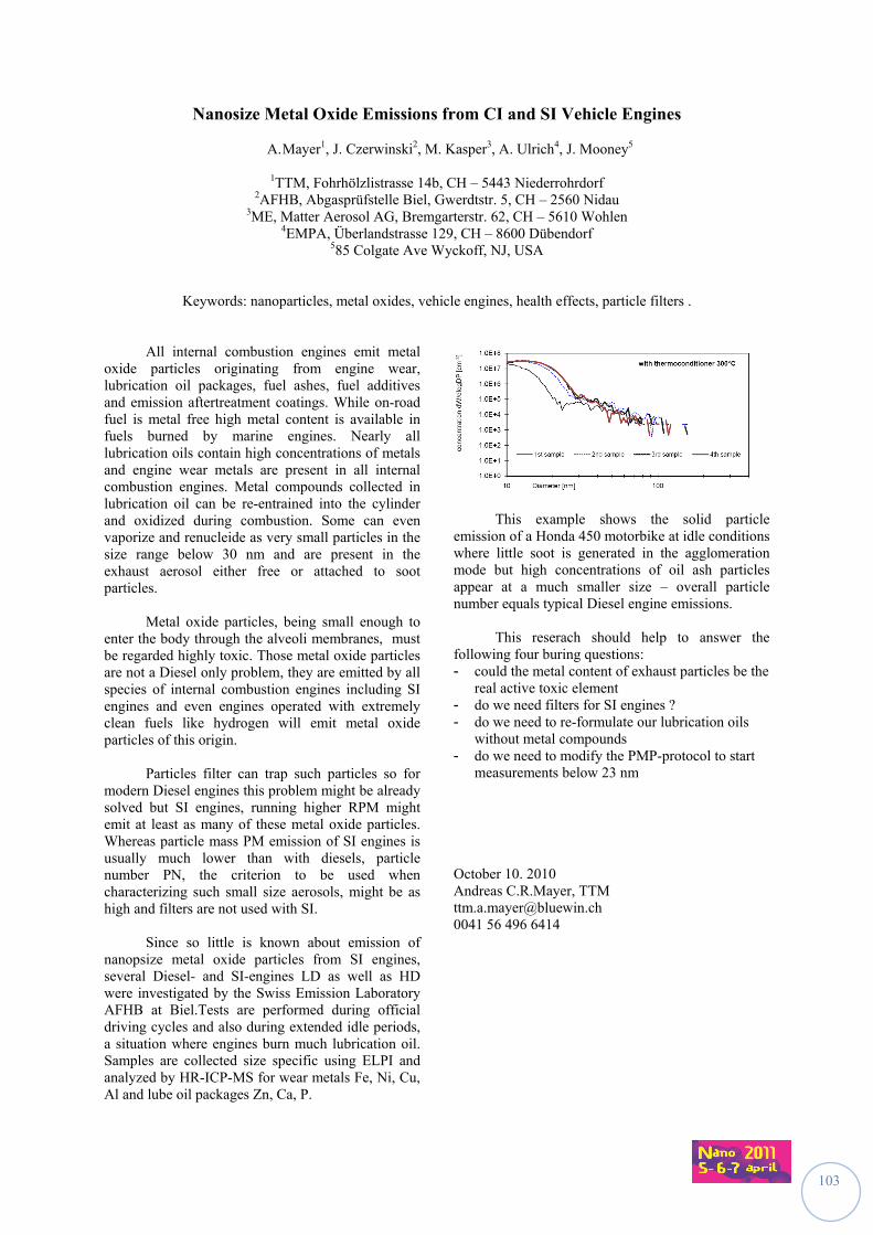
103
Nanosize Metal Oxide Emissions from CI and SI Vehicle Engines
A. Mayer1, J. Czerwinski2, M. Kasper3, A. Ulrich4, J. Mooney5
1TTM, Fohrhölzlistrasse 14b, CH – 5443 Niederrohrdorf 2AFHB, Abgasprüfstelle Biel, Gwerdtstr. 5, CH – 2560 Nidau
3ME, Matter Aerosol AG, Bremgarterstr. 62, CH – 5610 Wohlen 4EMPA, Überlandstrasse 129, CH – 8600 Dübendorf
585 Colgate Ave Wyckoff, NJ, USA
Keywords: nanoparticles, metal oxides, vehicle engines, health effects, particle filters .
All internal combustion engines emit metal oxide particles originating from engine wear, lubrication oil packages, fuel ashes, fuel additives and emission aftertreatment coatings. While on-road fuel is metal free high metal content is available in fuels burned by marine engines. Nearly all lubrication oils contain high concentrations of metals and engine wear metals are present in all internal combustion engines. Metal compounds collected in lubrication oil can be re-entrained into the cylinder and oxidized during combustion. Some can even vaporize and renucleide as very small particles in the size range below 30 nm and are present in the exhaust aerosol either free or attached to soot particles.
Metal oxide particles, being small enough to
enter the body through the alveoli membranes, must be regarded highly toxic. Those metal oxide particles are not a Diesel only problem, they are emitted by all species of internal combustion engines including SI engines and even engines operated with extremely clean fuels like hydrogen will emit metal oxide particles of this origin.
Particles filter can trap such particles so for
modern Diesel engines this problem might be already solved but SI engines, running higher RPM might emit at least as many of these metal oxide particles. Whereas particle mass PM emission of SI engines is usually much lower than with diesels, particle number PN, the criterion to be used when characterizing such small size aerosols, might be as high and filters are not used with SI.
Since so little is known about emission of
nanopsize metal oxide particles from SI engines, several Diesel- and SI-engines LD as well as HD were investigated by the Swiss Emission Laboratory AFHB at Biel.Tests are performed during official driving cycles and also during extended idle periods, a situation where engines burn much lubrication oil. Samples are collected size specific using ELPI and analyzed by HR-ICP-MS for wear metals Fe, Ni, Cu, Al and lube oil packages Zn, Ca, P.
This example shows the solid particle emission of a Honda 450 motorbike at idle conditions where little soot is generated in the agglomeration mode but high concentrations of oil ash particles appear at a much smaller size – overall particle number equals typical Diesel engine emissions.
This reserach should help to answer the following four buring questions: - could the metal content of exhaust particles be the
real active toxic element - do we need filters for SI engines ? - do we need to re-formulate our lubrication oils
without metal compounds - do we need to modify the PMP-protocol to start
measurements below 23 nm October 10. 2010 Andreas C.R.Mayer, TTM [email protected] 0041 56 496 6414
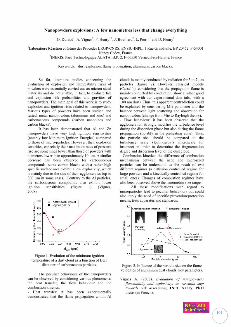
104
Nanopowders explosions: A few nanometres less that change everything
O. Dufaud1, A. Vignes2, F. Henry1,2, J. Bouillard2, L. Perrin1 and D. Fleury2
1Laboratoire Réaction et Génie des Procédés LRGP-CNRS, ENSIC-INPL, 1 Rue Grandville, BP 20452, F-54001 Nancy Cedex, France
2INERIS, Parc Technologique ALATA, B.P. 2, F-60550 Verneuil-en-Halatte, France
Keywords: dust explosion, flame propagation, aluminum, carbon blacks. So far, literature studies concerning the evaluation of explosion and flammability risks of powders were essentially carried out on micron-sized materials and do not enable, in fact, to evaluate fire and explosion risk probabilities and gravities of nanopowders. The main goal of this work is to study explosion and ignition risks related to nanopowders. Various types of powders have been studied and tested: metal nanopowders (aluminum and zinc) and carbonaceous compounds (carbon nanotubes and carbon blacks). It has been demonstrated that Al and Zn nanopowders have very high ignition sensitivities (notably low Minimum Ignition Energies) compared to those of micro-particles. However, their explosion severities, especially their maximum rates of pressure rise are sometimes lower than those of powders with diameters lower than approximately 10 µm. A similar decrease has been observed for carbonaceous compounds: some carbon blacks with a rather high specific surface area exhibit a low explosivity, which is mainly due to the size of their agglomerates (up to 300 µm in some cases). Contrary to the Al particles, the carbonaceous compounds also exhibit lower ignition sensitivities (figure 1) (Vignes, 2008).
Figure 1. Evolution of the minimum ignition
temperature of a dust cloud as a function of BET diameter of carbonaceous particles.
The peculiar behaviours of the nanopowders can be observed by considering various phenomena: the heat transfer, the flow behaviour and the combustion kinetics. - Heat transfer: it has been experimentally demonstrated that the flame propagation within Al
clouds is mainly conducted by radiation for 3 to 7 µm particles (figure 2). However classical models (Cassel’s), considering that the propagation flame is mainly conducted by conduction, show a rather good agreement with our experimental data (also with a 100 nm dust). Thus, this apparent contradiction could be explained by considering Mie parameter and the balance between light scattering and absorption for nanopowders (change from Mie to Rayleigh theory). - Flow behaviour: it has been observed that the agglomeration strongly modifies the turbulence level during the dispersion phase but also during the flame propagation (notably in the preheating zone). Thus, the particle size should be compared to the turbulence scale (Kolmogrov’s microscale for instance) in order to determine the fragmentation degree and dispersion level of the dust cloud. - Combustion kinetics: the difference of combustion mechanisms between the nano and microsized particles can be understood as the result of two different regimes (a diffusion controlled regime for large powders and a kinetically controlled regime for small ones). Changes of combustion regimes have also been observed above the nanometric size range.
All these modifications with regard to microparticles lead to peculiar behaviours but could also imply the need of specific prevention/protection means, tests apparatus and standards.
Figure 2. Influence of the particle size on the flame velocities of aluminium dust clouds: key parameters. Vignes A. (2008). Evaluation of nanopowders
flammability and explosivity: an essential step towards risk assessment, INPL Nancy, Ph.D thesis (in French).
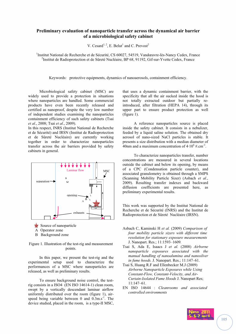
105
Preliminary evaluation of nanoparticle transfer across the dynamical air barrier of a microbiological safety cabinet
V. Cesard1, 2, E. Belut1 and C. Prevost2
1Institut National de Recherche et de Sécurité, CS 60027, 54519, Vandœuvre-lès-Nancy Cedex, France
2Institut de Radioprotection et de Sûreté Nucléaire, BP 68, 91192, Gif-sur-Yvette Cedex, France
Keywords: protective equipments, dynamics of nanoaerosols, containment efficiency.
Microbiological safety cabinet (MSC) are widely used to provide a protection in situations where nanoparticles are handled. Some commercial products have even been recently released and certified as nanoproof, despite the very low number of independent studies examining the nanoparticles containment efficiency of such safety cabinets (Tsai et al., 2008; Tsai et al., 2009). In this respect, INRS (Institut National de Recherche et de Sécurité) and IRSN (Institut de Radioprotection et de Sûreté Nucléaire) are currently working together in order to characterize nanoparticles transfer across the air barriers provided by safety cabinets in general.
Source of nanoparticle A Operator zone B Background zone Figure 1. Illustration of the test-rig and measurement
points.
In this paper, we present the test-rig and the experimental setup used to characterize the performances of a MSC where nanoparticles are released, as well as preliminary results.
To ensure background noise control, the test-
rig consists in a ISO4 (EN ISO 14614-1) clean room, swept by a vertically descendant laminar airflow uniformly distributed over the room (figure 1), air-speed being variable between 0 and 0.3m.s-1. The device studied, placed in the room, is a type-II MSC,
that uses a dynamic containment barrier, with the specificity that all the air sucked inside the hood is not totally extracted outdoor but partially re-introduced, after filtration (HEPA 14), through its upper part to ensure product protection as well (figure 1).
A reference nanoparticles source is placed
inside the safety cabinet. It consists in a nebulizer, feeded by a liquid saline solution. The obtained dry aerosol of nano-sized NaCl particles is stable. It presents a size distribution with a median diameter of 40nm and a maximum concentration of 4·106 #.cm-3.
To characterize nanoparticles transfer, number
concentrations are measured in several locations outside the cabinet and below its opening, by means of a CPC (Condensation particle counter), and associated granulometry is obtained through a SMPS (Scanning Mobility Particle Sizer) (Asbach et al., 2009). Resulting transfer indexes and backward diffusion coefficients are presented here, as preliminary experimental results.
This work was supported by the Institut National de Recherche et de Sécurité (INRS) and the Institut de Radioprotection et de Sûreté Nucléaire (IRSN). Asbach C, Kaminski H et al. (2009) Comparison of
four mobility particle sizers with different time resolution for stationary exposure measurements J. Nanopart. Res.; 11:1593–1609.
Tsai S, Ada E, Isaacs J et al. (2008) Airborne nanoparticle exposures associated with the manual handling of nanoalumina and nanosilver in fume hoods. J. Nanopart. Res.; 11:147–61.
Tsai S, Huang R.F and Ellenbecker M.J (2009) Airborne Nanoparticle Exposures while Using Constant-Flow, Constant-Velocity, and Air-Curtain-Isolated Fume Hoods J. Nanopart Res; 11:147–61.
EN ISO 14644 : Cleanrooms and associated controlled environments
separation
opening
Laminar flow
exhausts
B
A
MSC
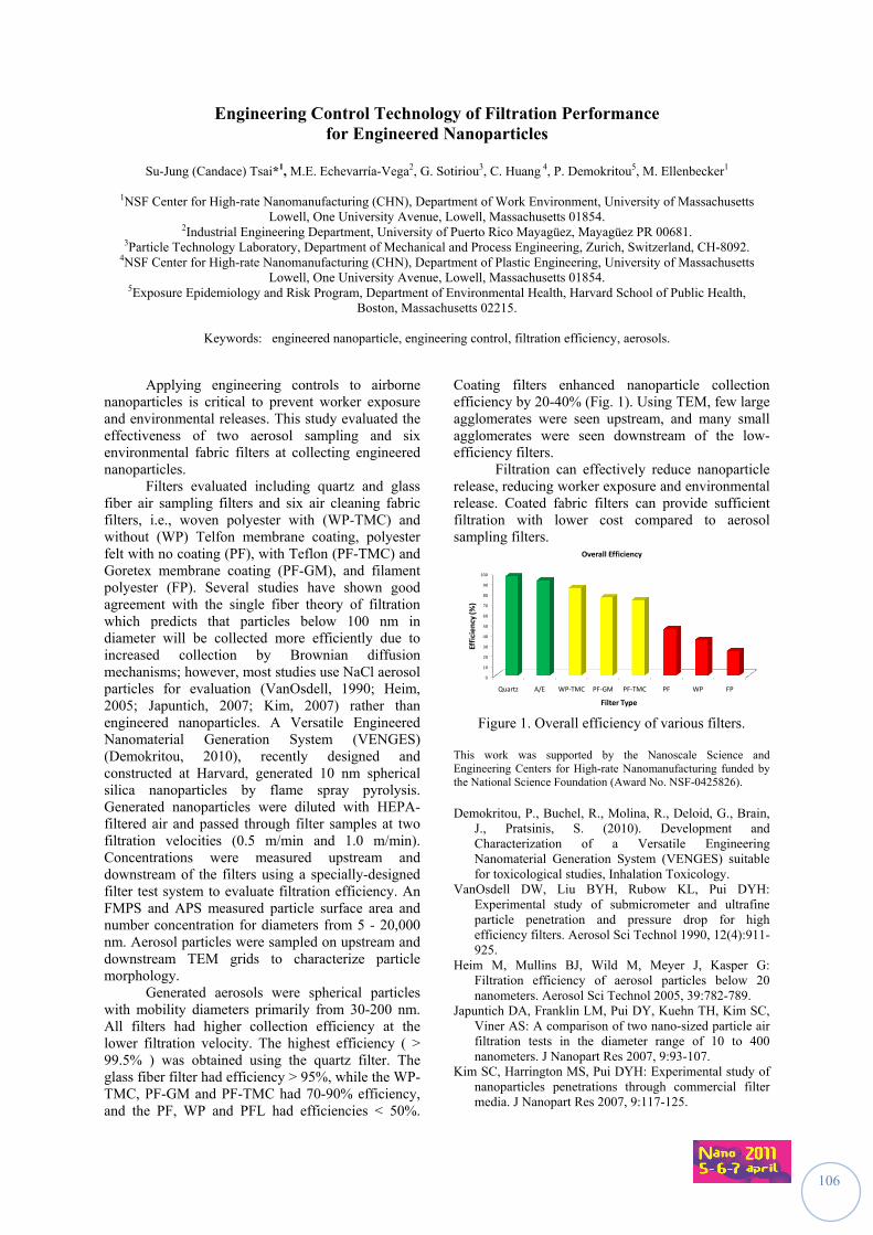
106
Engineering Control Technology of Filtration Performance for Engineered Nanoparticles
Su-Jung (Candace) Tsai*1, M.E. Echevarría-Vega2, G. Sotiriou3, C. Huang 4, P. Demokritou5, M. Ellenbecker1
1NSF Center for High-rate Nanomanufacturing (CHN), Department of Work Environment, University of Massachusetts Lowell, One University Avenue, Lowell, Massachusetts 01854.
2Industrial Engineering Department, University of Puerto Rico Mayagüez, Mayagüez PR 00681. 3Particle Technology Laboratory, Department of Mechanical and Process Engineering, Zurich, Switzerland, CH-8092.
4NSF Center for High-rate Nanomanufacturing (CHN), Department of Plastic Engineering, University of Massachusetts Lowell, One University Avenue, Lowell, Massachusetts 01854.
5Exposure Epidemiology and Risk Program, Department of Environmental Health, Harvard School of Public Health, Boston, Massachusetts 02215.
Keywords: engineered nanoparticle, engineering control, filtration efficiency, aerosols.
Applying engineering controls to airborne nanoparticles is critical to prevent worker exposure and environmental releases. This study evaluated the effectiveness of two aerosol sampling and six environmental fabric filters at collecting engineered nanoparticles.
Filters evaluated including quartz and glass fiber air sampling filters and six air cleaning fabric filters, i.e., woven polyester with (WP-TMC) and without (WP) Telfon membrane coating, polyester felt with no coating (PF), with Teflon (PF-TMC) and Goretex membrane coating (PF-GM), and filament polyester (FP). Several studies have shown good agreement with the single fiber theory of filtration which predicts that particles below 100 nm in diameter will be collected more efficiently due to increased collection by Brownian diffusion mechanisms; however, most studies use NaCl aerosol particles for evaluation (VanOsdell, 1990; Heim, 2005; Japuntich, 2007; Kim, 2007) rather than engineered nanoparticles. A Versatile Engineered Nanomaterial Generation System (VENGES) (Demokritou, 2010), recently designed and constructed at Harvard, generated 10 nm spherical silica nanoparticles by flame spray pyrolysis. Generated nanoparticles were diluted with HEPA-filtered air and passed through filter samples at two filtration velocities (0.5 m/min and 1.0 m/min). Concentrations were measured upstream and downstream of the filters using a specially-designed filter test system to evaluate filtration efficiency. An FMPS and APS measured particle surface area and number concentration for diameters from 5 - 20,000 nm. Aerosol particles were sampled on upstream and downstream TEM grids to characterize particle morphology.
Generated aerosols were spherical particles with mobility diameters primarily from 30-200 nm. All filters had higher collection efficiency at the lower filtration velocity. The highest efficiency ( > 99.5% ) was obtained using the quartz filter. The glass fiber filter had efficiency > 95%, while the WP-TMC, PF-GM and PF-TMC had 70-90% efficiency, and the PF, WP and PFL had efficiencies < 50%.
Coating filters enhanced nanoparticle collection efficiency by 20-40% (Fig. 1). Using TEM, few large agglomerates were seen upstream, and many small agglomerates were seen downstream of the low-efficiency filters.
Filtration can effectively reduce nanoparticle release, reducing worker exposure and environmental release. Coated fabric filters can provide sufficient filtration with lower cost compared to aerosol sampling filters.
0
10
20
30
40
50
60
70
80
90
100
Quartz A/E WP‐TMC PF‐GM PF‐TMC PF WP FP
Efficiency (%)
Filter Type
Overall Efficiency
Figure 1. Overall efficiency of various filters.
This work was supported by the Nanoscale Science and Engineering Centers for High-rate Nanomanufacturing funded by the National Science Foundation (Award No. NSF-0425826).
Demokritou, P., Buchel, R., Molina, R., Deloid, G., Brain,
J., Pratsinis, S. (2010). Development and Characterization of a Versatile Engineering Nanomaterial Generation System (VENGES) suitable for toxicological studies, Inhalation Toxicology.
VanOsdell DW, Liu BYH, Rubow KL, Pui DYH: Experimental study of submicrometer and ultrafine particle penetration and pressure drop for high efficiency filters. Aerosol Sci Technol 1990, 12(4):911-925.
Heim M, Mullins BJ, Wild M, Meyer J, Kasper G: Filtration efficiency of aerosol particles below 20 nanometers. Aerosol Sci Technol 2005, 39:782-789.
Japuntich DA, Franklin LM, Pui DY, Kuehn TH, Kim SC, Viner AS: A comparison of two nano-sized particle air filtration tests in the diameter range of 10 to 400 nanometers. J Nanopart Res 2007, 9:93-107.
Kim SC, Harrington MS, Pui DYH: Experimental study of nanoparticles penetrations through commercial filter media. J Nanopart Res 2007, 9:117-125.
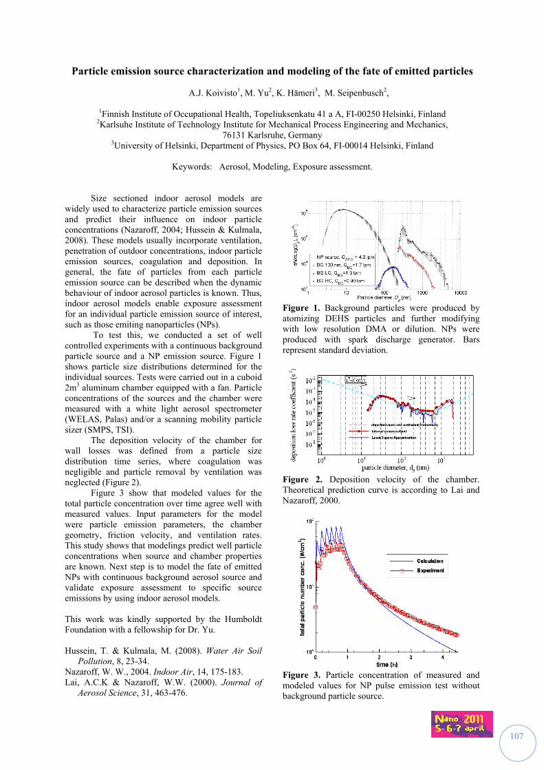
107
Particle emission source characterization and modeling of the fate of emitted particles
A.J. Koivisto1, M. Yu2, K. Hämeri3, M. Seipenbusch2,
1Finnish Institute of Occupational Health, Topeliuksenkatu 41 a A, FI-00250 Helsinki, Finland 2Karlsuhe Institute of Technology Institute for Mechanical Process Engineering and Mechanics,
76131 Karlsruhe, Germany 3University of Helsinki, Department of Physics, PO Box 64, FI-00014 Helsinki, Finland
Keywords: Aerosol, Modeling, Exposure assessment.
Size sectioned indoor aerosol models are widely used to characterize particle emission sources and predict their influence on indoor particle concentrations (Nazaroff, 2004; Hussein & Kulmala, 2008). These models usually incorporate ventilation, penetration of outdoor concentrations, indoor particle emission sources, coagulation and deposition. In general, the fate of particles from each particle emission source can be described when the dynamic behaviour of indoor aerosol particles is known. Thus, indoor aerosol models enable exposure assessment for an individual particle emission source of interest, such as those emiting nanoparticles (NPs). To test this, we conducted a set of well controlled experiments with a continuous background particle source and a NP emission source. Figure 1 shows particle size distributions determined for the individual sources. Tests were carried out in a cuboid 2m3 aluminum chamber equipped with a fan. Particle concentrations of the sources and the chamber were measured with a white light aerosol spectrometer (WELAS, Palas) and/or a scanning mobility particle sizer (SMPS, TSI). The deposition velocity of the chamber for wall losses was defined from a particle size distribution time series, where coagulation was negligible and particle removal by ventilation was neglected (Figure 2). Figure 3 show that modeled values for the total particle concentration over time agree well with measured values. Input parameters for the model were particle emission parameters, the chamber geometry, friction velocity, and ventilation rates. This study shows that modelings predict well particle concentrations when source and chamber properties are known. Next step is to model the fate of emitted NPs with continuous background aerosol source and validate exposure assessment to specific source emissions by using indoor aerosol models. This work was kindly supported by the Humboldt Foundation with a fellowship for Dr. Yu. Hussein, T. & Kulmala, M. (2008). Water Air Soil
Pollution, 8, 23-34. Nazaroff, W. W., 2004. Indoor Air, 14, 175-183. Lai, A.C.K & Nazaroff, W.W. (2000). Journal of
Aerosol Science, 31, 463-476.
Figure 1. Background particles were produced by atomizing DEHS particles and further modifying with low resolution DMA or dilution. NPs were produced with spark discharge generator. Bars represent standard deviation.
Figure 2. Deposition velocity of the chamber. Theoretical prediction curve is according to Lai and Nazaroff, 2000.
Figure 3. Particle concentration of measured and modeled values for NP pulse emission test without background particle source.
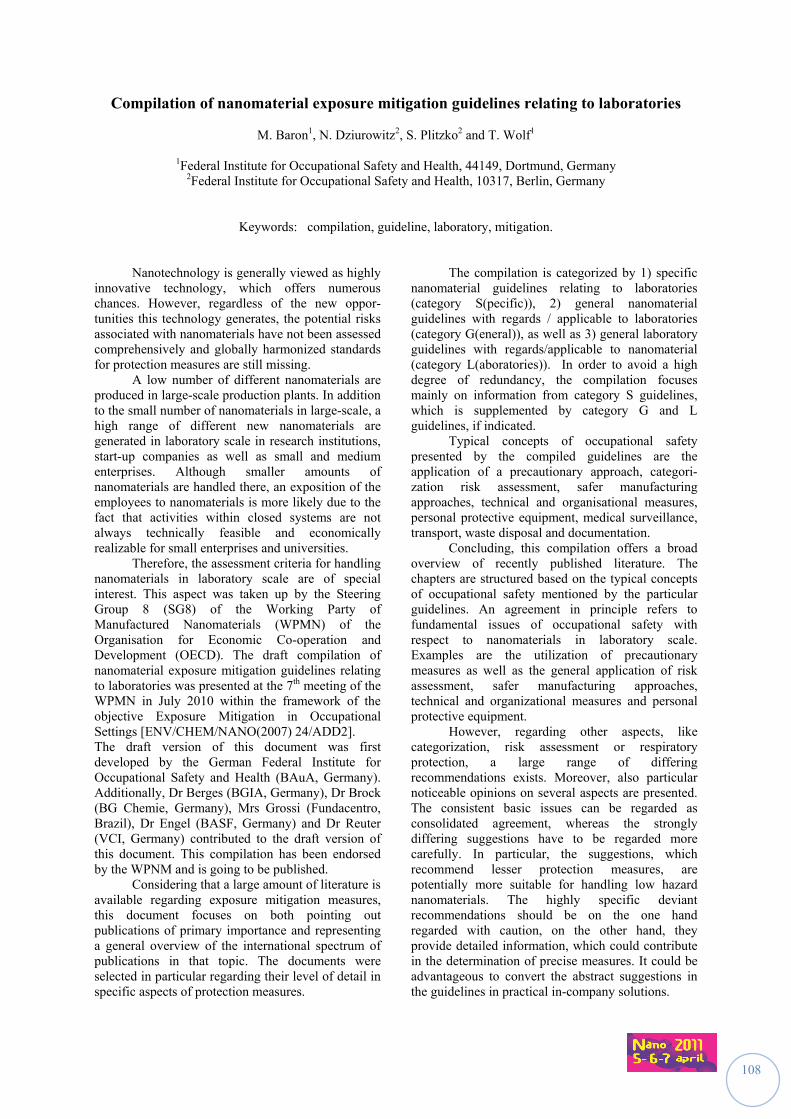
108
Compilation of nanomaterial exposure mitigation guidelines relating to laboratories
M. Baron1, N. Dziurowitz2, S. Plitzko2 and T. Wolf1
1Federal Institute for Occupational Safety and Health, 44149, Dortmund, Germany 2Federal Institute for Occupational Safety and Health, 10317, Berlin, Germany
Keywords: compilation, guideline, laboratory, mitigation. Nanotechnology is generally viewed as highly innovative technology, which offers numerous chances. However, regardless of the new oppor-tunities this technology generates, the potential risks associated with nanomaterials have not been assessed comprehensively and globally harmonized standards for protection measures are still missing. A low number of different nanomaterials are produced in large-scale production plants. In addition to the small number of nanomaterials in large-scale, a high range of different new nanomaterials are generated in laboratory scale in research institutions, start-up companies as well as small and medium enterprises. Although smaller amounts of nanomaterials are handled there, an exposition of the employees to nanomaterials is more likely due to the fact that activities within closed systems are not always technically feasible and economically realizable for small enterprises and universities. Therefore, the assessment criteria for handling nanomaterials in laboratory scale are of special interest. This aspect was taken up by the Steering Group 8 (SG8) of the Working Party of Manufactured Nanomaterials (WPMN) of the Organisation for Economic Co-operation and Development (OECD). The draft compilation of nanomaterial exposure mitigation guidelines relating to laboratories was presented at the 7th meeting of the WPMN in July 2010 within the framework of the objective Exposure Mitigation in Occupational Settings [ENV/CHEM/NANO(2007) 24/ADD2]. The draft version of this document was first developed by the German Federal Institute for Occupational Safety and Health (BAuA, Germany). Additionally, Dr Berges (BGIA, Germany), Dr Brock (BG Chemie, Germany), Mrs Grossi (Fundacentro, Brazil), Dr Engel (BASF, Germany) and Dr Reuter (VCI, Germany) contributed to the draft version of this document. This compilation has been endorsed by the WPNM and is going to be published. Considering that a large amount of literature is available regarding exposure mitigation measures, this document focuses on both pointing out publications of primary importance and representing a general overview of the international spectrum of publications in that topic. The documents were selected in particular regarding their level of detail in specific aspects of protection measures.
The compilation is categorized by 1) specific nanomaterial guidelines relating to laboratories (category S(pecific)), 2) general nanomaterial guidelines with regards / applicable to laboratories (category G(eneral)), as well as 3) general laboratory guidelines with regards/applicable to nanomaterial (category L(aboratories)). In order to avoid a high degree of redundancy, the compilation focuses mainly on information from category S guidelines, which is supplemented by category G and L guidelines, if indicated. Typical concepts of occupational safety presented by the compiled guidelines are the application of a precautionary approach, categori-zation risk assessment, safer manufacturing approaches, technical and organisational measures, personal protective equipment, medical surveillance, transport, waste disposal and documentation. Concluding, this compilation offers a broad overview of recently published literature. The chapters are structured based on the typical concepts of occupational safety mentioned by the particular guidelines. An agreement in principle refers to fundamental issues of occupational safety with respect to nanomaterials in laboratory scale. Examples are the utilization of precautionary measures as well as the general application of risk assessment, safer manufacturing approaches, technical and organizational measures and personal protective equipment. However, regarding other aspects, like categorization, risk assessment or respiratory protection, a large range of differing recommendations exists. Moreover, also particular noticeable opinions on several aspects are presented. The consistent basic issues can be regarded as consolidated agreement, whereas the strongly differing suggestions have to be regarded more carefully. In particular, the suggestions, which recommend lesser protection measures, are potentially more suitable for handling low hazard nanomaterials. The highly specific deviant recommendations should be on the one hand regarded with caution, on the other hand, they provide detailed information, which could contribute in the determination of precise measures. It could be advantageous to convert the abstract suggestions in the guidelines in practical in-company solutions.
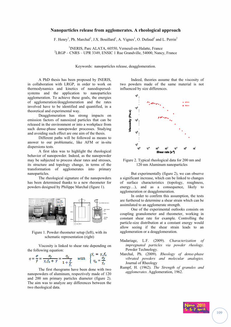
109
Nanoparticles release from agglomerates. A rheological approach
F. Henry1, Ph. Marchal2, J.X. Bouillard1, A. Vignes1, O. Dufaud2 and L. Perrin2
1INERIS, Parc ALATA, 60550, Verneuil-en-Halatte, France 2LRGP – CNRS – UPR 3349, ENSIC 1 Rue Grandville, 54000, Nancy, France
Keywords: nanoparticles release, deagglomeration.
A PhD thesis has been proposed by INERIS, in collaboration with LRGP, in order to work on thermodynamics and kinetics of nanodispersed-systems and the application to nanoparticles agglomeration. To achieve these goals, the energies of agglomeration/deagglomeration and the rates involved have to be identified and quantified, in a theoretical and experimental way. Deagglomeration has strong impacts on emission factors of nanosized particles that can be released in the environment or into a workplace from such dense-phase nanopowder processes. Studying and avoiding such effect are one aim of the thesis. Different paths will be followed as means to answer to our problematic, like AFM or in-situ dispersions tests. A first idea was to highlight the rheological behavior of nanopowder. Indeed, as the nanopowder may be subjected to process shear rates and stresses, its structure and topology change, in terms of the transformation of agglomerates into primary nanoparticles. The rheological signature of the nanopowders has been determined thanks to a new rheometer for powders designed by Philippe Marchal (figure 1).
Figure 1. Powder rheometer setup (left), with its schematic representation (right)
Viscosity is linked to shear rate depending on the following equation:
The first rheograms have been done with two nanopowders of aluminum, respectively made of 120 and 200 nm primary particles diameter (figure 2). The aim was to analyze any differences between the two rheological data.
Indeed, theories assume that the viscosity of two powders made of the same material is not influenced by size differences.
Figure 2. Typical rheological data for 200 nm and 120 nm Aluminum nanoparticles
But experimentally (figure 2), we can observe a significant increase, which can be linked to changes of surface characteristics (topology, roughness, energy…), and as a consequence, likely to agglomeration or deagglomeration. In order to confirm this assumption, the tests are furthered to determine a shear strain which can be assimilated to an agglomerate strength. One of the experimental outlooks consists on coupling granulometer and rheometer, working in constant shear rate for example. Controlling the particle-size distribution at a constant energy would allow seeing if the shear strain leads to an agglomeration or a deagglomeration. Madariage, L.F. (2009). Characterization of
impregnated particles via powder rheology. Powder Technology.
Marchal, Ph. (2009). Rheology of dense-phase vibrated powders and molecular analogies. Journal of Rheology
Rumpf, H. (1962). The Strength of granules and agglomerates. Agglomeration, 1962.
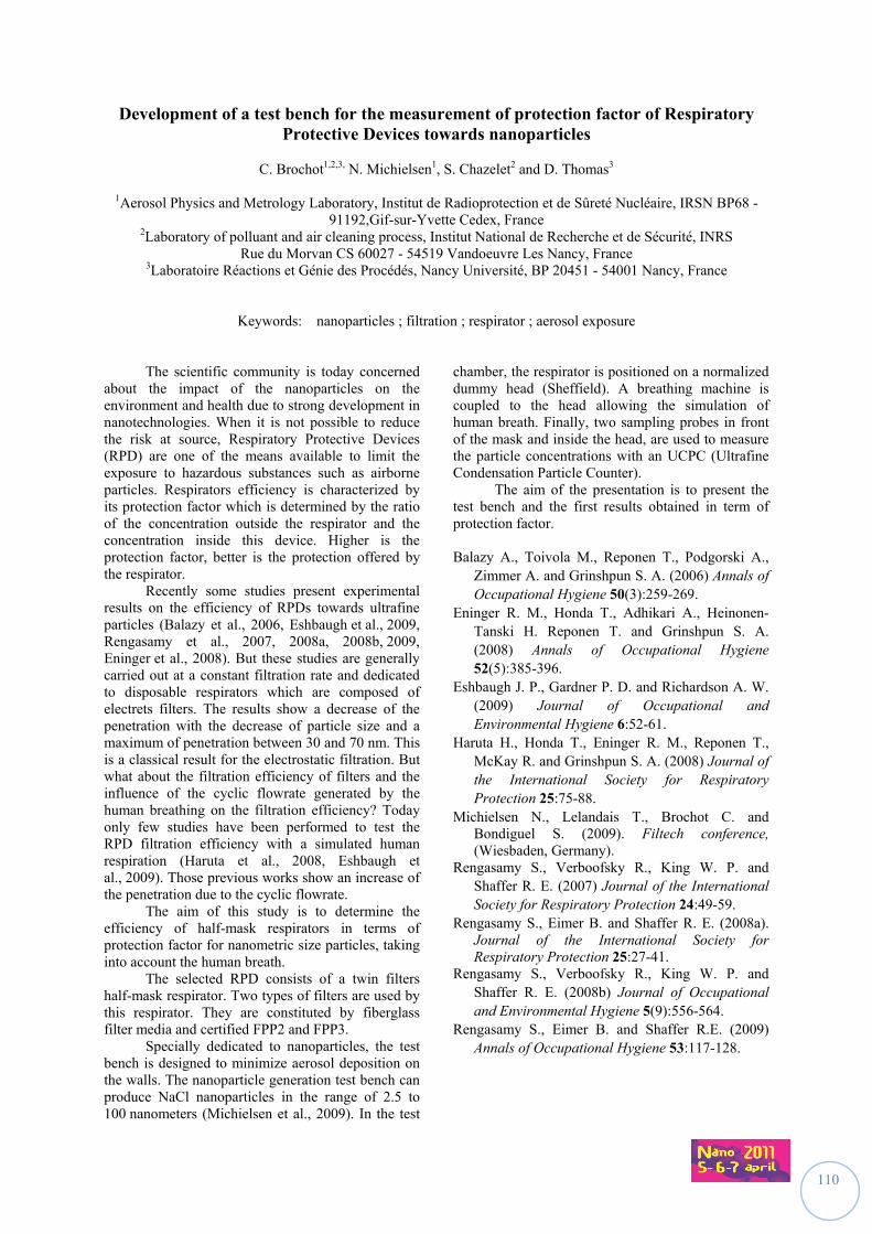
110
Development of a test bench for the measurement of protection factor of Respiratory Protective Devices towards nanoparticles
C. Brochot1,2,3, N. Michielsen1, S. Chazelet2 and D. Thomas3
1Aerosol Physics and Metrology Laboratory, Institut de Radioprotection et de Sûreté Nucléaire, IRSN BP68 -
91192,Gif-sur-Yvette Cedex, France 2Laboratory of polluant and air cleaning process, Institut National de Recherche et de Sécurité, INRS
Rue du Morvan CS 60027 - 54519 Vandoeuvre Les Nancy, France 3Laboratoire Réactions et Génie des Procédés, Nancy Université, BP 20451 - 54001 Nancy, France
Keywords: nanoparticles ; filtration ; respirator ; aerosol exposure
The scientific community is today concerned about the impact of the nanoparticles on the environment and health due to strong development in nanotechnologies. When it is not possible to reduce the risk at source, Respiratory Protective Devices (RPD) are one of the means available to limit the exposure to hazardous substances such as airborne particles. Respirators efficiency is characterized by its protection factor which is determined by the ratio of the concentration outside the respirator and the concentration inside this device. Higher is the protection factor, better is the protection offered by the respirator.
Recently some studies present experimental results on the efficiency of RPDs towards ultrafine particles (Balazy et al., 2006, Eshbaugh et al., 2009, Rengasamy et al., 2007, 2008a, 2008b, 2009, Eninger et al., 2008). But these studies are generally carried out at a constant filtration rate and dedicated to disposable respirators which are composed of electrets filters. The results show a decrease of the penetration with the decrease of particle size and a maximum of penetration between 30 and 70 nm. This is a classical result for the electrostatic filtration. But what about the filtration efficiency of filters and the influence of the cyclic flowrate generated by the human breathing on the filtration efficiency? Today only few studies have been performed to test the RPD filtration efficiency with a simulated human respiration (Haruta et al., 2008, Eshbaugh et al., 2009). Those previous works show an increase of the penetration due to the cyclic flowrate.
The aim of this study is to determine the efficiency of half-mask respirators in terms of protection factor for nanometric size particles, taking into account the human breath.
The selected RPD consists of a twin filters half-mask respirator. Two types of filters are used by this respirator. They are constituted by fiberglass filter media and certified FPP2 and FPP3.
Specially dedicated to nanoparticles, the test bench is designed to minimize aerosol deposition on the walls. The nanoparticle generation test bench can produce NaCl nanoparticles in the range of 2.5 to 100 nanometers (Michielsen et al., 2009). In the test
chamber, the respirator is positioned on a normalized dummy head (Sheffield). A breathing machine is coupled to the head allowing the simulation of human breath. Finally, two sampling probes in front of the mask and inside the head, are used to measure the particle concentrations with an UCPC (Ultrafine Condensation Particle Counter).
The aim of the presentation is to present the test bench and the first results obtained in term of protection factor. Balazy A., Toivola M., Reponen T., Podgorski A.,
Zimmer A. and Grinshpun S. A. (2006) Annals of Occupational Hygiene 50(3):259-269.
Eninger R. M., Honda T., Adhikari A., Heinonen-Tanski H. Reponen T. and Grinshpun S. A. (2008) Annals of Occupational Hygiene 52(5):385-396.
Eshbaugh J. P., Gardner P. D. and Richardson A. W. (2009) Journal of Occupational and Environmental Hygiene 6:52-61.
Haruta H., Honda T., Eninger R. M., Reponen T., McKay R. and Grinshpun S. A. (2008) Journal of the International Society for Respiratory Protection 25:75-88.
Michielsen N., Lelandais T., Brochot C. and Bondiguel S. (2009). Filtech conference, (Wiesbaden, Germany).
Rengasamy S., Verboofsky R., King W. P. and Shaffer R. E. (2007) Journal of the International Society for Respiratory Protection 24:49-59.
Rengasamy S., Eimer B. and Shaffer R. E. (2008a). Journal of the International Society for Respiratory Protection 25:27-41.
Rengasamy S., Verboofsky R., King W. P. and Shaffer R. E. (2008b) Journal of Occupational and Environmental Hygiene 5(9):556-564.
Rengasamy S., Eimer B. and Shaffer R.E. (2009) Annals of Occupational Hygiene 53:117-128.
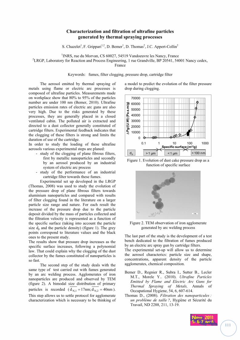
111
Characterization and filtration of ultrafine particles generated by thermal spraying processes
S. Chazelet1, F. Grippari1,2, D. Bemer1, D. Thomas2, J.C. Appert-Collin2
1INRS, rue du Morvan, CS 60027, 54519 Vandoeuvre les Nancy, France
2LRGP, Laboratory for Reaction and Process Engineering, 1 rue Grandville, BP 20541, 54001 Nancy cedex, France
Keywords: fumes, filter clogging, pressure drop, cartridge filter
The aerosol emitted by thermal spraying of metals using flame or electric arc processes is composed of ultrafine particles. Measurements made on workplace show that 80% to 95% of the particles number are under 100 nm (Bemer, 2010). Ultrafine particles emission rates of electric arc guns are also very high. Due to the risks generated by these processes, they are generally placed in a closed ventilated cabin. The polluted air is extracted and directed to a dust collector generally constituted of cartridge filters. Experimental feedback indicates that the clogging of these filters is strong and limits the duration of use of the cartridge. In order to study the loading of these ultrafine aerosols various experimental steps are planed:
- study of the clogging of plane fibrous filters, first by metallic nanoparticles and secondly by an aerosol produced by an industrial system of electric arc process
- study of the performance of an industrial cartridge filter towards these fumes.
Experimental set up developed in the LRGP (Thomas, 2008) was used to study the evolution of the pressure drop of plane fibrous filters towards aluminium nanoparticles and compared with results of filter clogging found in the literature on a larger particle size range and nature. For each result the increase of the pressure drop due to the particle deposit divided by the mass of particles collected and the filtration velocity is represented as a function of the specific surface (taking into account the particle size dp and the particle density) (figure 1). The grey points correspond to literature values and the black ones to the present study. The results show that pressure drop increases as the specific surface increases, following a polynomial law. That could explain why the clogging of the dust collector by the fumes constituted of nanoparticles is so fast. The second step of the study deals with the same type of test carried out with fumes generated by an arc welding process. Agglomerates of iron nanoparticles are produced and observed by TEM (figure 2). A bimodal size distribution of primary
particles is recorded ( 40nmd17nm;d N,2N,1 ).
This step allows us to settle protocol for agglomerate characterization which is necessary to be thinking of
a model to predict the evolution of the filter pressure drop during clogging.
0
10000
20000
30000
40000
50000
60000
70000
0.1 1 10 100 1000
P
g/(
U.W
) (P
a.s.
m/g
)
Specific surface (m²/g)
> 1 µm < 1 µm < 100 nmdp Figure 1. Evolution of dust cake pressure drop as a
function of specific surface
Figure 2. TEM observation of iron agglomerate
generated by arc welding process
The last part of the study is the development of a test bench dedicated to the filtration of fumes produced by an electric arc spray gun by cartridge filters. The experimental set-up will allow us to determine the aerosol characterics: particle size and shape, concentrations, apparent density of the particle agglomerates, chemical composition.
Bemer D., Regnier R., Subra I., Sutter B., Lecler M.T., Morele Y.. (2010). Ultrafine Particles Emitted by Flame and Electric Arc Guns for Thermal Spraying of Metals, Annals of Occupational Hygiene, 54, 6, 607-614.
Thomas D., (2008). Filtration des nanoparticules : un problème de taille ?, Hygiène et Sécurité du Travail, ND 2288, 211, 13-19.
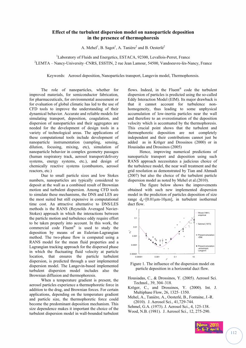
112
Effect of the turbulent dispersion model on nanoparticle deposition in the presence of thermophoresis
A. Mehel1, B. Sagot1, A. Tanière2 and B. Oesterlé2
1Laboratory of Fluids and Energetics, ESTACA, 92300, Levallois-Perret, France
2LEMTA – Nancy-University–CNRS, ESSTIN, 2 rue Jean Lamour, 54500, Vandoeuvre-lès-Nancy, France
Keywords: Aerosol deposition, Nanoparticles transport, Langevin model, Thermophoresis.
The role of nanoparticles, whether for improved materials, for semiconductor fabrication, for pharmaceuticals, for environmental assessment or for evaluation of global climatic has led to the use of CFD tools to improve the understanding of their dynamical behavior. Accurate and reliable models for simulating transport, deposition, coagulation, and dispersion of nanoparticles and their aggregates are needed for the development of design tools in a variety of technological areas. The applications of these computational tools include development of nanoparticle instrumentation (sampling, sensing, dilution, focusing, mixing, etc), simulation of nanoparticle behavior in complex geometry passages (human respiratory track, aerosol transport/delivery systems, energy systems, etc.), and design of chemically reactive systems (combustors, aerosol reactors, etc.)
Due to small particle sizes and low Stokes numbers, nanoparticles are typically considered to deposit at the wall as a combined result of Brownian motion and turbulent dispersion. Among CFD tools to simulate these mechanisms, the DNS and LES are the most suited but still expensive in computational time cost. An attractive alternative to DNS/LES methods is the RANS (Reynolds Averaged Navier-Stokes) approach in which the interactions between the particle motion and turbulence eddy require effort to be taken properly into account. In this work, the commercial code Fluent® is used to study the deposition by means of an Eulerian-Lagrangian method. The two-phase flow is computed using a RANS model for the mean fluid properties and a Lagrangian tracking approach for the dispersed phase in which the fluctuating fluid velocity at particle location, that ensures the particle turbulent dispersion, is predicted through a user implemented dispersion model. The Langevin-based implemented turbulent dispersion model includes also the Brownian diffusion and thermophoresis.
When a temperature gradient is present, the aerosol particles experience a thermophoretic force in addition to the drag, and Brownian forces. For certain applications, depending on the temperature gradient and particle size, the thermophoretic force could become the predominant deposition mechanism. This size dependence makes it important the choice of the turbulent dispersion model in wall-bounded turbulent
flows. Indeed, in the Fluent® code the turbulent dispersion of particles is predicted using the so-called Eddy Interaction Model (EIM). Its major drawback is that it cannot account for turbulence non-homogeneity, thus leading to some unphysical accumulation of low-inertia particles near the wall and therefore to an overestimation of the deposition velocity which is accentuated by the thermophoresis. This crucial point shows that the turbulent and thermophoretic deposition are not completely independent and their contributions cannot just be added as in Kröger and Drossinos (2000) or in Housiadas and Drossinos (2005)
Hence, improving numerical predictions of nanoparticle transport and deposition using such RANS approach necessitates a judicious choice of the turbulence model, the near wall treatment and the grid resolution as demonstrated by Tian and Ahmadi (2007) but also the choice of the turbulent particle dispersion model as noted by Mehel et al.(2010). The figure below shows the improvements obtained with such new implemented dispersion model in the prediction of particles deposition of size range dp=[0.01µm-10µm], in turbulent isothermal duct flow.
0.00001
0.0001
0.001
0.01
0.1
1
0.00001 0.001 0.1 10
u d+
Wood (1981) empirical
Sehmel (1973)
Present simulation -EIM
Present simulation -anisotropic Langevin
Figure 1. The influence of the dispersion model on
particle deposition in a horizontal duct flow.
Housiadas, C., & Drossinos, Y. (2005). Aerosol Sci. Technol., 39, 304–318.
Kröger, C., and Drossinos, Y. (2000). Int. J. Multiphase Flow, 26, 1325–1350.
Mehel, A., Tanière, A., Oesterlé, B., Fontaine, J.-R. (2010). J. Aerosol Sci., 41,729-744.
Sehmel, G.A. (1973). J. Aerosol Sci., 4, 125-138. Wood, N.B. (1981). J. Aerosol Sci., 12, 275-290.
113
.
Session IV
RRIISSKK AASSSSEESSSSMMEENNTT && RRIISSKK MMAANNAAGGEEMMEENNTT
Chairs: Sonia Desmoulin (CNRS, France) Claude Ostiguy (IRSST, Québec)
114
Time to shift paradigms? How to practice Nanotechnology risk governance
A.J. Dijkman1, J. Terwoert2, A.L. Hollander3
1Consultant, researcher health & safety policies and communication at TNO Work & Employment,
Polarisavenue 151, 2132 JJ Hoofddorp, The Netherlands. 2Consultant, researcher chemicals & occupational hygiene at TNO Work & Employment,
Polarisavenue 151, 2132 JJ Hoofddorp, The Netherlands. 3Senior researcher, programme manager at TNO Work & Employment,
Polarisavenue 151, 2132 JJ Hoofddorp, The Netherlands
Keywords: knowledge gaps, interventions, instruments Introduction As nanotechnology is becoming more and more of common use in products, the (public) debate about risks of nanotechnology is increasing. Risk governance estimates the importance of commitment of several stakeholders, the role of experts, and scientific knowledge on how to deal with uncertain risks. Due to the fast development in nanotechnology applications scientific research can no longer be the primary basis for taking for example political and policy decisions on risks. Also because nano applications are becoming important drivers for economic success, new and more interdisciplinary ways need to be explored in order to deal with uncertainty and new risks. The International Risk Governance Council (IRCG) has made a study on how to use risk governance in nanotechnology (Report IRCG, September 2008). In this study IRCG gives recommendations on how Risk governance needs to explore more interdisciplinary ways with a variety of stakeholders in order toFor the conference organized by INRS in association with PEROSH oral and poster presentations will be accepted. 1 Reinforce involvement and communication
about uncertainty and risks, 2 Stimulate the process of mapping and
endorsement of social commitment and acceptance
3 Reinforce collaboration, national and international
Research questions Based upon IRCG recommendations TNO has started a four years research program (2011-2014) on risk governance this year. The main purpose of this program is to develop (practical suitable) knowledge and guidance on how a variety of stakeholders (companies, government bodies, NGO's, employers' and employees' organizations, general public) can organize, communicate and deal with uncertainty of substances, e.g. of Nano substances. More specific: knowledge will be developed on what it means for (civil) society when results from research and information are not available (yet), and at the same time innovation must not come to a standstill. When
is it necessary to bring up the precautionary principle, how to deal with that principle, and what to do in circumstances when risks can not be foreseen? Is it time to shift paradigms? In other words, can we move to other thought and scientific approaches? Method A review of the literature and interviews with several stakeholders provides an overview of the ‘state-of-art’ and the main gaps of knowledge on the (practical) application of risk governance. The outcome will be presented and discussed. Concluding The presentation and discussion will lead to new ideas and initiatives on how to streamline and tune the vast amount of current initiatives, and on how to practice nanotechnology risk governance. TNO will take the opportunity to discuss the fragmentary landscape of European and global institutes, research groups and NGO’s dealing with risk governance. The TNO research program will seek active collaboration with EU partners in the next years to develop new methods and models to use in pilot studies on how to practice risk governance of new risks of substances in general, and more specifically of nanotechnology. Renn, O., (2005) White paper on risk governance-
Towards an integrative approach International Risk Governance Council (IRGC), Geneva
Crobe, A., Renn, O., Jaeger, A., (2008) Governance of Nanotechnology applications in food and cosmetics International Risk Governance Council (IRCG), Dialogic GmbH, Geneva
European Commission, (2001). European Governance, a white book. Brussels
115
Nanotechnology Occupational Safety and Health: Global Standards Development
V. Murashov,1 J. Howard2
1National Institute for Occupational Safety and Health, Permanent Mission of the United States of America, Route de Pregny 11, 1292, Geneva, Switzerland
2National Institute for Occupational Safety and Health, 395 E Street, SW, Washington, DC, 20201, USA
Keywords: standards, workplace, nanotechnology, nanomaterials.
Nanotechnology is a rapidly evolving and potentially transformative technology, which has the potential to greatly improve many areas of human life. As potentially transformative as nanotechnology may be, however, successful acceptance of any new technology, and its widespread commercial dissemination, requires strict attention to controlling potential risks. International standards can serve to protect both product users and product manufacturers. Historically, international standards that have been incorporated into international trade agreements or adopted into national laws have been developed by only a limited number of public (e.g. Organization for Economic Cooperation and Development [OECD], United Nations) and private organizations (e.g. International Organization for Standardization [ISO], ASTM International). In the last five years, almost all major standards developing organizations established such technical groups. For example, ISO established a technical committee for nanotechnologies, TC 229 in 2005, while OECD established Working Party on Manufactured Nanomaterials in 2006 (Murashov & Howard, 2011). Since workers bear the greatest health risk from exposure to any emerging technology, most organizations which develop safety and health standards for nanotechnology have focused their efforts initially on the workplace and especially the human health aspects including possible mitigation actions to allow for reduction of possible exposure. The workplace safety and health standards described in this presentation include voluntary, consensus-type standards adopted by the private sector as well as mandatory, or government regulatory, health-related standards that are designed to protect the health and safety of workers (Murashov et al., 2010; Murashov & Howard, 2009; Murashov et al., 2009).
Workplace safety and health standards for nanotechnology similar to other industries usually include the following elements: (1) quantitative occupational exposure limits, (2) hazard communication instructions, (3) standard practices, e.g. safety procedures or reference to codes of conduct, and (4) standard guidance, e.g. industrial hygiene guidance for safe handling of nanomaterials. Current national and international activities in these areas will be described in this presentation. Murashov, V., & Howard, J. (2011). Nanotechnology
Standards New York, USA: Springer Science+Business Media.
Murashov, V., & Howard, J. (2009). Nature Nanotech, 4, 467-470.
Murashov, V. et al. (2010). Industrial Health, to be published.
Murashov, V. et al. (2009). J. Nanopart. Res., 11, 1587-1591.
116
The role of the employer in prevention and compensation of risks associated to nanoparticles and nanomaterials
M. Bary1, N. Dedessus-Le-Moustier2 et A. Moriceau1
1Laboratoire IODE UMR CNRS 6262, Université de Rennes 1, France
2Laboratoire IREA EA 4251, Université de Bretagne sud, Vannes, France
Key words : prevention, potential risk, assessment, liability The risks associated to nanoparticles and nanomaterials have the particularity to be still predominantly potential. However, these substances are already used by industry. So the workers of private and public areas are yet exposed to it. Although prevention doesn’t stop developing, possible damages on the workers’s health and also, under the influence of dispersion of substances, transmission to their relatives is not to be excluded. Prevention of occupational hazards is at the very heart of the French system of social protection and work relations. In this scheme, the action of the employer, whether private or public, proves essential in order to protect the physical as well as the mental health of workers. Work legislation imposes on him the implementation of a genuine health policy at work. Whether it be a matter of safety requirements, risk assessment, information and formation, the employer must adopt active measures in order to protect the health of workers. Jurisprudence has considerably increased the scope of these obligations. In the absence of adequate protection of the workers against occupational hazards, the employer will see his rights to management and disciplinary power contained. How can these dispositions be possibly applied in the case of a hazard whose effects on health remain dubious ? Scientists and industrialists who show great interest in future applications regarding the use of nanomaterials and nanoparticles should take into account all the questions relating to health effects caused notably to workers who handle them. Studies on their toxicity are still not fully developed, though they are more and more used and commercialized. In the present context, what measures should the employer take to protect the health of workers and avoid a possible implication of his responsibility?
If risks of damages become a reality, the victims will want to be compensated. They could be by the regime of occupational diseases. However, this system awards basic allowance. To acquire an entire compensation, the victims could then prefer looking for the liability of the employer, as the development of litigation in this affair proves it. And this, especially as the employer will not be able to invoke the existence of a risk of development to exempt. It’s advisable to show, in French law, the “état de l’art” of the employer’s liability, private or public, in the case of workers’ exposition to nanoparticles and nanomaterials. In order to do that, it is necessary to wonder if the liability’s rules could apply in this case and if an indemnity of the victims is possible. In other words, are the present rules of liability adapted to risks associated to nanoparticles and nanomaterials or do they require to be changed or completed ? The interest of this study is to find the eventual reefs and insufficiencies of the labour law and employer’s liability law in purpose, at the same time, to improve at once the prevention and the indemnity of the victims, as well as to regulate the employer’s liability in presence of risks linked to nanoparticles and to nanomaterials. The confrontation from the specificities of nanoparticles and nanomaterials in the actual mechanisms of prevention and liability shows an uncertain legal protection of potential victims of risks linked to nanoparticles and nanomaterials. The study of the prevention and the employer’s liability faced to with the risks associated to nanoparticles and nanomaterials reveals the existence of several question settings relating to their implementation. For instance, is the risks’ assessment written in the « unique document » adapted to the taking account of uncertain risk ? Which prejudice could be recognized and which generative keep could be taken in the face of an uncertain risk ?
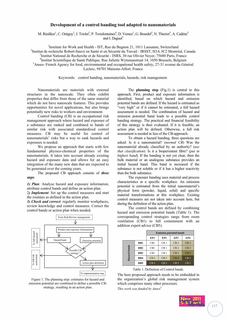
117
Development of a control banding tool adapted to nanomaterials
M. Riediker1, C. Ostiguy2, J. Triolet3, P. Troisfontaines4, D. Vernez1, G. Bourdel5, N. Thieriet5, A. Cadène5 and I. Daguet5
1Institute for Work and Health - IST, Rue du Bugnon 21, 1011 Lausanne, Switzerland
2Institut de recherche Robert-Sauvé en Santé et en Sécurité du Travail - IRSST, H3A 3C2 Montréal, Canada 3Institut National de Recherche et de Sécurité - INRS, 30 rue Olivier Noyer, 75680 Paris, France 4Institut Scientifique de Santé Publique, Rue Juliette Wytsmanstraat 14, 1050 Brussels, Belgium
5Anses- French Agency for food, environmental and occupational health safety, 27-31 avenue du Général Leclerc, 94701 Maisons-Alfort, France
Keywords: control banding, nanomaterials, hazards, risk management.
Nanomaterials are materials with external structures in the nanoscale. They often exhibit properties that differ from those of the same material which do not have nanoscale features. This provides opportunities for novel applications, but also brings potentially new risks to workers and environment. Control banding (CB) is an occupational risk management approach where hazard and exposure of a substance are ranked and combined to bands of similar risk with associated standardized control measures. CB may be useful for control of nanomaterials’ risks but a way to rank hazards and exposures is needed. We propose an approach that starts with few fundamental physico-chemical properties of the nanomaterials. It takes into account already existing hazard and exposure data and allows for an easy integration of the many new data that are expected to be generated over the coming years. The proposed CB approach consists of three steps:
1) Plan: Analyse hazard and exposure information, attribute control bands and define an action plan. 2) Implement: Set up the control measures and start the routines as defined in the action plan. 3) Check and correct: regularly monitor workplaces, review knowledge and control measures. Correct the control bands or action plan when needed.
Figure 1. The planning step: estimates for hazard and emission potential are combined to define a possible CB-
strategy, resulting in an action plan.
The planning step (Fig.1) is central to this approach. First, product and exposure information is identified, based on which hazard and emission potential bands are defined. If the hazard is estimated as “very high” or if it cannot be estimated, a full hazard assessment is needed. The combination of hazard and emission potential band leads to a possible control banding strategy. The practical and financial feasibility of this strategy is then evaluated. If it is feasible, an action plan will be defined. Otherwise, a full risk assessment is needed in lieu of the CB-approach. To obtain a hazard banding, three questions are asked: Is it a nanomaterial? (normal CB) Was the nanomaterial already classified by an authority? (use that classification). Is it a biopersistent fibre? (put in highest band). If the banding is not yet clear, then the bulk material or an analogous substance provides an initial hazard band. This band is increased if the substance is not soluble or if it has a higher reactivity than the bulk substance. The exposure banding uses material and process characteristics at a specific workplace. An emission potential is estimated from the initial nanomaterial’s physical form (powder, liquid, solid) and specific material transformations at this workplace. Existing control measures are not taken into account here, but during the definition of the action plan. The control bands are defined by combining hazard and emission potential bands (Table 1). The corresponding control strategies range from room ventilation (CB1) to full containment with an addition expert advice (CB5).
Emission potential bands
EP1 EP2 EP3 EP4
Haz
ard
ban
ds HB1 CB1 CB 1 CB 2 CB 3
HB2 CB1 CB 1 CB 2 CB 3
HB3 CB1 CB 1 CB 3 CB 4
HB4 CB 2 CB 2 CB 4 CB 5
HB5 CB 5 CB 5 CB 5 CB 5
Table 1: Definition of Control bands.
The here proposed approach needs to be embedded in the organization’s global risk management system which comprises many other processes. This work was funded by Anses5
Emission potential
Action plan definition
To implementation step
Possible CB strategy ?
Product and exposure informations
Hazard band
From Risk/Review management
To
full
haz
ard
asse
ssm
ent
To
full
ris
k as
sess
men
t
118
How to manage nanomaterials safety in research environment?
A. Groso1, A. Petri-Fink2, A. Magrez3, M. Riediker4, T. Meyer1
1Occupational Safety and Health, School of Basic Sciences, Ecole Polytechnique Fédérale de Lausanne, 1015, Lausanne, Switzerland
2Advanced Particles Group, Department of Chemistry, University of Fribourg, 1700, Fribourg, Switzerland 3Laboratory of Nanostructures and Novel Electronic Materials, Ecole Polytechnique Fédérale de Lausanne,
1015, Lausanne, Switzerland 4Institut universitaire romand de Santé au Travail, 1011, Lausanne, Switzerland
Keywords: nanomaterials, safety, risk, management
At the moment, there is not enough information on nano toxicology or studies on exposure to nanomaterials, making difficult a rigorous risk assessment. However, since preliminary scientific evaluations indicate that there are reasonable suspicions that activities involving nanomaterials might have damaging effects on human health; public and private institutions as well as industries have to adopt preventive and protective measures proportionate to the risk intensity and the desired level of protection. In this work, we present a practical procedure for a university-wide safety and health management of nanomaterials, developed as a multi-stakeholder effort (government, accident insurance, researchers and experts for occupational safety and health). The process starts using a schematic decision tree that allows classifying the nano laboratory into three hazard classes similar to a control banding approach (from Nano 3 - highest hazard to Nano1 - lowest hazard). Classifying laboratories into risk classes would require considering actual or potential exposure to the nanomaterial as well as statistical data on health effects of exposure. Due to the fact that these data (as well as exposure limits for each individual material) are not available yet, risk classes could not be determined. The main occupational exposure routes are the respiration tract and the skin. Consequently, the first differentiation in the decision tree for hazard class determination regards the environment, whether the process is carried out in a closed (complete process confinement) or open system. In case the process is not fully enclosed (glove box or completely sealed environment), different types of activities with nanomaterials are correspondingly discussed:
- Activity with nanofibers - Activity with nanoobjects in powder - Activity with nanoobjects in suspension - Activity with nanoobjects in solid matrix
Inside these categories, hazard classification is based on the quantity of nanomaterial as well as on the aggregation/agglomeration state (for activities with nanopowders). For nanopowders we also distinguish production and handling. Very often, particles are
supplied by other laboratories or external suppliers, where occupational safety and health team cannot control the process as well as for home-made particles. Furthermore, users manipulate such particles more often in confined spaces. Limits for hazards classes’ determination in case of handling are therefore lower than those for production. The hazards related to nanomaterials suspension is not only influenced by the nature of particles but also by the dispersant. The decision tree is organized accordingly: For manipulated quantities superior to 1 liter the nature of the used dispersant (flammable, toxic etc.) is considered. The preparation of composites is either treated as “Activity with nanoobjects in suspension” or “Activity with nanoobjects in powder” when performed in solution or in dry conditions, respectively. The laboratory is treated as Nano 1 if material characterization and post-preparation processing activities do not include any mechanical or thermal treatment. If dust can be released during the manipulation or if composites are friable, laboratory is treated as “Activity with nanoobjects in powder”. For each determined hazard level we then provide a list of required risk mitigation measures (technical, organizational and personal). The target ‘users’ of this safety and health methodology are researchers and safety officers. They can rapidly access the precautionary hazard class of their activities and the corresponding adequate safety and health measures. The proposed methodology and protective measures are provisional in nature pending the availability of more reliable scientific data. This methodology is being implemented at EPFL for research labs dealing with nanomaterials. It is our opinion that it would be useful to other research and academia institutions as well. J. Guzzardi, P. Pugeaud (STI, EPFL), SUVA (non-profit
insurance company under Swiss public law), SECO (State Secretariat for Economic Affairs) and Medical services EPFL are gratefully acknowledged for their valuable contribution. This work is partially supported by The Swiss National Science Foundation through the projects numbers 200021-115900 and 205321-125299.
119
NANOKEM -Risk assessment of nanoparticles in the paint and lacquer industry
F. Fotel2,5, A. Permin2, K.H. Cohr2, H.R. Lam2, A.T. Saber1, K.A. Jensen1, K.S. Hougaard1, I.K. Koponen1, S.T. Larsen1, N.R. Jacobsen1, R. Birkedal1, M. Roursgaard1,3, L. Mikkelsen3, P. Møller3,
S. Loft3, H. Wallin1,3 and U. Vogel1,4
1The National Research Centre for the Working Environment, Denmark 2DHI group, Denmark
3Department of Environmental Health, University of Copenhagen, Denmark 4Institute for Science, Systems, and Models, University of Roskilde, Denmark
5Presenting author
Keywords: risk assessment, occupational health, paint and lacquer industry, nanoparticles
The Nanokem project is a 3-year Danish research project that aims to identify the risk of exposure to nanoparticles in the paint and lacquer industry. The project is cofunded by the Danish Working Environment Research Fund, and participating institutions are; Aarhus University, University of Copenhagen, National Research Centre for the Working Environment, and DHI.
In cooperation with the Danish Paint and
lacquer industry, seven nanoparticle additives have been selected (Three TiO2 materials, Kaolinite, Carbon Black and two silica materials). Two types of paints (polyvinyl acetate and acryl), one lacquer and two fillers were studied as well. Paints were prepared by adding the nanoparticles in 2-10%, generally replacing equal amounts of pigments and/or fillers.
Exposure potentials from the handling of
nanoparticle powders were simulated by dustiness test. Exposure to nanoparticles during sanding operation was estimated in full scale simulation exposure chambers (Koponen IK et al.,2010; Koponen et al.,2009). The particles generated during sanding operations were collected and stored in a sample bank, and were used in the characterization of the toxic properties.
Characterization of nanoparticles:
The nanoparticles were analyzed for several characteristics believed to have a potential influence on the toxic properties; Crystalline phase, presence of coating, crystallite size, primary particle size, specific surface area, OH-radical formation and dustiness index.
Hazard identification of nanoparticles: Possible health effects of exposure to
nanoparticles were identified, trough the studies of genotoxic properties by intratracheal installation on mice, effects on vascular functions, embryonic effects as well as allergenic effects. In general the free nanoparticles induced inflammatory effects, as increase in number of neutrophils in BAL, whereas in a comet assay, only TiO2 seemed to induce DNA Damage (Larsen ST et al.,2010; Hougaard KS et al.,2010). The paint dusts also induced increased number of neutrophils, but no significant difference could be demonstrated between the reference paint dust and the nano-doped paints.
Further results will be presented and their
relevance will be discussed in the relation to development of a risk assessment of the exposure to nanoparticles in the paint and lacquer industry. Koponen IK, et al.(2010). Comparison of dust
released from sanding conventional and nanoparticle-doped wall and wood coatings. J Expo Sci Environ Epidemiol.
Koponen IK, et al. (2009). Sanding dust from nanoparticle-containing paints: physical characterisation. Journal of Physics: Conference Series, 151:1-9.
Larsen ST, et al. (2010). Nano titanium dioxide particles promote allergic sensitization and lung inflammation in mice. Basic Clin Pharmacol Toxicol,106(2):114-7
Hougaard KS, et al.(2010). Effects of prenatal exposure to surface-coated nanosized titanium dioxide (UV-Titan). A study in mice. Part Fibre Toxicol,7:16
120
From public to occupational health: Towards an inverse push-pull paradigm in nanotechnologies innovation?
P. Couleaud1, M. Verhille1,2, C. Frochot1, R. Vanderesse2, D. Bechet3, M. Barberi-Heyob3, C. Hervé4,
J. Bockzowski5, P. Chaskiel6 and J.C. André1,7
1LRGP-CNRS, Nancy-University, INPL, Nancy, F54000, France 2LCPM-CNRS, Nancy-University, INPL, Nancy, F54000, France
3CRAN-CNRS, Nancy-University, Centre Alexis Vautrin, Vandoeuvre-lès-Nancy, F54511, France 4Laboratoire d’éthique médicale, Université de Paris V, Paris, F75006, France
5U955 INSERM, IMRB, Henri Mondor Hospital, Creteil, F94010, France 6 CERTOP-CNRS Toulouse III University, F31000, Toulouse, France
7INSIS-CNRS Headquarter, F-75016, Paris, France
Keywords: nanodrugs, prevention, precautionary principle
Great expectations are placed on Nanotechnologies to enhance the future economic development of the Industrialized Countries because of their potential to improve many types of consumer products. Concerning nano-particles, considering the absence of specific EU regulation, As Low As Reasonably Achievable (ALARA) methods have to be used in order to protect workers against possible exposure, particularly in the absence of knowledge of a demonstrated hazard that they can induce (Precautionary Principle application). The population is asking stakeholders and governmental bodies to take the required measures for the “real” protection of the Society as a whole. Social pressure is now also expressed “vis-à-vis” the innovative enterprises, which have to prove, before the marketing stage, the absence of any risk for the users as well as the workers involved in the fabrication of nano-technological devices. This leads to a debate on scientists and their involvement with the “social body”, with research results, and with a new way of knowledge-making in order to “share the future”. The CNRS-INSIS Institute has addressed the issue and developed a Charter for a Socially Responsible Research (SRR), to be followed by all its research Units and Laboratories (André, 2008). This initiative, which applies to the LRGP at least, covers a total spectrum of complex responsibilities, INSIS being a key player in “science in society”. The Institute also takes into consideration the need to develop a comprehensive framework for the use of the precautionary principle. The goal is to prevent the disconnection observed between innovation and health hazards, as observed in the literature. Thus, the sciences are faced to the sociological constrains that may lead to a decrease in the financial funding that they receive, and researchers have to explain the objectives of their work, with a comprehensive rationale.
Debates, organized by the research units, research on toxicology can be successful in creating
operational tights between sciences, technologies and Society, in order to prioritize risk reduction as the ultimate common goal for all the stakeholders. The following step in the search for innovation is to include at the same time scientific and non-scientific parameters. The decision to pursue, or to stop, a specific research program on nano-materials, with applications and marketing purposes, should take into consideration its social acceptance. Besides this key general parameter, other data must also be monitored when implementing a research program with uncertainty concerning potential risks, the possible prevention and in particular, screening of the population involved in its development. Potential observed risks must also be analyzed in order to conclude on their severity, their reversibility, their consistency, and possible mean to reduce them. Ultimately, implementing SRR demonstrate a will to engage in a more win-win partnership between all stakeholders. From an applicative point of view, we can take the example of nanoparticles grafted with both photosensitizers involved in the photo destruction of tumor tissue by photodynamic therapy, and specific targeting units to increase the selectivity of the nano-objects for the targeted cells (Bechet, Couleaud et al., 2008). Several actions included in SRR will be explored in strong cooperation with toxicologists, ethicians, sociologists and societal stakeholders in order to develop a better confidence between scientific innovation and citizens. André, J.C., (2008) Environnement, Risques et Santé,
7, 47-54. Bechet D., Couleaud P., Frochot C., Viriot M.L., &
Barberi-Heyob M. (2008) Trends in Biotechnology, 26, 612-621.
121
NanoTrust – Contributing to an informed public risk debate on nanotechnologies
A. Gazsó1, R. Fries1 and R. Piringer2
1Institute of Technology Assessment, Austrian Academy of Sciences, Strohgasse 45/3, A-1030, Vienna, Austria 2Austrian Workers Compensation Board (AUVA), Adalbert-Stifter-Straße 65, A-1200, Vienna, Austria
Keywords: worker safety, risk perception, risk communication, technology assessment. Nanotechnology is an emerging branch of research and technology development. Up to now, safety aspects have not yet been thoroughly researched enough in order to allow for conclusive assessments regarding postulated risks. At the same time, concerns about potential risks are being raised and there are first signs of a public debate. Not least against the background of the experiences in the area of biotechnology, a foresighted nanotech policy is necessary, which is based on profound and well presented analyses. In 2005 the European Commission has adopted an action plan for Europe. In this document a “safe, integrated and responsible strategy” was proposed. The European Commission postulates that “risk assessment related to human health, the environment, consumer and workers should be responsibly integrated at all stages of the life cycle of the technology …”. Therefore, the European Action Plan for Nanotechnology and most of the subsequent national action plans suggest co-ordinated activities mainly in two areas, i.e. (1) increasing support for research on environmental, health and safety issues of nanotechnologies and (2) scientifically based risk communication to encourage an informed public debate. Different to most other new technologies nanotechnologies make specific demands to public discussion. Firstly, there is a lack of clarity regarding the term itself: There is no generally accepted definition of nanotechnologies, yet, and the use of several definitions is confusing. Nanotechnologies is an enabling technology with a wealth of conceivable applications which makes a categorisation of applications vage and ambiguous. Moreover, there exists a huge variety of development stages. Last but not least nanotechnologies is genuinely interdisciplinary research. The second challenge to public discussion is the risk knowledge gap, i.e. there exists a discrepancy between knowledge on risks and the state of development of certain application. Also timeframes can have wide ranges and are normally not communicated clearly. The third and most important demand is the lack of usable structured and therefore usable information while the demand of information by public interest is increasing. The Austrian Ministry of Transport, Innovation and Technology (BMVIT) aimed to meet the growing demand for information on
environmental, health and safety issues regarding the use of nanotechnology and nanomaterials by funding a national project. The project “NanoTrust” was established in October 2007 at the Institute of Technology Assessment and will be active until 2013. In the light of NanoTrust’s thematic focus on health and environmental risks as well as on societal aspects of nanotechnology, the team at the Institute of Technology Assessment is interdisciplinary (biology, physics, law, philosophy). The heart of the research project is to continually (1) survey, analyse and summarise the state of knowledge regarding potential health and environmental risks of nanotechnology, (2) to inform the interested public about these issues by publishing its results. The main goals of NanoTrust are: To elaborate dossiers on specific topics of interest
appearing in the public discussion on the risks of nanotechnologies;
To organize regular workshops and conferences on special topics such as communication on nanotechnologies or on open questions of risk assessment;
To set up a network with the core national and international actors and hence form an information platform;
To meet its goals in preparing suitable material for the interested public the institute establishes co-operations and transdisciplinary projects with partners preferably from regulatory bodies such as the Austrian Workers Compensation Board (AUVA), especially to identify the most pressing issues regarding worker safety. The Austrian workers compensation board is the largest public work accident insurance in Austria. AUVA has more than 4,3 Million insurants, predominantly employees, pupils and students, but also independent tradesman. This paper will present the structure and content of the work within NanoTrust, especially focusing on the inter- and transdisciplinary co-operation with AUVA to contribute to the risk communication on worker safety issues. European Commission (2004). Towards a European
strategy for nanotechnology (COM(2004)338) European Commission (2005). Nanosciences and
nanotechnologies: An action plan for Europe, 2005-2009 (COM(2005)243)
122
Technics and social life: case of nanoparticles
A. Mohamed1,2, Y. Schwartz2, D. Rouxel3
1INRS, Rue du Morvan, CS 60027 F - 54519 Vandoeuvre-les-Nancy Cedex, France 2Département de philosophie – ergologie, Université de Provence - 29, avenue R. Schuman –
13621 Aix-En-Provence Cedex 01 France 3Institut Jean Lamour - Département P2M - UMR CNRS n°7198 - Université Henri Poincaré - Nancy 1
BP 239 - 54506 Vandoeuvre-les-Nancy Cedex – France
Keywords: nanoparticles, standards, ergology, professionnal hazards.
This work deals, under the discipline of philosophy, in an ergologic posture, with the standards for nanoparticles (NPs) and the need to change the law (and standards in general) at a rate coordinated with the evolution of our scientific knowledge. These standards include the production of nanoparticles and their use in society. The general question is: how to produce evolving standards for protection against the risks of nanoparticles? The epistemological difficulties encountered by the traditional division between standards of conduct and theoretical rules (Amselek, 2009), push us to seek a generic concept of norm. On the other hand, nanotechnology and in particular the use of NPs are expected to grow in many sectors of society. And if “any activity is debate of standards”, we can assume that reality does not comply with the normative boundaries of our “market and law society” (Schwartz, 2000). Hence, the need for a new work on standard related to the emergence of inherently multidisciplinary nanotechnology. The seizure current NPs poses actually serious obstacles to the production methods of standard that is based primarily on scientific knowledge. For example you can see the difficulty of comparing toxicological studies (Hervé-Bazin, 2007), and the difficulty of comparing knowledge lab at the nanoparticles production stage and knowledge on NPs as nanoaerosols (C.P.P., 2006) for example, stage of contact with the workers. The chemical composition being clearly no longer the main criterion of individuation of product (but not necessarily the size either), it becomes necessary to add new criteria to characterize the NPs. Because a normative production target an object or group of objects identified according to their criteria of interaction with the environment. The manufactured NP is taken here as a new material technology. This novelty is due to new types of relationships between the subject and the medium and characteristics of the substance different from the bulk material. As an artifact, the NP raises two levels of inquiry on health before legal standard can be produced about it. First, the level of biological health for the individual worker, particularly in industry because of its
technical and scientific development. The answer to this question leads to legal standards (primarily labor law) to the nearest of activity, like for example the limit values for occupational exposure. In this work, we study some NPs (TiO2 …) in their evaluation process. The second level is the question of social acceptability of the artifact NP. It is therefore a question about the danger of nanotechnologies, as knowledge of production, manipulation and control of the matter at the nanometric state. The need of special legal norms is of course closely linked to the answer to the second question.
Production standards on NP-related activity must take into account both: - The criterion of adhesion of the recipients of the standard, which is necessary and sufficient (for it is met). This leads to the producers of standards taking into account the multitude of modes of production and design for each subject concerned; - The procedural criterion, which is contingent and relative (hierarchy of the standards); The major difficulty is to design a mode of production of standard that would work simultaneously on the two criteria. References : Schwartz Y., (2000). Le paradigme ergologique ou
un métier de philosophe, Toulouse, France, Octares éditions.
Hervé-Bazin B., et al, (2007). Les nanoparticules, un enjeu majeur pour la santé au travail ?, Paris, France, INRS- EDP Sciences.
Amselek P., (2009). Autopsie de la contrainte associée aux normes juridiques in Catherine Thibierge et al, La force normative. Naissance d’un concept, Paris, France, L.G.D.J, 3-11.
Comité de la prévention et de la précaution, (2006). Nanotechnologies, Nanoparticules, Quels dangers, quels risques ?, Paris, France, Ministère de l’écologie et du développement durable.
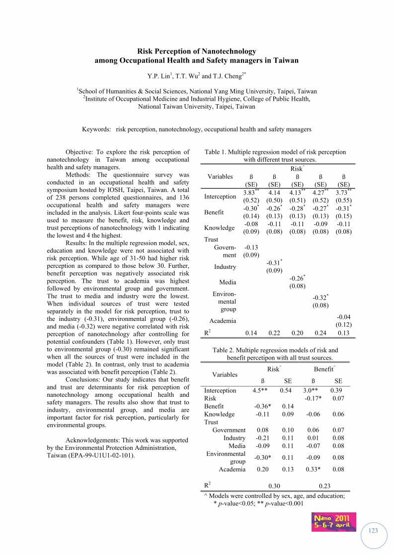
123
Risk Perception of Nanotechnology among Occupational Health and Safety managers in Taiwan
Y.P. Lin1, T.T. Wu2 and T.J. Cheng2*
1School of Humanities & Social Sciences, National Yang Ming University, Taipei, Taiwan
2Institute of Occupational Medicine and Industrial Hygiene, College of Public Health, National Taiwan University, Taipei, Taiwan
Keywords: risk perception, nanotechnology, occupational health and safety managers Objective: To explore the risk perception of nanotechnology in Taiwan among occupational health and safety managers. Methods: The questionnaire survey was conducted in an occupational health and safety symposium hosted by IOSH, Taipei, Taiwan. A total of 238 persons completed questionnaires, and 136 occupational health and safety managers were included in the analysis. Likert four-points scale was used to measure the benefit, risk, knowledge and trust perceptions of nanotechnology with 1 indicating the lowest and 4 the highest. Results: In the multiple regression model, sex, education and knowledge were not associated with risk perception. While age of 31-50 had higher risk perception as compared to those below 30. Further, benefit perception was negatively associated risk perception. The trust to academia was highest followed by environmental group and government. The trust to media and industry were the lowest. When individual sources of trust were tested separately in the model for risk perception, trust to the industry (-0.31), environmental group (-0.26), and media (-0.32) were negative correlated with risk perception of nanotechnology after controlling for potential confounders (Table 1). However, only trust to environmental group (-0.30) remained significant when all the sources of trust were included in the model (Table 2). In contrast, only trust to academia was associated with benefit perception (Table 2). Conclusions: Our study indicates that benefit and trust are determinants for risk perception of nanotechnology among occupational health and safety managers. The results also show that trust to industry, environmental group, and media are important factor for risk perception, particularly for environmental groups. Acknowledgements: This work was supported by the Environmental Protection Administration, Taiwan (EPA-99-U1U1-02-101).
Table 1. Multiple regression model of risk perception with different trust sources.
Variables Risk^
ß (SE)
ß (SE)
ß (SE)
ß (SE)
ß (SE)
Interception 3.83**
(0.52) 4.14
(0.50) 4.13** (0.51)
4.27** (0.52)
3.73** (0.55)
Benefit -0.30*
(0.14) -0.26* (0.13)
-0.28* (0.13)
-0.27* (0.13)
-0.31* (0.15)
Knowledge -0.08 (0.09)
-0.11 (0.08)
-0.11 (0.08)
-0.09 (0.08)
-0.11 (0.08)
Trust Govern-
ment -0.13 (0.09)
Industry -0.31* (0.09)
Media -0.26* (0.08)
Environ- mental group
-0.32* (0.08)
Academia -0.04 (0.12)
R2 0.14 0.22 0.20 0.24 0.13
Table 2. Multiple regression models of risk and
benefit percetipon with all trust sources.
Variables Risk^ Benefit^
ß SE ß SE
Interception 4.5** 0.54 3.0** 0.39 Risk -0.17* 0.07 Benefit -0.36* 0.14 Knowledge -0.11 0.09 -0.06 0.06 Trust
Government 0.08 0.10 0.06 0.07 Industry -0.21 0.11 0.01 0.08
Media -0.09 0.11 -0.07 0.08 Environmental
group -0.30* 0.11 -0.09 0.08
Academia 0.20 0.13 0.33* 0.08 R2 0.30 0.23
^ Models were controlled by sex, age, and education; * p-value<0.05; ** p-value<0.001
124
Equipping: an obligatory point of passage in nanoparticle-related risk management in research laboratories
H. Skaiky1, S. Caroly1, D. Vinck1 & E. Drais2
1Laboratoire PACTE, Université de Grenoble, Le patio, BP 47, 38040, Grenoble cedex 09, France
2INRS (Institut National de Recherche et de Sécurité), Rue du Morvan, CS 60027, 54519 Vandoeuvre, France
Keywords: equipment, risks, laboratory, nanoparticles Objectives:
The focus of this research is the potential risk emanating from nanoparticles in research laboratories. The aim of this paper is to underline the dynamics involved in nanoparticle risk prevention equipment and equipping work. Our approach in human and social sciences is a pragmatic one and therefore starts in the field. This empirical approach is also inspired by the wealth of literature stemming from laboratory studies where the process of scientific knowledge building is examined. Our paper contributes to laboratory studies focusing on the question of risk and, in particular, on the question of equipping/equipment and the controversial issues this raises with respect to risk management. In ergonomics, instruments are tied to a specific result, that of getting actors to buy into a tool (Rabardel, 1995), and to activity development. Ethnographic laboratory studies (Vinck, 2006) address this notion in order to report on the dynamics surrounding the equipping process. Methodology:
Material was collected directly from the field via interviews (N=7) with the actors concerned (researchers, health and safety officers, etc.). The information gathered was then completed by a day spent observing the activity of cleaning a plasma deposition reactor. This is an activity performed in the field studied, i.e. a plasma deposition laboratory. As part of an inter-disciplinary research project bringing together two research laboratories specialising in different disciplines (experimental physics and biology), the reactor cleaning activity acted as a focal point for the equipping process. Results:
The field proved to reveal much about the importance of the equipment and the equipping process in nanoparticle-related risk prevention. The cleaning activity brings to the fore the issue of the potential risk of nanoparticles in the laboratory space. This is because it is a manual activity that consists in scraping the internal walls of the reactor as well as its adjoining parts. The potential risk stems from the emanations of silver nanoparticles deposited on the sample as well as on the walls of the reactor.
The main research results are as follows: - The reactor cleaning activity opens a debate about the need for actors to be properly equipped in order to protect themselves from nanoparticle-related risks. -The equipment itself acts as an essential mediator in the prevention of nanoparticle-related risks. We underline how it becomes an object of negotiation in a three-way institutional discussion (between the two afore-mentioned laboratories and a health and safety organisation working with them to provide expertise in safety). - The use of equipment is not automatic. It is an object that is co-constructed by the various actors present in the field.
The cleaning activity involves the researchers in the dynamics of the equipping work required to prepare the reactor for an experiment: It invites the actors to create new activities (a nano-bin) to improve on nano-safety. The new category (the nano bin) does not solve the nano waste traceability problem as there is no specific treatment system for this waste. The calling in of external experts acts on the way the researchers involved in scraping the deposition reactor clean equip themselves with gestures. This gesture equipping process entails the modification of the gestures ordinarily adopted in the cleaning operation (wearing of masks, overalls, change in the way the reactor cleaning operation is normally done, etc.)
The former equipping process is questioned. This is something that the laboratory actors bring up when talking about the cleaning activity spatial layout, which is potentially risky for the person doing the experiment. Bibliography Rabardel, P. (1995). Les hommes et les technologies.
Paris : A. Colin. Vinck, D. (2006) L'équipement du chercheur.
Comme si la technique était déterminante. Ethnographique.org,9. http://www.ethnographiques.org/2006/Vinck.html
125
Outlining the regulatory context: National initiatives and the role of standards on nanotechnology
A. Ponce Del Castillo
Health and Safety Department, European Trade Union Institute, Brussels, 1210, Belgium
Key words: regulation, standards, national initiatives
By allowing humans to control the matter in the nano scale, nanotechnology confronts society with policy issues and opens a unique governance challenge.
The fact that current laws might not be
adequate to regulate nanotechnology, suggests the possibility that they cannot account for nanomaterials. As an interim solution, it is seen that current laws should be updated, as they would definitely be ready to be implemented.
In Europe one of the most important pieces of
regulation is REACH which in principle, governs the registration, authorization and evaluation of nanomaterials. However the nanomaterials will be treated insufficiently within REACH, and the information for its safe use will not be sufficient.
Alongside of the European regulations, there
are two other means that play a key role for the regulatory process of nanotechnologies: The national initiatives from Member States and the standards issued by international standardisation bodies.
In the view of starting regulatory initiatives at
the national level, countries like Belgium, France, Italy and the Netherlands have initiated diverse actions.
The common approach of those Member
states is to develop harmonised mandatory registers of nanomaterials and articles containing nanomaterials in their countries. Such registers could be the base for traceability, market surveillance, gaining knowledge for better risk prevention and for the improvement of the national and European legislative framework.
In particular this paper presents the proposals
from the Belgian Presidency of the Council of the European Union on the regulatory framework for nanomaterials; the nationwide public debates on nanotechnology in France and the implementation of the Grenelle Law; as well as the national register on nanomaterials in Italy and the Netherlands.
As an additional proposal to those national activities, this paper suggests that Members States could set up an exposure register of workers exposed to nanoparticles in association with health surveillance programmes. The register should list which workers have been exposed, the circumstances, duration and levels of the exposure and the protective measures applied.
Alongside the national initiatives, the role of standardisation is useful to support the implementation of European policy. The impact of standards is influential for the regulatory process on nanotechnology, mainly
Moreover the European Commission and the European Parliament have stressed the need of a harmonized terminology and definitions at the international level; hence the work on definition has become a priority for the European standardisation bodies.
In spite of the absence of specific regulation; this paper stresses out that standardisation should not be a substitute for regulation, and should only be reserved for technical specifications which are important for traceability. The role of standardisation should not extend towards health and safety, risk assessment methodology, risk management or any other societal issues.
The role of national regulatory initiatives and the direction standardisation bodies are taking at the moment with the development of standards are crucial components for the European regulatory framework on nanotechnologies.
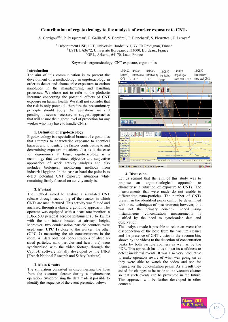
126
Contribution of ergotoxicology to the analysis of worker exposure to CNTs
A. Garrigou1et 2, P. Pasquereau1, P. Gaillard3, S. Bordère3, C. Blanchard1, S. Pierrettes1, F. Leroyer1
1 Département HSE, IUT, Université Bordeaux 1, 33170 Gradignan, France 2 LSTE EA3672, Université Bordeaux 2, 33000, Bordeaux France
3 GRL, Arkema, 64170, Lacq, France
Keywords: ergotoxicology, CNT exposure, ergonomics
Introduction The aim of this communication is to present the development of a methodology in ergotoxicology in order to detect and characterise exposures to carbon nanotubes in the manufacturing and handling processes. We chose not to refer to the plethoric literature concerning the potential effects of CNT exposure on human health. We shall not consider that the risk is only potential; therefore the precautionary principle should apply. As regulations are still pending, it seems necessary to suggest approaches that will ensure the highest level of protection for any worker who may have to handle CNTs.
1. Definition of ergotoxicology Ergotoxicology is a specialised branch of ergonomics that attempts to characterise exposure to chemical hazards and to identify the factors contributing to and determining exposure situations. Just as is the case for ergonomics at large, ergotoxicology is a technology that associates objective and subjective approaches of work activity analysis and also includes biological monitoring methods from industrial hygiene. In the case at hand the point is to detect potential CNT exposure situations while remaining firmly focused on activity analysis.
2. Method The method aimed to analyse a simulated CNT release through vacuuming of the reactor in which CNTs are manufactured. This activity was filmed and analysed through a classic ergonomic approach. The operator was equipped with a heart rate monitor, a PDR-1500 personal aerosol instrument (0 to 12µm) with the air intake located at airways height. Moreover, two condensation particle counters were used; one (CPC 1) close to the worker, the other (CPC 2) measuring the air concentrations in the room. All data obtained (concentrations of alveolar-sized particles, nano-particles and heart rate) were synchronised with the video footage through the Captiv® software initially developed by the INRS [French National Research and Safety Institute].
3. Main Results The simulation consisted in disconnecting the hose from the vacuum cleaner during a maintenance operation. Synchronising the data made it possible to identify the sequence of the event presented below:
14h04:12 Leak of CNTs
14h04:45 Detection by CPC 2
14h05:45 Detection by CPC 1
14h08:47 Particules peak
14h08:58 Beginning of nano peak CPC 2
14h09:47 Beginning of nano peak CPC 1
4. Discussion Let us remind that the aim of this study was to propose an ergotoxicological approach to characterise a situation of exposure to CNTs. The measurements that were made do not enable to differentiate nano-particles. The number of CNTs present in the identified peaks cannot be determined with these techniques of measurement; however, this was not the primary concern. Indeed using instantaneous concentration measurements is justified by the need to synchronise data and observation. The analysis made it possible to relate an event (the disconnection of the hose from the vacuum cleaner and the presence of CNT cluster in the vacuum bin, shown by the video) to the detection of concentration peaks by both particle counters as well as by the PDR. This approach has thus shown its usefulness to detect incidental events. It was also very productive to make operators aware of what was going on as they were able to watch the video and see for themselves the concentration peaks. As a result they asked for changes to be made to the vacuum cleaner so that such events can be prevented in the future. This approach will be further developed in other contexts.
127
Authors Index
A
Adiloglu, M. ................................................................... 56 Aguerre‐Chariol, O. ................................................. 80, 81 Akan, T. .......................................................................... 56 Aldeek, F. ....................................................................... 75 Alenius, H. ..................................................................... 57 Amodeo, T. .................................................................... 82 André, J.C. ................................................................... 120 Andreau, K. .................................................................... 31 Appert‐Collin, J.C. ........................................................ 111 Asbach, C. ................................................................ 62, 79 Assémat, E. .................................................................... 39 Audignon Durand, S. ..................................................... 77 Ausset, P. ....................................................................... 73 Authier, F‐J. ................................................................... 28 Aye, M. .......................................................................... 47
B
Backman, U. .................................................................. 79 Baeza, A. .................................................................. 46, 48 Baeza‐Squiban, A. .............................................. 31, 39, 51 Barberi‐Heyob, M. ................................................. 32, 120 Bard, D. .............................................................. 64, 79, 91 Barillet, S. ...................................................................... 24 Baron, M. ..................................................................... 108 Barthélémy, P. ............................................................... 47 Bary, M. ....................................................................... 116 Bastogne, T. ................................................................... 32 Bau, S. ................................................................ 71, 72, 94 Bauda, P. ....................................................................... 41 Bazzi, R. ......................................................................... 48 Bechet, D. .............................................................. 32, 120 Beck‐Schimmer, B.................................................... 25, 35 Bekker, C. ...................................................................... 86 Belut, E. ....................................................................... 105 Bemer, D...................................................................... 111 Benachour, H. ................................................................ 32 Berges, M. ............................................................... 64, 79 Berlin, K. ........................................................................ 62 Berque‐Bestel, I. ............................................................ 47 Bezdetnaya, L. ............................................................... 37 Bianchi, B. ...................................................................... 94 Billerbeck, U. ................................................................. 93 Billon‐Galland, M.A. ...................................................... 42 Binet, S. ................................................................... 23, 36 Birkedal, R. ............................................................ 33, 119 Blanchard, C. ............................................................... 126 Bockzowski, J. .............................................................. 120 Boggetto, N. .................................................................. 31 Bohgard, M. ................................................................... 65 Boivin, A. ....................................................................... 23 Boland, S. ........................................................... 44, 46, 48 Bomfim, J.A.S. .......................................................... 83, 84
Bordenave, L. ................................................................ 55 Bordère, S. .................................................................. 126 Borot, M.C. ......................................................... 31, 39, 46 Bossard, C. .................................................................... 31 Boublil, L. ...................................................................... 39 Bouillard, J.X. ............................................ 83, 84, 104, 109 Bourdel, G. .................................................................... 117 Bourdiol, F. .................................................................... 55 Boutou‐Kempf, O. ......................................................... 52 Bragaru, A. .................................................................... 53 Broberg, K. .................................................................... 65 Brochard, P. ............................................................ 42, 77 Brochot, C. .................................................................. 110 Brouwer, D. ........................................................ 64, 79, 86 Brun, E. .......................................................................... 58 Burdett, G. .................................................................... 91 Buresti, G. ..................................................................... 38 Burtscher, H. ........................................................... 60, 74 Busi, F. ........................................................................... 29
C
Cabuil, V. ....................................................................... 48 Cachier, H. ..................................................................... 51 Cadène, A. .................................................................... 117 Cadusseau, J. ................................................................. 28 Caroly, S. ..................................................................... 124 Carrière, M. ................................................................... 58 Casciardi, S. ............................................................. 38, 49 Catalán, J. ................................................................ 26, 45 Cavallo, D. ............................................................... 38, 49 Cederwall, T. ................................................................. 65 Cesard, V. .................................................................... 105 Chaffotte, A. .................................................................. 29 Chaskiel, P. .................................................................. 120 Chatelain, B. .................................................................. 43 Chazelet, S. .......................................................... 110, 111 Cheng, T.J. ................................................................... 123 Cheng, Y.S. .................................................................... 89 Chevillard, S. ................................................................. 22 Ciervo, A. ................................................................. 38, 49 Cohr, K.H. .................................................................... 119 Comte, P. ....................................................................... 34 Couleaud, P. .......................................................... 32, 120 Craciunoiu, F. ................................................................ 53 Crouzier, D. ................................................................... 55 Czerwinski, J. ......................................................... 34, 103
D
Daguet, I. ...................................................................... 117 Dahmann, D. ................................................................. 68 Dairou, J. ....................................................................... 29 Dalmasso, S. .................................................................. 70 Danila, M. ...................................................................... 53 Darne, C. ................................................................. 23, 36
128
De Gabory, L. ................................................................. 55 De Méo, M. ................................................................... 47 Debouzy, J.C. ................................................................. 55 Dedessus‐Le‐Moustier, N. ........................................... 116 Demokritou, P. ............................................................ 106 Deppert, K. .................................................................... 65 Derrough, S. .................................................................. 78 Di Giorgio, C................................................................... 47 Dietrich, S. ..................................................................... 62 Dijkman, A.J. ................................................................ 114 Dogné, J.M. ................................................................... 43 Donath, E. ...................................................................... 30 Dragan, G.C. .................................................................. 97 Drais, E. ....................................................................... 124 Dubertret, B. ................................................................. 37 Ducamp, S. .................................................................... 77 Dufaud, O. ........................................................... 104, 109 Dufour, J.P. .................................................................... 82 Dumortier, P. ................................................................. 42 Dupret, J.M. ................................................................... 29 Durand, C....................................................................... 78 Dutouquet, C. .................................................... 80, 81, 82 Dziurowitz, N. ........................................................ 62, 108
E
Echevarría‐Vega, M.E. ................................................. 106 Eichstädt, D. .................................................................. 93 Elgrabi, D. ...................................................................... 27 Ellenbecker, M. ............................................................ 106 Engel, R. ......................................................................... 86 Engel, S. ......................................................................... 93 Estrela Lopis, I. .............................................................. 30
F
Falck, G.C.‐M. ................................................................ 26 Fanizza, C. ...................................................................... 49 Ferecatu, I. .................................................................... 31 Fierz, M. ............................................................. 60, 63, 74 Fischer, R. ...................................................................... 93 Flahaut, E. ...................................................................... 55 Fleury, D. ......................................................... 83, 84, 104 Fontaine‐Aupart, M.‐P. .................................................. 75 Fort, Y. ........................................................................... 76 Fotel, F. ........................................................................ 119 Fransman, W. ................................................................ 79 Fregnaux, M. ................................................................. 70 Fréjafon, E. ........................................................ 80, 81, 82 Fresegna, A.M. ........................................................ 38, 49 Frick, R. .................................................................... 25, 35 Fries, R. ........................................................................ 121 Frochot, C. ............................................................. 32, 120
G
Gaillard, P. ................................................................... 126 Gao, Ch. ......................................................................... 30 Garrigou, A. ................................................................. 126 Garvas, M. ..................................................................... 54 Gaté, L. .................................................................... 23, 36 Gaumet, J.J. ................................................................... 70 Gauthier, L. .................................................................... 55 Gazsó, A. ...................................................................... 121
Gehr, P. ......................................................................... 34 Gensdarmes, F. ....................................................... 73, 94 George, I. ...................................................................... 51 Geszke‐Moritz, M. ......................................................... 40 Gherardi, R.K. ................................................................ 28 Girard, C. ....................................................................... 83 Götz, N.V. .................................................................... 102 Götz, U. ......................................................................... 62 Goutet, M. ..................................................................... 23 Graf, P. ........................................................................ 100 Grippari, F. .................................................................. 111 Gromb, S. ...................................................................... 42 Groso, A. ..................................................................... 118 Grzebyk, M. ................................................................... 95 Guadagnini, R. ......................................................... 44, 46 Gudmundsson, A. .......................................................... 87 Guichard, Y. ............................................................. 23, 36 Guillemin, F. ............................................................ 32, 37 Guiot, C. ........................................................................ 99 Gulsoy, N. ...................................................................... 56 Günther, D. ................................................................... 35 Guyot, A‐C. .................................................................... 58
H
H. Järventaus, H. ........................................................... 26 Haase, A. ..................................................................... 100 Hadji, R. ......................................................................... 76 Hagendorfer, H. .................................................... 69, 102 Hämeri, K. ................................................................... 107 Hannukainen, K.S. ......................................................... 45 Hattendorf, B. ............................................................... 35 Heinemann, M. ............................................................. 93 Helle, M. ........................................................................ 37 Hellweg, S. .................................................................... 63 Henry, F. .............................................................. 104, 109 Hervé, C. ...................................................................... 120 Hodson, L. ..................................................................... 79 Hollander, A.L.............................................................. 114 Honnert, B. .................................................................... 95 Hougaard, K.S. ............................................................. 119 Howard, J. ................................................................... 115 Huang, C. ..................................................................... 106 Hugounenq, P. .............................................................. 48 Hungerbühler, K. ......................................................... 102 Hussain, S. ............................................................... 29, 44
I
Iavicoli, S. ................................................................ 38, 49 Imbernon, E. ................................................................. 52 Isidore, Y. ...................................................................... 77 Itier, V. .......................................................................... 28
J
Jacobsen, N.R. ....................................................... 33, 119 Jammes, Y. .................................................................... 47 Janes, A. ........................................................................ 84 Jankowska, E. .......................................................... 64, 92 Järventaus, H. .......................................................... 45, 50 Jensen, K.A. ...................................................... 33, 90, 119 Jomini, S. ....................................................................... 41
129
K
Kaegi, R. ......................................................................... 69 Kaminski, H. ................................................................... 62 Karg, E. .......................................................................... 97 Kasper, G. ...................................................................... 61 Kasper, M. ............................................................. 34, 103 Keller, M. ................................................................. 66, 67 Kelly, A. .......................................................................... 91 Kerdine‐Römer, S. ......................................................... 24 Khan, Z. .......................................................................... 28 Kiesling, H.‐J. ................................................................. 62 Klages‐Büchner, S. ......................................................... 93 Kleps, I. .......................................................................... 53 Knobl, S. ......................................................................... 93 Koivisto, A.J. .......................................................... 26, 107 Kontturi, E. .................................................................... 45 Koponen, I.K. ................................................... 33, 90, 119 Kund, K. ......................................................................... 93
L
Lacourt, A. ............................................................... 42, 77 Lacroix, G. ...................................................................... 27 Laloy, J. .......................................................................... 43 Lam, H.R. ..................................................................... 119 Landsiedel, R. ................................................................ 98 Langlais, C. ..................................................................... 36 Larsen, S.T. .................................................................. 119 Laurenti, J.P. .................................................................. 70 Le Bihan, O. ................................................................... 81 Lecerf, P. ........................................................................ 82 Leroux, M. ..................................................................... 31 Leroyer, F..................................................................... 126 Lidén, G. ........................................................................ 87 Limbach, L.K. ................................................................. 25 Lin, Y.P. ........................................................................ 123 Lindberg, H.K. ................................................................ 26 Linse, S. .......................................................................... 65 Loft, S. ................................................................... 33, 119 Lorenz, C. ..................................................................... 102 Lucas, S. ......................................................................... 43 Luch, A. ........................................................................ 100 Ludwig, Ch. .................................................................... 69 Luechinger, N. ............................................................... 63 Lulek, J. .......................................................................... 40 Lux, F. ............................................................................ 32 Lynch, I. ......................................................................... 79
M
Mabondzo, A. ................................................................ 58 Macé, T. ......................................................................... 73 Magrez, A. ................................................................... 118 Maguhn, J. ..................................................................... 97 Maiello, R. ............................................................... 38, 49 Maillé, M. ...................................................................... 73 Mantion, A. .................................................................. 100 Marano, F. ......................................................... 31, 39, 44 Marbehant, C. ............................................................... 43 Marcelloni, A.M. ............................................................ 49 Marchal, F. ..................................................................... 37 Marchal, Ph. ................................................................ 109 Marchand, J.L. ............................................................... 52
Mark, D. ........................................................................ 64 Martinon, L. ............................................................ 39, 51 Masereel, B. .................................................................. 43 Mašić, A....................................................................... 100 Matikainen, S. ............................................................... 57 Mayer, A................................................................ 34, 103 Mehel, A. ..................................................................... 112 Meier, D. ....................................................................... 60 Meier, W. .................................................................... 100 Mejia, J. ......................................................................... 43 Messing, M.E. ................................................................ 65 Meunier, E. .................................................................... 55 Meyer, T. ..................................................................... 118 Michielsen, N. ....................................................... 73, 110 Micillino, J‐M. ................................................................ 36 Mikkelsen, L. ......................................................... 33, 119 Miu, M. ......................................................................... 53 Mohamed, A. .............................................................. 122 Möhlmann, C. ............................................................... 64 Møller, P. ............................................................... 33, 119 Monz, C. .................................................................. 62, 68 Mooney, J. ................................................................... 103 Mordon, S. .................................................................... 32 Moreau, K. .................................................................... 44 Moriceau, A. ................................................................ 116 Motzkus, C. ................................................................... 73 Mouchet, F. ................................................................... 55 Moya, S.E. ..................................................................... 30 Müller, L. ....................................................................... 34 Müller‐Edenborn, B. ...................................................... 35 Mullier, F. ...................................................................... 43 Murashov, V. ............................................................... 115 Murias, M. ..................................................................... 40 Mustin, C. ...................................................................... 75 Mutlu, O. ....................................................................... 56
N
Neubauer, N. ........................................................... 61, 67 Nguyen, V.S. .................................................................. 76 Nhim, C. ........................................................................ 24 Nordsieck, H. ................................................................. 97 Norppa, H. .......................................................... 26, 45, 50 Nykäsenoja, H. .............................................................. 26
O
Oesterlé, B. ................................................................. 112 Ostiguy, C. .................................................................... 117 Ouf, F.X. .................................................................. 72, 96
P
Paba, E. ......................................................................... 49 Pagels, J. ........................................................................ 65 Paget, V. ........................................................................ 22 Pagnout, C. .................................................................... 41 Pairon, J.C. .................................................................... 42 Pajk, S. ........................................................................... 54 Pallardy, M. ................................................................... 24 Palomäki, J. ................................................................... 57 Pasquereau, P. ............................................................ 126 Pelzer, J. .................................................................. 62, 64 Permin, A. ................................................................... 119
130
Pernot, M. ..................................................................... 32 Perrin, L. .............................................................. 104, 109 Petri‐Fink, A. ................................................................ 118 Pic, E. ............................................................................. 37 Pierrettes, S. ................................................................ 126 Pipy, B. ........................................................................... 55 Piret, J.P. ........................................................................ 43 Piringer, R. ................................................................... 121 Plendl, J. ...................................................................... 100 Plitzko, S. ..................................................................... 108 Ponce Del Castillo, A. ................................................... 125 Pons, T. .......................................................................... 37 Pontreau, S. ................................................................... 96 Prevost, C. ................................................................... 105 Prodi, L. ......................................................................... 37 Puech, P. ........................................................................ 55 Pylkkänen, L. .................................................................. 26
R
R’Mili, B. ............................................................ 80, 81, 84 Radauceanu, A............................................................... 52 Raemy, D. ...................................................................... 35 Ragot, J. ......................................................................... 93 Rampazzo, E. ................................................................. 37 Rastoix, O. ......................................................... 23, 36, 72 Ravanel, X. ..................................................................... 78 Reisinger, M. ................................................................. 93 Reuter, M. ..................................................................... 93 Riediker, M. ................................................... 79, 117, 118 Rinaldo, M. .............................................................. 42, 77 Rissler, J. ........................................................................ 65 Robert, S. ....................................................................... 43 Rodrigues‐Lima, F. ......................................................... 29 Rogerieux, F. .................................................................. 27 Rojas, E. ......................................................................... 30 Rolin, S. .......................................................................... 43 Rommert, A. .................................................................. 93 Rossi, E. ......................................................................... 26 Rothen‐Rutishauser, B. ............................................ 34, 35 Roursgaard, M. ............................................................ 119 Rousset, D. .............................................................. 23, 36 Rouxel, D. .............................................................. 76, 122
S
Saber, A.T. ............................................................. 33, 119 Sagot, B. ...................................................................... 112 Sanfins, E. ...................................................................... 29 Savolainen, K. ........................................ 26, 45, 50, 57, 66 Schierl, R. ....................................................................... 62 Schimmel, R. .................................................................. 86 Schlicker, A. ............................................................. 25, 35 Schmitt, J. ...................................................................... 23 Schneider, R. ........................................................... 40, 75 Schnelle‐Kreis, J. ............................................................ 97 Schröter, N. ................................................................... 93 Schwartz, Y. ................................................................. 122 Sébillaud, S. ................................................................... 36 Seipenbusch, M. .............................................. 61, 67, 107 Sellier, E. ........................................................................ 42 Sergent, J.A. ................................................................... 22 Seve, A. .......................................................................... 32 Siivola, K. ....................................................................... 50
Simion, M. ..................................................................... 53 Sirven, J.B. ..................................................................... 80 Sirviö, S.......................................................................... 66 Skaiky, H. ..................................................................... 124 Smerajec, M. ................................................................. 88 Sotiriou, G. .................................................................. 106 Spalla, O. ....................................................................... 99 Stark, W. ....................................................................... 35 Stark, W.J. ..................................................................... 25 Steigmeier, P. .......................................................... 60, 74 Steiner, S. ...................................................................... 34 Štrancar, J...................................................................... 54 Suhonen, S. ............................................................. 26, 50 Svensson, CR. ................................................................ 65 Swain, K. ........................................................................ 93
T
Tanière, A. ................................................................... 112 Taubert, A. .................................................................. 100 Terwoert, J. ................................................................. 114 Thieriet, N. .................................................................... 117 Thomas, D. .......................................................... 110, 111 Thorpe, A. ..................................................................... 64 Thuenemann, A.F. ....................................................... 100 Tillement, O. ........................................................... 28, 32 Tombolini, F. ........................................................... 38, 49 Toussaint, O. ................................................................. 43 Triolet, J. ....................................................................... 117 Troisfontaines, P. ........................................................... 117 Tromp, P. ....................................................................... 86 Trouiller, B. .................................................................... 27 Tsai, C.J. ......................................................................... 89 Tsai, S‐J. ....................................................................... 106 Tuinman, I. .................................................................... 86
U
Ulrich, A. ........................................................ 69, 102, 103 Umek, P. ........................................................................ 54 Urner, M. ...................................................................... 25 Ursini, C.L. ............................................................... 38, 49
V
Val, S. ............................................................................ 51 Välimäki, E. .................................................................... 57 van Duuren‐Stuurman, B. ............................................. 64 Vanderesse, R. ...................................................... 32, 120 Vanhala, E. .................................................................... 45 Vaslin‐Reimann, S. ........................................................ 73 Vaupotič, J. .................................................................... 88 Verhille, M. ................................................................. 120 Vernez, D. ..................................................................... 117 Vignes, A. ................................................. 83, 84, 104, 109 Vincent, B. ..................................................................... 76 Vinck, D. ...................................................................... 124 Vippola, M. .................................................................... 26 Vital, A. .......................................................................... 42 Voetz, M. ....................................................................... 93 Vogel, U. ................................................................ 33, 119 Von Barany, D. .............................................................. 62 von Gleich, A. ................................................................ 85 Vranic, S. ................................................................. 44, 46

131
W
Waher, J. ....................................................................... 87 Wake, D. ........................................................................ 64 Wallin, H. ............................................................... 33, 119 Walser, T. ...................................................................... 63 Weinand, R. ................................................................... 93 Weis, F. .......................................................................... 61 Wiench, K. ..................................................................... 98 Wigger, H. ...................................................................... 85 Witschger, O. ......................................... 52, 71, 72, 76, 94 Wohlleben, W. .............................................................. 98 Wolf, T. ........................................................................ 108
Wrobel, R. ..................................................................... 23 Wu, T.T. ....................................................................... 123
Y
Yon, J. ............................................................................ 96 Yu, M. .......................................................................... 107
Z
Zhou, Y. ......................................................................... 89 Zielecka, M. ................................................................... 92 Zimmermann, R. ........................................................... 97