Embed Size (px)
Citation preview
Université de Montréal
Modification de la spécificité de la transglutaminase par
une approche semi-aléatoire
Un nouvel outil pour la synthèse des peptides
par
Roberto Antonio Chica
Département de chimie
Faculté des arts et des sciences
Thèse présentée à la faculté des études supérieures
en vcte de l’obtention du grade de Philosophiœ Doctor (Ph. D.)
en chimie
Décembre, 2006
© Roberto Antonio Chica, 2006

LL) t o
o
Universitéde Montréal
Direction des bibliothèques
AVIS
L’auteur a autorisé l’Université de Montréal à reproduire et diffuser, en totalitéou en partie, par quelque moyen que ce soit et sur quelque support que cesoit, et exclusivement à des fins non lucratives d’enseignement et derecherche, des copies de ce mémoire ou de cette thèse.
L’auteur et les coauteurs le cas échéant conservent la propriété du droitd’auteur et des droits moraux qui protègent ce document. Ni la thèse ou lemémoire, ni des extraits substantiels de ce document, ne doivent êtreimprimés ou autrement reproduits sans l’autorisation de l’auteur.
Afin de se conformer à la Loi canadienne sur la protection desrenseignements personnels, quelques formulaires secondaires, coordonnéesou signatures intégrées au texte ont pu être enlevés de ce document. Bienque cela ait pu affecter la pagination, il n’y a aucun contenu manquant.
NOTICE
The author of this thesis or dissertation has granted a nonexclusive licenseallowing Université de Montréal to reproduce and publish the document, inpart or in whole, and in any format, solely for noncommercial educational andresearch purposes.
The author and co-authors if applicable retain copyright ownership and moralrights in this document. Neither the whole thesis or dissertation, norsubstantial extracts from it, may be printed or otherwise reproduced withoutthe author’s permission.
In compliance with the Canadian Privacy Act some supporting forms, contactinformation or signatures may have been removed from the document. Whilethis may affect the document page count, it does not represent any loss ofcontent from the document.
Université de Montréal
Faculté des études supérieures
Cette thèse intitulée
Modification de la spécificité de la transglutarninase par une approche semi-aléatoire
Un nouvel outil pour la synthèse des peptides
présentée par
Roberto Antonio Chica
a été évaluée par un jury composé des personnes suivantes:
Dr Andreea R. Schrnitzer, président-rapporteur
Dr Jodle N. Pelletier, directrice de recherche
Dr Jeffrey W. Keillor, codirecteur
Dr Robert Lortie, membre du jury
Dr Shabriar Mobashery, examinateur externe
Dr Andreea R. Schmitzer, représentant du doyen de la FES
111
Résumé
La synthèse chimique est la principale méthode employée pour produire les peptides
en industrie. Plusieurs étapes d’activation et de protectionldéprotection sont nécessaires, ce
qui diminue l’efficacité globale de leur synthèse en plus de générer de nombreux déchets
polluants. La synthèse enzymatique est une solution de rechange plus respectueuse de
l’environnement. Elle réduit largement le besoin d’activation et de protection des
groupements fonctionnels, en plus d’utiliser peu ou pas de solvants organiques polluants.
Les enzymes utilisées actuellement pour la synthèse des peptides sont les protéases. Un des
obstacles majeurs à l’utilisation de ces enzymes réside dans leur tendance à hydrolyser les
peptides synthétisés, ce qui diminue le rendement. De plus, le problème de spécificité
étroite des protéases empêche leur application à des réactions de couplage variées.
Pour s’attaquer à ces problèmes, nous proposons de développer de nouvelles
enzymes catalysant de façon plus efficace et spécifique la synthèse des peptides. Ainsi,
nous avons entrepris l’évolution dirigée, par une approche serni-aléatoire, de la
transglutarninase (TGase), une enzyme qui catalyse déjà la formation (et non l’hydrolyse)
des liens amides entre des peptides et des dérivés d’acides aminés. Nous voulons modifier
sa spécificité dans le but de permettre la catalyse de la formation de liens amides entre une
gamme d’acides x-aminés.
Pour ce faire, nous avons généré un modèle moléculaire de la TGase avec un
substrat lié au site actif pour identifier les résidus impliqués dans la spécificité. Ces résidus
ont été soumis à une mutagenèse semi-aléatoire pour générer cinq banques contenant
chacune jusqu’à $000 mutants. Une méthode d’expression de la TGase chez Escherichia
cou a été développée de sorte à obtenir de l’enzyme soluble et active. Une méthode de
criblage pour détecter les TGases mutantes ayant une spécificité désignée a été développée.
Cette méthode, basée sur des analogues d’acides aminés dérivés avec un groupement
fluorogène de type 7-hydroxycoumarine, a servi au criblage des cinq banques de mutants et
iv
a permis d’identifier un mutant. Trp332Phe. ayant une différente spécificité par rapport à
l’enzyme de type sauvage. Cette recherche a permis de développer un nouvel outil pour la
synthèse des peptides qui a une efficacité catalytique similaire à celle de la papaïne, une
protéase catalysant la synthèse des peptides.
Mots-clés transglutaminase, enzyme, enzymologie, évolution dirigée, biocatalyse.
modélisation moléculaire, cinétique, chimie bio-organique, chimie verte
V
Abstract
Chemical synthesis is the main method used to produce peptides in industry. It
requires various activation, protection and deprotection steps which decrease overali yields
and generate large quantities of waste that is hanuful for the environment. Enzymatic
peptide synthesis is an interesting alternative to chemical synthesis because it exploits the
high selectivity of enzymes which require no protection and deprotection of potentially
reactive functional groups, generating less waste. For this reason, enzymatic synthesis of
peptides is an environrnentally-friendly procedure. Proteases are cunently the enzymes of
choice for enzymatic peptide synthesis. However, they are prone to hydrolyse the peptide
products, decreasing the yields. furthermore, their narrow specificity prevents their
application to the synthesis of various products.
The main goal of this work is to develop new catalysts for enzyrnatic peptide
synthesis. These catalysts would be more efficient than proteases as they would not
hydrolyse the peptide products formed. To reach our goal. we have undertaken to rnodify
the specificity of a transglutaminase (TGase) by directed evolution in order to generate an
enzyme that can recognize a variety of amino acids as donor or acceptor substrates. This
would provide us with a novel tool for the synthesis of peptides as the bond forrned by the
enzyme would be peptidic rather than isopeptidic. The enzyme mechanism would not be
altered by directed evolution since the catalyzed reaction would rernain the same. namely
the formation of an amide bond. The difference would be in the range of recognized
substrates.
b modify the specificity of TGase by a semi-random approach, a protocol for the
efficient expression and purification of the recombinant enzyme in E. cou was developped.
Molecular modeling was applied to the generation of a model for the binding of peptide
substrates on TGase, since no substrate-bound crystal structure of TGase is currently
available. This model allowed identification of TGase residues that are involved in
vi
substrate binding. These residues were subjected to combinatorial mutagenesis, thus
generating five libraries of mutants, each containing up to $000 mutants. Following
screening of the mutants with a fluorimetric micropÏate-based assay that we deveÏopped,
mutant Trp332Phe was identified as having a new desired specificity for the acyl-acceptor
substrate. Mutant Trp332Phe can catalyze the formation of peptide bonds and represents a
novel tool for the synthesis of peptides in aqueous solution.
Keywords: transglutaminase, TGase, enzyme, enzyrnology, directed evolution,
biocatalysis, molecular modeling, kinetics, bioorganic chemistry, green chemistry
vii
Table des matières
CHAPITRE 1 Introduction 1
1.1 La chimie verte 2
1.1.1 Définition 2
1 .1.2 Les douze principes de la chimie verte 3
1.1.3 Labiocatalyse 4
1.2 La synthèse des peptides 6
1.2.1 La synthèse chimique 6
1.2.2 La biosynthèse 9
1.2.3 La synthèse enzymatique 9
1 .3 Les transglutaminases 11
1.3.1 Réaction catalysée et mécanisme catalytique 12
1.3.2 Classification 14
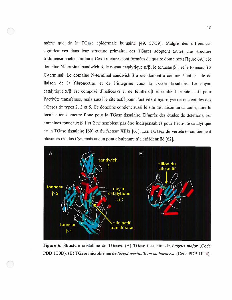
1.3.3 Structure 17
1.3.4 Spécificité 19
1.4 Description du projet de recherche 21
1.4.1 Objectif 21
1.4.2 Méthodologie générale 23
1.4.3 Étapes du projet 24
1.4.3.1 Identification des résidus de la TGase impliqués dans la liaison du substrat
donneur 25
1.4.3.2 Développement d’une méthode d’expression et de purification de la TGase
de foie de cobaye chez Escherichia cou 25
1.4.3.3 Développement d’un modèle d’homologie de la TGase de foie de cobaye et
mutagenèse de résidus du site actif 26
1.4.3.4 Évolution dirigée de la spécificité de la IGase de foie de cobaye par une
approche semi-aléatoire 26
viii
CHAPITRE 2 Évolution dirigée par une approche semi-aléatoire: revue de la
littérature 28
2.0 Préface 29
Article 1. Semi-rational approaches to engineering enzyme activity: combining the
benefits of directed evolution and rational design 30
2.1 Abstract 31
2.2 Introduction 31
2.3 Modifying enzyme activity using experimental semi-rational approaches 34
2.3.1 Targeted randomization of defined residues based on structural knowledge 35
2.3.2 Simultaneous random mutagenesis and site-saturation of defined residues 3$
2.3.3 Random mutagenesis followed by site-saturation of defined residues 39
2.4 Semi-rational and combinatorial design using computational approaches 41
2.5 Conclusions 43
2.6 Acknowledgements 44
2.7 References and recommended reading 44
CHAPiTRE 3 Identification des résidus de la TGase impliqués dans la liaison du
substrat donneur 51
3.0 Préface 52
Article 2. Tissue transglutaminase acylation: Proposed role of conserved active-site
Tyr and Trp residues revealed by molecular modeling of peptide substrate binding. 53
3.1 Abstract 54
3.2 Introduction 54
3.3 Resuits 5$
3.3.1 Kinetic characterization of the N-terminally derivatized peptides 5$
3.3.2 Modeling of substrate binding 59
3.3.3 Modeling ofthe tetrahedral and acyl-enzyme intermediate 6$
3.3.4 Structural analysis of Cbz-peptide binding 72
ix
3.4 Discussion .73
3.5 Materials and methods 79
3.5.1 Synthesis ofthe N-terminally derivatized peptide substrates 79
3.5.2 Kinetic measurements $0
3.5.3 Computational methods $0
3.5.4 Preparation ofthe protein structure $1
3.5.5 Computational construction of substrate molecules $1
3.5.6 Autornated docking of substrates on IGase 22
3.5.7 Manual docking of substrates on TGase 83
3.5.8 Calculations on the enzyme/substrate complex $3
3.5.9 Building ofthe acyl-enzyrne intermediate and the tetrahedral intermediate $4
3.6 Acknowledgrnents 85
3.7 References 85
CHAPITRE 4 Développement d’une méthode d’expression et de purification de la
TGase de foie de cobaye chez Escitericitia cou $9
4.0 Préface 90
Article 3. Expression and rapid purification of highly active hexahistidine-tagged
guinea pig liver transglutaminase 91
4.1 Abstract 92
4.2 Introduction 92
4.3 Materials and methods 94
4.3.1 Materials 94
4.3.2 Construction of expression plasmid 95
4.3.3 Sequence analysis 95
4.3.4 Overexpression ofTGase 96
4.3.4.1 Method A: expression with molecular chaperones DnaK and DnaJ 96
4.3.4.2 Method B: expression with chernical chaperone betaine 96
X
4.3.5 Purification ofHis6-tlGase.97
4.3.6 Protein concentrations 97
4.3.7 Determination of specific activity 98
4.3.8 Electrophoresis 9$
4.3.9 Enzyme purity 9$
4.3.10 Kinetic assays 99
4.3.10.1 Method I: DMPDA assay 99
4.3.10.2 Method II: glutamate dehydrogenase-linked assay (GDH) 99
4.4 Resuits and discussion 100
4.4.1 DNA sequencing 100
4.4.2 Expression and purification ofHis6-tTGase 101
4.4.3 Kinetic ofrecombinant guinea pig liver His6-tlGase 109
4.5 future work 110
4.6 Conclusions 111
4.7 Acknowledgements 111
4.8 References 111
CHAPITRE 5 Développement d’un modèle d’homologie de la TGase de foie de
cobaye et mutagenèse de résidus du site actif 116
5.OPréface 117
Article 4. Homology modeling of guinea pig liver transglutaminase and mutagenesis
ofconserved active-site residues Tyr519 and Cys336 118
5.1 Abstract 119
5.2 Introduction 119
5.3 Materials and methods 122
5.3.1 Homology modeling 122
5.3.2 Materials 123
5.3.3 Construction ofthe expression plasmid 123
xi
5.3.4 Mutagenesis of Cys336 and Tyr519.124
5.3.5 Overexpression and purification ofwild-type and mutant TGases 125
5.3.6 Synthesis ofCbz-Gly-7HC 125
5.3.7 Kinetic assays 126
5.3.7.1 Method I: DMPDA assay 126
5.3.7.2 Method II: 7-hydroxycournarin assay 12$
5.3.7.3 Method III: Hydroxarnate assay 128
5.4 Resuits and discussion 129
5.4.1 Homology rnodeling 129
5.4.2 Construction of plasmid pQE32-GTG 137
5.4.3 Role of guinea pig liver TGase residue Cys336 137
5.4.4 Role ofguineapig liver IGase residue 1yr519 141
5.5 Conclusion 143
5.6 Acknowledgements 143
5.7 References 144
CHAPITRE 6 Évolution dirigée de ]a spécificité de la TGase de foie de cobaye par
une approche semi-aléatoire 149
6.0 Préface 150
Article 5. Expansion of the peptide synthase specificity of guinea pig liver
transglutaminase by semi-random mutagenesis 151
6.1 Abstract 152
6.2 Introduction 153
6.3 Materials and Methods 156
6.3.1 Materials 156
6.3.2 Synthesis ofCbz-Gly-7HC, Cbz-Ala-7HC and Cbz-Phe-7HC 157
6.3.3 Overexpression and purification ofwild-type and mutant TGases 15$
6.3.4 Kinetic assays 15$
xii
6.3.5 Construction ofthe libraries of mutants.159
6.3.6 Library representation calculations 161
6.3.7 Preparation ofthe crude bacterial lysate for screening 161
6.3.8 Screening assay 162
6.4 Resuits 163
6.4.1 Cbz-Gly-7HC is hydrolyzed by wild-type guinea pig liver TGase 163
6.4.2 Wild-type guinea pig liver IGase can catalyse the formation of peptide bonds
164
6.4.3 Semi-random mutagenesis of guinea pig liver TGase 166
6.4.4 Library screening 16$
6.4.5 Enzyme kinetics of identified active mutants 171
6.5 Discussion 173
6.5.1 Wild-type guinea pig liver TGase has a peptide synthase activity 173
6.5.2 Modification of guinea pig liver TGase specificity 176
6.5.3 Structure/function analysis of active-site residues of guinea pig liver TGase.. 17$
6.6 Conclusion 181
6.7 References 181
CHAPITRE 7 Conclusion 186
7.1 Conclusion 187
7.2 Perspectives 189

XIII
Liste des tableaux
CHAPITRE I
Tableau 1. Les différentes TGases de mammifères 15
Tableau 2. Spécificité de la TGase tissulaire pour les substrats non naturels 20
CHAPITRE 2
Table 1. Comparison of approaches for engineering enzyme activity 33
CHAPITRE 3
Table 1. Kinetic constants for guinea pig liver IGase with various N-tenninaÏly derivatized
peptides 59
Table 2. Automated docking resuits for the docking of Cbz-Gln-Gly and Boc-Gln-Gly on
red sea bream TGase 63
Table 3. Manual docking resuits for Cbz-Gln-Gly on red sea bream TGase 65
Table 4. Manual docking resuits for Cbz-Gln-Gly, Cbz-Gln-Xaa and Boc-Gln-Gly 67
Table 5. Comparison ofthe conformation ofthe Gin side chain ofthe acyl-donor substrate
Cbz-Gln-Gly in the modelled Michaelis complex. acyl-enzyme and tetrahedral
intermediates 70
Table 6. Proposed interactions between acyl-donor substrates and tissue TGase 78
CHAPITRE 4
Table 1. Activity of recombinant guinea pig liver His6-tTGase from the two E. cou
expression systems 107
xiv
CHAPITRE 5
Table 1. Quality evaluation ofthe guinea pig liver TGase hornology models 131
Table 2. Distances between key atoms in human and red sea bream tissue TGases and in
horno!ogy mode! T2 of guinea pig !iver TGase 133
Table 3. Kinetic constants for Cbz-G!n-Gly of wild-type and mutant guinea pig liver
TGases determined using the DMPDA method 13$
CHAPITRE 6
Table 1. Kinetic constants of active guinea pig liver IGase mutants for hydro!ysis of
Cbz-G!y-7HC 164
Table 2. Kinetic constants of active guinea pig !iver TGase mutants for transarnidation
reaction of Cbz-Gly-7HC and N-AcLysOMe 166
Table 3. Libraries of mutant guinea pig liver TGases gcnerated in this study 167
Table 4. Active mutants identified by screening for transamidation of Cbz-G!y-711C and
N-AcLysOMe 170
Table 5. Acy!-acceptor substrate specificity of wi!d-type guinea pig !iver TGase and
mutant Trp332Phe 173
Table 6. Comparison ofguinea pig !iver TGase mutant Trp332Phe and papain for synthesis
ofGly-Phe peptide bonds 17$
xv
Liste des figures
CHAPITRE 1
Figure 1. Activation de la fonction carboxyle des acides aminés lors de la synthèse
peptidique $
Figure 2. Synthèse enzymatique des peptides catalysée par les protéases 11
Figure 3. Réaction catalysée par les TGases 12
Figure 4. Modifications post-traductioimelles des protéines catalysées par les TGascs 13
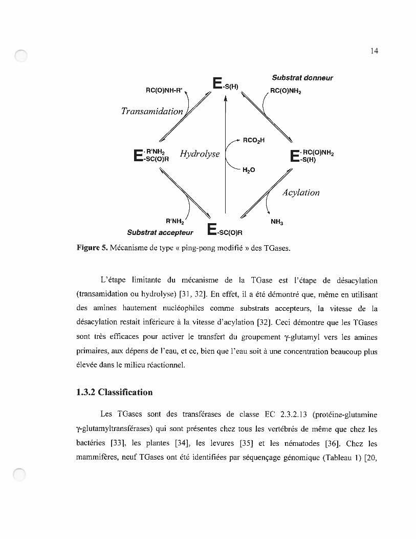
Figure 5. Mécanisme de type «ping-pong modifié » des TGases 14
Figure 6. Structure cristalline de IGases 18
Figure 7. Synthèse des peptides catalysée par une TGase modifiée 22
Figure 8. Évolution dirigée de la TGase par une approche semi-aléatoire 27
CHAPITRE 2
Figure 1. Selection of the prefened experimental approach for enzyme engineering based
on the availability of experirnental tools and prior knowledge of structure and
function 34
Figure 2. Residues in or near the enzyme active-site that were targeted for semi-rational
combinatorial mutagenesis 36
CHAPITRE 3
Scheme 1. Catalytic mechanism of tissue IGase 55
Figure 1. Crystal structure ofred sea bream TGase 57
Figure 2. Proposed binding clefi for the peptide substrate N-terminal functional group at
the surface ofred sea bream TGase 61
Figure 3. Model ofTGase with the acyl-donor Cbz-Gln-Gly 71
Scheme 2. Acylation step ofthe TGase rnechanisrn 79
xvi
CHAPITRE 4
Figure 1. TGase expression in the presence or absence of folding modulators 102
Figure 2. Specific activity of transglutarninase in the soluble fractions obtained from
lysates of E. cou expressing His6-tTGase, DnaK, and DnaJ, induced at different
temperatures 104
Figure 3. Quantity of His6-tTGase per litre of culture obtained afier purification, for
different induction temperatures 105
Figure 4. $DS—PAGE of fractions obtained during the purification 106
Figure 5. SDS—PAGE of fractions obtained after purification 108
CHAPITRE 5
Figure 1. Kinetic assays for tissue TGase used in this work 127
Figure 2. Stability of guinea pig liver TGase hornology model T2 during an unconstrained
300 ps molecular dynamics simulation at 300 K 134
Figure 3. Structures ofvarious tissue TGases 136
Figure 4. Activity loss of wild-type and Cys336 mutants of guinea pig liver TGase upon
incubation at 37 oc 140
CHAPITRE 6
Figure 1. Acyl-donor substrates of guinea pig liver TGase 154
Figure 2. Active-site residues ofguinea pig liver TGase 168
xvii
Liste des abréviations
7HC 7-hydroxycournarine
À Àngstr5rn
Ac groupement acétyle
ATP adénosine triphosphate
bp paire de base
BLAST Basic Local Aligornent Search Tool
Boc terr-butyloxycarbonyle
BSA albumine de sérum bovin
Cbz carbobenzyloxycarbonyle
CVFF constcint valence fiwcefielcl
d doublet
Da Dalton
DCC dicyclohexylcarbodiirnide
dITP désoxyinosine triphosphate
DMF N N-dirnéthy1fon;arnide
DMPDA NN-dirnéthyl- 1 ,4-phénylènediarnine
DNA acide désoxyribonucléique
dpi dots per inch (point par pouce)
DTT dithiothréitol
E. cou Escherichia cou
EC Enzyme Commission (numéro de classification des enzymes)
EDTA acide éthylènediaminetétraacétique
Enz enzyme
epPCR error-prone polymerase chain reaction
FAB bombardement d’atomes rapides
FACS fluorescence-activated ccli sorting
xviii
FG fiinctionaÏ group
fmoc 9-fluorénylméthylcarbamate
GABA acide y-aminobutyrique
GDH glutamate déshydrogénase
GDP guanosine diphosphate
GIP guanosine triphosphate
GP groupement protecteur
HBIU 2-( 1 H-benzotriazol- 1-yl)-l .1 ,3 ,3 -tétrarnethyluronium
hexafluorophosphate
HOAt 1 -hydroxy-7-azabenzotriazole
HOBt 1 -hydroxybenzotriazole
HRMS spectrométrie de masse à haute résolution
IMAC immobilized-metaÏ affinity chromatography
IPTG isopropyl- 3-D-thiogalactoside
LB Luria-Bertani
in multiplet
Mops acide 3-(N-morpholino)propanesulfonique
Mr masse moléculaire
NADH nicotinarnide adénine dinucléotide
NMR résonance magnétique nucléaire
NIA acide nitriloacétique
PCR réaction en chaîne de la polymérase
PDB Prote in data bank
PNP p-nitrophénolate
ppm partie par million
5 picoseconde
PyBOP benzotriazolyloxy-tris [pyrrolidino] -phosphonium
hexafluorophosphate
xix
rmsd root mean square deviation
rpm tours par minute
s singulet
SDS dodécylsulfate de sodium
SDS-PAGE électrophorèse sur gel de polyacrylamide avec dodécylsulfate
de sodium
t triplet
TGase transglutaminase
Tris tris(hydroxyméthyl)méthane
tRNA acide ribonucléique de transfert
tTGase TGase tissulaire
Xaa acide aminé
longueur d’onde d’émission
ex longueur d’onde d’ excitation
xx
A mi maUre A licia. No importa b
que suceda, siempre estaremosjuntos
xxi
Remerciements
Ces six dernières années passées au département de chimie de l’Université de
Montréal m’ont permis de grandir énormément en tant que scientifique et en tant que
personne. Durant ces années remplies d’efforts continus et de travail acharné, j’ai appris
beaucoup de choses sur moi-même en plus de vivre plusieurs moments heureux et
satisfaisants, et d’autres plus difficiles et douloureux. Néamoins, ces années d’études
doctorales en chimie ont été parmi les plus belles de ma vie. Tout ce que j’ai accompli
durant ces six dernières années n’aurait pas été possible sans la contribution de nombreuses
personnes proches de moi. J’aimerais leur manifester ma reconnaissance.
Je voudrais d’abord remercier mes parents, Alicia et Oscar. Sans l’énorme sacrifice
qu’ils ont fait en quittant leur El Salvador natal à catise de la guerre civile pour venir
recommencer leur vie à zéro au Canada, je ne serais pas ici en train de les remercier.
J’aimerais rendre hommage à ma mère qui m’a enseigné la signification réelle des mots
courage et espoir lors de sa lutte acharnée contre la leucémie... Elle a toujours été la plus
grande admiratrice de mes succès scolaires, et j’ai bénéficié de son amour inconditionnel.
J’aimerais saluer mon père parce qu’il m’a enseigné l’honnêteté et la ténacité, des valeurs
importantes pour tout scientifique.
Je voudrais remercier ma compagne, Isabelle, pour son amour et son soutien, surtout
lors de cette dernière année, où j’en ai eu particulièrement besoin. Un merci à mes frères
Oscar et Javier et à ma soeur Patricia pour leur aide et leur amour.
J’aimerais témoigner ma très grande reconnaissance envers mes directeurs de
recherche, Joelle et Jeff, aussi connus sous le nom de Moni & Dad. Ils ont été pour moi plus
que des directeurs de recherche : ils ont été des amis. Merci beaucoup de m’avoir guidé
dans mon apprentissage de la recherche scientifique et de m’avoir prêté votre assistance
tout au long de mon doctorat, tout en me laissant beaucoup de liberté. Merci aussi de
m’avoir montré que la recherche scientifique et la vie familiale n’étaient pas incompatibles.
xxii
Joelle, le temps fou que tu as consacré à m’aider à préparer des demandes de bourses, des
présentations à des conférences et ma thèse a contribué énormément à mes réussites. Jeff,
mon doctorat a été plus amusant grâce à notre complicité et à nos matchs de foot! Ce fut un
énorme plaisir de travailler à vos côtés durant toutes ces années. Finalement, merci de
m’avoir accordé le temps nécessaire pour que je m’occupe de mes responsabilités
familiales.
Je m’en voudrais de passer sous silence les étudiants, postdocs, stagiaires et
assistants de recherche des groupes Pelletier et Keillor avec qui j ‘ai eu la chance de
travailler durant mon doctorat. Ils sont trop nombreux pour que je les nomme tous. Sachez
que j ‘ai appris quelque chose de chacun d’entre vous et que vous avez contribué à créer une
belle ambiance dans l’aile F-500. Un merci particulier à mes proches collaborateurs,
Dr Steve « Sting » Gillet, Jessica Laroche et Farah-Jade Dryburgh avec qui j ‘ai eu le plaisir
de travailler lors de mon projet de doctorat.
Je veux absolument souligner l’apport de mes amis et collègues Nicolas « Softy »
Doucet, Jordan Volpato et Félix Doyon avec qui j ‘ai développé une grande camaraderie.
Ensemble, nous avons découvert les études supérieures.., en plus de faire de nombreuses
folies! Je garderai toujours de bons souvenirs de nos road trips, de nos «routes des CD »,
de nos soirées de poker et du vocabulaire que nous avons développé et popularisé
ensemble! C’est clé!
Un merci spécial à Marc Devleeschauwer, qui m’a initié à la recherche en chimie, et
à Pierre-Yves De Wals pour avoir été comme un petit frère au labo.
J’aimerais aussi remercier le Conseil de recherche en sciences naturelles et en génie
du Canada, le Fonds québécois de la recherche sur la nature et les technologies,
Boehringer-Ingelheim ainsi que le département de chimie de l’Université de Montréal pour
l’aide financière dont j’ai bénéficié.
CHAPITRE 1
Introduction
2
Depuis la synthèse de l’urée à partir du cyanate d’ammonium par Friedrich Whler
en 182$ [1], la chimie organique a connu un essor continu et fulgurant. De nos jours, les
chimistes sont capables de synthétiser une panoplie de molécules organiques ayant des
propriétés diverses provenant de la variété quasi infinie des structures possibles pour ces
composés à base de carbone et d’hydrogène. Que ce soit pour la synthèse de molécules
telles des antibiotiques, des plastiques, des colorants, des additifs alimentaires, des agents
de conservation et plusieurs autres composés d’utilité courante, les chimistes ont conçu
plusieurs méthodes de synthèse organique utilisées couramment en recherche, mais aussi en
industrie. La chimie organique contribue donc chaque jour à l’amélioration de notre qualité
devie.
Malgré son utilité évidente, la synthèse organique présente le désavantage de plus
en plus problématique de générer des déchets nocifs pour l’environnement et pour la santé
humaine. En effet, en 1993 aux États-Unis, l’industrie chimique a produit plus de 13
millions de tonnes de déchets, qui se sont retrouvés majoritairement dans l’air, l’eau et le
sol t2], et ce, malgré les efforts de gestion efficace des déchets mis de l’avant par
l’industrie. Pour s’attaquer à ce problème important de pollution environnementale par
l’industrie chimique, les chimistes, à travers le monde, oeuvrent de plus en plus à
l’élaboration d’une chimie moins nocive pour l’environnement, qu’on appelle chimie verte.
1.1 La chimie verte
1.1.1 Définition
La chimie verte est une philosophie d’application et d’utilisation plutôt qu’une sous
discipline de la chimie. Elle se penche sur les problèmes de pollution liés intrinsèquement à
l’application de la chimie par la conception de procédés et de produits chimiques
permettant de réduire ou d’éliminer l’utilisation et la production de substances nocives pour
l’être humain et pour l’environnement t31. La chimie verte tente donc de réduire et de
3
prévenir la pollution à la source, c’est-à-dire dans les réactions et les procédés chimiques
eux-mêmes. Elle touche plusieurs sous-disciplines de la chimie telles que la chimie
organique, la chimie inorganique, la chimie physique, la chimie analytique, la biochimie et
le génie chimique. Parmi les concepts principaux qu’elle préconise, on trouve la conception
de procédés chimiques maximisant la quantité du matériau de départ se retrouvant dans le
produit final, l’utilisation de solvants respectueux de l’environnement (en particulier l’eau),
la conception de procédés énergétiquement efficaces et le développement de méthodes
efficaces et préventives de gestion des déchets [2].
1.1.2 Les douze principes de la chimie verte
Pour aider à définir l’application de la philosophie de la chimie verte, Paul Anastas
et Jolrn Warner, de l’Agence de la protection environnementale des États-Unis, ont élaboré
douze principes qui résument ce qu’est la chimie verte et comment en faire l’application
[4]. Les douze principes sont:
Limiter les déchets: Planifier les synthèses chimiques afin de prévenir la production de
déchets qui devront être traités ou éliminés;
Concevoir des produits chimiques moins dangereux. Éviter de générer des produits
chimiques toxiques;
Concevoir des synthèses chimiques moins dangereuses: Planifier des synthèses chimiques
utilisant ou générant des produits peu ou pas nocifs pour l’environnement;
Utiliser des produits de départ renouvelables: Favoriser l’utilisation de produits de départ
provenant de ressources renouvelables tels des produits dérivés de l’agriculture ou
les déchets d’autres procédés chimiques plutôt que des produits dérivés du pétrole;
Privilégier les catalyseurs aux réactifs stoechiométriques: Les catalyseurs sont utilisés en
très petite quantité et participent à la réaction à plusieurs reprises tandis que les
réactifs stoechiométriques sont utilisés en excès et n’y participent qu’une fois;
4
Eviter autant que possible des dérivés chimiques tels les groupements protecteurs: Les
groupements protecteurs requièrent des réactifs additionnels et génèrent plus de
déchets;
Maximiser l’économie d’atomes: Concevoir les synthèses chimiques pour que les produits
contiennent le plus possible d’atomes de départ, ce qui limite les pertes;
Utiliser des solvants à risques réduits: Éviter l’utilisation de solvants, d’agents de
séparation et de tout autre produit chimique auxiliaire. Favoriser l’utilisation d’eau
comme solvant;
Augmenter Ï ‘efficacité énergétique. Effectuer les réactions chimiques à température et
pression ambiantes;
Concevoir des produits chimiques qui se dégradent après usage: Concevoir des produits
qui se dégradent lorsqu’ils sont en contact avec des substances inoffensives, pour
qu’ils ne s’accumulent pas dans la nature;
Faire les analyses en temps réel pour prévenir la pollution: Inclure un suivi en continu et
en temps réel dans les synthèses chimiques pour minimiser et éliminer la formation
de sous-produits de réaction;
Minimiser les risques d’accidents.• Concevoir les produits chimiques et gérer leur état
physique afin de minimiser les risques d’accidents tels les explosions, les incendies
et les déversements dans l’environnement.
Ces douze principes forment un vaste éventail de recommandations touchant plusieurs
aspects de la chimie moderne. Un aspect de la chimie moderne qui met en application
plusieurs des principes énoncés ci-dessus est la biocatalyse.
1.1.3 La biocatalyse
La biocatalyse est l’utilisation d’enzymes et de microorganismes pour la synthèse de
composés organiques utiles. Parmi les principes de la chimie verte qu’elle permet de mettre
en application, on retrouve l’utilisation de catalyseurs biodégradables au lieu de réactifs
5
stoechiornétriques et la substitution de solvants nocifs pour l’environnement par l’eau. De
plus, puisque les enzymes et les microorganismes fonctionnent à des températures
au-dessous de ioo oc et à pression atmosphérique, la biocatalyse permet d’augmenter
l’efficacité énergétique des réactions catalysées.
L’utilité de la biocatalyse dans une application de la chimie verte réside dans la très
haute sélectivité dont font preuve les biocatalyseurs comme les enzymes. Ces biomolécules
démontrent trois types de sélectivité : la chimiosélectivité, la régiosélectivité et
l’énantiosélectivité [5]. La chirniosélectivité des enzymes leur permet d’agir sur une seule
fonction chimique d’une molécule contenant plusieurs fonctions différentes qui réagiraient
néanmoins avec le même réactif chimique. La régiosélectivité des enzymes est leur capacité
de réagir spécifiquement sur une unique fonction chimique en présence d’autres fonctions
chimiques identiques sur la même molécule. L’énantiosélectivité des enzymes découle de
leur chiralité intrinsèque puisque ces biocatalyseurs sont fonTiés exclusivement d’acides
aminés ayant une configuration L. Les enzymes peuvent donc différencier les énantiomères
entre eux et réagir seulement avec un seul. Ces trois types de sélectivité des enzymes, en
particulier l’énantiosélectivité, en font des catalyseurs très utiles pour la synthèse
organique. De plus, la haute sélectivité des enzymes présente l’avantage de générer très peu
de sous-produits dont il faut se débarrasser par la suite, en plus de nécessiter peu ou pas de
groupements protecteurs. Donc, la haute sélectivité et la biodégradabilité des biocatalyseurs
en font des outils de choix dans l’application de la chimie verte.
Malgré l’utilité évidente des enzymes dans la biocatalyse, plusieurs problèmes
courants doivent encore être résolus pour qu’on bénéficie pleinement des avantages
procurés par ces biocatalyseurs. Par exemple, les enzymes ne sont pas toujours stables ou
assez actives pour être de bons biocatalyseurs sous les conditions des procédés industriels.
De plus, le biocatalyseur approprié pour la réaction désirée peut ne pas exister dans la
nature ou ne pas avoir été déjà isolé. Néanmoins, depuis quelques décennies, les
biocatalyseurs ont été utilisés avec succès dans la synthèse de produits chimiques courants
6
et spécialisés dans les industries chimique, pharmaceutique et agroalimentaire [6, 7]. Par
exemple, les biocatalyseurs sont utilisés dans la synthèse industrielle à grande échelle de
l’aspartame, de l’acrylarnide, de l’éphédrine, de l’acide aspartique et de pénicillines [3]. De
plus, ils remplacent déjà les catalyseurs chimiques traditionnels dans les processus de
blanchiment des pâtes et papiers [3] et d’hydrogénation des gras insaturés lors de la
préparation de la margarine [8]. Ils présentent donc une utilité économique en plus de
contribuer à l’application de la chimie verte. Une application supplémentaire de la
biocatalyse est la synthèse des peptides.
1.2 La synthèse des peptides
Les peptides sont des biomolécules formées d’acide aminés qui, dans la nature,
jouent plusieurs rôles physiologiques importants [9]. Ceci fait en sorte que les peptides ont
des applications variées et très en demande, en particulier dans les industries
pharmaceutique et agroalimentaire [10]. Par exemple, de nombreux composés
pharmaceutiques, tels des inhibiteurs d’enzymes, des honriones, des antibiotiques, des
antiviraux et des neurotransmetteurs, sont des peptides ou sont basés sur des peptides
biologiquement actifs [11-13]. De plus, certains dérivés peptidiques non naturels, comme
l’aspartame et autres succédanés de sucre hypocaloriques, figurent parmi les produits de
synthèse industrielle de premier plan [10, 14]. Pour ces raisons, l’importance de la synthèse
des peptides est en pleine croissance en recherche fondamentale tout comme dans
l’industrie.
1.2.1 La synthèse chimique
La synthèse chimique des peptides est la principale méthode de production de
peptides à l’échelle industrielle. Pour synthétiser un peptide. il faut procéder au couplage du
groupement carboxyle (C-terminus) d’un acide aminé et du groupement amino
(N-terminus) d’un deuxième acide aminé. Cette réaction de couplage nécessite l’activation
7
du groupement carboxyle et se fait avec des agents de couplage tels les carbodiirnides et les
oximes aromatiques. Les carbodiirnides, comme le dicyclohexylcarbodiirnide (DCC), ont
été les premiers agents de couplage peptidique développés. Ils activent les fonctions
carboxyles des acides aminés en permettant la formation d’urées O-acylées hautement
réactives (Figure lA). Le désavantage des carbodiimides est qu’ils sont trop réactifs ce qui
cause la racémisation des acides aminés [15]. Pour pallier ce problème, les oximes
aromatiques tels le 1 -hydroxybenzotriazole (HOBt) et le 1 -hydroxy-7-azabenzotriazole
(HOAt) ont été développés. Ces agents de couplage réagissent avec les urées O-acylées
synthétisées par les carbodiimides et forment des esters activés moins réactifs (Figure 1 B),
ce qui diminue la possibilité de racémisation [15]. De nouvelles méthodes d’activation ne
requérant plus l’utilisation des carbodiimides ont aussi été développées. L’ester activé est
introduit en tant que sel d’uronium ou de phosphonium d’un anion non nucléophile tel le
tétrafluoroborate ou l’hexafluorophosphate. Des agents de couplage de ce genre sont le
HBTU (2-( 1 H-benzotriazol- Ï -yl)- 1,1,3,3 -tétramethyluronium hexafluorophosphate) [16] et
le PyBOP (benzotriazolyloxy-tris[pyrrolidino]-phosphoniurn hexafluorophosphate) [17].
En plus de l’activation de la fonction carboxyle lors de la synthèse des peptides, il
faut procéder à la protection chimique de divers groupes fonctionnels que l’on veut
conserver intacts (-OH, -$H, -COOH, -NI-I2, etc.) et qui se retrouvent sur les chaînes
latérales des acides aminés. Plusieurs groupements protecteurs ont été développés à cette
fin, comme le groupement Fmoc (9-fluorénylméthylcarbamate), le groupement Boc
(tert-butoxycarbonyle) et le groupement Cbz (carbobenzyloxy). Évidemment, l’utilisation
de groupements protecteurs implique des étapes supplémentaires lors de la synthèse des
peptides, ce qui génère plus de déchets et entraîne l’achat de solvants et de réactifs
supplémentaires, engendrant des coûts additionnels. De plus, il faut une méthode
d’élimination et de gestion sécuritaire des solvants usés.
8
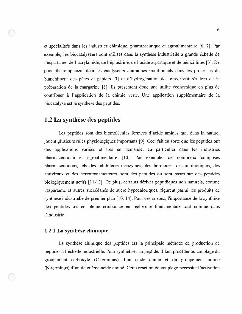
A H2N B H2N
CH3 CH3
Figure 1. Activation de la fonction carboxyle des acides aminés lors de la synthèse
peptidique. (A) Urée O-acylée formée par la réaction du DCC avec l’alanine. (B) Ester
activé formé par la réaction de l’urée O-acylée A avec le HOBt.
À la suite du couplage chimique, le cycle de protection, d’activation, de réaction et
de déprotection est répété pour allonger le peptide. Les rendements imparfaits obtenus à
chaque étape donnent vite lieu à une gamme de sous-produits indésirables. Notamment, des
sous-réactions de l’acide aminé activé avec l’eau, avec les groupes fonctionnels des chaînes
latérales d’autres acides aminés ou encore avec des bases diminuent l’efficacité et/ou la
pureté énantiomérique du produit final. En dernier lieu, il existe le problème d’isolation et
de purification du produit après chaque étape afin d’en éliminer les excès de réactifs, les
groupements protecteurs clivés et les autres sous-produits. Ce problème est partiellement
résolu par la synthèse sur support solide, qui facilite les étapes d’isolation et de purification.
La synthèse sur support solide est la méthode de choix pour la synthèse chimique
des peptides. Toutefois, cette méthode génère beaucoup de déchets, car, pour qu’elle donne
de bons rendements, il faut utiliser un excès de substrats et il faut laver la résine avec
beaucoup de solvant, sans compter qu’il faut quand même utiliser des groupements
protecteurs et des agents de couplage. La synthèse chimique des peptides est donc un
procédé polluant qui ne respecte pas les douze principes de la chimie verte. Des alternatives
à la synthèse chimique des peptides plus respectueuses de l’environnement sont la
biosynthèse et la synthèse enzymatique.
9
1.2.2 La biosynthèse
La biosynthèse est la synthèse de produits chimiques ayant lieu à l’intérieur
d’organismes vivants tels les microorganismes et les animaux transgéniques. Quoique utile,
la biosynthèse des peptides ne permet pas la synthèse de peptides formés d’acides aminés
non naturels. De plus, la production microbienne de petits peptides est limitée par la
présence de peptidases qui détruisent une partie des peptides produits, ce qui diminue les
rendements. Il peut aussi être difficile d’obtenir un produit de pureté satisfaisante à un coût
raisonnable avec cette méthode. Enfin, l’utilisation d’organismes transgéniques peut
présenter des problèmes éthiques, en plus de compliquer l’application à grande échelle de
cette méthode. Une alternative plus prometteuse est la synthèse in vitro à l’aide d’enzymes.
1.2.3 La synthèse enzymatique
La synthèse enzymatique des peptides réduit largement les besoins d’activation et de
protection chimique des acides aminés et assure la pureté énantiomérique des produits en
exploitant la très haute sélectivité des enzymes. Les deux principales approches de cette
méthode sont l’utilisation de peptidyltransférases, et la mise en oeuvre de protéases. La
première approche implique l’utilisation in vitro de composantes isolées de la machinerie
cellulaire [1$]: ribosornes, ARN messager et de nombreux ARN de transfert et autres
facteurs cellulaires, ainsi que des substrats coûteux comme l’ATP. Cette méthode est très
dispendieuse et demeure difficile dapplication à grande échelle.
L’utilisation de protéases pour la synthèse des peptides est la méthode
biocatalytique la plus répandue, étant déjà utilisée pour la synthèse à l’échelle industrielle
de l’aspartame et de l’insuline humaine [19]. La synthèse enzymatique de peptides à l’aide
de protéases peut se faire par contrôle thermodynamique ou cinétique (figure 2). Dans une
réaction à contrôle thermodynamique, on exploite l’inversion de la capacité des protéases à
hydrolyser les liaisons peptidiques. Par le contrôle des conditions réactionnelles, on peut
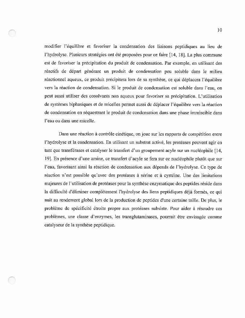
10
modifier l’équilibre et favoriser la condensation des liaisons peptidiques au lieu de
l’hydrolyse. Plusieurs stratégies ont été proposées pour ce faire [14, 18]. La plus commune
est de favoriser la précipitation du produit de condensation. Par exemple, en utilisant des
réactifs de départ générant un produit de condensation peu soluble dans le milieu
réactionnel aqueux, ce produit précipitera lors de sa synthèse. ce qui déplacera l’équilibre
vers la réaction de condensation. Si le produit de condensation est soltible dans l’eau, on
peut aussi utiliser des cosolvants non aqueux pour favoriser sa précipitation. L’utilisation
de systèmes biphasiques et de micelles permet aussi de déplacer l’équilibre vers la réaction
de condensation en séquestrant le produit de condensation dans une phase immiscible dans
l’eau ou dans une micelle.
Dans une réaction à contrôle cinétique, on joue sur les rapports de compétition entre
l’hydrolyse et la condensation. En utilisant un substrat activé, les protéases peuvent agir en
tant que transférases et catalyser le transfert d’un groupement acyle sur un nucléophile [14,
19]. En présence d’une amine, ce transfert d”acyle se fera sur ce nucléophile plutôt que sur
l’eau, favorisant ainsi la réaction de condensation aux dépends de l’hydrolyse. Ce type de
réaction n’est possible qu’avec des protéases à sérine et â cystéine. Une des limitations
majeures de l’utilisation de protéases pour la synthèse enzymatique des peptides réside dans
la difficulté d’éliminer complètement l’hydrolyse des liens peptidiques déjà formés, ce qui
nuit au rendement global lors de la production de peptides d’une certaine taille. De plus, le
problème de spécificité étroite propre aux protéases subsiste. Pour aider à résoudre ces
problèmes, une classe d’enzymes, les transglutaminases, pourrait être envisagée comme
catalyseur de la synthèse peptidique.
11
A R-COOH + H2N-R — R-CO-NH-R + H20
R-CO-N H-R + Enz-OH
_
RNy
B R-COX + Enz-OH - R-CO-Enz + HOX
H>\
R-000H + Enz-OH
Figure 2. Synthèse enzymatique des peptides catalysée par les protéases. (A) Synthèse sous
contrôle thermodynamique. (B) Synthèse sous contrôle cinétique [181.
1.3 Les transglutaminases
Les transglutaminases (TGases) composent une famille d’enzymes faisant partie de
la classe des transférases (EC 2.3.2.13), qui catalysent la réticulation post-traductioimelle
des protéines. La réticulation des protéines par les TGases génère des produits de haute
masse moléculaire très résistants à la dégradation protéolytique et au stress mécanique. Ces
produits se retrouvent dans plusieurs tissus et forment en partie la peau, les cheveux, les
matrices extracellulaires et les caillots sanguins [20]. Les TGases sont impliquées dans
plusieurs rôles physiologiques importants telles l’endocytose, la coagulation sanguine, la
formation de l’épiderme, l’apoptose et la régulation de la croissance cellulaire [2 1-23]. Une
mauvaise régulation de l’activité des TGases mène à plusieurs désordres physiologiques
tels la formation des cataractes [24], la maladie coeliaque [25] et le psoriasis [26], et
semblerait être impliquée dans le développement de maladies neurodégénératives telles les
maladies d’Alzheimer et de Huntington [27].
12
1.3.1 Réaction catalysée et mécanisme catalytique
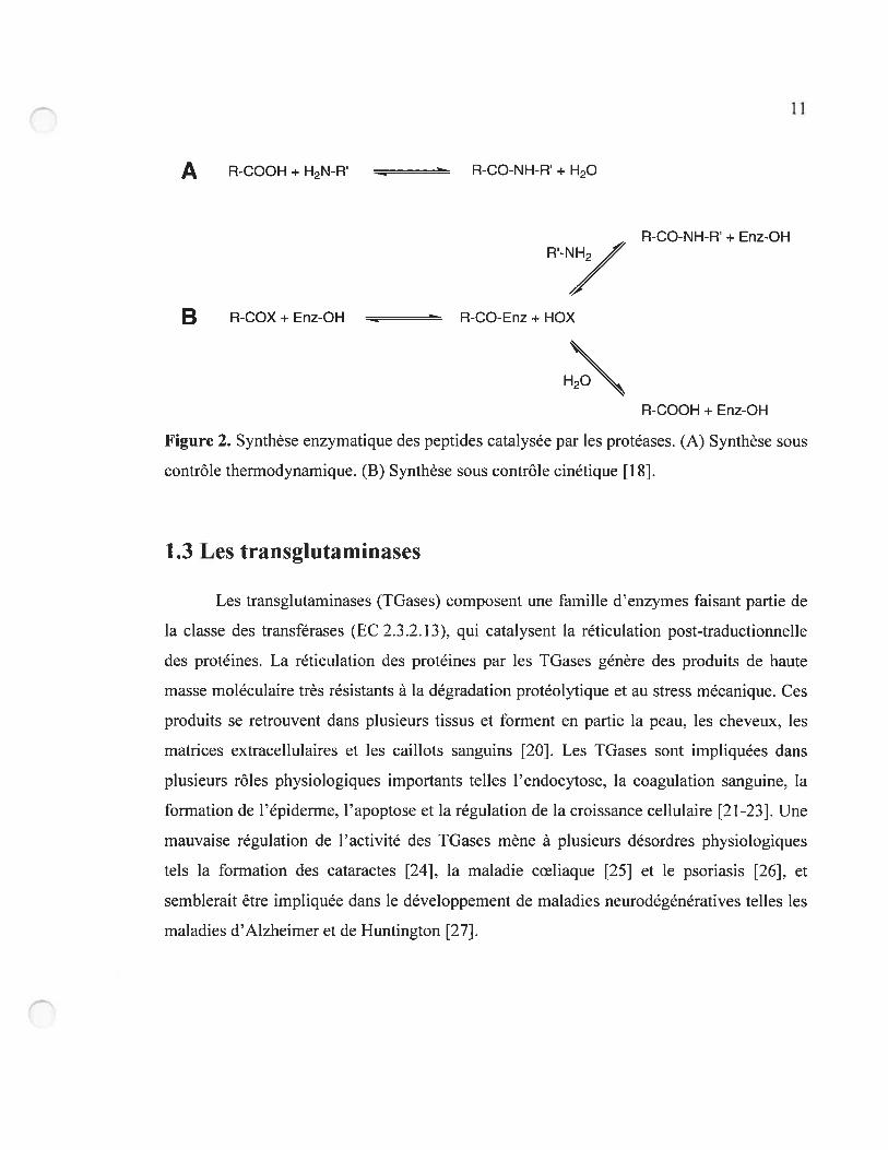
Les TGases réticulent les protéines en catalysant une réaction de transfert d’acyle
dépendante du Ca2 entre le groupement y-carboxamide d’un résidu Gin et le groupement
e-amino d’un résidu Lys, ce qui mène à la formation d’un lien isopeptidique
y-glutamyl-E-lysine (Figure 3). Ces liaisons isopeptidiques ne sont pas susceptibles de se
faire protéolyser. En plus de catalyser la formation de liens isopeptidiques entre des
peptides et des protéines, les TGases peuvent aussi catalyser d’autres modifications post
traductionneiles telles l’incorporation d’amines dans les protéines et la désamination sur
des sites spécifiques de résidus Gin (Figure 4).
N H2 + H2N
TGase
Substrat donneur (Gin) Substrat accepteur (Lys) Liaison isopeptidiqueNH3
figure 3. Réaction catalysée par les IGases.
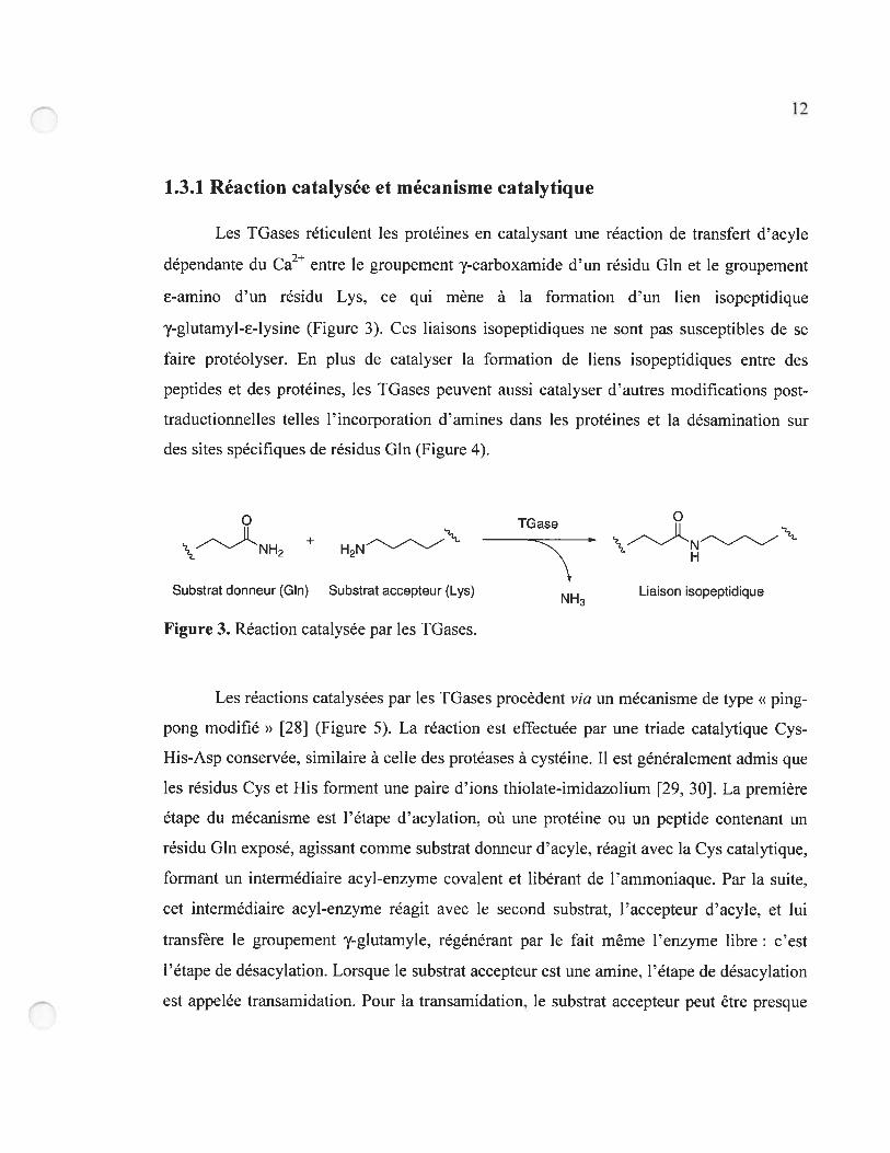
Les réactions catalysées par les TGases procèdent via un mécanisme de type «ping
pong modifié » [2$] (Figure 5). La réaction est effectuée par une triade catalytique Cys
His-Asp conservée, similaire à celle des protéases à cystéine. Il est généralement admis que
les résidus Cys et His forment une paire d’ions thiolate-imidazolium [29, 30]. La première
étape du mécanisme est l’étape d’acylation, où une protéine ou un peptide contenant un
résidu Gin exposé, agissant comme substrat donneur d’acyle, réagit avec la Cys catalytique,
formant un intermédiaire acyl-enzyme covalent et libérant de l’ammoniaque. Par la suite,
cet intermédiaire acyl-enzyme réagit avec le second substrat, l’accepteur d’acyle, et lui
transfère le groupement y-glutamyie, régénérant par le fait même l’enzyme libre c’est
l’étape de désacylation. Lorsque le substrat accepteur est une amine, l’étape de désacylation
est appelée transarnidation. Pour la transamidation, le substrat accepteur peut être presque
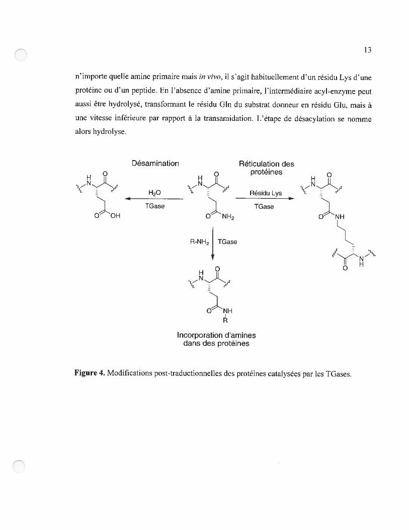
1—,li
n’importe quelle amine primaire mais in vivo, il s’agit habituellement d’un résidu Lys d’une
protéine ou d’un peptide. En l’absence OEamine primaire, Fintermédiaire acyl-enzyme peut
aussi être hydrolysé. transformant le résidu Gin du substrat donneur en résidu Glu, mais à
une vitesse inférieure par rapport à la transarnidation. L’étape de désacylation se nomme
alors hydrolyse.
Désamination Réticulation desprotéines
H20 Résidu Lys
TGase TGaseO OH
R-NH2 TGase
O
)NH
Incorporation d’aminesdans des protéines
Figure 4. Modifications post-traductionnelles des protéines catalysées par les TGases.
14
Substrat donneur
( )NH
E:R Hydrolyse E:)r2
/Ylation
NH3
Figure 5. Mécanisme de type « ping-pong modifié » des TGases.
L’étape limitante du mécanisme de la TGase est l’étape de désacylation
(transamidafion ou hydrolyse) [31, 321. En effet. il a été démontré que, même en utilisant
des amines hautement nucléophiles comme substrats accepteurs. la vitesse de la
désacylation restait inférieure à la vitesse d’acylation [32]. Ceci démontre que les TGases
sont très efficaces pour activer le transfert du groupement 7-glutamyl vers les amines
primaires, aux dépens de l’eau, et ce, bien que l’eau soit à une concentration beaucoup plus
élevée dans le milieu réactionnel.
1.3.2 Classification
Les TGases sont des transférases de classe EC 2.3.2.13 (protéine-glutamine
‘y-glutamyltransférases) qui sont présentes chez tous les vertébrés de même que chez les
bactéries [33], les plantes [34]. les levures [35] et les nématodes [361. Chez les
mammifères. neuf TGases ont été identifiées par séquençage génomique (Tableau 1) [20.
HO
RNH2
Substrat accepteur -SC(O)R
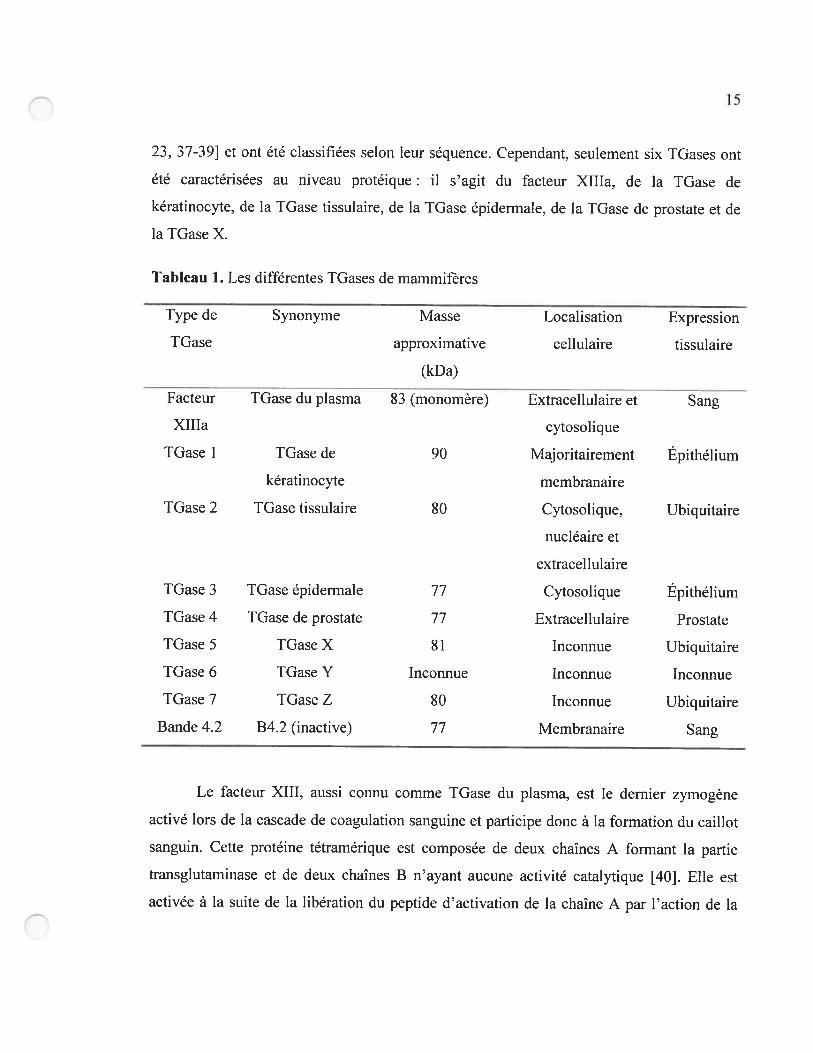
15
23, 37-39] et ont été classifiées selon leur séquence. Cependant, seulement six TGases ont
été caractérisées au niveau protéique il s’agit du facteur XIIIa, de la TGase de
kératinocyte, de la TGase tissulaire, de la IGase épiderrnale, de la TGase de prostate et de
la TGase X.
Tableau 1. Les différentes TGases de mammifères
Type de Synonyme Masse Localisation Expression
TGase approximative cellulaire tissulaire
(kDa)
Facteur TGase du plasma 83 (monomère) Extracellulaire et Sang
XIIIa cytosolique
TGase I TGase de 90 Majoritairement Épithélium
kératinocyte membranaire
IGase 2 TGase tissulaire $0 Cytosolique. Ubiquitaire
nucléaire et
extracellulaire
TGase 3 TGase épidennale 77 Cytosolique Épithélium
TGase 4 TGase de prostate 77 Extracellulaire Prostate
TGase 5 TGase X 81 Incoimue Ubiquitaire
TGase 6 TGase Y Inconnue Inconnue Inconnue
TGase 7 TGase Z $0 Inconnue Ubiquitaire
Bande 4.2 B4.2 (inactive) 77 Membranaire Sang
Le facteur XIII, aussi connu comme TGase du plasma. est le dernier zymogène
activé lors de la cascade de coagulation sanguine et participe donc à la formation du caillot
sanguin. Cette protéine tétramérique est composée de deux chaînes A formant la partie
transglutaminase et de deux chaînes B n’ayant aucune activité catalytique [40]. Elle est
activée à la suite de la libération du peptide d’activation de la chaîne A par l’action de la
16
thrombine, générant ainsi le facteur XIIIa. Le facteur XIIIa est la seule TGase de
mammifère qui est homodimérique chaque monomère est formé de 732 résidus et a une
masse moléculaire de $3 kDa. Cette IGase peut se retrouver dans le plasma et dans le
cytoplasme de plusieurs types de cellules.
La TGase de kératinocyte, ou IGase de type 1. est une enzyme monomérique de
90 kDa qui se retrouve liée, via une ancre lipidique, au côté cytoplasmique de la membrane
cellulaire des kératinocytes. C’est la plus grande TGase ($17 résidus, 90 kDa). car elle
possède une région d’ancrage à la membrane en N-terminal, qui contient un groupe de cinq
cystéines sur lesquelles des acides gras tels l’acide myristique et l’acide palmitique peuvent
former des liens thioester, donnant lieu à l’ancre lipidique [41]. Ses rôles biologiques sont
la différenciation des kératinocytes et la formation de la couche cornée de la peau. La
IGase de kératinocyte est protéolysée à deux endroits, ce qui l’active, et les trois fragments
ainsi générés restent associés entre eux [421.
La TGase tissulaire (IGase de type 2). une protéine monomérique de 690 résidus
($0 kDa), est le membre le plus répandu et le plus étudié de cette famille. Cette TGase se
retrouvant dans tous les tissus est majoritairement cytosolique, mais peut également se
retrouver dans le noyau [43] et dans le milieu extracellulaire [37]. Elle possède une activité
d’hydrolyse de nucléotides (GTP et ATP), ce qui en fait un membre de la famille des
protéines G [44]. La liaison au GTP inhibe son activité transférase. Cette IGase démontre
de plus des activités disulphide isornérase [45] et kinase [46], et contient des sites de liaison
à la fibronectine et à l’intégrine [47]. Le rôle physiologique précis de cette TGase n’est pas
encore clairement démontré, mais elle semble être impliquée dans Finduction de l’apoptose
[48], l’endocytose. l’adhésion cellulaire, le développement de la matrice extracellulaire et la
différenciation cellulaire [23. 37].
La TGase épidermale (TGase de type 3) est une TGase monomérique impliquée
dans la différenciation des kératinocytes et dans la formation de l’enveloppe cellulaire dans
l’épiderme et le follicule pileux. Cette enzyme consiste en deux chaînes polypeptidiques
17
qui sont exprimées en une seule proenzyme de 692 résidus (77 kDa) requérant une
protéolyse pour être activée. À la suite de cette protéolyse. deux fragments globulaires qui
demeurent associés sont générés [37]. La TGase épidermale, comme la TGase tissulaire,
peut aussi lier et hydrolyser le GTP. mais le rôle de cette activité demeure pour l’instant
inconnu [49].
Les autres TGases de mammifères sont encore peu étudiées. La TGase de prostate
(IGase de type 4) est impliquée dans la foniiation du caillot post-coïtal chez le rat. Son rôle
chez l’humain est inconnu. La TGase X (IGase de type 5) contribue à la formation de la
couche cornée des kératinocytes et peut lier le GTP [50]. La protéine Bande 4.2 de
l’érythrocyte fait partie de la famille des TGases mais n’a pas d’activité enzymatique. car la
Cys catalytique est remplacée par une Ala. C’est un composant majeur du cytosquelette des
érythrocytes et elle joue un rôle important dans la structure et les propriétés mécaniques de
ces cellules [37]. Le rôle physiologique des TGases de type 6 et 7 n’a pas encore été
élucidé.
Une TGase microbienne provenant de Streptoverticillium mobaraense a aussi été
étudiée. C’est la seule TGase ne provenant pas de vertébrés à avoir été isolée. Cette TGase
est très différente des TOases de mammifères, ne possédant aucune homologie de séquence
ni de structure avec ses homologues de mammifères. C’est une enzyme sécrétée, de 331
résidus (38 kDa), qui nécessite le clivage d’un propeptide pour être activée. Cette enzyme
possède une triade catalytique Cys-His-Asp et catalyse ta même réaction que les autres
TGases mais ne requiert pas de Ca2 pour être active, ce qui la distingue des TGases
provenant d’eucaryotes. Son rôle biologique n’est pas connu [51].
1.3.3 Structure
Les structures cristallines de quatre TGases de vertébrés appartenant à trois
différentes classes ont été résolues. Il s’agit de la TGase de plasma humaine (facteur XIIIa)
[30. 52-54], des TGase tissulaires humaine [55] et de poisson (Fagrits inqior) [56]. de
1$
même que de la TGase épidermale humaine [49. 57-59]. Malgré des différences
significatives dans leur structure primaire, ces IGases adoptent toutes une structure
tridimensionnelle similaire. Ces structures sont formées de quatre domaines (Figure 6A) : le
domaine N-terminal sandwich 13, le noyau catalytique a/f3, le tonneau [31 et le tonneau [3 2
C-terminal. Le domaine N-terminal sandwich f3 a été démontré comme étant le site de
liaison de la fibronectine et de l’intégrine chez la TGase tissulaire. Le noyau
catalytique u/[3 est composé d’hélices u et de feuillets f3 et contient le site actif pour
l’activité transférase. mais aussi le site actif pour l’activité d’hydrolyse de nucléotides des
IGases de types 2. 3 et 5. Ce domaine contient aussi le site de liaison au calcium, dont la
localisation demeure floue pour la TGase tissulaire. D’après des études de délétions, les
domaines tonneaux f3 1 et 2 ne semblent pas être indispensables pour l’activité catalytique
de la TGase tissulaire [60] et du facteur XIIIa [61]. Les TGases de vertébrés contiennent
plusieurs résidus Cys, mais aucun pont disulphure n’a été identifié [62].
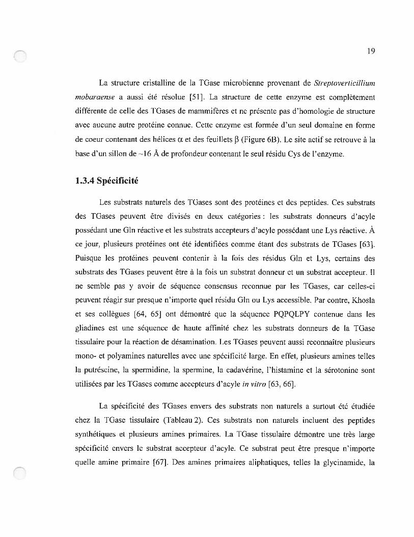
Figure 6. Structure cristalline de TGases. (A) TGase tissulaire de Fagrus major (Code
PDB 1 GOD). (B) IGase microbienne de Streptoverticillium mobaraense (Code PDB 1 1U4).
19
La structure cristalline de la IGase microbienne provenant de $treptoveriicillium
rnobaraense a aussi été résolue [51]. La structure de cette enzyme est complètement
différente de celle des TGases de mammifères et ne présente pas d’homologie de structure
avec aucune autre protéine connue. Cette enzyme est formée d’un seul domaine en forme
de coeur contenant des hélices Œ et des feuillets f3 (f igure 6B). Le site actif se retrouve à la
base d’un sillon de l6 À de profondeur contenant le seul résidu Cys de l’enzyme.
1.3.4 Spécificité
Les substrats naturels des TGases sont des protéines et des peptides. Ces substrats
des TGases peuvent être divisés en deux catégories : les substrats donneurs d’acyle
possédant une Gln réactive et les substrats accepteurs d’acyle possédant une Lys réactive. Àce jour, plusieurs protéines ont été identifiées comme étant des substrats de TGases [63].
Puisque les protéines peuvent contenir à la fois des résidus Gln et Lys, certains des
substrats des TGases peuvent être à la fois un substrat donneur et un substrat accepteur. Il
ne semble pas y avoir de séquence consensus reconnue par les TGases, car celles-ci
peuvent réagir sur presque n’importe quel résidu Gln ou Lys accessible. Par contre, Khosla
et ses collègues [64, 65] ont démontré que la séquence PQPQLPY contenue dans les
gliadines est une séquence de haute affinité chez les substrats donneurs de la TGase
tissulaire pour la réaction de désamination. Les TGases peuvent aussi reconnaître plusieurs
mono- et polyamines naturelles avec une spécificité large. En effet, plusieurs amines telles
la putréscine, la spermidine, la spermine, la cadavérine, l’histamine et la sérotonine sont
utilisées par les TGases comme accepteurs d’acyle in vitro [63, 66].
La spécificité des TGases envers des substrats non naturels a surtout été étudiée
chez la TGase tissulaire (Tableau 2). Ces substrats non naturels incluent des peptides
synthétiques et plusieurs amines primaires. La TGase tissulaire démontre une très large
spécificité envers le substrat accepteur d’acyle. Ce substrat peut être presque n’importe
quelle amine primaire [67]. Des amines primaires aliphatiques, telles la glycinarnide, la
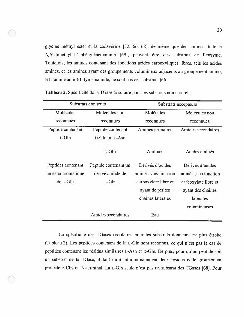
20
glycine méthyl ester et la cadavérine [32. 66, 68j. de même que des anilines, telle la
IV N-diméthyl- 1 ,4-phénylênediamine [69J. peuvent être des substrats de l’enzyme.
Toutefois, les amines contenant des fonctions acides carboxyliques libres, tels les acides
aminés, et les amines ayant des groupements volumineux adjacents au groupement amino,
tel l’amide aminé L-tyrosinamide, ne sont pas des substrats [661.
Tab]eau 2. Spécificité de la TGase tissulaire pour les substrats non naturels
Substrats donneurs Substrats accepteurs
Molécules Molécules non Molécules Molécules non
reconnues reconnues reconnues reconnues
Peptide contenant Peptide contenant Amines primaires Amines secondaires
L-Gln D-Glu ou L-Asn
L-Glu Anilines Acides aminés
Peptides contenant Peptide contenant un Dérivés dacides Dérivés d’acides
un ester aromatique dérivé anilide de aminés sans fonction aminés sans fonction
de L-Glu L-Gin carboxylate libre et carboxylate libre et
ayant de petites ayant des chafnes
chaînes latérales latérales
volumineuses
Amides secondaires Eau
La spécificité des TGases tissulaires pour les substrats donneurs est plus étroite
(Tableau 2). Les peptides contenant de la L-Glu sont reconnus, ce qui n’est pas le cas de
peptides contenant les résidus similaires L-Asn et D-GIn. De plus, pour qu’un peptide soit
un substrat de la TGase, il faut qu’il ait minimalement deux résidus et le groupement
protecteur Cbz en N-terminal. La L-Gln seule n’est pas un substrat des TGases [62]. Pour
21
cette raison, le peptide synthétique N-carbobenzyloxyglutaminylglycine (Cbz-Gln-Gly) est
le substrat donneur synthétique le plus couramment utilisé pour les études cinétiques des
TGases tissulaires. Ce composé a un KM élevé, d’environ 3 mM [70-72]. Il est aussi connu
que les esters aromatiques y-glutamyles sont de bons substrats des IGases. Par exemple, le
substrat articifiel N-carbobenzyloxy-L-glutamyl(y-p-nitrophényl ester)glycine [321 est
utilisé en analyses cinétiques et a un KM de 20 tM, ce qui est beaucoup plus bas que son
analogue Cbz-Gln-Gly. Par contre, les anilides et les amides secondaires ne sont pas des
substrats [73], ce qui démontre que la fonction y-carboxamide de la L-GIn est le seul amide
reconnu par l’enzyme. La TGase tissulaire peut donc reconnaître plusieurs dipeptides, tels
Cbz-Gln-Gly. comme substrats donneurs et plusieurs dérivés d’acides aminés, telle la
glycinamide. comme substrats accepteurs.
1.4 Description du projet de recherche
1.4.1 Objectif
L’objectif général du projet est de développer de nouvelles enzymes catalysant de
façon plus efficace et économiquement avantageuse la synthèse des peptides afin de
contribuer au développement de la chimie verte au Canada. L’objectif spécifique est de
modifier la spécificité de la TGase pour la rendre capable de catalyser le couplage entre la
fonction amide d’un amide aminé protégé (GP-Xaa-NH2) comme substrat donneur d’acyle
et le groupe a-amino de la forme amide ou ester d’un second acide aminé (Xaa-NH2 ou
Xaa-OMe) comme substrat accepteur d’acyle (figure 7).
L’avantage d’utiliser les TGases plutôt que les protéases pour la synthèse
enzymatique des peptides est qu’elles génèrent déjà des liaisons de type amide entre des
peptides et des dérivés d’acides aminés en plus de n’avoir aucune action hydrolytique
envers les produits de la réaction, ce qui pourrait donner de meilleurs rendements. De plus,
en utilisant la TGase comme enzyme de départ. la modification proposée n’impliquera
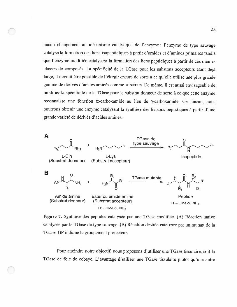
aucun changement au mécanisme catalytique de l’enzyme: l’enzyme de type sauvage
catalyse la fomiation des liens isopeptidiques à partir d’amides et «amines primaires tandis
que l’enzyme modifiée catalysera la formation des liens peptidiques à partir de ces mêmes
classes de composés. La spécificité de la TGase pour les substrats accepteurs étant déjà
large, il devrait être possible de l’élargir encore de sorte à ce qu’elle utilise une plus grande
gamme de dérivés d’acides aminés comme substrats. De même, il est aussi envisageable de
modifier la spécificité de la TGase pour le substrat dormeur de sorte à ce que cette enzyme
recoirnaisse une fonction Œ-carboxamide au lieu de y-carboxamide. Ce faisant. nous
pourrons obtenir une enzyme catalysant la synthèse des liaisons peptidiques à partir d’une
grande variété de dérivés d’acides aminés.
ATGasede o
type sauvage2 H
L-Gin L-Lys isopeptide(Substrat donneur) (Substrat accepteur)
B
GPNH2 + H2N-RTGase mutante
Amide aminé Ester ou amide aminé Peptide(Substrat donneur) (Substrat accepteur) R = OMe ou NH2
R = OMe ou NH2
Figure 7. Synthèse des peptides catalysée par une TGase modifiée. (A) Réaction native
catalysée par la TGase de type sauvage. (B) Réaction désirée catalysée par un mutant de la
TGase. GP indique le groupement protecteur.
Pour atteindre notre objectif, nous proposons d’utiliser une TGase tissulaire, soit la
TGase de foie de cobaye. Lavantage d’utiliser une IGase tissulaire plutôt qu’une autre
TGase est que cette classe de TGase a été la plus étudiée sa spécificité envers les substrats
donneurs et accepteurs non naturels est bien caractérisée, plusieurs méthodes d’expression
et de purification chez la bactérie ont été rapportées, des structures cristallines ont été
élucidées et leur mécanisme catalytique est bien établi. Parmi les TGases tissulaires, c’est la
IGase de foie de cobaye qui est la plus étudiée et pour cette raison, c’est elle qui est utilisée
tout au long de ce projet de recherche.
1.4.2 Méthodologie générale
Pour atteindre notre objectif, nous avons entrepris de modifier la spécificité de la
TGase de foie de cobaye. Puisque la IGase catalyse déjà la formation des liens
isopeptidiques à partir des amides primaires (substrat donneur) et des amines primaires
(substrat accepteur), nous proposons de modifier les sous-sites de liaison de ces substrats
sans modifier la réaction catalysée par l’enzyme. La modification du sous-site de liaison du
substrat donneur pourra permettre le remplacement du groupe y-carboxamide de la
glutarnine du substrat natif par le groupe Œ-carboxamide du substrat désiré. Ceci nécessitera
la création d’espace dans le site de liaison pour accommoder la chaîne latérale du résidu
N-terminal du peptide croissant (R1) (Figure 7). De façon similaire, le site de liaison du
substrat accepteur devra être agrandi pour permettre l’utilisation, comme substrat accepteur,
des acides aminés avec des chaînes latérales plus volumineuses (R2).
La modification de la spécificité de la IGase de foie de cobaye peut se faire par
évolution dirigée. L’évolution dirigée est le processus de création. dans un court laps de
temps, d’un grand nombre de protéines mutantes (une banque) qui sont criblées pour une
activité désirée. L’évolution dirigée imite donc les stratégies de l’évolution naturelle, en
accéléré [74-78]. Pour entreprendre l’évolution dirigée d’une enzyme de sorte à obtenir des
mutants ayant les nouvelles caractéristiques voulues, il faut procéder aux deux étapes
suivantes
24
1. La création de banques de mutants de la protéine d’intérêt, par des stratégies telles la
mutagenèse à haut taux d’erreurs ou encore le gene shuffling où les variantes d’une
séquence d’ADN sont fragmentées puis les fragments recombinés afin d’en obtenir
de nouvelles combinaisons [79, 80].
2. L’identification des mutants ayant la propriété désirée. Ceci peut être fait par
criblage, dont les conditions devraient diriger l’évolution vers le but désiré.
La création de banques d’ADN est maintenant aisément praticable dans les laboratoires
ayant l’expertise requise. Par contre. la stratégie d’identification des mutants voulus doit
être développée conformément à chaque but, de façon à s’assurer que les nouvelles
enzymes sélectionnées possèdent les caractéristiques spécifiquement requises.
L’évolution dirigée des enzymes peut se faire par une approche aléatoire ou par une
approche serni-aléatoire [81]. L’approche aléatoire consiste à effectuer la mutagenèse
aléatoire sur l’ensemble du gène encodant l’enzyme, c’est-à-dire introduire des mutations
au hasard dans l’espoir d’en trouver qui donnent l’activité désirée et qui n’auraient pas pu
être prédites. L’approche serni-aléatoire consiste à entreprendre une mutagenèse
semi-aléatoire, c’est-à-dire introduire des mutations au hasard, mais seulement dans une ou
des régions spécifiques du gène tels les résidus du site actif La mutagenèse semi-aléatoire
peut être combinatoire. Cette approche, de plus en plus répandue, a l’avantage d’exploiter
les données connues sur la structure et/ou la fonction de certains résidus, générant ainsi des
banques «intelligentes » ayant une meilleure probabilité de contenir des mutants aux
propriétés désirées. Cette approche serni-aléatoire, qui fait l’objet du chapitre 2, a été
choisie comme méthodologie générale pour atteindre notre objectif de recherche.
1.4.3 Étapes du projet
Afin de procéder à l’évolution dirigée de la TGase de foie de cobaye par une
aproche semi-aléatoire, il faut posséder de l’information structurelle et fonctionnelle ainsi
que des méthodes de mutagenèse semi-aléatoire et de criblage. L’information structurelle et
25
fonctionnelle a dû être acquise lors de ce projet de recherche, et les méthodes de
mutagenèse semi-aléatoire et de criblage ont dû être développées. Les étapes du projet de
recherche ont donc été les suivantes:
1. Identification des résidus de la TGase impliqués dans la liaison du substrat donneur;
2. Développement d’une méthode d’expression et de purification de la TGase de foie
de cobaye chez Escherichia cou;
3. Développement d’un modèle d’homologie de la TGase de foie de cobaye et
mutagenèse de résidus du site actif pour obtenir de l’information structurelle et
fonctionnelle supplémentaire:
4. Évolution dirigée de la spécificité de la TGase de foie de cobaye par une approche
semi-aléatoire.
Nous décrirons ces étapes dans les sections suivantes.
1.4.3.1 Identification des résidus de la TGase impliqués dans la liaison du substrat
donneur
Avant d’entreprendre l’évolution dirigée de la spécificité de la TGase de foie de
cobaye par une approche serni-aléatoire, il a fallu identifier les résidus appropriés à muter,
c’est-à-dire les résidus formant les sous-sites de liaison des substrats de l’enzyme. Puisque
aucune structure cristalline de cette enzyme avec un substrat lié au site actif n’est publiée,
nous avons généré un modèle de la TGase de Fagrus major avec un substrat donneur lié au
site actif Cette étude a été publiée en 2004 et forme le chapitre 3 de la présente thèse.
1.4.3.2 Développement d’une méthode d’expression et de purification de la TGase de
foie de cobaye chez Escherichia cou
Après avoir identifié les résidus de la IGase que nous voulions muter, nous avons
développé une méthode d’expression et de purification efficace de la IGase de foie de
cobaye chez E. cou pour obtenir rapidement, efficacement et avec un bon rendement de
26
l’enzyme soluble ayant une bonne activité spécifique. Ce protocole, développé avec le
Dr Steve Gillet, fut publié en 2004 et forme le chapitre 4.
1.4.3.3 Développement d’un modèle d’homologie de la TGase de foie de cobaye et
mutagenèse de résidus du site actif
Afin d’obtenir de l’information strticturelle et fonctionnelle supplémentaire, nous
avons généré un modèle d’homologie de la IGase de foie de cobaye. Ce modèle a permis
d’étudier le rôle de certains résidus du site actif. L’information recueillie par cette étude
décrite au chapitre 5 nous a servi dans la planification de la mutagenèse semi-aléatoire pour
l’évolution dirigée par une approche semi-aléatoire.
1.4.3.4 Évolution dirigée de la spécificité de la TGase de foie de cobaye par une
approche semi-aléatoire
À partir de l’information acquise par les études de modélisation des chapitres 3 et 5,
nous avons généré cinq banques de mutants de la TGase de foie de cobaye par mutagenèse
de saturation et/ou combinatoire. Par la suite, une méthode fluorimétrique de criblage en
plaques à 96 puits a été développée afin de permettre l’identification des mutants de ces
cinq banques ayant la spécificité désirée. Ces méthodes nous ont permis de procéder à
l’évolution dirigée de la spécificité de la TGase de foie de cobaye. qui est décrite au
chapitre 6.
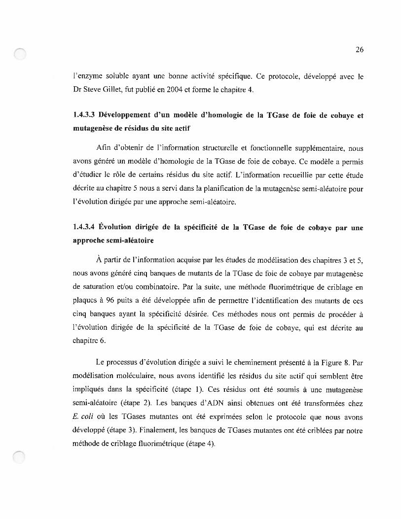
Le processus d’évolution dirigée a suivi le cheminement présenté à la Figure 8. Par
modélisation moléculaire. nous avons identifié les résidus du site actif qui semblent être
impliqués dans la spécificité (étape 1). Ces résidus ont été soumis à une mutagenèse
semi-aléatoire (étape 2). Les banques d’ADN ainsi obtenues ont été transformées chez
E. cou où les TGases mutantes ont été exprimées selon le protocole que nous avons
développé (étape 3). Finalement, les banques de TGases mutantes ont été criblées par notre
méthode de criblage fluorimétrique (étape 4).
27
in silico
1. Identification des résidusimpliqués dans la spécificité
4
in vitro—
2. Mutagenèsesemi-aléatoire
—
3. Expression des protéines
* ,
4%4. Criblage
Figure 8. Évolution dirigée de la IGase par une approche semi-aléatoire.
CHAPITRE 2
Evolution dirigée par une approche semi-aléatoire:
revue de la littérature
29
2.0 Préface
Plusieurs méthodes existent pour modifier les propriétés des enzymes selon nos
besoins. Pour modifier la spécificité de la TGase de foie de cobaye, nous avons opté pour la
méthode de l’évolution dirigée selon une approche semi-aléatoire. Cette approche a été
choisie pour deux raisons. Premièrement, nous voulions modifier les sous-sites de liaison
des substrats de la IGase sans modifier la réaction catalysée par l’enzyme. En ciblant la
mutagenèse au site actif. nous avons l’avantage dexploiter les données connues sur la
structure et/ou la fonction de certains résidus, générant ainsi des banques de mutants
« intelligentes ». Ces banques ont une meilleure probabilité de contenir des mutants aux
propriétés désirées. Deuxièmement. nous ne possédons pas de méthodes de criblage à haut
débit nous permettant de cribler des dizaines de milliers de mutants et plus. L’approche
semi-aléatoire semblait donc être le meilleur choix pour atteindre notre objectif.
Dans ce chapitre. la méthode d’évolution dirigée par une approche semi-aléatoire,
aussi connue sous le nom d’approche semi-rationnelle, est décrite sous la forme d’un article
de revue de la littérature publié en 2004 et intitulé « Semi-rational approaches to
engineering enzyme activity: combining the benefits of directed evolution and rational
design ». Cet article présente une vue d’ensemble d’articles récents où la modification
d’activités enzymatiques par cette approche a été démontrée avec succès. Cet article a été
écrit à contribution égale avec Nicolas Doucet. Ma contribution a été de rédiger
l’introduction de même que la section sur l’utilisation «outils informatiques pour
l’évolution dirigée par une méthode semi-aléatoire ( Semi-rational and combinatorial
design using computational approaches »).
nj
Article 1.
Semi-rational approaches to engineering enzyme activity:
combining the benefits of directed evolution and rational
design
Roberto A Chica1‘,
Nicolas Doucet’*
and Joelle N Pelletier”2
1Département de chimie, Université de Montréal, CP 612$, Succursale Centre-ViLle,
Montréal, Québec, H3C 3J7. Caiiada
2Département de biochimie. Université de Montréal. CP 6128. Succursale Centre-Ville,
MontréaL Québec, H3C 3J7, Canada
*
These authors made an equal contribution to this work.
Current Opinion in BiotecÏmology, 2005, 16, 37$-384
“Reprinted from Current Opinion in Biotechnology. Vol 16, Roberto A. Chica. Nicolas
Doucet and Joelle Pelletier, “Serni-rational approaches to engineering enzyme activity:
combining the benefits ofdirected evolution and rational design”, pages 378-3 $4,
Copyright (2005), with permission from Elsevier”.
9J
2.1 Abstract
Many research groups successfully rely on whole-gene random mutagenesis and
recombination approaches for the directed evolution of enzymes. Recent advances in
enzyme engineering have used a combination of these random methods of directed
evolution with elements of rational enzyme modification to successfully by-pass certain
limitations of both directed evolution and rational design. Semi-rational approaches that
target multiple, specific residues to mutate on the basis of prior structural or functional
knowledge create smart’ libraries that are more likely to yield positive resuits. Efficient
sampling of mutations likely to affect enzyme function has been conducted both
experimentally and, on a rnuch greater scale. computationally. with remarkable
improvements in substrate selectivity and specificity and in the de novo design of enzyme
activities within scaffolds of known structure.
2.2 Introduction
Enzymes are powerful catalysts that are able to increase reaction rates by up to 17
orders of magnitude [1 and 2]. Certain enzymes display perfect control over
stereochemistry and regioselectivity. while others display a breadth of specificity: either
feature may be attractive for industrial and synthetic applications [3]. However, few
enzymes naturally catalyze the reactions that chemists need under conditions that are
industrially convenient and economically advantageous. Many efforts are currently devoted
to the modification of enzyme activities to meet the needs oftoday’s chemists.
Multiple approaches have been developed to allow the identification of mutant
enzymes possessing desirable qualities such as increased activity, rnodified specificity,
selectivity or cofactor binding t41. hie earliest approach was rational design, which was
used to modify the specificity of enzymes [5. 6. 7. $ and 9]. This approach requires an in
depth knowledge of the structural features of the enzyme active-site and their contribution
32
to function. The cornplexity of the structure/function relationship in enzymes has proven to
be the factor limiting the general application of rational design. More recently, directed
evolution has proven to be a powerful tool for the modification of enzyme activities and has
become the most widely used approach. Briefly. directed evolution consists of the low
frequency introduction of randomly distributed mutations in a gene of interest, followed by
selection of the mutated proteins possessing the desired properties. Recent advances in this
area include the application of the immune system hypermutation’ process, which is
conducted entirely in live celis [10]. Directed evolution can enable the relatively rapid
engineering of enzymes without requiring au in-depth understanding of structure/function
relationships. unlike rational design. The numerous methodologies and successes of
directed evolution will flot be detailed here as recent reviews have been devoted to the topic
[11. 12, 13, 14 and 15] (also sec the article by AA Henry and FE Romesberg in this issue).
Because large numbers of mutants must generally be screened to obtain a significant,
desired effect on enzyme activity, the main limitation of directed evolution is the necessity
of developing a high-throughput screening rnethodoiogy that allows identification of the
desired property under relevant conditions. Not ail enzyme activities are readily amenable
to developing a high-throughput screening method. nor are ail screening methodologies
easy to implernent at the required scale.
A further limitation of directed evolution relates to the random nature of the
mutations introduced: in the case where functional information (from point mutations,
random mutagenesis or deduction by sequence alignrnent) or structural information exists.
it would be advantageous to exploit this by concentrating mutations where they might be
the most effective [16]. Analysis of enzyme modification resuits indicates that the majority
of mutations that beneficially affect certain enzyme properties (enantioselectivity. substrate
specificity and new catalytic activities) are located in or near the active-site, and more
specifically near residues that are implicated in binding or catalysis [15, 17, 18 and 19].
Other properties (stability and activity) can be improved by mutations either near or far
from the active-site, although there are many more mutations far from the active-site to test
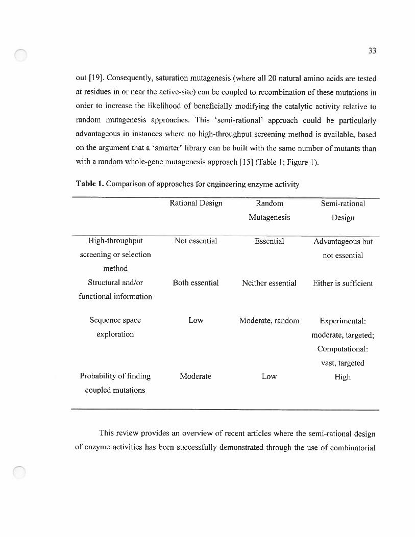
33
out [19]. Consequently, saturation mutagenesis (where ail 20 natural amino acids are tested
at residues in or near the active-site) can be coupled to recombination of these mutations in
order to increase the likelihood of beneficially modifying the catalytic activity relative to
random mutagenesis approaches. This semi-rational approach could be particularly
advantageous in instances where no high-throughput screening method is available. based
on the argument that a srnarter’ Hbrary can be built with the same number of mutants than
with a random whoie-gene mutagenesis approach [15] (Table 1; Figure 1).
Table 1. Comparison of approaches for engineering enzyme activity
Rational Design Random Serni-rational
Mutagenesis Design
High-throughput Not essential Essential Advantageous but
screening or selection not essential
method
Structural and/or Both essential Neither essential Either is sufficient
functional information
Sequence space Low Moderate, random Experimental:
exploration moderate, targeted;
Computational:
vast. targeted
Probability offinding Moderate Low High
coupled mutations
This review provides an overview of recent articles where the semi-rational design
of enzyme activities lias been successfully demonstrated through the use of combinatorial
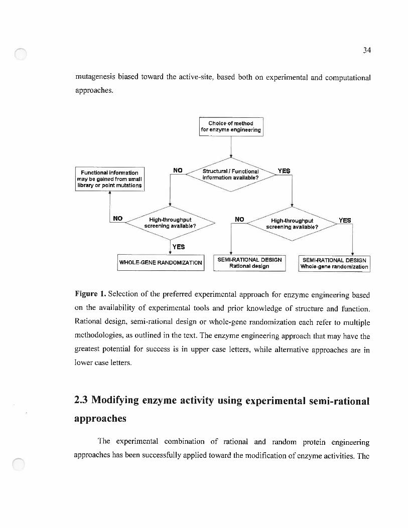
34
mutagenesis biased toward the active-site, based both on experimental and computational
approaches.
Figure 1. Selection of the preferred experimental approach for enzyme engineering based
on the availability of experirnental tools and prior knowledge of structure and function.
Rational design, semi-rational design or whole-gene randomization each refer to multiple
methodologies, as outlined in the text. The enzyme engineering approacli that may have the
greatest potential for success is in upper case letters, while alternative approaches are in
lower case letters.
2.3 Modifying enzyme activity using experimental semi-rational
approaches
The experimental combination of rational and random protein engineering
approacbes bas been successfully applied toward the modification of enzyme activities. The
Functional informationI may be gained from small
Iibrary or point mutations
YES
YES
SEMI-RATIONAL DESIGNWhole-gene randomization
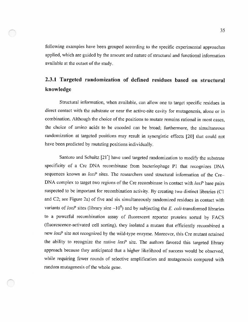
following examples have been grouped according to the specific experirnental approaches
applied, which are guided by the amount and nature of structural and functional information
available at the outset ofthe study.
2.3.1 Targeted randomization of defined residues based on structural
knowledge
Structural information. when available. can allow one to target specific residues in
direct contact with the substrate or near the active-site cavity for mutagenesis, alone or in
combination. Although the choice of the positions to mutate rernains rational in most cases,
the choice of amino acids to be encoded can be broad; furthermore, the simultaneous
randomization at targeted positions may resuit in synergistic effects {20] that could not
have been predicted by mutating positions individually.
Santoro and Schultz t21 ] have used targeted randomization to rnodify the substrate
specificity of a Cre DNA recombinase from bacteriophage Pi that recognizes DNA
sequences known as loxP sites. The researchers used structural information of the Cre—
DNA complex to target two regions of the Cre recombinase in contact with ÏoxP base pairs
suspected to be important for recombination activity. By creating two distinct libraries (CI
and C2; see Figure 2a) of five and six simultaneously randomized residues in contact with
variants of ÏoxF sites (library size —1O) and by subi ecting the E. coÏi-transfonned libraries
to a powerful recombination assay of fluorescent reporter proteins sorted by FACS
(fluorescence-activated ceil sorting). they isolated a mutant that efficiently recombined a
new loxF site not recognized by the wild-type enzyme. Moreover, this Cre mutant retained
the ability to recognize the native ÏoxF site. The authors favored this targeted library
approach because they anticipated that a higher Iikelihood of success would be observed.
while requiring fewer rounds of selective amplification and mutagenesis compared with
random mutagenesis ofthe whole gene.
36
A B
figure 2. Residues in or near the enzyme active-site that were targeted for serni-rational
combinatorial mutagenesis. The enzymes are shown as grey ribbon diagrams with space
fihling in serni-transparent gray. (A) Cre DNA recombinase from bacteriophagc P1 [21]
(PDB accession number 1CRX). Residues from libraries Cl (six residues) and C2 (five
residues) and the DNA substrate are colored yellow, green and blue, respectively. (B)
PBP2X DD-transpeptidase from S. pneumonia [30] (PDB accession number 1QMF). The
ten residues targeted for combinatorial site-directed mutagenesis are colored yellow and the
three subsequent unforeseen beneficial mutations obtained by epPCR are colored green.
Cefuroxime is acylated at the active-site residue Ser337 and is colored blue. (C) TEM-l f3-
lactamase from E. cou [43] (PDB accession number 1F QO). The 19 active-site residues
targeted for in siÏico mutagenesis using PDA are colored yellow. The benzylpenicillin
antibiotic is acylated at the active-site residue Ser7O and is colored blue. Figure generated
with Pyrnol Release 0. 9g (http://www.pyrnol.org).
C
Similarly, in a successful attempt to investigate the detoxification role of epoxide
hydrolases, Rui et aÏ. [22] used a combination ofrational design and saturation mutagenesis
at targeted active-site positions to expand the substrate range of an epoxide hydrolase from
Agrobacterium radiobacter AD 1 (EchA) to include chlorinated epoxides. On the basis of a
careful investigation ofthe active-site by structural comparison to related enzymes. residues
F108. C24$, 1219 and 1111 (in single-letter amino acid code), in the vicinity ofthe catalytic
triad, were separately randomized by saturation mutagenesis. As mutant C24$I showed a
slight increase in rate of cis-1,2-dichloroethylene (cis-DCE) mineralization (2.7-fold
increase with respect to the wild-type). it was used as a template for successive rounds of
saturation mutagenesis targeting residues F108 and 1219. The approach generated the triple
active-site area mutant F108L-1219L-C248L, which displayed a 10-fold enhancement in
cis-DCE mineralization relative to wild-type EchA.
Several other groups [23, 24, 25 and 26] have also successfully conducted
sirnultaneous or successive targeted randornization of multiple active-site positions based
on structural knowledge. as an efficient means to circumvent limitations inherent either to
site-directed mutagenesis or to whole-gene random mutagenesis. Recently. Schultz and
colleagues [27] cornbinatorially randomized two active-site residues of an aminoacyl-tRNA
synthetase as a step in the generation of an orthogonal synthetase/tRNA pair that efficiently
and selectively incorporates an unnatural amino acid into proteins.
In some cases. the structural information available is flot sufficient to rationally
select residues to be randornized. Thus. molecular modeling studies have been successfully
used to identify residues most likely to be in contact with substrate molecules. Our group
used this approach to perform combinatorial targeted mutagenesis of all 16 principal active-
site residues of type II R67 dihydrofolate reductase, allowing the selection of new, highly
modified active-site environrnents with activity at least as great as the native enzyme [22].
Similarly, the substrate specificity of BaciÏÏus stearotherinophihis SD I D-hydantoinase was
-D
modified by a semi-rational mutagenesis approach targeting residues chosen on the basis of
previous modeling studies [29].
2.3.2 Simultaneous random mutagenesis and site-saturation of defined
residues
Based on evolutionary protein-fold similarity between two related enzyme familles.
Peimbert and Segovia [30] introduced a f3-lactamase activity into PBP2X DD-transpeptidase
from Streptococciis pnetimonia through a combination of elTor-prone polymerase chain
reaction (epPCR) and saturation mutagenesis at targeted positions. This approach was used
to increase the odds of obtaining beneficial mutations at unpredictable locations in
combination with mutations at carefully chosen residues. This approach sliould increase the
return using the same number of mutants as a random whole-gene mutagenesis scherne.
Thus, two active-site residues (F450 and W374) were targeted for saturation mutagenesis in
combination with simultaneous amino acid replacernents at eight positions near the active-
site cavity (see Figure 2b). Afier the oligonucleotide-based mutagenesis, the gene was
ftirther arnplified with a 0.8 % random mutagenesis rate. This library was selected for
cefotaxirne resistance and mutants with 1 0-fold increased resistance relative to wild-type
PBP2X were obtained. The random mutagenesis resulted in additional mutations at
positions 312, 452 and 554, both proxirnal to and distal from the active-site. b assess the
impact of certain randomty inserted mutations, saturation mutagenesis was Lindertaken at
positions 312. 336. 450 and 452. Although no further increase in cefotaxime resistance was
observed compared with the original mutants. one new mutant showed F3-lactamase activity
without compromising the original DD-peptidase activity of PBP2X, thus generating a
mutant with dual substrate specificity.
j
2.3.3 Random mutagenesis fo]Iowed by site-saturation of defined residues
The structural or functional information necessary to make rational choices for the
residues to mutate is not always available. To circumvent this limitation, several research
groups have efficiently undertaken rounds of whole-gene randomization to provide ‘leads’
(i.e. residues identified as potentially advantageous when mutated) in conjunction with
fine-tuning of the lead mutants’ tbrough site-saturation mutagenesis of the identified
residues. Using sucb a cyclical randorn/targeted approach. Reetz and collaborators greatly
improved the enantioselectivity (E) of Pseudomonas aeruginosa lipases toward a
p-nitrophenyl ester in favour of the (25) enantiomer (reviewed in [31]). Initial
improvement from E = 1.1 (wild-type enzyme) to E = 11.3 was acbieved afier four rounds
of epPCR.
Using the mutational knowledge obtained by epPCR, they conducted saturation
mutagenesis at given mutational bot spots of the lipase. obtaining E = 20. Subsequent
rounds of epPCR increased E to 25. In addition, they developed a modified version of
Stemmer’s combinatorial multiple-cassette mutagenesis method [32] to mutate a defined
region of the lipase (residues 160—163) as well as several ‘bot spots’ (residues 155 and
162). Ibis semi-rational rnethod yielded tbeir most highly enantioselective lipase variant to
date (E = 51). Other groups have recently dernonstrated the power of similar semi-rational
approaches toward the evolution of enantioselective enzymes. As a resuit of the multiple
random mutagenesis steps, this approach bas also generated many mutations far from the
active-site cavity in addition to mutations designed within the active-site cavity [33, 34 and
35].
Geddie and Matsumura [36] have rnodified the substrate specificity of Escherichia
cou J3-glucuronidase through combinatorial site-saturation mutagenesis of several ‘hot
spots’, using knowledge previously obtained from DNA shuffling studies. The resulting
mutants displayed up to a 70-fold increase in xylosidase activity and were further improved
40
by several steps of whole-gene random mutagenesis using DNA shuffling, epPCR and
Staggered Extension Process (StEP) to generate mutants displaying 1 00-fold improvement
in xylosidase activity compared with their ancestor. Simitar studies were undertaken to
modify the enzymatic specificity and activity toward 7-aminodesacetoxycephalosporanic
acid (7-ADCA) of a glutaryl acylase of Pseudomonas SY-77 [37] and to modify and
improve fluorescent proteins fi-om Discosoma sp. [3$]. In ail of these studies, whole-gene
randornization was generally undertaken in a first round of mutagenesis, followed by fine
tuning’ of the most interesting mutants through single or combinatorial site-saturation
mutagenesis at targeted positions. Afierwards, mutants obtained were further improved
using subsequent rounds of random mutagenesis.
In the absence of structural or functional information. the Evolutionary Trace
rnethod [39] can pinpoint residues to mutate. This method entails coiielating evolutionary
variations within a gene of interest with divergences in the phylogenetic tree of that
sequence farnily. This has been shown to reveal the relative functional importance of
residues and to identify functional sites [40]. A recent review by Minshull et aï. [41]
provides a highly comprehensive overview of this and other approaches for identifying
residues that may be functionally relevant.
These recent examples demonstrate the power of structure-based semi-rational
approaches targeting active-site residues in enzyme engineering by enhancing our capacity
for rational design, while exploiting the advantageous aspects of random mutagenesis. We
can thus cycle between light sampling of randomly distributed mutations and saturation
mutagenesis at a limited number of positions likely to affect the property under study.
increasing the likelihood of identifying beneficial, cooperative effects with respect to
enzyme catalysis.
41
2.4 Semi-rational and combinatorial design using computational
approaclies
Although semi-rational, combinatorial mutagenesis directed at active-site residues
lirnits the number of variants generated relative to available sequence space and
theoretically focuses on an area of sequence space where improvernents have a greater
likelihood of occurring. it can nonetheless generate greater numbers of mutants than can be
screened. Indeed, combinatorial randomization of only five residues generates a library of
20 possibilities (3.2 x 10⁶ mutants), too large a number for manual screening. Thus, to
increase the power of semi-rational and combinatorial modification of enzyme activities,
computational methods have been developed based on protein design algorithrns. These
methods can either perform a virtual screening of a vast Iibrary or can be applied to the
design of enzyme active-sites.
The first method is the computational screening of mutant sequences of a virtual
library. Computational screening can screen libraries of 1080 variants [42], allowing for a
first layer of virtuai screening to eliminate mutations inconsistent with the protein fold.
This is an emerging area in protein design and has seldom yet been applied specifically to
enzyme design. However, its resounding success and great promise ment discussion herein.
Hayes et aï. [43J developed a strategy for the computationai screening of large libraries
called Protein Design Automation (PDA). for prediction of the optimal sequence that can
adopt a desired foid. PDA was used for prescreening large. virtual libraries of mutants(1023), thus decreasing the sequence space of interest by rnany orders of magnitude. PDA
allows ail, or a rationally defined set of residues, to change. The optimal sequence is chosen
based on its lowest conformational energy and is used to identify other near-optimal
sequences through Monte Carlo sirnulated aimealing. The mutations that occur most
frequentiy define the library to be experirnentally screened. Using PDA, Hayes and
colleagues pinpointed 19 residues of interest in TEM-1 f3-lactamase (see Figure 2c),

42
generated in siÏico 7 x 10²³ combinatorial mutants of these residues. and chose cut-offs to
define the library of roughly 200 000 lowest-energy mutants that were then generated
experimentally by mutagenesis and recombination. By selection against the antibiotic
cefotaxime, the authors identified a mutant harbouring six mutations in the vicinity of the
active-site, with a 1280-fold increase in resistance to cefotaxime that was attributed to an
increase in relative substrate specificity toward cefotaxime versus ampicillin.
The advantage of PDA is that it samples a vast sequence diversity and allows for
multiple mutations to be identified simultaneously, which is particularly beneficial when
the effect of multiple mutations is synergistic (non-additive) [20 and 44]. Furthermore,
PDA generates mutations at the tevel of the amino acid sequence rather than at the level of
the nucleotide sequence. Thus. there is no bias against mutations requiring two or three
nucleotide modifications. contrary to the important bias that exists in standard random
mutagenesis methods. Ibis approach vastly increases explorable sequence space, but has
flot been specifically designed for improvement of enzyme activity.
The second computational rnethod for semi-rational and combinatorial design,
which bas been specifically applied to the design of enzyme active-sites, was developed by
Hellinga and co-workers [45]. Ihis ground-breaking work. involving the prediction of
mutations that are necessary for the introduction of catalytically active-sites in non-catalytic
protein scaffolds, allowed the authors to achieve the rnost dramatic success in
computational enzyme design to date. In their report, Dwyer et al. [45] converted a non
catalytic ribose-binding protein (RBP) into an analog oftriose phosphate isomerase (TIM).
Their protein design algorithm first predicted mutations at the ribose-binding area to allow
binding of the 11M substrate. dihydroxyacetone phosphate (DHAP) [46]. The algorithrn
then positioned a set of TIM catalytic residues in the new DHAP-binding pocket. Finally,
14 virtual constructs were experimentally generated and tested for TIM activity. Seven of
the mutants possessed TIM activity greater than the background reaction. One of the seven
designs was particularly active and through further improvements by computational design
43
and random mutagenesis. allowed the generation of TIM analogs from a non-catalytic
protein. The computational design allowed for the definition of 13 to 21 mutations.
depending on the TIM analog. that introduce the necessary catalytic residues as well as
residues forming a stereochemically cornplementary substrate-binding cavity. The most
active TIM analogs displayed a 1 0—1 06-fold increase in reaction rate over background,
which is the largest increase in reaction rate for a rationally designed enzyme to date [47.
48 and 49]. and was sufficiently active to support growth of TIM-deficient E. cou. An
important contributing factor to the success of this work resuits from their superior
treatment of electrostatic interactions in a hetereogeneous protein enviroirnient [50].
Although this formidable achievement required prior knowledge of the precise chemical
and steric requirements of the target activity, it opens the door to the broader creation of
desirable catalytic activities within stable and well-behaved frameworks.
2.5 Conclusions
Targeted. combinatorial semi-rational mutagenesis is proving highly effective for
improving enzyme activities, as it readily allows the creation of neighbouring mutations. of
multiple simultaneous mutations and of mutations requiring multiple nucleotide
substitutions. This could be particularly advantageous in the modification of enzyme
activities, as active-site mutations are frequently coupled and have synergistic effects [20
and 44]. Important developments in computational methodologies promise to vastly
increase the searchable sequence space. Taken with a judicious choice of experimental
input, semi-rational mutagenesis peniiits the experimenter to focus mutations in areas more
likely to yield iead’ resuits. These methodologies pave the way to exciting areas of enzyme
research including efficient modification of existing activities, the development of new
activities within existing frameworks, as well as the evolution of promiscuous’ catalytic
activities allowing their efficient exploitation [51 and 52].
44
2.6 Acknowledgements
The authors thank Rornas J Kazlauskas for his careful reading of the manuscript and
his helpful comments.
2.7 References ami recommended reading
Papers of particular interest, published within the annual period of review, have
been highlighted as:
• of special interest
ofoutstanding interest
1 A. Radzicka and R. Wolfenden, A proficient enzyme, Science 267 (1995), pp. 90—93.
2 S.J. Benkovic and S. Hammes-Schiffer. A perspective on enzyme catalysis. Science 301
(2003), pp. 1196—1202.
3 0. Kirk. T.V. Borchert and C.C. Fugisang. Industrial enzyme applications, Curr Opin
Biotechnol 13 (2002), pp. 345—351.
4 U.T. Bornscheuer and M. Pohi, Improved biocatalysts by directed evolution and rational
protein design, Curr Opin Chem 31015 (2001), pp. 137—143.
5 N.S. Scrutton. A. Berry and R.N. Perham. Redesign of the coenzyme specificity of a
dehydrogenase by protein engineering. Nature 343 (1990). pp. 38—43.
6 C.S. Craik. C. Largrnan. T. Fletcher, S. Roczniak. P.J. Barr. R. Fletterick and W.J. Rutter.
Redesigning trypsin: alteration of substrate specifïcity, Science 22$ (1985). pp. 291—
297.
7 P. Carter, B. Niisson, J.P. Burnier, D. Burdick and J.A. Wells, Engineering subtilisin
BPN’ for site-specific proteolysis, &oteins 6 (1989). pp. 240—242.
45
2 J.A. Wells, D.B. Powers, R.R. Bott, T.P. Graycar and D.A. Estefl. Designing substrate
specificity by protein engineering of electrostatic interactions, Froc NatÏ Acad Sci
USA 84 (1987), pp. 12 19—1223.
9 F. Cedrone, A. Ménez and E. Quéméneur. Tailoring new enzyme functions by rational
redesign. Curr Opin Struct Biol 10 (2000), pp. 405—410.
10 L. Wang, W.C. Jackson, P.A. Steinbach and R.Y. Tsien. Evolution of new nonantibody
proteins via iterative somatic hypermutation, Froc NatÏ Acad Sci USA 101 (2004),
pp. 16745—16749.
11 K.E. Jaeger and T. Eggert. Enantioselective biocatalysis optimized by directed
evolution. Curr Opin BiotechnoÏ 15(2004). pp. 305—3 13.
12 J.L. Jestin and P.A. Kaminski. Directed enzyme evolution and selections for catalysis
based on product formation, JBiotecl7noï 113 (2004), pp. 85—1 03.
13 H. Tao and V.W. Cornish, Milestones in directed enzyme evolution, Curr Opin Chem
BioÏ 6 (2002), pp. 85$—864.
14 G.J. Williams, A.S. Nelson and A. Berry. Directed evolution of enzymes for biocatalysis
and the life sciences. Ccli Moi Life Sci 61(2004), pp. 3034—3046.
15 P.A. Daiby. Optirnising enzyme function by directed evolution. Curr Opin Struct BioÏ
13 (2003), pp. 500—505.
16 K. Chockalingam, Z. Chen. J.A. Katzenelienbogen and H. Zhao, Directed evolution of
specific receptor—ligand pairs for use in the creation of gene switches, Froc NatÏ
Acad$ci USA 102 (2005), pp. 5691—5696.
17 S. Park. K.L. Morley. G.P. Horsman. M. Holmquist. K. Huit and R.J. Kazlauskas.
Focusing mutations into the F. fluorescens esterase binding site increases
enantioselectivity more effectively than distant mutations, CÏ7e ni Biol 12 (2005), p?.
45—54.
1$• S.L. Strausberg, B. Ruan, K.E. Fisher, P.A. Aiexander and P.N. Bryan, Directed
coevolution of stability and catalytic activity in calcium-free subtilisin,
Biochemistiy 44 (2005). pp. 3272—3279.
46
A great example of serni-rational design of enzymatic activity and stability based on
structural and functional data previously obtained with subtilisin. Jointly rational and
semi-rational, the approach is based on a stepwise increase in the activity of mutants
obtained by a randomization oftargeted residues near the active-site cavity.
19 K.L. Morley and R.J. Kazlauskas, Tmproving enzyme properties: when are doser
mutations better?, Trends BiotechnoÏ 23 (2005), pp. 231—237.
20 A.S. Mildvan. Inverse thinking about double mutants of enzymes, Bioche,nisfly 43
(2004), pp. 14517—14520.
21• S.W. Santoro and P.G. Schultz. Directed evolution of the site specificity of Cre
recombinase, Froc NatÏAcad Sel USA 99(2002), pp. 4185—4190.
Using structural knowledge, residues of Cre recombinase from bacteriophage PI that are in
direct contact with variants of the ÏoxF DNA substrate were subjected to a semi-rational
saturation mutagenesis approach to successfully switch the substrate specificity of the Cre
enzyme. A powerful screening method using FACS is also described.
22 L. Rui, L. Cao, W. Chen. K.F. Reardon and T.K. Wood. Active-site engineering of the
epoxide hydrolase from Agrobacterium radiobacter AD 1 to enhance
aerobic mineralization of cis- 1 ,2-dichloroethylene in celis expressing an evolved
toluene ortho-rnonooxygenase, JBiol Chem 279 (2004), pp. 46810—46817.
23 C.M. Hill. W.S. Li, J.B. Thoden, H.M. Holden and F.M. Rausliel, Enhanced degradation
of chemical warfare agents through molecular engineering of the phosphotriesterase
active-site, JAm Chem Soc 125 (2003), pp. $990—8991.
24 M. Wilming, A. Iffland, P. Tafelmeyer, C. Arrivoli, C. Saudan and K. Jolmsson,
Examining reactivity and specificity of cytochrome e peroxidase by using
combinatorial mutagenesis, ChemBioChem 3 (2002). pp. 1097—1104.
25 N.M. Antikainen, P.J. Hergenrother, M.M. Harris, W. Corbett and S.F. Martin, Altering
substrate specificity of phosphatidylcholine-preferring phospholipase C of BaeiÏlus
47
cereïis by random mutagenesis of the headgroup binding site, Biochemistiy 42
(2003). pp. 1603—1610.
26 W.S. Yew, J. Akana. E.L. Wise. I. Rayment and J.A. Gent. Evolution of enzymatic
activities in the orotidine 5 ‘-monophosphate decarboxylase suprafamily: enhancing
the promiscuous D-arabino-hex-3-ulose 6-phosphate synthase reaction catalyzed by
3-keto- L-gulonate 6-phosphate decarboxylase. Biochemistiy 44 (2005), pp. 1807—
1815.
27 J.C. Anderson. N. Wu. S.W. Santoro. V. Lakshrnan. D.S. King and P.G. Schultz, An
expanded genetic code with a functional quadruplet codon. Froc Nail Acad Sci USA
101 (2004), pp. 7566—7571.
2$ A.R. Schmitzer, f. Lépine and J.N. Pelletier, Combinatorial exploration of the cataÏytic
site of a drug-resistant dihydrofolate reductase: creating alternative functional
configurations, Froteif? Eng Des Sel 17(2004). pp. 809—$19.
29 Y.H. Cheon, H.S. Park. J.H. Kim. Y. Kim and H.S. Kim, Manipulation ofthe active-site
loops of D-hydantoinase, a (WŒ)8-barrel protein. for modulation of the substrate
specificity, Biochemistry 43 (2004), pp. 74 13—7420.
30 M. Peimbert and L. Segovia, Evolutionary engineering ofa f3-lactarnase activity on a D
Ala D-Ala transpeptidase fold, Frotein Eng 16 (2003). pp. 27—35.
31•• M.T. Reetz, Controlling the enantioselectivity of enzymes by directed evolution:
practical and theoretical ramifications, Froc NatI Acad Sci USA 101 (2004), pp.
5716—5722.
An impressive example of enzyme evolution using a combination of semi-rational and
random mutagenesis approaches to overcome the respective limitations of each method.
The author consolidates the progression in modification of enantioselectivity in F.
aeruginosa lipases from E = 1.1 (wild-type) to E = 51, clearly describing improvements
obtained with each mutational step.
48
32 MJ. Reetz. S. Wilensek. D.X. Zha and K.E. Jaeger. Directed evolution of an
enantioselective enzyme through combinatori al multiple-cassette mutagenesis,
Angew Chem Int EdEngi 40 (2001), pp. 3529—359 1.
33 Y. Koga, K. Kato, H. Nakano and T. Yamane, Inverting enantioselectivity of
Burkholderia cepacia KWI-56 lipase by combinatorial mutation and high
throughput screening using single-molecule PCR and in vitro expression. JMoI Bio!
331 (2003). pp. 585—592.
34 G.P. Horsman, A.M.f. Liu, E. Henke. U.T. Bornscheuer and R.J. Kazlauskas. Mutations
in distant residues rnoderately increase the enantioselectivity of Pseudomonas
fluorescens esterase towards methyl 3-bromo-2-methylpropanoate and ethyl 3-
phenylbutyrate, Chemistry 9 (2003), pp. 1933—1 939.
35 B. Lingen, J. Grotzinger, D. Kolter, M.R. Kula and M. Pohl, Improving the carboligase
activity of benzoylformate decarboxylase from Pseudomonas putida by a
combination of directed evolution and site-directed mutagenesis, Protein Eng 15
(2002). pp. 585—593.
36 M.L. Geddie and I. Matsurnura. Rapid evolution of f3-glucuronidase specificity by
saturation mutagenesis of an active-site ioop. J Bio! Chein 279 (2004), pp. 26462—
2646$.
37 C.f. Sio, A.M. Riernens, J.M. van der Laan, R.M. Verhaert and W.J. Quax, Directed
evolution of a glutaryl acylase into an adipyl acylase, Fur J Biochem 269 (2002),
pp. 4495—4504.
38 N.C. Shaner. R.E. Campbell. P.A. Steinbach. B.N. Giepmans. A.E. Patmer and R.Y.
Tsien, Improved monomeric red, orange and yellow fluorescent proteins derived
from Discosoma sp. red fluorescent protein. Na! Biotechnol 22 (2004), pp. 1567—
1572.
Simultaneous saturation mutagenesis at many targeted positions coupled to epPCR allowed
the authors to generate an impressive array of multiple fluorescent and stable monomer
variants of Discosoma sp. fluorescent protein (DsRed) based solely on sequence
49
comparison of mutants of interest. These stable monomers are now applicable to
discrimination of celi types. transcriptional activities and/or fusion proteins. Although not
oriented toward catalysis. this example nevertheless dernonstrates the power of
semi-rational approaches.
39 0. Lichtarge, H.R. Boume and F.E. Cohen, An evolutionary trace rnethod defines
binding surfaces common to protein families, JMoÏ Biol 257 (1996). pp. 342—358.
40 0. Lichtarge. H. Yao, D.M. Kristensen. S. Madabushi and I. Mihalek, Accurate and
scalable identification of functional sites by evolutionary tracing, J Siruct fzinct
Genornics 4 (2003), pp. 159—1 66.
41 J. minshull, J.E. Ness, C. Gustafsson and S. Govindarajan, Predicting enzyme function
from protein sequence. Ctirr Opin Chein Biol 9 (2005). pp. 202—209.
42 B.I. Dahiyat, In silico design for protein stabilization, Ctirr Opin Biotechnol 10 (1999).
pp. 387—390.
43. R.J. Hayes, J. Bentzien, M.L. Ary, M.Y. Hwang, J.M. Jacinto, J. Vielmetter, A. Kundu
and B.I. Dahiyat, Combining computational and experimental screening for rapid
optimization of protein properties. Froc Nail Acad Sel USA 99 (2002). pp. 15926—
15931.
An iimovative approach to the use of computational tools in protein engineering. The
prescreen described here could be invaluable for the application of a semi-rational approach
to enzyme improvement.
44 C.A. Voigt. S. Kauffman and Z.G. Wang. Rational evolutionary design: the theory of in
vitro protein evolution. Adv Frotein Chem 55 (2000), pp. 79—160.
45.. M.A. Dwyer, LL. Looger and H.W. Hellinga, Computational design of a biologically
active enzyme, Science 304 (2004), pp. 1967—197 1.
Computational design of an enzymatic activity in a protein scaffold of known structure.
Ground-breaking in that it demonstrates the feasibility of creating new enzyrnatic activities
where there was none by introducing mutations at or near the substrate-binding site.
50
46 LL. Looger, M.A. Dwyer. H. Srnith and H.W. Hellinga, Computational design of
receptor and sensor proteins with novel functions, Nalure 423 (2003). pp. 185—190.
47 D. Hilvert, Critical analysis of antibody catalysis, Annu Rev Biochem 69 (2000), pp.
751—793.
4$ D.N. Bolon and S.L. Mayo. Enzyme-like proteins by computational design, Froc Na!!
AcadSci USA 98(2001). pp. 14274—14279.
49 D.N. Bolon, C.A. Voigt and S.L. Mayo, De novo design of biocatalysts, Curr Opin
Chem Bio! 6 (2002), pp. 125—129.
50 M.S. Wisz and H.W. Hellinga. An empirical model for electrostatic interactions in
proteins incorporating multiple geornetry-dependent dielectric constants, Proleins
51(2003), pp. 360—3 77.
51 R.J. Kazlauskas, Enhancing catalytic promiscuity for biocatalysis, Curr ()pin Chem Bio!
9(2005),pp. 195—201.
52 A. Aharoni, L. Gaidukov, O. Khersonsky, Q.G.S. Mc. C. Roodveldt and D.S. Tawfik,
The ‘evolvability’ of prorniscuous protein functions, Na! Genet 37 (2005), pp. 73—
76.
CHAPITRE 3
Identification des résidus de la TGase impliqués dans la
liaison du substrat donneur
52
3.0 Préface
Pour pouvoir entreprendre l’évolution dirigée d’une enzyme selon une approche
semi-aléatoire, il faut posséder de l’information structurelle et fonctionnelle. Pour notre
projet. l’information structurelle dont nous avions besoin était de connaître le mode de
liaison d’un substrat dans le site actif de la TGase. Puisque aucune structure cristalline de
TGase avec un substrat lié au site actif na été publiée, nous avons généré un modèle
moléculaire de ce complexe enzyme/substrat. Ce modèle nous a permis d’identifier les
résidus du site actif qui semblent être impliqués dans la liaison du substrat donneur et de
proposer des interactions importantes entre l’enzyme et son substrat lors de l’étape
d’acylation du mécanisme.
Cette étude de modélisation présentée au chapitre 3 a été publiée en 2004. Pour les
travaux qui ont mené à la rédaction du manuscrit « Tissue transglutarninase acylation:
proposed role of conserved active-site Tyr and Trp residues revealed by molecular
modeling of peptide substrate binding », j’ai effectué toutes les études de modélisation
moléculaire assistée par ordinateur à partir d’un protocole que j’ai mis au point, en plus de
rédiger le manuscrit. La contribution de Paul Gagnon a été de synthétiser et de caractériser
par cinétique enzymatique divers peptides agissant comme substrats donneurs de la TGase
de foie de cobaye.
53
Article 2.
Tissue transglutaminase acylation: Proposed role of
conserved active-site Tyr and Trp residues revealed by
molecular modeling of peptide substrate binding
Roberto A. Chica, Paul Gagnon, Jeffrey W. Keillor and Joelle N. Pelletier
Département de chimie, Université de Montréal. C.P. 612$. Succursale Centre-ville.
Montréal. Québec. Canada. H3C 3J7
Prolein Science, 2004, 13, 979-991
“Reprinted from Frotein Science, Vol 13, Roberto A. Chica, Paul Gagnon, Jeffrey W.
Keillor and Joelle N. Pelletier, “Tissue transglutaminase acylation: Proposed role of
conserved active-site Tyr and Trp residues revealed by molecular modeling of peptide
substrate binding”, pages 979-991, Copyright (2004). with permission from Cold Spring
Harbor Laboratory Press”.
54
3.1 Abstract
Transglutaminases (TGases) catalyze the cross-linking of peptides and proteins by
the formation of y-glutarnyl-E-lysyl bonds. Given the implication of tissue TGase in various
physiological disorders. development of specific tissue TGase inhibitors is of current
interest. b aid in the design of peptide-based inhibitors. a better understanding of the mode
of binding of model peptide substrates to the enzyme is required. Using a combined
kinetic/rnoiecular modeling approach. we have generated a model for the binding of small
acyl-donor peptide substrates to tissue IGase from red sea bream. Kinetic analysis of
various N-terminally derivatized Gln-Xaa peptides bas demonstrated that rnany
Cbz-Gln-Xaa peptides are typical in vitro substrates with Kr.i values between 1.9 mM and
9.4 mM, whereas Boc-Gln-Giy is not a substrate. demonstrating the importance of the Cbz
group for recognition. Our binding model of Cbz-Gln-Gly on tissue TGase has ailowed us
to propose the following steps in the acylation of tissue IGase. first, the active-site is
opened by dispiacement of conserved W329. Second, the substrate Gin side chain enters the
active-site and is stabilized by hydrophobie interaction with conserved residue W236.
Third, a hydrogen bond network is formed between the substrate Gin side chain and
conserved residues Y515 and the acid-base catalyst H332 that helps to orient and activate
the y-carboxamide group for nucleophilic attack by the catalytic suiphur atom. Finally, an
H-bond with Y5 15 stabiiizes the oxyanion formed during the reaction.
Keywords: tissue transglutarninase; molecular modeling; docking; enzyme kinetics;
peptide substrate: autodock; TGase
3.2 Introduction
Transglutarninases (TGases, EC 2.3.2.13) are enzymes that cataiyze the cross
linking of peptides and proteins by the formation of isopeptide bonds between the
y-carboxamide group of a glutamine side chain and the e-amino group of a lysine side
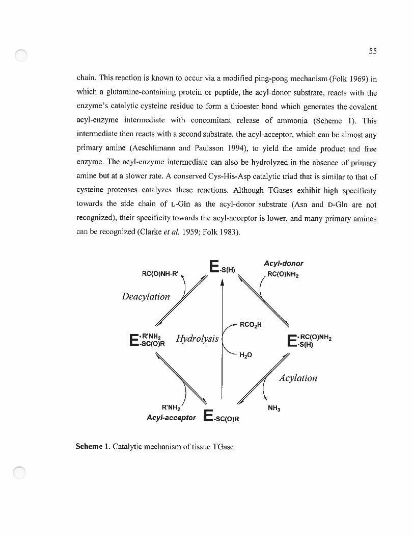
55
chain. This reaction is known to occur via a modified ping-pong mechanisrn (Folk 1969) in
which a glutamine-containing protein or peptide. the acyl-donor substrate. reacts with the
enzyme’s catalytic cysteine residue to forrn a thioester bond which generates the covaient
acyl-enzyme intermediate with concomitant release of ammonia (Scheme 1). This
intermediate then reacts with a second substrate, the acyl-acceptor, which can be almost any
primary amine (Aeschlimann and Pauïsson 1994), to yieid the amide product and free
enzyme. The acyi-enzyme intermediate can also be hydroiyzed in the absence of prirnary
amine but at a siower rate. A conserved Cys-His-Asp catalytic triad that is similar to that of
cysteine proteases catalyzes these reactions. Although TGases exhibit high specificity
towards the side chain of L-Gln as the acyl-donor substrate (Asn and D-Gin are not
recognized), their specificity towards the acyl-acceptor is lower, and rnany primary amines
can be recognized (Ciarke et cii. 1959: Folk 1983).
Deac,,/”
E. RC(O)NH2-S(H)
NH3
A cylation
E..sHÀ
A cyl-donor
RC(O)NH2
E.R.NH2HydroÏysis
-SC(O)R
HO
R N H2
AcyI-acceptor E-scfo)R
Scheme 1. Cataiytic mechanism of tissue TGase.
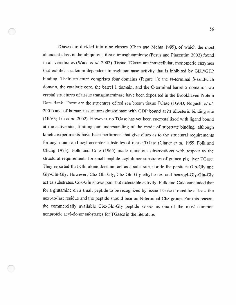
56
TGases are divided into fine classes (Chen and Mehta 1999), of which the most
abundant class is the ubiquitous tissue transglutaminase (Fesus and Piacentini 2002) found
in ail vertebrates (Wada et cii. 2002). Tissue TGases are intracellular. monorneric enzymes
that exhibit a calcium-dependent transglutaminase activity that is inliibited by GDP/GTP
binding. Their structure comprises four dornains (Figure 1): the N-terminal n-sandwich
domain, the catalytic core, the barrel 1 domain, and the C-terminal barrel 2 domain. Two
crystai structures of tissue transglutaminase have been deposited in the Brookhaven Protein
Data Bank. These are the structures of red sea bream tissue TGase (1 G0D; Noguchi cl ai.
2001) and of human tissue transglutaminase with GDP bound at its allosteric binding site
(1KV3; Liu et al. 2002). However, no TGase bas yet been cocrystallized with ligand bound
at the active-site. lirniting our understanding of the mode of substrate binding, although
kinetic experiments have been perforrned that give dues as to the structural requirements
for acyl-donor and acyl-acceptor substrates of tissue TGase (Clarke et ai. 1959; Folk and
Chung 1973). Folk and Cole (1965) made numerous observations with respect to the
structural requirements for small peptide acyt-donor substrates of guinea pig liver TGase.
They reported that Gin alone does not act as a substrate. nor do the peptides Gln-Gly and
Gly-Gln-Gly. However. Cbz-Gln-Glv. Cbz-Gln-Gly ethyl ester. and benzovl-Gly-GIn-Gly
act as substrates. Cbz-Gln shows poor but detectable activity. Folk and Cole concluded that
for a glutamine on a small peptide to be recognized by tissue TGase it must be at least the
next-to-last residue and the peptide should bear an N-terminal Cbz group. For this reason,
the commercially available Cbz-Gln-Gly peptide serves as one of the most common
nonproteic acyl-donor substrates for IGases in the literature.
57
Although there is no consensus as to their physiological role. tissue TGases have
been reported to act in endocytosis (Abe et al. 2000), apoptosis (Huo et al. 2003),
extracellular matrix modification (Priglinger et aÏ. 2003), and celi signaling (Singh et al.
2003). Tissue TGases have also been implicated in various physiological disorders such as
Alzheimer’s disease (Kim et al. 1999). cataract formation (Shridas et al. 2001), and more
recently, in Celiac sprue (Shan et al. 2002). As a resuit, development of inhibitors specific
to tissue IGases (as opposed to type 1 or 3 TGases; Chen and Mehta 1999) is of current
interest. However, to aid in inhibitor design, we need to gain a better understanding of the
mode of binding of mode! peptide substrates to the enzyme. To this end, we performed a
Figure 1. Crystal structure of red sea bream IGase tNoguchi et al. 2001). (A) Red sea
bream TGase with active-site residues circled. Front view. The P356—G369 loop is
represented in ribbon diagram. (B) Top view.
5$
combined experimental/molecular modeling strategy consisting of (1) the synthesis of a
series of Cbz- or Boc-derivatized peptides and the measurernent of their KM values, and (2)
molecular modeling of their binding at the active-site. Correlating the kinetic resuits with
molecular modeling of the ligands bound at the active-site cavity bas allowed us to gain a
better understanding of the requirements for productive binding of smali peptide acyl-donor
substrates.
3.3 Results
3.3.1 Kinetic characterization ofthe N-terrninally derivatized peptides
The synthesis ofthe derivatized peptide substrates has been described (Gagnon et aï.
2002). The kinetic pararneters KM and kcat of guinea pig liver TGase were deterrnined for a
series of Cbz-derivatized Gin-Xaa peptides as well as for the Boc-derivatized Gln-Gly
dipeptide (Table 1). The seven Cbz-peptides assayed gave sirnilar KM and keai values.
whereas the Boc-dipeptide did flot appear to act as a donor substrate, showing no reactivity
at concentrations up to 40 mM. the limit of its solubility. The similar KM and kcat values
obtained for ail the Cbz-derivatized Gln-Xaa peptides indicate that the identity of the
C-terminai amino acid does not significantly affect productive binding or turnover. Thus,
these substrates resuit in a similar catalytic efficiency. Because arnino acids having side
chains with widely differing chernical and structurai attributes were assayed and because we
observed that the nature of these side chains does not affect substrate recognition or
efficiency, we can conclude that substrate binding does not depend on the nature of the
C-terminal amino acid. However. the nature of the N-terminal functional group, Boc or
Cbz, has an important effect on substrate recognition by the enzyme. Namely. Boc-Gln-Gly
does not serve as a donor substrate, whereas Folk and Cole (1966) observed that Cbz-Gln
does act as a substrate of tissue type TOase. albeit a poor one. aithough Gln-Gly does not.
Our kinetic data confirm that the presence of the Cbz group contributes importantly to
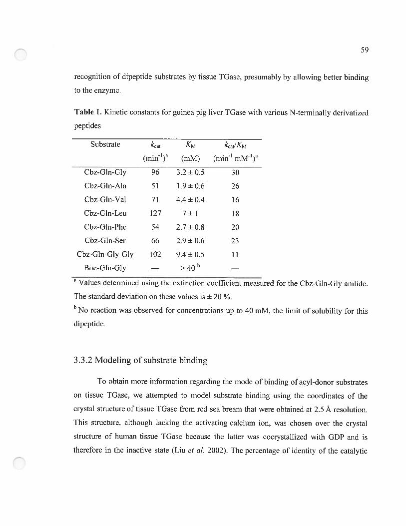
59
recognition of dipeptide substrates by tissue IGase, presumably by allowing better binding
to the enzyme.
Table 1. Kinetic constants for guinea pig liver TGase with various N-terrninally derivatized
peptides
Substrate kcat KM kcat/KM
(min_l)a (mM) (min mIA_l)a
Cbz-Gln-Gly 96 3.2 + 0.5 30
Cbz-Gln-Ala 51 1.9 ± 0.6 26
Cbz-Gln-Val 71 4.4 + 0.4 16
Cbz-Gln-Leu 127 7 ± 1 18
Cbz-Gln-Phe 54 2.7 ± 0.8 20
Cbz-Gln-Ser 66 2.9 + 0.6 23
Cbz-GÏn-Gly-Gly 102 9.4 + 0.5 11
Boc-Gln-Gly — > 40 b
a Values determined using the extinction coefficient measured for the Cbz-Gln-Gly anilide.
The standard deviation on these values is + 20 %.b No reaction was observed for concentrations up to 40 mM, the lirnit of solubility for this
dipeptide.
3.3.2 Modeling of substrate binding
To obtain more information regarding the mode of binding ofacyl-donor substrates
on tissue TGase. we attempted to model substrate binding using the coordinates of the
crystal structure of tissue TGase from red sea bream that were obtained at 2.5 À resolution.
This structure, although lacking the activating calcium ion, was chosen over the crystal
structure of human tissue TGase because the latter was cocrystallized with GDP and is
therefore in the inactive state (Liu et aÏ. 2002). The percentage of identity of the catalytic
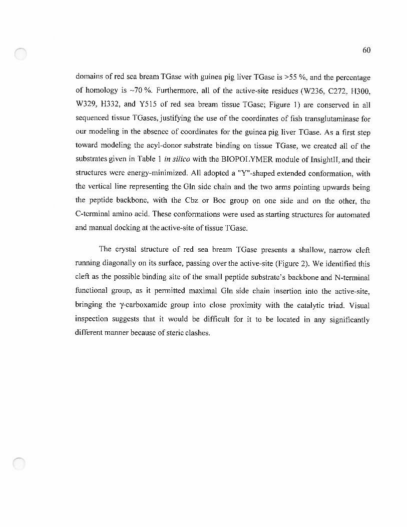
60
domains of red sea bream TGase with guinea pig liver TGase is >55 ¾, and the percentage
of hornology is 70 ¾. Furthermore, ail of the active-site residues (W236, C272. H300,
W329, H332, and Y515 of red sea bream tissue TGase; Figure I) are conserved in au
sequenced tissue TGases. justifying the use of the coordinates of fish transglutarninase for
our modeling in the absence of coordinates for the guinea pig liver TGase. As a first step
toward modeling the acyl-donor substrate binding on tissue TGase, we created ail of the
substrates given in Table 1 in siÏico with the BIOPOLYMER module of Insightll, and their
structures were energy-minimized. Ail adopted a ‘Y”-shaped extended conformation, with
the vertical une representing the Gin side chain and the two arms pointing upwards being
the peptide backbone, with the Cbz or Boc group on one side and on the other. the
C-terminal amino acid. These conformations were used as starting structures for automated
and manual docking at the active-site of tissue TGase.
The crystal structure of red sea bream TGase presents a shailow, narrow clefi
running diagonally on its surface, passing over the active-site (Figure 2). We identified this
cieft as the possible binding site of the small peptide subslrate’s backbone and N-terminai
functionai group, as it permitted maximai Gin side chain insertion into the active-site.
bringing the y-carboxamide group into close proximity with the catalytic triad. Visuai
inspection suggests that it would be difficuit for it to be iocated in any significantly
different manner because of steric ciashes.
61
jFigure 2. Proposed binding clefi for the peptide substrate N-terminal functionaÏ group at
the surface of red sea bream IGase. The diagonal clefi running over the active-site cavity is
in yeltow; the Gin side chain of the Cbz-Gln-Gly substrate that enters the active-site cavity
away from the viewer is represented by X. Negative charges are in red; positive are in blue.
The four positions tested for manual docking of the N-terminal Cbz group are nurnbered.
The Cbz group is drawn to scale.
To test this hypothetical binding mode, we perforrned automated docking
experiments using AutoDock 3.0 software (Morris et aÏ. 1998). Preliminary docking
experirnents were performed using the PDB file I GOD. We observed that the Cbz-Gln-Gly
and Boc-Gln-Gly substrates neyer entered the active-site during the 50 runs performed.
Indeed, the active-site of the enzyme is visibly blocked by a Trp residue. W329 (figure 1)
which is conserved in ail tissue TGases as weil as in Factor XIII (Yee et al. 1994).
epidermal (Ahvazi et aÏ. 2002). and keratinocyte TGases. This apparently results from the
structure having been resolved in the absence of substrate. Because automated docking is
performed without movement of the protein atoms, it is unlikely to succeed in identifying a
binding mode for the substrate Gin side chain inside the active-site. Thus, we manually
62
increased the accessibility of the active-site of the structure of red sea bream TGase by
introducing torsions in the C—C (-83.13°) and C—C (+48.52°) bonds of residue W329.
These torsions opened the active-site cavity while minimizing steric clashes between W329
and the rest of the protein. With this starting structure. the AutoDock procedure resulted in
several structures with the substrate Gin entering the active-site (Table 2). This indicates
that W329 may prevent entry to the active-site. acting as a ‘gate’ that could regulate TGase
activity. 0f the 50 runs performed with Cbz-GIn-Gly. oniy four generated a structure where
the substrates Gin side chain had entered the active-site (Table 2). This low number of hits
is expected because the substrate was allowed a high degree of conformational flexibility.
Three of the four bound structures positioned the Cbz group at position 0; the last one
positioned the Cbz at position 1 (Figure 2). With the Boc-Gln-Gly substrate, oniy six of the
50 runs generated a structure where the Gin side chain had entered the active-site. 0f these
six structures, two had the Boc at position 2. three at position 0, and one at position I. It
should be noted that these positions are not preciseiy deflned but represent restricted zones
within which positioning was observed. These AutoDock resuits allowed us to establish
three potential binding sites at the surface of the enzyme for the N-terminai firnctionai
group. Position 0. which is in the diagonal clefi. appears to be favoured for binding. This
suggests that positioning of the N-terminal functionai group in the clefi is favourable to
productive binding of the Gln side chain inside the active-site cavity. The low number of
runs that resuited in a structure where Gln was bound in the active-site reflects the generally
weak affinity of tissue TGase for these dipeptide substrates. This in tum is manifested in
their miilimolar KM values. We also observed that afler energy minimization of these 10
structures using Insightll. 75 % (3/4) ofthe Cbz-Gin-Giy structures formed an H-bond with
conserved residue Y515 of the active-site, whereas only 16 % (1/6) of the Boc-Gln-Gly
structures had formed this H-bond (Table 2).
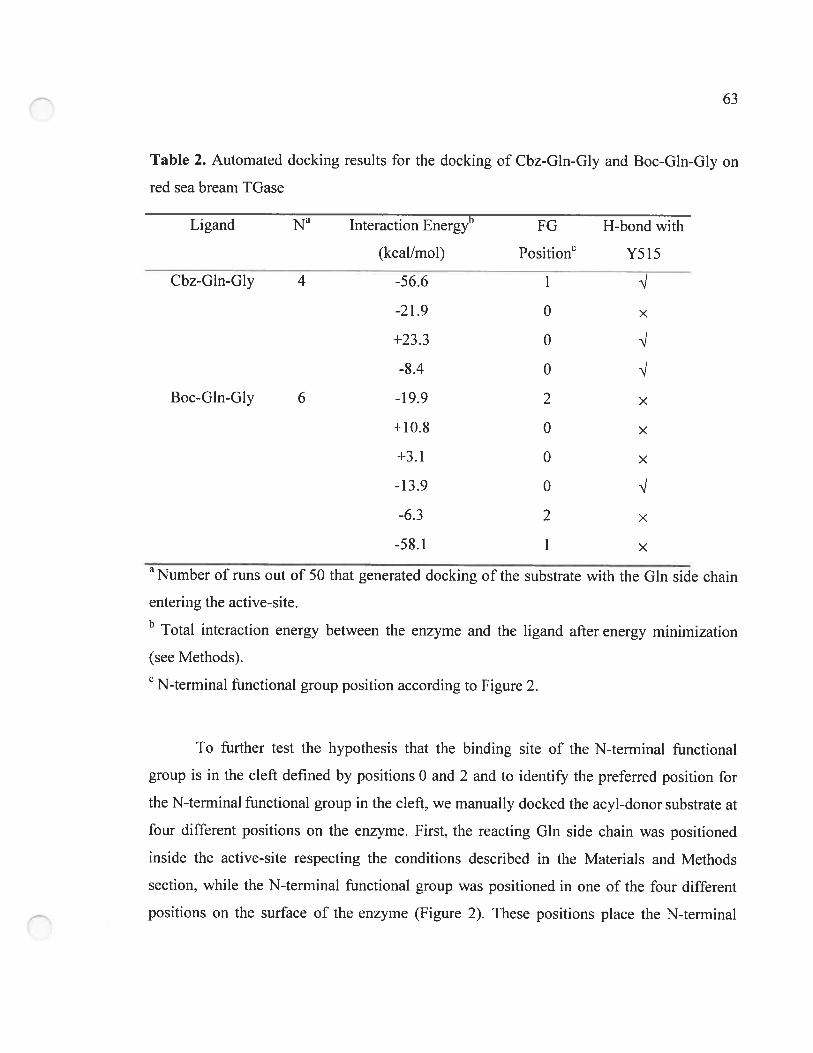
63
Table 2. Automated docking resuits for the docking of Cbz-Gln-Giy and Boc-Gln-Gly on
red sea bream TGase
Ligand N’ Interaction Energy” FG H-bond with
(kcal/rnol) Positionc y5j5
Cbz-Gin-Gly 4 -56.6 1
-21.9 0 x
o q
-8.4 0
Boc-Gln-Giy 6 -19.9 x
+10.8 0 x
+3.1 0 x
-Ii.
-0.2 L X
-58.1 1 x
Number of runs out of 50 that generated docking of the substrate with the Gin side chain
entering the active-site.b Total interaction energy between the enzyme and the ligand afler energy minirnization
(see Methods).
N-terminal functional group position according to Figure 2.
To further test the hypothesis that the binding site of the N-terminai functional
group is in the cleft defined by positions O and 2 and to identify the preferred position for
the N-terminal functionai group in the ciefi. we manualiy docked the acyl-donor substrate at
four different positions on the enzyme. First, the reacting Gin side chain was positioned
inside the active-site respecting the conditions described in the Materials and Methods
section, whiie the N-terminal functionai group was positioned in one of the four different
positions on the surface of the enzyme (Figure 2). These positions place the N-terminal
64
functional group alternatively in the clefi (positions O and 2), in a small cavity perpendicular
to the clefi (position 1), or directly on top of residue W236 (position 3). Steric clashes were
more important in position 3. which was chosen as a negative control for the molecular
modeling procedure. Then. steric clashes between the enzyme and the substrate were
energy-minimized while the enzyme/substrate distance was constrained. as described in
Materials and Methods. Ibis caused the active-site to open up as residue W329 was
displaced, indicating that this residue is very mobile, because it readily moves away from
the active-site after merely an energy minimization.
A lO-picosecond molecular dynarnics simulation was then performed on the
generated enzyme-substrate complexes to explore the immediate conformational space. One
hundred conformers were thus generated. The conformer with the lowest energy was further
energy-minimized, and the resulting structure was analyzed. Ihis resulting structure with
each substrate was always of lower energy than the structure generated before the molecular
dynamics simulation. The backbone of the enzyme was maintained fixed during Trial 1 of
manual docking (Table 3) to determine the most plausible binding site for the N-terminal
functional group in order for the srnaÏl Cbz-Gln-Gly substrate to adopt a conformation that
best fit the enzyme’s structure and not the opposite. Ihis experiment was also performed
two more times with varying conditions, including independent manual substrate
positioning, a 25 À or 40 À layer of water covering the active-site, and with no constraint
on the enzyme backbone, which generated similar results (Table 3). In ail three independent
trials with Cbz-Gin-Giy. the enzyrne-substrate interaction energy was the lowest and was
consistently negative when the Cbz was positioned in the upper left part of the clefi
(position 0; figure 2), indicating that it is the only one ofthe four positions tested that is
energetically favourable and reproducible for binding the Cbz group for a small peptide
substrate. Thus, it appears that the preferred position for docking the Cbz group on the
surface of the enzyme is located in the upper lefi corner of the diagonal clefi (position 0).
Ibis position aiso aiiows better insertion of the reacting Gin side chain into the active-site
based on the anti-conformation of the Gln side chain in the active-site compared to the
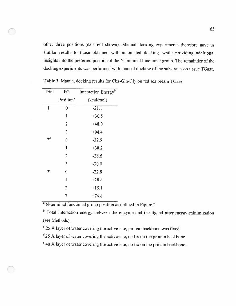
65
other three positions (data not shown). Manual docking experirnents therefore gave us
sirnilar resufts to those obtained with autornated docking, while providing additional
insights into the preferred position ofthe N-terminal functional group. The remainder ofthe
docking experiments was performed with manual docking of the substrates on tissue TGase.
Table 3. Manual docking resuits for Cbz-Gln-Gly on red sea bream IGase
Trial FG Interaction Energyb
Positiona (kcal/mol)
le 0 -21.1
1 +36.5
2 +48.0
3 +94.4
0 -32.9
1 +38.2
2 -26.6
3 -30.03e 0 -22.8
1 +28.8
2 +15.1
3 +74.8
a N-terminal functional group position as deflned in Figure 2.b Total interaction energy between the enzyme and the ligand afier energy minimization
(see Methods).
25 À layer of water covering the active-site. protein backbone was fixed.U25 À layer of water covering the active-site, no fix on the protein backbone.e 40 À layer of water covering the active-site, no fix on the protein backbone.
66
following the docking studies on the putative positioning of the Cbz group using
Cbz-Gln-Gly, binding of the Cbz-Gln-Xaa substrates was modeied with the N-terminal
functional group in position 0. The Boc-Gln-Gly substrate was modeied as a control to
ensure that the modeling resuits are consistent with its iack of reactivity as a substrate
(Table 1). For each test. the substrate was manually positioned in an independent manner.
During the caiculations. only the residues including atoms within 20 À of the cataiytic S
atom of C272. the water moiecules, and the substrate were mobile. Constraints were applied
as described in Materials and Methods to retain the substrate Gin proximal to the cataiytic
residues, thus increasing the likelihood of model ing productive enzyrne/substrate
complexes. Afier the energy minirnizationlrnolecuiar dynamics methodology was
performed, the interaction energy between the enzyme and each substrate was analyzed. For
the Cbz-Gln-Gly substrate. anaiyzed in 13 tests, we found that the interaction cnergy (AG)
between ligand and protein was negative. thus favourable. in ail but one case (Table 4). In
the single case where the interaction energy was positive. the electrostatic component of the
interaction energy between enzyme and substrate was very high (+158.9 kcal/mole). In this
case, the carboxylate of Cbz-Gln-Gly was found to be in doser proxirnity than usual to a
conserved, negatively charged residue of red sea bream TGase, Glu360. which could resuit
in such an unfavourable electrostatic energy. Nonetheless. the average of the 13 data sets
suggests that there is a favourable interaction between the enzyme and the Cbz-Gln-Gly
substrate. The average van der Waais energy between the enzyme and the Cbz-Gln-Giy
ligand was -53 + 5 kcal/mole. Whiie exarnining the interactions between the enzyme and
Cbz-Gln-Gly. we foundthat each ofthe 13 structures generated showed an H-bond between
the 0D proton of Y5 15 and the 06 of Gin from Cbz-Gln-Gly. Also, 77 % of the structures
t 10/13) displayed an H-bond between N of H332 and HE of GIn side chain. The proxirnity
and orientation displayed in this H-bond pairing resembles the interaction that takes place
during catalysis when the acid/base catalyst, F1332, protonates the ammonia leaving group.
The high prevalence of these H-bonds suggests that they are important in binding the
glutaminyl residue at the active-site.
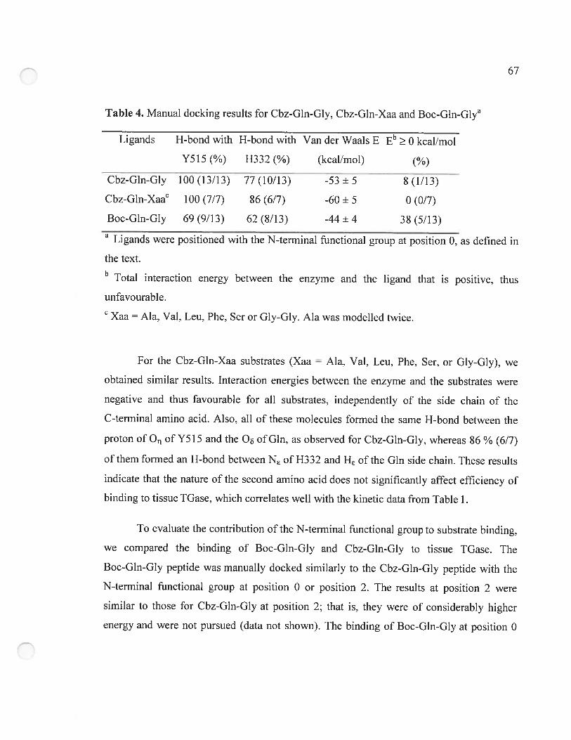
67
Table 4. Manual docking resuits for Cbz-Gln-Gly, Cbz-Gln-Xaa and BocGlnG1ya
Ligands H-bond with H-bond with Van der Waals E Et O kcal/rnol
Y515 (%) H332 (%) (kcaÏ/mol) (%)
Cbz-Gln-Gly 100(13/13) 77(10/13) -53+5 8(1/13)
CbzGlnXaac 100 (7/7) $6 (6/7) -60 + 5 0 (0/7)
Boc-Gin-Giy 69(9/13) 62(8/13) -44+4 3$ (5/13)
Ligands were positioned with the N-terminal functionai group at position O. as defined in
the text.b Total interaction energy between the enzyme and the ligand that is positive, thus
unfavourable.
Xaa Ala, Val, Leu, Phe. Ser or Giy-G1 . Ala was modeiied twice.
For the Cbz-Gin-Xaa substrates (Xaa = Ala, Val, Leu, Phe, Ser, or Gly-Gly). we
obtained similar results. Interaction energies between the enzyme and the substrates were
negative and thus favourabie for ail substrates. independently of the side chain of the
C-terminal amino acid. Aiso, all of these molecules fonned the sarne H-bond between the
proton of O of Y5 15 and the O of Gin, as observed for Cbz-Gln-Gly, whereas 26 % (6/7)
of them forrned an H-bond between NE of H332 and HE of the Gin side chain. These resuits
indicate that the nature of the second amino acid does flot significantly affect efficiency of
binding to tissue TGase. which colTelates well with the kinetic data from Table 1.
To evaluate the contribution of the N-terminal functional group to substrate binding,
we cornpared the binding of Boc-Gln-Gly and Cbz-Gln-Gly to tissue TGase. The
Boc-GIn-Gly peptide was manually docked sirnilarly to the Cbz-GIn-Giy peptide with the
N-terminal functional group at position O or position 2. The resuits at position 2 were
sirnilar to those for Cbz-Gin-Gly at position 2; that is. they were of considerably higher
energy and were flot pursued (data flot shown). The binding of Boc-Gln-Giy at position O
68
did not generate reproducible resuits as Cbz-Gln-Xaa did. Indeed, sorne resuits generated
poor interaction energy between enzyme and substrate. whereas others gave as good an
interaction as the Cbz peptides. 0f the 13 trials. 38 % (5/13) generated a positive interaction
energy, indicating unfavourabie binding. Furtherrnore, the H-bond between the
Boc-Gln-Giy substrate and Y5 15 was found in only 69 % (9/13) of the trials, whereas the
H-bond with H332 was found in 62 % of the trials (8/1 3), which is significantiy iess than
for the Cbz-derivatized peptides. In addition, the average van der Waals energy is
-44 ±4 kcai/mole, which is more than 10 kcal/moie higher than for the average of ail
Cbz-derivatized peptides (Cbz-Gln-Gly and Cbz-Gln-Xaa). Taken together, these resuits
suggest that the presence ofthe Boc group does not allow for binding that is as constant and
reproducible as for the Cbz-Gln-Xaa substrates. It thus appears that the Boc group is
deleterious to proper binding, because it does not allow a constant productive insertion of
Gin side chain into the active-site.
3.3.3 Modeling of the tetrahedral and acyl-enzyrne intemiediate
To minimize any potential bias arising from the manuai docking undertaken during
the modeling study and to investigate more thoroughly the importance of the H-bond
network with Y5 15 and H332 identified for the binding of the Cbz-peptides, we created in
silico the tetrahedral and acyl-enzyrne intermediates for the reaction of tissue IGase with
Cbz-Gin-Gly. These intermediates have a covaient bond between the C atom of Gin from
the donor substrate and S of the catalytic Cys residue. thus eliminating the requirement for
manual docking of the substrate. The acyi-enzyrne and tetrahedrai intermediates were
constructed respecting the xi—x2 and x2—x plots dihedral angles (Janin et aï. 197$) for the
substrate Gin and C272. Two starting structures for the acyl-enzyme intermediate were
constructed, one with the Cbz group at position O and another at position 2. These dihedrai
angles stiil respected the Xi—X2 and plots at the end ofthe simulation, indicating that
the generated structure was coherent with regards to the conformation of the two residues
involved in the covaient bond between enzyme and substrate (data not shown). Afier the
69
simulation, a lower energy for the modeled system was obtained for the acyl-enzyrne
intermediate with the Cbz group at position O cornpared to position 2 (-4513 kcal/mole
versus -4461 kcal/mole). This is consistent with the hypothesis that position O represents the
actual Cbz binding site because it is energetically more favorable.
While analyzing the covalent structures generated. we observed that the acyl portion
of the molecule adopted a conformation very similar to the model of TGase with the
noncovalently docked substrate. Indeed. the RMSD of the Gin side chains of Cbz-Gln-Gly
in the Michaelis complex and the acyl portion of the acyl-enzyrne and tetrahedral
intermediates are very low (Table 5), indicating near-identical positioning of the Gin side
chain inside the active-site for these three rnodeled structures (Figure 3A—C). Also, the
dihedral angles l and 2 are nearly identical among the three structures, which
demonstrates that the conformation of the Gln side chain in the Michaelis complex is the
same as in the tetrahedral and acyl-enzyrne intermediates. This suggests that the insertion of
the Gin side chain of the docked substrate is coherent and thus there was no bias in
positioning during manual docking. The H-bond between the reactive Gln ‘y-carboxarnide
group and the enzyme Y5 15 residue was present in the acyl-enzyme intermediate. The
H-bond between N of H332 and HE of Gln side chain was not present because the Gln side
chain in the acylenzyme intermediate no longer carnes the NH2 group.
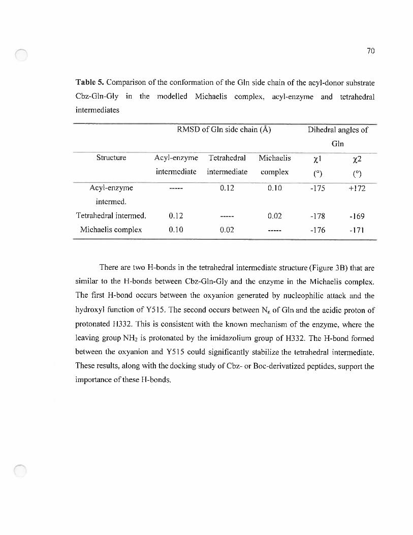
70
Table 5. Comparison ofthe conformation ofthc Gin side cliain ofthe acyl-donor substrate
Cbz-Gln-Gly in the rnodelled Michaelis complex, acyl-enzyrne and tetrahedrai
intermediates
RMSD of Gin side chain (À) Dihedrai angles of
Gin
Structure Acyl-enzyrne Tetrahedral Michaelis xiintenriediate interrnediate compiex (0) (0)
Acyi-enzyrne 0.12 0.10 -175 +172
intermed.
Tetrahedral intermed. 0.12 0.02 -178 -169
Michaeiis complex 0.10 0.02 -176 -171
There are two H-bonds in the tetrahedrai internwdiate structure (Figure 33) that are
similar to the H-bonds between Cbz-Gln-Giy and the enzyme in the Michaeiis complex.
The first H-bond occurs between the oxyanion generated by nucleophiiic attack and the
hydroxyl function of Y5 15. The second occurs between N of Gin and the acidic proton of
protonated H332. This is consistent with the known rnechanism of the enzyme, where the
leaving group NH2 is protonated by the irnidazolium group of H332. The H-bond formed
between the oxyanion and Y5 15 could significantly stabiiize the tetrahedrai intermediate.
These resuits, along with the docking study of Cbz- or Boc-derivatized peptides. support the
importance ofthese H-bonds.
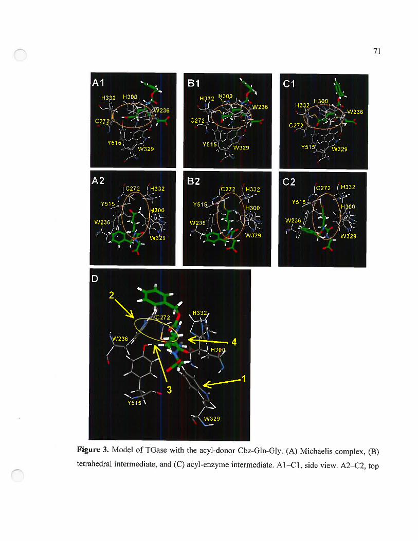
71
Figure 3. Model of TGase with the acyl-donor Cbz-Gln-Gly. (A) Michaelis complex. (B)
tetrahedral intermediate, and (C) acyl-enzyrne intermediate. Al—Cl, side view. A2—C2, top
72
view. In C2, the NH3 molecuie lias been released. (D) Interactions proposed to be important
in the rnodeied Michaeiis compiex ofTGase with Cbz-GIn-Giy: (1) Dispiacement ofW329
toward the bottom to allow entrance of substrate Gin side chain into the active-site; (2)
Hydrophobie interaction ofthe indole group of W236 with the methylene groups ofthe Gin
side chain; (3) H-bond of the Gin y-carboxarnide with Y515; (4) H-bond of the Gin
7-carboxamide with H332.
3.3.4 Structural analysis of Cbz-peptide binding
The 13 structures generated by molecular dynamics foiiowing manuai docking of
Cbz-Gin-Gly peptides and the seven structures generated with the Cbz-Gin-Xaa substrates
were anaiyzed as a group. The Gin side chain enters the active-site airnost perpendiculariy
with respect to the center of mass of the enzyme. figure 3D illustrates one of the trial
structures, chosen because it was typicai and representative. The conformation of the
substrate Gin side chain is the iow-energy anti-conformation. The l and 2 dihedrai angies
of the substrate Gin are near 180e. whicli gives a trans-conformation to the side cliain that
is common for amino acids. Three aromatic residues. W236. W329, and H300 that appear to
form a tunnel leading to the catalytic residues, surround the side chain. As previously
discussed, residue W329 is displaced by the insertion of the substrate Gin side chain into
the active-site so as to open the active-site that it btocks in the unbound state. This
dispiacement places the W329 side chain in a position very simiiar to that achieved
manuaiiy as part of the automated docking experiments (C-C = 95•530 and
= +82.4°). The roof of the tunnei is formed by a ioop of residues P356—G369
(figure 1). The two methyiene groups of the Gin side chain of the substrate are in contact
with the indoie group of residue W236. This maxirnizes the hydrophobie interaction
between the indoie group ofW236 and the methyiene groups ofthe substrate Gin side chain
during the course ofthe simulation—as evidenced by the concerted movernent of these two
groups during the dynamics simuiation. The substrate y-carboxamide, as mentioned earlier,
73
adopts a conformation allowing it to forrn H-bonds with residues Y515 and H332. In this
conformation, the nucleophilic attack by the thiolate can only be achieved from the top or
the bottom of the substrate y-carboxamide, in keeping with an addition-displacement
mechanism. If the attack occurs from the top, the oxyanion generated could form a
stabilizing H-bond with the hydroxyl group of Y5 15. Such a bond is observed in the model
of the tetrahedral intermediate.
3.4 Discussion
No existing crystai structures of tissue TGase (Noguchi et aÏ. 2001; Liu et aÏ. 2002)
have been resolved with ligand bound at the active-site, limiting our understanding of the
binding of acyl-donor substrates. The only structural elernents that have been shown to be
important for productive binding of small peptide acyl-donor substrates are (1) the iength of
the side chain (Asn versus GIn). (2) the stereochemistry ofthe Gin side chain (D-Gin versus
L-GIn). (3) the length of the peptide substrate (Clarke et al. 1959; Folk 1983). and (4) to a
lesser extent, the position ofthe reactive Gin in the peptide/protein sequence (Ohtsuka et aï.
2000). Thus, it is known that for srnail acyl-donor peptide substrates, the reactive Gin
residue must be at ieast the second residue from the C terminus and the third from the
N terminus (Folk and Cole 1965), where Cbz can replace the flrst two arnino acids.
Molecules such as Gln and Gln-Gly do not act as substrates, whereas Cbz-Gln-Giy is the
most widely used substrate.
b gain insight into the binding of smaii peptide acyl-donor substrates on tissue
TGase. we used a cornbined kinetic/molecuiar modeiing approach. Kinetic tests were
performed on guinea pig liver transglutaminase. because it is the most extensively
characterized tissue IGase. Molecular modeiing was performed on the ciystai structure of
red sea bream tissue TGase. The catalytic core domain of this enzyme bas a sequence
homology of -70 % to that of guinea pig liver, and ail but four residues composing the clefi
(Figure 2) are conserved between the two enzymes, and thus the two structures can be
74
considered very similar. Ail of the active-site residues (W236, C272. H300. W329, H332.
and Y5 15 of red sea bream tissue TGase) are conserved in ail tissue IGases, inciuding the
human enzyme, of which the catalytic dornain has 58 % identity and 70 % hornology with
the red sea bream enzyme. This justifies the use of the coordinates of red sea bream
transglutaminase for our modeiing in the absence of coordinates for the guinea pig iiver
TGase. as well as the pertinence of these studies to future drug design. Furthermore.
Cbz-Gin-Giy is an efficient acyl-donor substrate of both red sea bream IGase and guinea
pig liver TGase (Ohtsuka et aÏ. 2000) and is therefore appropriate for docking studies on
this structure.
Molecular modeiing suggests that there is a hydrophobic interaction between the
indole group of the side chain of W236 and the methyiene groups of the Gin side chain of
the substrate. Molecuiar dynamics simulations also confirm the concerted movement of
these two groups. Residue W236 (W241 in gwnea pig liver TGase and human tissue
IGase) is conserxed in ail eukaryotic TGases that have been sequenced. In site-directed
mutagenesis studies of human tissue TGase. replacement of W24 1 with Ala. Gin, His, Phe,
or Tyr resulted in a substantiai loss of activity. demonstrating the importance of this Irp to
catalysis (Murthy et al. 2002). Significantly, the mutants W241H, W241F, and W241Y,
possessing aromatic side chains, showed low but readiiy measurabie activities of
approximately one-tenth that of wiid-type and around 20-fold higher than that of the non
aromatic mutants W241A and W241Q (Murthy et al. 2002). In consideration of these
results and the contacts revealed by our modeiing studies of red sea bream TGase, we
propose that the acyi-donor substrate Gin residue is stabilized in the active-site through
hydrophobie interactions of the methylene groups of the substrate side chain with the
aromatic indole group of residue W236. This could partiy account for the fact that peptide
or protein-bound Asn is not a substrate of TGase because its shorter and iess hydrophobie
side chain, iacking one methylene unit cornpared to Gin, would flot aiiow these stabilizing
interactions.
75
Molecular modeling suggests that residue Y5 15 is crucial in acyl-donor substrate
binding and reactivity. The presence ofa ubiquitous H-bond between the hydroxyl group of
Y5 15 and the carbonyl oxygen from Gin side chain for ail of the efficient substrates (Cbz
peptides) indicates that Y515 participates in stabilizing the substrate in the active-site. This
tyrosine residue is strictiy conserved in ail tissue TGases. It beiongs to the barrei 1 domain
but in the foided structure is iocated within the active-site and in close proxirnity to the
cataiytic C272 in the core domain. Taken together. these data suggest that Y5 15 plays an
important role in this enzyme. It has been proposed to form a reguiatory H-bond with the
catalytic suiphur atom, preventing nucieophulic attack by the suiphur atom on the substrate
carboxamide group (Yee et al. 1994g Noguchi et al. 2001). However, our findings suggest
that it could orient the substrate y-carboxamide for nucleophilic attack by the thiolate and
that the H-bond formation couid activate the carbonyi towards this nucieophiiic attack by
rendering it more electrophilic. Our studies aiso reveal the presence of this H-bond in the
structures of the acyi-enzyrne and tetrahedrai intermediates, further suggesting its
invoivement in stabilizing the oxyanion generated during catalysis. We are currentiy
undertaking mutagenesis of guinea pig liver IGase at this position to verify our hypothesis.
The H-bond network identified in our modeiing study, invoiving residues Y5 15 and
H332 of red sea bream tissue TGase and the y-carboxarnide group of the donor substrate,
couid be criticai for transgiutaminase activity. This network is highiy prevaient in the
structures generated by modeling with efficient substrates (Cbz-peptides) and is present at a
significantiy iower percentage in the structures generated with the unreactive dipeptide
Boc-Gln-Gly. This H-bond network could catalyze acylation by orienting the
y-carboxamide group relative to the two cataiytic residues C272 and H332, activating the
‘y-carbonyi group to nucieophiiic attack by C272 by rendering it more electrophiiic and
stabiiizing the oxyanionic transition state. Moiecuiar modeling showed that the H-bond
between the O proton of Y5 15 and the O of Gin from Cbz-Gin-Gly was present 100 % of
the time with ail efficient substrates as weIl as in the acyi-enzyrne and tetrahedral
76
intermediates. However, this H-bond was flot aiways present when a poor substrate.
Boc-Gln-Gly, was modeled.
Residue W329 blocks the active-site in the crystal structure of red sea bream TGase
and human TGase (W332 in human tissue TGase), preventing substrate entry. Indeed.
automated docking experiments with AutoDock 3.0 using the crystal structure ofthe red sea
bream enzyme whose active-site is blocked by residue W329 did flot generate structures
with substrate in the active-site. However, we have shown by molecular dynarnics
simulation over 10 5 that this residue readily moves to allow insertion of the side chain
into the active-site. This tryptophan residue is also strictly conserved in ail of the tissue
TGases and is located in a loop comprising residues $321 to W329. It could therefore act as
a ‘gate’ that could close the active-site and exciude water. W329 could then move to open
the active-site upon substrate binding, allowing substrates to enter. The opening of the
active-site by dispiacement of this residue may be mediated by allosteric calcium binding,
which is essential for TGase activity. This hypothesis is indirectly supported by resuits from
crystallographic studies of TGase 3, showing that calcium binding induces conformational
changes that expose the corresponding W327 in a way that should increase its dynarnic
mobility and interactions with incoming substrates (Ahvazi et aÏ. 2002).
The difference in recognition reftected by the varied KM values for Gln-Gly
dipeptides having different N-terminal frmnctional groups, Boc or Cbz, could be attributed to
various factors. first, the van der Waals energy of interaction between the enzyme and the
N-terminally derivatized Gln-Gly dipeptide substrate varies by almost 10 kcal/mole,
depending on the N-terminal functional group. This difference in interaction energy
correlates well with the poorer binding of the Boc-derivatized dipeptides. Furthermore,
modeling of the binding of Boc-derivatized dipeptides gave unfavorable, positive
interaction energies far more frequently than Cbz-derivatized dipeptides. Finally, the
aforementioned H-bond network is frequently absent in the structures generated with the
Boc-dipeptides. These differences in binding could be the result of differences in the shape
77
and size between the Cbz and Boc groups. because the Chz is pianar whereas the Boc is
globular. The Boc group is also shorter in that the bulky teri-butyi group is attached directiy
to the carbamate oxygen, whereas the bulky phenyl group ofthe Cbz group is attached to an
additionai methylene unit, increasing its iength and conformational flexibitity. It is aiso
possible that good recognition of Cbz-Gin-Giy by tissue TGase is conferred by the
aromaticity of the Cbz group. We are currenti undertaking kinetic studies of various
substrates containing differing N-terminal functional groups to explore the importance of
the nature ofthis group to binding afflnity.
Summarized in Table 6 and Scheme 2 are the interactions that we propose for the
acylation of tissue TGase by srnaii peptide substrates. First, the active-site is opened by
dispiacement of residue W329. This dispiacement couid occur afier calcium binding and/or
upon binding of acyl-donor substrate to the enzyme. The Gin side chain then enters the
active-site. Its insertion is favoured and stabilized by hydrophobic interactions between the
methylene groups ofthe Gin side chain and the indole group ofresidue W236. Ibis residue
could also be partiy responsibie for the speciflcity towards Gin rather than Asn. The
y-carboxamide group of the substrate Gin then forms the H-bond network with residues
Y5 15 and H332. Ibis network orients the y-carboxamide group for nucleophilic attack by
the catalytic thiol and increases eiectrophiiicity of the y-carboxamide, making it more
reactive. Afier nucleophilic attack, the resulting oxyanion is stabiiized by the H-bond
formed with the hydroxyl group of Y5 15, whiie the NI-I2 group of the y-carboxamide forrns
an H-bond with the acidic proton of the H332 irnidazoiiurn. Then, ammonia is reieased on
formation of the acyl-enzyme intermediate.
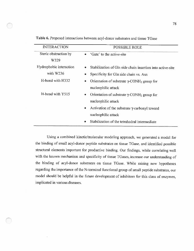
78
Table 6. Proposed interactions between acyl-donor substrates and tissue TGase
E’ffERACTION POSSIBLE ROLE
Steric obstruction by • Gate’ to the active-site
W329
Hydrophobie interaction • Stabilization of Gin side chain insertion into active-site
with W236 • Specificity for Gin side chain vs. Asn
H-bond with H332 • Orientation of substrate y-CONH2 group for
nucleophiiic attack
H-bond with Y515 • Orientation of substrate ‘-CONH2 group for
nucleophiiic attack
• Activation of the substrate y-carbonyl toward
nucleophilic attack
• Stabilization of the tetrahedral intermediate
Using a combined kinetic/moiecuiar modeiing approach, we generated a model for
the binding of small acyl-donor peptide substrates on tissue TGase. and identified possible
structural elernents important for productive binding. Our findings, while correiating weIi
with the known rnechanism and specificity of tissue IGases, increase our understanding of
the binding of acyl-donor substrates on tissue IGase. Whiie raising new hypotheses
regarding the importance of the N-terminal functional group of smali peptide substrates. our
mode! shouid be helpfui in the future development of inhibitors for this class of enzymes,
impiicated in various diseases.
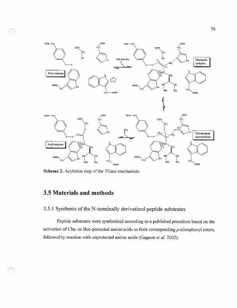
79
Y515—CH-
C272
H332/
CH2
NHNH
HC—W329
riihaeIiscomplex
H
H2CNW329
Tetrahedralintermediate
H
W329
3.5 Materials and metliods
3.5.1 Synthesis ofthe N-terrninaÏÏy derivatized peptide substrates
Peptide substrates were synthesized according to a published procedure based on the
activation of Cbz- or Boc-protected amino acids as their corresponding p-nitrophenyl esters,
folÏowed by reaction with unprotected amino acids (Gagnon et aï. 2002).
H332/
C272
CH2
SH
W236
CH1
J
Scheme 2. Acylation step ofthe TGase rnechanism.
$0
3.5.2 Kinetic measurements
Kinetic parameters were rneasured using a kinetic protocol previously developed in
our laboratory (de Macédo et al. 2000). According to this rnethod. N.N-dimethyl- 1.4-
phenylene diamine (DMPDA) is used as an acceptor substrate for the TGase-mediated
transamidation from Gin-containing donor substrates. For Cbz-Gln-Gly, the anilide
resulting from the transarnidation reaction with DMPDA has been shown to have an
extinction coefficient of $940 Mcm’ at 27$ nrn. Although the specific extinction
coefficients of each of the corresponding anilides formed from the other substrates used in
this study were flot determined. they were assumed to flot differ significantly. This was
corroborated by the observation that similar concentrations of different anilide products
gave similar absorbance values. and the kcat values shown in Table J were calculated based
on this assumption. A KM value was rneasured for each substrate using this method at 37 °C
and pH 7.0, over substrate concentrations that ranged from 0.2- to fivefold KM. By way of
comparison, a value of 3.2 mM was thus determined for the KM of Cbz-Gln-Gly, in
accordance with that determined in the literature by various rnethods (Folk and Chung
1985; Day and Keillor 1999: de Macédo et ai. 2000).
3.5.3 Computational methods
Computations were perforrned using the Insightll package (version 2000, Accelrys).
The BIOPOLYMER module was used to build or modify molecular structures. and all
energy minimizations and molecular dynamics calculations were performed with the
DISCOVER module using the consistent valence force field (CVFF). Interaction energies
between the enzyme and the ligands were calculated with the DOCKTNG module of
Insightll. Autornated docking was performed with AutoDock 3.0 software (Scripps).
$1
3.5.4 Preparation ofthe protein structure
The starting coordinates were taken from the PDB file 1GOD ofred sea bream tissue
transglutarninase (Noguchi et aÏ. 2001). The crystallographic water molecules as well as the
sulfate ion were removed. Hydrogen atoms were added using the BIOPOLYMER module at
the normal ionization state of the arnino acids at pH 7.0. Two dihedral angle torsions
involving hydrogen atoms were introduced in the protein structure in order to render it more
consistent with the catalytic process. The first involves the hydrogen atom of the hydroxyl
group of Y5 15. which was turned towards the suiphur atom of the catalytic C272. This
torsion was introduced according to the suggested existence of a regulatory H-bond between
the catalytic Cys and Y515 of red sea bream tissue TGase (Noguchi et aÏ. 2001). The
second involves the hydrogen atom of the thiol group of C272. which was turned towards
N ofH332. This torsion was introduced according to the knowncatalytic mechanism ofthe
enzyme in which the general acid-base catalyst, H332, deprotonates the catalytic Cys
residue prior to or during catalysis (Case and Stem 2003). Energy minirnization of the
added hydrogen atoms was then performed to remove bad contacts in the protein structure.
One thousand steps of steepest descents energy minimization were perforrned, followed by
a conjugate gradients energy minimization until convergence of 0.01 kcal/mole!À. Both
energy minirnizations were performed while keeping the heavy atoms of the protein fixed
and using a dielectric constant of 1. This generated the structure gOd300.
3.5.5 Computational construction of substrate molecules
Substrates were built using fragments from the BIOPOLYMER module fragment
library. The resulting structures were energy-minimized using 1000 steps of steepest
descents energy minirnization followed by a conjugate gradients energy minimization until
convergence of 0.01 kcal/rnole/À with a distance-dependent dielectric constant of $0 to
mimic implicit water. These resulting molecules served as starting structures for the
docking experiments.
$2
3.5.6 Automated docking ofsubstrates on TGase
AutoGrid was used to generate geornetry-centered grid maps with 0.375 À spacing
based on a region that contained ail residues including atoms within a 15 À radius centered
about the catalytic suiphur atom in the structure gOd300. A distance-dependent dielectric
constant of 4.0 was used to mimic the interior of the protein. The genetic algorithm
implemented in AutoDock 3.0 (Morris et aï. 199$) was appiied with a starting population
size of 50. fifty runs were performed for each ligand (Cbz-GIn-Gly and Boc-Gln-Giy) with
a maximum number of energy evaluations of 250,000 and a maximum number of
generations of 27,000. Auto-Tors was used to define the available torsions for the ligands.
However, we restricted the torsions of the Gln side chain (CŒ—C, C—C and C-—C) tO
define an extended conformation that maxirnizes its length. The torsions of the methyl
groups of the Boc functionai group were aiso deleted, as thev do not affect the structure of
the molecuie. There remained seven possible torsions for Cbz-Gin-Gly and six for
Boc-GÏn-Gly. Afier automated docking, the resuiting enzyme/substrate structures were
energy-minimized using the DISCOVER modu]e of Insightll. A simulation zone containing
ail residues including atoms within 20 À of the sulphur atom of the cataiytic cysteine
residue was used, with the remainder of the protein being fixed. An assembly was created
between the enzyme and the substrate, and a layer of 25 À of expiicit water was added
around the substrate. Ibis layer of water completeiy covered the active-site region. A
nonbond cutoff of ii À was used to speed up calculations. and a distance restraint was
applied to maintain the substrate’s reactive group (y-carboxamide group of Gin) at a
maximal distance of 3 À from the cataiytic S of C272 and N of H332. A dielectric
constant of 1 was applied. A multistage energy minimization was performed on the
enzyme/substrate compiex to further refine the compiex. One thousand iterations of steepest
descents energy minimization was performed, foiiowed by a conjugate gradients energy
minimization until convergence of 0.001 kcal/mole/À.
o-,Ô-,
3.5.7 Manual docking ofsubstrates on TGase
Manuai docking of the minimized substrates on IGase was performed as foiiows.
First, the reactive Gin side chain of the rigid substrates was inserted into the weli-known
active-site cavity(Yee et aÏ. 1994) perpendicuiariv to the center of mass oftheprotein so as
to maximize the depth of insertion whiie maintaining ail protein atoms fixed. The side chain
was piaced in a sterically unhindered position that is consistent with Gin side chain
insertion into the active-site cavity. Second, the distance between the reactive atoms of the
enzyme and the substrates (S of C272 with C of the Gin side chain and NE of Gin side
chain with N of H332) was kept at vaiues iower than 3 À. Third, the peptide backbone and
the N-terminai functional group of the substrates were placed in a stericaiiy unhindered
position on the surface of the enzyme, with the functionai group in one of the four tested
positions (Figure 2). Finaily, an assembiy was created between the enzyme and the
substrate, and a layer of either 25 À or 40 À of expiicit water was added around the
substrate. This iayer of water compietely covered the active-site region.
3.5.8 Calculations on the enzyrne/substrate complex
For ail reaction steps, a simuiation zone containing ail residues inciuding atoms
within 20 À of the suiphur atom of the cataiytic C272 residue was used. with the rernainder
of the protein being fixed. A nonbond cutoff of ii À was used to speed up caicuiations, and
a distance restraint was applied to maintain the substrate’s reactive group (y-carboxamide
group of Gin) ciose to the cataiytic residues at a maximal distance of 3 À from S- of C272
and N of H332. For the moiecular dynamics experiments, an additional restraint of
100 kcai/mole on the oxygen atoms of the water moiecules was added to prevent them from
“boiiing off” A dielectric constant of 1 was applied.
A multistage energy minimization was perforrned on the enzyme/substrate complex
to remove bad contacts between the substrate and the enzyme. First, 1000 iterations of
$4
steepest descents energy minimization was performed, followed by a conjugate
gradients energy minirnization until convergence of 0.001 kcal/rnole/À. This first energy
minimization was followed by a molecular dynamics simulation in which the molecular
system was allowed to equilibrate at 300 K for 1 ps. followed by the actual simulation to
explore conformational space for 10 ps whule maintaining the sarne temperature. Snapshots
were taken every 0.1 ps, generating 100 different conformers. The conformer with the
lowest energy was energy-minimized with 100 steps of steepest descents followed by a
conjugate gradients energy minimization to convergence of 0.00 1 kcal/mole/À. At this
point, the structures were analyzed.
3.5.9 Building of the acyl-enzyme intermediate and the tetrahedral
intermediate
As a starting point for the energy calculations, the acyl-enzyme and tetrahedral
intermediates were built using the BIOPOLYMER module. While building these
molecules, we introduced torsions at the l, 2. and dihedral angles of the catalylic
C272 and the reactive Gln of the substrate. These angles (xl = -30° and 2 = -60° for Cys.
xl = -160°, 2 = 180°, and = 0° for Gln) were selected from published I—2 and2—X3
plots for amino acid side chains (Janin et aÏ. 1978). The angles allowed the positioning of
the N-terminal functional group in position O as defined in Figure 2, whereas the angles
xl = -45° and 2 = -45° for Cys, l = 180°. 2 = 180°, and x3 = 180° for Gln allowed the
positioning of the N-terminal ftinctional group in position 2. For the tetrahedral
intermediate. the catalytic His residue was protonated, a formal charge of- 1 was introduced
at the oxyanion, and the partial charges were calculated using the CVfF force fleld. These
molecules were then minimized and subjected to a molecular dynamics simulation in the
same manner as the enzyme/substrate intermediates.
85
3.6 Acknowledgments
We thank Dr. Andreea Schrnitzer for assistance in molecular modeling. R.A.C. and
P.G. were recipients of Fonds Québécois de recherche sur la nature et les technologies
(FQRNT) fellowships. This research was funded by FQRNT Grant 80357 (jointly held by
J.N.P. andJ.W.K.).
3.7 References
Abe, S.. Yamashita. K.. Kolmo. H., and Ohkubo, Y. 2000. Involvement oftransglutaminase
in the receptor-mediated endocytosis of mouse peritoneal macrophages. BioÏ.
Pharrn. BulÏ. 23: 1511—1513.
Aeschlimairn, D. and Paulsson, M. 1994. Transglutarninases: Protein cross-Iinking enzymes
in tissues and body fluids. Thromb. Haemost. 71: 402—415.
Ahvazi, B.. Kim. H.C., Kee, S.H.. Nemes. Z.. and Steinert. P.M. 2002. Three-dirnensional
structure ofthe human transglutaminase 3 enzyme: Binding of calcium ions changes
structure for activation. EMBOJ 21: 2055—2067.
Case, A. and Stem, R.L. 2003. Kinetic analysis ofthe action of tissue transglutarninase on
peptide and protein substrates. Biochemistiy 42: 9466—948 1.
Chen, J.S. and Mehta, K. 1999. Tissue transglutarninase: An enzyme with a spiit
personality. Int. J Biochem. Ccli Biol. 31: 8 17—836.
Clarke, D.D., Mycek, M.J.. Neidie, A.. and Waelsh, H. 1959. The incorporation of amines
into proteins. Arch. Biochem. Biophys. 79: 338—354.
Day. N. and Keillor. J.W. 1999. A continuous spectrophotometric linked enzyme assay for
transglutaminase activity. Anal. Biochem. 274: 141—144.
de Macédo, P., Marrano, C., and Keillor, J.W. 2000. A direct continuous
spectrophotometric assay for transglutarninase activity. Anal. Biochem. 285: 16—20.
Fesus, L. and Piacentini, M. 2002. Transglutaminase 2: An enigmatic enzyme with diverse
functions. Trends Biochem. Sel. 27: 534—539.
$6
Folk, J.E. 1969. Mechanism of action of guinea pig liver transglutarninase. VI. Order of
substrate addition. I Bio!. Chem. 244: 3707—3713.
1983. Mechanism and basis for specificity of transglutaminase-catalyzed epsilon
(7-glutamyl) lysine bond formation. Adv. Enzvmol. ReÏat. Areas Mol. Bio!. 54: 1—56.
Folk. i.E. and Chung, S.I. 1973. Molecular and catalytic properties of transgÏutaminases.
Adv. EnzymoÏ. ReÏat. Areas Mol. Biol. 3$: 109—191.
1985. Transglutarninases. Methods EnzymoÏ. 113: 358—375.
Folk, i.E. and Cole, P.W. 1965. Structural requirernents of specific substrates for guinea
pig liver transglutarninase. I Bio!. Chem. 240: 2951—2960.
1966. Transglutaminase: Mechanistic features of the active-site as determined by
kinetic and inhibitor studies. Biochim. Biophys. Acta 122: 244—264.
Gagnon, P., Huang, X.. Therrien. E., and Keillor. J.W. 2002. Peptide coupling of
unprotected amino acids through in situ p-nitrophenyl ester formation. Tetrahedron
Let!. 43: 7717—7719.
Huo, J., Metz, S.A., and Li. G. 2003. Role of tissue transglutaminase in GTP depletion
induced apoptosis of insulin-secreting (HIT-T 15) ceils. Biochem. Pharmacol. 66:1’7. I 3—LL..
Janin, J., Wodak. S., Levitt, M.. and Maigret, B. 197$. Conformation of amino acid side
chains in proteins. I Mc)!. Bio!. 125: 357—386.
Kim, S.Y., Grant, P., Lee, J.H., Pant, H.C., and Steinert, P.M. 1999. Differential expression
of multiple transglutaminases in human brain. Increased expression and cross
linking by transglutaminases 1 and 2 in Alzheimer’s disease. I Bio!. Chem. 274:
30715—30721.
Liu, S., Cerione, R.A., and Clardy, J. 2002. Structural basis for the guanine nucleotide
binding activity of tissue transglutaminase and its regulation of transarnidation
activity. Froc. Nati. Acad. $ci. 99: 2743—2747.
87
Morris, G.M., Goodsell, D.S.. Halliday, R.S., Huey, R., Hart, W.E., Belew, R.K., and
Oison, A.J. 1998. Automated docking using a iarnarckian genetic algorithrn and an
empiricai binding free energy function. I Comput. Chem. 19: 1639—1662.
Murthy, S.N.P.. Iismaa. S.. Begg. G.. Freyrnann. D.M.. Graham. R.M., and Lorand, L.
2002. Conserved tryptophan in the core dornain of transglutaminase is essential for
catalytic activity. Froc. Na!!. Acad. Sel. 99: 2738—2742.
Noguchi, K., Ishikawa, K., Yokoyama, K., Ohtsuka, T., Nio, N., and Suzuki, E. 2001.
Crystal structure of red sea bream transglutarninase. I Biol. Chem. 276: 12055—
12059.
Ohtsuka, T.. Ota. M., Nio. N.. and Motoki. M. 2000. Comparison of substrate specificities
of transglutarninases using synthetic peptides as acyi donors. Biosci. BiotechnoÏ.
Biochem. 64: 2608—2613.
Priglinger, S.G., May, C.A.. Neubauer. A.S., Alge, C.S., Schônfeld, C.L., Karnpik, A., and
Welge-Lussen, U. 2003. Tissue transgiutarninase as a modifying enzyme of the
extracellular matrix in PVR membranes. Invest. OphthaÏmol. Vis. Sel. 44: 355—364.
Shan. L.. Molberg. 0., Pan-ot, I., Hausch. f., filiz. f., Gray. G.M.. Sollid, L.M., and
Khosla, C. 2002. Structural basis for gluten intoierance in ceiiac sprue. Science 297:
2275—2279.
Shridas, P., Sharma, Y., and Balasubrarnanian, D. 2001. Transgiutaminase-mediated cross
linking of x-crystallin: Structural and functionai consequences. FF35 Lett. 499:
245—250.
Singh, U.S.. Pan, J.. Kao. Y.L., Joshi. S.. Young. K.L.. and Baker, K.M. 2003. Tissue
transglutarninase mediates activation of RhoA and MAP kinase pathways during
retinoic acid-induced neuronal differentiation of SH-SY5Y cells. I Biol. Chem.
27$: 391—399.
Wada, F., Nakamura, A., Masutani, T., Ikura, K., Maki, M., and Hitomi, K. 2002.
Identification of mammalian-type transglutaminase in Physarum polycephalum.
82
Evidence from the cDNA sequence and involvernent of GTP in the regulation of
transamidating activity. Eur. I Biochem. 269: 345 1—3460.
Yee, V.C., Pedersen, L.C., Le Trong, I., Bishop, P.D.. Stenkamp, R.E., and Teller, D.C.
1994. Three-dimensional structure of a transglutaminase: Human blood coagulation
factor XIII. Froc. NatÏ. Acad Sci. 91: 7296—7300.
CHAPITRE 4
Développement d’une méthode d’expression et de
purification de la TGase de foie de cobaye chez
Esciterichia coil
90
4.0 Préface
Afin de pouvoir développer les méthodes expérimentales nécessaires à ce projet, il
nous fallait une source de TGase de foie de cobaye recombinante. Pour ceci, nous avons
conçu notre propre protocole d’expression et de purification de l’enzyme chez Escherichia
cou. Lors du développement de ce protocole. nous avons dû nous attaquer au problème de
faible solubilité intrinsèque de la TGase de foie de cobaye lorsqu’elle est exprimée chez E.
cou. Ce protocole nous a permis d’obtenir rapidement. efficacement et avec un bon
rendement la TGase de foie de cobaye soluble ayant l’activité spécifique la plus élevée
rapportée dans la littérature. De plus. le fait de l’exprimer de manière recombinante offre la
possibilité d’effectuer des manipulations génétiques. Ce protocole a fait l’objet d’un article.
publié en 2004, intitulé « Expression and rapid purification of highly active hexahistidine
tagged guinea pig liver transglutaminase ». qui est présenté au chapitre 4.
Pour cet article, j’ai préparé et séquencé le vecteur d’expression pour le gène de la
transglutaminase par biologie moléculaire en plus de participer à l’optimisation de
l’expression et de la purification de l’enzyme recombinante avec le Dr Steve Gillet. un
chercheur postdoctoral que j’ai formé en lui enseignant les techniques de biologie
moléculaire et de biochimie utilisées dans le laboratoire. Le Dr Steve Gillet a effectué
toutes les expériences de cinétique enzymatique et a rédigé majoritairement le manuscrit, à
l’exception des sections rapportant les méthodes et les résultats de la construction du
vecteur pQE32-TGase, dont j’ai été l’auteur.
91
Article 3.
Expression and rapid pnrification of highly active
liexahistidine-tagged guinea pig lïver transglutaminase
Steve M. f. G. Gillet, Roberto A. Chica, Jeffrey W. Keillor and JoeÏÏe N.
Pelletier
Département de Chimie, Université de Montréal, C.P. 6128, Succursale Centre-Ville.
Montréal. Québec. Canada H3C 3J7
Protein Expression anti Purification, 2004, 33, 256-264
“Reprinted from Protein Expression and Purificatio,7. Vol 33, Steve M. F. G. Gillet.
Roberto A. Chica, Jeffrey W. Keillor and Joelle N. Pelletier, “Expression and rapid
purification of highly active hexahi stidine-tagged guinea pig liver transgIutaminase’. pages
256-264, Copyright (2004), with permission from Elsevier”.
92
4.1 Abstract
Tissue transglutarninase bas been identified as a contributor to a wide variety of
diseases, including cataract formation and Celiac disease. Guinea pig tissue
transglutarninase has a very broad substrate specificity and therefore is useful for kinetic
studies using substrate analogues. Here. we report the expression in Escherichia cou of a
hexahistidine-tagged guinea pig liver tissue transglutaminase (His6-tTGase) allowing rapid
purification by imrnobilized-metal affinity chromatograpby. Using this procedure we have
obtained the highest reported specific activity (17 U/rng) combined with a high yield
(22 mg/L of culture) for recombinant TGase using a single-step purification protocol. Using
two independent spectrophotometric assays, we detennined that the KM value of the
recombinant enzyme with the substrate Cbz—Gln—Gly is in the same range as values
reported in the literature for the native enzyme. We have thus developed a rapid and
reproducible protocol for the preparation of high quality tissue TGase.
Author Keywords: Transglutaminase: Hexahistidine tag; Chaperone; IMAC;
Betaine
4.2 Introduction
Transglutaminases (amine y-glutamyltransferases, EC 2.3.2.13) are a family of
enzymes that catalyse Ca2tdependent acyl transfer reactions. Catalytic activity is exhibited
toward the y-carboxamide moiety of a protein or peptide-bound glutarninyl residue, which
serves as acyl donor to eitber protein or peptide-bound lysyl residues or free amines.
resulting in new intermolecular crosslinks [1. 2 and 3]. Tissue transglutaminase (TGase) is
widely distributed in animal tissues including liver. kidney, lung. spleen, brain. and the
endothelial and smootb muscle cells of arteries. veins, and capillaries [4].
( -
‘ii
Tissue transglutaminase bas been identified as a contributor to cataracts and Celiac
disease, and a growing body ofevidence suggests that it may be involved in atherosclerosis.
inflammation, fibrosis. diabetes. cancer metastases, autoimmune diseases, lamellar
ichthyosis. and psoriasis (for a review. see [5]). TGase is also suspected to play a role in
neurodegenerative diseases (such as Huntington s disease, Alzheimer’ s disease,
Parkinson’s disease. and supranuclear palsy) associated with an increase in polyglutamine
containing peptides in the brain [6. 7 atid 8].
Tissue TGase has been purified from many sources, including mammalian livers, rat
testicular tissue, and human erythrocytes t91. Currently, the most widely cited source of
native TGase for research is guinea pig liver, due to its rich abundance of endogenous
TGase in comparison to other sources ofmarnrnalian liver [10]. As much as 25 mg can be
obtained from 140 g of guinea pig liver [11]. with a specific activity of 1$ U/mg. up to 6- to
I 0-fold higher than the typical commercial native guinea pig tissue TGase (Sigma). As the
guinea pig liver TGase shov’s $8 % sequence homology ($0 % sequence identity) at the
amino acid level (using standard protein—protein BLAST) with human tissue TGase, its use
in medical research is pertinent [12].
The establishment of an efficient expression system of the recombinant mammalian
tissue-type transglutaminase would allow the enzyme to be obtained in the high yield and
purity that are essential to the structure/function studies required for mechanistic studies
and the subsequent development of mechanism-based inhibitors. Since tissue TGases
require no post-translational modification for activity. [13] they are a good target for
recombinant expression in bacterial celis. Recombinant human endothelial, guinea pig liver
and red sea bream (Pagrus major) tissue transglutarninases have been expressed and
purified [14, 15 and 16]. Expression in Eseherichia cou and purification of hexahistidine
tagged human tissue transglutaminase have recently been reported by Shi et al. [17]. The
specific activity reported in the literature for the recombinant human transglutaminases was
approximately 2- to 4-fold lower than that of the commercial native guinea pig tissue
94
IGase (Sigma) and the quantity of enzyme obtained per litre of bacterial culture was in the
range of 0.6 mg [17 and 18]. The activity ofthe recombinant guinea pig liver tissue TGase
expressed in E. cou obtained by Ikura et aÏ. [19] was approximately 7-fold lower than that
of the native guinea pig liver tissue TGase, but the quantity of enzyme produced per litre of
culture was in the range of 37.5 mg/L. This very high yield indicates the pertinence of
pursuing expression in E. cou. although improvement of specific activity is essential to our
research.
We sought to combine the practical nature of a hexahistidine tag with an improved
yield of soluble enzyme to obtain high yields of recombinant guinea pig liver TGase with
specific activity that should at Ieast approach that reported for the highly purified native
enzyme [li]. Herein, we report two rnethods for expression in E. cou of guinea pig liver
tissue transglutaminase with a hexahistidine tag at the N-terminus allowing rapid
purification by immobilized-rnetal affinity chromatography (IMAC).
The first method, based on that of Yokoyarna et aÏ. [16], relies on the chaperones
DnaK and DnaJ to achieve a high yield of soluble protein. The second method relies on the
use of betaine. a chemical chaperone. which bas been sbown to improve the soluble yield
during recombinant protein expression [20]. Using our rnodified procedure we have
obtained for the first time a hexahistidine-tagged tissue IGase with the best specific
activity (17 U/mg) combined with a high yield (22 mg/L of culture) for recombinant TGase
using a single-step purification protocol.
4.3 Materials ami methods
4.3.1 Materials
All reagents used were of the highest available purity. Glutamate dehydrogenase
(GDH) was purchased from Roche, restriction enzymes and DNA-modifying enzymes were
from New England Biolabs and MBI Fermentas. Ihe Sequenase 2.0 DNA sequencing kit as
95
weII as the dJTP Nucleotide kit were purchased from Amersham Biosciences. Synthetic
oligonucleotides were obtained from A1phaDNA, the infra-red dye labelled sequencing
prirners were from Li-Cor Biotechnology Division. and QIAFXII gel extraction kit and
Ni-NTA agarose resin were obtained from Qiagen. Ail aqueous solutions were prepared
using water purified with a Millipore BioCeil system.
4.3.2 Construction of expression plasmid
The guinea pig liver transglutaminase gene was PCR-arnplified from plasrnid
pKTG1 t131 with the prirners $phIGTGaseF-Met 5’-(CACACAGCATGCAGGCAG-
AGGATCTGATCCTG), which airneals to the 5’-end ofthe coding region and introduces a
unique SphI restriction site (underlined) while removing the start codon. and
pKTGI_GTGaseR 5-(ATCTTCTCTCATCCGCCAAA). aimealing —20 bp downstrearn
from the stop codon and the PstI restriction site, which imrnediately follows it. The PCR
product was digested by FstI/$phl and purified by agarose gel electrophoresis followed by
extraction with a QIAEXII kit (Qiagen). The TGase gene was inserted in pQE32 expression
vector (Qiagen) via $phl/FstI yielding the plasrnid pQE32-TGase. This plasrnid encodes an
N-terminal His6-tag (underlined) followed by a five amino acid iinker sequence
(MRGSHHHHHHGIRMQ) preceding the TGase, resulting in a 704 amino acid protein
with a calculated molecular weight of 78.7 kDa. The plasmid was then transfornEjed into E.
cou XL-1 Blue ceils or E. cou XL-] Blue ceils containing the plasmid pDnaKJ [16]. which
encodes the molecular chaperones DnaK and DnaJ. The entire SphI/PstI fragment,
including the whole coding region, was verifled by DNA sequencing.
4.3.3 Sequence analysis
DNA sequencing was performed using a dITP Nucleotide kit for Sequenase version
2.0 DNA polymerase (Amersharn—Pharmacia Biotech) with a Li-Cor 1R2 autornated
system.
96
4.3.4 Overexpression of TGase
4.3.4.1 Method A: expression with molecular chaperones DnaK and DnaJ
E. cou. ceils from frozen stock. harbouring the expression plasmids pQE32-TGase
and pDnaKJ (expressing both DnaK and DnaJ chaperones; [16]) were grown overnight in
50 mL LB medium containing 100 tg/mL ampicillin for maintenance ofpQE32-TGase and
30 ig/rnL chloramphenicol for maintenance of pDnaKJ. A portion (20 mL) of the bacterial
suspension was then inoculated into 400 mL of fresh LB medium and the ceils were
incubated at 37 oc with agitation (250 rprn). When 0D600 reached approximately 0.6, the
expression of His6-tTGase was induced by addition of IPTG to a final concentration of
1 mM. The culture was incubated further for 20 h at 2$ °c with shaking at 240 rpm.
4.3.4.2 Method B: expression with chemical chaperone betaine
E. cou ceils. from frozen stock. harbouring the expression plasmid pQE32-TGase
were grown ovemight in 50 mL LB medium containing 100 tg/rnL ampicillin. A portion
(20 mL) of the bacterial suspension was then inoculated into 400 mL of fresh LB medium
containing 1 M sorbitol and 2.5 mM betaine and induction of His6-tTGase expression was
undertaken as in Method A.
Ail of the following procedures were carried out at 0—4 °c. Affer induction of His6-
tTGase, the bacterial suspension was centrifuged at lSOOg for 30 min. The celi pellet was
resuspended in $ mL of extract buffer (0.1 M potassium phosphate buffer, pH 8.0.
containing 5 mM imidazole) and disrupted by sonication (three pulses of 200 W for 30 s
with a tapered rnicro-tip). The celi debris were removed by centrifugation at 2000g for
30 min. The supernatant fraction obtained was used as the ceil extract.
97
4.3.5 Purification of His6-tTGase
Ail procedures were carried out at 0—4 °C. b the ce!! extract, 500 tL Ni—NTA
agarose, equilibrated with extract buffer, was added and rnixed gently (100 rprn on a rotary
shaker) for 60 min. The lysate-Ni—NTA mixture was loaded into a colunrn (Bio-Rad Poly
Prep Chromatography Columns 0.8 x 4 cm). The flow-through was collccted and the
column was washed successively with 6 mL of extract buffer, 6 mL of washing buffer A
(0.1 M potassium phosphate buffer, pH 8.0, containing 10 mM irnidazole), and 6 mL of
washing buffer B (0.1 M potassium phosphate buffer, pH 8.0, containing 20 mM
imidazole). The column was e!uted with 4 mL elution buffer (0.1 M potassium phosphate
buffer, pH 7.5. containing 250 mM imidazole). The eluant was transferred to an Econo-Pac
10-DG column (Bio-Rad) and the tissue TGase was desalted by elution with 10 mM Mops
buffer (pH 7.0). Aliquots of this purified eluant were assayed for tissue TGase activity as
describcd above. Fractions with specific activities higher than 10 U/rng were pooied. The
solution was then aliquoted (each fraction containing approximately 500 tg of enzyme)
into microcentrifuge tubes and flash-frozen on dry ice for 5 min. The samples were
lyophilized using a Labconco Freeze Dry System and stored at —20 °C.
4.3.6 Protein concentrations
Concentrations of total protein were generally detennined using the Bio-Rad protein
assay, a method based on the Bradford assay [21], using BSA as a standard. Colour
developrnent took place over 15 min and absorbance values were measured using a Varian
Cary 100 Bio. Standard curves were perfonned daily. For fractions obtained afier elution
from the Ni—NTA colurnn. in which TGase was considered to be of high purity, protein
concentration was determined using the extinction coefficient for native TGase
(A28910”°=15.8) [22].
98
4.3.7 Determination of specific activity
IGase activity was measured by the colorimetric hydroxamate assay procedure
using 30 mM benzyloxycarbonyl-L-glutaminylglycine, 1 mM EDIA, 5 mM CaC12, and
0.1 M hydroxylamine in 0.2 M Tris—acetate at pH 6.0 and 37 °C [23]. The activity assay
solution was prepared fresh daily. One unit of enzyme activity (U) is defined as the amount
of TGase. which catalyses the formation of 1 tmol of y-glutarnylhydroxamic acid
per minute. Specific activity is given as units per milligram ofprotein.
4.3.8 Electrophoresis
Separation of proteins by sodium dodecyl sulphate—polyacrylarnide gel
electrophoresis (SDS—PAGE) was conducted using the fisher scientific electrophoresis cell
according to procedures of Laernmli [24] using minigels (105 x $5 mm dimensions)
prepared with 9 % acrylamide resolving gel at pH 8.8, 4 % acrylamide stacking gel at
pH 6.8. and 2.7 % crosslinker concentrations. The protein sample (10 1iL) was mixed with
10 tL of loading buffer [50 mM Tris—Hcl (pH 6.8). 100 mM DTT. 2% w/v SDS, 0.1% w/v
bromophenol blue, and 10% v/v glycerol] and boiled for 5 min prior to loading. Protein
bands were revealed using 250R coomassie blue staining technique [25]. coonassie blue
stained gels were dried. The SDS—PAGE broad-range molecular weight rnarkers (Bio-Rad)
were used as standards.
4.3.9 Enzyme purity
The dried gels were scanned using a flatbed document scanner at 400 dpi resolution.
The scanned images were analysed using the program Scion Image for Windows (Scion
cooration), which has extensive image analysis capabilities for one-dimensional
electrophoretic gels. The background was removed by using the ]D Vertical Subtract
Background command. We used the macros Gelplot2 to plot the lanes profiles. Each peak
99
was manually delimited on the baseline with the une drawing tool and then measurements
ofthe areas were taken with the Azitomatic Oullining Tool.
4.3.10 Kinetic assays
4.3.10.1 Method I: DMPDA assay
The kinetic properties of His6-tTGase were deterrnined using a continuous
spectrophotometric assay [26]. Ail assays were perforrned in triplicate using six different
concentrations of donor substrate ranging from 0.1 to 3 KM. Nonspecific reactivity was
observed above these concentrations. A standard stock buffer solution was prepared by
combining 12 mL of 1 M Tris—acetate buffer (pH 7.0), 3 mL of 0.1 M CaCb. 3 mL of
0.02 M EDTA. and 48 mL ofwater. To 340 1iL ofthis stock solution were added 20 tL of
20 mM N.N-dimethyl-1.4-phenylenediarnine (DMPDA), 25—120 pi of 5 or 50 mM
Cbz-Gln-Gly and the final volume was adjusted to 480 jiL through the addition of 0—95 tL
ofwater. The reaction solution was preincubated for 3 min at 37 oc prior to initiation ofthe
enzymatic reaction upon addition of 20 j.tL (---$jig) of purified His6-tTGase, giving a final
volume of 500 tL. Reaction progress was followed as a linear increase of absorbance
measured at 27$ iirn in a Varian Cary 100 spectrophotometer thermostated at 37 °c. For the
blank reactions. 20 pi of water was substituted for TGase enzyme. Siopes were rneasured
over the first <10 % substrate conversion to product and were converted into initiai rates by
dividing by the extinction coefficient of the authentic reaction product, carbobenzyloxy-L
glutarnyl--y-[4-(dimethylamino)anilido]glycine (8940 + 55 M cm1) [261.
4.3.10.2 Method II: glutamate dehydrogenase-linked assay (GDH)
Kinetic pararneters were determined using a continuous spectrophotometric linked
enzyme assay [27]. Ail assays were performed in tripiicate using seven different
concentrations of donor substrate ranging from 0.1 to 3 KM. Nonspecific reactivity was
observed above these concentrations. A standard stock buffer solution of 400 mM Mops
100
buffer (pi-1 7.0). 10 mM CaC12, and 2 mM EDTA was prepared. To 250 jiL of this stock
solution were added 20 1tL of 12.5 mM NADH. 20 jiL of 250 mM Œ-ketoglutarate. 20 tL
ofGDH (2.4 U), 20 tL of 83.5 mM N-acetyl lysine methyl ester, 25—1 20 IIL of 5 or 50 mM
Cbz—Gln—Gly and the final volume was adjusted to 480 jiL through the addition of
30-125 iL of water. The reaction solution was preincubated for 3 min at 37 °C prior to
initiation of the enzymatic reaction upon the addition of 20 jiL (—8 tg) of His6-tlGase,
giving a final volume of 500 jiL. for the corresponding blank solution. the TGase solution
was replaced by water. The decrease in absorbance due to the oxidation of NADH was
followed against a blank at 340 mii in a Varian Cary 100 spectrophotorneter therrnostated at
37 °C. Afier an initial lag, linear slopes of absorbance versus tirne were rneasured over the
first <10 % of substrate conversion to product and were converted into initial rates by
dividing by the extinction coefficient ofNADH (6220 M’ cm1) [27].
4.4 Results and discussion
4.4.1 DNA sequencing
DNA sequencing using the automated Li-Cor system revealed a silent mutation at
residue Arg76 (CGC instead of CGG) relative to the published sequence of the guinea pig
liver TGase gene [28]. Other mutations relative to the published sequence [28] were also
detected. A two base pair inversion (TC to CI) in adjacent nucleotides from codons
encoding residues Prol4l and Arg142 was observed resulting in a suent mutation at the
codon encoding Pro 141 (CCC instead of CET) and a missense mutation at the codon
encoding Arg 142 (IGG instead of CGG), which results in a Arg to Trp mutation. Trp may
be the correct residue at this position. considering that all eukaryotic TGases that have been
sequenced encode a Trp residue at this position. A second inversion of two base pairs was
detected in the codon encoding residue Arg436 (CAG instead of CGA), which resuits in a
Arg to Gin mutation. While Arg436 exists in other sequences, Gln is found in TGase 1
101
from Canis /nniÏiaris at the corresponding position. It is unclear if the G1n436 sequenced
here represents the correct native residue. a mutation or a polyrnorphism.
4.4.2 Expression and purification of His6-tTGase
Since large amounts of highly active tissue TGase are required for structure/activity
studies or for mutation of the enzyme. the establishment of an efficient expression system
of the recombinant mammalian tissue-type TGase was needed. We report the development
of a rapid and easy one-step purification protocol for the reproducible preparation of tissue
TGase with a good yield and high purity. Furtherrnore, the presence ofthe hexahistidine tag
permits the use of a common purification protocol should mutated enzymes be produced.
The E. cou ceils harbouring the expression plasmid pQE32-TGase were initially
tested for production of the His6-tagged guinea pig liver transglutaminase in the absence of
additional factors prornoting protein folding (Figure 1, lanes 1—3). SDS—PAGE analysis of
the supernatant and cell pellet following cell lysis by sonication showed a bold protein band
migrating slightly more slowly than the native guinea pig liver TGase (as a result of the
additional hexahistidine tag (resuits not shown)). Whule the transglutaminase band was
detected in the soluble fraction, the rnajority of the expressed enzyme was present in the
insoluble fraction. first, we attempted to convert the inclusion bodies to soluble, active
enzyme, beginning with denaturation of the inclusion bodies in urea or guanidinium
chloride. However, ail attempts at refolding by immobilized metal-ion affinity
chrornatography (Ni—NTA) while applying a decreasing gradient of guanidinium chloride.
urea or pH [29 and 30] with or without 5% polyethylene glycol t19] failed to regenerate
active enzyme from the inclusion bodies (data not shown).
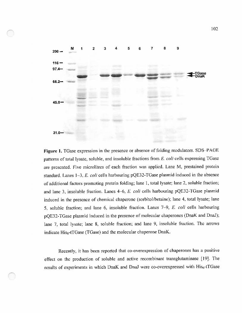
102
M 1 2 3 4 5 6 7 8 9200 —
116—
974—TGaseDnaK
66.2—
45.0—
31.0—
Figure 1. TGase expression in the presence or absence of folding modulators. SDS—PAGE
patterns of total lysate, soluble, and insoluble fractions from E. cou ceils expressing IGase
are presented. Five microlitres of each fraction was applied. Lane M. prestained protein
standard. Lanes 1—3, E. cou celis harbouring pQE32-TGase plasmid induced in the absence
ofadditional factors promoting protein folding; lane 1. total lysate; lane 2. soluble fraction;
and lane 3, insoluble fraction. Lanes 4—6, E. cou ceils harbouring pQE32-TGase plasmid
induced in the presence of chernical chaperone (sorbitol/betaine); lane 4, total lysate; lane
5, soluble fraction; and lane 6. insoluble fraction. Lanes 7—9. E. cou celis harbouring
pQE32-TGase plasmid induced in the presence ofrnolecular chaperones (DnaK and DnaJ);
lane 7, total lysate; lane 8, soluble fraction; and lane 9, insoluble fraction. The arrows
indicate His6-tTGase (TGase) and the molecular chaperone DnaK.
Recently, it has been reported that co-overexpression of chaperones has a positive
effect on the production of soluble and active recombinant transglutaminase [19]. The
resuits of experiments in which DnaK and DnaJ were co-overexpressed with His6-tlGase
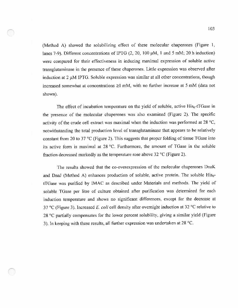
103
(Method A) showed the solubilizing effect of these molecular chaperones (Figure 1,
lanes 7-9). Different concentrations of IPTG (2. 20, 100 jiM, 1 and 5 mM; 20 h induction)
were cornpared for their effectiveness in inducing maximal expression of soluble active
transglutaminase in the presence of these chaperones. Littie expression was observed after
induction at 2 tM IPTG. Soluble expression was similar at ah other concentrations, though
increased somewhat at concentrations 1 mM. with no further increase at 5 mM (data not
shown).
The effect of incubation temperature on the yield of soluble, active His6-tTGase in
the presence of the molecular chaperones was also examined (Figure 2). The specific
activity of the crude ecU extract was maximal when the induction was perforn-ied at 2$ °C,
notwithstanding the total production level of transglutaminase that appears to be relatively
constant from 20 to 37 °C (Figure 2). This suggests that proper folding of tissue TGase into
its active forrn is maximal at 2$ °C Furtherrnore, the arnount of TGase in the soluble
fraction decreased rnarkedly as the temperature rose above 32 oc (Figure 2).
The results showed that the co-overexpression of the molccular chaperones DnaK
and DnaJ (Method A) enhances production of soluble, active protein. The soluble His6-
tlGase was purified by IMAC as described under Materials and methods. The yield of
soluble TGase per litre of culture obtained afier purification was determined for each
induction temperature and shows no significant differences. except for the decrease at
37 °C (Figure 3). Increased E. cou ceil density afier overnight induction at 32 oc relative to
2$ °C partially compensates for the lower percent solubility. giving a similar yield (Figure
3). In keeping with these resufts, ail further expression was undertaken at 2$ °C.
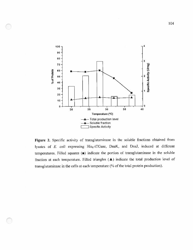
D
2>o
o
oG,D.
90
4
3
60 u—•-
20
10
o
104
Temperature (°C)
 Total production level• Soluble fraction
I Specific Activity
Figure 2. Specific activity of transglutaminase in the soluble fractions obtained from
lysates of f. cou expressing His6-tlGase, DnaK, and DnaJ, induced at different
temperatures. Filled squares (u) indicate the portion of transglutarninase in the soluble
fraction at each temperature. FilIed triangles (Â) indicate the total production level of
transglutarninase in the cells at each temperature (% of the total protein production).
20 25 30 35 40o
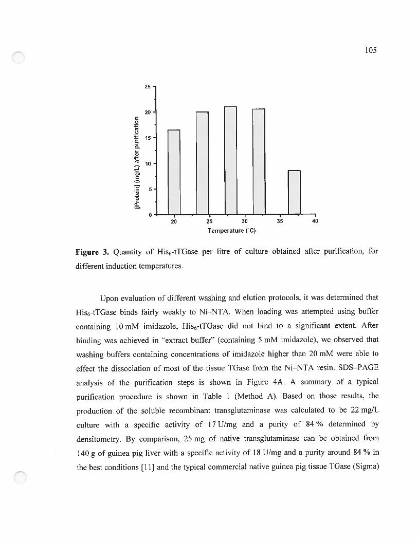
105
25
20
o
Q
‘ 15
o.G)
10-J
E
.E 5Q
oL.
o
0-— r —‘- — — . —r— i
20 25 30 35 40
Temperature (C)
Figure 3. Quantity of His6-tTGase per litre of culture obtained afler purification, for
different induction temperatures.
Upon evaluation of different washing and elution protocols. it was determined that
His6-tTGase binds fairly weakly to Ni—NTA. When loading was attempted using buffer
containing 10 mM imidazole, His6-tTGase did flot bind to a significant extent. Afier
binding was achieved in “extract buffer” (containing 5 mM imidazole), we observed that
washing buffei-s containing concentrations of imidazole higher than 20 mM were able to
effect the dissociation of most of the tissue TGase from the Ni—NTA resin. SDS—PAGE
analysis of the purification steps is shown in Figure 4A. A summary of a typical
purification procedure is shown in Table 1 (Method A). Based on those resuits, the
production of the soluble recombinant transglutaminase was calculated to be 22 rng/L
culture with a specific activity of 17 U/rng and a purity of $4 % determined by
densitometry. By comparison. 25 mg of native transglutaminase can be obtained from
140 g of guinea pig liver with a specific activity of 1$ U/mg and a purity around $4 % in
the best conditions [111 and the typical commercial native guinea pig tissue TGase (Sigma)
106
bas a specific activity of 2-3 U/mg. Two principal impurities in the elution fraction were
observed by SDS—PAGE, one migrating at approxirnately 70 kDa (Figure 4A: irnrnediate!y
below His6-tTGase) and the second at approxirnately 66 kDa. In Figure 5. the elution
fraction of the contro! purification of ceils expressing DnaK and DnaJ but flot the His6-
tTGase (lane 2) shows the same band that migrates near 70 kDa. This is apparently due to
non-specific binding of DnaK (Mr 72.000) on the Ni—NTA resin as evidenced by the
absence of this impurity in the elution fraction of celis lacking pDnaKJ plasmid (lanes 3
and4).
A B1 2 3456 789M 1 2 34 5 6 789M
—200 —200
—116- —116
—97.4 - —97.4
— TGase_. —66.2
-45.0 -45.0
—31.0
figure 4. SDS—PAGE of fractions obtained during the purification. Expression with
molecu!ar chaperones DnaK (70 kDa) and DnaJ (40 kDa) (A) or with chemical chaperone
(betaine) (B). Ten microlitres of each fraction was applied. For both panels, lane M,
prestained protein standards; !ane 1. negative control: ce!! lysate from E. cou without
expression plasmid; lane 2. negative control: cell lysate from E. cou harbouring pDnaKJ
p!asmid alone; !ane 3, ce!! lysate from either expression system; lane 4, flow-through ofthe
Ni—NIA; lane 5, first washing fraction (5 mM irnidazo!e); !ane 6, second washing fraction
(10 mM imidazole); lane 7. third washing fraction (20 mM imidazole): lane 8. elution
fraction (250 mM irnidazo!e); and !ane 9, native guinea pig liver transglutarninase purified
according to the procedure ofLeblanc e! al. [11]. The arrows indicate His6-tTGase (TGase)
and the rno!ecu!ar chaperone DnaK.
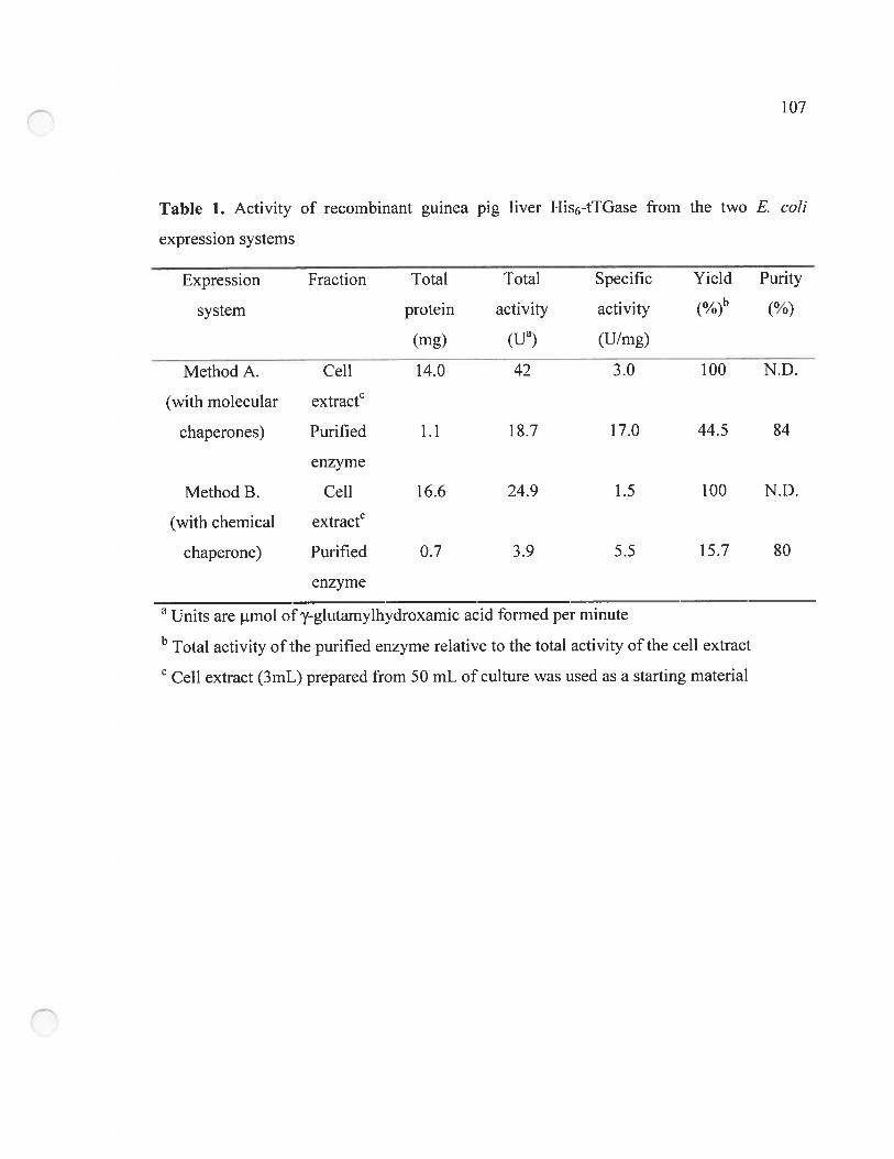
107
Table 1. Activity of recombinant guinea pig liver His6-tTGase from the two E. cou
expression systems
Expression Fraction Total Total Specific Yield Purity
system protein activity activity (%)b (%)
(mg) tua) (U/mg)
Method A. Ce!! 14.0 42 3.0 100 N.D.
(with molecular extractc
chaperones) Purified 1.1 18.7 17.0 44.5 84
enzyme
MethodB. Ce!! 16.6 24.9 1.5 100 N.D.
(with chemical extractc
chaperone) Purified 0.7 3.9 5.5 15.7 80
enzyme
a Units are iimol of’-glutarny1hydroxamic acid forrned per minute
b Total activity ofthe purified enzyme relative to the total activity ofthe celi extract
C Ceil extract (3mL) prepared from 50 mL of culture was used as a starting material
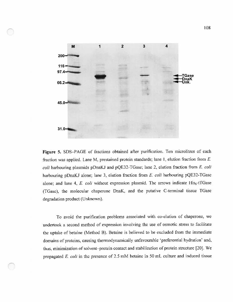
108
M 1 2 3 4
200
116 ——._
97.4—i-—J TGase
DnaK66.2—--. Unk.
45.0—
31 .0—
Figure 5. SDS—PAGE of fractions obtained after purification. Yen microlitres of each
fraction was applied. Lane M, prestained protein standards; lane I, elution fraction from E.
cou harbouring plasmids pDnaKJ and pQE32-TGase; lane 2. elution fraction from E. cou
harbouring pDnaKJ alone; lane 3. elution fraction from E. cou harbouring pQE32-TGase
alone: and lane 4, E. cou without expression plasmid. The arrows indicate His6-tTGase
(IGase). the molecular chaperone DnaK. and the putative C-terminal tissue TGase
degradation product (Unknown).
To avoid the purification problems associated with co-elution of chaperone. we
undertook a second rnethod of expression involving the use of osmotic stress to facilitate
the uptake of betaine (Method B). Betaine is believed to be excluded from the immediate
domains of proteins, causing thermodynamicaÏÏy unfavourable preferential hydration and,
thus, minimization of solvent—protein contact and stabilization of protein structure [201. We
propagated E. cou in the presence of 2.5 mM betaine in 50 mL culture and induced tissue
109
TGase production at 2$ °C for 20 h in 1 M sorbitol LB-based medium (Method B).
Sorbitot/betaine (S/B) treatment enhances production of soluble, active protein relative to
expression of His6-tlGase in the absence of any chaperone (Figure 1, lanes 4—6). The
soluble His6-tTGase was purified by IMAC as described under Materials and rnethods.
SD$—PAGE analysis of the purification steps is shown in Figure 4B. Table 1 shows a
surnmary of a typical purification procedure. Based on these resuits. the production of the
soluble recombinant transglutarninase was calculated to be 14 mg/L culture with a specific
activity of 5.5 U/mg and a purity of $0 %. While this strategy elirninated the impurity
around 70 kDa, the band migrating at approximately 66 kDa was stiil present. This
irnpurity was absent from the elution fraction of the purification from ceils not expressing
the His6-tTGase (Figure 5. lanes 2 and 4). This band may be due to the partial C-terminal
degradation of the recombinant tissue TGase. While the use of chernical chaperones [20]
increased the yield of soluble enzyme relative to expression in the absence of molecular
chaperones, the specific activity of the resulting purified tissue IGase was lower than that
obtained by Method A. As we required enzyme of the highest possible specific activity for
our research needs, we did flot further investigate Method B. although it provides enzyme
of reasonable quality with the potential advantage of carrying no trace of contaminating
DnaK chaperone.
Expression of His6-tlGase in E. cou by combination of Methods A and B (with
DnaK, DnaJ, and sorbitol/betaine medium) did not show any enhancernent of solubility
relative to Method A (data flot shown).
4.4.3 Kinetic of recombinant guinea pig tiver His6-tTGase
The specific activity of purified His6-tlGase was determined according to the
hydroxamate assay using Cbz—Gln—GIy and hydroxylarnine [23]. This value (17 U/rng) was
similar to that of the highly purified native transglutarninase (1$ U/mg LI 1] or 14 U/mg
[23]) previously determined using the same method. Using the DMPDA assay (Method I)
110
t261 we determined KM and for the weIl-characterized donor substrate Cbz—Gln—Gly.
Nonlinear regression to the Michaelis—Menten equation yielded a KM for Cbz—Gln—Gly as
5 + 2 mM and a kcat as 60 rnin. Using the GDH-linked assay (Method II). with the sarne
donor substrate, we determined a KM of 3.9 ± 0.4 mM and a kcat of 44 min1. The KM value
reported in the literature for the native guinea pig tissue TGase for this donor substrate
using Method I is 2.40 + 0.05 mM [26] and using Method 11 is 5.7 ± 0.6 mM [31], which
are comparable to the values we have obtained. In the literature, the kcat value reported for
the native guinea pig tissue TGase for Cbz—Gln—Gly using Method II is 52 ± 2 rnin1 [31].
This indicates that the hexahistidine tag and the potential mutations or polymorphisrns at
positions 142 and 436 detected by automated DNA sequencing do not appear to affect the
catalytic activity of the recombinant enzyme since the kcat and KM values are not
significantly different than those ofthe tissue TGase purified from guinea pig liver.
4.5 Future work
The yield. purity. and specific activity of the TGase purified by Method A are the
highest reported to date using a single-step purification protocol. indicating that this rapid
method yields enzyme of high enough quality for most research applications. Its kinetic
properties do flot significantly differ from those of the native enzyme. As detailed above, it
seems probable that the two principal impurities are the chaperone DnaK and a C-terminal
degradation product of the tissue TGase. DnaK couÏd be rapidly eliminated by adding a
FLAG tag (DYKDDDDK) that would allow elimination of the tagged chaperone by
passing the sample over an anti-fLAG affinity column [32] or by adding any other such
affinity label. One could also consider using other chaperones than DnaK and DnaJ. The
putative C-terminal tissue TGase degradation product could be eliminated by transferring
the hexahistidine tag from the N- to the C-terminus such that C-terminally degraded tissue
TGase will flot bind to Ni—NTA. Transferring the hexahistidine tag to the C-terminus may
also have the added benefit of improving the affinity of His6-tlGase toward Ni—NIA,
111
considering the relatively poor affinity we observed for the N-terminally tagged tissue
IGase (Figure 4).
4.6 Conclusions
In conclusion. the procedures for the expression and purification of recombinant
guinea pig liver transglutaminase presented in this work represent an efficient. easily
reproducible, and rapid method to obtain highly purified enzyme with high specific activity.
While the use of sorbitol and betaine as chernical chaperones [20] increased the yield of
soluble enzyme relative to expression in the absence of molecular chaperones, the specific
activity of the resulting purified tissue TGase was not high enougli to warrant further
investigation. Expression in the presence of the molecular chaperone DnaK and DnaJ [15],
coupled to rapid affinity purification, resulted in a good yield ofhighly active enzyme.
4.7 Acknowledgements
We thank Dr. H. Yasueda (Central Research Laboratories. Ajinomoto Co. Inc.,
Japan) for pDnaKJ plasmid. Dr. K. Ikura (Department 0f Applied Biology. Kyoto Institute
Teclmology. Kyoto, Japan) for the pKTGI plasrnid, C. Gravel (Université de Montréal) for
the native enzyme, N. Doucet (Université de Montréal) for critical reading of the
manuscript, and D. Halim (Université de Montréal) for assistance with the kinetic assays.
This work was supported by VRQ Grant 2201-147 and FQRNT Grant ER-$0357. jointly
held by J.W.K. and J.N.P. R.A.C. is also grateful to FQRNT.
4.8 References
1. J.E. Folk and P.W. Cole. Transglutaminases: mechanistic features of the active-site as
deterrnined by kinetic and inhibitor studies. Biochim. Biophvs. Acta 122 (1996), pp.
244—264.
112
2. K.E. Achyuthan. T.F. Slaughter. M.A. Santiago. J.J. Enghild and C.A. Greenberg. Factor
XIIIa-derived peptides inhibit transglutaminase activity. Localization of substrate
recognition sites. J Biol. Chem. 26$ (1993), pp. 212X4—21292.
3. C.S. Greenberg, P.J. Birkbichler and R.H. Rice, Transglutaminases: multifunctional
cross-Iinking enzymes that stabilize tissues. fASEBJ 5(1991), pp. 3071—3077.
4. D. Aeschlimann and M. Paulsson. Transglutarninases: protein cross-linking enzymes in
tissues and body fluids. Thromb. Haemostasis 71 (1994), pp. 402—415.
5. S.-Y. Kim, T.M. Jeitner and P.M. Steinert, Transglutaminases in disease. Neurochem.
Int. 40 1 (2002), pp. $5—103.
6. A.J.L. Cooper, T.M. Jeitner, V. Gentile and J.P. Blass, Cross linldng of polyglutarnine
domains catalyzed by tissue transglutaminase is greatly favored with pathological
length repeats: does transglutaminase play a role in (CAG),1/Q,1-expansion disease?.
Neurochem. Int. 40 1 (2002). pp. 53—67.
7. S.M. Singer, G.M. Zainelli, M.A. Norlund, J.M. Lee and N.A. Muma, Transglutaminase
bonds in neurofibrillary tangles and paired helical filament tau early in Alzheimer’s
disease. Neurochem. In!. 40 1 (2002). pp. 17—30.
8. M.V. Karpuj. M.W. Becker and L. Steinrnan. Evidence for a role for transglutaminase in
Huntington’s disease and the potential therapeutic implications. Neurochem. Int. 40
1 (2002), pp. 3 1—36.
9. B. Wilhelrn, A. Meinhardt and J. Seitz. Transglutaminases: purification and activity
assays. J Chromatogr. B 684 (1996), pp. 163—1 77.
10. 1. Abe. S.I. Chung. R.P. DiAugustine and J.E. Folk. Rabbit liver transglutaminase:
physical, chemical and catalytic properties. Biochemistiy 16 (1977), pp. 5495—5501.
11. A. Leblanc, N. Day. A. Ménard and J.W. Keillor, Guinea pig liver transglutaminase: a
modified purification procedure affording enzyme with superior activity in greater
yield. Protein Expr. Purif 17 (1999). pp. 89—95.
1I
12. W. Dietrich. T. Elmis. M. Bauer. P. Donner, U. Volta, E.O. Riecken and D. Schuppan.
Identification of tissue transglutarninase as the autoantigen of celiac disease. Nat.
lied 3 (1997), pp. 797—$01.
13. K. Ikura, Y. Tsuchiya, R. Sasaki and H. Chiba, Expression of guinea-pig liver
transglutarninase cDNA in Fscherichia cou. Amino-terminal N-alpha-acetyl group
is not essential for catalytic function of transglutarninase. Fur. .1 Biochem. 187
(l99O)..pp. 705—711.
14. K.N. Lee, S.A. Arnold, P.J. Birckbilcher, M.K. Patterson, Jr., B.M. fraij, Y. Takeuchi
and H.A. Carter, Site-directed mutagenesis of hurnan tissue transglutarninase: Cys
277 is essential for transglutarninase activity but flot for GTPase activity. Biochim.
Biophvs. Acta 1202 (1993). pp. 1—6.
15. K. Ikura. Y. Tsuchiya. R. Sasaki and H. Chiba, Expression of guinea-pig liver
transglutaminase cDNA in Escherichia cou. Arnino-terminal N-Œ-acetyl group is
not essential for catalytic function oftransglutaminase. Fur. I Biochem. 187 (1990),
pp. 705—711.
16. K.-L Yokoyarna, Y. Kikuchi and H. Yasueda, Overproduction of DnaJ in Fscherichia
colt improves in vivo solubility of the recombinant fish-derived transglutaminase.
Biosci. Bioteclmoï. Biochem. 62 6 (1998). pp. 1205—1210.
17. Q. Shi. S.-Y. Kim, J.P. Blass and A.J.L. Cooper. Expression in Escherichia cou and
purification of hexahistidine-tagged human tissue transglutarninase. Prote in Expr.
Furif 24 (2002), pp. 366—373.
1$. A. Ambrus and L. Fésfls, Polyethylene glycol enhanced refolding of the recombinant
human tissue transglutaminase. Prep. Biochem. Biotechîlol. 311 (2001), pp. 59—70.
19. K. Ikura, T. Kokubu. S. Natsuka. A. Ichikawa. M. Adachi, K. Nishiara, H. Yanagi and
S. Utsumi. Co-overexpression of folding modulators improves the solubility of the
recombinant guinea pig liver transglutarninase expressed in Escherichia coït. Prep.
Biochem. Biotechnoï. 32 2 (2002), pp. 189—205.
114
20. J.R. Blackwell and R. Horgan. A novel strategy for production of a highly expressed
recombinant protein in an active form. fEBSLett. 295 1—3 (1991), pp. 10—12.
21. M. Bradford, A rapid and sensitive rnethod for the quantitation of microgram quantities
of protein utilizing the principle of protein—dye binding. Anal. Biochem. 72 (1976),
pp. 248—254.
22. J.E. folk and S.I. Chung. Transglutaminases. Methods EnzymoÏ. 113 (1985), pp. 358—
364.
23. J.E. Folk and P.W. Cole, Mechanisrn of action of guinea pig liver transglutaminase. I.
Purification and properties of the enzyme: identification of a functional cysteine
essential for activity. J Bio!. Chem. 241 (1966), pp. 55 18—5525.
24. U.K. Laernrnli. Cleavage of structural proteins during the assernbly of the head of
bacteriophage T4. Na! tire 227 (1970). pp. 680—685.
25. C. fernandez-Patron. L Castellanos-Serra and P. Rodriguez, Reverse staining of
sodium dodecyl sulfate polyacrylamide gels by imidazole-zinc salts: sensitive
detection ofunrnodified proteins. Biotechniques 12 (1992), pp. 564—573.
26. P. de Macédo, C. Marrano and J.W. Keillor, A direct continuous spectrophotornetric
assay for transglutaminase activity. Anal. Biochem. 285 (2000). pp. 16—20.
27. N. Day and J.W. Keillor. A continuous spectrophotometric linked enzyme assay for
transglutaminase activity. Anal. Biochem. 274 (1999), pp. 14 1—144.
2$. K. Ikura, T. Nasu, H. Yokota, Y. Tsuchiya, R. Sasaki and H. Chiba, Amino acid
sequence of guinea pig liver transglutaminase from its cDNA sequence.
Biochernistry 27 8 (1988), pp. 2898—2905.
29. C. Panagabko, S. Morley, S. Neely, H. Lei, D. Manor and J. Atkinson, Expression and
refolding of recombinant hurnan Œ-tocopherol transfer protein capable of specific x
tocopherol binding. Protein Expr. Furf 24 (2002), pp. 395—403.
30. Z. Gu, M. Weidenhaupt, N. Ivanova, M. Pavlov, B. Xu, Z.-G. Su and J.-C. Janson,
Chromatographic methods for the isolation of, and refolding of proteins from,
Escherichia cou inclusion bodies. Protein Expr. Purif 25 (2002), pp. 174—179.
115
31. N. Day, Études mécanistiques de l’étape d’acylation de la transglutaminase tissulaire de
foie de cobaye. Mémoire de thèse, Université de Montréal, 1999
32. T.P. Hopp. K.S. Prickett, V.L. Price. R.T. Libby, C.J. March, D.P. Cerretti, D.L. Urdal
and P.J. Conlon, A short polypeptide marker sequence useful for recombinant
protein identification and purification. BiotechnoÏogy 6 (1988). pp. 1204—12 10.
CHAPITRE 5
Développement d’un modèle d’homologie de la TGase de
foie de cobaye et mutagenèse de résidus du site actif
117
5.0 Préface
Pour pouvoir entreprendre l’évolution dirigée de la TGase de foie de cobaye par une
approche semi-aléatoire. nous avions besoin d’information structurel le et fonctionnelle
additionelle. Dans ce chapitre. nous présentons le premier modèle par homologie de la
TGase de foie de cobaye, qui nous a permis d’acquérir de l’information structurelle
supplémentaire. De plus, de l’information fonctionnelle a été acquise lors de cette étude par
des expériences de mutagenèse dirigée et combinatoire de certains résidus du site actif de la
TGase de foie de cobaye. dont le rôle est mal défini dans la littérature. Cette information a
été d’une grande utilité pour l’application de notre approche semi-aléatoire.
Pour le manuscrit « Hornology modeling of guinea pig liver transglutarninase and
mutagenesis of conserved active-site residues Tyr5 19 and Cys336 ». présenté au chapitre 5.
j’ai créé le modèle d’homologie de la TGase de foie de cobaye, j’ai construit le vecteur
d’expression pQE32-GTG et j’ai réalisé toutes les expériences de cinétique enzymatique et
la majorité des expériences de mutagenèse dirigée. en plus de rédiger le manuscrit. Le
Dr Steve Gillet a développé la méthode de cinétique enzymatique à la 7-hydroxycoumarine
en plus de synthétiser le composé Cbz-Gly-7HC. farah-Jade Dryburgh m’a assisté lors de
la création des mutants Cys336 et Tyr5 19 de la TGase de foie de cobaye durant son stage
sous ma supervision. Ce manuscrit était soumis pour publication au moment du dépôt de
cette thèse.
118
Article 4.
Homology modeling of guinea pig liver transglutaminase
and mutagenesis of conserved active-site residues Tyr519
and Cys336
Roberto A. Chica, Steve M.F.G. Gillet, Farah-Jade Dryburgh, Jeffrey W.
Keillor and Joelle N Pelletier
Département de chimie, Université de Montréal, CP 612$, Succursale Centre-Ville.
Montréal. Québec, H3C 3J7, Canada
Manuscrit soumis
119
5.1 Abstract
Transglutaminases (TGases) cross-link peptides and proteins by catalyzing the
formation of 7-glutamyl-e-lysyl bonds. Crystal structures from TGases ail show a
conserved Tyr within hydrogen bonding distance ofthe catalytic Cys. In tissue IGases, the
catalytic Cys is located between this conserved Tyr and a second conserved Cys. It has
been proposed that rernoval of this conserved Tyr would lead to immediate inactivation of
the enzyme as the catalytic Cys wouid form a disulphide bridge with this second Cys
residue. In addition, our previous resuits from molecular modeling suggested that this Tyr
forms a critical H-bond with the acyl-donor substrate. b investigate these hypotheses, we
turned to the widely-studied guinea pig liver TGase. Because its structure has not been
reported, we generated a hornology model for this enzyme. Owing to its 43 % sequence
identity with red sea bream tissue TGase, the model exhibits high structural sirnilarity with
this and other tissue TGases. justifying the use of guinea pig liver TGase as our
experimental system. Mutagenesis of the conserved Tyr5 19 and Cys336 resuited in only a
two- to four-fold decrease in catalytic efficiency. indicating that these two residues are not
essential for activity. furthermore. wild-type and Cys336 mutants lose activity upon
incubation at 37 oc Activity was not restored by addition of a reducing agent, invalidating
the hypothesis that residue Cys336 fonns an inactivating disuiphide bond with the catalytic
Cys. Our work provides the first mode! of guinea pig liver TGase and demonstrates that its
active-site is surprisingly accommodating of mutations.
Keywords: tissue transglutaminase, TGase, homo!ogy modeling. site-directed
mutagenesis, combinatorial mutagenesis, enzyme kinetics
5.2 Introduction
Eukaryotic transglutaminases (protein-glutarnine y-glutamyltransferases, EC
2.3.2.13) are a family of enzymes that catalyze the cross-linking ofpeptides and proteins by
120
Ca2-dependent acyl transfer reactions [Ï-5]. They catalyze the formation of isopeptide
bonds between the y-carboxamide group of a protein- or peptide-bound glutamine side
chain and the a-amino group of a protein- or peptide-bound lysine side chain via a modified
ping-pong mechanism [6]. The reaction is carried out by a conserved Cys-His-Asp catalytic
triad that is sirnilar to that of cysteine proteases: residues Cys and His are believed to form
a thiolate-irnidazolium ion pair. The first step of the mechanism is the acylation step where
a glutarnine-containing protein or peptide acting as an acyl-donor reacts with the enzymes
catalytic cysteine thiolate, releasing NI-I3 and generating the covalent acyl-enzyme
intermediate. This intermediate then reacts with a second substrate, the acyl-acceptor,
which can be almost any primary amine [7]. to yield the amide product and free enzyme in
the second step of the TGase mechanisrn. the deacylation step. The acyl-enzyrne
intermediate can also undergo hydrolysis in the absence of primary amine but at a siower
rate. It has been shown that the deacylation step of the TGase mcchanisrn is the rate
lirniting step [8. 9] even when the acceptor substrate is highly nucleophilic. demonstrating
the efficiency with which IGases activate the acyl-transfer from the glutamine
‘y-carboxamide group.
Eukaryotic TGases are divided into nine classes [5]. Thus far. crystal structures
from only four TGases belonging to three different classes have been determined: plasma
TGase (factor XIIIa) [10-13], tissue TGase [14. 15] and epidermal TGase [16-191. Plasma
TGase is a homodimeric protein that requires cleavage of a propeptide by thrombin for
activation. Tissue TGase and epidermal TGase are intracellular, monomeric enzymes that
are inhibited by GTP [14. 17]. Epidermal TGase also requires proteolytic activation for
activity, unlike tissue TGase. Ail eukaryotic TGases share a common structure that consists
of four dornains: the N-terminal 3-sandwich domain, the catalytic core which contains the
conserved catalytic triad, the barrel I domain and the C-terminal barrel 2 domain.
The crystal structures ofhurnan factor XIIIa [12], red sea bream tissue TGase [15],
human tissue TGase [14] and human epidermal TGase [16] ail show a conserved Tyr
121
residue that is within hydrogen bonding distance of the catalytic Cys residue. It bas been
proposed that prior to catalysis, the putative hydrogen bond between the conserved Tyr and
the catalytic Cys must 5e broken in order to give the glutamine-containing substrate access
to tbe catalytic thiolate [12]. However. our previous molecular modeling study performed
on red sea bream tissue TGase suggested that this Tyr residue instead forrns a critical
H-bond with the y-carboxarnide group of the acyl-donor substrate, orienting and activating
it for the nucÏeophiÏic attack in the acylation step of the TGase mechanism [20]. In tissue
IGases, the catalytic Cys is located between this conserved Tyr and a second, conserved
Cys. It bas also been proposed that rernoval of the conserved Tyr of red sea bream tissue
TGase wou!d !ead to immediate inactivation as tbe catalytic Cys would form a disuiphide
bridge with this second Cys [15]. Sucb a disuiphide bridge bas not been observed but
indirect observations made by Folk and coworkers [21, 22] support this hypothesis.
Narnely, they reported that another tissue TGase. guinea pig liver TGase. loses activity
steadily during storage: rnuch of the lost activity can be restored by addition of the reducing
agent dithiothreitol, consistent with inactivation having been the resuit of disulpbide
bridging ofthe catalytic Cys.
To investigate these hypotheses. a homology mode! for guinea pig liver TGase was
developed since no crystal structure of this enzyme has been reported. This model
demonstrated that guinea pig liver TGase bas an active-site geometry and overali structure
that is similar to that of red sea bream tissue TGase from whicb the functional hypotheses
were derived. Thus, we generated mutants of the most extensively studied TGase, guinea
pig liver TGase. at the positions of the conserved active-site Tyr (TyrS 19) and the second
Cys residue (Cys336). Cys336 was mutated to A!a and Val to verify if it participates in
oxidation ofthe catalytic residue Cys277 by formation ofa disu!phide bond. Tyr519 was
mutated to Leu, Phe, G!n or Ris to modify the putative H-bond with the donor substrate and
to verify if removal of this residue inactivates the enzyme by allowing disu!phide bond
formation with the catalytic Cys277.
122
5.3 Materials ami metliods
5.3.1 Homology modeling
The homology mode! of guinea pig liver TGase was prepared, using as templates
the PDB coordinates ofthe crystal structures ofhurnan tissue IGase (1KV3), red sea bream
tissue TGase (1GOD), human epidermal TGase (1L9M) and hurnan factor XIIIa (1EVU).
Crystallized water molecules, heterologous molecules and extra monorners were removed.
The primary sequences of these TGases were extracted from their crystal structures using
the Homology module of Insightll (Acceirys, San Diego, CA). Using these sequences, an
aligmrient was derived using either the Tcoffee server [23] or ClustalW [24], with default
parameters. f ive 3D models for guinea pig liver TGase were derived for each sequence
alignrnent file using the crystal structure coordinates by Modeller 8v1 [25] with default
pararneters for generation of loop conformations. The quality of the proposed models was
assessed using the Molprobity t26] web server, Whatcheck [27] and Prosail t2$]. Ail ten
models were then refined using a series of energy minirnizations perforrned with the
Discover module of Insightll. We applied a tether restraint of 100 kcal/À2 on all atoms
using 1000 steps of steepest descents energy minimization followed by a conjugate
gradients energy minimization until convergence of 0.001 kcal rnoF’Â’ with a distance
dependent dielectric constant of 4 to mimic the interior of proteins. The resulting structures
were again analyzed using Molprobity, Whatcheck and Prosail. On the basis of those
results, mode! T2 was selected as the representative model. It was subjected to an
unconstrained molecular dynarnics simulation consisting of a 1 ps equilibration of the
molecular system at 300 K followed by the actual simulation to explore conformational
space for 300 ps while maintaining the sarne temperature. A nonbond cutoff of 15 À was
used to speed up calculations. The radius of gyration of the structure during the simulation
was measured using the Decipher module of Insightll.
123
5.3.2 Materials
Ail reagents used were of the highest available purity. Restriction enzymes and
DNA-modifying enzymes were from New England Biolabs (Mississauga, ON) and MBI
Fermentas (Burlington, ON). Synthetic oligonucleotides were obtained from Alpha DNA
(Montreal, QC) and Integrated DNA Technologies (Coralville, lA). infra-red dye labeÏled
sequencing prirners were from Li-Cor Biotechnology Division (Lincoin. N3). and Ni-NTA
agarose resin was obtained from Qiagen (Mississauga, ON). Ail aqucous solutions were
prepared using water purified with a Millipore BioCeli system. DNA sequencing was
perforrned using a kit for Sequenase version 2.0 DNA polymerase (Arnersham—Pharmacia
Biotech) with a Li-Cor 1R2 automated system.
5.3.3 Construction ofthe expression ptasmid
The pQE32-GTG expression plasrnid that contains the guinea pig Jiver TGase gene
was constructed using the site overlap extension mutagenesis method [29] starting with the
pQE32-TGase plasmid [30]. The pQE32-GTG plasmid differs from pQE32-TGase by the
presence of a SalI restriction site at position 799 bp (numbering according to the origin of
replication of plasmid pQE32-TGase), the presence of an AgeI restriction site at position
1728 bp and the absence of a GC-rich 41 bp sequence containing a SalI restriction site 3’
from the stop codon and 5’ from the vector PstI restriction site. To introduce the SalI
restriction site at position 799 bp, the guinea pig liver TGase gene was PCR-amplified from
plasmid pQE32-TGase using the primer pairs SphIGTGaseF-Met
(5’-CACACAGCATGCAGGCAGAGGATCTGATCCTG-3’) with GTG-SalI-799-R
(5 ‘-CACGTAGACAGGTCGAUFGCGGCGCGAGCA-3’), and GTG-SalI-799-F (5’-
TGCTCGCGCCGCAGTCGACCTGTCTACGTG-3’) with pQE32Reverse
(5’-GTTCTGAGGTCATTACTGG-3’). These primer pairs introduce a SalI restriction site
(bold italic) through two suent mutations (underlined) at codons Ser2 15 and Arg2 16 of the
guinea pig liver TGase gene. The 2069 bp full length guinea pig liver TGase gene was
124
generated by a third PCR reaction using the two amplicons and the external prirners
SphIGTGaseF-Met and pQE32Reverse. This full length PCR product was digested with
PstI/$phl, isolated by agarose gel electrophoresis and extracted with a QIAEXII kit
(Qiagen). The mutated gene was inserted into pQE32 expression vector (Qiagen) via
SphI/PsII yielding the plasmid pQE32-TGase(Sa/1). To introduce the AgeI restriction site at
position 172$ bp. the guinea pig liver TGase gene was PCR-amplified from plasrnid
pQE32-TGase(SalI) using the primer pairs GTG-AgeI-1 728-F
(5 ‘-AATGGAGTCCTGGGACCGGTGTGCAGCACCAAC-3’) with GTG-PstI-2234-R
(5 ‘-ACACACCTGCAGTTAGGCGGGGCCGATGAT-3’) and SphIGTGaseF-Met with
GTG-AgeI-1 72$-R (5’-GTTGGTGCTGCAcAcCGGTCCCAGGACTCCATT-3’). These
primer pairs introduce an AgeI restriction site (bold italic) through two suent mutations
(underlined) at codons G1y524 and Pro525 of the guinea pig liver TGase gene while also
removing the SalI-containing 41 bp sequence between the stop codon of the TGase gene
and the PstI site of plasmid pQE32-TGase(SaÏI). The 2069 bp full length guinea pig liver
TGase gene was generated by a third PCR reaction using the two amplicons and the
external primers described above. This PCR product was digested with SphI/PstI, isolated
by agarose gel electrophoresis and extracted. The mutated gene was inserted into pQE32
expression vector via SphI/FstI yielding the plasrnid pQE32-GTG. The entire SphI/FstI
fragment that includes the whole coding region was verified by DNA sequencing.
5.3.4 Mutagenesis ofCys336 and Tyr519
The Cys336Ala and Cys336Val mutations of the guinea pig liver TGase gene were
introduced by a pair of complementary degenerate oligonucleotides (onÏy the coding strand
is shown): GTG-GYT336 (5’-ATCTGGAACTTCCACGYTTGGGTGGAGTCGT-3’).
Codon GYT (Y = C or T; bold) encodes both Ala and Val. A third PCR reaction using
these two amplicons and the extemal primers GTG-$alI-799-F and GTG-AgeI-172$-R was
performed to obtain the 92$ bp guinea pig liver TGase gene fragment located between
restriction sites SalI and AgeI. For mutations Tyr5l9Phe. Tyr5l9Leu. TyrSl9flis and
125
Tyr5l9GIn. the primer pair GTG-SalI-799-f with GTG-YWS5I9
(5 ‘-GAGAACCGGTCCCAGGACTCCATTSWRGCTGACGATGCGT-3’) was used.
Primer GTG-YWS5 19 contains the degenerate codon YWS (non-coding strand SWR where
S = C or G. W = A or T and R = A or G; bold) that encodes Leu. Ris, Gin. Phe. Tyr and the
amber stop codon. The PCR products were digested by S’alliAgel, isolated by agarose gel
electrophoresis and extracted. The resulting 92$ bp fragments were inserted into
pQE32-GTG expression vector via S’alliAgel yielding the plasmid pQE32-GTG expressing
the mutant TGases. The plasmids were then transforrned into E. cou XL-1 hue ceils or E.
cou XL-1 Blue celis containing the plasmid pDnaKJ [31] that encodes the molecular
chaperones DnaK and DnaJ. The desired mutants were identified by DNA sequencing and
the integrity of the entire coding region was verified. The Cys336 and TyrS 19 double
mutants were generated by a similar procedure.
5.3.5 Overexpression and purification ofwild-type and mutant TGases
Wild-type and mutated recombinant guinea pig liver TGases were overexpressed
and purified from E. cou according to a protocol developed in our laboratory [30] with the
following modifications. Afier Ni-NIA purification. the eluant was transferred to an
Amicon Ultra 15 mL tube (Millipore) with a molecular weight cutoff of 30 kDa for
desalting by centrifugation with 25 mM Tris-acetate buffer (pH 7.0) containing 0.5 mM
EDTA. The TGase samples were aliquoted at a final concentration of 250 tgimL, snap
frozen on dry ice and stored at —$0 °C. Typical yields were 1.5-10 rngiL of $5 % pure
protein, as estimated from Coomassie Blue staining following SDS-PAGE.
5.3.6 Synthesis of Cbz-Gly-7HC
Cbz-Gly-7HC (Figure lB) was synthesized based on a previously reported protocol
[32]. Namely, 0.2 g (1 mmol) of CbzGly and 0.4 g (2.5 mmol) of 7-hydroxycoumarin were
dissolved in 10 mL ofethyl acetate. Then 0.22 mL (0.2 g. 2 mmol) ofN-methylmorpholine
126
and 0.8 mL (0.63 g. 5 mmol) of N.N-diisopropylcarbodiimide were added with stirring at
room temperature. Stirring was continued until the complete disappearance of CbzGly. as
followed by thin layer chromatography (ethyl acetate). The reaction mixture was then
washed once with 1 M NaOH. three tirnes with 0.1 M NaOH, 3 times with 0.1 M HC1. once
with saturated NaHCO3 and once with brine. The organic phase was then dried over
MgSO4, fiitered and evaporated under reduced pressure. The resuiting residue was purified
by silica gel chrornatography (ethyl acetate) to rernove traces of diisopropylurea, giving the
desired ester in 70% yield (0.25 g). 1H NMR (300 MHz, CDCl) (Y 4.30 (2H, d), 5.18
(2H, s), 5.33 (1H, s), 6.44 (1H, d), 7.0$ (1H, d), 7.10 (1H, s), 7.37 (5H, m), 7.51 (1H, d),
7.70 (1H, d). 13C NMR (75 MHz, CDC13) ô 168.5, 160.6, 156.7, 154.9, 152.9, 143.1, 136.3,
129.0, 128.9, 128.6, 128.5, 118.4, 117.2, 116.6, 110.5, 67.7, 43.2. HRMS (FAB) calculated
for C19H16N06 ([M+H]): 354.0972, found 354.0968.
5.3.7 Kinetic assays
5.3.7.1 Method I: DMPDA assay
The kinetic properties of the mutant TGases were determined using a continuous
spectrophotometric assay (Figure lA) [33]. Ail kinetic runs werc performed in triplicate at
six different concentrations of donor substrate. A standard stock buffer solution (182 mM
Tris-acetate buffer pH 7.0, 5 mM CaC12. and 0.9 mM EDTA) was prepared. 0.8 mM of
N,N-dimethyl-1 ,4-phenylenediamine (DMPDA) and 0.5-30 mM of
N-carbobenzyloxyglutaminylglycine (Cbz-Gln-Gly) (Sigma) were added to the stock buffer
solution. The reaction solution was preincubated for 3 min at 37 oc prior to initiation ofthe
enzymatic reaction by addition of --$ tg of purified His6-TGase. Reaction progress was
followed as a linear increase of absorbance measured at 27$ nrn with a Cary 100
spectrophotometer (Varian) thermostated at 37 °C. For the reaction bianks. water was
substituted for TGase enzyme. Slopes were rneasured over the first <10 % substrate
conversion to product and were converted into initial rates by dividing by the extinction
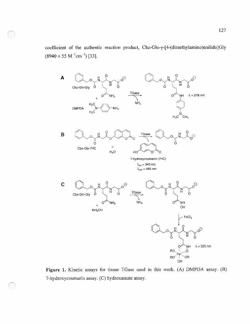
127
coefficient of the authentic reaction product, Cbz-G1u-y-[4-(dirnethy1amino)ani1ido]G1y
($940 ± 55 Mcm1) [33].
A 0Je Q0j0eCbz-GIn-GIy O O O O
TGaseO NH O NH ?=278nm
DMPDAH3C H3
H3C’ “CH3
B OoJo-OI)o
Cbz-GIy-7HC+120 HO O O
7-hydroxycoumarin (7HC)
= 340 nm
em 465 nm
TG eCbz-GIn-GIy O O
_____
O O
O NH2 NH3 O NH
+ OHNH2OH
FeCI3
H —
OÏNN
O- NH ?L=525nm
R0.Fe:
RO’ ‘OR
OR
Figure 1. Kinetic assays for tissue TGase used in this work. (A) DMPDA assay. (B)
7-hydroxycournarin assay. (C) hydroxamate assay.
128
5.3.7.2 Method II: 7-hydroxycoumarin assay
The decrease of activity of wild-type, Cys336Ala and Cys336Val guinea pig liver
TGases as a function of tirne was measured using the following procedure. Frozen purified
enzyme was thawed at room temperature then incubated at 37 °C in its storage buffer.
Aliquots of 10 .tL (3 tg) were periodically sarnpled for immediate measurement of
residual activity using a continuous fluorometric assay (f igure lB) [32] described in the
next paragraph. Afier three hours incubation at 37 °c, dithiothreitol was added to the
purified enzyme to a final concentration of 5 mM and a further 10 tL aliquot was sampled.
Residual activity was measured at 25 °c by adding 3 tg of iGase to each well of a
ici Luminescence 96-well microtiter plate (Thermo Electron) containing a 100 iM
solution of the donor substrate cbz-Gly-7Hc in stock buffer solution (100 mM Mops
buffer pH 7.0, 5 mM caCl2, and 0.05 mM EDTA, 5 % DMF). All assays were perforrned in
triplicate. ilie increase in fluorescence due to the release of 7-hydroxycoumarin was
followed against a blank at 340 uni and 2Lern 465 nm in a FluoStar Optima microtiter
plate reader (BMG Labtecli). for the aliquots containing dithiothreitol, a blank containing
the same amount of dithiothreitol but with water replacing enzyme was used. Linear siopes
of fluorescence versus tirne were measured over the first <10 % conversion of substrate to
product. To convert the slopes into initial rates, a “fluorescence coefficient” was
determined, on a daily basis, by measuring the arbitrary fluorescence intensities
corresponding to five concentrations of 7-hydroxycoumarin at concentrations ranging from
0.05 to 0.5 iM in 5 % DMF in the stock buffer solution at 25 °c. The value of this
“fluorescence coefficient” varied only slightly (<5 %) each day.
5.3.7.3 Method III: ilydroxamate assay
ihe inactivation of iGase activity as a function of time was also deterrnined using
the discontinuous hydroxamate assay (Figure 1C) [34]. Incubation of enzymes at 37 °C and
129
addition of dithiothreitol afier 3 h were as described above. For residual activity assays, a
stock solution containing 37.5 mM Cbz-Gln-Gly, 1.25 mM EDTA, 125 mM hydroxylamine
and 6.25 mM CaC12 in 250 mM Tris-acetate buffer (pH 6.0) was prepared. To this solution,
25 tg of enzyme was added and the reaction was allowed to proceed for 10 minutes after
which it was quenched with a ferric chloride reagent solution (1.67 % FeC1y6H2O, 5 %
trichloroacetic acid and 0.83 M HC1). The absorbance was measured at 525 nm on a
Biotech Ultrospec 2000 (Pharmacia) table-top spectrophotometer and was converted into
initial rates using the extinction coefficient of the hydroxamate-containing product
(340 M1cm’) t21]. For the aliquots containing dithiothreitol. a blank containing the same
amount ofreducing agent but with water replacing enzyme was used.
5.4 Resuits and discussion
To test the hypotheses reported in the literature [15, 20] of the putative interactions
between the catalytic Cys residue and conserved active-site residues TyrS 15 and Cys333 of
red sea bream tissue TGase, site-directed mutagenesis of guinea pig liver IGase was
envisaged. Guinea pig liver IGase was chosen to test these hypotheses because it is the
most widely characterized tissue TGase, allowing correlation with previous results.
However, despite numerous studies of this tissue IGase, its structure has not yet been
reported. Thus, in order to more confidently guide site-directed mutagenesis of guinea pig
liver TGase, a homology model for this enzyme was generated to verify that the positioning
ofthe homologous Tyr and Cys residues (Tyr519 and Cys336, respectively) as well as the
overall structure of the enzyme was sufficiently similar to that of red sea bream tissue
TGase, from which these functional hypotheses were derived.
5.4.1 Homology modeling
To generate the homology model of guinea pig liver IGase, we used the crystal
structures of four TGases as templates: human tissue TGase [14], red sea bream tissue
130
TGase [15], hurnan epidermal IGase [161 and human factor XIIIa [351. Guinea pig liver
TGase bas 83 % residue identity with human tissue TGase. 43 % with red sea bream tissue
TGase, 39 % with human epidermal TGase and 36 % with human factor XIIIa. Residue
homology is 90 %, 59 ¾, 56 % and 51 %, respectively, which is reflected in the conserved
fold of the four TGases. These structures show sufficiently high sequence and structural
similarity to justify their use as templates for a homology model of guinea pig liver TGase.
Sequence alignrnents between these four IGases and guinea pig liver TGase were
performed. Two distinct sequence alignrnents were generated. The first was derived using
the Tcoffee server [23] and the second using ClustalW [24], both with default pararneters.
As expected from the higli homology between the templates sequences, the two resulting
alignment files were identical except in the areas defining gaps. The gaps are sirnilar but
differ slightly in their extremities and should thus have littie effect on hornology modeling.
Using the sequence alignments. ten models were generated by MoUcher: five using
the Tcoffee alignments and five using the ClustalW aiignments (Table 1). These models
were analyzed using three different criteria: backbone conformation. residue contacts and
residue interactions. Backbone confonnation was evaiuated using Molprobity [26], by
verifying torsional angles. A low percentage (2.0 %) ofresidues having torsional angles in
the disallowed regions of the Ramachandran plot (Table 1) was obtained, coherent with
properly folded models. furtherrnore, the percentages obtained for the ten models were in
the same range as the values for the template crystal structures. Residue contacts were
verified by Whatcheck. No deleterious contacts that were not already present in the
tempiate crystal structures were detected. again indicating that the models were well folded.
Lastly. analysis of residue interactions was perforrned using Prosall [28], an algorithm that
measures the interaction energies of residues with the remainder of the protein. Ail models
had regions with unfavourable residue interactions and had to be further refined.
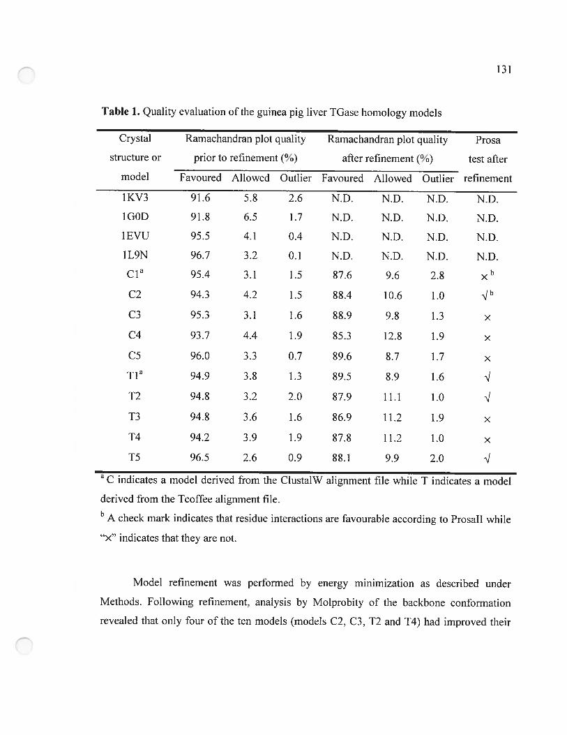
1—,I-,
Table 1. Quality evaluation ofthc guinea pig liver TGase hornology models
Crystal Ramachandran plot quality Ramachandran plot quality Prosa
structure or prior to refinement (%) afler refinement (%) test afier
model Favoured Allowed Outlier Favoured Allowed Outiier refinement
1KV3 91.6 5.8 2.6 N.D. N.D. N.D. N.D.
IG0D 91.8 6.5 1.7 N.D. N.D. N.D. N.D.
1EVU 95.5 4.1 0.4 N.D. N.D. N.D. N.D.
ÎL9N 96.7 3.2 0.1 N.D. N.D. N.D. N.D.
c1’ 95.4 3.1 1.5 87.6 9.6 2.8
C2 94.3 4.2 1.5 $8.4 10.6 1.0 q”
C3 95.3 3.1 1.6 88.9 9.8 1.3 x
C4 93.7 4.4 1.9 85.3 12.8 1.9 x
C5 96.0 3.3 0.7 89.6 8.7 1.7 x
TIa 94.9 3.8 1.3 89.5 8.9 1.6
12 94.8 3.2 2.0 87.9 11.1 1.0
T3 94.8 3.6 1.6 86.9 11.2 1.9 x
T4 94.2 3.9 1.9 87.8 11.2 1.0 x
15 96.5 2.6 0.9 88.1 9.9 2.0
a C indicates a mode! derived from the Clusta!W alignment file while T indicates a model
derived from the Tcoffee alignment file.b A check mark indicates that residue interactions are favourable according to Prosali while
.><‘ indicates that they are flot.
Mode! refinement was perforrned by energy minimization as described under
Methods. Following refinement, analysis by Molprobity of the backbone conformation
revealed that only four ofthe ten models (models C2. C3, 12 and T4) had improved their
132
backbone conformation, as evidenced by a decreased percentage of residues with angles in
the disallowed regions of the Rarnachandran plot (Table 1). 0f these four models. only two
(C2 and T2) no longer had regions with unfavourable residue interactions as measured by
Prosail. Analysis of residue contacts by Whatcheck did not reveal any deleterious contacts
that were not already present in the templates when refined by the same energy
minirnization protocol (data not shown).
Models C2 and T2 were selected as the most representative models. Low rmsd
values of 1.6 À for the main chain atoms and 2.2 À for ail heavy atoms were calculated
between the two models. demonstrating that these two structures are very sirnilar.
Inspection of the active-site residues (Trp241, Cys277, His305, Asp306, Trp332. His335,
Cys336, Thr360-G1u363, Tyr5 19 and Asn520) demonstrates that the active-site structure of
the two models is very similar. having an rmsd of 1.3 Â. The conformation of residue
Cys336 is different in the two models. In mode! C2. the ‘ and X2 angles of Cys336 are
-176.3° and 61.4°, respectively. while they are -47.2° and -73.1° in mode! T2. Thus,
Cys336 is in a trans conformation in mode! C2 while it is in the most abundant gauche
conformation in model T2. This difference in conformation of the Cys336 side chain
modifies the distance between the S atoms ofCys336 and Cys277: it is 5.3 À for mode! C2
and 4.5 À for mode! 12 (Table 2). These two conformations are allowed according to the
X1-X2 plot for Cys [36] demonstrating that more than one conformation of this residue is
allowed in the TGase fold. However. only the gaziche conformation is observed in the four
crystal structures used as templates. so model 12 was selected as the best model and
analyzed ftirther.
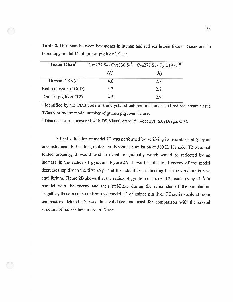
1lii
Table 2. Distances between key atoms in human and red sea bream tissue TGases and in
homology model T2 of guinea pig liver TGase
Tissue TGasea Cys277 S - Cys336 S’ Cys277 S - Tyr5i9 O
(À) (À)
4.6 2.8
4.7 2.8
4.5 2.9
Human (IKV3)
Red sea bream (1GOD)
Guinea pig liver (T2)
Identified by the PDB code of the crystal structures for human and red sea bream tissue
TGases or by the model number of guinea pig liver TGase.b Distances were measured with DS Visualizer vi .5 (Acceirys. San Diego, CA).
A final validation ofmodel T2 was performed by verifying its overail stability by an
unconstrained, 300-ps long molecular dynarnics simulation at 300 K. If model T2 were not
folded properly, it would tend to denature gradually which would be reflected by an
increase in the radius of gyration. Figure 2A shows that the total energy of the model
decreases rapidly in the first 25 ps and then stabilizes. indicating that the structure is near
equilibrium. Figure 23 shows that the radius of gyration of model 12 decreases by ---i À in
parallel with the energy and then stabilizes during the remainder of the simulation.
Together, these resuits confirm that model T2 of guinea pig liver TGase is stable at room
temperature. Model T2 was thus validated and used for comparison with the crystal
structure ofred sea bream tissue TGase.
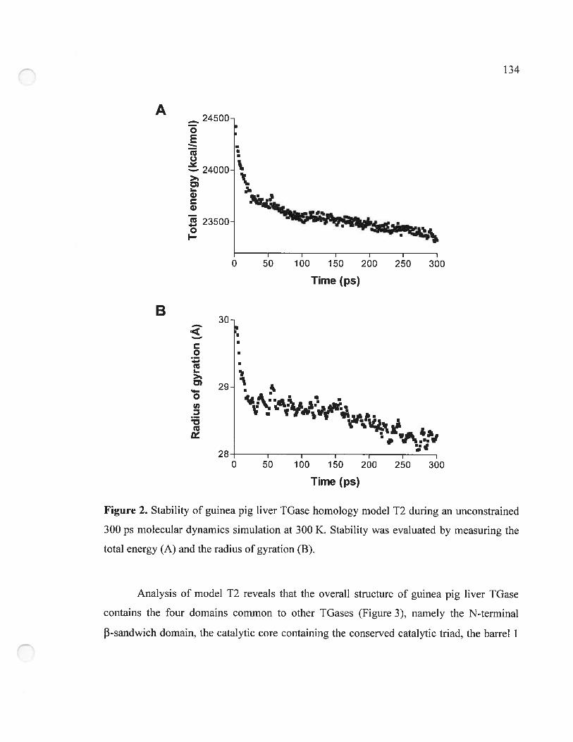
Figure 2. Stability of guinea pig liver IGase homology mode! T2 during an unconstrained
300 ps molecular dynamics simulation at 300 K. Stability was evaluated by measuring the
total energy (A) and the radius of gyration (B).
Analysis of model T2 reveals that the overall structure of guinea pig liver TGase
contains the four domains common to other TGases (Figure 3), namely the N-terminal
13-sandwich domain, the catalytic core containing the conserved catalytic triad, the barrel Y
1,13
..
A
B
24000 ‘I
•1.
r
24500o
tuo.
>1
D)I.ww
23500oI
O
30
otuI
D)
o(nz
tu
I
50 100 150 200
Tïme f ps)
250 300
I
.
“nI I I I I
0 50 100 150 200 250 300
Tïme f ps)
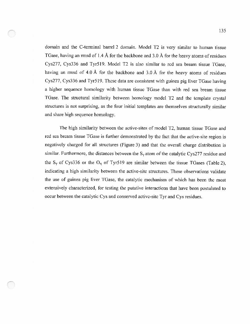
135
dornain and the C-terminal barre! 2 dornain. Mode! 12 is very similar to human tissue
TGase, having an rrnsd of 1.4 À for the backbone and 3.0 À for the heavy atoms ofresidues
Cys277, Cys336 and Tyr519. Mode! T2 is a!so similar to red sea bream tissue TGase,
having an rrnsd of 4.0 À for the backbone and 3.0 À for the heavy atoms of residues
Cys277, Cys336 and Tyr519. These data are consistent with guinea pig liver TGase having
a higher sequence homology with human tissue TGase than with red sea bream tissue
TGase. The structural sirnilarity between hornology model T2 and the template crystal
structures is flot surprising. as the four initial templates are thernselves structurally sirnilar
and share high sequence hornology.
The high simi!arity between the active-sites of model T2, hurnan tissue TGase and
red sea bream tissue TGase is further demonstrated by the fact that the active-site region is
negatively charged for all structures (Figure 3) and that the overall charge distribution is
sirnilar. furtherrnore, the distances between the S atom of the catalytic Cys277 residue and
the S of Cys336 or the O of Tvr519 are sirnilar between the tissue TGases (Table 2),
indicating a high sirni!arity between the active-site structures. These observations va!idate
the use of guinea pig liver TGase. the catalytic mechanism of which has been the most
extensively characterized. for testing the putative interactions that have been postulated to
occur between the catalytic Cys and conserved active-site Tyr and Cys residues.
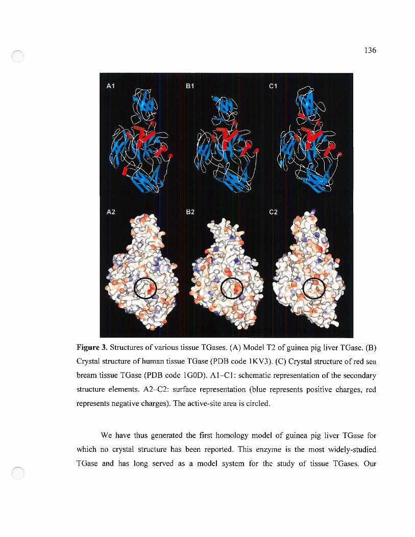
We have thus generated the first hornology model of guinea pig liver IGase for
which no crystal structure has been reported. This enzyme is the most widely-studied
TGase and has long served as a model system for the study of tissue TGases. Our
ÇI—,li
Figure 3. Structures of various tissue IGases. (A) Model T2 of guinea pig liver TGase. (B)
Crystal structure ofhuman tissue TGase (PDB code 1KV3). (C) Crystal structure ofred sea
bream tissue TGase (PDB code 1GOD). Al—Cl: schematic representation of the secondary
structure elements. A2—C2: surface representation (blue represents positive charges, red
represents negative charges). The active-site area is circled.
137
homology model will find general application in the study of structure-function
relationships and in the design ofnew substrates and inhibitors.
5.4.2 Construction of plasmid pQE32-GTG
Expression plasmid pQE32-GTG was constructed to facilitate our mutagenesis
experiments on the 2000-bp long guinea pig liver IGase gene. The SalI and AgeI
restriction sites were introduced at positions 799 bp and 172$ bp of plasrnid pQE32-TGase
through silent mutations of the guinea pig liver TGase gene to allow PCR mutagenesis of a
92$-bp DNA fragment instead of the entire 2000-bp gene. This precaution was taken to
minimize the occurrence of non-specffic mutations during the subsequent mutagenic site
overlap extension PCR. In addition, the 41 -bp sequence between the stop codon of the
guinea pig liver TGase gene and the PstI site of plasmid pQE32-TGase was removed to
eliminate an undesired SalI restriction site and to facilitate DNA sequencing from the 3’ end
ofthe gene which had been hindered due to its high GC content (7$ %, including a 12-hp G
repeat). These mutations have no effect on the amino acid sequence of the wild-type guinea
pig liver TGase, nor on its specific activity when expressed in E. cou (data not shown).
5.4.3 Role of guinea pig liver TGase residue Cys336
It has been reported that guinea pig liver TGase loses activity steadily during
storage and that much of the lost activity can be restored by addition of 1-5 mM of
dithiothreitol [21, 22]. This suggests that oxidation is the cause of inactivation. For this
reason, we store TGase samples at -$0 °C in srnall aliquots that are thawed and discarded
afier a 1 -day experiment. Recently, it has been proposed that the S atom of the catalytic
Cys residue ofred sea bream tissue TGase (Cys272) can form a disulphide bond with the S
atom of a second conserved Cys residue (Cys333) located 4.7 À away according to the
crystal structure [15]. Disulphide bond formation would inactivate the enzyme. This
hypothesis is consistent with the time-dependent loss of activity of guinea pig liver TGase,
1—,Ii
also a tissue TGase. To test the hypothesis of oxidative enzyme inactivation via disuiphide
bridge formation, we rnutated the second conserved Cys in guinea pig liver TGase (Cys336,
corresponding to red sea bream tissue TGase Cys333). The guinea pig liver enzyme was
rnutated as it is the rnost extensively characterized tissue TGase, allowing a correlation with
previous mechanistic work. Cys336 was mutated to Ala or Val; no undesired secondary
mutations were observed. We detenuined the catalytic parameters of the Cys336 mutants
using the DMPDA assay (Table 3) because it uses a glutamine-containing peptide as a
donor substrate, closely resembling the natural substrates of the enzyme. The catalytic
efficiency of both mutants is approximately 50 % that of the wild-type enzyme. For mutant
Cys336Ala, an increase in KM accounts for most ofthe decreased catalytic efficiency. while
a decrease in kcat accounts for most of the decreased activity in the case of mutant
Cys336Val. These results confirm that residue Cys336 does not play a direct role in
catalysis nor does the mutation ofthis active-site residue have a large effect on activity.
Table 3. Kinetic constants for Cbz-Gln-Gly of wild-type and mutant guinea pig liver
TGases determined using the DMPDA method
TGase KM kcat kcat/KM kcat/KM mutant / kcat/KM WT
(mM) (rnin1) (rnin’mM’)
Wild-type 7+1 116+7 17 1.0
Cys336Ala 17+2 110+7 6 0.4
Cys336Val 5+1 47+5 9 0.5
Tyr5l9Phe 8+1 85+4 11 0.6
Tyr5l9His 4+2 19+2 5 0.3
Tyr5l9Leu 6+2 67+10 11 0.6
Tyr5l9Gln 7 ± 2 48 ± 4 7 0.4
Cys336Val + Tyr5l9His 5 + 1 44 + 5 9 0.5
Cys336Val + Tyr5l9Leu 8 + 2 43 + 5 5 0.3
Cys336Val+Tyr5l9Gln 4.3+0.6 46+2 11 0.6
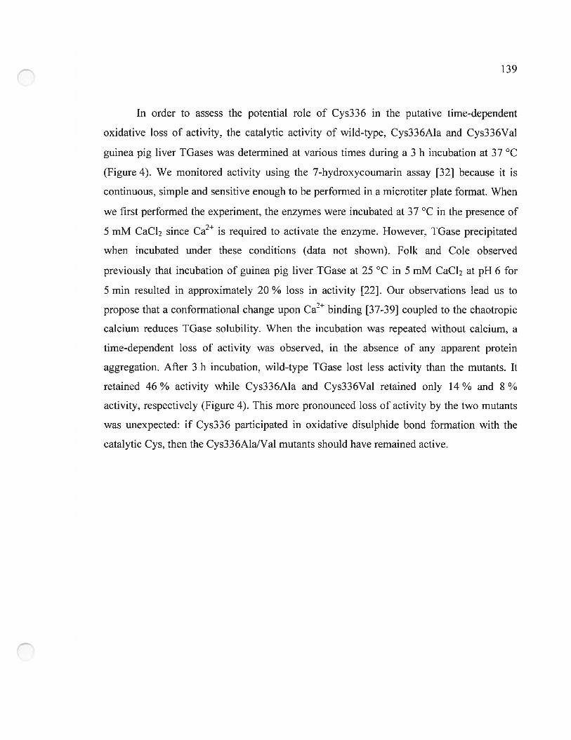
139
In order to assess the potential role of Cys336 in the putative tirne-dependent
oxidative loss of activity, the catalytic activity of wild-type, Cys336Ala and Cys336Val
guinea pig liver IGases was determined at various times during a 3 h incubation at 37 oc
(Figure 4). We monitored activity using the 7-hydroxycoumarin assay [32] because it is
continuous, simple and sensitive enough to be perforrned in a microtiter plate format. When
we first performed the experiment, the enzymes were incubated at 37 °c in the presence of
5 mM cacl2 since Ca2 is required to activate the enzyme. However, TGase precipitated
when incubated under these conditions (data not shown). Folk and cole observed
previously that incubation of guinea pig liver IGase at 25 oc in 5 mM CaC12 at pH 6 for
5 min resulted in approxirnately 20 ¾ loss in activity [22]. Our observations lead us to
propose that a conformational change upon Ca2 binding [37-39] coupled to the chaotropic
calcium reduces IGase solubility. When the incubation was repeated without calcium, a
time-dependent loss of activity was observed, in the absence of any apparent protein
aggregation. Afier 3 h incubation, wild-type TGase lost less activity than the mutants. It
retained 46 ¾ activity while Cys336Ala and Cys336Val retained only 14 % and $ %
activity, respectively (Figure 4). This more pronounced loss of activity by the two mutants
was unexpected: if cys336 participated in oxidative disulphide bond formation with the
catalytic cys, then the Cys336AlaIVal mutants should have remained active.
140
w
___
L
w>
w
Figure 4. Activity loss of wild-type and Cys336 mutants of guinea pig liver TGase upon
incubation at 37 °C. The assay used to measure activity was the 7-hydroxycoumarin assay.
Following incubation at 37 oc for 3 h. 5 mM dithiothreitol was added to the enzyme
aliquots. Dithiothreitol was expected to reduce the putative cys277-cys336 disuiphide
bond in the inactivated wild-type TGase. However, dithiothreitol did flot allow recovery of
activity for the wiÏd-type TGase (data not shown). While the 7-hydroxycoumarin assay is
rapid and sensitive, it features an aromatic ester as a donor substrate for TGase. This ester is
also sensitive to nucleophilic attack by dithiothreitol. To eliminate the possibility that the
experimental controls were flawed, the experiment was repeated using the hydroxamate
assay which is flot sensitive to dithiothreitol. Using this assay, we observed that afier 3 h
incubation, wild-type TGase retained 53 ¾ activity while cys336AIa and cys336val
retained 25 % and 12 % activity, respectively, consistent with our previous resuits. The
confirmation that mutants Cys336Ala and Cys336Val are inactivated more rapidly than the
wild-type invalidates the hypothesis of oxidation of the catalytic Cys residue through the
• WiId type—o-— Cys336AIa
• Cys336VaI
0.0 0.5 1.0 1.5 2.0 2.5 3.0
Incubation time (h)
141
formation of a disuiphide bridge with Cys336. Furthermore, according to our bomology
model, no other Cys residue is located within disuiphide-forming distance of the catalytic
Cys residue. Again, addition of 5 mM dithiothreitol did not resuit in the recovery of activity
of the wild-type TGase. These resuits dernonstrate that inactivation of IGase over time
does not result from formation of a Cys277-Cys336 disuiphide bond. However, oxidation
of the catalytic Cys residue to a species that cannot be reduced by dithiothreitol is stili
possible. Not unexpectedly, no reactivation ofthe Cys336AlalVal mutants was achieved by
addition of dithiothreitol. The more rapid inactivation and greater extent of inactivation of
the two mutants may result from less stable folding.
5.4.4 Role ofguinea pig liver TGase residue Tyr519
Molecular modeling performed on the crystal structure of red sea bream tissue
TGase bas suggested that residue TyrS 15 forrns a critical H-bond with the donor substrate
Cbz-Gln-Gly during the acylation step of the TGase rnechanisrn [20]. This H-bond would
orient the substrate y-carboxarnide group for nucleophilic attack by the catalytic thiol
(Cys272) and increase electrophilicity of the 7-carboxamide. making it more reactive. Afier
nucleophilic attack, the resulting oxyanion would also be stabilized by the H-bond forrned
with the hydroxyl group of the conserved TyrS 15, stabilizing the tetrahedral interniediate
formed during the acylation step. To test this hypothesis, homologous residue Tyr519 of
guinea pig liver TGase was mutated to amino acids that cannot form a H-bond with the
substrate (Phe or Leu), as well as to amino acids that have the potential to fonn a H-bond
with the substrate (His or GIn). According to our hypothesis [20], if loss of the H-bond
reduces the rate of the acylation step to the point where it becomes rate-lirniting, the Leu
and Phe mutants would lose activity while the Gln and Ris mutants may retain activity.
The DMPDA kinetic assay was used to measure the effect of these mutations on the
catalytic activity. This assay was chosen because it is continuous, direct and. rnost
importantly, uses the modelled Cbz-Gln-Gly donor substrate that would require the
142
proposed H-bond with Tyr5 19 to be activated. This is flot the case for direct continuous
assays that use aromatic esters as donor substrates such as the 7-hydroxycournarin assay
[32] or the Cbz-L-Glu(y-p-nitrophenyl ester)Gly assay [8]. Ail single mutants showed a
two- to four-fold decrease in catalytic efficiency relative to wild-type, mostly as a resuit of
decreased kcat (Table 3). Thus, contrary to our hypothesis regarding residue Tyr5l 9, arnino
acids with different potentials for H-bond formation yielded similar decreases in activity.
Clearly, TyrS 19 is flot critical for catalytic activity, nor does it appear that H-bonding by
Tyr5 19 plays a large role in stabilization of the transition state of the rate-limiting
deacylation step. Furthermore, contrary to what lias been proposed in the literature [15],
mutation of this Tyr to an amino acid that cannot forrn H-bonds does flot lead to immediate
inactivation of tissue TGase via disulphide bridging ofthe catalytic Cys.
We [8] and others [9] have observed the acceleration of the steady-state rate of
transamidation mediated by TGase, using a series of increasingly nucleophilic acceptor
substrates. The fact that the steady-state rate increases without attaining a plateau of activity
is consistent with the acylation step being faster than deacylation, even with the most
nucleophilic acceptor substrates chosen [8, 9]. We postulate that Tyr519 may play an
important role in activation of the donor substrate during the acylation step, but the loss of
activity caused by the TyrS 19 mutations is not sufficient to render the acylation step
significantly slower than the deacylation step. The observed modest decreases in the
steady-state rate constants are more likely due to the slight decrease in rate of deacylation,
through the disruption of transition state stabilisation by H-bonds or other dipole or
hydrophobic interactions. The activity assays used in this study are not amenable to
measuring the rate constants of the separate acylation and deacylation steps, but we are
currently undertaking rapid-mix quench experiments to quantitatively analyze the “burst”
phase to measure rate constants for the acylation step ofthese mutants.
Mutation of TyrS 19 in combination with Cys336 yields results sirnilar to mutation
of either residue alone, indicating that mutation of two residues in very close proximity to
1 4“-f-,
the catalytic Cys residue does flot inactivate the enzyme. No co-operative effect is observed
that is dïfferent from the resuits of the single mutations. Our homology model and the
crystal structures available for other TGases suggest that these residues are in close
proximity to the catalytic Cys residue, so it is surprising that they can be combinatorially
replaced and yield mutants with near-native activity. Thus, the active-site of guinea pig
liver TGase appears to be permissive to mutations ofnon-catalytic residues.
5.5 Conclusion
In summary, we have generated the first model of guinea pig liver TGase, the most
widely studied tissue TGase. The quality of this model was validated using four different
criteria: backbone conformation, residue contacts, residue interactions and overall stability.
Ah values for these criteria fail within the himits of reliable structures. The model was
similar to the crystal structures of other tissue TGases, vahidating the use of guinea pig hiver
IGase for mutagenesis experiments based on structure/function considerations. Mutation of
active-site residues Cys336 and Tyr519, alone or in combination, yiehded mutant enzymes
that had only a two- to four-fold decrease in catalytic efficiency relative to wild-type,
suggesting that the active-site of tissue IGase is tolerant to mutations. Furthermore, we
demonstrated that residue Cys336 does not form an inactivating disulphide bridge with the
catalytic Cys residue as had previously been suggested.
5.6 Acknowledgements
R.A.C. was recipient of a Natural Sciences and Engineering Research Council of
Canada graduate scholarship. This research was funded by a grant from the Fonds
Québécois de la recherche sur la nature et les technologies (jointly held by J.N.P. and
J.W.K.).
144
5.7 References
1 Lorand, L. and Graham, R. M. (2003) Transglutarninases: crosslinking enzymes
with pleiotropic functions. Nature Reviews Molecular Ceil Biology 4, 140-156
2 Griffin, M., Casadio, R. and Bergarnini, C. M. (2002) Transglutarninases: nature’s
biological glues. Biochernical Journal 368, 3 77-396
3 Beninati, S. and Piacentini. M. (2004) The transglutaminase family: an overview:
minireview article. Amino Acids 26, 367-3 72
4 Fesus, L. and Piacentini, M. (2002) Transglutaminase 2: an enigmatic enzyme with
diverse functions. Trends in Biochemical Sciences 27, 534-539
5 Mehta, K. (2005) Mammalian transglutarninases: a family portrait. Progress in
Experimental Tumor Research 38. 1-1$
6 Connellan, J. M. and Folk, J. E. (1969) Mechanism ofthe inactivation of guinea pig
liver transglutaminase by 5,5-dithiobis-(2-nitrobenzoic acid). Journal of Biological
Chemistry 244, 3 173-3181
7 Aeschlimairn, D. and Paulsson, M. (1994) Transglutaminases: protein cross-linking
enzymes in tissues and body fluids. Thrornb. Haemost. 71, 402-415
$ Leblanc, A., Gravel, C., Labelle, J. and Keillor, j• W. (2001) Kinetic studies of
guinea pig liver transglutaminase reveal a general-base-catalyzed deacylation
mechanism. Biochemistry 40, $335-8342
9 Case, A. and Stem, R. L. (2003) Kinetic analysis of the action of tissue
transglutaminase on peptide and protein substrates. Biochemistry 42, 9466-94$ 1
10 Fox, B. A., Yee, V. C., Pedersen, L. C., Le Trong, I.. Bishop, P. D., Stenkamp, R. E.
and Teller, D. C. (1999) Identification of the calcium binding site and a novel
ytterbium site in blood coagulation factor XIII by x-ray crystallography. Journal of
Biological Chemistry 274, 49 17-4923
145
11 Pedersen, L. C., Yee, V. C., Bishop, P. D., Le Trong, I., Teller, D. C. and Stenkarnp,
R. E. (1994) Transglutaminase factor XIII uses proteinase-like catalytic triad to
crosslink macromolecules. Protein Science 3, 113 1-1 135
12 Yee, V. C., Pedersen, L. C., Le Trong, I.. Bishop, P. D., Stenkarnp, R. E. and Teller,
D. C. (1994) Three-dirnensional structure of a transglutarninase: human blood
coagulation factor XIII. Proceedings of the National Academy of Sciences of the
United States ofAmerica 91. 7296-7300
13 Weiss, M. S., Metzner, H. J. and Hilgenfeld, R. (1998) Two non-proline cis peptide
bonds may be important for factor XIII function. FEBS Letters 423, 291-296
14 Liu, S., Cerione, R. A. and Clardy, J. (2002) Structural basis for the guanine
nucleotide-binding activity of tissue transglutaminase and its regulation of
transamidation activity. Proceedings of the National Acaderny of Sciences of the
United States ofArnerica 99, 2743 -2747
15 Noguchi, K., Ishikawa, K., Yokoyama. K., Ohtsuka, T., Nio, N. and Suzuki. E.
(2001) Crystal structure of red sea bream transglutaminase. Journal of Biological
Chemistry 276, 12055-12059
16 Ahvazi, B., Kim, H. C., Kee, S. H., Nernes, Z. and Steinert, P. M. (2002) Three
dimensional structure of the hurnan transglutaminase 3 enzyme: binding of calcium
ions changes structure for activation. EMBO Journal 21, 2055-2067
17 Ahvazi, B., Boeshans, K. M. and Steinert, P. M. (2004) Crystal structure of
transglutaminase 3 in complex with GMP: structural basis for nucleotide specificity.
Journal ofBiological Chemistry 279, 26716-26725
1$ Ahvazi, B., Boeshans, K. M., Idler, W., Baxa, U. and Steinert, P. M. (2003) Roles
of calcium ions in the activation and activity of the transglutaminase 3 enzyme.
Journal ofBiological Chemistry 278, 23834-23841
19 Ahvazi, B., Boeshans, K. M., Idler, W., Baxa, U.. Steinert, P. M. and Rastinejad, f.
(2004) Structural basis for the coordinated regulation of transglutaminase 3 by
146
guanine nucleotides and calcium/magnesium. Journal of Biological Chemistry 279,
7180-7192
20 Chica, R. A., Gagnon, P., Keillor, J. W. and Pelletier, J. N. (2004) Tissue
transglutaminase acylation: Proposed role of conserved active-site Tyr and Trp
residues revealed by molecular modeling of peptide substrate binding. Protein
Science 13, 979-991
21 Folk, J. E. and Chung, S. I. (1985) Transglutaminases. Methods in Enzymology
113, 358-375
22 Folk, J. E. and Cole, P. W. (1966) Transglutaminase: mechanistic features of the
active-site as determined by kinetic and inhibitor studies. Biochimica & Biophysica
Acta 122, 244-264
23 Notredame, C., Higgins, D. G. and Heringa, J. (2000) T-Coffee: A novel method for
fast and accurate multiple sequence alignment. Journal of Molecular Biology 302,
205-217
24 Thornpson, J. D., Higgins, D. G. and Gibson, T. J. (1994) CLUSTAL W: improving
the sensitivity of progressive multiple sequence alignment through sequence
weighting, position-specific gap penalties and weight matrix choice. Nucleic Acids
Research 22, 4673-4680
25 Sali, A. and Blundeil, T. L. (1993) Comparative Protein Modeling by Satisfaction of
Spatial Restraints. Journal ofMolecular Biology 234, 779-8 15
26 Lovell, S. C., Davis, I. W., Arendall, W. B., 3rd, de Bakker, P. I., Word, J. M.,
Prisant, M. G., Richardson, J. S. and Richardson, D. C. (2003) Structure validation
by Caipha geometry: phi,psi and C beta deviation. Proteins 50, 437-450
27 Hoofi, R. W. W., Vriend, G., Sander, C. and Abola, E. E. (1996) Errors in Protein
Structures. Nature 381, 272
28 Sippl, M. J. (1993) Recognition ofErrors in 3-Dimensional Structures ofProteins.
Proteins 17, 355-362
147
29 Ho, S. N., Hunt, H. D., Horton, R. M., Pullen, J. K. and Pease, L. R. (1989) Site
directed mutagenesis by overlap extension using the polyrnerase chain reaction.
Gene 77, 5 1-59
30 Gillet, S. M., Chica, R. A., Keillor, J. W. and Pelletier, J. N. (2004) Expression and
rapid purification of highly active hexahistidine-tagged guinea pig liver
transglutarninase. Protein Expression & Purification 33, 256-264
31 Yokoyama, K., Kikuchi, Y. and Yasueda, H. (199$) Overproduction of DnaJ in
Eseherichia cou improves in vivo solubility of the recombinant fish-derived
transglutaminase. Bioscience, Biotechnology & Biochemistry 62, 1205-1210
32 Gillet, S. M., Pelletier, J. N. and Keillor, J. W. (2005) A direct fluorometric assay
for tissue transglutaminase. Analytical Biochernistry 347, 22 1-226
33 de Macedo, P., MalTano, C. and Keillor, J. W. (2000) A direct continuous
spectrophotometric assay for transglutarninase activity. Analytical Biochemistry
285, 16-20
34 Folk, J. E. and Cole, P. W. (1966) Identification of a functional cysteine essential
for the activity of guinea pig liver transglutaminase. Journal of Biological
Chemistry 241, 3238-3240
35 Garzon, R. J., Pratt, K. P., Bishop. P. D., Le Trong, I., Stenkarnp, R. E. and Teller,
D. C. (2000) Tryptophan 279 is Essential for the Transglutaminase Activity of
Coagulation Factor XIII: Functional and Structural Characterization. To be
published
36 Janin, J. and Wodak, S. (1978) Conformation ofarnino acid side-chains in proteins.
Journal ofMolecular Biology 125, 357-3 86
37 Casadio, R., Polverini. E., Mariani, P.. Spinozzi, F., Carsughi, F., fontana, A.,
Polverino de Laureto, P., Matteucci, G. and Bergarnini, C. M. (1999) The structural
basis for the regulation of tissue transglutaminase by calcium ions. European
Journal ofBiochemistry 262. 672-679
142
38 Di Venere, A.. Rossi. A.. De Matteis, F.. Rosato. N.. Agro. A. F. and Mei. G. (2000)
Opposite effects of Ca(2+) and GTP binding on tissue transglutarninase tertiary
structure. Journal ofBiological Chemistry 275. 3915-3921
39 Mariani. P.. Carsughi, F.. Spinozzi, F.. Rornanzetti, S., Meier, G., Casadio, R. and
Bergarnini, C. M. (2000) Ligand-induced conformational changes in tissue
transglutaminase: Monte Carlo analysis of srnall-angle scattering data. Biophysical
Journal 78, 3240-3251
CHAPITRE 6
Évolution dirigée de la spécificité de la TGase de foie de
cobaye par une approche semi-aléatoire
150
6.0 Préface
À la suite de l’obtention d’information structurelle et fonctionnelle et du
développement d’une méthode d’expression et de purification de la IGase de foie de
cobaye, il nous fallait développer une méthode de criblage pour détecter les mutants ayant
une spécificité modifiée, afin de pouvoir faire l’évolution dirigée de l’enzyme. Dans ce
chapitre, nous décrivons la création de cinq banques de mutants et leur criblage avec de
nouveaux composés fluorogènes, durant le processus d’évolution dirigée selon une
approche semi-aléatoire de la TGase de foie de cobaye.
Pour le manuscrit « Expansion of the peptide synthase specificity of guinea pig liver
transglutaminase by serni rational mutagenesis» présenté au chapitre 6, j’ai effectué la
majorité des expériences d’expression et de purification des TGases, de cinétique
enzymatique, de construction des banques de mutants et de criblage, en plus de rédiger le
manuscrit. Jessica Laroche et Philippe Lesage m’ont assisté dans les expériences
d’expression et de purification des IGases, de cinétique enzymatique, de construction des
banques de mutants et de criblage en tant que stagiaires sous ma supervision. Le Dr Steve
Gillet a développé le protocole de criblage en plaques à 96 puits en plus de synthétiser les
composés Cbz-Gly-7HC, Cbz-Ala-7HC et Cbz-Phe-7HC. Le Pr Joelle Pelletier a effectué
les calculs de représentativité des banques de mutants.
151
Article 5.
Expansion of the peptide synthase specificity of guinea
pig liver transglutaminase by semi-random mutagenesis
Roberto A. Chica, Steve M.F.G. Gillet, Jessica Laroche, Philippe Lesage,
Jeffrey W. Keillor and Joelle N. Pelletier
Département de chimie. Université de Montréal, CP 612$, Succursale Centre-Ville,
Montréal. Québec, H3C 3J7, Canada
Manuscrit prêt à être soumis
152
6.1 Abstract
Transglutarninases (TGases) catalyze the cross-linking of proteins and peptides by
the formation of y-glutarnyl-E-lysyl bonds between an acyl-donor and an acyl-acceptor
substrate. In vivo, the acyl-donor substrate is a glutamine-containing protein while the acyl
acceptor is a lysine-containing protein. The acyl-acceptor substrate can be replaced in vitro
by prirnary amines. Recently, we dernonstrated the use of a novel class of acyl-donor
substrates of guinea pig liver TGase that contain 7-hydroxycoumarin esters. In this study,
we explored the substrate specificity of guinea pig liver TGase by assessing its reactivity
toward a variety ofpotential acyl-donor substrates having a 7-hydroxycournarin ester on the
Œ-carboxyl group ofvarious amino acids. We also assessed its reactivity toward a variety of
amino acid derivatives as acyl-acceptor substrates. We observed that the wild-type enzyme
can react directly at the a-carboxyl group of N-carbobenzyloxyglycine cournarin-7-yl ester,
dernonstrating for the first tirne that guinea pig liver TGase bas a peptide synthase activity
as it can catalyze the formation of peptide bonds between Gly and a series of small polar
and hydrophobie arnino acid derivatives. To further broaden its specificity, we performed
serni-random mutagenesis by perforrning saturation and ofien combinatorial mutagenesis at
conserved active-site residues (Trp241; His305 and Asp306; Trp332; Thr360, G1n362 and
G1u363; TyrS 19 and Asn520). We developed a microtiter plate-based screening assay that
allowed identification of mutants with altered specificity. Mutant Trp332Phe recognized
L-Phe and L-Tyr derivatives as acyl-acceptor substrates while the wild-type enzyme did not,
demonstrating that serni-random mutagenesis of the active-site rapidly yielded changes in
acyl-acceptor substrate specificity.
Keywords: tissue transglutarninase, TGase, semi-random mutagenesis, peptide
synthase, combinatorial mutagenesis, enzyme kinetics
1I 3.)
6.2 Introduction
Transglutarninases (IGases. EC 2.3.2.13) catalyze the Ca2-dependent cross-linking
ofpeptides and proteins vici the formation ofy-glutamyl-E-lysyl isopeptide bonds [1-3]. The
catalytic reaction follows a modified ping-pong rnechanism in which a glutamine
containing protein or peptide, the acyl-donor substrate, reacts with the catalytic cysteine
residue to form a thioester bond, thus generating a covalent acyl-enzyrne intermediate. The
acyl-enzyme intermediate then reacts with a second substrate, the acyl-acceptor, to yield the
isopeptide-containing product and free enzyme in a transamidation reaction. In the absence
of an acyl-acceptor. the acyl-enzyme intermediate is hydrolyzed. transforming the acyl
donor glutamine residue into glutamate and regenerating the free enzyme [4]. Catalysis is
carried out by a Cys-His-Asp catalytic triad sirnilar to that of cysteine proteases. TGases are
divided into nine classes [5] of which the tibiquitous tissue TGase found in all vertebrates
t61 is the rnost widely distributed in eukaryotic organisrns. Tissue IGases. such as guinea
pig liver TGase. are intracellular and monomeric enzymes whose transarnidation activity is
inhibited by GTP.
Tissue TGases exhibit a broad specificity towards the acyl-acceptor as almost any
prirnary amine can serve as a substrate [7] although in nature it is generally a lysine
containing protein or peptide. Many primary amines, such as glycinarnide [8, 9], and
anilines, such as ÀN-dirnethyl-1,4-phenylenediarnine [10], can also function as acyt
acceptor substrates. However, amines containing free carboxylic acid groups, such as free
amino acids. and amines with adjacent bulky substituents, such as L-tyrosinamide. do not
act as substrates [9].
Tissue TGase displays narrower specificity for its acyl-donor substrates. The side
chain of protein-bound L-Gln residue is the native substrate while the side chains of similar
amino acids such as L-Asn and D-Gin are not acyl-donor substrates [8]. Synthetic peptides
containing L-Gln are also acyl-donor substrates of TGases. For this reason, the dipeptide
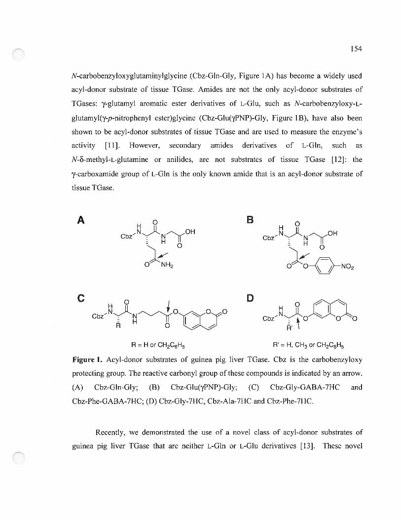
154
N-carbobenzyioxygiutaminyigiycine (Cbz-Gin-Giy, Figure lA) has become a wideiy used
acyl-donor substrate of tissue TGase. Amides are flot the oniy acyl-donor substrates of
IGases: y-giutamyi aromatic ester derivatives of L-Glu, such as N-carbobenzyioxy-L
giutamyi(y-p-nitrophenyi ester)giycine (Cbz-Glu(’yPNP)-Giy, Figure 1 B), have also been
shown to be acyl-donor substrates of tissue IGase and are used to measure the enzyme’s
activity j1 1]. However, secondary amides derivatives of L-Gin, such as
N-8-methyi-L-giutamine or anilides, are flot substrates of tissue TGase [121: the
y-carboxarnide group of L-Gin is the only known amide that is an acyl-donor substrate of
tissue TGase.
A BCbzNfl0H
ONO2
H°D
CbzN
CbzN
R = H or CH2C6H5 R = H, CH3 or CH2C6H5
Figure 1. Acyi-donor substrates of guinea pïg liver TGase. Cbz is the carbobenzyioxy
protecting group. The reactive carbonyi group ofthese compounds is indicated by an arrow.
(A) Cbz-Gin-Giy; (B) Cbz-Glu(yPNP)-Giy; (C) Cbz-Giy-GABA-7HC and
Cbz-Phe-GABA-7HC; (D) Cbz-Gly-7HC, Cbz-Ala-7HC and Cbz-Phe-7HC.
Recentiy, we demonstrated the use of a novel class of acyi-donor substrates of
guinea pig liver TGase that are neither L-Gin or L-Giu derivatives [13]. These novei
155
substrates, 4-(N-carbobenzyloxyglycylamino)-butyric acid coumarin-7-y! ester and
4-(N-carbobenzyloxyphenyla!any!amino)-butyric acid coumarin-7-yl ester
(Cbz-Gly-GABA-7HC and Cbz-Phe-GABA-7HC, respectively, figure 1 C), have scaffolds
that resemble those of recent!y published TGase irreversible inhibitors [14, 151. Upon
reaction with TGase, 7-hydroxycournarin is released, resulting in an increase in
fluorescence that makes these compounds useful for measuring the rates of the hydrolysis
or transamidation reactions catalyzed by guinea pig liver TGase. The scaffolds of these
substrates differ from the typical L-Glu arornatic ester acyl-donor substrates of tissue
TGases in that the reactive ester function is located on the main chain instead of the side
chain, thus giving rise to products that do not contain a y-glutamyl-E-lysyl isopeptide bond.
These resuits illustrate that specificity for acyl-donor substrates with aromatic ester
functions is broader than had previously been supposed and demonstrate that the enzyme
can generate products with nove! scaffo!ds.
In this study, we exp!ored the substrate specificity of guinea pig !iver TGase by
assessing its reactivity toward a variety of potentia! acy!-donor substrates having an
aromatic ester function on the a-carboxy! group of various amino acids (Figure I D). As
we!!, we assessed its reactivity toward a variety of amino acid derivatives as acy!-acceptor
substrates. We observed that the wild-type enzyme can react directly on the Œ-carboxyl
group of N-carbobenzyloxyg!ycine cournarin-7-y! ester (Cbz-Gly-7HC, Figure 1 D),
demonstrating for the first time that guinea pig liver IGase has an intrinsic peptide synthase
activity as it can cata!yze the formation of peptide bonds between Gly and a series of sma!!
polar and hydrophobic amino acid derivatives.
Furthermore, we performed serni-random mutagenesis of guinea pig !iver TGase to
further broaden its specificity. five libraries of mutants were thus created by performing
saturation and often combinatoria! mutagenesis at conserved active-site residues (Trp24 1;
His305 and Asp306; Trp332; 1hr360, G!n362 and G!u363; Tyr519 and Asn520). To
determine the activity of >3000 mutants from the five libraries, we deve!oped a rapid
156
microtiter plate-based screening assay. Using this assay, we identified a mutant with altered
specificity for the acyl-acceptor substrate. Single mutant Trp332Phe recognized L-Phe and
L-Tyr derivatives as acyl-acceptor substrates while the wild-type enzyme did flot,
demonstrating that semi-random mutagenesis of the active-site rapidly yielded changes in
acyl-acceptor substrate specificity.
6.3 Materials and Methods
6.3.1 Materials
Ail reagents used were of the highest available purity. Restriction and DNA
modifying enzymes were purchased from New England Biolabs (Mississauga, ON) and
MBI fermentas (Burlington, ON). Cellytic B buffer, lysozyme, N-Œ-acetyl-L-lysine methyl
ester hydrochloride (N-AcLysOMe), glycinamide (G1yNH2) and L-amino acid methyl ester
hydrochiorides (ValOMe, SerOMe, PheOMe, TyrOMe, TrpOMe and ProOMe) were
purchased from Sigma-Aldrich (Oakville, ON). L-phenylalaninamide (PheNH2) and
L-alaninamide hydrochiorides (A1aNH2) were purchased from Novabiochem (Mississauga,
ON). QIAPREP plasmid purification kit, QIAEXII agarose gel extraction kit and Ni-NTA
agarose resin were purchased from Qiagen (Mississauga, ON). Synthetic oligonucleotides
were obtained from Alpha DNA (Montreal, QC) and Integrated DNA Technologies
(Coralville, TA). Infra-red dye-labelled sequencing primers were from Li-Cor
Bioteclmology Division (Lincoïn, NB). Ail aqueous solutions were prepared using water
purified with a Millipore BioCeli system. DNA sequencing was performed using a kit for
Sequenase version 2.0 DNA polyrnerase (Amersham—Pharmacia Biotech) with a Li-Cor
1R2 autornated system.
157
6.3.2 Synthesis of Cbz-GIy-7HC, Cbz-Ala-7HC and Cbz-Phe-7HC
Cbz-Gly-7HC was synthesized based on a previously reported protocol [13].
Narnely, 0.2 g (1 mmol) of CbzGly and 0.4 g (2.5 mrnol) of 7-hydroxycoumarin (7HC)
were dissolved in 10 mL of ethyl acetate. Then 0.22 mL (0.2 g, 2 mmol) of
N-methylmorpholine and 0.8 mL (0.63 g, 5 mrnol) of N,N-diisopropylcarbodiimide were
added with stirring at room temperature. Stirring was continued until the complete
disappearance of CbzGly, as followed by thin layer chromatography (ethyl acetate). The
reaction mixture was then washed once with I M NaOH, three tirnes with 0.1 M NaOH, 3
times with 0.1 M HC1, once with saturated NaHCO3 and once with brine. The organic
phase was then dried over MgSO4, filtered and evaporated under reduced pressure. The
resulting residue was purified by silica gel chromatography (ethyl acetate) to rernove traces
of diisopropylurea, giving the desired ester in 70 % yield (0.25 g). Cbz-Ala-7HC and
Cbz-Phe-7HC were synthesized according to a similar protocol.
‘H NMR (300 MHz, CDCÏ3) 64.30 (2H. d), 5.1$ (2H, s), 5.33 (1H, s), 6.44 (1H, d),
7.0$ (1H, d), 7.10 (1H, s), 7.37 (5H, m), 7.51 (1H, d), 7.70 (1H, d). ‘3C NMR (75 MHz,
CDC13) ô 168.5, 160.6, 156.7, 154.9, 152.9, 143.1, 136.3, 129.0, 128.9, 128.6, 128.5, 118.4,
117.2, 116.6, 110.5, 67.7, 43.2. HRMS (FAB) calculated for C,9H,6N06 ([M+Hf):
354.0972, found 354.096$. Cbz-Ala-7HC: ‘H NMR (300 MHz, CDC13) 61.63 (3H, d),
4.62 (1H, m), 5.18 (2H, s), 5.33 (1H, d), 6.42 (1H, d), 7.07 (1H, d), 7.09 (1H, s), 7.39
(5H, m), 7.51 (1H, d), 7.70 (1H, U). ‘3C NMR (75 MHz, CDCJ3) ô 171.5, 160.5, 156.0,
154.9, 153.1, 143.1, 136.4, 129.0, 128.9, 128.6, 128.5, 118.4, 117.2, 116.6, 110.5, 67.5,
50.2, 18.5. HRMS (fAB) calculated for C20H,8N06 ([M+H]j: 368.1129, found 368.1118.
Cbz-Phe-7HC: ‘H NMR (300 MHz, CDC13) 63.26 (2H, m), 4.91 (1H, m), 5.19 (2H, s),
5.35 (1H, d), 6.93 (1H, m), 7.22 (2H, m), 7.39 (9H, m), 7.68 (1H, d), 7.72 (1H, d).
‘3C NMR (75 MHz, CDC13) ô 170.1, 160.5, 156.2, 154.9, 152.9, 143.1, 136.3, 135.4, 129.7,
158
129.2, 128.9, 128.6, 128.5, 127.9, 118.4, 117.2, 116.6, 110.5, 67.6, 55.4, 38.5. HRMS
(FAB) calculated for C26H22N06 ([M+Hf): 444.1442, found 444.1432.
6.3.3 Overexpression and purification ofwild-type and mutant TGases
Wild-type and specific rnutated recombinant guinea pig liver TGases of interest
were overexpressed and purified from Escherichia cou on a 1 L scale according to a
protocol developed in our laboratory [16] with the following modifications. Afier Ni-NTA
purification, the eluant was transferred to an Amicon Ultra 15 mL tube (Millipore) with a
molecular weight eut-off of 30 kDa and the TGase solution was desalted by centrifugation
with 25 mM Tris-acetate buffer (pH 7.0) containing 0.5 mM EDTA. The samples were
aliquoted, snap-frozen on dry ice and stored at -$0 °C. Typical yields were 1.5-10 mg/L of
$5 % pure protein, as estimated from Coomassie Blue staining following SDS-PAGE.
6.3.4 Kinetic assays
Ah assays were performed in triphicate. The following solutions were prepared: a
standard stock buffer solution (100 mM Mops buffer pH 7.0, 5 mM CaC12, and 0.05 mM
EDTA), a 2 mM solution of acyl-donor substrate Cbz-Gly-7HC in DMF, 200 mM solutions
of acyl-acceptor substrates N-AcLysOMe, G1yNH2, A1aNH2 and SerOMe in water and
$00 mM stock solutions ofValOMe, PheNH2, PheOMe, TyrOMe, ProOMe and TrpOMe in
50 % DMF in water. Prior to performing the assays, a ‘fluorescenee coefficient” was
determined daily by measuring the arbitrary fluorescence intensities corresponding to five
concentrations of 7-hydroxycoumarin at concentrations ranging from 0.05 to 0.5 jiM in 5 %
DMF in the stock buffer solution at 25 °C. The value of this “fluorescence coefficient”
varied only slightly (<5 %) eaeh day. For the hydrolysis reaction, activity was measured by
adding 1-3 tg of the purified enzymes to each well of a TCT Luminescence 96-well
microtiter plate (Thermo Electron) containing a 0.5-200 tM solution of the acyl-donor
substrate in stock buffer. For the transamidation reaction, the same amount of the purified
159
enzymes was added to 0.5-80 mM of the acyl-acceptor substrate in stock buffer solution
containing 100 tM of acyl-donor substrate Cbz-Gly-7HC. The acyl-acceptor substrate was
replaced by water or 50 % DMf in water in the blank. DMF was present at 5-7.5 % in the
final reaction mixtures. The increase in fluorescence due to the release of
7-hydroxycoumarin was followed against a blank at 2ex 340 nrn and 7efl1 465 flfl1 fl a
FluoStar Optima microtiter plate reader (BMG Labtech). Linear siopes of fluorescence
versus time were measured over the first <10 % conversion of substrate to product and
were converted into initial rates using the fluorescence coefficient.
6.3.5 Construction of the libraries of mutants
Five distinct guinea pig liver TGase mutant libraries were created by site-overlap
extension PCR [17] by mutating the following conserved active-site residues: Trp241
(library 1); His305 and Asp306 (library 2); Trp332 (library 3); Thr360, G1n362 and G1u363
(library 4) as well as Tyr519 and Asn520 (library 5). The template used was plasmid
pQE32-GTG [1$] that contains the wild-type guinea pig liver IGase gene with a SalI
restriction site at 799 bp and an AgeI restriction site at position 1728 bp (numbering
according to the origin of replication of plasmid pQE32-GTG). All mutations were
introduced using primers containing the degenerate “NNS” codon (where N = A, C, G or T
and S = C and G) that encodes all twenty amino acids in addition to the amber stop codon.
The arnplified fragments spanned the portion of the TGase sequence included between the
unique SalI and AgeI restriction sites of the template. They were generated with the
external primers GTG-SaÏI-799-F (5 ‘-TGCTCGCGCCGCAGTCGACCTGTCT
ACGTG-3’) and GTG-AgeI-1 728-R (5’-GTTGGTGCTGCACACCGGTCCCAGGACT-
CCATT-3’) containing the SalI and AgeI restriction sites (underlined), respectively. The
external primers were used in combination with sets of complementary pairs of degenerate
oligonucleotides that introduced mutations for libraries 1 to 4 (only the coding strands are
shown): Library 1 (GTG-W241-NNS: 5’-GCTTCAGGGACGCNNSGACAACAAC-
160
TACAGT-3’), Library 2 (GTG-H305-D306-NNS: 5’-AACTTTAACTCA-
GCCNNSNNSCAGAACAGCAACCT-3’), Library 3 (GTG-W332-M’JS:
5’-AGAGCGAGATGATCNNSAACTTCCACTGCTGG-3’) and Library 4 (GTG-Loopln:
5’-AGGCCCTGGACCCCNNSCCCNNSNNSAAGAGTGAAGGGACA-TA-3’). For
library 4, a previous step of excision of four codons at positions 360-363 (Thr360, Pro361,
G1n362 and G1u363) of the guinea pig liver TGase gene was performed using a
complernentary pair of oligonucicotides (only the coding strand is shown): GTG-LoopOut
(5’-CTGGACCCCAAGAGTGAAGGGACATACT-3’). For library 5, the mutations were
introduced into the TGase gene using a direct PCR amplification with primer
GTG-$aÏI-799-F and the degenerate oligonucleotide GTG-Y519-N520-NNS-R
(5’-CACACAACCGGTCCCAGGACTCCSNNSNNGCTGACGATGCGT-3’, AgeI
restriction site underlined).
The full-length SaluAgel amplicons were digested with SalI!AgeI, isolated by
agarose gel electrophoresis and extracted. The fragment libraries were ligated into
pQE32-GTG expression vector via SalI/AgeI yielding libraries of pQE32-GTG plasmids
expressing the mutant TGases. The libraries were transforned by electroporation into
E. cou SS320. An average of 5 x 1 0 colonies per library was obtained. Library quality was
evaluated by DNA sequencing of 16 to 32 colonies per library following propagation on LB
medium containing 100 jig/rnL of ampicillin.
Following library validation, all colonies from a given library were pooled in 1 mL
fresh LB medium supplemented with ampicillin and were snap-frozen at -so oc following
addition of 500 .tL of a sterile 50 % glycerol 1 % Nacl solution. The frozen stocks were
used to inoculate cultures for the extraction and purification of the pQE32-GTG plasrnids
harbouring the mutant TGase libraries. The purified plasmids were transformed into E. cou
XL-1 Blue cells containing the plasmid pDnaKJ t191 that encodes the molecular
chaperones DnaK and DnaJ. An average of —1 0 colonies per library was obtained.
161
Colonies were picked into individual wells of Costar 3357 polypropylene 96-well
microtiter plates (Coming) containing 100 iL of medium (LB with 100 jig/mL ampicillin
and 30 ig/rnL chloramphenicol; no ampicillin for the negative control). In each plate, well
Al contained a positive control (E. cou XL-1 Blue/pDnaKJ/pQE32-GTG expressing the
wild-type guinea pig liver TGase) whule well A2 contained a negative control (E. cou XL-1
Blue/pDnaKJ). Two plates each for libraries 1 and 3 (188 clones per library) as well as ten
plates each for libraries 2, 4 and 5 (940 clones per library) were thus prepared. The plates
were covered with a sterile Breathe-Easy gas permeable sealing membrane (Sigma) and
incubated ovemight at 37 °C with shaking at 250 rpm. After incubation, 50 .tL of a sterile
50 % glycerol 1 ¾ NaCÏ solution was added to all wells and each plate was covered with a
Storage Mat III (Coming) previously sterilized in ethanol. The resulting “mother” plates
were snap-frozen and stored at -80 °C.
6.3.6 Library representation calculations
Library representation was calculated based on a Poisson distribution, as follows.
The libraries were designed to encode n possible different theoretical variants at the DNA
level (Table 3, number of encoded genes; some redundancy exists at the protein level).
Library screening entailed sampling in colonies, randornly. from this theoretical pool; we
thus formed experimental samples of in variants per library. We calculated 7, the expected
number of missing theoretical variants within the in variants: 2 n (1h/,,) “ [20]. Library
representation is given as 1 - (?fl/) and is expressed as a percentage.
6.3.7 Preparation of the crude bacterial lysate for screening
The mother plates containing the libraries were used to inoculate sterile, round
bottom Costar 3799 (Coming) 96-well microtiter plates (“daughtcr” plates) containing
100 iL of LB per well, supplernented with the appropriate antibiotics. The daughter plates
were incubated ovemight (37 °C, 250 rpm). The bacterial cultures (5 tL) were used to
162
inoculate 100 jiL of fresh medium in new 96-well microtiter plates. The plates were
incubated (37 °C, 250 rpm) for 3 h, at which tirne protein expression was induced by
addition of 1 mM isopropyl-I3-D-thiogalactoside with overnight incubation (30 °c,250 rpm).
For celi lysis, the plates were snap-frozen on crushed dry ice and then thawed at
37 °C. Lysis buffer (20 pi ofa 6 rng/rnL lysozyrne in clltk B buffer solution) was added
to each well. The plates were incubated at 30 oc for 30 min. The crude bacterial lysate was
used directly in the screening assay.
6.3.8 Screening assay
A screening procedure was developed to measure the activity of the five mutant
libraries. This procedure utilizes cbz-xaa-7Hc esters (f igure 1 D) as acyl-donor substrates.
A standard stock buffer solution (100 mM Mops buffer pH 7.0, 5 mM cacl2, and 0.05 mM
EDTA), 1.5 mM stock solutions of acyl-donor substrates Cbz-Gly-7Hc and Cbz-Ala-7Hc
in DMF and 200 mM stock solutions of acyl-acceptor substrates N-AcLysOMe in water
and PheNH2 in 50 % DMf were prepared. Transamidation activity was measured by adding
20 tL of the crude bacterial lysates to 180 pi of reaction buffer containing 75 tM of the
acyl-donor substrate and 10 mM of the acyl-acceptor substrate in the stock buffer solution
in each well of a TCT Luminescence 96-well microtiter plate (Therrno Electron). Water
replaced acyl-acceptor substrate for the hydrolysis reaction. DMF was present at 5-7.5 % in
the final reaction mixtures. The increase in fluorescence due to the release of
7-hydroxycoumarin was followed at 2ex 340 nrn and 2em 465 nrn in a HTS 7000 plate reader
(Perkin Elmer). Linear slopes of fluorescence versus time were measured over 15 min.
163
6.4 Resuits
6.4.1 Cbz-Gly-7HC is hydrolyzed by wild-type guinea pig liver TGase
We previousiy demonstrated that guinea pig liver TGase does not require the L-Gin
or L-Glu scaffold in its acyl-donor substrates when the leaving group is an arornatic
alcohol: the enzyme reacted with Cbz-Gly-GABA-7HC as weii as Cbz-Phe-GABA-7HC
(Figure 1C) with KM values of 7-9 M and kcat values of 45-75 rnin1 [13]. b verify if
guinea pig liver TGase could react with analogs of these compounds having the
7-hydroxycoumarin leaving group directly bound to the Œ-carboxyl group of an amino acid,
N-carbobenzyloxyarnino acid coumarin-7-yl esters with substituents of varying bulk
(Cbz-Gly-7HC, Cbz-Ala-7HC and Cbz-Phe-7HC, Figure I D) were synthesized and their
reactivity was evaluated in the hydroiysis reaction. Michaelis-Menten kinetics
demonstrated that only compound Cbz-Gly-7HC is an acyl-donor substrate of wild-type
guinea pig liver TGase, having a KM of 14 + 3 tM and a kcat of 6.8 + 0.5 rnin1 (Table 1).
This KM is sirnilar to that of Cbz-Gly-GABA-7HC (9 ± 2 jiM) while the kcat is l0-fold
iower (75 + 5 min1) [13]. The lower kcat appears to be due to the absence of the
y-aminobutyric acid (GABA) linker in Cbz-Gly-7HC relative to Cbz-Gly-GABA-7HC,
which may resuit in a decreased accessibility of the substrate reactive carbonyl group for
nucleophilic attack by the catalytic thiol. An alternative explanation would be that the
absence of the GABA iinker hinders hydrolysis of the acyl-enzyme intermediate formed
during the reaction by modifïing accessibility or nucleophilicity of the acceptor water
molecule. Cbz-Ala-7HC did flot react with guinea pig liver TGase and Cbz-Phe-7HC was
not soluble enough to test against guiflea pig liver TGase. The iack of reactivity of
Cbz-Aia-7HC appears to resuit from the methyl side chain of the Ala residue that may
hinder productive binding at the active-site.
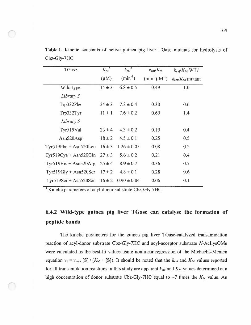
164
Table 1. Kinetic constants of active guinea pig liver TGase mutants for hydrolysis of
Cbz-Gly-7HC
TGase KMa kcata kcat!KM kcat/KM WT /
(tM) (rnin’) (min’IM1) kcat/KM mutant
Wild-type 14±3 6.8±0.5 0.49 1.0
Libraiy 3
Trp332Phe 24 ± 3 7.3 ± 0.4 0.30 0.6
Trp332Tyr 11 ± 1 7.6±0.2 0.69 1.4
Libraiy 5
Tyr5l9Val 23±4 4.3±0.2 0.19 0.4
Asn52OAsp 18 ± 2 4.5 ± 0.1 0.25 0.5
Tyr5l9Phe+Asn520Leu 16±3 1.26±0.05 0.08 0.2
Tyr5l9Cys + Asn52OGln 27 ± 3 5.6 ± 0.2 0.21 0.4
Tyr5l9His + Asn52OArg 25 ± 4 8.9 ± 0.7 0.36 0.7
Tyr5l9Gly + Asn52OSer 17 ± 2 4.8 ± 0.1 0.28 0.6
Tyr5l9Ser+Asn52OSer 16±2 0.90±0.04 0.06 0.1
a Kinetic parameters of acyl-donor substrate Cbz-Gly-7HC.
6.4.2 Wild-type guinea pig liver TGase can catalyse the formation of
peptide bonds
The kinetic parameters for the guinea pig liver TGase-catalyzed transamidation
reaction of acyl-donor substrate Cbz-Gly-7HC and acyl-acceptor substrate N-AcLysOMe
were calculated as the best-fit values using nonlinear regression of the Michaelis-Menten
equation VO = Vrnax tS] / (KM + tSl). It should be noted that the kcat and KM values reported
for alI transamidation reactions in this study are apparent kcat and KM values deterrnined at a
high concentration of donor substrate Cbz-Gly-7HC equal to 7 times the KM value. An
165
increase in the rate of liberation of 7-hydroxycoumarin from Cbz-Gly-7HC in the presence
of the widely used acyl-acceptor substrate N-AcLysOMe was observed. Wild-type guinea
pig liver iGase catalyzes the formation of a covalent bond between the x-carboxyl group
of glycine in Cbz-Gly-7HC and the e-amino group of lysine in N-AcLysOMe with KM and
kcat values of 4 + I mM and 12 + 1 min1, respectively (Table 2). As tissue TGase can use
amino acid derivatives as acyl-acceptor substrates [9], we tested a variety of arnino acid
derivatives for their potential as acyl-acceptor substrates. The bulky aromatic amino acid
derivatives PheNH2, PhcOMe, TyrOMe and TrpOMe were not substrates of wild-type
guinea pig liver IGase; neither was ProOMe (data flot shown). This correlates with
previous observations that amines with adjacent bulky substituents are not acyl-acceptor
substrates [9]. However, the small hydrophobie arnino acid derivatives G1yNH2 and
A1aNH2 acted as substrates. G1yNH2 had a KM of 1.9 + 0.3 mM and a kcat of
11.0 + 0.4 min1 while A1aNH2 had a KM of 4 + 1 mM and a kcat of 4.2 + 0.2 min1. G1yNH2
and A1aNH2 have been identified previously as acyl-acceptor substrates of guinea pig liver
IGase [9]. ValOMe had an inhibitory effect at concentrations higher than 10 mM. SerOMe,
a small polar amino acid derivative, was also found to serve as a substrate of the enzyme
with a KM of 5 + 2 mM and a kcat of 11 + 1 min1. These results demonstrate that wild-type
guinea pig liver IGase can catalyze the formation of Cbz-Gly-Xaa dipeptides (where Xaa
is Gly, Ala or Ser) through a peptide synthase activity.
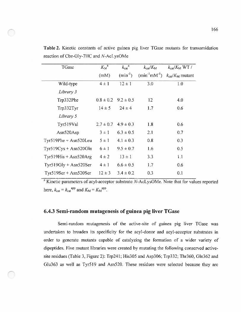
166
Table 2. Kinetic constants of active guinea pig liver TGase mutants for transamidation
reaction of Cbz-Gly-7HC and N-AcLysOMe
TGase KMa kcat kcat/KM kcat/KM WT I
(mlvi) (min’) (min’irilVf1) kcat/KM mutant
Wild-type 4±1 12+1 3.0 — 1.0
Libraiy 3
Trp332Phe 0.8 + 0.2 9.2 + 0.5 12 4.0
Trp332Tyr 14+5 24+4 1.7 0.6
Libraiy 5
Tyr5l9Val 2.7±0.7 4.9±0.3 1.8 0.6
Asn52OAsp 3 + 1 6.3 ± 0.5 2.1 0.7
Tyr5l9Phe + Asn52OLeu 5 ± 1 4.1 + 0.3 0.8 0.3
Tyr5l9Cys + Asn52OGln 6 + 1 9.5 + 0.7 1.6 0.5
Tyr5l9His + Asn52OArg 4 ± 2 13 + 1 3.3 1.1
Tyr5l9Gly+Asn52OSer 4+1 6.6±0.5 1.7 0.6
Tyr5l9Ser+Asn52OSer 12+3 3.4+0.2 0.3 0.1
a Kinetic parameters of acyl-acceptor substrate N-AcLysOMe. Note that for values reported
here, kcat = kcatP and KM KM.
6.4.3 Semi-random mutagenesis of guinea pig ilver TGase
Semi-random mutagenesis of the active-site of guinea pig liver TGase was
undertaken to broaden its specificity for the acyl-donor and acyl-acceptor substrates in
order to generate mutants capable of catalyzing the formation of a wider variety of
dipeptides. Five mutant libraries were created by mutating the following conserved active-
site residues (Table 3, Figure 2): Trp241; His305 and Asp306; Trp332; Thr360, G1n362 and
G1u363 as well as Tyr519 and Asn520. These residues were selected because they are
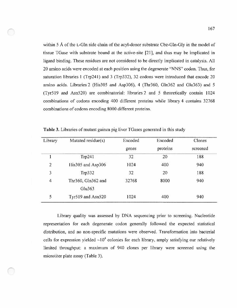
167
within 5 À of the L-Gin side chain of the acyl-donor substrate Cbz-Gln-Gly in the model of
tissue TGase with substrate bound at the active-site [21], and thus may be irnplicated in
ligand binding. These residues are not considered to be directly irnplicated in catalysis. Ail
20 amino acids were encoded at each position using the degenerate “NNS” codon. Thus, for
saturation libraries 1 (Trp241) and 3 (Trp332), 32 codons were introduced that encode 20
amino acids. Libraries 2 (His305 and Asp306), 4 (1hr360, G1n362 and G1u363) and 5
(1yr519 and Asn520) are combinatorial: libraries 2 and 5 theoretically contain 1024
combinations of codons encoding 400 different proteins while library 4 contains 3276$
combinations of codons encoding $000 different proteins.
Table 3. Libraries of mutant guinea pig liver TGases generated in this study
Library Mutated residue(s) Encoded Encoded Clones
genes proteins screened
1 1rp241 32 20 18$
2 His3O5andAsp3O6 1024 400 940
3 Trp332 32 20 188
4 Thr360, G1n362 and 3276$ $000 940
G1u363
5 Tyr5l9andAsnS2O 1024 400 940
Library quality was assessed by DNA sequencing prior to screening. Nucleotide
representation for each degenerate codon generally followed the expected statistical
distribution, and no non-specific mutations were observed. Transformation into bacterial
ceils for expression yielded i04 colonies for each library, amply satisfying our reiatively
limited throughput: a maximum of 940 clones per library were screened using the
microtiter plate assay (Table 3).
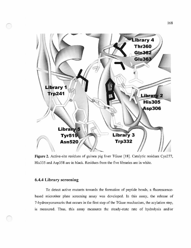
16$
6.4.4 Library screening
b detect active mutants towards the formation of peptide bonds, a fluorescence
based microtiter plate screening assay was deveÏoped. In this assay, the release of
7-hydroxycoumarin that occurs in the first step of the TGase mechanism, the acylation step,
is measured. Thus, this assay measures the steady-state rate of hydrolysis and/or
Ti-- ïbrary4
Thr360
Figure 2. Active-site residues of guinea pig liver TGase [1$]. Catalytic residues Cys277,
His335 and Asp358 are in black. Residues from the five libraries are in white.
169
transamidation. In each case, TGase deacylation is the rate-limiting step [11, 22]. 188
clones of libraries 1 and 3 were screened, approximately 6 tirnes the library size (32 DNA
variants) giving a representation of>99 % ofthe whole library. For libraries 2, 4 and 5, 940
clones were screened, resulting in approximately 60 ¾ library coverage for libraries 2 and 5
(1024 DNA variants) and <3 % for library 4 (32768 DNA variants). Library representation
was calculated as described under Methods. It should be noted that this screening assay is
also easily arnenable to automation for screening larger numbers of mutants in a library
versus library approacli (mutants versus commercially-available acyl-acceptor substrates).
For each library, we first rneasured the rate of hydrolysis using Cbz-Gly-7HC or
Cbz-Ala-7HC as acyl-donor substrates. As noted above, Cbz-Gly-7HC is a substrate of
wild-type guinea pig liver TGase. It was used to detect active mutants among the libraries.
Cbz-Ala-7HC is not a substrate of wild-type guinea pig liver TGase and was used to screen
for mutants with a different specificity for the acyl-donor substrate. Cbz-Phe-7HC was not
used for screening due to its Ïow aqueous solubility (<50 tM).
None of the >3 000 clones screened were capable of hydrolyzing Cbz-Ala-7HC at a
detectable rate. Thus, the serni-random mutagenesis performed was not consistent with this
particular modification of enzyme specificity for the acyl-donor substrate. However, library
screening with Cbz-Gly-7HC in the presence or absence of acyl-acceptor N-AcLysOMe
yielded active mutants from libraries 3 and 5 for the hydrolysis and transarnidation
reactions. DNA sequencing of the active mutants (Table 4) revealed that library 3 (single
mutation) yielded only two active mutants (Trp332Phe and Trp332Tyr), suggesting that
neutral aromatic amino acids are the only residues that can be introduced at this position to
give a detectable activity. Library 5 (double mutations) yielded 20 active mutants, three of
which were single mutants. Although residues Tyr5 19 and Asn520 are conserved in nature,
they are more permissive to mutations than the other positions screened as a variety of
hydrophobic, uncharged polar and charged arnino acids were identified at each position.
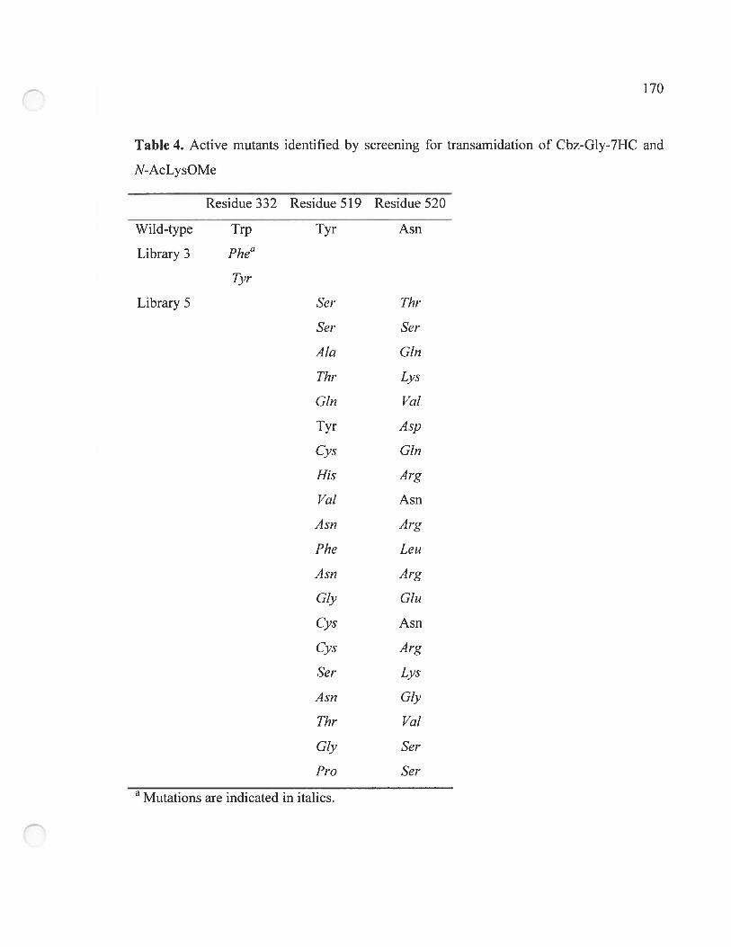
170
Table 4. Active mutants identified by screening for transamidation of Cbz-Gly-7HC and
N-AcLysOMe
Residue 332 Residue 519 Residue 520
Wild-type Trp Tyr Asn
Library 3 Phe”
Tyr
Library 5 8cr Thr
8cr 8cr
AÏa Gin
Thr Lys
Gin Val
Tyr Asp
Cys Gin
His Arg
Val Asn
Asn Arg
Fhe Leu
Asn Arg
Gly Glu
Cys Asn
Cys Arg
8cr Lys
Asn Gly
Thr Val
Gly 8cr
Pro 8cr
a Mutations are indicated in italics.
171
Indeed, 11 out of 20 encoded amino acids were identified at each position (Gly, Ala,
Val, Pro, Ser, Thr, Cys, Asn, Gin, Tyr and His for position 519 and Gly, Val, Leu, Ser, Thr,
Asn, Gin, Asp, Glu, Lys and Arg for position 520) demonstrating the high tolerance to
sequence variation at these positions. An even higher variability of amino acids would have
been expected at these positions if our library screening had been exhaustive. Throughout
screening, the wild-type enzyme was aiso identified as active. No active mutants from
libraries 1 (Trp241), 2 (His305 and Asp306) and 4 (Thr360, G1n362 and Glu363) were
identified.
Screening of the five mutant libraries using acyl-donor Cbz-Gly-7HC with the
potentiai acyl-acceptor PheNH2 was also performed. As PheNH2 is not an acyl-acceptor
substrate of wild-type guinea pig liver TGase. this screening was perforrned to identify
mutants with altered specificity. However, this screening did not allow us to detect mutants
that had an increased rate of transamidation relative to hydrolysis in the presence of
PheNH2, potentially due to our non-exhaustive screening and to the iow sensitivity of our
assay that allows only mutants with near-native activity to be identified.
6.4.5 Enzyme kinetics of identified active mutants
Kinetic constants for acyl-donor substrate Cbz-Gly-7HC in the hydroiysis reaction
and acyl-acceptor substrate N-AcLysOMe in the transamidation reaction were determined
for ail the active mutants from iibrary 3 and for the seven most active mutants from
library 5 identified by screening (Tables 1 and 2). for the hydrolysis reaction, ail mutants
except mutant Trp332Tyr exhibited a 1.5- to l0-fold decrease in catalytic efficiency.
Mutant Trp332Tyr was slightly more catalytically efficient than the wild-type enzyme. For
the transamidation reaction with N-AcLysOMe, ail mutants except single mutant
Trp332Phe and double mutant Tyr5l9His+Asn52OArg, had a 1.5- to 10-fold decrease in
catalytic efficiency, demonstrating a good coiielation between the relative catalytic
efficiency of hydrolysis and transamidation for most mutants. Double mutant
172
Tyr5l9His + Asn520Arg was found to be as catalytically efficient as the witd-type enzyme
with respect to transamidation.
The mutations at position 332 from the two identified active mutants resulted in
different effects. Whereas mutant Trp332Tyr vas more efficient at catalyzing the
hydrolysis of Cbz-Gly-7HC than the wild-type, it was Iess efficient than the wild-type
enzyme for catalysis of the transamidation reaction with N-AcLysOMe. However, mutant
Trp332Phe showed an impressive 4-fold increase in catalytic efficiency of transamidation
relative to the wild-type while being only slightly less efficient than wild-type for
hydrolysis. This suggests that the Trp332Phe mutation improves transarnidation efficiency
without significantly affecting hydrolytic efficiency. The opposing resuits for these two
mutants are surprising since both display uncharged arornatic arnino acids at position 332.
Kinetic parameters for the potential acyl-acceptor substrate PheNH2 were also
measured for both active mutants from library 3 and for the seven most active mutants of
library 5. Although no mutants showed activity by the plate-based screening method using
this compound. mutant Trp332Phe showed significant transamidation activity with
PheNH2. The KM and kcat values determined were 12 + 5 mM and 1.6 ± 0.2 min1,
respectively (Table 5). The resulting catalytic efficiency value of 0.1 min1 rnM’ is 120-fotd
lower than the corresponding value with acyl-acceptor N-AcLysOMe (12 min1mM’:
Table 2) and is apparently below the limit of detection of the plate-based screening assay.
The absolute transamidation efficiency of mutant Trp332Phe relative to that of the wild
type likely accounts for it being the only mutant with detectable PheNH2 transarnidation.
Mutant Trp332Phe also recognized PheOMe with similar KM and kcat, demonstrating that
the C-terminal amide or methyl ester functional groups of the L-Phe derivatives have no
significant effect on the kinetic parameters. This is in sharp contrast with the charged
carboxylate group that renders amino acids unreactive as acyl-acceptor substrates [91.
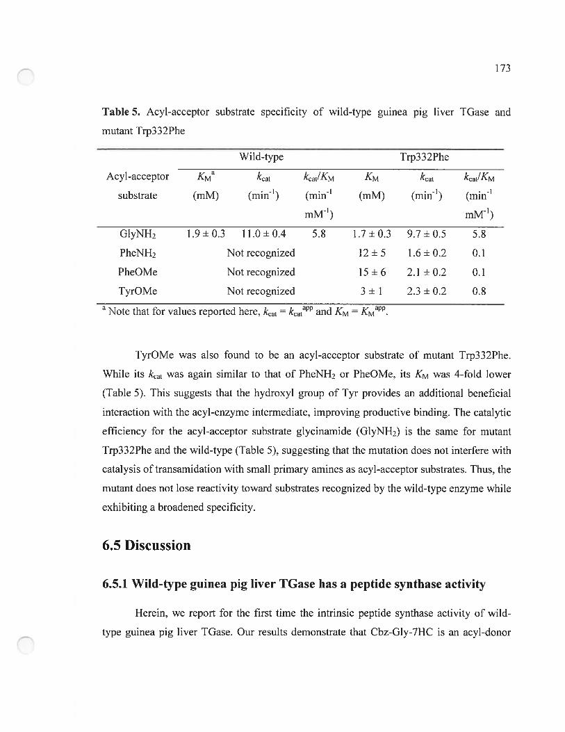
173
Table 5. Acyl-acceptor substrate specificity of wild-type guinea pig liver TGase and
mutant Trp332Phe
Wild-type Trp332Phe
Acyl-acceptor KMa kcat kcat/KM KM kcat kcat/KM
substrate (mM) (min1) (min1 (rril\4) (min’) (min1
mIVf’) mM1)
G1yNH2 1.9+0.3 11.0+0.4 5.8 1.7+0.3 9.7+0.5 5.8
PheNH2 Not recognized 12 + 5 1.6 + 0.2 0.1
PheOMe Not recognized 15 + 6 2.1 + 0.2 0.1
TyrOMe Not recognized 3 + 1 2.3 + 0.2 0.8a Note that for values reported here, kcat kcat and KM = KMaPP.
TyrOMe was also found to be an acyl-acceptor substrate of mutant Trp332Phe.
While its kcat was again similar to that of PheNH2 or PheOMe, its KM was 4-fold lower
(Table 5). This suggests that the hydroxyl group of Tyr provides an additional beneficial
interaction with the acyl-enzyme intermediate, improving productive binding. The catalytic
efficiency for the acyl-acceptor substrate glycinamide (GIyNH2) is the same for mutant
Trp332Phe and the wild-type (Table 5), suggesting that the mutation does not interfere with
catalysis oftransamidation with small prirnary amines as acyl-acceptor substrates. Thus, the
mutant does not lose reactivity toward substrates recognized by the wild-type enzyme while
exhibiting a broadened specificity.
6.5 Discussion
6.5.1 Wi]d-type guinea pig liver TGase has a peptide synthase activity
Herein, we report for the first time the intrinsic peptide synthase activity of wild
type guinea pig liver TGase. Our results demonstrate that Cbz-Gly-7HC is an acyl-donor
174
substrate of wild-type guinea pig liver TGase and that various srnall arnino acid derivatives
are acyl-acceptor substrates. Arornatic esters acting as acyl-donor substrates of guinea pig
liver TGase has been previously demonstrated [11, 13]. furthermore, Clarke et aï. [9]
reported that glycinamide-N’5 can serve as an acyl-acceptor substrate for guinea pig liver
TGase, proceeding with retention of the amide N’5 atom. This indicates that formation of
the covalent bond between glycinamide-N’5 and the L-Gin residue of the acyl-donor
substrate occurred through the Œ-arnino group of glycinarnide rather than through the amide
function. These previously reported observations provide insight for interpreting the
peptide synthase activity of wild-type guinea pig liver TGase reported herein.
The peptide synthase activity of guinea pig liver TGase we observed resuits from its
transferase activity: the enzyme can transfer an a-Gly moiety onto the Œ-arnino group of
various small amino acid derivatives, thus forrning peptide bonds. This transferase activity
of IGases relies on their capacity to exciude water from the active-site [23]. If water has
free access to the thioester bond of the covalent acyl-enzyrne intermediate, amine acyl
acceptor substrates would not be able to compete with it for the acyl transfer reaction, as
water is much more abundant. Thus, the intermediate thioester must be sequestered in the
active-site long enough for amines to enter and act as acyl-acceptor substrates. It is believed
that the exclusion of water results from hydrophobic residues forming a tunnel leading to
the catalytic residues. These hydrophobic residues have been identified as residues Trp24 1,
His305 and Trp332 which form the walls of the active-site tunnel [21]. This ability to
exclude water from the active-site differentiates TGases from the cysteine proteases, such
as papain, that share a simiiar segment of a-helix and 13-sheet containing an identical
catalytic triad [24].
Proteases are known to catalyze the synthesis of peptides under certain conditions.
In a thermodynamically-controlled process, amino acid condensation is favoured over
peptide hydrolysis by shifting equiiibria through product precipitation or decreasing water
activity. In a kinetically-controlled process, the aminolysis of an acyl-enzyme formed from
175
an activated acyl-donor substrate is favoured over its hydrolysis by using high
concentrations of acyl-acceptor substrate. The kinetic process requires a protease such as
serine or cysteine proteases that can form a covalent acyl-enzyme intermediate [25], as is
the case with TGases. Competition between hydrolysis and aminolysis is aiways present
during the degradation of this acyl-enzyme intermediate, resulting in lower yields for the
synthesis of peptides, since proteases are not efficient at exciuding water from thc active-
site — rather, tliey are designed to bind and activate water as part of their native catalytic
cycle. Furthermore, proteases can hydrolyse their peptide products, further lowering the
overall yield of synthetic peptide.
Proteases used in kinetically-controlled peptide synthesis have a
transamidationlhydrolysis ratio in the range of 1 0- 1 [26, 27] whereas guinea pig liver
IGase has a similar 102 increase in rate of transamidation relative to hydrolysis when the
acceptor substrate is hydroxylamine [11]. The catalytic efficiency of wild-typc guinea pig
liver IGase for the synthesis of the Cbz-Gly-G1yNH2 and Cbz-Gly-A1aNH2 dipeptides is
5.8 and 1.1 min’mM1, respectively. Papain, a cysteine protease that has been used in
peptide synthesis, lias catalytic efficiencies of 0.3 and 2.9 minmM’ for the synthesis of
the Boc-Gly-Phe-N2H2Ph dipeptide and the Boc-Tyr(Bzl)-Gly-Gly-Phe-Leu-N2H2Ph
pentapeptide, respectively [2$]. These catalytic efficiencies are within the same order of
magnitude than those of wild-type guinea pig liver TGase, suggesting that TGase could be
used as a catalyst for the synthesis of peptide bonds. An advantage of guinea pig liver
TGase-catalyzed peptide synthesis is that it requires no organic co-solvent. Indeed, the 5 %
DMf used in the transamidation assay of Cbz-Gly-7HC and G1yNH2 by TGase is required
only to help solubilize the acyl-donor substrate. This is not the case with papain, which
catalyzed the synthesis of the Boc-Tyr(Bzl)-Gly-Gly-Phe-Leu-N2H2Ph peptide in a mixture
containing 40 % ethanol [29] to decrease the activity of water, since hydrolysis is the
preferred, native reaction of the protease. A further advantage of TGase for the synthesis of
peptide bonds is that TGase camot recognize secondary amides as acyl-donor substrates,
thus limiting hydrolysis ofthe dipeptide product and potentially increasing yields.
176
We tested wild-type guinea pig liver TGase with the amide analog of Cbz-Gly-7HC,
Cbz-glycinamide (Cbz-GÏyNH2), to verify if it could act as an acyl-donor substrate. This
compound does flot react with IGase at concentrations up to 50 mM. The KM value for this
compound is apparently greater than its solubility lirnit. It has previously been observed
that aromatic ester acyl-donor substrates of TGase have a lower KM value than the
corresponding amide: Cbz-Gln-Gly (Figure lA) has a KM of 3.2 mM [21] whereas its
aromatic ester analog Cbz-Glu(yPNP)-Gly (Figure 13) has a KM of 0.02 mM [11]. The
160-fold lower KM ofthe arornatic ester could be due to irnproved binding conferred by the
p-nitrophenol aromatic leaving group. This irnproved binding could also occur in
Cbz-Gly-7HC relative to Cbz-G1yNH2 through beneficial 7t-stacking interactions between
the aromatic leaving group and the aromatic side chains of the three tunnel-wall residues of
guinea pig liver TGase (Trp241, His305 and Trp332). This apparent need of an activated
leaving group in acyl-donor substrates for peptide synthesis by native guinea pig liver
TGase represents a serious limitation for their application to peptide synthesis. This
limitation is flot observed for proteases, for example, which can react directly on the
carboxylate group of acyl-donors.
6.5.2 Modification of guinea pig liver TGase specificity
As discussed above, guinea pig liver TGase may be useful for peptide synthesis.
However, to improve its usefulness, the enzyme specificity should be broadened. Wild-type
guinea pig liver TGase caimot recognize Cbz-Ala-7HC as an acyl-donor substrate, therefore
preventing it from forming Cbz-Ala-Xaa dipeptides. Furthermore, amino acid derivatives
having bulky side-chains are not acyl-acceptor substrates ofthe enzyme. To further broaden
its specificity for acyl-donor and acyl-acceptor substrates, we performed semi-random
mutagenesis of conserved active-site residues coupled to screening with desired cornpounds
such as the aforementioned Cbz-Ala-7HC. Our rationale was that modification of the
177
specificity would be achieved more readily through a semi-random approach by mutating
residues that directly interact with the substrate.
Although through this approach we did not identify any mutants with altered acyl
donor specificity of the enzyme, we rapidly identified a point mutant, Trp332Phe, with a
different specfficity for the acyl-acceptor substrate. This mutant can forrn peptide bonds
between Cbz-Gly and the neutral aromatic amino acids L-Plie and L-Tyr, and represents the
first example of a TGase that can utilize an amine with an adjacent bulky substituent as an
acyl-acceptor substrate. Indeed, this mutant catalyzed the formation of the
Cbz-Gly-PheNH2, Cbz-Gly-PheOMe and Cbz-Gly-TyrOMe dipeptides. However, the
catalytic efficiency of mutant Trp332Phe with these substrates (0.1-0.8 rnin1mM1) is one
order of magnitude lower than that with acyl-acceptor substrates such as G1yNH2 and
N-AcLysOMe (5.8 min1mM1 and 12.0 rnin1rnM1, respectively).
Comparison ofthe catalytic efficiency of guinea pig liver TGase mutant Trp332Phe
for the formation of the Gly-Phe peptide bond (0.1 min1rnM1) with the corresponding
value for papain (0.3 min’mM1; Table 6) [28] reveals that they are in the same order of
magnitude. Thus, Trp332Phe, whilc being less efficient at forming Gly-Phe peptide bonds
than Gly-Gly peptide bonds (Table 5), is roughly as efficient as papain for the formation of
Gly-Phe peptide bonds. This dernonstrates that mutant Trp332Phe of guinea pig liver
TGase could be useful for peptide synthesis.
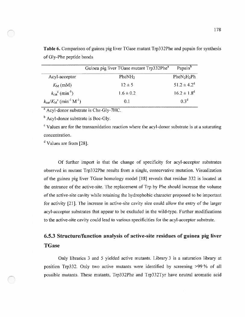
172
Table 6. Comparison of guinea pig liver IGase mutant Trp332Phe and papain for synthesis
of Gly-Phe peptide bonds
Guinea pig liver TGase mutant Trp332Phea papaiib
PheNH2 PheN2H2Ph
12+551742d
1.6 + 0.2 16.2 + l.8
-iU.1 U..)
Acyl-acceptor
KM (mM)
kcatC (1rLin1)
kcat/KM’ (min’ M’)
a Acyl-donor substrate is Cbz-Gly-7HC.b Acyl-donor substrate is Boc-Gly.C Values are for the transamidation reaction where the acyl-donor substrate is at a saturating
concentration.U Values are from [28].
0f further import is that the change of specificity for acyl-acceptor substrates
observed in mutant Trp332Phe resuits from a single, conservative mutation. Visualization
ofthe guinea pig liver IGase homology model [18] reveals that residue 332 is located at
the entrance of the active-site. The replacernent of Trp by Phe should increase the volume
of the active-site cavity while retaining the hydrophobic character proposed to be important
for activity [21]. The increase in active-site cavity size could allow the entry of the larger
acyl-acceptor substrates that appear to be excluded in the wild-type. further modifications
to the active-site cavity could lead to various specificities for the acyl-acceptor substrate.
6.5.3 Structure/function analysis of active-site residues of guinea pig liver
TGase
Only libraries 3 and 5 yielded active mutants. Library 3 is a saturation library at
position Trp332. Only two active mutants were identified by screening >99 ¾ of ail
possible mutants. These mutants, Trp332Phe and Trp332Tyr have neutral aromatic acid
179
substitutions at position 332 indicating that the enzyme requires a bulky aromatic residue at
position 332 to maintain its activity, stability or proper folding. Previous experimental
evidence supports this. Conserved active-site residue 1rp332 of human tissue TGase was
previously mutated to Ala or Phe [30]. Mutant Trp332Ala did not allow recovery of the
recombinant protein possibly due to improper folding reducing its solubility. Residue
Trp332 is located at the entrance of the hydrophobic active-site tunnel and has been
proposed to form a “gate” to the active-site [21]. Replacernent of this residue with a srnaii
residue such as Ala might expose the hydrophobic tunnel and decrease enzyme solubiiity.
Mutant Trp332Phe of human tissue IGase was found to have a transamidation activity
similar to wild-type [30]. This observation correlates with our results of the near-native
catalytic efflciency of guinea pig liver TGase mutant Trp332Phe. Unfortunately, no kinetic
parameters were reported for human tissue TGase mutant Trp332Phe preventing a more
thorough comparison with our mutant.
Screening of combinatorial library 5 aliowed us to identify 20 mutants with near
native activity. These mutants had 11 out of 20 possible encoded amino acids at either
position 519 or 520, with no bias towards polar, hydrophobic, charged or aromatic amino
acids, indicating that these positions are very permissive to mutations. Our previous
mutagenesis studies of guinea pig liver TGase [1$] have demonstrated the perrnissibility to
mutations of conserved active-site residue TyrS 19 which was mutated to amino acids with
side chains of diverse polarity (Leu, Phe, Gin and His). These mutagenesis experiments
demonstratcd that TyrS 19 is not essential for the hydrolysis or transarnidation reactions
cataiyzed by tissue TGase although it is conserved. Furthermore, mutation of residue
Tyr5 19 in combination with mutation of residue Cys3 36, which is located next to the
catalytic Cys residue, yielded mutants with near-native catalytic efficiency, again implying
that the active-site of guinea pig liver TGase is toierant to mutations at these positions [18].
Thus, it is not surprising that we identified 11 out of 20 possible mutations at position 519
during our screening. Since we only screened 940 mutants, representing approximately
180
60 ¾ of the library, we could expect to identify more mutants with near-native activity with
a more exhaustive screen.
Library 1 yielded no mutants with detectable, near-native hydrolysis or
transarnidation activities. This library is a saturation library of residue Trp241 and was
screened with the same representation of >99 % as library 3. Residuc Trp241 of human
tissue TGase was previously mutated to various amino acids [31]. It was observed that non
aromatic substitutions at this position (Trp24 lGly and Trp24 1 Ala) resulted in mutants with
hardly any detectable activity while aromatic substitutions (Trp24lPhe, Trp24lTyr and
Trp24lHis) yielded active mutants that nonetheless showed a 25- to 165-fold decrease in
catalytic efficiency relative to wiid-type. The sensitivity of our screening assay allowed us
to detect only mutants displaying near-native activity, therefore increasing its stringency.
Mutants having a 25- to 165-fold decrease in catalytic efflciency would not be detected.
Thus, although mutants Trp241Phe, Trp24lTyr and Trp24lHis were present in library 1,
we did not identify them as active during our screening.
Library 2 is a combinatorial library at positions His305 and Asp306. It contains the
same number of mutants as iibrary 5 and thus our screening should have represented
approximately 60 ¾ of the whole library. Unlike library 5, library 2 did not yieid mutants
with any detectabie hydrolysis or transamidation activities, suggesting that conserved
residues His305 and Asp306 may play an important role in the stability or activity of the
enzyme. These residues have been postuiated to be implicated in exclusion of water from
the active-site through the formation ofa sait bridge between them [23]. Furthermore, these
residues have been postulated to play an important role in the positioning and deprotonation
of the amine substrates to the active-site [32, 33]. Thus, the lack of active mutants may
resuk from the loss of important interactions between the enzyme and the acyl-acceptor
substrates during the deacylation step, which is the step we measure.
Library 4 is a combinatorial library containing 32768 genetic variants encoding for
$000 mutant proteins and was the largest library. It contained mutants at positions Thr360,
1$’
G1n362 and G1u363: none of these residues have been mutated previously. Consequently, it
is the library with the lowest representation during screening: only 940 clones were
screened which represents <3 % of the whole library. No active mutants from this library
were identified due to the limited range of our screening. As a resuit, it is difficult to
propose a role for residues Thr360, G1n362 and G1u363. Nonetheless, these residues belong
to the same loop as catalytic residue Asp358 (Figure 2). Mutations of this loop might
disrupt the proper positioning ofthis catalytic residue, affecting activity.
6.6 Conclusion
Herein we demonstrate for the first time the peptide synthase activity of wild-type
guinea pig liver TGase. This enzyme can forrn Gly-Xaa peptide bonds between Cbz
protected Gly and a variety of small polar or hydrophobie amino acid derivatives with a
similar catalytic efficiency as the cysteine protease papain. Although the specificity of
guinea pig liver TGase for peptide bond formation is lirnited, it could be a new tool for the
enzymatic synthesis of peptides and complernent the known specificities of other proteases.
To expand its speciflcity, we perfonTled semi-random mutagenesis of active-site residues
and rapidly identified mutant Trp332Phe by a fluorimetric microplate-based assay. This
mutant has a different specificity for the acyl-acceptor substrate. The semi-random
mutagenesis also allowed us to acquire structure/function information on the role of the
active-site residues of guinea pig liver tissue TGase.
6.7 References
1 Fesus, L. and Piacentini, M. (2002) Transglutarninase 2: an enigmatic enzyme with
diverse functions. Trends in Biochernical Sciences 27, 534-539
2 Griffin, M., Casadio, R. and Bergarnini, C. M. (2002) Transglutaminases: natures
biological glues. Biochemical Journal 368. 377-3 96
182
3 Lorand, L. and Graham, R. M. (2003) Transglutaminases: crosslinking enzymes
with pleiotropic functions. Nature Reviews Molecular Celi Biology 4, 140-156
4 Folk, J. E., Cole, P. W. and Mullooly, J. P. (196$) Mechanim of action of guinea pig
liver transglutaminase. V. The hydrolysis reaction. Journal of Biological Chemistry
243, 41$-427
5 Mehta, K. (2005) Mammalian transglutaminases: a family portrait. Progress in
Experimental Tumor Research 38, 1-18
6 Wada, F., Nakamura, A., Masutani, T., Ikura, K., Maki, M. and Hitomi, K. (2002)
Identification of mammalian-type transglutaminase in Physarum polycephalum.
Evidence from the cDNA sequence and involvement of GTP in the regulation of
transarnidating activity. European Journal of Biochemistry 269, 3451-3460
7 Aeschlimann, D. and Paulsson, M. (1994) Transglutaminases: protein cross-linking
enzymes in tissues and body fluids. Thrombosis & Haemostasis 71, 402-415
2 folk, J. E. and Chung, S. I. (1985) Transglutaminases. Methods in Enzymology
113, 358-375
9 Clarke, D. D., Mycek, M.J., Neidle, A. and Waelsch, H. (1959) The Incorporation
of Amines into Proteins. Archives of Biochemistry And Biophysics 79, 33$-354
10 de Macédo, P., Mairano, C. and Keillor, J. W. (2000) A direct continuous
spectrophotometric assay for transglutarninase activity. Analytical Biochernistry
285, 16-20
11 Leblanc, A., Gravel, C., Labelle, J. and Keillor, J. W. (2001) Kinetic studies of
guinea pig liver transglutaminase reveal a general-base-catalyzed deacylation
mechanism. Biochemistry 40, 8335-8342
12 Halim, D., Caron, K. and Keillor, J. W. (2007) Synthesis and evaluation ofpeptidic
maleirnides as transglutaminase inhibitors. Bioorg Med Chem Lett 17, 305-8
13 Gillet, S. M., Pelletier, J. N. and Keillor, J. W. (2005) A direct fluorometric assay
for tissue transglutaminase. Analytical Biochemistry 347, 221-226
1 OI Ô3
14 Choi, K., Siegel, M.. Piper, J. L, Yuan, L., Cho, E., Strnad, P., Omary, B., Rich, K.
M. and Khosla, C. (2005) Chernistry and biology of dihydroisoxazole derivatives:
selective inhibitors of human transglutaminase 2. Chernistry & Biology 12, 469-475
15 Pardin, C., Gillet, S. M. and Keillor, J. W. (2006) Synthesis and evaluation of
peptidic irreversible inhibitors of tissue transglutarninase. Bioorg Med Chem 14,
$379-8385
16 Gillet, S. M., Chica, R. A., Keillor, J. W. and Pelletier, J. N. (2004) Expression and
rapid purification of highly active hexahistidine-tagged guinea pig liver
transglutarninase. Protein Expression & Purification 33, 256-264
17 Ho, S. N., Hunt, H. D., Horton, R. M., Pullen. J. K. and Pease, L. R. (1989) Site
directed mutagenesis by overlap extension using the polymerase chain reaction.
Gene 77, 5 1-59
1$ Chica, R. A., Gillet, S. M., Dryburgh, F.-J., Keillor, J. W. and Pelletier, J. N. (2006)
Homology modeling of guinea pig liver transglutaminase and mutagenesis of
conserved active-site residues Tyr5 19 and Cys336. submitted.
19 Yokoyama, K., Kikuchi, Y. and Yasueda, H. (1998) Overproduction of DnaJ in
Eseherichia cou improves in vivo solubility of the recombinant fish-derived
transglutaminase. Bioscience, Bioteclmology & Biochemistry 62, 1205-12 10
20 Pelletier, J. N. and Denault. M. G. (2007) Protein library design and screening:
Working out the probabilities. In Methods Mol. Biol.: Protein Engineering
Protocols, Humana Press
21 Chica, R. A., Gagnon, P., Keillor, J. W. and Pelletier, J. N. (2004) Tissue
transglutaminase acylation: Proposed role of conserved active-site Tyr and Trp
residues revealed by molecular modeling of peptide substrate binding. Protein
Science 13, 979-991
22 Case, A. and Stem, R. L. (2003) Kinetic analysis of the action of tissue
transglutaminase on peptide and protein substrates. Biochernistry 42, 9466-948 1
184
23 Nernes, Z., Petrovski, G., Csosz, E. and Fesus, L. (2005) Structure-function
relationships of transglutaminases--a contemporary view. Progress in Experimental
Tumor Research 38, 19-36
24 Kashiwagi, T., Yokoyarna, K., Ishikawa, K., Ono, K., Ejima, D., Matsui, H. and
Suzuki, E. (2002) Crystal structure of microbial transglutarninase from
$treptoverticiÏlium mobaraense. Journal of Biological Chemistry 277, 44252-44260
25 Lombard, C., Saulnier, J. and Wallach, J. M. (2005) Recent trends in protease
catalyzed peptide synthesis. Protein & Peptide Letters 12, 62 1-629
26 Kumar, D. and Bhalla, T. C. (2005) Microbial proteases in peptide synthesis:
approaches and applications. Appi. Microbiol. Biotechnol. 68, 726-736
27 Riechrnann, L. and Kasche, V. (1985) Peptide synthesis catalyzed by the serine
proteinases chymotrypsin and trypsin. Biochimica & Biophysica Acta 830, 164-172
2$ Kulimaim, W. (1984) Kinetics of chymotrypsin- and papain-catalysed synthesis of
[leucine]enkephalin and [methionine]enkephalin. Biochemical Journal 220, 405-416
29 Kullmann, W. (1980) Proteases as catalysts for enzyrnic syntheses of opioid
peptides. Journal ofBiological Chemistry 255, 8234-8238
30 Murthy, S. N., Iisrnaa, S.. Begg, G., Freymann, D. M.. Graham, R. M. and Lorand,
L. (2002) Conserved tryptophan in the core domain of transglutaminase is essential
for catalytic activity. Proceedings of the National Academy of Sciences of the
United States ofAmerica 99, 273 8-2742
31 Iisrnaa, S. E., Holman, S., Wouters, M. A., Lorand, L., Graham, R. M. and Husain,
A. (2003) Evolutionary specialization of a tryptophan indole group for transition
state stabilization by eukaryotic transglutaminases. Proceedings of the National
Academy of Sciences of the United States ofAmerica 100, 12636-12641
32 Pedersen, L. C., Yee, V. C., Bishop, P. D.. Le Trong. I., Teller, D. C. and Stenkamp,
R. E. (1994) Transglutaminase factor XIII uses proteinase-like catalytic triad to
crosslink macromolecules. Protein Science 3, 113 1-1135
185
33 Yee, V. C., Pedersen, L. C., Le Trong, I., Bishop. P. D., Stenkamp, R. E. and Teller,
D. C. (1994) Three-dirnensional structure of a transglutarninase: hurnan blood
coagulation factor XIII. Proceedings of the National Acaderny of Sciences of the
United States ofArnerica 91, 7296-7300
CHAPITRE 7
Conclusion
187
7.1 Conclusion
Le but de notre projet de recherche était de développer de nouveaux biocatalyseurs
pour la synthèse des peptides. Pour atteindre cet objectif général, nous avons procédé à la
modification de la spécificité de la IGase de foie de cobaye par évolution dirigée selon une
approche semi-aléatoire. Cette approche a été choisie, car elle exploite les données connues
sur la structure et/ou la fonction de certains résidus, générant ainsi des banques de mutants
«intelligentes » ayant une plus grande probabilité de contenir des mutants aux propriétés
désirées. Ceci a l’avantage de ne pas requérir une méthode de criblage à haut débit, puisque
la taille des banques de mutants est limitée selons nos besoins. L’approche semi-aléatoire
semblait donc être le meilleur choix pour atteindre notre objectif
Afin d’appliquer cette approche, nous avions besoin d’information structurelle et
fonctionnelle qui n’était pas disponible dans la littérature. Nous avons obtenu plus
d’information structurelle par l’identification des résidus du site actif de la TGase tissulaire
impliqués dans la liaison des substrats. Pour identifier ces résidus, nous avons utilisé une
approche combinant la cinétique enzymatique et la modélisation moléculaire qui nous a
permis de développer un modèle moléculaire de la IGase tissulaire avec le substrat
donneur d’acyle Cbz-Gln-Gly lié au site actif Ce modèle nous a permis de proposer une
séquence d’événements lors de l’étape d’acylation du mécanisme des TGases. De plus, il
nous a permis d’identifier les résidus du site actif qui sont en contact direct avec le substrat.
Par la suite, nous avons conçu une méthode efficace et rapide d’expression et de
purification de la TGase de foie de cobaye chez E. cou de sorte à pouvoir obtenir l’enzyme
nécessaire pour le développement des méthodes expérimentales essentielles au projet. Ce
protocole nous a permis d’obtenir de l’enzyme hautement pure ayant l’activité spécifique la
plus élevée rapportée dans la littérature. De plus, l’expression en présence des chaperones
DnaK et DnaJ nous a permis d’obtenir une grande quantité d’enzyme recombinante ayant
des paramètres cinétiques similaires à ceux de l’enzyme native purifiée à partir d’organes
18$
animaux. La conception de cette méthode présente aussi l’avantage de permettre les
modifications génétiques de l’enzyme recombinante.
Toujours dans le but d’obtenir de l’information structurelle et fonctionnelle requise
pour développer des banques « intelligentes », nous avons généré le premier modèle par
homologie de la TGase de foie de cobaye puisque aucune structure cristalline n’avait été
résolue pour cette enzyme. Ce modèle a démontré que la structure de la TGase de foie de
cobaye est très similaire à celle de la TGase tissulaire de Pagrus major que nous avons
utilisée pour développer le modèle de liaison du substrat donneur Cbz-Gln-Gly. Le modèle
représente un nouvel outil pour toute étude structurelle de l’enzyme de foie de cobaye pour
laquelle aucune structure cristalline n’a été résolue. Nous avons aussi procédé à la
mutagenèse des résidus Tyr519 et Cys336 du site actif de la TGase de foie de cobaye, seuls
ou en combinaison, et avons démontré la tolérance aux mutations du site actif. Le modèle et
les résultats de mutagenèse ont validé l’utilisation de la TGase de foie de cobaye pour
l’évolution dirigée par une approche semi-aléatoire.
Enfin, ces avancées nous ont permis de développer une méthode de criblage
fluorimétrique basée sur des substrats donneurs contenant un ester 7-hydroxycoumaryl
permettant de détecter le changement de spécificité de la TGase. Nous avons découvert que
la TGase de foie de cobaye de type sauvage catalysait déjà la formation de liaisons
peptidiques entre un dérivé ester 7-hydroxycoumaryl de la Gly et des dérivés de petits
acides aminés polaires ou hydrophobes grâce à une activité peptide synthase intrinsèque. La
spécificité de l’activité peptide synthase de la TGase de foie de cobaye a été modifiée par
évolution dirigée selon une approche semi-aléatoire. En exploitant l’information
structurelle obtenue par modélisation moléculaire, nous avons procédé à la création de cinq
banques de mutants ciblant certains résidus du site actif Plus de 3000 clones provenant de
ces cinq banques ont été criblés, ce qui nous a permis d’identifier un mutant (Trp332Phe)
pouvant former des liens peptidiques entre la Gly et des dérivés de L-Phe et de L-Tyr, ce
qui n’est pas le cas de l’enzyme de type sauvage. Ce mutant a une efficacité catalytique
189
similaire à celle de la papaïne pour la synthèse du lien peptidique Gly-Phe et ne devrait pas
hydrolyser le produit, ce qui démontre qu’il pourrait être avantageux comme biocatalyseur
pour la synthèse enzymatique de peptides.
En conclusion, nous avons démontré l’activité peptide synthase intrinsèque de la
TGase de foie de cobaye de type sauvage et avons modifié sa spécificité par évolution
dirigée selon une approche semi-aléatoire. Bien que sa spécificité demeure étroite, cette
enzyme représente un nouvel outil pour la synthèse enzymatique des peptides et sa
spécificité pourrait complérnenter celle des protéases utilisées pour la biocatalyse de ce type
de réactions.
7.2 Perspectives
Ce projet a permis de démontrer que la TGase de foie de cobaye représente un outil
prometteur pour la synthèse enzymatique des peptides. Afin qu’elle soit pleinement
exploitée, une caractérisation plus approfondie est en cours. Cette caractérisation
inclut l’analyse cinétique avec une plus grande variété de substrats accepteurs potentiels de
sorte à clarifier les limites de la spécificité de la TGase et la mesure du rendement de la
réaction de synthèse peptidique catalysée par l’enzyme de sorte à connaître son efficacité.
À plus long terme, plusieurs propriétés de la TGase pourraient être améliorées ou
modifiées pour qu’elle devienne un meilleur catalyseur de la synthèse des peptides. Des
changements supplémentaires à la cavité du site actif pourraient être envisagées pour que la
spécificité envers les substrats donneurs et accepteurs soit modifiée. Pour ce faire, les cinq
banques pourraient être combinées entre elles et de toutes nouvelles banques de mutants
ciblant d’autres résidus pourraient être créées. Aussi, les cinq banques pourraient être
criblées avec de nouveaux substrats donneurs ou accepteurs potentiels afin que de nouvelles
spécificités soient identifiées. Une méthode de criblage ayant une limite de détection plus
basse et étant basée sur le principe du FRET (fluorescence resonance energy transfer)
190
pourrait être conçue. Une méthode de ce genre permettrait de détecter la formation directe
de liens peptidiques plutôt que la libération d’un fluorophore à l’étape d’acylation, comme
c’est le cas avec la méthode présentée au chapitre 6. finalement, l’enzyme pourrait être
soumise à une évolution dirigée selon une approche aléatoire afin que son efficacité
catalytique ou sa stabilité soit augmentée.
I
Bibliographie
1 Ege, S. N. (1994) Organic chemistry. D.C. Heath and Cornpany. Toronto
2 Anastas. P. T. and Williamson. T. C. (1996) Green chemistry: An overview. In
Green chemistry: Designing chernistry for the environrnent (Anastas. P. T. and
WiÏÏiamson. T. C.. eds.). pp. I-17. American Chemical Society. Washington D.C.
3 Alcalde, M., Ferrer, M., Plou, f. J. and Ballesteros, A. (2006) Environmental
biocatalysis: from remediation with enzymes to nove! green processes. Trends in
Biotechnology 24, 28 1-287
4 Anastas, P. T. and Warner, J. C. (1998) Green chemistry. Theory and practice.
Oxford University Press. New York
5 Wescott. C. R. and Klibanov, A. M. (1994) The solvent dependence of enzyme
specificity. Biochimica et Biophysica Acta (BBA) - Protein Structure and Molecular
Enzymology 1206. 1-9
6 Zhao. H.. Chockalingam. K. and Chen. Z. (2002) Directed evolution of enzymes
and pathways for industrial biocatalysis. Current Opinion in Biotechnology 13, 104-
110
7 Panke. S., Held, M. and Wubbolts. M. (2004) Trends and innovations in industrial
biocatalysis for the production of fine chemicals. Current Opinion in Biotechno!ogy
15, 272-279
8 Zhang, H., Xu, X., Mu, H., Niisson. J., Adier-Nissen, J. and Hoy, C. (2000)
Lipozyme IM-catalyzed interesterification for the production of margarine fats in a
1 kg scale stirred tank reactor. European Journal of Lipid Science & Techno!ogy
102, 411-418
9 Voet, D. and Voet. J. G. (1995) Biochemistry. John Wiley & Sons, Inc., Toronto
10 Erbeldinger, M.. Ni. X. and Ha!ling, P. J. (2001) Kinetics of enzymatic solid-to
so!id peptide synthesis: synthesis of Z-aspartaine and control of acid-base
conditions by using inorganic saits. Biotechno!ogy & Bioengineering 72, 69-76
II11 Andersson, L., Blomberg, L., Flegel, M., Lepsa, L., Niisson, B. and Verlander, M.
(2000) Large-scale synthesis ofpeptides. Biopolymers 55, 227-250
12 Mayo, K. H. (2000) Recent advances in the design and construction of synthetic
peptides: for the love of basics or just for the teclmology of it. Trends in
Bioteclrnology 18,212-217
13 Jenssen, H., Harniil, P. and Hancock, R. E. (2006) Peptide antimicrobial agents.
Clinical Microbiology Reviews 19, 491-511
14 Kurnar, D. and BhaÏla, T. C. (2005) Microbial proteases in peptide synthesis:
approaches and applications. Applied Microbiology & Biotechnology 68, 726-73 6
15 Carpino, L. A. (1993) 1-Hydroxy-7-azabenzotriazole. An Efficient Peptide
Coupling Additive. Journal ofthe Arnerican Chemical Society 115, 4397-4398
16 Knorr, R., Trzeciak, A., Bairnwarth, W. and Gillessen, D. (1989) New coupling
reagents in peptide chemistry. Tetrahedron Letters 30, 1927-1930
17 Coste, J., Le-Nguyen, D. and Castro, B. (1990) PyBOP(R): A new peptide coupling
reagent devoid oftoxic by-product. Tetrahedron Letters 31, 205-208
1$ Kullrnann, W. (1987) Enzyrnatic Peptide Synthesis. CRC Press, Inc., Boca Raton,
Florida
19 Lombard, C., Saulnier, J. and Wal!ach, J. M. (2005) Recent trends in protease
catalyzed peptide synthesis. Protein & Peptide Letters 12, 621-629
20 Griffin, M., Casadio, R. and Bergarnini, C. M. (2002) Transglutarninases: natures
biological glues. Biochemical Journal 368. 3 77-396
21 Autuori, f., fanace, M. G., Oliverio, S., Piredda. L. and Piacentini, M. (199$)
“Tissue” transglutarninase and apoptosis. Advances in Biochernical Engineering &
Biotechnology 62, 129-136
22 Abc, S., Yamashita, K., Kohrio, H. and Ohkubo, Y. (2000) Involvernent of
transglutarninase in the receptor-rnediated endocytosis of mouse peritoneal
macrophages. Biological and Pharmaceutical Bulletin 23, 1511-1513
23 Chen, J. S. and Melita, K. (1999) Tissue transglutaminase: an enzyme with a spiit
personality. International Journal ofBiochernistry & Cell Biology 31, $17-$36
III24 Shridas, P., Sharma, Y. and Balasubramanian, D. (2001) Transglutarninase
mediated cross-linking of aipha-crystallin: structural and functional consequences.
FEB$ Letters 499, 245-250
25 Shan, L., Molberg, 0., Parrot, I., Hausch, f., Filiz, F., Gray, G. M., Sollid, L. M.
and Khosla, C. (2002) Structural basis for gluten intolerance in celiac sprue. Science
297, 2275-2279
26 Schroeder, W. T., Thacher, S. M., Stewart-Galetka, S., Annarella, M., Cherna, D.,
Siciliano, M. J., Davies. P. J., Tang, H. Y., Sowa, B. A. and Duvic, M. (1992) Type
I keratinocyte transglutarninase: expression in human skin and psoriasis. Journal of
Investigative Derrnatology 99, 27-34
27 Lorand, L. (1996) Neurodegenerative diseases and transglutaminase. Proceedings of
the National Academy of Science ofthe United States of America 93, 14310-14313
28 folk, J. E. (1969) Mechanisrn of action of guinea pig liver transglutarninase. VI.
Order of substrate addition. Journal ofBiological Chernistry 244, 3707-3713
29 Ahvazi, B. and Steinert, P. M. (2003) A model for the reaction mechanisrn of the
transglutaminase 3 enzyme. Experimental & Molecular Medicine 35, 228-242
30 Yee, V. C., Pedersen, L. C., Le Trong. I., Bishop, P. D., Stenkamp, R. E. and Teller,
D. C. (1994) Three-dirnensional structure of a transglutaminase: human blood
coagulation factor XIII. Proceedings of the National Academy of Sciences of the
United States of America 91, 7296-7300
31 Case, A. and Stem, R. L. (2003) Kinetic analysis of the action of tissue
transglutaminase on peptide and protein substrates. Biochemistry 42. 9466-94$ 1
32 Leblanc, A., Gravel. C., Labelle, J. and Keillor, J. W. (2001) Kinetic studies of
guinea pig liver transglutaminase reveal a general-base-catalyzed deacylation
mechanism. Biochemistry 40, 8335-8342
33 Pasternack, R., Dorsch, S., Otterbach, J. T., Robenek, I. R., Wolf, S. and
Fuchsbauer, H. L. (199$) Bacterial pro-transglutaminase from Streptoverticillium
mobaraense--purification, characterisation and sequence of the zymogen. European
Journal ofBiochemistry 257, 570-576
Iv34 Della Mea, M., Caparros-Ruiz, D.. Claparols, I., Serafini-fracassini, D. and
Rigau, J. (2004) AtPnglp. The first plant transglutaminase. Plant Physiology 135,
2046-2054
35 Iranzo, M., Aguado, C., Pallotti, C., Canizares, J. V. and Mormeneo, 5. (2002)
Transglutaminase activity is involved in Saccharomyces cerevisiae wall
construction. Microbiology 148, 1329-1334
36 Madi, A., Punyiczki, M., di Rao, M., Piacentini, M. and Fesus, L. (199$)
Biochemical characterization and localization of transglutarninase in wild-type and
cell-death mutants of the nematode Caenorhabditis elegans. European Journal of
Biochemistry 253, 583-590
37 Mehta, K. (2005) Mammalian transglutaminases: a family portrait. Progress in
Experimental Tumor Research 38, 1-18
38 Cooper, A. J. L., Jeitner, T. M. and Blass, J. P. (2002) The role oftransglutaminases
in neurodegenerative diseases: overview. Neurochernistry International 40, 1-5
39 Lorand, L. and Graham, R. M. (2003) Transglutaminases: crosslinking enzymes
with pleiotropic functions. Nature Reviews Molecular Cell Biology 4, 140-156
40 Lai, T. S., Slaughter. T. F., Peoples, K. A. and Greenberg, C. S. (1999) Site-directed
mutagenesis of the calcium-binding site of blood coagulation factor XIIIa. Journal
ofBiological Chemistry 274, 24953-24958
41 Steinert, P. M., Kim, S. Y., Chung, S. I. and Marekov, L. N. (1996) The
transglutaminase 1 enzyme is variably acylated by myristate and palmitate during
differentiation in epidermal keratinocytes. Journal of Biological Chemistry 271,
26242-26250
42 Boeshans, K. M., Mueser, T. C. and Ahvazi, B. (2007) A three-dimensional model
of the human transglutaminase 1: insights into the understanding of lamellar
ichthyosis. Journal ofMolecular Modeling 13, 233-246
43 Lesort, M., Attanavanich, K., Zhang, J. and Jolmson, G. V. (1998) Distinct nuclear
localization and activity of tissue transglutaminase. Journal of Biological Chemistry
273, 11991-11994
V44 Nakaoka, H., Perez, D. M.. Baek, K. J., Das, T., Husain, A., Misono, K., 1m, M. J.
and Graham, R. M. (1994) Gh: a GTP-binding protein with transglutaminase
activity and receptor signaling function. Science 264, 1593-1596
45 Hasegawa, G., Suwa, M., Ichikawa, Y.. Ohtsuka, T., Kurnagai, S., Kikuchi, M.,
Sato, Y. and Saito, Y. (2003) A novel function of tissue-type transglutarninase:
protein disuiphide isornerase. Biochemical Journal 373, 793-$03
46 Mishra, S. and Murphy, L. J. (2004) Tissue transglutarninase lias intrinsic kinase
activity: identification of transglutaminase 2 as an insulin-like growth factor
binding protein-3 kinase. Journal ofBiological Chemistry 279, 23863-23 86$
47 Fesus, L. and Piacentini, M. (2002) Transglutarninase 2: an enigmatic enzyme with
diverse functions. Trends in Biochemical Sciences 27, 534-539
42 Fesus, L., Thomazy, V. and Falus, A. (1987) Induction and activation of tissue
transglutaminase during prograrnmed ceil death. FEBS Letters 224, 104-108
49 Ahvazi, B., Boeshans, K. M. and Steinert, P. M. (2004) Crystal structure of
transglutaminase 3 in complex with GMP: structural basis for nucleotide specificity.
Journal ofBiological Chemistry 279, 267 16-26725
50 Candi, E., Paradisi, A., Teiiinoni, A., Pietroni, V., Oddi, S., Cadot, B., Jogini, V.,
Meiyappan, M., Clardy, J., Finazzi-Agro, A. and Melino, G. (2004)
Transglutaminase 5 is regulated by guanine-adenine nucleotides. Biochemical
Journal 381, 313-319
51 Kashiwagi, T., Yokoyama, K., Ishikawa. K., Ono, K., Ejima, D., Matsui, H. and
Suzuki, E. (2002) Crystal structure of microbial transglutaminase from
Streptoverticillium mobaraense. Journal of Biological Chemistry 277, 44252-44260
52 Fox, B. A., Yee, V. C., Pedersen, L. C., Le Trong, I.. Bishop, P. D., Stenkamp, R. E.
and Teller, D. C. (1999) Identification of the calcium binding site and a novel
ytterbium site in blood coagulation factor XIII by x-ray crystallography. Journal of
Biological Chemistry 274, 49 17-4923
53 Pedersen, L. C., Yee, V. C., Bishop, P. D., Le Trong, I., Teller, D. C. and Stenkamp,
R. E. (1994) Transglutaminase factor XIII uses proteinase-like catalytic triad to
crosslink macromolecules. Protein Science 3, 1131-1135
VI54 Weiss, M. S., Metzner. H. J. and Hilgenfeld, R. (1998) Two non-proline cis
peptide bonds may be important for factor XIII function. FEBS Letters 423, 291-
296
55 Liu, S., Cerione, R. A. and Clardy, J. (2002) Structural basis for the guanine
nucleotide-binding activity of tissue transglutaminase and its regulation of
transamidation activity. Proceedings of the National Academy of Sciences of the
United States ofArnerica 99, 2743-2747
56 Noguchi, K., Ishikawa, K., Yokoyarna, K., Ohtsuka, T., Nio, N. and Suzuki, E.
(2001) Crystal structure of red sea bream transglutaminase. Journal of Biological
Chernistry 276, 12055-12059
57 Ahvazi, B., Kim, H. C., Kee, S. H., Nemes, Z. and Steinert, P. M. (2002) Three
dirnensional structure of the human transglutarninase 3 enzyme: binding of calcium
ions changes structure for activation. EMBO Journal 21, 2055-2067
58 Ahvazi, B., Boeshans, K. M., Idier, W., Baxa, U. and Steinert, P. M. (2003) Roles
of calcium ions in the activation and activity of the transglutaminase 3 enzyme.
Journal offliological Chemistry 278, 23834-23841
59 Ahvazi, B., Boeshans, K. M., Idler, W., Baxa, U., Steinert, P. M. and Rastinejad, f.
(2004) Structural basis for the coordinated regulation of transglutaminase 3 by
guanine nucleotides and calcium/magnesiurn. Journal of Biological Chemistry 279,
7180-7192
60 Lai, T. S., Slaughter, T. F., Koropchak. C. M., Haroon, Z. A. and Greenberg, C. S.
(1996) C-terminal deletion of human tissue transglutaminase enhances magnesium
dependent GTP/ATPase activity. Journal of Biological Chemistry 271, 31191-
31195
61 Lai, T. S., Achyuthan, K. E., Santiago, M. A. and Greenberg, G. S. (1994)
Carboxyl-terminal truncation of recombinant factor XIII A-chains. Characterization
of minimum structural requirement for transglutaminase activity. Journal of
Biological Chemistry 269, 24596-2460 1
VII62 Wilming, M., Iffland. A., Tafelmeyer, P., Arrivoli, C., Saudan, C. and Johusson,
K. (2002) Examining reactivity and specificity of cytochrome c peroxidase by using
combinatorial mutagenesis. Chembiochem 3, 1097-1104
63 Facchiano, F. and Facchiano, A. (2005) Transglutaminases and their substrates.
Progress in Experirnental Tumor Research 38, 37-57
64 Choi, K., Siegel, M., Piper, J. L., Yuan, L., Cho, E., Stmad, P., Omary, B., Rich, K.
M. and Khosla, C. (2005) Chemistry and biology of dihydroisoxazole derivatives:
selective inhibitors ofhuman transglutaminase 2. Chemistry & Biology 12, 469-475
65 Hausch, F., Haittunen, T., Maki, M. and Khosla, C. (2003) Design, synthesis, and
evaluation of gluten peptide analogs as selective inhibitors of human tissue
transglutaminase. Chernistry & Biology 10, 225-231
66 Clarke, D. D., Mycek, M.J., Neidle, A. and Waelsch, H. (1959) The Incorporation
of Amines into Proteins. Archives ofBiochemistry and Biophysics 79, 338-354
67 Aeschlimann, D. and Paulsson, M. (1994) Transglutaminases: protein cross-linking
enzymes in tissues and body ftuids. Thrornbosis & Haemostasis 71, 402-415
6$ Folk, J. E. and Chung, S. I. (1985) Transglutaminases. Methods in Enzymology
113, 358-375
69 de Macedo, P., Marrano, C. and Keillor, J. W. (2000) A direct continuous
spectrophotometric assay for transglutarninase activity. Analytical Biochemistry
285, 16-20
70 Chica, R. A., Gagnon, P., Keillor, J. W. and Pelletier, J. N. (2004) Tissue
transglutaminase acylation: Proposed role of conserved active site Tyr and Trp
residues revealed by molecular modeling of peptide substrate binding. Protein
Science 13, 979-99 1
71 Gillet, S. M., Chica, R. A., Keillor, J. W. and Pelletier, J. N. (2004) Expression and
rapid purification of highly active hexahistidine-tagged guinea pig liver
transglutaminase. Protein Expression & Purification 33, 256-264
72 Day, N. and Keillor, j• W. (1999) A continuous spectrophotometric linked enzyme
assay for transglutaminase activity. Analytical Biochemistry 274, 141-144
VIII73 Halim. D.. Caron. K. and Keillor. J. W. (2006) Synthesis and evaluation of
peptidic maleimides as transglutaminase inhibitors. Bioorganic & Medicinal
Chemistry Letters
74 Dalby, P. A. (2003) Optimising enzyme ftmction by directed evolution. Current
Opinion in Structural BioÏogy 13. 500-505
75 Jaeger. K. E. and Eggert. T. (2004) Enantioselective biocatalysis optimized by
directed evolution. Current Opinion in Bioteclmology 15. 305-3 13
76 Jestin, J. L. and Karninski, P. A. (2004) Directed enzyme cvolution and selections
for catalysis based on product formation. Journal ofBiotechnology 113. 85-103
77 Tao. H. and Cornisli, V. W. (2002) Milestones in directed enzyme evolution.
Cuiient Opinion in Chemical Biology 6, 85$-$64
7$ Williams, G. J., Nelson. A. S. and Berry. A. (2004) Directed evolution of enzymes
for biocatalysis and the life sciences. Cellular and Molecular Life Sciences 61.
3034-3046
79 Stemmer, w. p• (1994) Rapid evolution of a protein in vitro by DNA shuffling.
Nature 370, 389-39 1
$0 Stemmer. w. . (1994) DNA shuffling by random fragmentation and reassernbly: in
vitro recombination for molecular evolution. Proceedings of the National Acaderny
of Sciences ofthe United States ofAmerica 91, 10747-1075 1
$1 Koga, Y., Kato. K., Nakano, H. and Yamane, T. (2003) Inverting enantioselectivity
of Burkholderia cepacia KWI-56 lipase by combinatorial mutation and high
throughput screening using single-molecule PCR and in vitro expression. Journal of
Molecular Biology 331, 585-592