Embed Size (px)
Citation preview
Prise en charge de l’anomalie de naissance de la coronaire gauche
Le point de vue du cardiopédiatre
ALCAPA
Abnormal Left Coronary Artery into Pulmonary Artery
Dr Daniela Laux UE3C-Paris et M3C-CCML
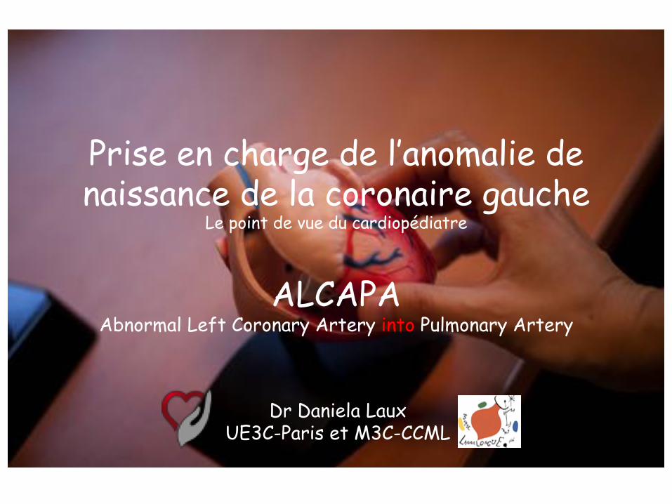
Repères chronologiques
Formation valves semilunaires Délamination valve tricuspide
Convergence
Bourgeons endocardiques Entonnoir tricuspidien
Elongation voie éjection Arcs Ao 4et 6
Connexion coronaires - aorte
J23 40 50 44 42 30
LOOP
Septation cardiaque
Wedging
J18
Croissant cardiaque
Corne D du sinus veineux Apparition VP 1° Arcs Ao 2 et 3
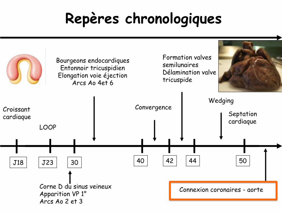
Embryologie
Bogers AJJC. Anat Embryol 1989
Les artères coronaires ne naissent pas de l�aorte (notion ancienne de bourgeons coronaires)
…mais se connectent à l�aorte
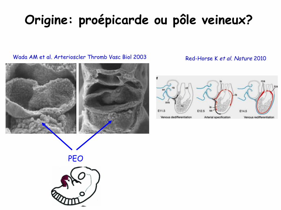
Origine: proépicarde ou pôle veineux?
PEO
Wada AM et al. Arterioscler Thromb Vasc Biol 2003 Red-Horse K et al. Nature 2010
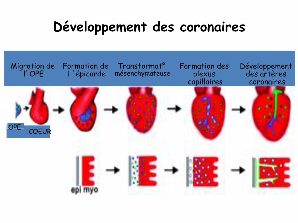
Développement des coronaires
OPE COEUR
Formation de l �épicarde
Transformat° mésenchymateuse
Formation des plexus
capillaires Développement
des artères coronaires
Migration de l�OPE
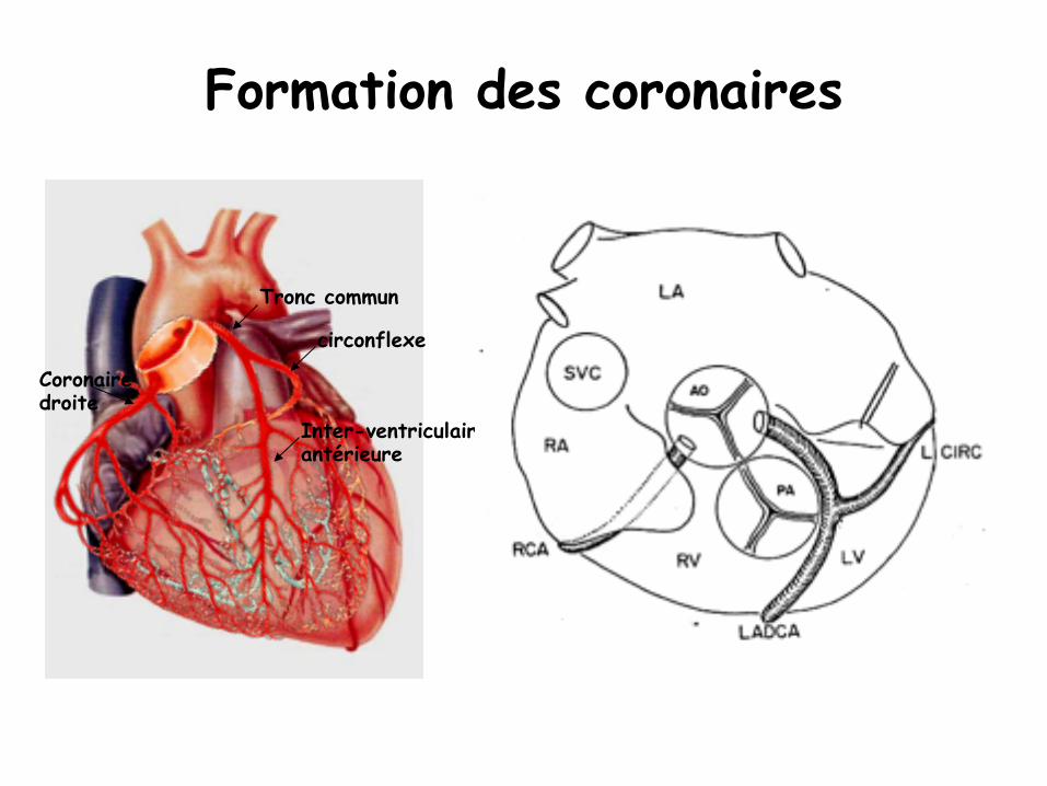
Tronc commun
circonflexe
Inter-ventriculaire antérieure
Coronaire droite
Formation des coronaires
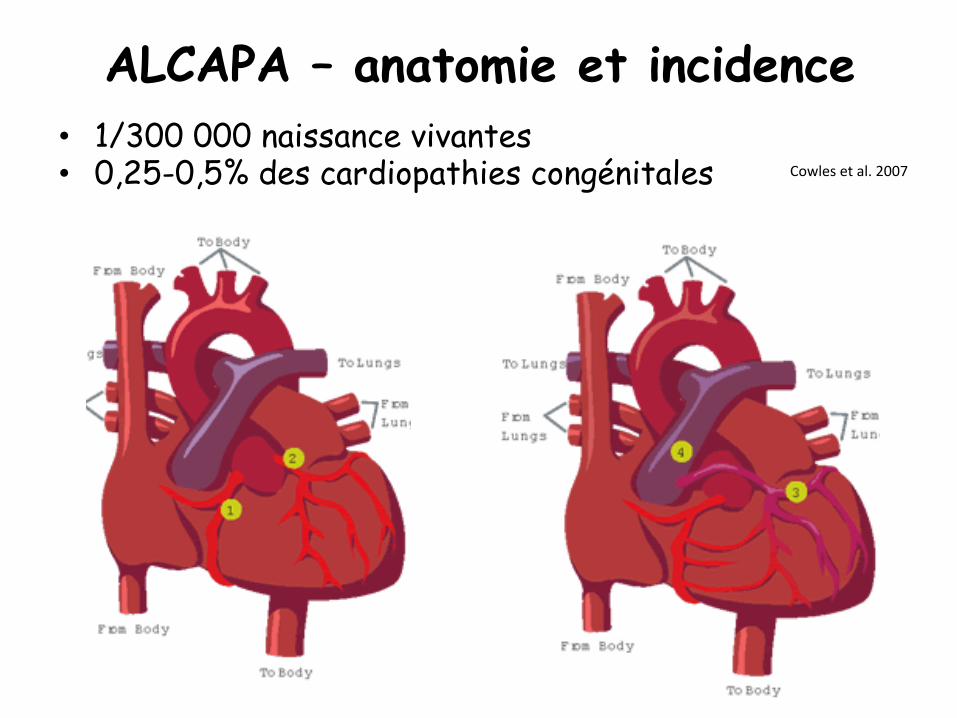
ALCAPA – anatomie et incidence • 1/300 000 naissance vivantes • 0,25-0,5% des cardiopathies congénitales Cowlesetal.2007
cardiac surgery was performed with a median delay of3.5 months after the initial repair. Table 2 summarizes the
moment of diagnosis as related to surgery, the diagnostic
method, and the surgery performed for all the patients.
Outcome and Follow-Up Period
Of the 12 patients, 7 (58 %) died after surgery. Death
occurred with a median delay of 0.5 months (range, 1 dayto 36 months) after the last surgery (complete repair or
second surgery for direct coronary reimplantation). The
reasons for early postoperative death were hemodynamicinstability such as low cardiac output syndrome, severe left
ventricular dysfunction with pulmonary hypertension cri-
sis, and cardiorespiratory arrest after accidental extubation.All the fatal events occurred in the ICU. For three
patients, ALCAPA still was unrecognized at the moment of
death, and the correct anatomic diagnosis was establishedat autopsy. The only exception was the newborn with
hypoplastic left heart syndrome, in whom the surgeon haddiscovered an abnormally connected infundibular coronary
branch while reconstructing the neo-aorta during Norwood
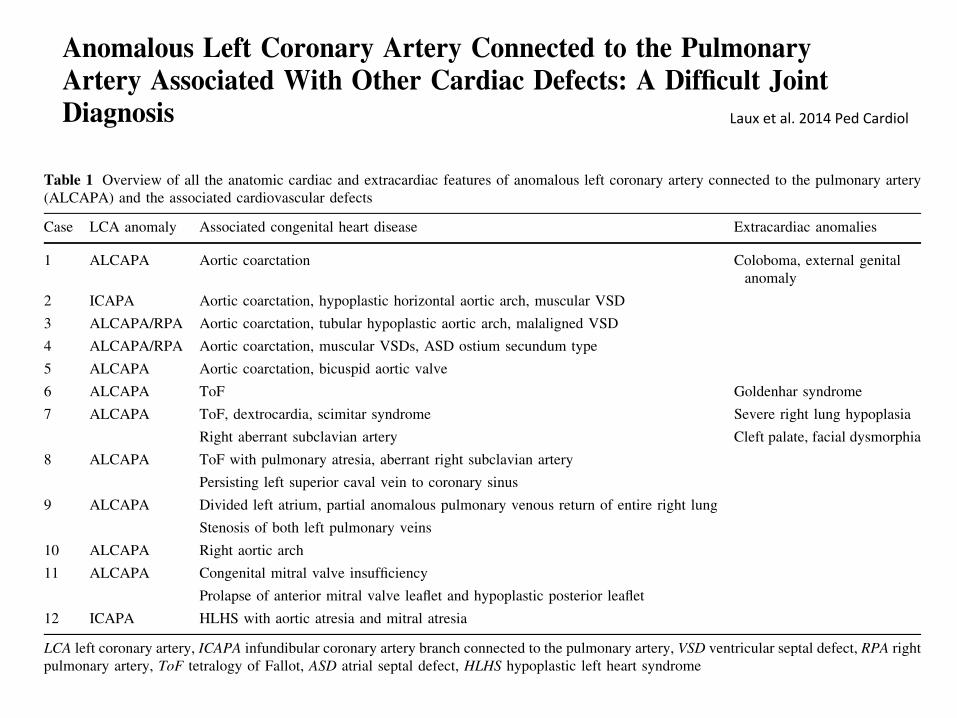
Table 1 Overview of all the anatomic cardiac and extracardiac features of anomalous left coronary artery connected to the pulmonary artery(ALCAPA) and the associated cardiovascular defects
Case LCA anomaly Associated congenital heart disease Extracardiac anomalies
1 ALCAPA Aortic coarctation Coloboma, external genitalanomaly
2 ICAPA Aortic coarctation, hypoplastic horizontal aortic arch, muscular VSD
3 ALCAPA/RPA Aortic coarctation, tubular hypoplastic aortic arch, malaligned VSD
4 ALCAPA/RPA Aortic coarctation, muscular VSDs, ASD ostium secundum type
5 ALCAPA Aortic coarctation, bicuspid aortic valve
6 ALCAPA ToF Goldenhar syndrome
7 ALCAPA ToF, dextrocardia, scimitar syndrome Severe right lung hypoplasia
Right aberrant subclavian artery Cleft palate, facial dysmorphia
8 ALCAPA ToF with pulmonary atresia, aberrant right subclavian artery
Persisting left superior caval vein to coronary sinus
9 ALCAPA Divided left atrium, partial anomalous pulmonary venous return of entire right lung
Stenosis of both left pulmonary veins
10 ALCAPA Right aortic arch
11 ALCAPA Congenital mitral valve insufficiency
Prolapse of anterior mitral valve leaflet and hypoplastic posterior leaflet
12 ICAPA HLHS with aortic atresia and mitral atresia
LCA left coronary artery, ICAPA infundibular coronary artery branch connected to the pulmonary artery, VSD ventricular septal defect, RPA rightpulmonary artery, ToF tetralogy of Fallot, ASD atrial septal defect, HLHS hypoplastic left heart syndrome
Fig. 1 Patient 3: Computer tomographic (CT) images of aorticcoarctation and anomalous left coronary artery connected to thepulmonary artery (ALCAPA). a Sagittal CT image showing an aorticcoarctation (CoA, black arrow) bypassed by a large persistent arterial
duct (PDA). The horizontal aortic arch is hypoplastic. b Coronal CTimage with the anomalous left coronary artery (ALCAPA, blackarrow) connected to the right pulmonary artery (RPA), a rather rareanatomic variant. PAT pulmonary artery trunk
Pediatr Cardiol
123
Author's personal copy
ORIGINAL ARTICLE
Anomalous Left Coronary Artery Connected to the PulmonaryArtery Associated With Other Cardiac Defects: A Difficult JointDiagnosis
Daniela Laux • Claire Bertail • Fanny Bajolle •
Lucile Houyel • Younes Boudjemline •
Damien Bonnet
Received: 7 December 2013 / Accepted: 25 April 2014! Springer Science+Business Media New York 2014
Abstract Anomalous left coronary artery connected tothe pulmonary artery (ALCAPA) can be associated rarely
with other congenital heart defects. The preoperative joint
diagnosis is challenging. From 1987 to 2012, a retrospec-tive bicentric assessment of 12 patients with ALCAPA
related to other cardiac defects focused on the associated
heart defect, the moment of complete diagnosis related tosurgery, and outcome. Coarctation was the most frequently
associated heart defect (n = 5) followed by tetralogy of
Fallot with or without pulmonary atresia (n = 3). Thestudy group comprised one case of hypoplastic left heart
syndrome, one right aortic arch, one congenital mitral
malformation, and one infant with divided left atrium andanomalous pulmonary venous return. Only four patients
had a complete diagnosis of both the cardiac defect and the
coronary abnormality before surgery. In two cases, thecoronary anomaly was discovered during surgery per-
formed for another cardiac defect and treated at the same
time. The diagnosis of the six remaining patients wasdetermined after cardiac repair. Of the 12 patients, 7
(58 %) died after surgery. Half of these patients died withinthe first 30 days after repair. At this writing, the remaining
patients are in good health after a median follow-up period
of 5.4 years (range, 2.1–8.5 years). This study confirmed
that ALCAPA associated with other cardiac defects often ismisdiagnosed before surgery, mostly due to specific
hemodynamics masking myocardial ischemia preopera-
tively. Survival was compromised due to the unrecognizeddiagnosis of an associated coronary abnormality but also
because of midterm complications related to the other
cardiac defects.
Keywords Anomalous left coronary artery connected to
the pulmonary artery ! ALCAPA ! Aortic coarctation !Tetralogy of Fallot ! Congenital heart disease ! Cardiacdefect
Introduction
Anomalous left coronary artery connected to the pulmon-ary artery (ALCAPA) is a congenital coronary anomaly
responsible for cardiac failure due to myocardial infarction
in early infancy [17]. Anatomic variants such as circum-flex, left anterior descending coronary artery, infundibular
branch, right coronary artery (ARCAPA), single coronaryartery, and both coronary arteries connected to the pul-
monary trunk or one of its branches, especially the right
pulmonary artery, have been reported.Although ALCAPA is most often an isolated entity, it can
be associated rarely with other congenital heart defects
(CHDs). Numerous associated CHDs including atrial andventricular septal defect (VSD), persistent arterial duct,
aortic coarctation/aortic arch abnormalities, and conotruncal
defects such as tetralogy of Fallot or common arterial trunkhave been described in the medical literature since the first
isolated clinical description in 1933 [1, 4–9, 12–16].
The positive preoperative diagnosis of ALCAPA andother cardiac defects is a challenge for the pediatric
D. Laux (&) ! C. Bertail ! L. HouyelCentre de Reference Malformations Cardiaques CongenitalesComplexes-M3C-CCML, Department of Congenital CardiacSurgery, Centre Chirurgical Marie Lannelongue, 133 Avenue dela Resistance, Le Plessis Robinson, Francee-mail: [email protected]
F. Bajolle ! Y. Boudjemline ! D. BonnetCentre de Reference Malformations Cardiaques CongenitalesComplexes-M3C-Necker, Pediatric Cardiology, 149 Rue deSevres, Hopital Necker Enfants Malades, Paris, France
123
Pediatr Cardiol
DOI 10.1007/s00246-014-0916-4
Author's personal copy
Lauxetal.2014PedCardiol
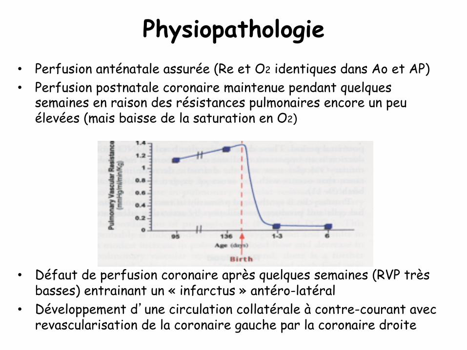
Physiopathologie • Perfusion anténatale assurée (Re et O2 identiques dans Ao et AP) • Perfusion postnatale coronaire maintenue pendant quelques
semaines en raison des résistances pulmonaires encore un peu élevées (mais baisse de la saturation en O2)
• Défaut de perfusion coronaire après quelques semaines (RVP très
basses) entrainant un « infarctus » antéro-latéral • Développement d�une circulation collatérale à contre-courant avec
revascularisation de la coronaire gauche par la coronaire droite
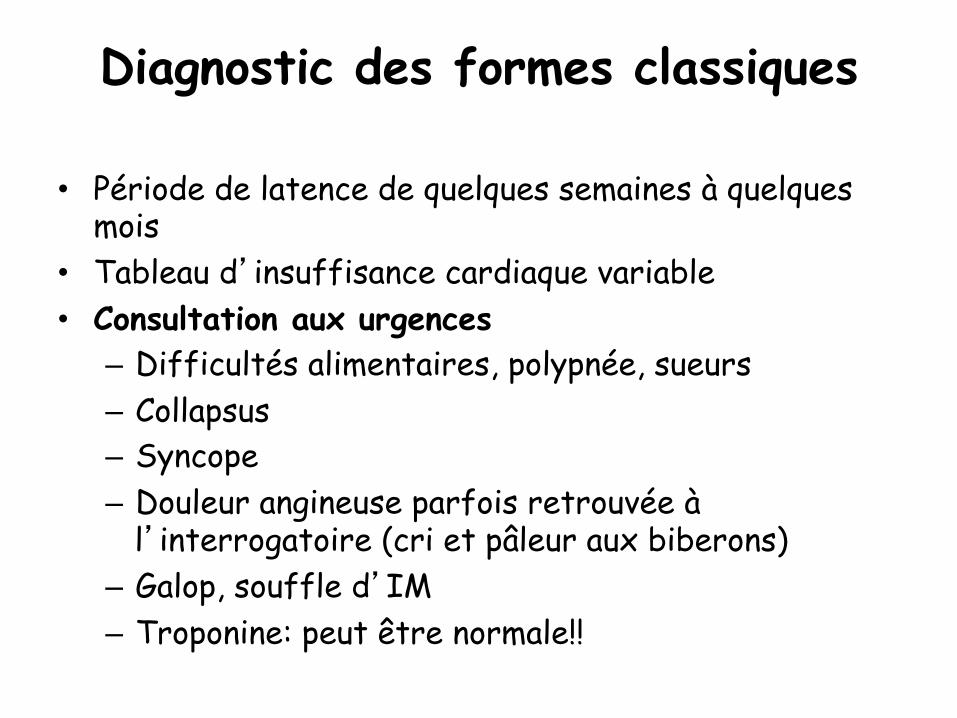
Diagnostic des formes classiques
• Période de latence de quelques semaines à quelques mois
• Tableau d�insuffisance cardiaque variable • Consultation aux urgences – Difficultés alimentaires, polypnée, sueurs – Collapsus – Syncope – Douleur angineuse parfois retrouvée à
l�interrogatoire (cri et pâleur aux biberons) – Galop, souffle d�IM – Troponine: peut être normale!!
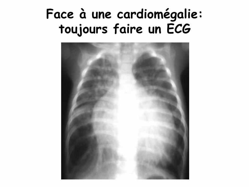
Face à une cardiomégalie: toujours faire un ECG
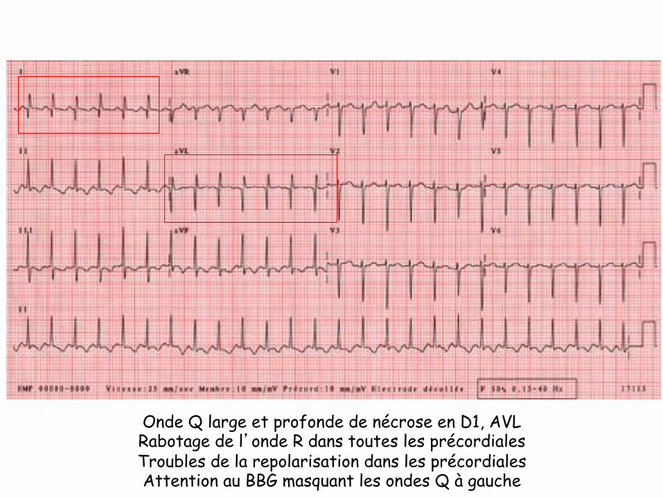
Onde Q large et profonde de nécrose en D1, AVL Rabotage de l�onde R dans toutes les précordiales Troubles de la repolarisation dans les précordiales Attention au BBG masquant les ondes Q à gauche
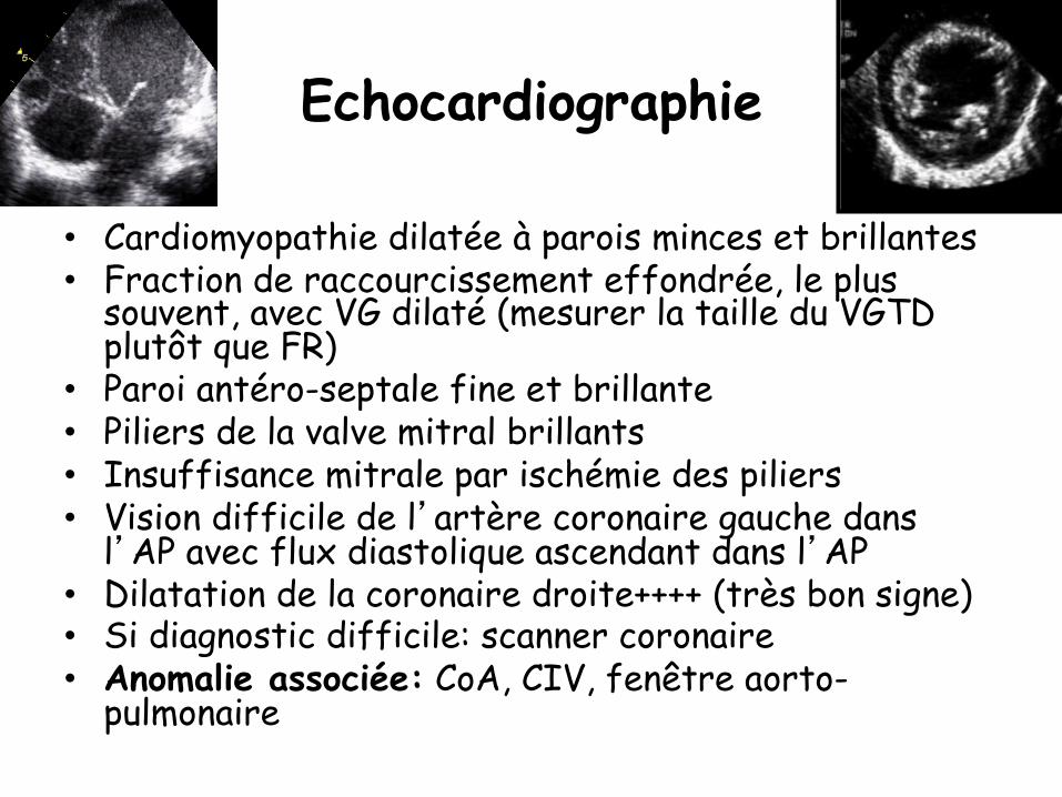
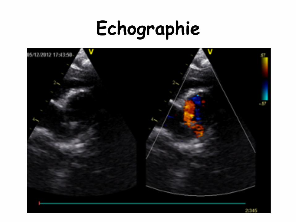
Echocardiographie
• Cardiomyopathie dilatée à parois minces et brillantes • Fraction de raccourcissement effondrée, le plus
souvent, avec VG dilaté (mesurer la taille du VGTD plutôt que FR)
• Paroi antéro-septale fine et brillante • Piliers de la valve mitral brillants • Insuffisance mitrale par ischémie des piliers • Vision difficile de l�artère coronaire gauche dans
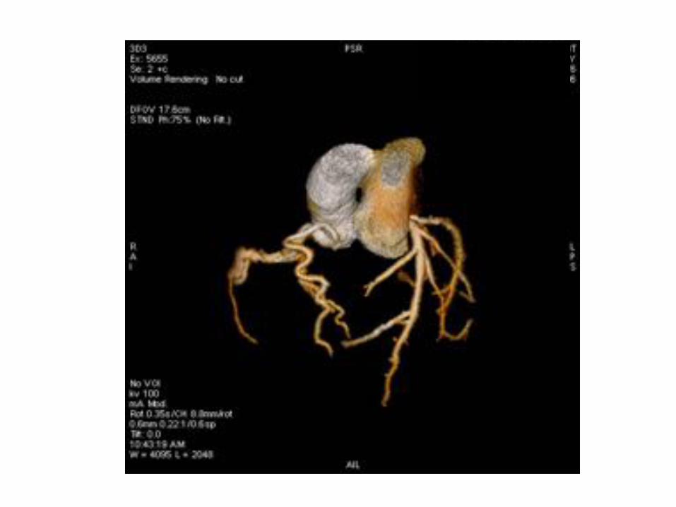
l�AP avec flux diastolique ascendant dans l�AP • Dilatation de la coronaire droite++++ (très bon signe) • Si diagnostic difficile: scanner coronaire • Anomalie associée: CoA, CIV, fenêtre aorto-
pulmonaire
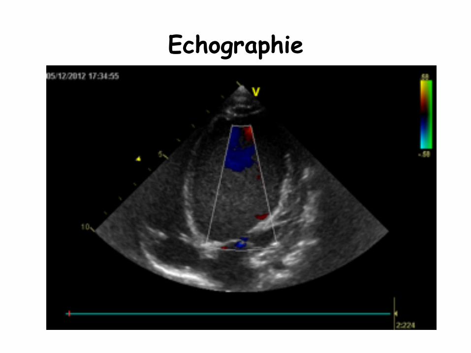
Echographie
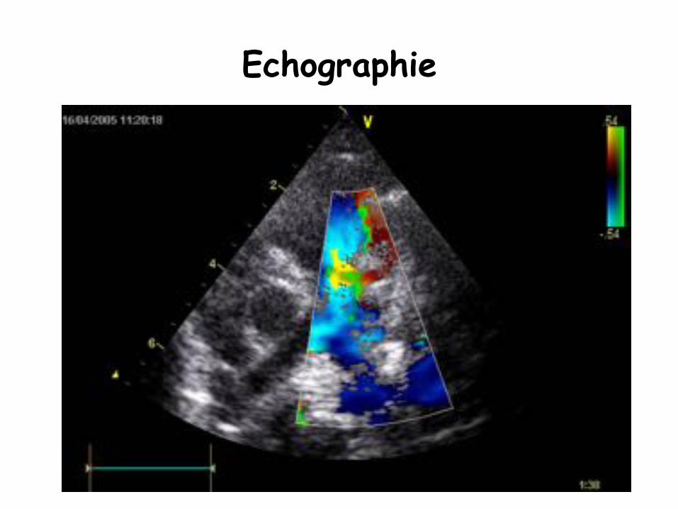
Echographie
Echographie
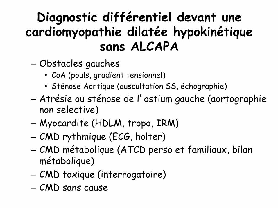
Diagnostic différentiel devant une cardiomyopathie dilatée hypokinétique
sans ALCAPA – Obstacles gauches
• CoA (pouls, gradient tensionnel) • Sténose Aortique (auscultation SS, échographie)
– Atrésie ou sténose de l�ostium gauche (aortographie non selective)
– Myocardite (HDLM, tropo, IRM) – CMD rythmique (ECG, holter) – CMD métabolique (ATCD perso et familiaux, bilan
métabolique) – CMD toxique (interrogatoire) – CMD sans cause
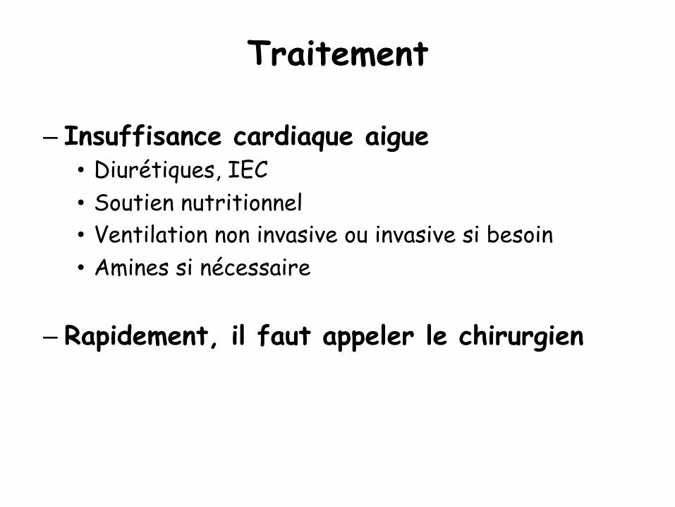
Traitement
– Insuffisance cardiaque aigue • Diurétiques, IEC • Soutien nutritionnel • Ventilation non invasive ou invasive si besoin • Amines si nécessaire
– Rapidement, il faut appeler le chirurgien
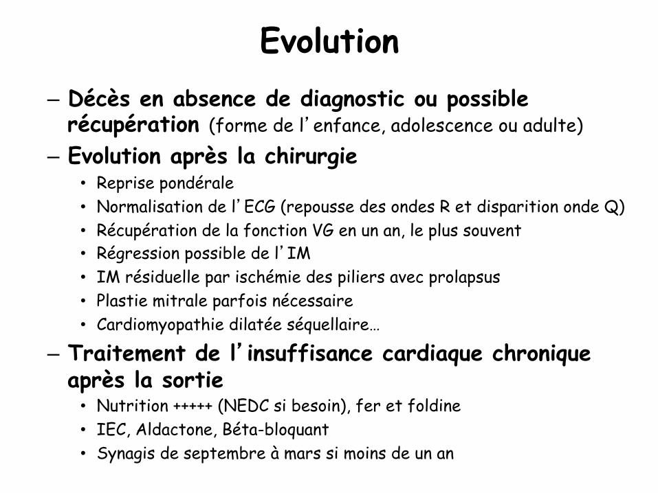
Evolution – Décès en absence de diagnostic ou possible
récupération (forme de l�enfance, adolescence ou adulte) – Evolution après la chirurgie
• Reprise pondérale • Normalisation de l�ECG (repousse des ondes R et disparition onde Q) • Récupération de la fonction VG en un an, le plus souvent • Régression possible de l�IM • IM résiduelle par ischémie des piliers avec prolapsus • Plastie mitrale parfois nécessaire • Cardiomyopathie dilatée séquellaire…
– Traitement de l�insuffisance cardiaque chronique après la sortie • Nutrition +++++ (NEDC si besoin), fer et foldine • IEC, Aldactone, Béta-bloquant • Synagis de septembre à mars si moins de un an
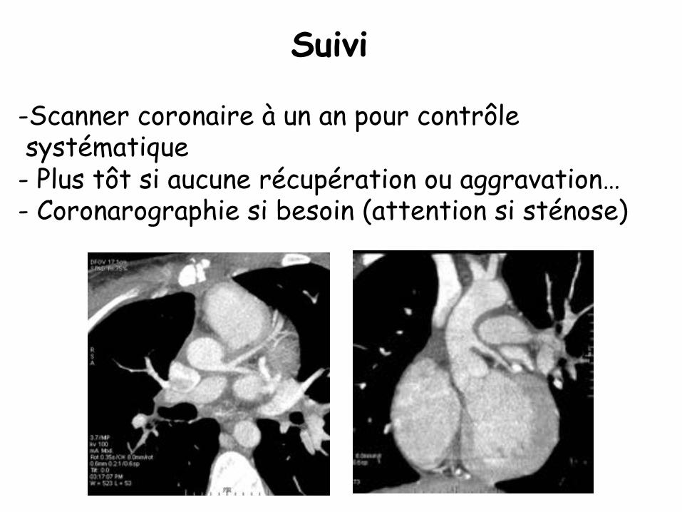
Suivi
- Scanner coronaire à un an pour contrôle systématique - Plus tôt si aucune récupération ou aggravation… - Coronarographie si besoin (attention si sténose)
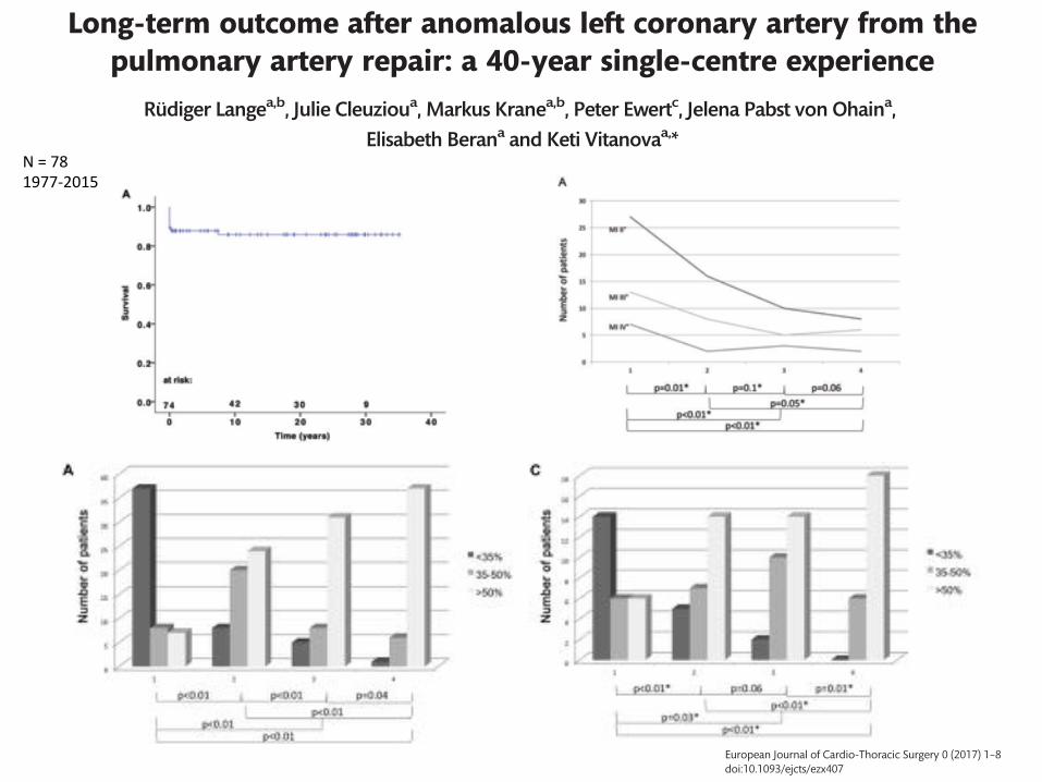
Cite this article as: Lange R, Cleuziou J, Krane M, Ewert P, Pabst von Ohain J, Beran E et al. Long-term outcome after anomalous left coronary artery from thepulmonary artery repair: a 40-year single-centre experience. Eur J Cardiothorac Surg 2017; doi:10.1093/ejcts/ezx407.
Long-term outcome after anomalous left coronary artery from thepulmonary artery repair: a 40-year single-centre experience
Rudiger Langea,b, Julie Cleuzioua, Markus Kranea,b, Peter Ewertc, Jelena Pabst von Ohaina,Elisabeth Berana and Keti Vitanovaa,*
a Department of Cardiovascular Surgery, German Heart Centre Munich, Technische Universitat Munchen, Munich, Germanyb German Heart Center Munich–DZHK Partner Site Munich Heart Alliance, Munich, Germanyc Department of Pediatric Cardiology and Congenital Heart Disease, German Heart Centre Munich, Technische Universitat Munchen, Munich, Germany
* Corresponding author. Department of Cardiovascular Surgery, German Heart Centre Munich, Lazarettstrasse 36, 80636 Munich, Germany. Tel: +49-89-12182962;fax: +49-89-12184123; e-mail: [email protected] (K. Vitanova).
Received 6 July 2017; received in revised form 9 October 2017; accepted 23 October 2017
Abstract
OBJECTIVES: An anomalous left coronary artery from the pulmonary artery (ALCAPA) is a rare congenital anomaly, often associated withseverely impaired left ventricular (LV) contractility and functional mitral valve (MV) regurgitation. Current data suggest that earlier correc-tion of ALCAPA may result in a more complete recovery of LV function. By analysing the results of a large single-centre ALCAPA cohort, wesought to investigate whether these treatment paradigms remain valid.
METHODS: A retrospective study was performed evaluating all patients undergoing repair of ALCAPA over a period of almost 40 years. Allpreoperative and postoperative echocardiographic reports were reviewed, focusing on the recovery of LV and MV function.
RESULTS: The study cohort included 78 patients who underwent ALCAPA repair between 1977 and 2015, who were divided into 2 groupsbased on patient age at initial repair: Group A (n = 52, age <1 year) and Group B (n = 26, age >1 year). Following repair, systolic LV and MVfunction improved significantly (P < 0.01) in both groups. Patient age at the time of initial surgery had no significant influence on theimprovement of LV function. Early mortality (within 30 days) was 10% (n = 8). No 30-day mortality was reported in the past 20 years.Survival at 20 years following ALCAPA repair was 86 ± 4%.
CONCLUSIONS: Following ALCAPA repair, LV function significantly improved, regardless of age at the time of repair. In addition, preoper-ative functional MV regurgitation decreased over time. Concomitant mitral valve surgery at the time of ALCAPA repair is required inpatients with structural abnormalities of the MV.
Keywords: Anomalous left coronary artery from the pulmonary artery repair • Left ventricular function • Coronary collateralization
INTRODUCTION
An anomalous left coronary artery from the pulmonary artery(ALCAPA) is a rare congenital anomaly with an incidence of 1 in300 000 live births, which corresponds to 0.25–0.5% of all con-genital heart disease [1, 2]. The pathological condition of ALCAPAwas first described by Brooks in 1882 [3]. In 1933, Bland, Whiteand Garland published the first clinical description.
The formation of collaterals between the right coronaryartery and the left coronary artery along with decreasing pul-monary vascular resistance after birth leads to a coronary stealphenomenon by left to right shunting. In the absence ofadequate collateralization, these changes result in severe myo-cardial ischaemia and dysfunction [4]. If left uncorrected, thenatural course of the disease in infants reaches a mortality rateof 90% [4]. Extensive collateral arteries may enable somepatients to survive beyond infancy. However, subendocardial
ischaemia may promote sudden death due to ventriculararrhythmia [5].
Since the first correction was performed in 1953 by Mustard,several surgical techniques have been described for repair ofALCAPA including ligation of the anomalous artery [6], saphenousvein and mammary artery grafting from the aorta [7], anastomo-sis of the subclavian artery, performed in an end-to-end fashionafter excision of the coronary button from the pulmonary artery[8], transpulmonary baffling (Takeuchi procedure [9]) and coro-nary translocation into the aorta [10]. Coronary translocationallows a true anatomical restoration and is considered as the pro-cedure of choice for repair of ALCAPA in modern times.
Restoration of true anatomy leads to rapid recovery of leftventricular (LV) function [11, 12] within the 1st postoperative year[13]. There remains a paucity of information in the literatureregarding the influence of patient age at the time of ALCAPArepair on the improvement of LV function. Moreover, only a few
CO
NG
ENIT
AL
VC The Author 2017. Published by Oxford University Press on behalf of the European Association for Cardio-Thoracic Surgery. All rights reserved.
European Journal of Cardio-Thoracic Surgery 0 (2017) 1–8 ORIGINAL ARTICLEdoi:10.1093/ejcts/ezx407
Downloaded from https://academic.oup.com/ejcts/advance-article-abstract/doi/10.1093/ejcts/ezx407/4652928by Technische Universitaet Muenchen useron 18 December 2017
Figure 2: (A) ALCAPA patient without coronary collateralization (infant type) and (B) coronary collateralization in an ALCAPA patient (adult type). ALCAPA: anomalousleft coronary artery from the pulmonary artery; RCA: right coronary artery.
Figure 3: (A) Freedom from cardiac death in 78 anomalous left coronary artery from the pulmonary artery patients and (B) freedom from cardiac death in 37 anoma-lous left coronary artery from the pulmonary artery patients who underwent repair after 1995.
4 R. Lange et al. / European Journal of Cardio-Thoracic Surgery
Downloaded from https://academic.oup.com/ejcts/advance-article-abstract/doi/10.1093/ejcts/ezx407/4652928by Technische Universitaet Muenchen useron 18 December 2017
Prior to ALCAPA repair, mild FMR was identified in 31 (40%)patients and moderate-to-severe FMR was identified in 47 (60%)patients (Table 3). MV function had improved significantly at thefinal follow-up at a median time period of 18 years (range 2–39 years) following repair (Fig. 5A).
Only 1 patient of the 78 patients had a concomitant MV repairat the time of ALCAPA repair; this was performed due to prolapseof the anterior mitral leaflet. Four (5%) patients underwent a totalof 8 reoperations for MV repair at a median time perod of6 months (range 11 days–14 months) after initial ALCAPA repair.
In 1 patient, an MV replacement was necessary at the third reop-eration. Freedom from MI >_2 was 88 ± 4%, 84 ± 5% and 83 ± 5%after 5, 10 and 20 years, respectively (95% CI 25.2–31.8; Fig. 5B).
DISCUSSION
ALCAPA patients require surgical correction as soon as the diag-nosis is made, regardless of symptoms or the degree of inter-coronary collateralization [20]. The surgical method of ALCAPA
Figure 4: (A) Left ventricular ejection fraction in patients from Group A at different time points; (B) left ventricular ejection fraction in patients from Group B at differ-ent time points; (C) correlation between DEF and the patient ages; (D) file 3: correlation between DEF and the patient ages in all patients from Group B. *statistical sig-nificance. DEF: difference between EF at the final follow-up and preoperative EF; 1: preoperative; 2: postoperative; 3: at the time of hospital discharge; 4: at the finalfollow-up; EF: ejection fraction.
Table 2: EF in 52 patients (Group A) and 26 patients (Group B) at different time points following ALCAPA repair
Time point Preoperative, n (%) Postoperative, n (%) Discharge, n (%) Final follow-up, n (%)
Group A (n = 52) B (n = 26) A (n = 52) B (n = 26) A (n = 44) B (n = 26) A (n = 44) B (n = 24)
EF (%)<35 37 (71) 14 (54) 8 (15) 5 (19) 5 (11) 2 (8) 1 (2) 035–50 8 (16) 6 (23) 20 (39) 7 (27) 8 (18) 10 (38) 6 (14) 6 (25)>50 7 (13) 6 (23) 24 (46) 14 (54) 31 (70) 14 (54) 37 (84) 18 (75)
P-value 0.06 0.4 0.3 0.3
ALCAPA: anomalous left coronary artery from the pulmonary artery; EF: ejection fraction.
CO
NG
ENIT
AL
5R. Lange et al. / European Journal of Cardio-Thoracic Surgery
Downloaded from https://academic.oup.com/ejcts/advance-article-abstract/doi/10.1093/ejcts/ezx407/4652928by Technische Universitaet Muenchen useron 18 December 2017
repair has evolved over time and the translocation technique hasbecome a standard modern procedure for most patients. Incases of an exceptional distance of the left coronary ostium fromthe aorta, the transpulmonary baffling (Takeuchi) procedure is avalid alternative. This study showed that to date, long-termresults after initial repair are excellent. In our study, overall sur-vival at 10 and 20 years was 86 ± 4%. No early deaths occurredamong the 37 patients who underwent repair after 1995. Amongpatients repaired after 1995, freedom from death at 20 yearspost-repair was 97± 3%. This long-term results of the study (upto 40 years postoperatively) in a large patient population areunique and have not been published by other investigators.Naimo et al. [14] recently reported 98% survival in 42 patients at20 years following ALCAPA repair. Survival rates after 10 yearshave been described by different authors to range between 82%and 100% [15, 21].
In all, 84% (Group A) and 79% (Group B) of our patients exhib-ited normal LV function at the final follow-up, with no differencein outcomes whether ALCAPA had been corrected before orbeyond 1 year of age. FMR was addressed in only 1 patient at theinitial operation and only 5% of the patients required a laterintervention on the MV. In all the remaining patients, FMRimproved in the long term with ALCAPA repair alone.
LV damage due to myocardial ischaemia in patients withALCAPA is associated with a range of pathological findings, suchas endocardial and subendocardial fibrosis of the papillarymuscles, myocardial necrosis and ventricular dilatation [22]. As aconsequence, MV incompetence develops in the majority ofcases [23]. In our study, 65% of patients had severely impaired LVfunction and 60% of patients presented with FMR Grade 2 ormore before ALCAPA repair. Systolic function improved signifi-cantly after repair and remained normal in 80% of patients.There was no significant difference in the improvement of LVfunction whether patients had undergone repair of ALCAPAbefore or beyond 1 year of age.
The mechanisms for an age-independent regeneration of LVfunction remain speculative [24]. Within the 1st week after birth,mammalian hearts are able to regenerate with complete restora-tion of cardiac geometry after resection of the ventricular apex[25]. This capability is lost with increasing age. Additionally,Bergmann et al. [26] showed cardiomyocyte turnover of around1% in young probands, which also declines with increasing age.As ALCAPA repair leads to an ‘age-independent’ full recovery ofLV function, it can be hypothesized that the major mechanism ofimpaired LV function is hypoperfusion/hypoxia rather than a def-inite loss of cardiomyocytes. Conversely, Shivalkar et al. [13]
Figure 5: (A) Mitral valve insufficiency in patients with anomalous left coronary artery from the pulmonary artery at different time points and (B) freedom from mitralvalve insufficiency (Grade 2 or more) after anomalous left coronary artery from the pulmonary artery repair. *statistical significance. 1: preoperative; 2: postoperative;3: at the time of hospital discharge; 4: at the final follow-up; MI: mitral valve insufficiency.
Table 3: Echocardiographic parameters in 78 ALCAPA patients
Time point Preoperative Postoperative Discharge Final follow-up
MI, grade, n (%)0 7 (9) 19 (24) 22 (32) 21 (31)1 24 (31) 33 (42) 30 (43) 31 (45)2 27 (35) 16 (21) 10 (14) 8 (12)3 13 (17) 8 (10) 5 (7) 6 (9)4 7 (8) 2 (3) 3 (4) 2 (3)
LVESD (mm), median (range) 33 (19–48) 32 (16–44) 25 (13–41) 28 (17–41)LVEDD (mm), median (range) 37 (22–68) 37 (25–58) 33 (17–61) 39 (27–56)
ALCAPA: anomalous left coronary artery from the pulmonary artery; LVEDD: left ventricle end-diastolic dimension; LVESD: left ventricle end-systolic dimen-sion; MI: mitral valve insufficiency.
6 R. Lange et al. / European Journal of Cardio-Thoracic Surgery
Downloaded from https://academic.oup.com/ejcts/advance-article-abstract/doi/10.1093/ejcts/ezx407/4652928by Technische Universitaet Muenchen useron 18 December 2017
Cite this article as: Lange R, Cleuziou J, Krane M, Ewert P, Pabst von Ohain J, Beran E et al. Long-term outcome after anomalous left coronary artery from thepulmonary artery repair: a 40-year single-centre experience. Eur J Cardiothorac Surg 2017; doi:10.1093/ejcts/ezx407.
Long-term outcome after anomalous left coronary artery from thepulmonary artery repair: a 40-year single-centre experience
Rudiger Langea,b, Julie Cleuzioua, Markus Kranea,b, Peter Ewertc, Jelena Pabst von Ohaina,Elisabeth Berana and Keti Vitanovaa,*
a Department of Cardiovascular Surgery, German Heart Centre Munich, Technische Universitat Munchen, Munich, Germanyb German Heart Center Munich–DZHK Partner Site Munich Heart Alliance, Munich, Germanyc Department of Pediatric Cardiology and Congenital Heart Disease, German Heart Centre Munich, Technische Universitat Munchen, Munich, Germany
* Corresponding author. Department of Cardiovascular Surgery, German Heart Centre Munich, Lazarettstrasse 36, 80636 Munich, Germany. Tel: +49-89-12182962;fax: +49-89-12184123; e-mail: [email protected] (K. Vitanova).
Received 6 July 2017; received in revised form 9 October 2017; accepted 23 October 2017
Abstract
OBJECTIVES: An anomalous left coronary artery from the pulmonary artery (ALCAPA) is a rare congenital anomaly, often associated withseverely impaired left ventricular (LV) contractility and functional mitral valve (MV) regurgitation. Current data suggest that earlier correc-tion of ALCAPA may result in a more complete recovery of LV function. By analysing the results of a large single-centre ALCAPA cohort, wesought to investigate whether these treatment paradigms remain valid.
METHODS: A retrospective study was performed evaluating all patients undergoing repair of ALCAPA over a period of almost 40 years. Allpreoperative and postoperative echocardiographic reports were reviewed, focusing on the recovery of LV and MV function.
RESULTS: The study cohort included 78 patients who underwent ALCAPA repair between 1977 and 2015, who were divided into 2 groupsbased on patient age at initial repair: Group A (n = 52, age <1 year) and Group B (n = 26, age >1 year). Following repair, systolic LV and MVfunction improved significantly (P < 0.01) in both groups. Patient age at the time of initial surgery had no significant influence on theimprovement of LV function. Early mortality (within 30 days) was 10% (n = 8). No 30-day mortality was reported in the past 20 years.Survival at 20 years following ALCAPA repair was 86 ± 4%.
CONCLUSIONS: Following ALCAPA repair, LV function significantly improved, regardless of age at the time of repair. In addition, preoper-ative functional MV regurgitation decreased over time. Concomitant mitral valve surgery at the time of ALCAPA repair is required inpatients with structural abnormalities of the MV.
Keywords: Anomalous left coronary artery from the pulmonary artery repair • Left ventricular function • Coronary collateralization
INTRODUCTION
An anomalous left coronary artery from the pulmonary artery(ALCAPA) is a rare congenital anomaly with an incidence of 1 in300 000 live births, which corresponds to 0.25–0.5% of all con-genital heart disease [1, 2]. The pathological condition of ALCAPAwas first described by Brooks in 1882 [3]. In 1933, Bland, Whiteand Garland published the first clinical description.
The formation of collaterals between the right coronaryartery and the left coronary artery along with decreasing pul-monary vascular resistance after birth leads to a coronary stealphenomenon by left to right shunting. In the absence ofadequate collateralization, these changes result in severe myo-cardial ischaemia and dysfunction [4]. If left uncorrected, thenatural course of the disease in infants reaches a mortality rateof 90% [4]. Extensive collateral arteries may enable somepatients to survive beyond infancy. However, subendocardial
ischaemia may promote sudden death due to ventriculararrhythmia [5].
Since the first correction was performed in 1953 by Mustard,several surgical techniques have been described for repair ofALCAPA including ligation of the anomalous artery [6], saphenousvein and mammary artery grafting from the aorta [7], anastomo-sis of the subclavian artery, performed in an end-to-end fashionafter excision of the coronary button from the pulmonary artery[8], transpulmonary baffling (Takeuchi procedure [9]) and coro-nary translocation into the aorta [10]. Coronary translocationallows a true anatomical restoration and is considered as the pro-cedure of choice for repair of ALCAPA in modern times.
Restoration of true anatomy leads to rapid recovery of leftventricular (LV) function [11, 12] within the 1st postoperative year[13]. There remains a paucity of information in the literatureregarding the influence of patient age at the time of ALCAPArepair on the improvement of LV function. Moreover, only a few
CON
GEN
ITA
L
VC The Author 2017. Published by Oxford University Press on behalf of the European Association for Cardio-Thoracic Surgery. All rights reserved.
European Journal of Cardio-Thoracic Surgery 0 (2017) 1–8 ORIGINAL ARTICLEdoi:10.1093/ejcts/ezx407
Downloaded from https://academic.oup.com/ejcts/advance-article-abstract/doi/10.1093/ejcts/ezx407/4652928by Technische Universitaet Muenchen useron 18 December 2017
N=781977-2015
CONGENITAL – Original Submission
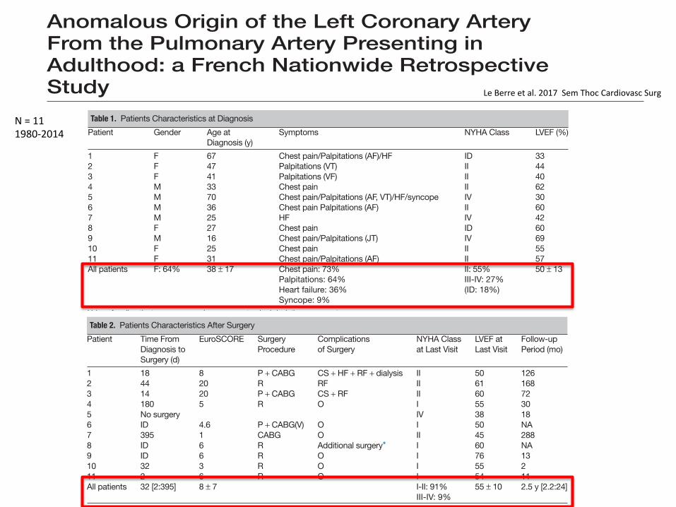
Anomalous Origin of the Left Coronary ArteryFrom the Pulmonary Artery Presenting inAdulthood: a French Nationwide RetrospectiveStudyLaura Le Berre, MD,* Alban-Elouen Baruteau, MD,†,‡ Alain Fraisse, MD, PhD,§,‖
Dominique Boulmier, MD,¶ Maria Jimenez, MD,# Bruno Gallet, MD, PhD,**Karine Warin Fresse, MD,†† Jacques Mansourati, MD,* and Patrice Guerin, MD, PhD††
Anomalous origin of the left coronary artery from the pulmonary artery (ALCAPA)is a rare congenital heart disease usually diagnosed during the first monthsof life. Without surgical treatment, ALCAPA carries a high mortality risk, anddisease presentation in adulthood is rare. We describe the diagnosis and man-agement of patients presenting with ALCAPA in adulthood. This multicenterFrench nationwide retrospective study included adult patients diagnosed from1980 to 2014. Eleven adult patients (mean age: 38 ± 17 years) were ana-lyzed. All patients were symptomatic, presenting with chest pain, palpitations,heart failure, or syncope. Electrocardiogram was abnormal in 8 (73%) pa-tients. Echocardiogram showed a mildly depressed left ventricular ejectionfraction of 50 ± 13%, kinetic abnormalities in 5 (45%) patients, and signifi-cant mitral regurgitation in 8 (73%) patients. Coronary angiography wasperformed in 10 (91%) patients and confirmed the diagnosis. Computerizedtomography scan, magnetic resonance imaging, and myocardial scintigra-phy were performed when deemed necessary. Ten patients underwentreconstructive surgery, but 1 patient was not operated because of age. Fourpatients experienced postoperative complications including cardiogenic shock,heart failure, renal failure, or additional surgery. After a median follow-up of2.5 years, all 10 operated patients were alive and asymptomatic, and thenonoperated patient had died at the age of 70 from syncope related to ven-tricular tachycardia. ALCAPA may be diagnosed in adults. Althoughcomplications may occur postoperatively, long-term outcome is favorable inadult patients undergoing surgical correction. Surgery should be discussedas first-line therapy in adults with ALCAPA.
Semin Thoracic Surg ■■:■■–■■ © 2017 Elsevier Inc. All rights reserved.
Keywords: ALCAPA, Bland-White-Garland syndrome, congenital heartdisease, myocardial infarction, congenital heart disease
*Brest University Hospital, Western Brittany University, Brest, France†Marie-Lannelongue Hospital, Paris-Sud University, Paris, France‡Morgan Stanley Children’s Hospital, New York Presbyterian, ColumbiaUniversity Medical Center, New York, New York§AP-HM—La Timone Children Hospital, Marseille, France‖Royal Brompton Hospital, Harefield NHS Trust, Imperial College London,London, UK¶Rennes University Hospital, Rennes-1 University, Rennes, France#Clinique Saint Augustin, Bordeaux, France**Argenteuil Hospital, Argenteuil, France††L’Institut du Thorax, Nantes University Hospital, Nantes, France
Date and number of the IRB approval: 24/10/12 Comité de Protectiondes Personnes Ouest VI.
Address reprint requests to Laura Le Berre, MD, Service de CardiologieClinique de Keraudren rue Ernestine de Tremaudan, Dherbécourt, 29200Brest, France. E-mail: [email protected]
CT scan showing abnormal origin of the left coronaryartery from the pulmonary artery (white arrow).
Central Message
ALCAPA is rarely diagnosed in adults, but sur-gical correction in adulthood appears to beassociated with favorable long-term outcome.
Perspective Statement
ALCAPA is a rare congenital heart diseaseusually diagnosed during the first months of life.Without surgical treatment, mortality is high andpresentation in adulthood is rare.We describe the management of 11 adult pa-tients with ALCAPA. Although complicationsmay occur postoperatively, long-term outcomeis favorable, suggesting surgery should be con-sidered as first-line therapy in adults withALCAPA.
1043-0679/$-see front matter © 2017 Elsevier Inc. All rights reserved. 1https://doi.org/10.1053/j.semtcvs.2017.08.018
diagnosis (all 4 tests were necessary in 1 patient, 2 or 3 in 6 pa-tients, and 1 in 4 patients).
Coronary angiography revealed the presence of giant arteries inthe right coronary system, which irrigated the left system, as wellas in several collateral arteries. MRI allowed analyzing the myo-cardial perfusion. Anomalous origin of the left coronary wasconfirmed, and left ventricular function was assessed. Lack ofcollaterals or their localization was reported (in 2 cases, collateralswere present in the anterior territory). No pericardial abnormali-ty was found. Non-compaction of the left myocardium was foundin 1 patient. Myocardial scintigraphy (exercise myocardial perfu-sion imaging) was also used to assess myocardial irrigation by
collaterals. All patients were found to have perfusion abnormali-ties, mainly in the anteroseptal and anterolateral territories.
Patient ManagementTen (91%) patients underwent surgery (Table 2). The mean
EuroSCORE was 8 ± 7. One patient was denied surgery becauseof age (70 years old) and comorbidities. In 6 patients out of 10operated (60%), the left coronary system was reimplanted on theascending aorta. The other patients underwent closure of the anom-alous origin of the coronary artery by a pericardium patch and asingle coronary artery bypass grafting (the left anterior descendingcoronary by either internal mammary artery in 3 patients or
Table 1. Patients Characteristics at Diagnosis
Patient Gender Age atDiagnosis (y)
Symptoms NYHA Class LVEF (%)
1 F 67 Chest pain/Palpitations (AF)/HF ID 332 F 47 Palpitations (VT) II 443 F 41 Palpitations (VF) II 404 M 33 Chest pain II 625 M 70 Chest pain/Palpitations (AF, VT)/HF/syncope IV 306 M 36 Chest pain Palpitations (AF) II 607 M 25 HF IV 428 F 27 Chest pain ID 609 M 16 Chest pain/Palpitations (JT) IV 6910 F 25 Chest pain II 5511 F 31 Chest pain/Palpitations (AF) II 57All patients F: 64% 38 ± 17 Chest pain: 73%
Palpitations: 64%Heart failure: 36%Syncope: 9%
II: 55%III-IV: 27%(ID: 18%)
50 ± 13
Values for all patients are expressed as mean ± standard deviation or percentage.AF, atrial fibrillation; F, female; HF, heart failure; ID, indeterminate ; JT, junctional tachycardia; LVEF, left ventricular ejection fraction; M, male; NYHA,New York Heart Association; VF, ventricular fibrillation; VT, ventricular tachycardia.
Table 2. Patients Characteristics After Surgery
Patient Time FromDiagnosis toSurgery (d)
EuroSCORE SurgeryProcedure
Complicationsof Surgery
NYHA Classat Last Visit
LVEF atLast Visit
Follow-upPeriod (mo)
1 18 8 P + CABG CS + HF + RF + dialysis II 50 1262 44 20 R RF II 61 1683 14 20 P + CABG CS + RF II 60 724 180 5 R O I 55 305 No surgery IV 38 186 ID 4.6 P + CABG(V) O I 50 NA7 395 1 CABG O II 45 2888 ID 6 R Additional surgery* I 60 NA9 ID 6 R O I 76 1310 32 3 R O I 55 211 2 6 R O I 54 11All patients 32 [2:395] 8 ± 7 I-II: 91%
III-IV: 9%55 ± 10 2.5 y [2.2:24]
Values for all patients are expressed as median with range or mean ± standard deviation or percentage.*Patient required additional surgery for left coronary artery and pulmonary artery truncus stenosis with tricuspid regurgitation.CABG, coronary artery bypass grafting; CS, cardiogenic shock; HF, heart failure; NA, not available; P, pericardium patch; R, reimplantation onthe ascending aorta; RF, renal failure; V, venous.
CONGENITAL – ANOMALOUS ORIGIN OF THE LEFT CORONARY ARTERY
Seminars in Thoracic and Cardiovascular Surgery • Volume ■■, Number ■■ 3
LeBerreetal.2017SemThocCardiovascSurg
N=111980-2014

• Laurie, née le 13/11/95 • Aucun antécédent personnel • ETT normale à 4 ans pour souffle • Douleur thoracique rétro-sternale constrictive à
l�effort depuis deux ans avec blockpnée, sans dyspnée ni orthopnée, ni syncope, ni palpitation
• Majoration récente des douleurs (sport+++) • Epreuve d�effort au centre de l�asthme!!
Diagnostic des formes « pièges »
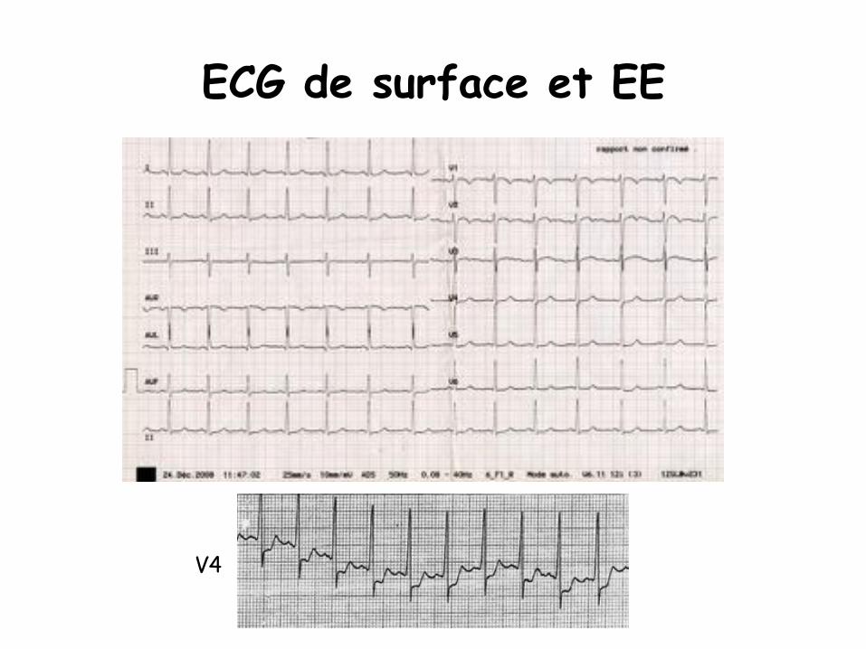
ECG de surface et EE
V4
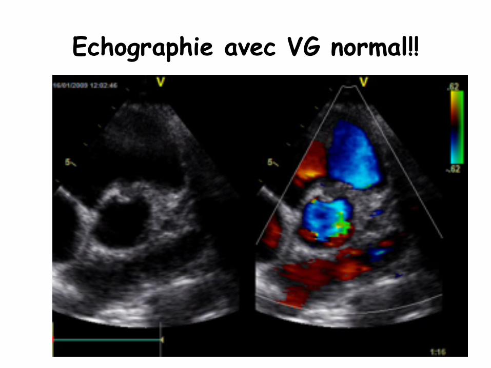
Echographie avec VG normal!!

Echographie
ALCAPA avec sténose de l�ostium gauche Rôle de la sténose de l�ostium gauche dans la bonne tolérance : évite le vol diastolique dans l�AP?
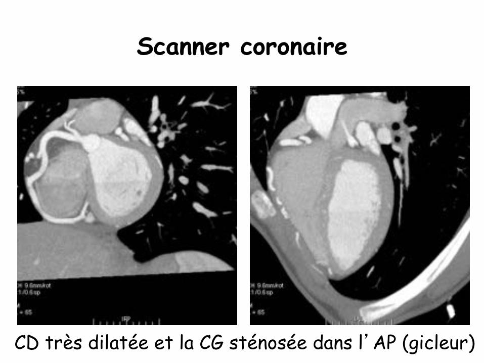
Scanner coronaire
CD très dilatée et la CG sténosée dans l�AP (gicleur)
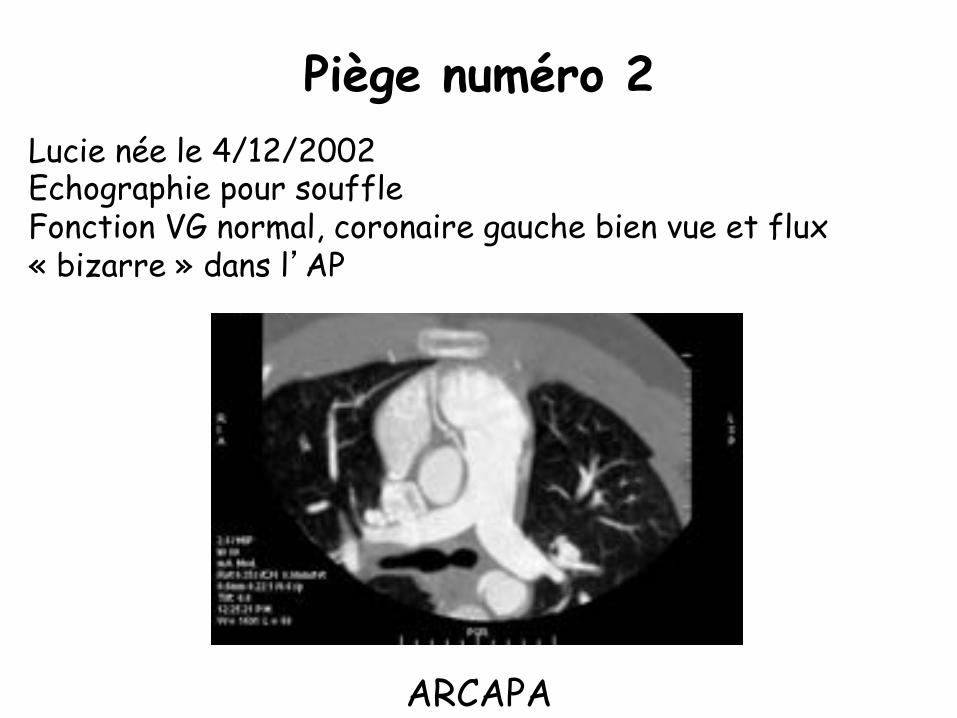
Piège numéro 2 Lucie née le 4/12/2002 Echographie pour souffle Fonction VG normal, coronaire gauche bien vue et flux « bizarre » dans l�AP
ARCAPA
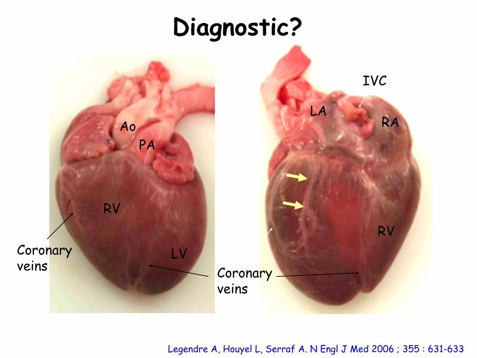
Ao PA
RV
LV RV
RA LA
IVC
Coronary veins
Coronary veins
Legendre A, Houyel L, Serraf A. N Engl J Med 2006 ; 355 : 631-633
Diagnostic?