Embed Size (px)
Citation preview
1
Title: AUGMENTED REALITY IN A TUMOR RESECTION MODEL
Short running head: Augmented reality in partial nephrectomy
Pauline Chauvet1,2, Toby Collins2, Clément Debize2, Lorraine Novais-Gameiro3, Bruno
Pereira4, Adrien Bartoli2, Michel Canis1,2, Nicolas Bourdel1,2
1 Department of Gynecologic Surgery, Clermont-Ferrand University Hospital Estaing, place Lucie
et Raymond Aubrac, Clermont-Ferrand, France
2 Faculté de Médecine- ALCoV UMR6284 CNRS / Université d'Auvergne, ISIT, Clermont-Ferrand,
France.
3 CICS Centre Imagerie Cellulaire Santé Faculté de Médecine, 28 Place Henri Dunant,
CLERMONT-FERRAND, France
4 University Hospital Clermont-Ferrand, Biostatistics unit (DRCI), 58, Rue Montalembert, 63003
Clermont-Ferrand cedex, France
Corresponding author: Nicolas Bourdel [email protected] +33676713113
Department of Gynecologic Surgery, Clermont-Ferrand University Hospital Estaing, place Lucie et
Raymond Aubrac, Clermont-Ferrand, France
2
Abstract
Background: Augmented Reality (AR) guidance is a technology that allows a surgeon to see sub-
surface structures, by overlaying preoperative imaging data on a live laparoscopic video. Our
objectives were to evaluate a state-of-the-art AR guidance system in a tumor surgical resection
model, comparing the accuracy of the resection with and without the system. Our system has
three phases. Phase 1: using the MRI images, the kidney’s and pseudotumor’s surfaces are
segmented to construct a 3D model. Phase 2: the intra-operative 3D model of the kidney is
computed. Phase 3: the pre-operative and intra-operative models are registered, and the
laparoscopic view is augmented with the pre-operative data.
Methods: We performed a prospective experimental study on ex vivo porcine kidneys. Alginate
was injected into the parenchyma to create pseudotumors measuring 4-10mm. The kidneys were
then analyzed by MRI. Next, the kidneys were placed into pelvictrainers, and the pseudotumors
were laparoscopically resected. The AR guidance system allows the surgeon to see tumors and
margins using classical laparoscopic instruments, and a classical screen. The resection margins
were measured microscopically to evaluate the accuracy of resection.
Results: 90 tumors were segmented: 28 were used to optimize the AR software, 62 were used to
randomly compare surgical resection: 29 tumors were resected using AR, and 33 without AR. The
analysis of our pathological results showed 4 failures (tumor with positive margins) (13.8%) in the
AR group, and 10 (30.3%) in the Non-AR group. There was no complete miss in the AR group,
while there were 4 complete misses in the non-AR group. In total, 14 (42.4%) tumors were
completely missed or had a positive margin in the non-AR group.
Conclusions: Our AR system enhance the accuracy of surgical resection, particularly for small
tumors. Crucial information such as resection margins and vascularization could also be
displayed.
Keywords: Augmented Reality, laparoscopy, partial nephrectomy, resection margins.
3
INTRODUCTION
Laparoscopic surgery has brought considerable changes to all fields of surgery by introducing the
concept of minimal invasive surgery. The main problems for surgeons dealing with minimal
invasive surgery techniques are hand–eye disconnection, reduced depth perception due to the
two-dimensional vision offered on the flat screen with the angle of view and haptic feedback being
sometimes limited (1).
Augmented Reality (AR) is a technology allowing a surgeon to see sub-surface structures in an
endoscopic video image (1,2,3). This works by overlaying information obtained from preoperative
imaging, such as MRI and fusing it in real-time with the endoscopic images (1,2). AR guidance
systems have been successfully developed to assist surgical procedures including adrenalectomy
(2), prostatectomy (4), liver resection and neurosurgery (5). AR has not been developed however
for soft and deformable organs in laparoscopic surgery, and despite considerable research,
robust systems capable of handling soft tissue deformation are yet to be created. Furthermore,
quantitative evaluation is both difficult and limited in the literature.
We have already evaluated AR with a phantom uterus model, with small intramural myomas in a
synthetic 3D printed uterus model. The results of this study showed that the AR system can
significantly improve the localization of myomas (6). We then demonstrated the feasibility of our
AR system in a clinical situation (7). To continue the development of our system, for small inner
tumors we decided to evaluate it in an animal tumor resection model. The aim of our work was to
evaluate an AR system for surgery, comparing the accuracy of the resection with and without AR.
This is the first pre-clinical study that systematically evaluates the benefit of a state-of-the-art
laparoscopic AR guidance system, by performing complete resections and measuring
positive/negative margin statistics.
4
MATERIALS & METHODS
FIRST STEP: DEVELOPMENT OF A NEW PSEUDOTUMOR MODEL
The study protocol was covered by a request for authorization to use animals for scientific
purposes (Ministry of Higher Education and Research, Ethics Veterinarian Committee). All the
procedures were performed in the operating room of the International Center of Endoscopic
Surgery (CICE), approval number: C63 18 113 (certificate of authorization to trade in live animals
number B 63-168). We started the procedure with ex-vivo tests, on porcine kidneys, recovered
from pigs operated in a context of resident training (laparoscopic nephrectomy).
Development of the Optimal Pseudotumor Material
To determine the optimal pseudotumor agent to be used, various substances were injected. For
each substance, we looked at ease of preparation and injection, reproducibility, and physical
palpability. The objectives were to create a pseudotumor mixture with a viscosity that would
minimize extravasation from the injection site while still allowing injection through the needle. We
tried silicone (Silicone plomb, Dalbe®, ref. 477001100) but the mixture was too viscous and
difficult to implant. We also tried polyurethane resin (Résine polyuréthane - 2x500gr – Dalbe® ref.
477001700) but the mixture was also difficult to use as the solidification phase takes less than a
minute. Finally, to create a model that would include all the characteristics required, we used
alginate, a hydrocolloid (Alginate nourrisson – DTM®, ref. 477008500). The purpose was to create
tumors between 5 and 10 mm in size, in order to test the system with really small inner tumors.
We created two or three tumors in each kidney, depending on the kidney’s size.
Pseudotumor creation
The alginate mixture was prepared by adding lukewarm water to the alginate powder and stirring
for 1 minute. Solidification took between 1 to 4 minutes, after 3 minutes the mixture reached a
semisolid state and was no longer injectable through the needle. The optimal concentration of
alginate mixture was assessed by injecting different concentrations of mixtures in ex vivo porcine
5
kidneys. Finally, a concentration of 0.5 gram/mL proved to result in a mixture that was easily
injected and formed firm tumours in the kidney parenchyma. After mixing, the alginate mixture was
loaded into a 5mL syringe and 1-2mL of the mixture was slowly injected into the renal parenchyma
using an 18-gauge needle.
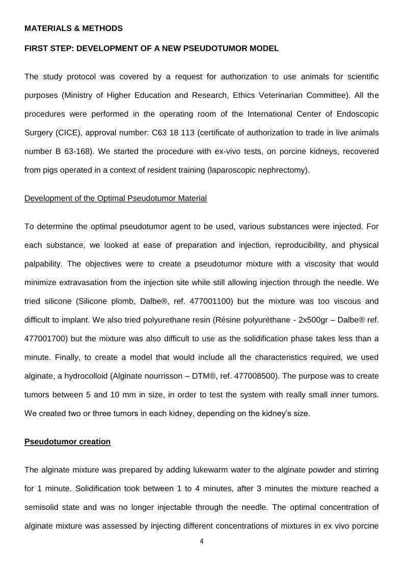
MRI: Imaging of pseudotumors
T1-weighted MRI examinations were made of the kidneys with a 3T MR scanner along the three
planes (axial, coronal and sagittal). We adjusted MRI settings to have a 0.4mm resolution, with a
slice thickness of 1.5mm. Pseudotumors were easily identified as hypointense focal lesions
(Figure 1).
SECOND STEP: EVALUATION WITH A STATE-OF-THE-ART AUGMENTED REALITY
SYSTEM
AR software
For soft organs, AR is very challenging and robust systems capable of handling soft tissue
deformation are yet to be created. To achieve AR in this context, three main challenges have to be
overcome:
6
- In the segmentation phase with the MRI images, the kidney and pseudotumor surfaces are
determined in order to construct a 3D mesh model. This process is the least time-critical
because it can be done before the intervention and typically performed with semi-automatic
facilities (8). This segmentation phase was performed with the use of interactive
segmentation software (Medical Imaging Interaction Toolkit; German Cancer Research
Center).
- The second challenge is real-time registration, where the goal is to transpose the pre-
operative organ model into the per-operative model. To conform to the deformation
between the pre and the surgical model, we used a biomechanical model. The objectives of
the registration paradigm is to construct a pre-operative biomechanical model from pre-
operative data and then to texture the external surface of this pre-operative biomechanical
model. The initial registration stage is non-live. There is then a need to follow the organ
deformation and movements. It is the live stage which is called tracking. For the tracking
stage, we used an existing method based on ‘feature-matching’ (9) proposed in our
group. This method is called Wide-Baseline Multi-Texturemap Registration.
Visualization with the AR software
The 3D pre-operative model is automatically deformed, positioned (or “registered” whatever the
movements of the organ) and fused with the laparoscopic view of the kidneys (9). This blending
gives the impression that the kidney is semi-transparent and the surgeon can see the exact
location of the tumor inside (Figure 1). The goal of the visualization is to augment the laparoscope
image with data from the organ model in order to guide the surgeon. Our AR software ran on a
mid-range Intel i7 desktop workstation with an NVidia 980Ti GPU and visualizations shown on a
26-inch monitor. The software was improved while we developed and used it (6). Our AR system
guides surgeons, by showing them how to access the tumor with an incision tool. The main
improvement we brought is called Tool-port Projection visualization. What the surgeon actually
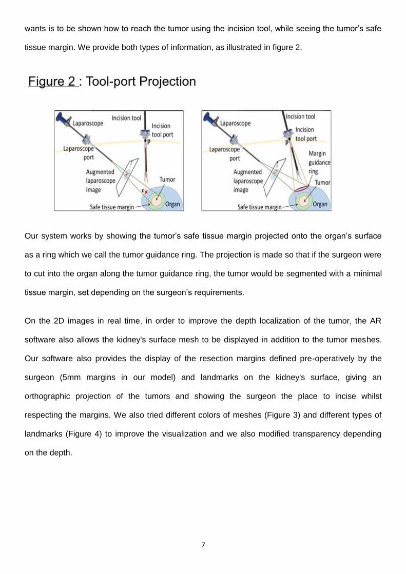
7
wants is to be shown how to reach the tumor using the incision tool, while seeing the tumor’s safe
tissue margin. We provide both types of information, as illustrated in figure 2.
Our system works by showing the tumor’s safe tissue margin projected onto the organ’s surface
as a ring which we call the tumor guidance ring. The projection is made so that if the surgeon were
to cut into the organ along the tumor guidance ring, the tumor would be segmented with a minimal
tissue margin, set depending on the surgeon’s requirements.
On the 2D images in real time, in order to improve the depth localization of the tumor, the AR
software also allows the kidney's surface mesh to be displayed in addition to the tumor meshes.
Our software also provides the display of the resection margins defined pre-operatively by the
surgeon (5mm margins in our model) and landmarks on the kidney's surface, giving an
orthographic projection of the tumors and showing the surgeon the place to incise whilst
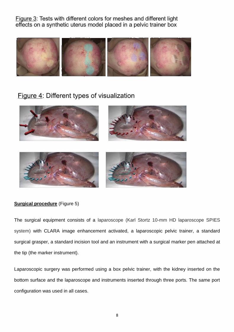
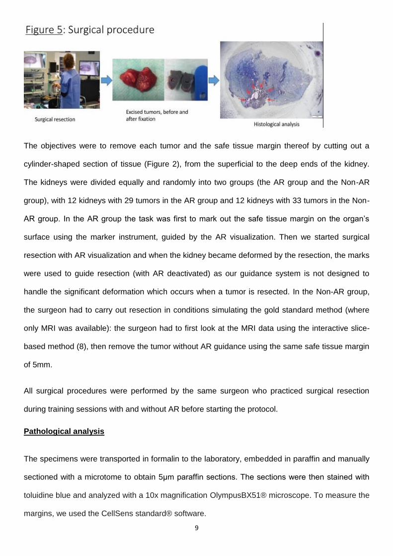
respecting the margins. We also tried different colors of meshes (Figure 3) and different types of
landmarks (Figure 4) to improve the visualization and we also modified transparency depending
on the depth.
8
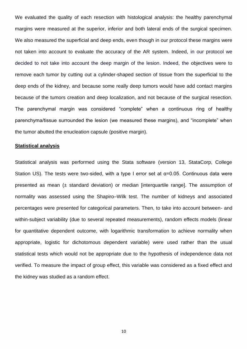
Surgical procedure (Figure 5)
The surgical equipment consists of a laparoscope (Karl Stortz 10-mm HD laparoscope SPIES
system) with CLARA image enhancement activated, a laparoscopic pelvic trainer, a standard
surgical grasper, a standard incision tool and an instrument with a surgical marker pen attached at
the tip (the marker instrument).
Laparoscopic surgery was performed using a box pelvic trainer, with the kidney inserted on the
bottom surface and the laparoscope and instruments inserted through three ports. The same port
configuration was used in all cases.
9
The objectives were to remove each tumor and the safe tissue margin thereof by cutting out a
cylinder-shaped section of tissue (Figure 2), from the superficial to the deep ends of the kidney.
The kidneys were divided equally and randomly into two groups (the AR group and the Non-AR
group), with 12 kidneys with 29 tumors in the AR group and 12 kidneys with 33 tumors in the Non-
AR group. In the AR group the task was first to mark out the safe tissue margin on the organ’s
surface using the marker instrument, guided by the AR visualization. Then we started surgical
resection with AR visualization and when the kidney became deformed by the resection, the marks
were used to guide resection (with AR deactivated) as our guidance system is not designed to
handle the significant deformation which occurs when a tumor is resected. In the Non-AR group,
the surgeon had to carry out resection in conditions simulating the gold standard method (where
only MRI was available): the surgeon had to first look at the MRI data using the interactive slice-
based method (8), then remove the tumor without AR guidance using the same safe tissue margin
of 5mm.
All surgical procedures were performed by the same surgeon who practiced surgical resection
during training sessions with and without AR before starting the protocol.
Pathological analysis
The specimens were transported in formalin to the laboratory, embedded in paraffin and manually
sectioned with a microtome to obtain 5μm paraffin sections. The sections were then stained with
toluidine blue and analyzed with a 10x magnification OlympusBX51® microscope. To measure the
margins, we used the CellSens standard® software.
10
We evaluated the quality of each resection with histological analysis: the healthy parenchymal
margins were measured at the superior, inferior and both lateral ends of the surgical specimen.
We also measured the superficial and deep ends, even though in our protocol these margins were
not taken into account to evaluate the accuracy of the AR system. Indeed, in our protocol we
decided to not take into account the deep margin of the lesion. Indeed, the objectives were to
remove each tumor by cutting out a cylinder-shaped section of tissue from the superficial to the
deep ends of the kidney, and because some really deep tumors would have add contact margins
because of the tumors creation and deep localization, and not because of the surgical resection.
The parenchymal margin was considered “complete” when a continuous ring of healthy
parenchyma/tissue surrounded the lesion (we measured these margins), and “incomplete” when
the tumor abutted the enucleation capsule (positive margin).
Statistical analysis
Statistical analysis was performed using the Stata software (version 13, StataCorp, College
Station US). The tests were two-sided, with a type I error set at α=0.05. Continuous data were
presented as mean (± standard deviation) or median [interquartile range]. The assumption of
normality was assessed using the Shapiro–Wilk test. The number of kidneys and associated
percentages were presented for categorical parameters. Then, to take into account between- and
within-subject variability (due to several repeated measurements), random effects models (linear
for quantitative dependent outcome, with logarithmic transformation to achieve normality when
appropriate, logistic for dichotomous dependent variable) were used rather than the usual
statistical tests which would not be appropriate due to the hypothesis of independence data not
verified. To measure the impact of group effect, this variable was considered as a fixed effect and
the kidney was studied as a random effect.

11
RESULTS
Tumor creation and MRI
In total we created 128 tumors: 38 were not segmented because of faults in the pseudo-tumor
creation phase (tumor visible on kidney surface, or mixture accidentally injected in the urinary
tract). We thus finally segmented 90 tumors. 28 were used to test the AR software, improve
visualization and for surgeon training.
Next, 62 tumors were surgically resected (29 with AR/33 without AR). The pigs’ kidneys ranged in
size from 63.88 to 85.05mm. The measurements were not statistically different between our two
groups. At MRI, the tumors ranged in size from 4.03 to 15.4 mm. We also analyzed the kidney and
pseudotumor volume estimation after segmentation (Table 1).
Table 1: kidney and tumor volume statistics
Pathological analysis
The volume of the specimens was analyzed macroscopically and the specimens ranged in size
from 0.720 to 8.57cm3 (mean 3.39 ± 1.67). There were no differences between the two groups
(p=0.44).
We considered a resection to be a failure if either the tumor was completely absent from the core
(a complete miss), or if it was intersected by the perimeter of the core (a positive margin).
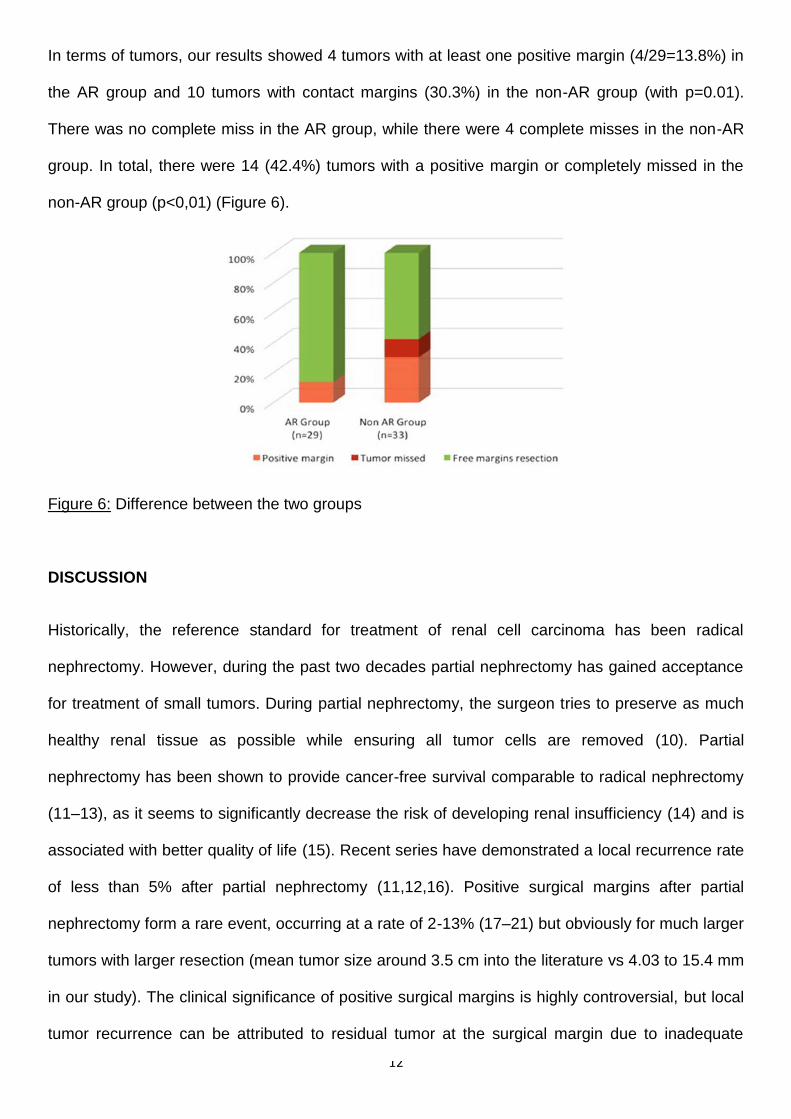
- Complete miss and positive margin (Figure 6)
12
In terms of tumors, our results showed 4 tumors with at least one positive margin (4/29=13.8%) in
the AR group and 10 tumors with contact margins (30.3%) in the non-AR group (with p=0.01).
There was no complete miss in the AR group, while there were 4 complete misses in the non-AR
group. In total, there were 14 (42.4%) tumors with a positive margin or completely missed in the
non-AR group (p<0,01) (Figure 6).
Figure 6: Difference between the two groups
DISCUSSION
Historically, the reference standard for treatment of renal cell carcinoma has been radical
nephrectomy. However, during the past two decades partial nephrectomy has gained acceptance
for treatment of small tumors. During partial nephrectomy, the surgeon tries to preserve as much
healthy renal tissue as possible while ensuring all tumor cells are removed (10). Partial
nephrectomy has been shown to provide cancer-free survival comparable to radical nephrectomy
(11–13), as it seems to significantly decrease the risk of developing renal insufficiency (14) and is
associated with better quality of life (15). Recent series have demonstrated a local recurrence rate
of less than 5% after partial nephrectomy (11,12,16). Positive surgical margins after partial
nephrectomy form a rare event, occurring at a rate of 2-13% (17–21) but obviously for much larger
tumors with larger resection (mean tumor size around 3.5 cm into the literature vs 4.03 to 15.4 mm
in our study). The clinical significance of positive surgical margins is highly controversial, but local
tumor recurrence can be attributed to residual tumor at the surgical margin due to inadequate
13
tumor resection, so margin negativity remains an oncologic imperative to decrease this risk of local
recurrence (22,23). The traditional practice has been to excise an additional 1 cm of tumor
parenchyma, although recent studies have indicated that narrower margins are sufficient (24).
Currently, the precise amount of normal renal parenchyma that must be removed to ensure a safe
surgical margin is still a subject of great concern. The only imperative rule is negativity of the
margins. If the tumor is completely excised with a surrounding margin of normal renal tissue, the
width of the resection margin does not correlate with long-term disease progression (25). Accurate
localization of small renal tumors could allow the surgeon to excise only the tumor with a small
healthy margin and could help to reduce the learning curve for partial nephrectomy.
In our study, the choice of small tumors was made in order to model cases where tumors do not
deform the surface of the kidney, making them harder to localize.
Inaccurate tumor localization can lead to the excision of an inordinate amount of normal
parenchyma in an effort to obtain a complete resection or, even worse, could result in positive
margins. AR allows real-time and precise localization of the lesion and adjustment of the extent of
the resection accordingly. Virtual exploration with AR may assist the surgeon during the
preliminary phase of a surgical procedure, through interactive and visual planning of the operative
strategy, which can be simulated and corrected at every step.
Surgical resection and pathological analysis
With our AR guidance system, we first evaluate the potential benefit of the use of AR to localize
small inner tumors.
Our study shows that AR significantly improved the mean accuracy of tumor resection. The
localization of tumors during laparoscopy can be simple when a superficial deformation is present,
but for small tumors, it can be difficult since there is no tactile feedback. Moreover, although MR or
CT provides a good cartography of tumors to transpose it for intra-operative navigation, using 2D
vision remains challenging. The radiologist’s anatomical landmarks are sometimes different to
those used by the surgeons (26). The use of our AR guidance system provides significant benefits
14
and allows for accurate localization of very small tumors. Other important information (like
vascularization, ureters and other anatomical structures) could also be displayed.
Our current AR system and the literature (Table 2)
Research on the use of AR with mobile and deformable organs has not been reported. One of the
reasons is most likely the technical challenge. There is no automatic segmentation available for
MRI and the segmentation phase is still manual, even if achieved with the help of semi-automatic
facilities (8). The process is time consuming, but not truly challenging and can be carried out
before the intervention.
The most challenging phase remains the registration phase. The main problem is to achieve
registration accurately, reliably and in real time. In our AR system, we do not need any external
navigation tracking systems or preplaced surface markers. In our study, we describe an AR
guidance system based on an existing system (6) in which we use a two-phase approach (Wide-
Baseline Multi-Texturemap Registration).
Unlike SLAM-based tracking methods, which form the main classical tracking approach, our
method does not use frame-to-frame tracking. Instead it performs tracking-by-detection.
Consequently, our method can be used without any other hardware such as magnetic or optical
tracking devices (4,27), and is usable when the surgical scene is approximately rigid. This allows
it to register over long durations and can trivially recover when the organ is not visible for
certain periods, such as when the surgeon removes and then reinserts the laparoscope
or cleans the lens. In cases when the organ is assumed to be fixed relative to background
structures, we can track using features from both the organ and background structures.
Moreover, SLAM during laparoscopy is still proving challenging, due to the repeated nature of the
tissue’s texture, rapid camera motion, blur and appearance changes caused by blood or
coagulation. Our novel two-phase approach (Wide-Baseline Multi- Texturemap Registration) was
shown to significantly outperform SLAM (9).
15
Our system has also undergone several important improvements. The main improvement is a
considerably better way to visually guide the surgeon, by showing how to access the tumor with an
incision tool. We provide both types of information with what we call Tool-port Projection, shown in
figure 2. Tool-port Projection works by showing the tumor’s safe tissue margin projected onto the
organ’s surface as a ring, which we call the tumor guidance ring. The projection is made such that
if the surgeon were to cut into the organ along the tumor guidance ring, the tumor would be
resected with a minimal tissue margin, set depending on the surgeon’s requirements.
During partial nephrectomy, there are two stages in which an AR environment offers a potential
clinical advantage. First, to facilitate rapid and accurate anatomic identification of important
neighboring structures (major vessels and the renal vasculature). Second, to facilitate
unambiguous dissection during tumor resection ensuring negative surgical margins while
achieving a maximally nephron-sparing operation.
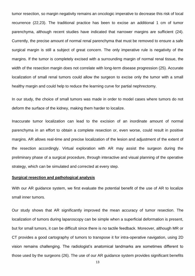
Around 20 cases of partial nephrectomy with AR have been reported, with different AR techniques
(28) (Table 2) (different methods of registration: manual registration, surface-based registration,
fiducial-based registration, 3D-CT stereoscopic image registration) and different methods for
tracking, but no tracking system seems to be truly reliable, especially in case of kidney
deformation. Our system does not require artificial landmarks and unlike other systems (10,27,29)
it does not fail with motion blur or when the laparoscope is removed (e.g. for cleaning) and then
reinserted. Our system also solves the most challenging stages of tracking and fusion in real time.
In their study, Hughes-Hallett et al. explain that the ideal AR image guidance system would use a
pre-operative scan and incorporate the triad of automated registration, tracking and tissue
deformation. Our system seems to be the one that agrees most with this ideal definition, because
other systems have no biomechanical model, no real-time tracking and fusion and are defeated by
motion blur.
16
The cost of a system may be a weakness. However, our system runs on a standard Intel i7
desktop PC at a cost of less than 1000 Euros (which is dropping each year for the same level of
hardware) and does not need any other device.
Table 2: the state of the art
Limitations
One of the limitations is that we used MRI. We know that Computed Tomography (CT) has
traditionally been the imaging technique of choice for evaluating potential solid renal tumors (30).
We emphasize that our AR guidance system does not require any fundamental changes to use
pre-operative CT images rather than MRI. For the most part, CT is limited to characterization
based upon the attenuation and enhancement characteristics of a lesion and requires exposure of
patients to ionizing radiation. For these reasons, MRI is being increasingly used to characterize
solid renal masses (30), especially in case of lesions deemed to be too small to characterize by
CT (31). To add to these arguments, segmentation is easier using MRI due to the precision of the
resolution. In the future, US segmentation will be possible using the same technique as we have
described, probably adding to the cost-effectiveness of our technique. Another study has already
17
shown the feasibility of the navigation system with transrectal ultra-sonography information
superimposed via AR in real time for laparoscopic prostatectomy (4).
The imaging technique of choice for peroperative evaluating potential solid renal tumors is still
discussed (30). The use of an intraoperative ultrasound guidance need the use specific materials,
with a important cost. A learning curve for the use of this technique is also necessary. Our AR
guidance system does not require any fundamental changes to the MRI acquisition. The cost of
the material of our AR system is also reduced. The next step in the development of AR will be the
comparison with EUS.
In our study, the software was developed and improved while we used it. This continuous
improvement could have influence the results but the main evolution concerned only the rapidity
and didn’t modify the principle of the fusion and the tracking phases.
Another limitation is that during surgical procedure, AR systems are not designed actually to
handle the deformation that occurs after incision of the organ surface. In the next future, the
technical evolution including new biomechanical model (32) will allow to maintain AR guidance
after the initial incision.
Future perspectives
In this study, we show the significant benefit of the use of this AR guidance system, in this
surgical model. The next step will be to work on the other anatomical structures: AR could be
helpful to localize not only the tumor itself, but also all the anatomic landmarks and surrounding
organs (ureter, main vessels, rectum…). AR could also improve planning the surgery for a specific
case: it can be used for visualizing during surgery a preoperative optimized incision plan (which
takes into account vascularization, access to all the tumors, tool ports etc.). Other surgical
indication with large dissection and distortion of the normal anatomy (Deep Infiltrating
endometriosis, oncologic procedure, uterine scar niche, etc.) could also benefit from this
technology.
18
CONCLUSION
There is a significant benefit in the use of our AR guidance system in our surgical model. It
provides accurate localization of very small tumors. Crucial information such as vascularization
could also be displayed. In the near future, the intra-operative localization of all anatomical
structures (including the ureters, arteries and complete vascularization of the tumor) will become
possible. In the broader context, this study is the first to systematically evaluate an AR tumor
guidance system in laparoscopic surgery by performing complete resections and measuring
resection margin errors.
DISCLOSURES
Dr P. Chauvet, T. Collins, C. Debize, L. Novais-Gameiro, B. Pereira, Prs A. Bartoli and M.
Canis, Dr N. Bourdel have no conflicts of interest or financial ties to disclose.
ACKNOWLEDGEMENT
This research has received funding from the EU’s FP7 through the ERC research grant 307483
FLEXABLE.
REFERENCES 1. Pessaux P, Diana M, Soler L, Piardi T, Mutter D, Marescaux J. Robotic duodenopancreatectomy
assisted with augmented reality and real-time fluorescence guidance. Surg Endosc. août 2014;28(8):2493‑8.
2. Marescaux J, Rubino F, Arenas M, Mutter D, Soler L. Augmented-reality-assisted laparoscopic
adrenalectomy. JAMA. 10 nov 2004;292(18):2214‑5.
3. Pessaux P, Diana M, Soler L, Piardi T, Mutter D, Marescaux J. Towards cybernetic surgery: robotic
and augmented reality-assisted liver segmentectomy. Langenbecks Arch Surg Dtsch Ges Für Chir. avr
2015;400(3):381‑5.
4. Simpfendörfer T, Baumhauer M, Müller M, Gutt CN, Meinzer H-P, Rassweiler JJ, et al. Augmented
reality visualization during laparoscopic radical prostatectomy. J Endourol Endourol Soc. déc
2011;25(12):1841‑5.
5. Grimson WL, Ettinger GJ, White SJ, Lozano-Perez T, Wells WM, Kikinis R. An automatic
registration method for frameless stereotaxy, image guided surgery, and enhanced reality visualization.
IEEE Trans Med Imaging. 1996;15(2):129‑40.
6. Bourdel N, Collins T, Pizarro D, Bartoli A, Da Ines D, Perreira B, et al. Augmented reality in
gynecologic surgery: evaluation of potential benefits for myomectomy in an experimental uterine model.
Surg Endosc. 29 avr 2016;
7. Bourdel N, Collins T, Pizarro D, Debize C, Grémeau A, Bartoli A, et al. Use of augmented reality in
19
laparoscopic gynecology to visualize myomas. Fertil Steril. janv 2017;
8. Wolf I, Vetter M, Wegner I, Böttger T, Nolden M, Schöbinger M, et al. The medical imaging
interaction toolkit. Med Image Anal. déc 2005;9(6):594‑604.
9. Collins T, Pizarro D, Bartoli A, Canis M, Bourdel N. Realtime Wide-Baseline Registration of the
Uterus in Laparoscopic Videos Using Multiple Texture Maps. In: Liao H, Linte CA, Masamune K, Peters
TM, Zheng G, éditeurs. Augmented Reality Environments for Medical Imaging and Computer-Assisted
Interventions [Internet]. Springer Berlin Heidelberg; 2013 [cité 10 oct 2015]. p. 162‑71. (Lecture Notes in
Computer Science). Disponible sur: http://link.springer.com/chapter/10.1007/978-3-642-40843-4_18
10. Teber D, Guven S, Simpfendörfer T, Baumhauer M, Güven EO, Yencilek F, et al. Augmented
Reality: A New Tool To Improve Surgical Accuracy during Laparoscopic Partial Nephrectomy? Preliminary
In Vitro and In Vivo Results. Eur Urol. août 2009;56(2):332‑8.
11. Hafez KS, Fergany AF, Novick AC. Nephron sparing surgery for localized renal cell carcinoma:
impact of tumor size on patient survival, tumor recurrence and TNM staging. J Urol. déc
1999;162(6):1930‑3.
12. Herr HW. Partial nephrectomy for unilateral renal carcinoma and a normal controlateral kidney: 10-
year followup. J Urol. janv 1999;161(1):33‑5.
13. Lee CT, Katz J, Shi W, Thaler HT, Reuter VE, Russo P. Surgical management of renal tumors 4 cm
or less in a contemporary cohort. J Urol. mars 2000;163(3):730‑6.
14. McKiernan J, Simmons R, Katz J, Russo P. Natural history of chronic renal insufficiency after partial
and radical nephrectomy. Urology. juin 2002;59(6):816‑20.
15. Clark PE, Schover LR, Uzzo RG, Hafez KS, Rybicki LA, Novick AC. Quality of life and
psychological adaptation after surgical treatment for localized renal cell carcinoma: impact of the amount of
remaining renal tissue. Urology. févr 2001;57(2):252‑6.
16. Hafez KS, Novick AC, Campbell SC. Patterns of Tumor Recurrence and Guidelines for Followup
After Nephron Sparing Surgery for Sporadic Renal Cell Carcinoma. J Urol. juin 1997;157(6):2067‑70.
17. Breda A, Stepanian SV, Liao J, Lam JS, Guazzoni G, Stifelman M, et al. Positive Margins in
Laparoscopic Partial Nephrectomy in 855 Cases: A Multi-Institutional Survey From the United States and
Europe. J Urol. juill 2007;178(1):47‑50.
18. Gill IS, Kavoussi LR, Lane BR, Blute ML, Babineau D, Colombo JR, et al. Comparison of 1,800
Laparoscopic and Open Partial Nephrectomies for Single Renal Tumors. J Urol. juill 2007;178(1):41‑6.
19. Yossepowitch O, Thompson RH, Leibovich BC, Eggener SE, Pettus JA, Kwon ED, et al. Positive
Surgical Margins at Partial Nephrectomy: Predictors and Oncological Outcomes. J Urol. juin
2008;179(6):2158‑63.
20. Ani I, Finelli A, Alibhai SMH, Timilshina N, Fleshner N, Abouassaly R. Prevalence and impact on
survival of positive surgical margins in partial nephrectomy for renal cell carcinoma: a population-based
study: Positive surgical margins after partial nephrectomy for RCC. BJU Int. juin 2013;111(8):E300‑5.
21. Tabayoyong W, Abouassaly R, Kiechle JE, Cherullo EE, Meropol NJ, Shah ND, et al. Variation in
Surgical Margin Status by Surgical Approach among Patients Undergoing Partial Nephrectomy for Small
Renal Masses. J Urol. déc 2015;194(6):1548‑53.
22. Vermooten V. Indications for conservative surgery in certain renal tumors: a study based on the
growth pattern of the cell carcinoma. J Urol. août 1950;64(2):200‑8.
23. Russo P. Renal cell carcinoma: presentation, staging, and surgical treatment. Semin Oncol. avr
2000;27(2):160‑76.
24. Sutherland SE, Resnick MI, Maclennan GT, Goldman HB. Does the size of the surgical margin in
partial nephrectomy for renal cell cancer really matter? J Urol. janv 2002;167(1):61‑4.
25. Castilla EA, Liou LS, Abrahams NA, Fergany A, Rybicki LA, Myles J, et al. Prognostic importance
of resection margin width after nephron-sparing surgery for renal cell carcinoma. Urology. déc
2002;60(6):993‑7.
26. Rizzo S, Calareso G, De Maria F, Zanagnolo V, Lazzari R, Cecconi A, et al. Gynecologic tumors:
how to communicate imaging results to the surgeon. Cancer Imaging Off Publ Int Cancer Imaging Soc.
2013;13(4):611‑25.
27. Nakamoto M, Nakada K, Sato Y, Konishi K, Hashizume M, Tamura S. Intraoperative magnetic
20
tracker calibration using a magneto-optic hybrid tracker for 3-D ultrasound-based navigation in laparoscopic
surgery. IEEE Trans Med Imaging. févr 2008;27(2):255‑70.
28. Hughes-Hallett A, Mayer EK, Marcus HJ, Cundy TP, Pratt PJ, Darzi AW, et al. Augmented Reality
Partial Nephrectomy: Examining the Current Status and Future Perspectives. Urology. févr
2014;83(2):266‑73.
29. Simpfendörfer T, Gasch C, Hatiboglu G, Müller M, Maier-Hein L, Hohenfellner M, et al.
Intraoperative Computed Tomography Imaging for Navigated Laparoscopic Renal Surgery: First Clinical
Experience. J Endourol. oct 2016;30(10):1105‑11.
30. Allen BC, Tirman P, Jennings Clingan M, Manny J, Del Gaizo AJ, Leyendecker JR. Characterizing
solid renal neoplasms with MRI in adults. Abdom Imaging. avr 2014;39(2):358‑87.
31. Fananapazir G, Lamba R, Lewis B, Corwin MT, Naderi S, Troppmann C. Utility of MRI in the
Characterization of Indeterminate Small Renal Lesions Previously Seen on Screening CT Scans of Potential
Renal Donor Patients. AJR Am J Roentgenol. août 2015;205(2):325‑30.
32. Paulus CJ, Haouchine N, Kong S-H, Soares RV, Cazier D, Cotin S. Handling topological changes
during elastic registration: Application to augmented reality in laparoscopic surgery. Int J Comput Assist
Radiol Surg. mars 2017;12(3):461‑70.
![[DEMO] On-Site Augmented Collaborative Architecture Visualizationfar.in.tum.de/pub/toennis2014ismarArch/toennis2014ismarArch.pdf · IEEE International Symposium on Mixed and Augmented](https://img.pdfslide.fr/doc/110x75/5f0b5b887e708231d4301ea8/demo-on-site-augmented-collaborative-architecture-ieee-international-symposium.jpg)