Embed Size (px)
Citation preview
RESEARCH ARTICLE
Use of ECG and Other Simple Non-Invasive
Tools to Assess Pulmonary Hypertension
Gabor Kovacs1,2*, Alexander Avian2,3, Vasile Foris1,2, Maria Tscherner1,2,
Xhylsime Kqiku1, Philipp Douschan1,2, Gerhard Bachmaier3, Andrea Olschewski2,4,
Marco Matucci-Cerinic5, Horst Olschewski1,2
1 Medical University of Graz, Department of Internal Medicine, Division of Pulmonology, Graz, Austria,
2 Ludwig Boltzmann Institute for Lung Vascular Research, Graz, Austria, 3 Medical University of Graz,
Institute for Medical Informatics, Statistics and Documentation, Graz, Austria, 4 Medical University of Graz,
Institute for Physiology, Graz, Austria, 5 Department of Biomedicine, Division of Rheumatology, AOUC and
Department of Experimental and Clinical Medicine, University of Florence, Florence, Italy
Abstract
Background
There is a broad consensus that pulmonary hypertension (PH) is to be diagnosed by right
heart catheterization (RHC) and that the most important non-invasive tool is echocardiogra-
phy. However, the role of simple non-invasive tools in the work-up of PH is not clearly
defined. We hypothesized that the use of simple non-invasive techniques may help to guide
important decisions in the diagnostics of pulmonary hypertension.
Objectives
We aimed to develop an algorithm with the use of simple, non-invasive tools in order to iden-
tify patients with very high or very low likelihood of PH.
Methods
We retrospectively analyzed all consecutive patients undergoing RHC between 2005 and
2010 in our center and performed logistic regression of simple non-invasive parameters
regarding detection and exclusion of PH and derived a two-step algorithm. In a prospective
study we evaluated this algorithm between 2011 and 2013.
Results
The retrospective cohort consisted of n = 394 patients of which 49% presented with PH.
Right axis deviation in the ECG was present in 90/394 patients and had a positive predictive
value (PPV) of 93% for PH. The combination of non-right axis deviation, N-terminal pro
brain natriuretic peptide (NT-proBNP)<333pg/ml, arterial oxygen saturation (SO2)�95.5%
and WHO functional class I-II was present in 69/394 patients and excluded PH with a nega-
tive predictive value (NPV) of 96%. The prospective study confirmed these results in a
cohort of n = 168 patients (PPV:92%, NPV:97%). Taken together, simple non-invasive tools
PLOS ONE | DOI:10.1371/journal.pone.0168706 December 28, 2016 1 / 12
a1111111111
a1111111111
a1111111111
a1111111111
a1111111111
OPENACCESS
Citation: Kovacs G, Avian A, Foris V, Tscherner M,
Kqiku X, Douschan P, et al. (2016) Use of ECG and
Other Simple Non-Invasive Tools to Assess
Pulmonary Hypertension. PLoS ONE 11(12):
e0168706. doi:10.1371/journal.pone.0168706
Editor: Masataka Kuwana, JAPAN
Received: July 2, 2016
Accepted: December 4, 2016
Published: December 28, 2016
Copyright: © 2016 Kovacs et al. This is an open
access article distributed under the terms of the
Creative Commons Attribution License, which
permits unrestricted use, distribution, and
reproduction in any medium, provided the original
author and source are credited.
Data Availability Statement: All relevant data are
within the paper and its Supporting Information
files.
Funding: The authors received no specific funding
for this work.
Competing Interests: Dr. Gabor Kovacs reports
personal fees and non-financial support from
Actelion, Bayer, MSD, GSK, Pfizer, AOP,
Boehringer Ingelheim, Astra Zeneca, Takeda,
Novartis and Chiesi, outside the submitted work.
Dr. Alexander Avian has nothing to disclose. Dr.
Vasile Foris reports non-financial support from
GlaxoSmithKlein, Actelion Pharmaceuticals Ltd,
allowed a prediction regarding the presence or absence of PH in 42% of patients with sus-
pected PH.
Conclusion
ECG, NT-proBNP, SO2 and WHO functional class may predict the presence or absence of
PH in almost half of the patients with suspected PH, suggesting an important role for these
variables in the work-up of patients at risk for PH.
Clinical Trial Registration
NCT01607502
Introduction
Pulmonary hypertension (PH) is a severe hemodynamic condition of the pulmonary circula-
tion eventually leading to right heart failure and death [1]. Independent of its etiology, devel-
opment of PH is associated with worse prognosis of the underlying disease [2–4]. PH is
diagnosed by right heart catheterization [1], while Doppler echocardiography, with all its limi-
tations [5], is widely accepted as the most specific non-invasive screening tool. Other simple,
widely available, non-invasive tools such as ECG, chest X-ray, pulmonary function tests (PFT),
blood gas analysis (BGA) and laboratory tests are recommended to be performed during the
diagnostic process [1], but their specificity and sensitivity is generally considered to be low,
often restricting these methods to delineate co-morbid conditions, or, in the best case, to sup-
port the suspicion of a pulmonary vascular disease. A recent study in systemic sclerosis
patients, however, revealed that a combination of simple non-invasive markers performed bet-
ter than echocardiography alone and recommended a complex algorithm that helps to avoid
some echocardiographic and right heart catheter investigations [6]. As limitations, the study
included only systemic sclerosis patients with a low diffusing capacity for CO (DLCO) and the
results may not be relevant for other populations than systemic sclerosis patients. In our study,
we derived a two-step algorithm from a retrospective evaluation and we applied this simple
algorithm in a prospective cohort of patients. The algorithm is based on a combination of sim-
ple, investigator-independent, non-invasive examinations that may be available in almost
every outpatient clinic and may help to guide further diagnostic decisions in the work-up of
pulmonary hypertension.
Patients and Methods
The study consisted of a retrospective part, in which a combination of parameters was identi-
fied by logistic regression and based on which an algorithm was developed, and a validation
part, in which the proposed algorithm was tested in a prospective manner. Between 2005 and
2010, patients of the Medical University of Graz undergoing right heart catheterization due to
suspected pulmonary hypertension were included into the retrospective part of the study. In
the prospective part, patients undergoing right heart catheterization (RHC) between 2011 and
2013 were evaluated. All patients gave written informed consent. All patients had either unex-
plained dyspnea or had an established risk factor for PAH (e.g. systemic sclerosis) and the
diagnostic work-up followed the recommendations of current international PH guidelines
[7,8]. The study has been approved by the local ethics review board. In all patients, right heart
ECG and Other Simple Non-Invasive Tools in PH
PLOS ONE | DOI:10.1371/journal.pone.0168706 December 28, 2016 2 / 12
Pfizer Inc., Bayer, Eli Lilly, VitalAire and Novartis,
outside the submitted work. Dr. Maria Tscherner
has nothing to disclose. Dr. Xhylsime Kqiku has
nothing to disclose. Dr. Philipp Douschan reports
non-financial support from Bayer, GSK, Menarini
and from Actelion outside the submitted work. Dr.
Gerhard Bachmaier has nothing to disclose. Dr.
Andrea Olschewski reports personal fees from
Pfizer outside the submitted work. Dr. Horst
Olschewski reports grants and personal fees from
Actelion, grants and personal fees from Bayer,
personal fees from Gilead, personal fees from GSK,
personal fees from Novartis, personal fees from
Pfizer, outside the submitted work. Dr. Matucci-
Cerinic has nothing to disclose. This does not alter
the authors’ adherence to PLOS ONE policies on
sharing data and materials.
catheterization and a routine non-invasive assessment [1] including physical examination, his-
tory, ECG, blood gas analysis, pulmonary function tests, laboratory tests and six-minute walk
test was performed. As the goal of the study was the development of a simple algorithm, based
on the routine work-up of PH patients, the following simple parameters were evaluated: ECG:
the presence of right axis deviation (RAD) characterized by an electrical heart axis greater than
+90˚ (the amplitude of the S wave bigger than the amplitude of the R wave in lead I); blood gas
analysis: arterial partial pressure of oxygen (pO2), arterial partial pressure of carbon dioxide
(pCO2), arterial oxygen saturation (SO2); pulmonary function tests: forced expiratory volume
in the first second (FEV1), forced vital capacity (FVC), diffusion capacity for carbon monoxide
(DLCO); laboratory tests: N-terminal pro brain natriuretic peptide (NT-proBNP), uric acid;
six-minute walk distance, Borg dyspnea score at the start and the end of the six-minute walk
test; WHO functional class.
ECG was reviewed for RAD by two independent physicians; unclear cases were decided by
consensus. Blood gas analysis of arterialized ear lobe capillary blood was performed with an
ABL 800 Flex (Radiometer; Copenhagen, Denmark) blood gas analyzer. Pulmonary function
test was performed with a Jaeger MS PFT Analyzer, NT-proBNP and uric acid levels were
determined by commercially available kits. Six-minute walk tests were performed according to
the recommendations of the American Thoracic Society [9]. RHC examinations were per-
formed with a 7F quadruple-lumen, balloon-tipped, flow-directed Swan-Ganz catheter (Bax-
ter) in the supine position using the transjugular approach. The reference point was set at the
level of the anterior axillary line [10].
The study was approved by the Ethics Committee of the Medical University of Graz (NR:
23–408 ex 10/11). Participants provided their written informed consent to participate in this
study. The ethics committee approved this consent procedure.
Statistics
Data are presented as mean and standard deviation or median and interquartile range for
continuous data and absolute and relative frequency for categorical data, respectively. To
identify patients with a high risk for PH and patients with a very low risk for PH in the ret-
rospective analysis, a two-step algorithm was developed. In the first step, patients with right
axis deviation (electrical axis of the heart greater than +90˚) were selected as high risk
patients. In the second step, in the remaining patient group, possible predictors (arterial
SO2, pO2, pCO2, DLCO, FEV1, FVC, Borg dyspnea score at the end of the six minute walk
test, NT-proBNP, uric acid, 6 Minute Walking distance and WHO functional class) for
exclusion of PH were analyzed. In the univariate logistic regression analysis the predictors’
ability for discriminating patients with and without PH (mean pulmonary arterial pressure
(mPAP) � 25mmHg vs. mPAP < 25mmHg) was analyzed. Univariate significant variables
were checked for multicollinearity. Multicollinearity was verified by correlation matrices: a
correlation >0.4 was used as the cutoff for multicollinearity. After excluding multicollinear-
ity, remaining variables were selected for multivariate logistic regression. Variables in the
final model were selected with a forward stepwise procedure. The decision to include vari-
ables was based on a likelihood-ratio test. To calculate the best cut off score of each variable
we used the Youden-Index. Therefore the value of a variable which maximizes sensitivity
and specificity was calculated by using ROC analysis. The result was evaluated in the pro-
spective cohort. Sensitivity, specificity, negative predictive value and positive predictive
value were calculated for both steps and for the final model. A p-value <0.05 was consid-
ered significant. Statistical analysis was performed with IBM SPSS Statistics (Release 20.0.0.
2011. Chicago (IL), USA: SPSS Inc., an IBM Company) software.
ECG and Other Simple Non-Invasive Tools in PH
PLOS ONE | DOI:10.1371/journal.pone.0168706 December 28, 2016 3 / 12
Results
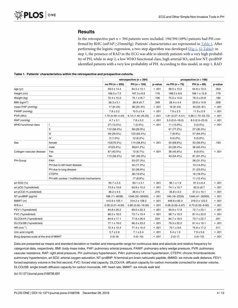
In the retrospective part n = 394 patients were included. 194/394 (49%) patients had PH con-
firmed by RHC (mPAP�25mmHg). Patients’ characteristics are represented in Table 1. After
performing the logistic regression, a two-step algorithm was developed (Fig 1, S1 Table): in
step 1, the presence of RAD in the ECG was able to identify patients with a very high probabil-
ity of PH, while in step 2, a low WHO functional class, high arterial SO2 and low NT-proBNP
identified patients with a very low probability of PH. According to this model, in step 1, RAD
Table 1. Patients’ characteristics within the retrospective and prospective cohorts.
retrospective (n = 394) prospective (n = 168)
no PH (n = 200) PH (n = 194) p-value no PH (n = 79) PH (n = 89) p-value
age (yr) 59.0 ± 14.4 64.5 ± 13.1 < .001 58.5 ± 13.3 64.6 ± 13.3 .003
Height (cm) 166.0 ± 7.5 167.3 ± 8.8 .116 168.5 ± 8.6 166.1 ± 12.8 .179
Weight (kg) 72.4 ± 15.0 75.1 ±16.7 .106 75.0 ± 14.6 79.3 ± 24.8 .180
BMI (kg/m2) 26.3 ± 5.1 26.8 ±5.7 .349 26.4 ± 4.4 29.6 ± 14.8 .058
mean PAP (mmHg) 17 (8–24) 38 (25–91) < .001 16 (9–24) 40 (25–81) < .001
PAWP (mmHg) 7.8 ± 3.2 10.5 ± 5.4 < .001 7.9 ± 3.3 11.9 ± 5.9 < .001
PVR (WU) 1.70 (0.59–4.43) 6.12 (1.40–25.25) < .001 1.61 (0.27–4.51) 5.86 (1.70–23.72) < .001
RAP (mmHg) 4.7 ± 3.1 7.8 ± 5.2 < .001 5.0 (0.0–18.0) 8.0 (2.0–23.0) < .001
WHO functional class I 27 (13.5%) 1 (0.5%) < .001 11 (13.9%) 0 (0.0%) < .001
II 112 (56.0%) 58 (29.9%) 61 (77.2%) 27 (30.3%)
III 59 (29.5%) 123 (63.4%) 7 (8.9%) 57 (64.0%)
IV 2 (1.0%) 12 (6.2%) 0 (0.0%) 5 (5.6%)
Sex female 153(76.5%) 114 (58.8%) < .001 55 (69.6%) 53 (59.6%) .193
male 47(23.5%) 80(41.2%) 24 (30.4%) 36 (40.4%)
Collagen vascular disease Yes 87 (43.5%) 13 (6.7%) < .001 36 (45.6%) 8 (9.0%) < .001
No 113 (56.5%) 181 (93.3%) 43 (54.4%) 81 (91.0%)
PH-Group PAH 53 (27.3%) 28 (31.5%)
PH due to left heart disease 34 (17.5%) 13 (14.6%)
PH due to lung disease 52 (26.8%) 21 (23.6%)
CTEPH 38 (19.6%) 16 (18.0%)
PH with unclear / multifactorial mechanisms 17 (8.8%) 11 (12.4%)
art SO2 (%) 95.7 ± 2.0 93.1 ± 3.1 < .001 96.1 ± 1.9 91.0 ± 6.4 < .001
art pO2 (%predicted) 73.9 ± 10.6 63.8 ± 10.2 < .001 74.1 ± 10.7 62.9 ±9.7 < .001
art pCO2 (% predicted) 36.2 ± 4.5 36.9 ± 7.4 .270 35.8 ± 4.0 37.2 ± 10.1 < .001
NT-proBNP (pg/ml) 188 (11–6038) 1248 (32–35000) < .001 146 (16–2259) 1348 (41–35000) < .001
6MWT (m) 410.9 ± 105.1 314.2 ± 128.2 < .001 449.8 ± 82.4 319.0 ± 123.5 < .001
R/S in I 0.06 (0.01–9.00) 0.80 (0.02–19.00) < .001 0.06 (0.02–2.67) 0.73 (0.02–9.00) < .001
FEV1 (%predicted) 84.8 ± 20.2 69.0 ± 22.3 < .001 90.9 ± 17.9 72.7 ± 23.1 < .001
FVC (%predicted) 86.2 ± 18.5 73.7 ± 19.4 < .001 98.7 ± 19.3 81.4 ± 23.5 < .001
DLCOcVA (%predicted) 84.6 ± 17.1 77.8 ± 26.0 .024 84.7 ± 18.5 73.7 ± 22.7 .001
DLCOcSB (%predicted) 77.1 ± 19.0 66.3 ± 23.2 < .001 79.3 ± 22.2 61.4 ± 19.1 < .001
HR (min-1) 72.4 ± 12.4 77.4 ± 15.0 < .001 70.1 ± 9.6 75.6 ± 17.2 .011
Uric acid (mg/dl) 5.7 ± 2.6 7.1 ± 2.4 < .001 5.4 ± 1.9 7.9 ± 5.6 < .001
Borg dyspnea scale at the end of 6MWT 2 (0–9) 4 (0–10) < .001 2 (0–7) 3 (0–10) < .001
Data are presented as means and standard deviation or median and interquartile range for continuous data and absolute and relative frequency for
categorical data, respectively. BMI: body mass index, PAP: pulmonary arterial pressure, PAWP: pulmonary artery wedge pressure, PVR: pulmonary
vascular resistance, RAP: right atrial pressure, PH: pulmonary hypertension, PAH: pulmonary arterial hypertension, CTEPH: chronic thromboembolic
pulmonary hypertension, art SO2: arterial oxygen saturation, NT-proBNP: N terminal pro brain natriuretic peptide, 6MWD: six minute walk distance, FEV1:
forced expiratory volume in the first second, FVC: forced vital capacity, DLCOcVA: diffusion capacity for carbon monoxide corrected for alveolar volume,
DLCOcSB: single breath diffusion capacity for carbon monoxide, HR: heart rate, 6MWT: six minute walk test
doi:10.1371/journal.pone.0168706.t001
ECG and Other Simple Non-Invasive Tools in PH
PLOS ONE | DOI:10.1371/journal.pone.0168706 December 28, 2016 4 / 12
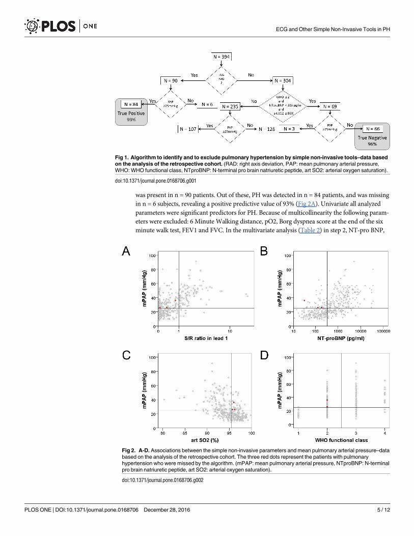
was present in n = 90 patients. Out of these, PH was detected in n = 84 patients, and was missing
in n = 6 subjects, revealing a positive predictive value of 93% (Fig 2A). Univariate all analyzed
parameters were significant predictors for PH. Because of multicollinearity the following param-
eters were excluded: 6 Minute Walking distance, pO2, Borg dyspnea score at the end of the six
minute walk test, FEV1 and FVC. In the multivariate analysis (Table 2) in step 2, NT-pro BNP,
Fig 1. Algorithm to identify and to exclude pulmonary hypertension by simple non-invasive tools–data based
on the analysis of the retrospective cohort. (RAD: right axis deviation, PAP: mean pulmonary arterial pressure,
WHO: WHO functional class, NTproBNP: N-terminal pro brain natriuretic peptide, art SO2: arterial oxygen saturation).
doi:10.1371/journal.pone.0168706.g001
Fig 2. A-D. Associations between the simple non-invasive parameters and mean pulmonary arterial pressure–data
based on the analysis of the retrospective cohort. The three red dots represent the patients with pulmonary
hypertension who were missed by the algorithm. (mPAP: mean pulmonary arterial pressure, NTproBNP: N-terminal
pro brain natriuretic peptide, art SO2: arterial oxygen saturation).
doi:10.1371/journal.pone.0168706.g002
ECG and Other Simple Non-Invasive Tools in PH
PLOS ONE | DOI:10.1371/journal.pone.0168706 December 28, 2016 5 / 12
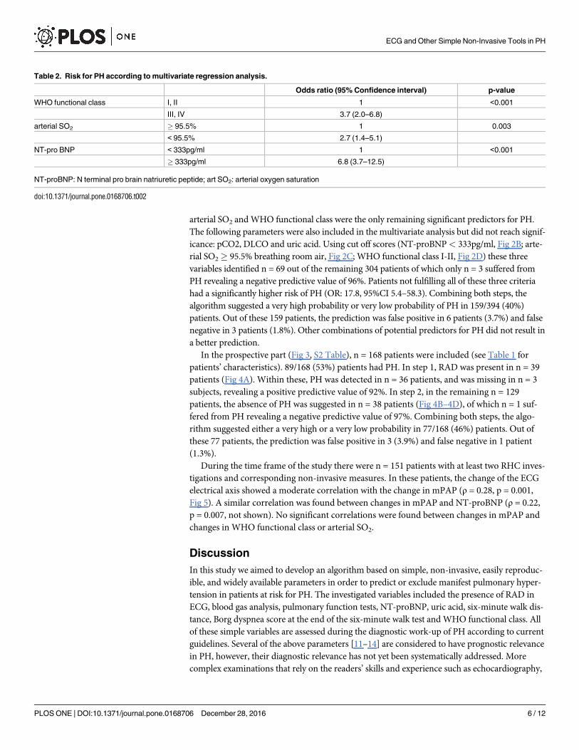
arterial SO2 and WHO functional class were the only remaining significant predictors for PH.
The following parameters were also included in the multivariate analysis but did not reach signif-
icance: pCO2, DLCO and uric acid. Using cut off scores (NT-proBNP< 333pg/ml, Fig 2B; arte-
rial SO2� 95.5% breathing room air, Fig 2C; WHO functional class I-II, Fig 2D) these three
variables identified n = 69 out of the remaining 304 patients of which only n = 3 suffered from
PH revealing a negative predictive value of 96%. Patients not fulfilling all of these three criteria
had a significantly higher risk of PH (OR: 17.8, 95%CI 5.4–58.3). Combining both steps, the
algorithm suggested a very high probability or very low probability of PH in 159/394 (40%)
patients. Out of these 159 patients, the prediction was false positive in 6 patients (3.7%) and false
negative in 3 patients (1.8%). Other combinations of potential predictors for PH did not result in
a better prediction.
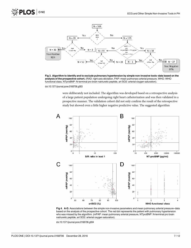
In the prospective part (Fig 3, S2 Table), n = 168 patients were included (see Table 1 for
patients’ characteristics). 89/168 (53%) patients had PH. In step 1, RAD was present in n = 39
patients (Fig 4A). Within these, PH was detected in n = 36 patients, and was missing in n = 3
subjects, revealing a positive predictive value of 92%. In step 2, in the remaining n = 129
patients, the absence of PH was suggested in n = 38 patients (Fig 4B–4D), of which n = 1 suf-
fered from PH revealing a negative predictive value of 97%. Combining both steps, the algo-
rithm suggested either a very high or a very low probability in 77/168 (46%) patients. Out of
these 77 patients, the prediction was false positive in 3 (3.9%) and false negative in 1 patient
(1.3%).
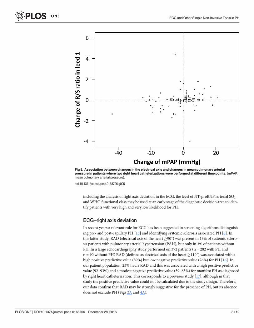
During the time frame of the study there were n = 151 patients with at least two RHC inves-
tigations and corresponding non-invasive measures. In these patients, the change of the ECG
electrical axis showed a moderate correlation with the change in mPAP (ρ = 0.28, p = 0.001,
Fig 5). A similar correlation was found between changes in mPAP and NT-proBNP (ρ = 0.22,
p = 0.007, not shown). No significant correlations were found between changes in mPAP and
changes in WHO functional class or arterial SO2.
Discussion
In this study we aimed to develop an algorithm based on simple, non-invasive, easily reproduc-
ible, and widely available parameters in order to predict or exclude manifest pulmonary hyper-
tension in patients at risk for PH. The investigated variables included the presence of RAD in
ECG, blood gas analysis, pulmonary function tests, NT-proBNP, uric acid, six-minute walk dis-
tance, Borg dyspnea score at the end of the six-minute walk test and WHO functional class. All
of these simple variables are assessed during the diagnostic work-up of PH according to current
guidelines. Several of the above parameters [11–14] are considered to have prognostic relevance
in PH, however, their diagnostic relevance has not yet been systematically addressed. More
complex examinations that rely on the readers’ skills and experience such as echocardiography,
Table 2. Risk for PH according to multivariate regression analysis.
Odds ratio (95% Confidence interval) p-value
WHO functional class I, II 1 <0.001
III, IV 3.7 (2.0–6.8)
arterial SO2 � 95.5% 1 0.003
< 95.5% 2.7 (1.4–5.1)
NT-pro BNP < 333pg/ml 1 <0.001
� 333pg/ml 6.8 (3.7–12.5)
NT-proBNP: N terminal pro brain natriuretic peptide; art SO2: arterial oxygen saturation
doi:10.1371/journal.pone.0168706.t002
ECG and Other Simple Non-Invasive Tools in PH
PLOS ONE | DOI:10.1371/journal.pone.0168706 December 28, 2016 6 / 12
were deliberately not included. The algorithm was developed based on a retrospective analysis
of a large patient population undergoing right heart catheterization and was then validated in a
prospective manner. The validation cohort did not only confirm the result of the retrospective
study but showed even a little higher negative predictive value. The suggested algorithm
Fig 3. Algorithm to identify and to exclude pulmonary hypertension by simple non-invasive tools–data based on the
analysis of the prospective cohort. (RAD: right axis deviation, PAP: mean pulmonary arterial pressure, WHO: WHO
functional class, NTproBNP: N-terminal pro brain natriuretic peptide, art SO2: arterial oxygen saturation).
doi:10.1371/journal.pone.0168706.g003
Fig 4. A-D. Associations between the simple non-invasive parameters and mean pulmonary arterial pressure–data
based on the analysis of the prospective cohort. The red dot represents the patient with pulmonary hypertension
who was missed by the algorithm. (mPAP: mean pulmonary arterial pressure, NTproBNP: N-terminal pro brain
natriuretic peptide, art SO2: arterial oxygen saturation).
doi:10.1371/journal.pone.0168706.g004
ECG and Other Simple Non-Invasive Tools in PH
PLOS ONE | DOI:10.1371/journal.pone.0168706 December 28, 2016 7 / 12
including the analysis of right axis deviation in the ECG, the level of NT-proBNP, arterial SO2
and WHO functional class may be used at an early stage of the diagnostic decision-tree to iden-
tify patients with very high and very low likelihood for PH.
ECG–right axis deviation
In recent years a relevant role for ECG has been suggested in screening algorithms distinguish-
ing pre- and post-capillary PH [15] and identifying systemic sclerosis associated PH [6]. In
this latter study, RAD (electrical axis of the heart�90˚) was present in 13% of systemic sclero-
sis patients with pulmonary arterial hypertension (PAH), but only in 3% of patients without
PH. In a large echocardiography study performed on 372 patients (n = 282 with PH and
n = 90 without PH) RAD (defined as electrical axis of the heart�110˚) was associated with a
high positive predictive value (89%) but low negative predictive value (26%) for PH [16]. In
our patient population, 23% had a RAD and this was associated with a high positive predictive
value (92–93%) and a modest negative predictive value (59–65%) for manifest PH as diagnosed
by right heart catheterization. This corresponds to a previous study [17], although in that
study the positive predictive value could not be calculated due to the study design. Therefore,
our data confirm that RAD may be strongly suggestive for the presence of PH, but its absence
does not exclude PH (Figs 2A and 4A).
Fig 5. Association between changes in the electrical axis and changes in mean pulmonary arterial
pressure in patients where two right heart catheterizations were performed at different time points. (mPAP:
mean pulmonary arterial pressure).
doi:10.1371/journal.pone.0168706.g005
ECG and Other Simple Non-Invasive Tools in PH
PLOS ONE | DOI:10.1371/journal.pone.0168706 December 28, 2016 8 / 12
In previous studies also other ECG parameters such as P wave amplitude, P wave axis, signs
of right and left ventricular strain etc. were analyzed [15]. In our study we deliberately used
only one single robust and very simple ECG parameter, which appeared to be relevant based
on the earlier studies and allows automatic interpretation.
We also looked for possible confounders in our analysis, which may have influenced the
electrical axis of the heart. Neither age (p = 0.786) nor pulmonary arterial wedge pressure
(PAWP) as a variable for left heart disease (p = 0.667) was significantly associated with the
electrical axis of the heart. Systemic blood pressure was weakly negatively associated with RAD
(ρ = -0.279, p<0.001) and males presented more often with RAD than females in the retro-
spective (p = 0.001) but not in the prospective cohort (p = 0.222).
Recent studies suggested that ECG may indicate disease progression [18] and therapy
response [19] in PAH. We also found a modest correlation between the change in the electrical
axis of the heart and mPAP between baseline and follow-up examinations. Although this cor-
relation was comparable to the correlation between mPAP and NT-proBNP changes, the clini-
cal relevance of such findings remains low.
NT-proBNP
Brain natriuretic peptide (BNP) and NT-proBNP are established biomarkers with prognostic
relevance in PAH [20–22], although they may depend on age and renal function [23]. Besides
being recommended as a marker of clinical progression, NT-proBNP has been suggested to be
included in screening algorithms for PH. In different clinical settings, various NT-proBNP
thresholds may be clinically relevant. In the DETECT study [6], a continuous scale, but no sin-
gle threshold for NT-proBNP was used, while another recent -PAH screening algorithm in sys-
temic sclerosis recommended a threshold of 210 pg/ml [24]. In the study of Bonderman et al.,
a low NT-proBNP threshold (80pg/ml) distinguished pre- and post-capillary PH [15]. In our
algorithm, as part of a combination of parameters, the optimal threshold was found to be in a
higher range (333pg/ml).
WHO functional class and arterial SO2
The most frequently used, easy and well established method to describe the physical limitation
in patients with PH is the WHO functional class. Although it is very subjective, and the stratifi-
cation strategy may vary among centers and individual physicians, the WHO functional class
performs surprisingly well in clinical studies as secondary end-point and represents one of the
most important prognostic parameters in PAH [25]. The measurement of oxygen saturation is
simple and can be taken from arterial blood gas analysis or non-invasively from pulse oxime-
try. Both WHO functional class and arterial SO2 are unspecific for pulmonary hypertension
[26]. However, in combination with our other parameters they proved to be very useful to
identify patients with no PH.
Clinical relevance of our findings
The group of patients with very high probability for PH were those with RAD (electrical axis
of the heart>90˚) in the ECG, revealing a positive predictive value of 92–93%. This may sug-
gest that in dyspnea patients or a disease associated with PAH, a right axis deviation in the
ECG is highly suspicious for PH and further clinical examinations (echocardiography, eventu-
ally right heart catheterization) are strongly recommended.
Patients with very low probability for PH were characterized by any ECG axis other than
RAD, low NT-proBNP, good oxygen saturation and low WHO functional class. Our results
may thus suggest a reasonable role for these parameters in an active decision against a
ECG and Other Simple Non-Invasive Tools in PH
PLOS ONE | DOI:10.1371/journal.pone.0168706 December 28, 2016 9 / 12
diagnostic work-up for PH and support the concept of clinical and laboratory patterns facili-
tating specific diagnostic decisions [6,15,27–29]. Of course, such criteria cannot replace spe-
cific methods like echocardiography and right heart catheterization. On the other hand, in
many clinical situations we discuss diagnostic procedures with patients at moderate risk for
PH and both the physician and the patient may profit from prediction rules to guide the shared
decision making. Therefore, we believe that our results may be useful for guiding the decisions
towards specific diagnostics in individual patients at risk for pulmonary hypertension.
Limitations
As the most important limitation of our study, our patient collective was typical for a PH cen-
ter but may be different in primary care settings or specialized heart or lung clinics. All patients
had either unexplained dyspnea or an established risk factor for PH and the findings (includ-
ing reported PPV and NPV) may not be valid in patients without these characteristics or in the
general population. In addition, we have to accept that besides correctly predicting PH or “no
PH” in about half of the patients, our algorithm was not able to provide additional help to indi-
cate the presence or absence of pulmonary hypertension in the other half.
Conclusion
Our suggested 2-step algorithm recognizes patients with either a very high or a very low proba-
bility for pulmonary hypertension in nearly half of the patients at risk for PH. This result can
be achieved by the systematic use of four simple non-invasive parameters: right axis deviation
in ECG, SO2, NT-proBNP and WHO functional class.
Supporting Information
S1 Table. Individual data of patients assessed in the retrospective part of the study.
(SAV)
S2 Table. Individual data of patients assessed in the prospective part of the study.
(SAV)
Author Contributions
Conceptualization: GK HO.
Data curation: GK VF MT XK PD.
Formal analysis: AA GB.
Funding acquisition: AO HO.
Investigation: GK VF MT XK PD.
Methodology: GK AA HO.
Project administration: GK.
Resources: GK VF MT XK PD HO.
Software: AA GB.
Supervision: AO HO.
Validation: AA.
ECG and Other Simple Non-Invasive Tools in PH
PLOS ONE | DOI:10.1371/journal.pone.0168706 December 28, 2016 10 / 12

Visualization: GK AA.
Writing – original draft: GK AA.
Writing – review & editing: GK MM AO HO.
References1. Galie N, Humbert M, Vachiery JL, Gibbs S, Lang I, Torbicki A, et al. 2015 ESC/ERS Guidelines for the
diagnosis and treatment of pulmonary hypertension: The Joint Task Force for the Diagnosis and Treat-
ment of Pulmonary Hypertension of the European Society of Cardiology (ESC) and the European
Respiratory Society (ERS): Endorsed by: Association for European Paediatric and Congenital Cardiol-
ogy (AEPC), International Society for Heart and Lung Transplantation (ISHLT). Eur Heart J. 2016; 37:
67–119. doi: 10.1093/eurheartj/ehv317 PMID: 26320113
2. Abramson SV, Burke JF, Kelly JJ Jr, Kitchen JG 3rd, Dougherty MJ, Yih DF, et al. Pulmonary hyperten-
sion predicts mortality and morbidity in patients with dilated cardiomyopathy. Ann Intern Med. 1992;
116: 888–895. PMID: 1580444
3. Lam CS, Borlaug BA, Kane GC, Enders FT, Rodeheffer RJ, Redfield MM. Age-associated increases in
pulmonary artery systolic pressure in the general population. 2009; 119: 2663–70.
4. Oswald-Mammosser M, Weitzenblum E, Quoix E, Moser G, Chaouat A, Charpentier C, et al. Prognostic
factors in COPD patients receiving long-term oxygen therapy. Importance of pulmonary artery pressure.
1995; 107: 1193–8. PMID: 7750305
5. Fisher MR, Forfia PR, Chamera E, Housten-Harris T, Champion HC, Girgis RE, et al. Accuracy of
Doppler echocardiography in the hemodynamic assessment of pulmonary hypertension. 2009; 179:
615–21. doi: 10.1164/rccm.200811-1691OC PMID: 19164700
6. Coghlan JG, Denton CP, Grunig E, Bonderman D, Distler O, Khanna D, et al. Evidence-based detection
of pulmonary arterial hypertension in systemic sclerosis: the DETECT study. Ann Rheum Dis. 2014; 73:
1340–1349. doi: 10.1136/annrheumdis-2013-203301 PMID: 23687283
7. Galie N, Hoeper MM, Humbert M, Torbicki A, Vachiery JL, Barbera JA, et al. Guidelines for the diagno-
sis and treatment of pulmonary hypertension: The Task Force for the Diagnosis and Treatment of Pul-
monary Hypertension of the European Society of Cardiology (ESC) and the European Respiratory
Society (ERS), endorsed by the International Society of Heart and Lung Transplantation (ISHLT). 2009;
30: 2493–537. doi: 10.1093/eurheartj/ehp297 PMID: 19713419
8. Galie N, Torbicki A, Barst R, Dartevelle P, Haworth S, Higenbottam T, et al. Guidelines on diagnosis
and treatment of pulmonary arterial hypertension. The Task Force on Diagnosis and Treatment of Pul-
monary Arterial Hypertension of the European Society of Cardiology. 2004; 25: 2243–78.
9. ATS Committee on Proficiency Standards for Clinical Pulmonary Function Laboratories. ATS state-
ment: guidelines for the six-minute walk test. Am J Respir Crit Care Med. 2002; 166: 111–117. doi: 10.
1164/ajrccm.166.1.at1102 PMID: 12091180
10. Kovacs G, Olschewski H. Cardiac Catheterization. In: Peacock AJ, Naeije R, Rubin LJ, editors. Pulmo-
nary Circulation.: CRC Press Taylor & Francis Group; 2016. pp. 186–196.
11. Olsson KM, Sommer L, Fuge J, Welte T, Hoeper MM. Capillary pCO2 helps distinguishing idiopathic
pulmonary arterial hypertension from pulmonary hypertension due to heart failure with preserved ejec-
tion fraction. Respir Res. 2015; 16: 34-015-0194-6.
12. Hoeper MM, Pletz MW, Golpon H, Welte T. Prognostic value of blood gas analyses in patients with idio-
pathic pulmonary arterial hypertension. Eur Respir J. 2007; 29: 944–950. doi: 10.1183/09031936.
00134506 PMID: 17301100
13. Foris V, Kovacs G, Tscherner M, Olschewski A, Olschewski H. Biomarkers in pulmonary hypertension:
what do we know? Chest. 2013; 144: 274–283. doi: 10.1378/chest.12-1246 PMID: 23880678
14. Miyamoto S, Nagaya N, Satoh T, Kyotani S, Sakamaki F, Fujita M, et al. Clinical correlates and prog-
nostic significance of six-minute walk test in patients with primary pulmonary hypertension. Comparison
with cardiopulmonary exercise testing. 2000; 161: 487–92. doi: 10.1164/ajrccm.161.2.9906015 PMID:
10673190
15. Bonderman D, Wexberg P, Martischnig AM, Heinzl H, Lang MB, Sadushi R, et al. A noninvasive algo-
rithm to exclude pre-capillary pulmonary hypertension. Eur Respir J. 2011; 37: 1096–1103. doi: 10.
1183/09031936.00089610 PMID: 20693249
16. Al-Naamani K, Hijal T, Nguyen V, Andrew S, Nguyen T, Huynh T. Predictive values of the electrocardio-
gram in diagnosing pulmonary hypertension. Int J Cardiol. 2008; 127: 214–218. doi: 10.1016/j.ijcard.
2007.06.005 PMID: 17651847
ECG and Other Simple Non-Invasive Tools in PH
PLOS ONE | DOI:10.1371/journal.pone.0168706 December 28, 2016 11 / 12

17. Rich S, Dantzker DR, Ayres SM, Bergofsky EH, Brundage BH, Detre KM, et al. Primary pulmonary
hypertension. A national prospective study. Ann Intern Med. 1987; 107: 216–223. PMID: 3605900
18. Tonelli AR, Baumgartner M, Alkukhun L, Minai OA, Dweik RA. Electrocardiography at diagnosis and
close to the time of death in pulmonary arterial hypertension. Ann Noninvasive Electrocardiol. 2014; 19:
258–265. doi: 10.1111/anec.12125 PMID: 24372670
19. Henkens IR, Gan CT, van Wolferen SA, Hew M, Boonstra A, Twisk JW, et al. ECG monitoring of treat-
ment response in pulmonary arterial hypertension patients. 2008; 134: 1250–7. doi: 10.1378/chest.08-
0461 PMID: 18641107
20. Leuchte HH, Holzapfel M, Baumgartner RA, Ding I, Neurohr C, Vogeser M, et al. Clinical significance of
brain natriuretic peptide in primary pulmonary hypertension. J Am Coll Cardiol. 2004; 43: 764–770. doi:
10.1016/j.jacc.2003.09.051 PMID: 14998614
21. Leuchte HH, Holzapfel M, Baumgartner RA, Neurohr C, Vogeser M, Behr J. Characterization of brain
natriuretic peptide in long-term follow-up of pulmonary arterial hypertension. Chest. 2005; 128: 2368–
2374. doi: 10.1378/chest.128.4.2368 PMID: 16236896
22. Fritz JS, Blair C, Oudiz RJ, Dufton C, Olschewski H, Despain D, et al. Baseline and follow-up 6-min
walk distance and brain natriuretic peptide predict 2-year mortality in pulmonary arterial hypertension.
Chest. 2013; 143: 315–323. doi: 10.1378/chest.12-0270 PMID: 22814814
23. Leuchte HH, El Nounou M, Tuerpe JC, Hartmann B, Baumgartner RA, Vogeser M, et al. N-terminal pro-
brain natriuretic peptide and renal insufficiency as predictors of mortality in pulmonary hypertension.
Chest. 2007; 131: 402–409. doi: 10.1378/chest.06-1758 PMID: 17296640
24. Thakkar V, Stevens W, Prior D, Youssef P, Liew D, Gabbay E, et al. The inclusion of N-terminal pro-
brain natriuretic peptide in a sensitive screening strategy for systemic sclerosis-related pulmonary arte-
rial hypertension: a cohort study. Arthritis Res Ther. 2013; 15: R193. doi: 10.1186/ar4383 PMID:
24246100
25. Sitbon O, Humbert M, Nunes H, Parent F, Garcia G, Herve P, et al. Long-term intravenous epoprostenol
infusion in primary pulmonary hypertension: prognostic factors and survival. 2002; 40: 780–8. PMID:
12204511
26. Galie N, Rubin L, Hoeper M, Jansa P, Al-Hiti H, Meyer G, et al. Treatment of patients with mildly symp-
tomatic pulmonary arterial hypertension with bosentan (EARLY study): a double-blind, randomised con-
trolled trial. 2008; 371: 2093–100. doi: 10.1016/S0140-6736(08)60919-8 PMID: 18572079
27. Benza RL, Gomberg-Maitland M, Miller DP, Frost A, Frantz RP, Foreman AJ, et al. The REVEAL Regis-
try risk score calculator in patients newly diagnosed with pulmonary arterial hypertension. Chest. 2012;
141: 354–362. doi: 10.1378/chest.11-0676 PMID: 21680644
28. Nickel N, Golpon H, Greer M, Knudsen L, Olsson K, Westerkamp V, et al. The prognostic impact of fol-
low-up assessments in patients with idiopathic pulmonary arterial hypertension. Eur Respir J. 2012; 39:
589–596. doi: 10.1183/09031936.00092311 PMID: 21885392
29. Kane GC, Maradit-Kremers H, Slusser JP, Scott CG, Frantz RP, McGoon MD. Integration of clinical
and hemodynamic parameters in the prediction of long-term survival in patients with pulmonary arterial
hypertension. Chest. 2011; 139: 1285–1293. doi: 10.1378/chest.10-1293 PMID: 21071530
ECG and Other Simple Non-Invasive Tools in PH
PLOS ONE | DOI:10.1371/journal.pone.0168706 December 28, 2016 12 / 12