Embed Size (px)
Citation preview
International Journal of
Molecular Sciences
Review
Anti-Inflammatory Drugs as Anticancer Agents
Silvia Zappavigna 1 , Alessia Maria Cossu 1,2, Anna Grimaldi 1, Marco Bocchetti 1,2 ,Giuseppe Andrea Ferraro 3, Giovanni Francesco Nicoletti 3, Rosanna Filosa 4,5,* andMichele Caraglia 1,2
1 Department of Precision Medicine, University of Campania “Luigi Vanvitelli”, 80138 Naples, Italy;[email protected] (S.Z.); [email protected] (A.M.C.); [email protected] (A.G.);[email protected] (M.B.); [email protected] (M.C.)
2 Biogem Scarl, Institute of Genetic Research, Laboratory of Molecular and Precision Oncology,83031 Ariano Irpino, Italy
3 Multidisciplinary Department of Medical and Dental Specialties, University of Campania, “Luigi Vanvitelli”,Plastic Surgery Unit, 80138 Naples, Italy; [email protected] (G.A.F.);[email protected] (G.F.N.)
4 Department of Science and Technology, University of Sannio, 82100 Benevento, Italy5 Consorzio Sannio Tech-AMP Biotec, 82030 Apollosa, Italy* Correspondence: [email protected]
Received: 18 March 2020; Accepted: 7 April 2020; Published: 9 April 2020�����������������
Abstract: Inflammation is strictly associated with cancer and plays a key role in tumor developmentand progression. Several epidemiological studies have demonstrated that inflammation can predisposeto tumors, therefore targeting inflammation and the molecules involved in the inflammatory processcould represent a good strategy for cancer prevention and therapy. In the past, several clinical studieshave demonstrated that many anti-inflammatory agents, including non-steroidal anti-inflammatorydrugs (NSAIDs), are able to interfere with the tumor microenvironment by reducing cell migration andincreasing apoptosis and chemo-sensitivity. This review focuses on the link between inflammationand cancer by describing the anti-inflammatory agents used in cancer therapy, and their mechanismsof action, emphasizing the use of novel anti-inflammatory agents with significant anticancer activity.
Keywords: cancer; COX-2 inhibitors; embelin; inflammation-associated cancer; 5-LOX inhibitors;NSAIDs
1. Introduction
Inflammation is strongly related to cancer and plays a key role in tumor development andprogression. It is now clear that chronic inflammation promotes carcinogenesis by inducing proliferation,angiogenesis and metastasis and reducing the response to the immune system and chemotherapeuticagents [1]. A microenvironment rich in inflammatory cells, growth factors and DNA-damage-promotingagents contributes to sustained and enhanced cell proliferation and survival, therefore promotingneoplastic risk [2]. Several epidemiological studies have demonstrated a strong correlation betweeninflammation and cancer. At their genesis, tumor cells are phenotypically similar to inflammatorycells since they express cytokines, chemokines and their receptors. The persistent secretion of theseinflammatory mediators can induce tissue and DNA injury that leads to an accumulation of mutationsin epithelial cells by promoting their growth. Mutated cells continue to produce cytokines andrecruit inflammatory cells by generating a tumor inflammatory microenvironment, that contributes toangiogenesis, migration and metastasis. Inflammatory mediators were found to be more expressedin tumors than in normal tissues [2]. Thus, the use of anti-inflammatory agents, either alone or incombination with the chemotherapeutic agents, is essential for the prevention and treatment of cancer.
Int. J. Mol. Sci. 2020, 21, 2605; doi:10.3390/ijms21072605 www.mdpi.com/journal/ijms
Int. J. Mol. Sci. 2020, 21, 2605 2 of 29
Many anti-inflammatory agents, including NSAIDs (non-steroidal anti-inflammatory drugs), are ableto interfere with tumor microenvironment by reducing cell migration and increasing apoptosis andchemo-sensitivity. In subjects undergoing to long-term NSAID therapy, a low incidence of primary orrecurrent tumors was recorded. Moreover, mortality was significantly reduced in cancer patients aftercombination therapy with NSAIDs [3].
This review discusses the link between inflammation and cancer by describing the role ofthe main inflammatory mediators in tumorigenesis, angiogenesis and metastasis. In addition,the anti-inflammatory agents used in cancer therapy, and their mechanism of action will be described.The review will focus on the future perspectives regarding the use of novel anti-inflammatory agentsand the related mechanisms at the basis of their significant anticancer activity.
2. Inflammation
Inflammation is a physiologic process activated afterwards microbial pathogen infection, and/orwound healing. In response to tissue damage, neutrophils are rapidly recruited in the inflammatorysites by the activated endothelium and macrophages and mast cells present in the tissues through thesecretion of specific mediators. Neutrophils represent the first effectors of the inflammatory responseand are recruited by a four-step mechanism including L-, P-, and E-selectin-mediated activationto promote cell rolling along the vascular endothelium, leukocyte integrin activation, neutrophilimmobilization on the vascular endothelium and transmigration to the inflammatory sites [1–4].
Once activated, macrophages are responsible for the production of growth factors and cytokinesthat attract several types of inflammatory cells to the inflamed sites. All these effectors of theinflammatory response are involved in sustaining the defense against injury.
The inflammation response is self-limiting and its duration is regulated by several molecules witha dual activity that is both pro-inflammatory and anti-inflammatory [4]. One of these molecules isthe anti-inflammatory mediator TGF-β (transforming growth factor-β) that is secreted in responseto the phagocytosis of apoptotic cells and contributes to the resolution of inflammation through arapid clearance of inflammatory cells [4]. If the inflammatory response lasts too long it might shift tochronic inflammation, characterized by the presence of lymphocytes and macrophages with abnormalmorphology that continuously secrete growth factors and cytokines. The persistent production ofinflammatory mediators can lead to tissue and DNA damage by generating a microenvironment thatpromotes cell proliferation and predisposes to cancer [2].
3. Inflammation and Cancer
Cancer can be related to several etiologic factors including environmental stress and genomicinstability [5]. Cancer development is a multi-step process, firstly initiated by genetic alterationsinduced by viral or chemical carcinogens and successively promoted by the exposure to chemicalirritants, hormones or inflammatory mediators that induce cell proliferation and reduce the DNArepair process [5]. Finally, cells acquire a growth advantage and transform to malignant cancer cellswith unregulated proliferation and enhanced angiogenesis [5].
Several epidemiological and clinical trials demonstrated a positive correlation betweeninflammation and cancer [4]. For instance, ulcerative colitis and Crohn’s disease [6] can increase theneoplastic risk and this process is, on the other hand, reduced by the use of anti-inflammatory agentsfor colitis [7]. Moreover, inflammation is often induced by microbial agents or chemical irritants; in fact,Helicobacter pylori infection or hepatitis B and C viruses can predispose to cancer [8].
Chronic inflammation is strictly related to cancer risk since it is characterized by an increased cellproliferation and reduced DNA repair [9]. In this context, macrophages and other leukocytes present ininflammatory sites secrete a great amount of reactive oxygen species (ROS) and mutagenic agents againstmicrobial agents that induce a persistent tissue damage and cause DNA alterations [10]. Moreover,macrophages and T lymphocytes can produce tumor necrosis factor-α (TNF-α) and macrophagemigration inhibitory factor (MIF) that interfere with the p53- and Rb-E2F pathways, contributing to
Int. J. Mol. Sci. 2020, 21, 2605 3 of 29
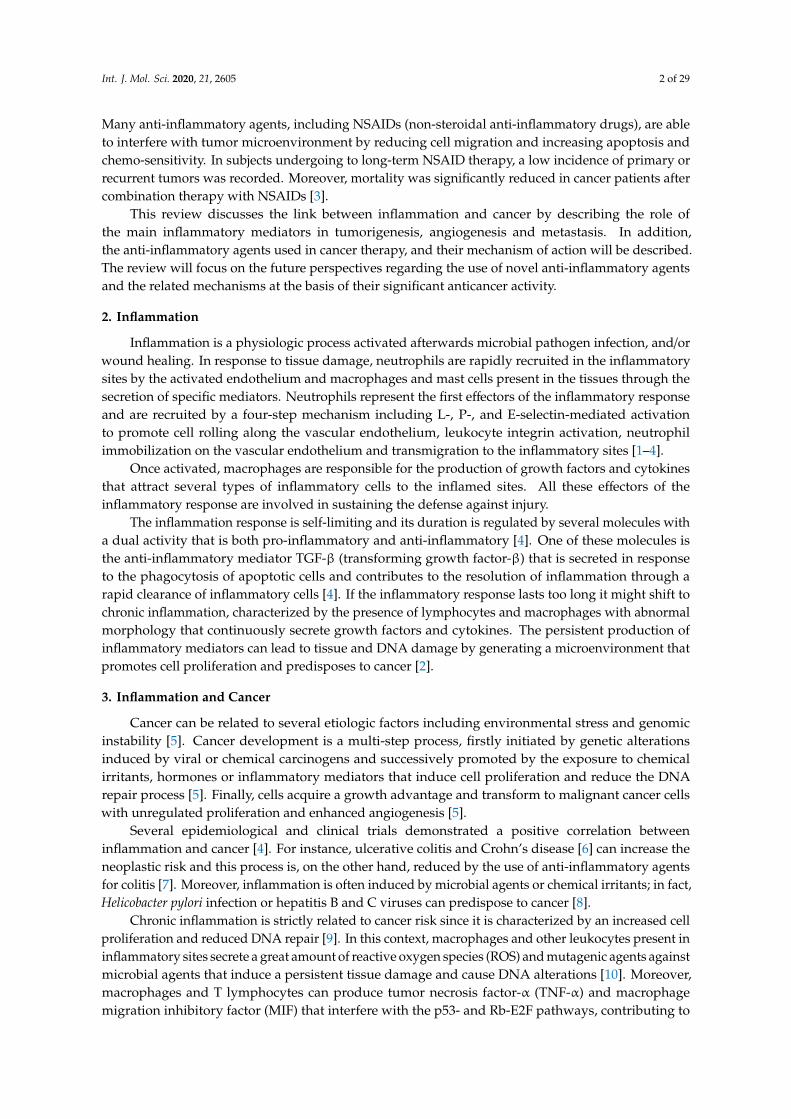
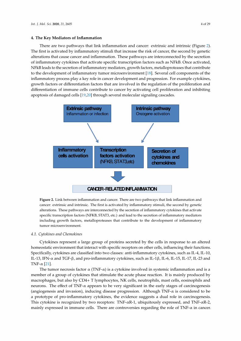
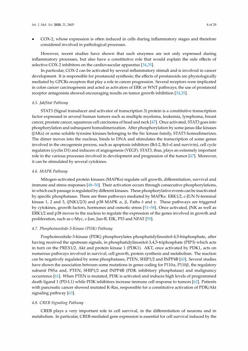
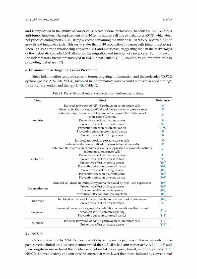
tumorigenesis [11,12]. The shift from initiated cells to malignant cells requires many genetic andepigenetic events also related to chronic inflammation. Chronic inflammation is characterized bya continuous tissue and DNA injury that leads to an accumulation of mutations in epithelial cells(Figure 1) [13].
Int. J. Mol. Sci. 2020, 21, x FOR PEER REVIEW 3 of 30
genetic and epigenetic events also related to chronic inflammation. Chronic inflammation is characterized by a continuous tissue and DNA injury that leads to an accumulation of mutations in epithelial cells (Figure 1) [13].
Figure 1. Inflammation and cancer. Various inflammatory and carcinogenic agents can activate the transcription factor NFkB. Once activated, it binds to specific DNA sequences in the nucleus and induces the production of pro-inflammatory cytokines and COX enzymes. Activated immune cells produce specific cytokines (IL-6, VEGF, etc.) and metalloproteinases (MMP-2 and MMP-9). IL-6 and growth factors can induce STAT3 activation by leading to cell proliferation and survival while metalloproteases degrade the membrane basement, promoting cell invasion. Moreover, macrophages secrete a great amount of reactive oxygen species (ROS) and mutagenic agents against microbial agents that induce a persistent tissue damage and cause DNA alterations by contributing to tumorigenesis.
Mutated cells are able to generate a tumor inflammatory microenvironment [2] rich in macrophages, neutrophils, eosinophils, dendritic cells, mast cells, and lymphocytes that play a key role in inflammation-associated cancers [1,2]. In particular, tumor-associated macrophages (TAM) can promote tumor progression through the secretion of specific factors such as cytokines (IL-10) and growth factors (vascular endothelial growth factor (VEGF), endothelin-2, and urokinase-type plasminogen activator) that contribute to the angiogenesis [13] and suppress the immune response. Moreover, TAMs produce metalloproteinases (MMP-2 and MMP-9) that degrade the membrane basement by promoting cell invasion and metastasis [13]. Also, mast cells and tumor-associated neutrophils potentiate tumor progression by releasing cytokines and growth factors that are involved in angiogenesis, invasion and metastasis [14]. These cytokines secreted in tumor sites are specific signals to recruit lymphocytes but their specific role in tumor development is under investigation [15–17].
Figure 1. Inflammation and cancer. Various inflammatory and carcinogenic agents can activate thetranscription factor NFkB. Once activated, it binds to specific DNA sequences in the nucleus and inducesthe production of pro-inflammatory cytokines and COX enzymes. Activated immune cells producespecific cytokines (IL-6, VEGF, etc.) and metalloproteinases (MMP-2 and MMP-9). IL-6 and growthfactors can induce STAT3 activation by leading to cell proliferation and survival while metalloproteasesdegrade the membrane basement, promoting cell invasion. Moreover, macrophages secrete a greatamount of reactive oxygen species (ROS) and mutagenic agents against microbial agents that induce apersistent tissue damage and cause DNA alterations by contributing to tumorigenesis.
Mutated cells are able to generate a tumor inflammatory microenvironment [2] rich in macrophages,neutrophils, eosinophils, dendritic cells, mast cells, and lymphocytes that play a key role ininflammation-associated cancers [1,2]. In particular, tumor-associated macrophages (TAM) can promotetumor progression through the secretion of specific factors such as cytokines (IL-10) and growth factors(vascular endothelial growth factor (VEGF), endothelin-2, and urokinase-type plasminogen activator)that contribute to the angiogenesis [13] and suppress the immune response. Moreover, TAMs producemetalloproteinases (MMP-2 and MMP-9) that degrade the membrane basement by promoting cellinvasion and metastasis [13]. Also, mast cells and tumor-associated neutrophils potentiate tumorprogression by releasing cytokines and growth factors that are involved in angiogenesis, invasion andmetastasis [14]. These cytokines secreted in tumor sites are specific signals to recruit lymphocytes buttheir specific role in tumor development is under investigation [15–17].
Int. J. Mol. Sci. 2020, 21, 2605 4 of 29
4. The Key Mediators of Inflammation
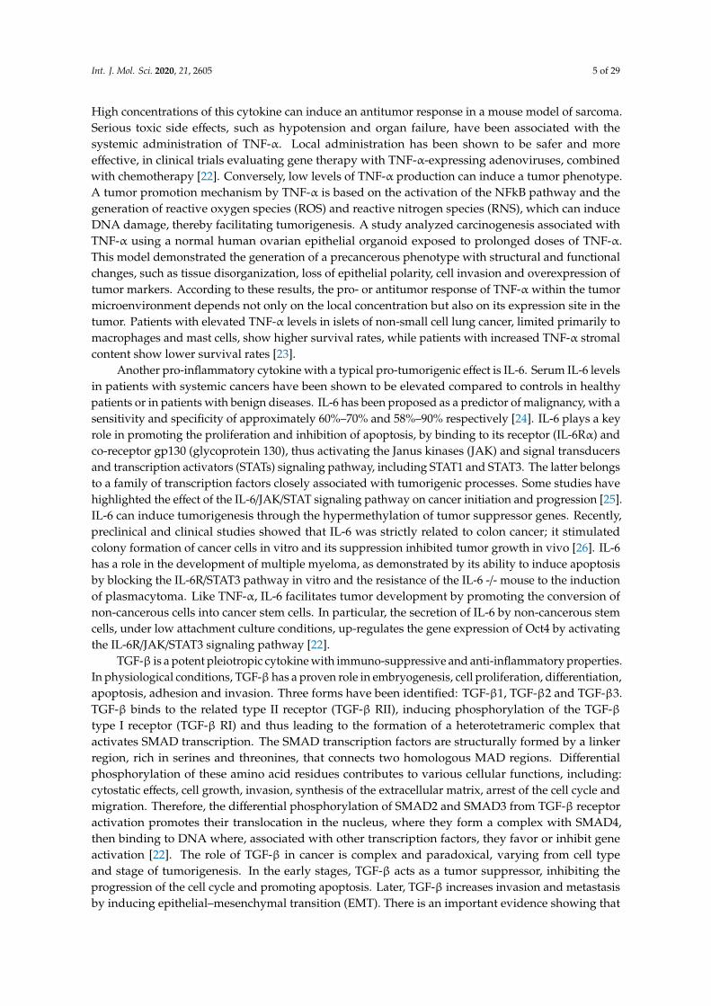
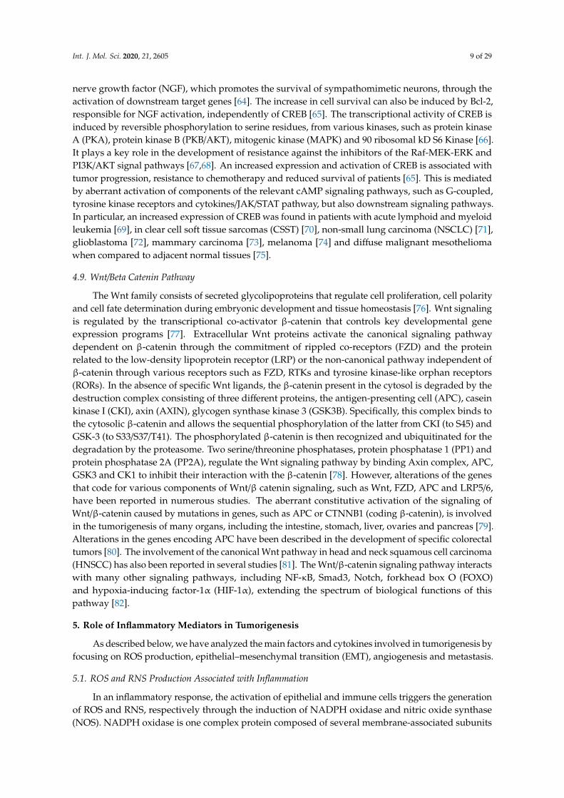
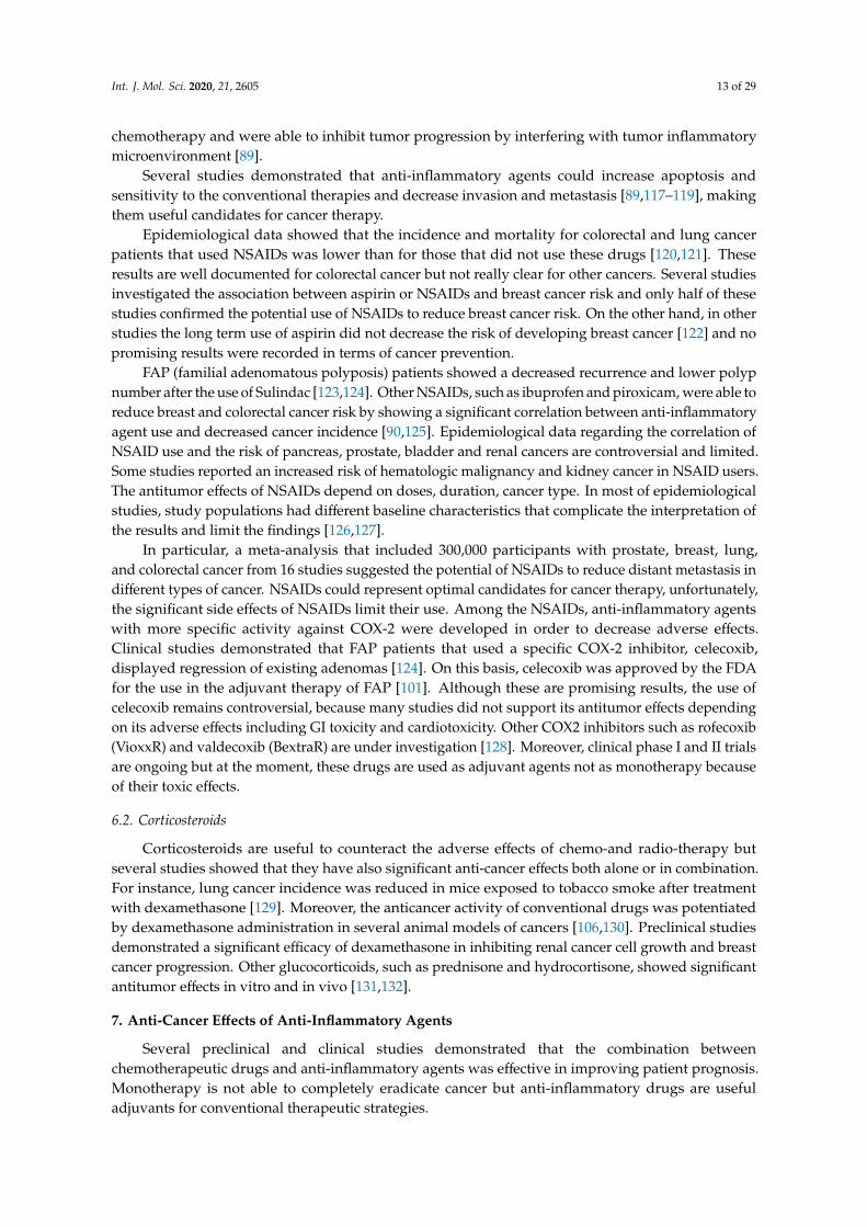
There are two pathways that link inflammation and cancer: extrinsic and intrinsic (Figure 2).The first is activated by inflammatory stimuli that increase the risk of cancer, the second by geneticalterations that cause cancer and inflammation. These pathways are interconnected by the secretionof inflammatory cytokines that activate specific transcription factors such as NFkB. Once activated,NFkB leads to the secretion of inflammatory mediators, growth factors, metalloproteases that contributeto the development of inflammatory tumor microenvironment [18]. Several cell components of theinflammatory process play a key role in cancer development and progression. For example cytokines,growth factors or differentiation factors that are involved in the regulation of the proliferation anddifferentiation of immune cells contribute to cancer by activating cell proliferation and inhibitingapoptosis of damaged cells [19,20] through several molecular signaling cascades.
Int. J. Mol. Sci. 2020, 21, x FOR PEER REVIEW 4 of 30
4. The Key Mediators of Inflammation
There are two pathways that link inflammation and cancer: extrinsic and intrinsic (Figure 2). The first is activated by inflammatory stimuli that increase the risk of cancer, the second by genetic alterations that cause cancer and inflammation. These pathways are interconnected by the secretion of inflammatory cytokines that activate specific transcription factors such as NFkB. Once activated, NFkB leads to the secretion of inflammatory mediators, growth factors, metalloproteases that contribute to the development of inflammatory tumor microenvironment [18]. Several cell components of the inflammatory process play a key role in cancer development and progression. For example cytokines, growth factors or differentiation factors that are involved in the regulation of the proliferation and differentiation of immune cells contribute to cancer by activating cell proliferation and inhibiting apoptosis of damaged cells [19,20] through several molecular signaling cascades.
Figure 2. Link between inflammation and cancer. There are two pathways that link inflammation and cancer: extrinsic and intrinsic. The first is activated by inflammatory stimuli, the second by genetic alterations. These pathways are interconnected by the secretion of inflammatory cytokines that activate specific transcription factors (NFKB, STAT3, etc.) and lead to the secretion of inflammatory mediators including growth factors, metalloproteases that contribute to the development of inflammatory tumor microenvironment.
4.1. Cytokines and Chemokines
Cytokines represent a large group of proteins secreted by the cells in response to an altered homeostatic environment that interact with specific receptors on other cells, influencing their functions. Specifically, cytokines are classified into two classes: anti-inflammatory cytokines, such as IL-4, IL-10, IL-13, IFN-α and TGF-β, and pro-inflammatory cytokines, such as IL-1β, IL-6, IL-15, IL-17, IL-23 and TNF-α [21].
The tumor necrosis factor α (TNF-α) is a cytokine involved in systemic inflammation and is a member of a group of cytokines that stimulate the acute phase reaction. It is mainly produced by macrophages, but also by CD4+ T lymphocytes, NK cells, neutrophils, mast cells, eosinophils and neurons. The effect of TNF-α appears to be very significant in the early stages of carcinogenesis (angiogenesis and invasion), inducing disease progression. Although TNF-α is considered to be a prototype of pro-inflammatory cytokines, the evidence suggests a dual role in carcinogenesis. This
Figure 2. Link between inflammation and cancer. There are two pathways that link inflammation andcancer: extrinsic and intrinsic. The first is activated by inflammatory stimuli, the second by geneticalterations. These pathways are interconnected by the secretion of inflammatory cytokines that activatespecific transcription factors (NFKB, STAT3, etc.) and lead to the secretion of inflammatory mediatorsincluding growth factors, metalloproteases that contribute to the development of inflammatorytumor microenvironment.
4.1. Cytokines and Chemokines
Cytokines represent a large group of proteins secreted by the cells in response to an alteredhomeostatic environment that interact with specific receptors on other cells, influencing their functions.Specifically, cytokines are classified into two classes: anti-inflammatory cytokines, such as IL-4, IL-10,IL-13, IFN-α and TGF-β, and pro-inflammatory cytokines, such as IL-1β, IL-6, IL-15, IL-17, IL-23 andTNF-α [21].
The tumor necrosis factor α (TNF-α) is a cytokine involved in systemic inflammation and is amember of a group of cytokines that stimulate the acute phase reaction. It is mainly produced bymacrophages, but also by CD4+ T lymphocytes, NK cells, neutrophils, mast cells, eosinophils andneurons. The effect of TNF-α appears to be very significant in the early stages of carcinogenesis(angiogenesis and invasion), inducing disease progression. Although TNF-α is considered to bea prototype of pro-inflammatory cytokines, the evidence suggests a dual role in carcinogenesis.This cytokine is recognized by two receptors: TNF-αR-1, ubiquitously expressed, and TNF-αR-2,mainly expressed in immune cells. There are controversies regarding the role of TNF-α in cancer.
Int. J. Mol. Sci. 2020, 21, 2605 5 of 29
High concentrations of this cytokine can induce an antitumor response in a mouse model of sarcoma.Serious toxic side effects, such as hypotension and organ failure, have been associated with thesystemic administration of TNF-α. Local administration has been shown to be safer and moreeffective, in clinical trials evaluating gene therapy with TNF-α-expressing adenoviruses, combinedwith chemotherapy [22]. Conversely, low levels of TNF-α production can induce a tumor phenotype.A tumor promotion mechanism by TNF-α is based on the activation of the NFkB pathway and thegeneration of reactive oxygen species (ROS) and reactive nitrogen species (RNS), which can induceDNA damage, thereby facilitating tumorigenesis. A study analyzed carcinogenesis associated withTNF-α using a normal human ovarian epithelial organoid exposed to prolonged doses of TNF-α.This model demonstrated the generation of a precancerous phenotype with structural and functionalchanges, such as tissue disorganization, loss of epithelial polarity, cell invasion and overexpression oftumor markers. According to these results, the pro- or antitumor response of TNF-α within the tumormicroenvironment depends not only on the local concentration but also on its expression site in thetumor. Patients with elevated TNF-α levels in islets of non-small cell lung cancer, limited primarily tomacrophages and mast cells, show higher survival rates, while patients with increased TNF-α stromalcontent show lower survival rates [23].
Another pro-inflammatory cytokine with a typical pro-tumorigenic effect is IL-6. Serum IL-6 levelsin patients with systemic cancers have been shown to be elevated compared to controls in healthypatients or in patients with benign diseases. IL-6 has been proposed as a predictor of malignancy, with asensitivity and specificity of approximately 60%–70% and 58%–90% respectively [24]. IL-6 plays a keyrole in promoting the proliferation and inhibition of apoptosis, by binding to its receptor (IL-6Rα) andco-receptor gp130 (glycoprotein 130), thus activating the Janus kinases (JAK) and signal transducersand transcription activators (STATs) signaling pathway, including STAT1 and STAT3. The latter belongsto a family of transcription factors closely associated with tumorigenic processes. Some studies havehighlighted the effect of the IL-6/JAK/STAT signaling pathway on cancer initiation and progression [25].IL-6 can induce tumorigenesis through the hypermethylation of tumor suppressor genes. Recently,preclinical and clinical studies showed that IL-6 was strictly related to colon cancer; it stimulatedcolony formation of cancer cells in vitro and its suppression inhibited tumor growth in vivo [26]. IL-6has a role in the development of multiple myeloma, as demonstrated by its ability to induce apoptosisby blocking the IL-6R/STAT3 pathway in vitro and the resistance of the IL-6 -/- mouse to the inductionof plasmacytoma. Like TNF-α, IL-6 facilitates tumor development by promoting the conversion ofnon-cancerous cells into cancer stem cells. In particular, the secretion of IL-6 by non-cancerous stemcells, under low attachment culture conditions, up-regulates the gene expression of Oct4 by activatingthe IL-6R/JAK/STAT3 signaling pathway [22].
TGF-β is a potent pleiotropic cytokine with immuno-suppressive and anti-inflammatory properties.In physiological conditions, TGF-β has a proven role in embryogenesis, cell proliferation, differentiation,apoptosis, adhesion and invasion. Three forms have been identified: TGF-β1, TGF-β2 and TGF-β3.TGF-β binds to the related type II receptor (TGF-β RII), inducing phosphorylation of the TGF-βtype I receptor (TGF-β RI) and thus leading to the formation of a heterotetrameric complex thatactivates SMAD transcription. The SMAD transcription factors are structurally formed by a linkerregion, rich in serines and threonines, that connects two homologous MAD regions. Differentialphosphorylation of these amino acid residues contributes to various cellular functions, including:cytostatic effects, cell growth, invasion, synthesis of the extracellular matrix, arrest of the cell cycle andmigration. Therefore, the differential phosphorylation of SMAD2 and SMAD3 from TGF-β receptoractivation promotes their translocation in the nucleus, where they form a complex with SMAD4,then binding to DNA where, associated with other transcription factors, they favor or inhibit geneactivation [22]. The role of TGF-β in cancer is complex and paradoxical, varying from cell typeand stage of tumorigenesis. In the early stages, TGF-β acts as a tumor suppressor, inhibiting theprogression of the cell cycle and promoting apoptosis. Later, TGF-β increases invasion and metastasisby inducing epithelial–mesenchymal transition (EMT). There is an important evidence showing that
Int. J. Mol. Sci. 2020, 21, 2605 6 of 29
TGF-β signaling changes are involved in human cancers. An increase in mRNA and TGF-β proteinhas been observed in gastric cancer, non-small cell lung cancer and colorectal and prostate cancers,and TGF-β receptor deletion or mutations have been associated with colorectal, prostate, brain andbladder cancers, in correlation with a more invasive and advanced carcinoma, with a higher degreeof invasion and poor prognosis. In the tumor microenvironment, common sources of TGF-β arerepresented by tumor and stromal cells, including immune cells and fibroblasts. The bone matrix isalso an abundant source of TGF-β and a common site for metastasis in many tumors, in correlationwith tumor promotion and the invasive effects of these cytokines [22].
Interleukin 10 is known to be a powerful anti-inflammatory cytokine. Almost all immune cellsproduce IL-10 including T cells, B cells, monocytes, macrophages, mast cells, granulocytes, dendriticcells and keratinocytes. Cancer cells can also secrete IL-10, as can tumor-infiltrating macrophages.When IL-10 binds to its receptor it activates on the cytoplasmic side the tyrosine kinases Jak1 and Tyk2,which phosphorylate an intracellular domain of IL-10R, allowing the interaction of this with STAT1,STAT3 and STAT5, and favoring the translocation of STATs in the nucleus and the induction of targetgene expression. Several studies have found that IL-10 has both pro- and anti-tumor effects. The IL-10inhibits NF-kB; therefore, this may downregulate the expression of pro-inflammatory cytokines andact as an anti-tumor cytokine. In addition, IL-10 can exert anti-tumor activity in gliomas, melanomas,and brain and ovarian tumors, through a mechanism that involves the down-regulation of MHC-1,thus inducing cell lysis of tumor mediated by NK. Thanks to its immunosuppressive effect on dendriticcells and macrophages, IL-10 can attenuate antigen presentation, cell maturation and differentiation,allowing cancer cells to circumvent the mechanisms of immunosurveillance. Furthermore, as previouslydescribed for the IL-6, STAT3 can also be activated by IL-10, although the contradictory responses ofthe cytokines are determined by the receptor and by the time of activation of STAT. In particular, IL-6leads to a transient, rapid decline in phosphorylation and nuclear localization of STAT3, while IL-10induces sustained phosphorylation of STAT3. Through the activation of STAT3, IL-10 can also have apro-tumorigenic effect, mediated by an autocrine-paracrine loop that involves the up-regulation ofBcl-2 and the activation of resistance to apoptosis. Similarly, elevated IL-10 levels are associated withpoor prognosis [27].
Other important inflammatory mediators are chemokines, chemiotactic small cytokines thatexpress their action by binding specific receptors expressed on endothelial cells or immune systemcells. Chemokine production is stimulated by cytokines; they are able to control leukocyte infiltrationin the tumor, regulate immune angiogenesis and act as growth factors [28]. Chemotactic factors areinvolved in cancer promotion. Several studies showed that chemokines stimulate cell growth andmetastasis and induce angiogenesis in various tumors. In particular, chemokines can induce tumorcell migration by increasing the expression of MMPs that degrade extracellular matrix in a similarmanner they stimulate leukocyte migration in inflammatory sites [29].
4.2. NFkB Transcription Factor
It has been shown that various inflammatory and carcinogenic agents, including TNF-α, cigarettesmoking, lipopolysaccharides (LPS), interleukins (IL-1) and hydrogen peroxide (H2O2), activate NFκB,a nuclear transcription factor involved in tumorigenesis, inflammation, proliferation, carcinogenesisand apoptosis [30]. NF-κB comprises a family of conserved and structurally related proteins includingRelA/P65, Rel/cRel, RelB, NF-κB1/p50 and NF-B2/p52. When inactivated, NFκB is found in the cytosolbound to an IκB inhibitor protein (IκBα). Through the involvement of membrane receptors, a varietyof extracellular signals can activate the IκB kinase (IKK) enzyme. IKK, in turn, phosphorylates IκBαprotein leading to its ubiquitination and degradation by the proteasome [31]. In this way, NFκB isactivated and moved to the nucleus where it binds to specific DNA sequences called response elements(RE). This mechanism leads to a change in cell functions, such as the production of pro-inflammatorycytokines. NFκB, also, activates transcription of the coding mRNA for its IκB inhibitor subunit,thus generating a negative feed-back circuit. NFkB is activated by several cytokines as well and plays
Int. J. Mol. Sci. 2020, 21, 2605 7 of 29
a key role in inflammatory process. In cancer, it is often constitutively activated and able to inducesurvival and promote cancer progression through the activation of genes coding for proteins thatregulate the progression of the cell cycle (e.g., Ciclina D, c-myc) and apoptosis (e.g., CIAP, A1/BFL1, Bcl2,c-Flip) [30,31]. It is responsible for the secretion of ROS that cause DNA damage and prevent mutatedcells from being destroyed. This mediator strictly links inflammation and cancer since it producescytokines, growth factors and adhesion molecules that have pro-cancer effects. In fact, several in vitroand in vivo studies showed the involvement of NFkB in cancer promotion. For instance, knockout ofIKK leaded to NFkB inactivation and decreased tumor growth in mouse model of colitis-associatedcancer [32].
4.3. iNOS and NO Secretion
iNOS is an enzyme that catalyzes NO production and is overexpressed in several chronicinflammatory processes and cancers. iNOS is activated by pro-inflammatory cytokines or NFkBand induces DNA damage, reduces DNA repair and promotes cancer development [33]. Moreover,it stimulates angiogenesis and metastasis and can induce COX-2, an important mediator in the linkbetween inflammation and cancer [34]. It has been reported that iNOS inhibitors were able to reducetumorigenesis in vivo [33].
4.4. LOX and COX Pathways
Leukotrienes (LT) have been recognized among the various mediators of a wide range ofinflammatory and allergic reactions such as rheumatoid arthritis, intestinal inflammatory diseases,psoriasis, allergic rhinitis although their primary pathophysiological implication is related to bronchialasthma [35]. Their biosynthesis requires a cell activation that stimulates the conversion of arachidonicacid into biologically active messengers. In the presence of an external stimulus, phospholipase A2releases arachidonic acid from the membrane phospholipids. This intermediate can be attacked by twodifferent enzymes: cyclooxygenases (COX-1 and COX-2) leading to the formation of prostaglandins(PG) and thromboxane A2 (TXA2) or lipoxygenases (5-, 8-, 12-, 15-LOX) responsible for the synthesis ofleukotrienes [36]. Human 5-LOX is predominantly present in mature leukocytes including granulocytes,monocytes/macrophages, mast cells, lymphocytes B and dendritic cells in which the ability to expressthe enzyme is acquired during cell maturation [36]. Numerous evidences suggest the involvement ofthe 5-LOX pathway in the proliferation and survival of tumor cells [37–44]: (I) the enzymes necessary forthe biosynthesis of LTs, as well as the LTs receptors, are present or even over-expressed in transformedcells or neoplastic tissues; (II) a substantial formation of 5-LOX products occurs at these sites; (III) theaddition of 5-LOX products from the outside stimulates the proliferation and survival of tumor cells;(IV) pharmacological or genetic 5-LOX inhibition inhibits tumor cell growth and induces apoptosis.5-LOX was abundantly detected in human or animal cancer cell lines such as brain [37], breast [38],colon [39], renal [40], mesothelium [41] esophageal mucosa [42], pancreas [43], and prostate [44], and inmost of these studies there is also a concomitant increase in 5-LOX products. Recent studies [45] haveshown that the expression of 5-LOX in papillary thyroid carcinoma (PTC) promotes carcinogenesis bythe induction of metalloproteinases (MMPs). These enzymes, activated by both 5-LOX and its product,5-hydroxyoxyacetanic acid, are able to degrade and remodel the extracellular matrix, promoting cellinvasion. While 5-LOX uses arachidonic acid for the formation of leukotrienes, prostaglandin Hsynthase (PGHS) provides conversion to prostaglandin (PG) and thromboxane (TX) A2 [46]. PGHSis an enzyme that, like 5-LOX, catalyzes two coupled reactions: an initial cyclo-oxygenation and asubsequent hydroperoxide formation. However, since drugs that inhibit prostaglandin formationgenerally inhibit the first cyclooxygenation reaction, prostaglandin H synthase is functionally andpharmacologically described as cyclooxygenase (COX) [34]. Among these enzymes there are twoisoforms:
• COX-1, constitutively expressed in many cells and mainly involved in the prostanoidphysiological production;
Int. J. Mol. Sci. 2020, 21, 2605 8 of 29
• COX-2, whose expression is often induced in cells during inflammatory stages and thereforeconsidered involved in pathological processes.
However, recent studies have shown that such enzymes are not only expressed duringinflammatory processes, but also have a constitutive role that would explain the side effects ofselective COX-2 inhibitors on the cardiovascular apparatus [34,35].
In particular, COX-2 can be activated by several inflammatory stimuli and is involved in cancerdevelopment. It is responsible for prostanoid synthesis; the effects of prostanoids are physiologicallymediated by GPCRs receptors that play a role in cancer progression. Several receptors were implicatedin colon cancer carcinogenesis and acted as activators of ERK or WNT pathways; the use of prostanoidreceptor antagonists showed encouraging results on tumor growth inhibition [34,35].
4.5. Jak/Stat Pathway
STAT3 (Signal transducer and activator of transcription 3) protein is a constitutive transcriptionfactor expressed in several human tumors such as multiple myeloma, leukemia, lymphoma, breastcancer, prostate cancer, squamous cell carcinoma of head and neck [47]. Once activated, STAT3 goes intophosphorylation and subsequent homodimerization. After phosphorylation by some janus-like kinases(JAKs) or some soluble tyrosine kinases belonging to the Src kinase family, STAT3 homodimerizes.The dimer moves into the nucleus, binds to DNA and stimulates the transcription of some genesinvolved in the oncogenesis process, such as apoptosis inhibitors (Bcl-2, Bcl-xl and survivin), cell cycleregulators (cyclin D1) and inducers of angiogenesis (VEGF). STAT3, thus, plays an extremely importantrole in the various processes involved in development and progression of the tumor [47]. Moreover,it can be stimulated by several cytokines.
4.6. MAPK Pathway
Mitogen-activated protein kinases (MAPKs) regulate cell growth, differentiation, survival andimmune and stress responses [48–50]. Their activation occurs through consecutive phosphorylations,in which each passage is regulated by different kinases. These phosphorylative events can be inactivatedby specific phosphatases. There are three pathways mediated by MAPKs: ERK1/2, c-JUN N-terminalkinase 1, 2 and 3, (JNK1/2/3) and p38 MAPK α, β, Paths δ and γ. These pathways are triggeredby cytokines, growth factors, hormones and osmotic stress [51–58]. Once activated, JNK as well asERK1/2 and p38 moves to the nucleus to regulate the expression of the genes involved in growth andproliferation, such as c-Myc, c-Jun, Jun-B, EIK, P53 and NFAT [59].
4.7. Phosphoinositide-3-Kinase (PI3K) Pathway
Posphoinositide-3-kinase (PI3K) phosphorylates phosphatidylinositol-4,5-bisphosphate, afterhaving received the upstream signals, in phosphatidylinositol-3,4,5-triphosphate (PIP3) which actsin turn on the PREX1/2, Akt and protein kinase 1 (PDK1). AKT, once activated by PDK1, acts onnumerous pathways involved in survival, cell growth, protein synthesis and metabolism. The reactioncan be negatively regulated by some phosphatases, PTEN, SHIP1/2 and INPP4B [60]. Several studieshave shown the association between some mutations in genes coding for P110α, P110β, the regulatorysubunit P85α and, PTEN, SHIP1/2 and INPP4B (PI3K inhibitory phosphatase) and malignancyoccurrence [61]. When PTEN is mutated, PI3K is activated and induces high levels of programmeddeath ligand 1 (PD-L1) while PI3K inhibitors increase immune cell response to tumors [62]. Patientswith pancreatic cancer showed mutated K-Ras, responsible for a constitutive activation of PI3K/Aktsignaling pathway [63].
4.8. CREB Signaling Pathway
CREB plays a very important role in cell survival, in the differentiation of neurons and inmetabolism. In particular, CREB-mediated gene expression is essential for cell survival induced by the
Int. J. Mol. Sci. 2020, 21, 2605 9 of 29
nerve growth factor (NGF), which promotes the survival of sympathomimetic neurons, through theactivation of downstream target genes [64]. The increase in cell survival can also be induced by Bcl-2,responsible for NGF activation, independently of CREB [65]. The transcriptional activity of CREB isinduced by reversible phosphorylation to serine residues, from various kinases, such as protein kinaseA (PKA), protein kinase B (PKB/AKT), mitogenic kinase (MAPK) and 90 ribosomal kD S6 Kinase [66].It plays a key role in the development of resistance against the inhibitors of the Raf-MEK-ERK andPI3K/AKT signal pathways [67,68]. An increased expression and activation of CREB is associated withtumor progression, resistance to chemotherapy and reduced survival of patients [65]. This is mediatedby aberrant activation of components of the relevant cAMP signaling pathways, such as G-coupled,tyrosine kinase receptors and cytokines/JAK/STAT pathway, but also downstream signaling pathways.In particular, an increased expression of CREB was found in patients with acute lymphoid and myeloidleukemia [69], in clear cell soft tissue sarcomas (CSST) [70], non-small lung carcinoma (NSCLC) [71],glioblastoma [72], mammary carcinoma [73], melanoma [74] and diffuse malignant mesotheliomawhen compared to adjacent normal tissues [75].
4.9. Wnt/Beta Catenin Pathway
The Wnt family consists of secreted glycolipoproteins that regulate cell proliferation, cell polarityand cell fate determination during embryonic development and tissue homeostasis [76]. Wnt signalingis regulated by the transcriptional co-activator β-catenin that controls key developmental geneexpression programs [77]. Extracellular Wnt proteins activate the canonical signaling pathwaydependent on β-catenin through the commitment of rippled co-receptors (FZD) and the proteinrelated to the low-density lipoprotein receptor (LRP) or the non-canonical pathway independent ofβ-catenin through various receptors such as FZD, RTKs and tyrosine kinase-like orphan receptors(RORs). In the absence of specific Wnt ligands, the β-catenin present in the cytosol is degraded by thedestruction complex consisting of three different proteins, the antigen-presenting cell (APC), caseinkinase I (CKI), axin (AXIN), glycogen synthase kinase 3 (GSK3B). Specifically, this complex binds tothe cytosolic β-catenin and allows the sequential phosphorylation of the latter from CKI (to S45) andGSK-3 (to S33/S37/T41). The phosphorylated β-catenin is then recognized and ubiquitinated for thedegradation by the proteasome. Two serine/threonine phosphatases, protein phosphatase 1 (PP1) andprotein phosphatase 2A (PP2A), regulate the Wnt signaling pathway by binding Axin complex, APC,GSK3 and CK1 to inhibit their interaction with the β-catenin [78]. However, alterations of the genesthat code for various components of Wnt/β catenin signaling, such as Wnt, FZD, APC and LRP5/6,have been reported in numerous studies. The aberrant constitutive activation of the signaling ofWnt/β-catenin caused by mutations in genes, such as APC or CTNNB1 (coding β-catenin), is involvedin the tumorigenesis of many organs, including the intestine, stomach, liver, ovaries and pancreas [79].Alterations in the genes encoding APC have been described in the development of specific colorectaltumors [80]. The involvement of the canonical Wnt pathway in head and neck squamous cell carcinoma(HNSCC) has also been reported in several studies [81]. The Wnt/β-catenin signaling pathway interactswith many other signaling pathways, including NF-κB, Smad3, Notch, forkhead box O (FOXO)and hypoxia-inducing factor-1α (HIF-1α), extending the spectrum of biological functions of thispathway [82].
5. Role of Inflammatory Mediators in Tumorigenesis
As described below, we have analyzed the main factors and cytokines involved in tumorigenesis byfocusing on ROS production, epithelial–mesenchymal transition (EMT), angiogenesis and metastasis.
5.1. ROS and RNS Production Associated with Inflammation
In an inflammatory response, the activation of epithelial and immune cells triggers the generationof ROS and RNS, respectively through the induction of NADPH oxidase and nitric oxide synthase(NOS). NADPH oxidase is one complex protein composed of several membrane-associated subunits
Int. J. Mol. Sci. 2020, 21, 2605 10 of 29
that catalyze the superoxide anion (O2−), leading to the production of peroxide of hydrogen (H2O2)mediated by superoxide dismutase (SOD-). On the other hand, NOS generates nitric oxide (NO) fromL-arginine, which can be converted into RNS such as nitrogen dioxide (NO2), Peroxynitrite (ONOO-),and nitrogen trioxide (N2O3). Different of NOS are produced according to the cell type: inducibleNOS (iNOS) in phagocytes and constitutively in endothelial and neuronal cells (eNOS and nNOS).ROS and RNS have a powerful antimicrobial role in phagocytes and also act as second messengersin signal transduction. Activation of phagocytes can directly induce reactive oxygen and nitrogenspecies (collectively called RONS), activating NOX2, NADPH oxidase, and iNOS. Furthermore,TNF-α, IL-6, and TGF-β trigger the generation of RONS in non-phagocytic cells. The increasedexpression of NADPH oxidase and NOS and their RONS products has been identified in severalcancers, suggesting that free radicals have a role in the genesis and malignant progression. ElevatedRON levels have been observed in various chronic inflammatory diseases, such as H. pylori-associatedgastritis and inflammatory bowel disease (IBD), suggesting a role in cancer risk. Several mechanismshave been proposed to clarify the participation of RONS in cancer development. RONS inducecellular oxidative stress and damage to lipids, proteins, and DNA, as well as the production of 8-oxo-7,8-dihydro-2′-deoxyguanosine (8-oxodG), currently used as a marker of damage to DNA. Identifyingthese DNA damage markers in chronic inflammatory processes, such as gastritis associated withH. pylori, hepatitis, and ulcerative colitis, underlines the relevance of RONS in pathologies with anincreased risk of cancer. An increase in iNOS, 3-nitrotyrosine, and 8-oxodG has been found in thelivers of patients with primary sclerosing cholangitis. In addition, RNS interferes with DNA repair,as is demonstrated in iNOS-overexpressing cells that are unable to repair modified 8-oxodG. RONS aregenerated by cellular stress and by modification of macromolecules, although they are also involvedin the regulation of signaling pathways, such as cell survival and proliferation through Akt, Erk1/2,and the activation of hypoxia-inducible factor 1 (HIF-1) [22].
5.2. Tumor Growth Associated with Inflammation
As repeatedly stressed, inflammation is important in generating malignancy through the exposureof pro-inflammatory cytokines and the sustained activation of signaling pathways such as NF-kB andSTAT3. After the transformation in the malignant cells, these cytokines are also involved in tumorgrowth by stimulating the proliferation of neoplastic cells and eluding immunosurveillance. Severalcytokines have growth factor activity. In one study it was noted that the silencing of TNF-α in a cell lineof the gallbladder cancer decreased cell proliferation and invasion by an autocrine effect, influencingthe signaling pathways of TNF-α/NF-kB/AKT/ Bcl-2 in these cells [22]. The pro-tumorigenic roleof IL-17 has also been implicated in other types of cancer. Mice with carcinogenic-induced uterinetumors deficient in the IL-17 receptor showed a lower tumor incidence and reduced tumor size.Other molecules have been reported in cancer that can influence tumor growth through regulationof the IL-6/STAT3 signaling pathway. Inflammatory mediators such as Hmgbl, IL-23 and IL-17can promote tumor growth by activating the IL-6/STAT3 pathway in a mouse model of melanoma.In cholangiocarcinoma, a high expression of the oncoprotein, gankirin, promotes tumor proliferation,invasion and metastasis through the activation of the IL-6/STAT3 signaling pathway [83].
5.3. Epithelial–Mesenchymal Transition (EMT) Associated with Inflammation
The epithelial–mesenchymal transition (EMT) is a process in which, following a chronic stimulus,epithelial cells with basal–apical polarity lose their phenotype and acquire the characteristics ofnon-polarized and migrating mesenchymal cells [84]. Molecular markers have been identified to assesswhether or not an epithelial cell has gone through the EMT process. The main marker is the lossof E-cadherin, an event associated with the destruction of cell–cell junctions. E-cadherin (epithelialcadherin) is necessary for the formation of strong and stable adherent junctions and therefore for themaintenance of the epithelial phenotype and normal tissue architecture of the adult. A reduction in theexpression of E-cadherin, as has been shown in various malignancies, plays a crucial role in the loss of
Int. J. Mol. Sci. 2020, 21, 2605 11 of 29
cell differentiation and in dissemination. It can be said that E-cadherin appears to be the custodian ofthe epithelial phenotype. N-cadherin (neural cadherin) is normally found only in cells of the nervoussystem, but is produced in some carcinoma cells that have lost the expression of E-cadherin and,in this cellular context, is associated with an increased invasive potential [85]. A relevant inflammatorymediator in EMT is TGF-β and extensive evidences support the concept that EMT can be induced bypro-inflammatory cytokines. TNF-α and IL-6 may urge the TGF-β signaling pathway through theprogression of EMT. Both cytokines promote the activation of NF-kB, which regulates the expressionof the transcription factors involved in EMT, coordinating the effects of Snail1, Snail2, Twist, ZEB1and ZEB2. Furthermore, IL-6 induces cell invasiveness in EMT, through the increased expression ofvimentin (it is the main component of the cytoskeleton of mesenchymal cells) and the down-regulatedexpression of E-cadherin, both mediated by signaling JAK/STAT3/Snail, as shown in head and neckcancer. Finally, ROS production can promote EMT: therefore, exposing kidney epithelial cells to ROSinduces the expression of TGF-β, the SMAD signaling pathway, and EMT [22].
5.4. Angiogenesis Associated with Inflammation
Angiogenesis includes the processes that lead to the generation of new blood vessels from analready existing vascular network. This angiogenic process is important in tumor developmentbecause new blood vessels penetrate and supply nutrients and oxygen to cancer cells. Severalangiogenic factors secreted by tumor cells have been identified, in particular the vascular endothelialgrowth factor (VEGF), which is expressed in response to cytokines and growth factors. Furthermore,the characterization of tumor associated macrophages (TAM) obtained from metastatic lymph nodes(MLN) in an animal model of melanoma, has shown that MLN are mainly constituted by infiltratingmacrophages TIE2/CD31. This subpopulation overexpresses in a significant way VEGF and is directlyrelated to angiogenesis. Some studies have shown that TNF-α may have a double-edged role inangiogenesis, depending on the used doses. High doses of TNF-α inhibit angiogenesis in mice whilelow doses promote vascularization of the area. The anti-angiogenic effect of TNF-α is linked tothe down-regulation of ανβ3 (adhesion molecule) and of the angiotensin signaling pathway, whilepro-angiogenic responses have been associated with the increased expression of VEGF, VEGFR, IL-8,and FGF. Therefore, low levels of TNF-α increase tumor growth, induce angiogenesis of several tumorsin mice, and stimulate a sub-population of tumor-associated myeloid cells and the coexpression ofendothelial and myeloid markers with pro-angiogenic/pro-vascular properties. The tumor source ofTNF-α can be derived from myeloid or tumor cells and through autocrine activation can stimulatetumor growth and angiogenesis. Another important angiogenic factor is IL-6, which induces theexpression of VEGF in a dose-dependent manner in gastric cancer cell lines. Similarly, IL-6 promotesangiogenesis through activation of the STAT3 pathway in cervical cancer. Together, the secretion ofIL-6 and the subsequent phosphorylation of STAT3 are involved in the up-regulation of angiogenicmediators, such as VEGF, HIF1α, the VEGFR2 co-receptor and neuropilin 2 (NRP2) [22].
5.5. Metastases Associated with Inflammation
Metastases are malignant cells that detach themselves from the primary tumor and spread toother organs where they can reproduce and generate new tumors. The metastatic cascade is modulatedby the action of several cytokines released by surrounding cells such as tumor associated macrophages(TAMs), tumor infiltrating lymphocytes (TIL), and cancer associated fibroblasts (CAFs), promotingthe escape of cancer cells and dissemination; the influence of TNF-α has been studied in variousexperimental animal models. The administration of this cytokine leads to a significant increase in thenumber of lung metastases. In some studies, it has been proposed that cancer cells activate myeloid cellsto generate a favorable microenvironment for metastasis. In Lewis lung cancer (LLC) cells, high levelsof IL-6 and TNF-α have been induced in bone marrow-derived macrophages. TNF-α -/- and non-IL-6-/- mice injected with LLC cells showed increased survival and a reduction in lung tumor multiplicity,suggesting a key role of TNF-α in LLC metastasis. IL-6, in turn, is up-regulated in several cancers
Int. J. Mol. Sci. 2020, 21, 2605 12 of 29
and is implicated in the ability of cancer cells to create bone metastases. In contrast, IL-10 exhibitsanti-tumor function. The replacement of IL-10 to the human cell line of melanoma A375P, which doesnot produce endogenous IL-10, using a vector containing the murine IL-10 cDNA, reversed tumorgrowth and lung metastasis. This result states that IL-10 production by cancer cells inhibits metastasis.There is also a strong relationship between EMT and metastasis, suggesting that, in the early stagesof the metastatic cascade, EMT allows for the migration and invasion of cancer cells. For this reason,the inflammatory mediators involved in EMT, in particular TGF-β, could play an important role inpromoting metastasis [22].
6. Inflammation as Target for Cancer Prevention
Since inflammation can predispose to tumor, targeting inflammation and the molecules (COX-2cyclooxygenase 2, NF-kB, VEGF,) involved in inflammatory process could represent a good strategyfor cancer prevention and therapy [1–4], (Table 1).
Table 1. Preventive and anticancer effects of anti-inflammatory drugs.
Drug Effect Reference
Aspirin
Induced activation of NF-kB pathway in colon cancer cells [86]Induced activation of caspase8/Bid and Bax pathway in gastric cancer [87]
Induced apoptosis in neuroblastoma cells through the inhibition ofproteasome function [88]
Preventive effect on bladder cancer [89]Preventive effect on breast cancer [90]
Preventive effect on colorectal cancer [91,92]Preventive effect on esophageal cancer [93]
Preventive effect on lung cancer [94]
Colecoxib
Induced apoptosis in prostate cancer cells [95]Induced endoplasmic reticulum stress in hepatoma cells [96]
Inhibited the expression of survivin via the suppression of promoter activityin human colon cancer cells [97]
Preventive effect on bladder cancer [98]Preventive effect on breast cancer [99]Preventive effect on cervix cancer [100]
Preventive effect on colorectal cancer [101]Preventive effect on lung cancer [102]
Preventive effect on neuroblastoma [103]Preventive effect on prostate cancer [104]
Dexamethasone
Induced cell death in multiple myeloma mediated by miR-125b expression [105]Preventive effect on breast cancer [106]Preventive effect on rectal cancer [107]
Preventive effect on multiple myeloma [108]
Ibuprofen Inhibited activation of nuclear β-catenin in human colon adenomas [109]Preventive effect on breast cancer [90]
PiroxicamPrevented colon carcinogenesis by inhibition of membrane fluidity and
canonical Wnt/β-catenin signaling [110]
Preventive effect on colorectal cancer [111]
SulindacInduced activation of NF-kB pathway in colon cancer cells [112]
Preventive effect on breast cancer [113]
6.1. NSAIDs
Cancer prevention by NSAIDs mostly works by acting on the pathway of the eicosanoids. In thepast, several clinical studies have demonstrated that NSAIDs had anti-tumor activity [114,115] andtheir long-term use reduced the incidence of colorectal, esophageal, breast, and lung cancers [116].NSAIDs showed toxicity and non-specific effects that were lower than those induced by conventional
Int. J. Mol. Sci. 2020, 21, 2605 13 of 29
chemotherapy and were able to inhibit tumor progression by interfering with tumor inflammatorymicroenvironment [89].
Several studies demonstrated that anti-inflammatory agents could increase apoptosis andsensitivity to the conventional therapies and decrease invasion and metastasis [89,117–119], makingthem useful candidates for cancer therapy.
Epidemiological data showed that the incidence and mortality for colorectal and lung cancerpatients that used NSAIDs was lower than for those that did not use these drugs [120,121]. Theseresults are well documented for colorectal cancer but not really clear for other cancers. Several studiesinvestigated the association between aspirin or NSAIDs and breast cancer risk and only half of thesestudies confirmed the potential use of NSAIDs to reduce breast cancer risk. On the other hand, in otherstudies the long term use of aspirin did not decrease the risk of developing breast cancer [122] and nopromising results were recorded in terms of cancer prevention.
FAP (familial adenomatous polyposis) patients showed a decreased recurrence and lower polypnumber after the use of Sulindac [123,124]. Other NSAIDs, such as ibuprofen and piroxicam, were able toreduce breast and colorectal cancer risk by showing a significant correlation between anti-inflammatoryagent use and decreased cancer incidence [90,125]. Epidemiological data regarding the correlation ofNSAID use and the risk of pancreas, prostate, bladder and renal cancers are controversial and limited.Some studies reported an increased risk of hematologic malignancy and kidney cancer in NSAID users.The antitumor effects of NSAIDs depend on doses, duration, cancer type. In most of epidemiologicalstudies, study populations had different baseline characteristics that complicate the interpretation ofthe results and limit the findings [126,127].
In particular, a meta-analysis that included 300,000 participants with prostate, breast, lung,and colorectal cancer from 16 studies suggested the potential of NSAIDs to reduce distant metastasis indifferent types of cancer. NSAIDs could represent optimal candidates for cancer therapy, unfortunately,the significant side effects of NSAIDs limit their use. Among the NSAIDs, anti-inflammatory agentswith more specific activity against COX-2 were developed in order to decrease adverse effects.Clinical studies demonstrated that FAP patients that used a specific COX-2 inhibitor, celecoxib,displayed regression of existing adenomas [124]. On this basis, celecoxib was approved by the FDAfor the use in the adjuvant therapy of FAP [101]. Although these are promising results, the use ofcelecoxib remains controversial, because many studies did not support its antitumor effects dependingon its adverse effects including GI toxicity and cardiotoxicity. Other COX2 inhibitors such as rofecoxib(VioxxR) and valdecoxib (BextraR) are under investigation [128]. Moreover, clinical phase I and II trialsare ongoing but at the moment, these drugs are used as adjuvant agents not as monotherapy becauseof their toxic effects.
6.2. Corticosteroids
Corticosteroids are useful to counteract the adverse effects of chemo-and radio-therapy butseveral studies showed that they have also significant anti-cancer effects both alone or in combination.For instance, lung cancer incidence was reduced in mice exposed to tobacco smoke after treatmentwith dexamethasone [129]. Moreover, the anticancer activity of conventional drugs was potentiatedby dexamethasone administration in several animal models of cancers [106,130]. Preclinical studiesdemonstrated a significant efficacy of dexamethasone in inhibiting renal cancer cell growth and breastcancer progression. Other glucocorticoids, such as prednisone and hydrocortisone, showed significantantitumor effects in vitro and in vivo [131,132].
7. Anti-Cancer Effects of Anti-Inflammatory Agents
Several preclinical and clinical studies demonstrated that the combination betweenchemotherapeutic drugs and anti-inflammatory agents was effective in improving patient prognosis.Monotherapy is not able to completely eradicate cancer but anti-inflammatory drugs are usefuladjuvants for conventional therapeutic strategies.
Int. J. Mol. Sci. 2020, 21, 2605 14 of 29
The mode of action at the basis of the anti-inflammatory drug antitumor effects is not completelydefined but three different potential mechanisms have been described.
7.1. Chemoprotection
One of the problems related to the conventional anticancer therapies is the side effect profile.Chemotherapy often induces toxicity to both tumor and several normal tissues, reducing patientquality of life. Several studies showed that the combination between conventional therapiesand anti-inflammatory agents could decrease the side effects of chemotherapeutics. For example,the concomitant administration of celecoxib and docetaxel to patients with metastatic prostate cancerreduced toxicity to bone marrow [133] and the combination of celecoxib and FOLFIRI (folinic acid,fluorouracil and irinotecan) or capecitabine decreased diarrhea episodes [99,134]. Recently, the GECO(Gemcitabine-Coxib) study evaluated the effects of refecoxib in combination with gemcitabine inpatients with NSCLC and showed that refecoxib use for 3 months ameliorated the quality of life [121].
Recent studies focused on dexamethasone, commonly used as anti-emetic, and demonstratedthat it was able to reduce hematologic toxicity induced by gemcitabine and carboplatin. Anotherglucocorticoid, budesonide, in combination with irinotecan decreased diarrhea episodes [135,136].
Other anti-inflammatory agents such as aspirin can act as anti-thrombotic drugs, decrease arterialthrombosis by facilitating the passage of chemotherapeutics and ameliorate prognosis.
Anti-inflammatory drugs such as COX-2 inhibitors or dexamethasone reduced neurotoxicityby inhibiting the expression and activity of matrix metalloproteinases (MMPs) 3 and 9 and VEGF,therefore stabilizing blood-brain barrier [137].
7.2. Alterations in Pharmacokinetics or Metabolism
Anti-inflammatory agents are able to modify the pharmacokinetics of the other drugs. In fact,several studies showed that dexamethasone decreased hematologic toxicity of conventional therapies(gemcitabine, carboplatin and doxorubicin) in mice, probably by changing drugpharmacokinetics[106,130].Glucocorticoids induced no significant differences in plasma pharmacokinetics but altered the uptake ofgemcitabine or carboplatin by the spleen and bone marrow and increased the amount of drugsthat reached the tumor [130]. The combination between dexamethasone and adriamycin showedsimilar results [106]; glucocorticoids can interfere with the pharmacokinetics of conventional drugs byincreasing their anticancer effects and decreasing their side effects.
In addition, anti-inflammatory agents can alter the metabolism of chemotherapeutic drugs; forinstance, rofecoxib acts as CYP1A2 inhibitor [138] and induces changes in concentration, half-lifeand clearance of the other drugs that are metabolized by CYP1A2. Dexamethasone induces CYP2D6activity and celecoxib inhibits it [139], thus interfering with the efficacy of tamoxifen, a substrateof CYP2D6. Diclofenac inhibits the glucuronidation of DMXAA by blocking its metabolism andincreasing its plasma concentration [140]. The tumor interstitial fluid pressure (IFP) is responsible forthe decreased uptake of chemotherapeutic drugs into the tumor site and minor chemoresponsivity [141].Several studies demonstrated that VEGF and PDGF antagonists, colecoxib and dexamethasone reducedIFP, thus increasing the amount of drugs that reached the tumor [100,142]. Anti-inflammatory agentscan alter metabolism of other drugs, improving their efficacy and decreasing their toxicity, but it isimportant to consider that cytochrome p450 enzymes that play a key role in metabolism of the majorpart of the drugs can be polymorphic and lead to unexpected results in terms of toxicity and efficacy ofthe drugs.
7.3. Chemosensitization
In addition to the ability of anti-inflammatory drugs to decrease toxicity of conventionaldrugs by altering their metabolism, the effective anticancer effects of the combination betweenanti-inflammatory agents and chemotherapeutic drugs are probably due also to the chemosensitizationby anti-inflammatory agents. Several preclinical studies showed that the combinations between
Int. J. Mol. Sci. 2020, 21, 2605 15 of 29
celecoxib and etoposide, doxorubicin, vincristine or irinotecan were additive or synergistic in vitro.In particular, celecoxib in combination with irinotecan or doxorubicin induced tumor growth inhibitionin rats models of neuroblastoma [103]. Moreover, celecoxib increased the response of prostate cancercells to docetaxel in vitro and in vivo and sensitized gliomas to radiation [143].
Dexamethasone was also able to increase the efficacy of carboplatin and gemcitabine in severalxenograft models probably interfering with pharmacokinetics [106,130]. In addition, dexamethasone isan immunosuppressive drug that blocks cytokines secretion and reduces lymphocyte proliferationby protecting bone marrow and spleen from the action of chemotherapeutic drugs that targetproliferating cells.
Several studies have demonstrated that NSAIDs induced apoptosis in different tumor types bydirectly acting on NFkB pathway. Aspirin and sulindac are able to sequester NFkB in the nucleolusand block the transcription of NFkB targets, such as cytokines, growth factors, adhesion molecules,etc. [86]. Celecoxib increased the anticancer activity of doxorubicin probably increasing IkB expressionthus inhibiting NFkB activity [144]. Also, dexamethasone was able to suppress NFkB pathway [145].
Anti-inflammatory agents are able to induce apoptosis in cancer cells also interfering with proteinsinvolved in programmed cell death. For example, celecoxib potentiated the efficacy of docetaxelby activating caspases and PARP and decreasing XIAP activity [103]. Aspirin acted on caspasesand pro-apoptotic proteins [87] and probably inhibited NFkB activity by blocking proteasome [88].Dittmann et al. demonstrated that celecoxib sensitized cancer cells to radiation by inhibiting EGFRin an independent manner by COX-2 [146]. Many anti-inflammatory agents retain their antitumoractivity also if they do not act as COX-2 inhibitor. Another mode of action of NSAIDs is the abilityto inhibit drug resistance molecules; sulindac or COX-inhibitors inhibited the P-glycoprotein (P-gp)expression and activity [147,148]. On the other hand, celecoxib increased expression of multidrugresistance proteins MRP4 and MRP5 and decreased the anticancer activity of conventional drugs [148].New clinical trials that evaluate the use of several anti-inflammatory agents as monotherapy or incombination in cancer therapy are under investigation.
8. Novel Anti-Inflammatory Drugs with Anti-Cancer Activity
Several clinical trials evaluating the use of anti-inflammatory agents in combination withchemotherapeutic drugs for cancer prevention and therapy have been performed and anti-inflammatoryagents showed encouraging results in terms of efficacy and toxicity. Preclinical and clinical studies thatevaluate the anti-cancer effects of new anti-inflammatory agents and their mode of action are ongoing.
8.1. Anti-Cancer Agents Based on COX-2 Inhibitors
Several studies that evaluated the use of COX-2 inhibitors in cancer therapy showed that theantitumor effects of these agents were independent from their ability to inhibit COX-2, therefore newagents that retained the antitumor activity but had minor side effects compared with COX-2 inhibitorswere developed [149].
Several novel agents based on celecoxib did not act as specific COX-2 inhibitors but showeda significant efficacy and decreased toxicity in preclinical studies. In particular, these new agentsinduced in vitro and in vivo cancer growth inhibition through different mechanisms compared toCOX-2 specific agents. They were able to induce anoikis, cell cycle arrest by targeting AKT, MAPK orSTAT3 pathways [149,150].
8.2. NO-Donating NSAIDs
NO donating NSAIDs represent analogs of NSAIDs with decreased side effects. These agents retainthe active component able to induce anti-inflammatory effects linked by a spacer to NO. The potencyof NO-NSAIDs composed of aromatic spacers was higher compared to those with aliphatic ones.NO, once released, protects GI from injury induced by the active drug by reducing GI toxicity [151].Diclofenac, naproxen, aspirin, sulindac, ibuprofen have been modified in order to obtain NO donating
Int. J. Mol. Sci. 2020, 21, 2605 16 of 29
molecules [152]. Several NO-donating NSAIDs showed significant anticancer effects in preclinicalstudies; they induced apoptosis, growth inhibition or cell cycle arrest on different types of cancerin vitro and in vivo [151,152]. All the preclinical studies about NO-donating NSAIDs showed a higherefficiency compared to NSAIDs but additional studies are required to better understand their potentialin cancer prevention [151]. NO-ASA (NO-acetylsalicylic acid) showed synergic or additive effectsin combination with oxaliplatin or 5-fluoruracil on colon cancer models and reduced cancer risk inmodel of induced pancreatic cancer. Moreover, the ortho and para-isomers of NO-ASA were moreeffective than meta-isomers in inhibiting cell growth and survival in colon cancer. The use of NCX4016(NO-acetylsalicylic acid) for colorectal cancer prevention was evaluated in a phase I study but thepossible genotoxicity of this agent induced the premature termination of the study [152].
8.3. Natural Products
Different foods or natural products have shown anti-inflammatory effects such as grapes/red wine(resveratrol), garlic (various compounds), curry powder (curcumin) [153–157]. Moreover, these naturalcompounds showed anti-cancer effects on cancer cells or xenografts due to the induction of apoptosisor cell cycle arrest.
These compounds act as anti-inflammatory agents by targeting NFKB, MAPK and JNK pathwaysor inhibiting VEGF or COX enzymes and probably these effects contribute to their anticanceractivity [153–155]. Recently, clinical studies showed that the use of natural products as adjuvant agentsfor conventional therapies gave encouraging results for patients. The combination between curcuminand vinorelbine or 5-fluorouracil was synergistic in inhibiting cancer cell proliferation. Ginseng saponinsincreased the response of cancer cells to chemotherapeutic drugs and reduced hematologic toxicityafter radiotherapy [155]. Garlic compounds were able to potentiate the anticancer effects of cytarabineand fludarabine in myeloid leukemia cells [156] and increased the response of prostate cancer cellsand xenograft tumors to docetaxel with decreased side effects [157]. The combination of naturalproducts with the conventional anti-inflammatory agents increased their efficacy and decreased theirtoxicity [158]. In fact, garlic compound S-allylmercaptocysteine potentiated the in vitro effects ofsulindac on cell growth inhibition and apoptosis induction [158]. Further investigations showedthat Barberine, present in plants of the genera Coptis, acts as anti-inflammatory, anticarcinogenic,and proapoptotic agent via inhibition of transcription factor NFκB and downregulation of COX-2 [42].It inhibited COX-2 transcriptional activity in colon cancer cells at concentrations higher than 0.3µM [39]. Moreover, Barberine was able to reduce the metastatic potential of melanoma cells byinducing COX-2 inhibition and reactive oxygen species (ROS) production which in turn increasedAMPK phosphorylation [45].
8.4. LOX Inhibitors
To confirm the implication of 5-LOX in the pathophysiology of cancer, many researchershave applied the use of pharmacological instruments such as 5-LOX inhibitors (Zileuton, ZYflo,ABT-761) [159–161], FLAP inhibitors (MK-886) [162,163] or LTA4 hydrolase [164] and LTs antagonists(Zafirlukast and Montelukast) [159,165,166] in blocking cell proliferation and inducing apoptosisin vitro and in vivo. Despite strong potential for anti-LTs therapy in cancer prevention and treatment,so far few clinical trials have been conducted to evaluate the efficacy of 5-LOX inhibitory drugs inantitumor therapy. It has been shown that treatment with LOX inhibitors in some pancreatic celllines significantly reduced cell proliferation [159]; however, this contrasted with the simultaneousadministration of 5-HETE and 12-HETE metabolites [159–161]. The potential use of LOX inhibitors inthe prevention and treatment of pancreatic cancer has also been demonstrated by in vivo studies withathymic mice in which LOX inhibitors led to a decreased tumor growth [160].
A study by Gosh showed that 5-LOX inhibition by MKAP1, a FLAP inhibitor, induced apoptosisin LNCaP and PC3 cell lines [163].
Int. J. Mol. Sci. 2020, 21, 2605 17 of 29
5-LOX inhibitors resulted inducers of apoptosis in esopharyngeal cancer cells [164] and 12-LOXinhibitors had antiproliferative and pro-apoptotic effects on gastric cancer cells [167]. Finally, few studieson hematopoietic cells showed that the 5-LOX AA861 inhibitor induced apoptosis in the P388 leukemiacell line [168]. Natural inhibitors, derived from plants, have been tested to evaluate their effectivenessin blocking the synthesis of LTs in cells isolated from animal or human organs. In 1981, a study waspublished on the NDGA, a polyphenolic derivative extracted from a mexican plant (Larrea divaricata)as the first 5-LOX inhibitor of natural origin [169]. Later on, other compounds were isolated fromthe Chinese plant Artemisia Rubris: caffeic acid, eupatiline and 4-dimethylnaphthylin inhibited theactivity of 5-LOX both directly on purified enzyme and in mastocytoma cell assays [170]. In the sameyear, esculetine, present in many plants, was identified as a 5-LOX inhibitor [171]. A recent study byWeiz showed that the embelin acted as a 5-LOX inhibitor because it could interfere with the arachidonicacid (AA) metabolism by blocking the activity of 5-LOX [172].
8.5. Embelin and Its Derivatives
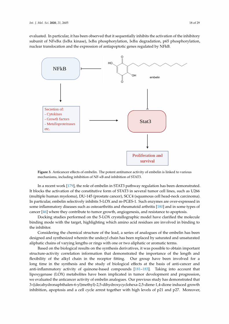
Embelin is a benzoquinone derivative able to interfere with the arachidonic acid metabolism,whose structure consists of a polar 2,5-dihydroxy-1,4-benzoquinone nucleus linked at position 3 toa long hydrophobic alkyl chain that gives solubility in the apolar phase and allows the moleculeto cross the cellular barrier. Thanks to a study carried out in collaboration with prof. Oliver Werz(University of Jena, Germany), it has been highlighted that this molecule can block the activity ofboth 5-LOX and mPGES-1 with IC50 values of 0.06 and 0.2 µM respectively [172]. The chinonicstructure of the membrane is crucial for the inhibitory activity. In fact, it has been shown that themolecule is able to inhibit the enzyme without suffering a hydroquinone reduction process. Dockingstudies demonstrated that embelin did not act as a chelant of iron, but it fits with its undecylatedchain in the hydrophobic channel in which normally the iron-catalyzed oxygenation reaction of theAA is carried out. The benzoquinone ring co-ordinates with three amino acids (Gln363, Gln557 andTyr181) by determining hydrogen bonds that stabilize the enzyme-molecule interaction. The bindingbetween the embelin and 5-LOX is mediated by two water molecules that co-crystallize with thecomplex and form hydrogen bonds with two amino acids such as Asn425 and Thr364. Recent studiesshowed that embelin and its derivatives had antioxidant, anti-inflammatory, antitumor and analgesicproperties [173]. In particular, they were able to induce apoptosis in human myeloid cells HL byinteracting with microtubule proteins and activate caspases in pancreatitis. It has been shown thatnaturally occurring embelin derivatives (5-O-methyl embelin and 5-O-ethyl embelin) [174] induced cellcycle arrest of HL-60 cells at G0/G1 stage in a dose- and time-dependent manner. The potent antitumoractivity of embelin is linked to various mechanisms, such as: (i) Inhibition of XIAP, (ii) Inhibition ofc-FLIP expression, (iii) Activation of PPARγ, (iv) Inhibition of NF-κB, (v) Inhibition of STAT3 (Figure 3).
Several studies have demonstrated that Embelin binds to XIAP protein and induces apoptosis.A preclinical study performed on PC-3 and LNCaP prostate cancer cells that expressed high levels ofXIAP, healthy human fibroblasts (WI-38) and normal prostate human epithelial cells (PrEC) used ascontrols confirmed the selectivity profile for cancer cells; in fact, IC50 values for PC3 and DU werelower than those of healthy cell lines. Further biological studies have demonstrated that embelincan induce apoptosis in prostate PC-3 and leukemia HL-60 cells by activating caspase 9 throughdown-regulation of XIAP protein [174,175].
In two recent studies, it has been shown that embelin is able to induce TRAIL-induced apoptosisboth in glioblastoma cells and pancreatic cancer cells if associated with FLIP antisense oligonucleotidesby down-regulating anti-apoptotic protein FLIP [176]. Various studies have shown that PPARγactivation inhibits cell growth and induces differentiation and apoptosis in colon cancer cells [177];embelin was able to reduce cell proliferation and induce apoptosis both in HCT116 and HT-29 coloncancer cells, by increasing PPARγ receptor expression. Moreover, it showed a chemopreventive effecton mice after the induction of colorectal cancer through PPARγ upregulation. In a work carriedout by Ahn and collaborators [178], the ability of the embelin to modulate NFκB pathway was
Int. J. Mol. Sci. 2020, 21, 2605 18 of 29
evaluated. In particular, it has been observed that it sequentially inhibits the activation of the inhibitorysubunit of NFκBα (IκBα kinase), IκBα phosphorylation, IκBα degradation, p65 phosphorylation,nuclear translocation and the expression of antiapoptotic genes regulated by NFkB.Int. J. Mol. Sci. 2020, 21, x FOR PEER REVIEW 18 of 30
Figure 3. Anticancer effects of embelin. The potent antitumor activity of embelin is linked to various mechanisms, including inhibition of NF-κB and inhibition of STAT3.
Several studies have demonstrated that Embelin binds to XIAP protein and induces apoptosis. A preclinical study performed on PC-3 and LNCaP prostate cancer cells that expressed high levels of XIAP, healthy human fibroblasts (WI-38) and normal prostate human epithelial cells (PrEC) used as controls confirmed the selectivity profile for cancer cells; in fact, IC50 values for PC3 and DU were lower than those of healthy cell lines. Further biological studies have demonstrated that embelin can induce apoptosis in prostate PC-3 and leukemia HL-60 cells by activating caspase 9 through down-regulation of XIAP protein [174,175].
In two recent studies, it has been shown that embelin is able to induce TRAIL-induced apoptosis both in glioblastoma cells and pancreatic cancer cells if associated with FLIP antisense oligonucleotides by down-regulating anti-apoptotic protein FLIP [176]. Various studies have shown that PPARγ activation inhibits cell growth and induces differentiation and apoptosis in colon cancer cells [177]; embelin was able to reduce cell proliferation and induce apoptosis both in HCT116 and HT-29 colon cancer cells, by increasing PPARγ receptor expression. Moreover, it showed a chemopreventive effect on mice after the induction of colorectal cancer through PPARγ upregulation. In a work carried out by Ahn and collaborators [178], the ability of the embelin to modulate NFκB pathway was evaluated. In particular, it has been observed that it sequentially inhibits the activation of the inhibitory subunit of NFκBα (IκBα kinase), IκBα phosphorylation, IκBα degradation, p65 phosphorylation, nuclear translocation and the expression of antiapoptotic genes regulated by NFkB.
In a recent work [179], the role of embelin in STAT3 pathway regulation has been demonstrated. It blocks the activation of the constitutive form of STAT3 in several tumor cell lines, such as U266 (multiple human myeloma), DU-145 (prostate cancer), SCC4 (squamous cell head-neck carcinoma). In particular, embelin selectively inhibits 5-LOX and m-PGES-1. Such enzymes are over-expressed in some inflammatory diseases such as osteoarthritis and rheumatoid arthritis [180] and in some types of cancer [46] where they contribute to tumor growth, angiogenesis, and resistance to apoptosis.
Docking studies performed on the 5-LOX crystallographic model have clarified the molecule binding mode with the target, highlighting which amino acid residues are involved in binding to the inhibitor.
Figure 3. Anticancer effects of embelin. The potent antitumor activity of embelin is linked to variousmechanisms, including inhibition of NF-κB and inhibition of STAT3.
In a recent work [179], the role of embelin in STAT3 pathway regulation has been demonstrated.It blocks the activation of the constitutive form of STAT3 in several tumor cell lines, such as U266(multiple human myeloma), DU-145 (prostate cancer), SCC4 (squamous cell head-neck carcinoma).In particular, embelin selectively inhibits 5-LOX and m-PGES-1. Such enzymes are over-expressed insome inflammatory diseases such as osteoarthritis and rheumatoid arthritis [180] and in some types ofcancer [46] where they contribute to tumor growth, angiogenesis, and resistance to apoptosis.
Docking studies performed on the 5-LOX crystallographic model have clarified the moleculebinding mode with the target, highlighting which amino acid residues are involved in binding tothe inhibitor.
Considering the chemical structure of the lead, a series of analogues of the embelin has beendesigned and synthesized wherein the undecyl chain has been replaced by saturated and unsaturatedaliphatic chains of varying lengths or rings with one or two aliphatic or aromatic terms.
Based on the biological results on the synthesis derivatives, it was possible to obtain importantstructure-activity correlation information that demonstrated the importance of the length andflexibility of the alkyl chain in the receptor fitting. Our group have been involved for along time in the synthesis and the study of biological effects at the basis of anti-cancer andanti-inflammatory activity of quinone-based compounds [181–183]. Taking into account thatlipoxygenase (LOX) metabolites have been implicated in tumor development and progression,we evaluated the anticancer activity of embelin analogues. Our previous study has demonstrated that3-((decahydronaphthalen-6-yl)methyl)-2,5-dihydroxycyclohexa-2,5-diene-1,4-dione induced growthinhibition, apoptosis and a cell cycle arrest together with high levels of p21 and p27. Moreover,
Int. J. Mol. Sci. 2020, 21, 2605 19 of 29
it induced a significant cleavage of caspases 8, 9, 3 and 7, disrupted the interaction cIAP2/XIAP bydegrading XIAP and inhibited NF-kB pathway.
Moreover we found that the orthoquinone EA-100C(4,5-dimethoxy-3-alkyl-1,2-benzoquinone)and its reduced form EA-100C red (3-tridecyl-4,5-dimethoxybenzene-1,2-diol), synthesized with a C13n-alkyl chain lacking hydroxyl groups, showed IC50 values of 10 and 60 nM, respectively, in cell-freeassays representing the most potent 5-LO inhibitors [184].
The anticancer activity of these novel 5-LO inhibitor derivatives have been demonstrated onglioblastoma (GBM) cells. EA-100C was able to induce apoptosis, cell cycle modulation, and autophagy;on the other hand, EA-100C red induced ER stress-mediated apoptosis associated to autophagy throughCHOP and Beclin1 up-regulation and activation of caspases 3, 9, JNK and NFkB pathway [185].
8.6. Other Therapeutic Agents Targeting Inflammation
Statins have been developed as HMG-CoA reductase inhibitors therefore, inhibiting cholesterolsynthesis. They have also shown to have anti-inflammatory and anti-cancer effects when usedboth as monotherapy or in combination with other anti-inflammatory or chemotherapeutic agents(5-fluorouracil, doxorubicin and cisplatin) [186–188].
Blocking chemokine receptors can represent a good therapeutic strategy for cancer. Severalchemokines and their receptors are overexpressed in cancer and strictly related to cancer progression andmetastases such as CCR7, CXCR4 and CXCR7. Preclinical studies showed that agents targeting CXCR4receptor including antibodies, siRNAs or antagonists were able to inhibit tumor growth and metastasis.In particular, they showed encouraging results in cancer of the head and neck and inhibited angiogenesisin colon cancer [189,190]. Other therapeutic agents targeting inflammatory microenvironment or hostimmune response include Toll-like receptor agonists or antagonists, antibodies (rituximab, zerumboneand CXCR4), or other agents directed against pro-inflammatory molecules or their receptors.
9. Conclusions
Inflammation is strictly related to cancer and plays a key role in tumor development andprogression. Targeting inflammation, either alone or in combination with the chemotherapeutic agents,is therefore essential for the prevention and treatment of cancers. Several studies have shown thatthe use of anti-inflammatory drugs is associated with decreased cancer incidence and recurrence.Many anti-inflammatory agents can be used as adjuvants for conventional therapies but additionalstudies are needed to better understand their potential as anticancer drugs. Early diagnosis andtreatment of chronic inflammation could be useful to decrease cancer development. Anti-inflammatoryagents that obtained FDA approval have a limited use in cancer therapy for the presence of off-targeteffects and toxicities. Therefore, combinations, changes in doses or regimens or development of newagents could allow to overcome these limitations and represent a new therapeutic strategy in cancer.It is also important to develop safe and effective agents that reduce the risk of cancer. For example,NSAIDs exhibit several side effects, so analogs with lesser adverse effects have been developed,whose anti-tumor effects are still to be determined. Cancer is a complex pathology and requires atargeted approach: knowledge of the biochemical pathways involved in cell proliferation has allowedthe design of new target-based drugs. It is important to consider that these new drugs should betested in preclinical and in vivo studies and then be transplanted into clinical use without losing theireffectiveness and safety. The idea of targeting the inflammatory tumor micro-environment is innovativebut a better understanding of the molecular events underlying the activity of the new synthesizedagents can be helpful in developing new effective therapeutic strategies.
Author Contributions: S.Z. conceived the design of the manuscript. S.Z. and A.M.C. were involved in drafting ofthe article. A.G., G.A.F., G.F.N. and M.B. contributed in formatting of the manuscript. M.B. revised the figures andthe tables. R.F. and M.C. revised the final version. All authors have read and agreed to the published version ofthe manuscript.
Funding: This research received no external funding.
Int. J. Mol. Sci. 2020, 21, 2605 20 of 29
Conflicts of Interest: The authors declare no conflict of interest.
Abbreviations
AA arachidonic acidCOX cyclooxygenasesFAP familial adenomatous polyposisH2O2 hydrogen peroxideJAK janus-like kinasesIκBα IκB inhibitor proteinIKK IκB kinaseIL interleukinsLT LeukotrienesLOX lipoxygenasesLPS lipopolysaccharidesMIF macrophage migration inhibitory factorMMP metalloproteinasesNO-ASA no-aspirinNSAIDs non-steroidal anti-inflammatory drugsPTC papillary thyroid carcinomaP-gp P-glycoproteinPG prostaglandinsPGHS prostaglandin H synthaseRE response elementSTAT3 Signal transducer and activator of transcription 3TAM tumor-associated macrophagesTGF-β transforming growth factor-βTNF-α tumor necrosis factor-αTXA2 thromboxane A2.
References
1. Balkwill, F.; Mantovani, A. Inflammation and cancer: Back to Virchow? Lancet 2001, 357, 539–545. [CrossRef]2. Philip, M.; Rowley, D.A.; Schreiber, H. Inflammation as a tumor promoter in cancer induction.
Semin. Cancer Biol. 2004, 14, 433–439. [CrossRef] [PubMed]3. Rayburn, E.R.; Ezell, S.J.; Zhang, R. Anti-inflammatory agents for cancer therapy. Mol. Cell Pharm. 2009, 1,
29–43. [CrossRef] [PubMed]4. Hoepelman, A. Inflammation. Basic Principles and Clinical Correlates, 3rd ed.; Gallin, J.I., Snyderman, R., Eds.;
Lippincott Williams & Wilkins Publishers: Philadelphia, PA, USA, 2001; Volume 58, p. 1335. ISBN 0397517599.[CrossRef]
5. Hanahan, D.; Weinberg, R.A. The hallmarks of cancer. Cell 2000, 100, 57–70. [CrossRef]6. Seril, D.N. Oxidative stress and ulcerative colitis-associated carcinogenesis: Studies in humans and animal
models. Carcinogenesis 2003, 24, 353–362. [CrossRef]7. Qiu, X.; Ma, J.; Wang, K.; Zhang, H. Chemopreventive effects of 5-aminosalicylic acid on inflammatory bowel
disease-associated colorectal cancer and dysplasia: A systematic review with meta-analysis. Oncotarget 2017,8, 1031–1045. [CrossRef]
8. Van Tong, H.; Brindley, P.J.; Meyer, C.G.; Velavan, T.P. Parasite infection, carcinogenesis and humanmalignancy. EBioMedicine 2017, 15, 12–23. [CrossRef]
9. Guo, Y.; Nie, Q.; MacLean, A.L.; Li, Y.; Lei, J.; Li, S. Multiscale modeling of inflammation-inducedtumorigenesis reveals competing oncogenic and oncoprotective roles for inflammation. Cancer Res. 2017, 77,6429–6441. [CrossRef]
10. Kawanishi, S.; Ohnishi, S.; Ma, N.; Hiraku, Y.; Murata, M. Crosstalk between DNA damage and inflammationin the multiple steps of carcinogenesis. Int. J. Mol. Sci. 2017, 18, 1808. [CrossRef]
Int. J. Mol. Sci. 2020, 21, 2605 21 of 29
11. Zhao, H.; Liu, J.; Song, L.; Liu, Z.; Han, G.; Yuan, D.; Wang, T.; Dun, Y.; Zhou, Z.; Liu, Z.; et al. Oleanolic acidrejuvenates testicular function through attenuating germ cell DNA damage and apoptosis via deactivationof NF-κB, p53 and p38 signalling pathways. J. Pharm. Pharmacol. 2016, 69, 295–304. [CrossRef]
12. Petrenko, O.; Moll, U.M. Macrophage migration inhibitory factor MIF interferes with the Rb-E2F pathway.Mol. Cell 2005, 17, 225–236. [CrossRef] [PubMed]
13. Riabov, V.; Gudima, A.; Wang, N.; Mickley, A.; Orekhov, A.; Kzhyshkowska, J. Role of tumor associatedmacrophages in tumor angiogenesis and lymphangiogenesis. Front. Physiol. 2014, 5, 75. [CrossRef] [PubMed]
14. Lin, E.Y.; Pollard, J.W. Role of infiltrated leucocytes in tumour growth and spread. Br. J. Cancer 2004, 90,2053–2058. [CrossRef]
15. Haqqani, A.S.; Sandhu, J.K.; Birnboim, H.C. Expression of interleukin-8 promotes neutrophil infiltration andgenetic instability in mutatect tumors. Neoplasia 2000, 2, 561–568. [CrossRef] [PubMed]
16. Bromwich, E.J.; McArdle, P.A.; Canna, K.; McMillan, D.C.; McNicol, A.M.; Brown, M.; Aitchison, M.The relationship between T-lymphocyte infiltration, stage, tumour grade and survival in patients undergoingcurative surgery for renal cell cancer. Br. J. Cancer 2003, 89, 1906–1908. [CrossRef] [PubMed]
17. Canna, K.; McArdle, P.A.; McMillan, D.C.; McNicol, A.M.; Smith, G.W.; McKee, R.F.; McArdle, C.S.The relationship between tumour T-lymphocyte infiltration, the systemic inflammatory response andsurvival in patients undergoing curative resection for colorectal cancer. Br. J. Cancer 2005, 92, 651–654.[CrossRef]
18. Kashfi, K. Anti-inflammatory agents as cancer therapeutics. In Advances in Pharmacology; Elsevier: Cambridge,MA, USA, 2009; pp. 31–89. [CrossRef]
19. Dranoff, G. Cytokines in cancer pathogenesis and cancer therapy. Nat. Rev. Cancer 2004, 4, 11–22. [CrossRef]20. Bando, H.; Toi, M. Tumor angiogenesis, macrophages, and cytokines. In Advances in Experimental Medicine
and Biology; Springer: Boston, MA, USA, 2000; pp. 267–284. [CrossRef]21. Balkwill, F. Tumour necrosis factor and cancer. Nat. Rev. Cancer 2009, 9, 361–371. [CrossRef]22. Landskron, G.; De la Fuente, M.; Thuwajit, P.; Thuwajit, C.; Hermoso, M.A. Chronic inflammation and
cytokines in the tumor microenvironment. J. Immunol. Res. 2014, 2014, 149185. [CrossRef]23. Micheau, O.; Tschopp, J. Induction of TNF receptor I-mediated apoptosis via two sequential signaling
complexes. Cell 2003, 114, 181–190. [CrossRef]24. Xu, J.; Ye, Y.; Zhang, H.; Szmitkowski, M.; Makinen, M.J.; Li, P.; Xia, D.; Yang, J.; Wu, Y.; Wu, H. Diagnostic and
prognostic value of serum interleukin-6 in colorectal cancer. Medicine 2016, 95, e2502. [CrossRef] [PubMed]25. Gao, S.P.; Mark, K.G.; Leslie, K.; Pao, W.; Motoi, N.; Gerald, W.L.; Travis, W.D.; Bornmann, W.; Veach, D.;
Clarkson, B.; et al. Mutations in the EGFR kinase domain mediate STAT3 activation via IL-6 production inhuman lung adenocarcinomas. J. Clin. Investig. 2007, 117, 3846–3856. [CrossRef] [PubMed]
26. Smyth, M.J.; Cretney, E.; Kershaw, M.H.; Hayakawa, Y. Cytokines in cancer immunity and immunotherapy.Immunol. Rev. 2004, 202, 275–293. [CrossRef] [PubMed]
27. Iyer, S.S.; Cheng, G. Role of interleukin 10 transcriptional regulation in inflammation and autoimmunedisease. Crit. Rev. Immunol. 2012, 32, 23–63. [CrossRef]
28. Ardestani, S.K.; Inserra, P.; Solkoff, D.; Watson, R.R. The role of cytokines and chemokines on tumorprogression: A review. Cancer Detect. Prev. 1999, 23, 215–225. [CrossRef]
29. Strieter, R.M. Chemokines: Not just leukocyte chemoattractants in the promotion of cancer. Nat. Immunol.2001, 2, 285–286. [CrossRef]
30. Viatour, P.; Merville, M.-P.; Bours, V.; Chariot, A. Phosphorylation of NF-κB and IκB proteins: Implications incancer and inflammation. Trends Biochem. Sci. 2005, 30, 43–52. [CrossRef]
31. Tergaonkar, V.; Bottero, V.; Ikawa, M.; Li, Q.; Verma, I.M. IkappaB kinase-independent IkappaBalphadegradation pathway: Functional NF-kappaB activity and implications for cancer therapy. Mol. Cell. Biol.2003, 23, 8070–8083. [CrossRef]
32. Guttridge, D.C.; Albanese, C.; Reuther, J.Y.; Pestell, R.G.; Baldwin, A.S., Jr. NF-kappaB controls cell growthand differentiation through transcriptional regulation of cyclin D1. Mol. Cell. Biol. 1999, 19, 5785–5799.[CrossRef]
33. Hussain, S.P.; Trivers, G.E.; Hofseth, L.J.; He, P.; Shaikh, I.; Mechanic, L.E.; Doja, S.; Jiang, W.; Subleski, J.;Shorts, L.; et al. Nitric oxide, a mediator of inflammation, suppresses tumorigenesis. Cancer Res. 2004, 64,6849–6853. [CrossRef]
Int. J. Mol. Sci. 2020, 21, 2605 22 of 29
34. Williams, C.S.; Mann, M.; DuBois, R.N. The role of cyclooxygenases in inflammation, cancer, and development.Oncogene 1999, 18, 7908–7916. [CrossRef] [PubMed]
35. Funk, C.D. Prostaglandins and leukotrienes: Advances in eicosanoid biology. Science 2001, 294, 1871–1875.[CrossRef] [PubMed]
36. Werz, O. 5-lipoxygenase: Cellular biology and molecular pharmacology. Curr. Drug Target. Inflamm. Allergy2002, 1, 23–44. [CrossRef] [PubMed]
37. Boado, R.J.; Pardridge, W.M.; Vinters, H.V.; Black, K.L. Differential expression of arachidonate 5-lipoxygenasetranscripts in human brain tumors: Evidence for the expression of a multitranscript family. Proc. Natl. Acad.Sci. USA 1992, 89, 9044–9048. [CrossRef] [PubMed]
38. Przylipiak, A.; Hafner, J.; Przylipiak, J.; Köhn, F.M.; Runnebaum, B.; Rabe, T. Influence of 5-lipoxygenase onin vitro growth of human mammary carcinoma cell line MCF-7. Gynecol. Obstet. Investig. 1998, 46, 61–64.[CrossRef] [PubMed]
39. Wächtershäuser, A.; Steinhilber, D.; Loitsch, S.M.; Stein, J. Expression of 5-lipoxygenase by human colorectalcarcinoma Caco-2 cells during butyrate-induced cell differentiation. Biochem. Biophys. Res. Commun. 2000,268, 778–783. [CrossRef] [PubMed]
40. Matsuyama, M.; Yoshimura, R.; Mitsuhashi, M.; Tsuchida, K.; Takemoto, Y.; Kawahito, Y.; Sano, H.;Nakatani, T. 5-Lipoxygenase inhibitors attenuate growth of human renal cell carcinoma and induce apoptosisthrough arachidonic acid pathway. Oncol. Rep. 2005, 14, 73–79.
41. Avis, I.M.; Jett, M.; Boyle, T.; Vos, M.D.; Moody, T.; Treston, A.M.; Martínez, A.; Mulshine, J.L. Growth controlof lung cancer by interruption of 5-lipoxygenase-mediated growth factor signaling. J. Clin. Investig. 1996, 97,806–813. [CrossRef]
42. Hoque, A. Increased 5-lipoxygenase expression and induction of apoptosis by its inhibitors in esophagealcancer: A potential target for prevention. Carcinogenesis 2005, 26, 785–791. [CrossRef]
43. Knab, L.M.; Grippo, P.J.; Bentrem, D.J. Involvement of eicosanoids in the pathogenesis of pancreatic cancer:The roles of cyclooxygenase-2 and 5-lipoxygenase. World J. Gastroenterol. 2014, 20, 10729–10739. [CrossRef]
44. Ghosh, J.; Myers, C.E. Central role of arachidonate 5-lipoxygenase in the regulation of cell growth andapoptosis in human prostate cancer cells. In Advances in Experimental Medicine and Biology; Springer: Boston,MA, USA, 1999; pp. 577–582. [CrossRef]
45. Kummer, N.T.; Nowicki, T.S.; Azzi, J.P.; Reyes, I.; Iacob, C.; Xie, S.; Swati, I.; Darzynkiewicz, Z.; Gotlinger, K.H.;Suslina, N.; et al. Arachidonate 5 lipoxygenase expression in papillary thyroid carcinoma promotes invasionvia MMP-9 induction. J. Cell. Biochem. 2012, 113, 1998–2008. [CrossRef] [PubMed]
46. Hara, S.; Kamei, D.; Sasaki, Y.; Tanemoto, A.; Nakatani, Y.; Murakami, M. Prostaglandin E synthases:Understanding their pathophysiological roles through mouse genetic models. Biochimie 2010, 92, 651–659.[CrossRef] [PubMed]
47. Hodge, D.R.; Hurt, E.M.; Farrar, W.L. The role of IL-6 and STAT3 in inflammation and cancer. Eur. J. Cancer2005, 41, 2502–2512. [CrossRef]
48. Dong, C.; Davis, R.J.; Flavell, R.A. MAP kinases in the immune response. Annu. Rev. Immunol. 2002, 20,55–72. [CrossRef] [PubMed]
49. Liu, J.; Dietz, T.; Carpenter, S.R.; Alberti, M.; Folke, C.; Moran, E.; Pell, A.N.; Deadman, P.; Kratz, T.;Lubchenco, J.; et al. Complexity of coupled human and natural systems. Science 2007, 317, 1513–1516.[CrossRef] [PubMed]
50. Arthur, J.S.; Ley, S.C. Mitogen-activated protein kinases in innate immunity. Nat. Rev. Immunol. 2013, 13,679–692. [CrossRef] [PubMed]
51. Owens, D.M.; Keyse, S.M. Differential regulation of MAP kinase signalling by dual-specificity proteinphosphatases. Oncogene 2007, 26, 3203–3213. [CrossRef]
52. Kyriakis, J.M.; Avruch, J. Mammalian MAPK signal transduction pathways activated by stress andinflammation: A 10-year update. Physiol. Rev. 2012, 92, 689–737. [CrossRef]
53. Kolch, W. Meaningful relationships: The regulation of the Ras/Raf/MEK/ERK pathway by protein interactions.Biochem. J. 2000, 351 Pt 2, 289–305. [CrossRef]
54. Chambard, J.C.; Lefloch, R.; Pouyssegur, J.; Lenormand, P. ERK implication in cell cycle regulation.Biochim. Biophys. Acta 2007, 1773, 1299–1310. [CrossRef]
55. Knight, T.; Irving, J.A. Ras/Raf/MEK/ERK pathway activation in childhood acute lymphoblastic leukemiaand its therapeutic targeting. Front. Oncol. 2014, 4, 160. [CrossRef] [PubMed]
Int. J. Mol. Sci. 2020, 21, 2605 23 of 29
56. Johnson, G.L.; Lapadat, R. Mitogen-activated protein kinase pathways mediated by ERK, JNK, and p38protein kinases. Science 2002, 298, 1911–1912. [CrossRef] [PubMed]
57. Yang, Y.; Kim, S.C.; Yu, T.; Yi, Y.S.; Rhee, M.H.; Sung, G.H.; Yoo, B.C.; Cho, J.Y. Functional roles of p38mitogen-activated protein kinase in macrophage-mediated inflammatory responses. Mediat. Inflamm. 2014,2014, 352371. [CrossRef]
58. Salojin, K.; Oravecz, T. Regulation of innate immunity by MAPK dual-specificity phosphatases: Knockoutmodels reveal new tricks of old genes. J. Leukoc. Biol. 2007, 81, 860–869. [CrossRef] [PubMed]
59. Soares-Silva, M.; Diniz, F.F.; Gomes, G.N.; Bahia, D. The mitogen-activated protein kinase (MAPK) pathway:Role in immune evasion by trypanosomatids. Front. Microbiol. 2016, 7, 183. [CrossRef] [PubMed]
60. Akinleye, A.; Avvaru, P.; Furqan, M.; Song, Y.; Liu, D. Phosphatidylinositol 3-kinase (PI3K) inhibitors ascancer therapeutics. J. Hematol. Oncol. 2013, 6, 88. [CrossRef]
61. Fruman, D.A.; Chiu, H.; Hopkins, B.D.; Bagrodia, S.; Cantley, L.C.; Abraham, R.T. The PI3K pathway inhuman disease. Cell 2017, 170, 605–635. [CrossRef]
62. Brown, K.K.; Toker, A. The phosphoinositide 3-kinase pathway and therapy resistance in cancer.F1000prime Rep. 2015, 7, 13. [CrossRef]
63. Ebrahimi, S.; Hosseini, M.; Shahidsales, S.; Maftouh, M.; Ferns, G.A.; Ghayour-Mobarhan, M.; Hassanian, S.M.;Avan, A. Targeting the Akt/PI3K signaling pathway as a potential therapeutic strategy for the treatment ofpancreatic cancer. Curr. Med. Chem. 2017, 24, 1321–1331. [CrossRef]
64. Sandoval, S.; Pigazzi, M.; Sakamoto, K.M. CREB: A key regulator of normal and neoplastic hematopoiesis.Adv. Hematol. 2009, 2009, 634292. [CrossRef]
65. Steven, A.; Seliger, B. Control of CREB expression in tumors: From molecular mechanisms and signaltransduction pathways to therapeutic target. Oncotarget 2016, 7, 35454–35465. [CrossRef] [PubMed]
66. Du, K.; Montminy, M. CREB is a regulatory target for the protein kinase Akt/PKB. J. Biol. Chem. 1998, 273,32377–32379. [CrossRef] [PubMed]
67. Johannessen, C.M.; Johnson, L.A.; Piccioni, F.; Townes, A.; Frederick, D.T.; Donahue, M.K.; Narayan, R.;Flaherty, K.T.; Wargo, J.A.; Root, D.E.; et al. A melanocyte lineage program confers resistance to MAP kinasepathway inhibition. Nature 2013, 504, 138–142. [CrossRef] [PubMed]
68. Phuong, N.T.; Lim, S.C.; Kim, Y.M.; Kang, K.W. Aromatase induction in tamoxifen-resistant breast cancer:Role of phosphoinositide 3-kinase-dependent CREB activation. Cancer Lett 2014, 351, 91–99. [CrossRef][PubMed]
69. Cheng, J.C.; Kinjo, K.; Judelson, D.R.; Chang, J.; Wu, W.S.; Schmid, I.; Shankar, D.B.; Kasahara, N.; Stripecke, R.;Bhatia, R.; et al. CREB is a critical regulator of normal hematopoiesis and leukemogenesis. Blood 2008, 111,1182–1192. [CrossRef] [PubMed]
70. Antonescu, C.R.; Nafa, K.; Segal, N.H.; Dal Cin, P.; Ladanyi, M. EWS-CREB1: A recurrent variant fusion inclear cell sarcoma—Association with gastrointestinal location and absence of melanocytic differentiation.Clin. Cancer Res. Off. J. Am. Assoc. Cancer Res. 2006, 12, 5356–5362. [CrossRef] [PubMed]
71. Xia, Y.; Zhan, C.; Feng, M.; Leblanc, M.; Ke, E.; Yeddula, N.; Verma, I.M. Targeting CREB pathway suppressessmall cell lung cancer. Mol. Cancer Res. MCR 2018, 16, 825–832. [CrossRef]
72. Tan, X.; Wang, S.; Yang, B.; Zhu, L.; Yin, B.; Chao, T.; Zhao, J.; Yuan, J.; Qiang, B.; Peng, X. The CREB-miR-9negative feedback minicircuitry coordinates the migration and proliferation of glioma cells. PLoS ONE 2012,7, e49570. [CrossRef]
73. Fan, C.F.; Mao, X.Y.; Wang, E.H. Elevated p-CREB-2 (ser 245) expression is potentially associated withcarcinogenesis and development of breast carcinoma. Mol. Med. Rep. 2012, 5, 357–362. [CrossRef]
74. Jean, D.; Bar-Eli, M. Regulation of tumor growth and metastasis of human melanoma by the CREBtranscription factor family. Mol. Cell. Biochem. 2000, 212, 19–28. [CrossRef]
75. Yamada, T.; Amann, J.M.; Fukuda, K.; Takeuchi, S.; Fujita, N.; Uehara, H.; Iwakiri, S.; Itoi, K.; Shilo, K.; Yano, S.;et al. Akt kinase-interacting protein 1 signals through CREB to drive diffuse malignant mesothelioma.Cancer Res. 2015, 75, 4188–4197. [CrossRef] [PubMed]
76. Logan, C.Y.; Nusse, R. The Wnt signaling pathway in development and disease. Annu. Rev. Cell Dev. Biol.2004, 20, 781–810. [CrossRef] [PubMed]
77. MacDonald, B.T.; Tamai, K.; He, X. Wnt/beta-catenin signaling: Components, mechanisms, and diseases.Dev. Cell 2009, 17, 9–26. [CrossRef] [PubMed]
Int. J. Mol. Sci. 2020, 21, 2605 24 of 29
78. He, X.; Semenov, M.; Tamai, K.; Zeng, X. LDL receptor-related proteins 5 and 6 in Wnt/beta-catenin signaling:Arrows point the way. Development 2004, 131, 1663–1677. [CrossRef]
79. Liu, L.J.; Xie, S.X.; Chen, Y.T.; Xue, J.L.; Zhang, C.J.; Zhu, F. Aberrant regulation of Wnt signaling inhepatocellular carcinoma. World J. Gastroenterol. 2016, 22, 7486–7499. [CrossRef]
80. Zhang, L.; Shay, J.W. Multiple roles of APC and its therapeutic implications in colorectal cancer. J. Natl.Cancer Inst. 2017, 109. [CrossRef]
81. Le, P.N.; Keysar, S.B.; Miller, B.; Eagles, J.R.; Chimed, T.S.; Reisinger, J.; Gomez, K.E.; Nieto, C.; Jackson, B.C.;Somerset, H.L.; et al. Wnt signaling dynamics in head and neck squamous cell cancer tumor-stromainteractions. Mol. Carcinog. 2019, 58, 398–410. [CrossRef]
82. Ma, B.; Hottiger, M.O. Crosstalk between Wnt/beta-catenin and NF-kappaB signaling pathway duringinflammation. Front. Immunol. 2016, 7, 378. [CrossRef]
83. Saito, H.; Yamada, Y.; Takaya, S.; Osaki, T.; Ikeguchi, M. Clinical relevance of the number ofinterleukin-17-producing CD 8+ T cells in patients with gastric cancer. Surg. Today 2015, 45, 1429–1435.[CrossRef]
84. Yang, J.; Weinberg, R.A. Epithelial-mesenchymal transition: At the crossroads of development and tumormetastasis. Dev. Cell 2008, 14, 818–829. [CrossRef]
85. Nieman, M.T.; Prudoff, R.S.; Johnson, K.R.; Wheelock, M.J. N-cadherin promotes motility in human breastcancer cells regardless of their E-cadherin expression. J. Cell Biol. 1999, 147, 631–644. [CrossRef] [PubMed]
86. Stark, L.A.; Din, F.V.N.; Zwacka, R.M.; Dunlop, M.G. Aspirin-induced activation of the NF-κB signalingpathway: A novel mechanism for aspirin-mediated apoptosis in colon cancer cells. FASEB J. 2001, 15,1273–1275. [CrossRef] [PubMed]
87. Gu, Q.; Wang, J.D.; Xia, H.H.X.; Lin, M.C.M.; He, H.; Zou, B.; Tu, S.P.; Yang, Y.; Liu, X.G.; Lam, S.K.;et al. Activation of the caspase-8/Bid and Bax pathways in aspirin-induced apoptosis in gastric cancer.Carcinogenesis 2005, 26, 541–546. [CrossRef] [PubMed]
88. Dikshit, P.; Chatterjee, M.; Goswami, A.; Mishra, A.; Jana, N.R. Aspirin induces apoptosis through theinhibition of proteasome function. J. Biol. Chem. 2006, 281, 29228–29235. [CrossRef] [PubMed]
89. De Groot, D.J.A.; De Vries, E.G.E.; Groen, H.J.M.; De Jong, S. Non-steroidal anti-inflammatory drugs topotentiate chemotherapy effects: From lab to clinic. Crit. Rev. Oncol. Hematol. 2007, 61, 52–69. [CrossRef][PubMed]
90. Harris, R.E.; Chlebowski, R.T.; Jackson, R.D.; Frid, D.J.; Ascenseo, J.L.; Anderson, G.; Loar, A.; Rodabough, R.J.;White, E.; McTiernan, A.; et al. Breast cancer and nonsteroidal anti-inflammatory drugs: Prospective resultsfrom the Women’s Health Initiative. Cancer Res. 2003, 63, 6096–6101.
91. Flossmann, E.; Rothwell, P.M. Effect of aspirin on long-term risk of colorectal cancer: Consistent evidencefrom randomised and observational studies. Lancet 2007, 369, 1603–1613. [CrossRef]
92. Thun, M.J.; Namboodiri, M.M.; Heath, C.W. Aspirin use and reduced risk of fatal colon cancer. N. Engl.J. Med. 1991, 325, 1593–1596. [CrossRef]
93. Corley, D.A.; Kerlikowske, K.; Verma, R.; Buffler, P. Protective association of aspirin/NSAIDs and esophagealcancer: A systematic review and meta-analysis. Gastroenterology 2003, 124, 47–56. [CrossRef]
94. Bardia, A.; Ebbert, J.O.; Vierkant, R.A.; Limburg, P.J.; Anderson, K.; Wang, A.H.; Olson, J.E.; Vachon, C.M.;Cerhan, J.R. Association of aspirin and nonaspirin nonsteroidal anti-inflammatory drugs with cancerincidence and mortality. JNCI J. Natl. Cancer Inst. 2007, 99, 881–889. [CrossRef]
95. Dandekar, D.S.; Lopez, M.; Carey, R.I.; Lokeshwar, B.L. Cyclooxygenase-2 inhibitor celecoxib augmentschemotherapeutic drug-induced apoptosis by enhancing activation of caspase-3 and -9 in prostate cancercells. Int. J. Cancer 2005, 115, 484–492. [CrossRef] [PubMed]
96. Maeng, H.J.; Song, J.-H.; Kim, G.-T.; Song, Y.-J.; Lee, K.; Kim, J.-Y.; Park, T.-S. Celecoxib-mediated activationof endoplasmic reticulum stress induces de novo ceramide biosynthesis and apoptosis in hepatoma HepG2cells mobilization. BMB Rep. 2017, 50, 144–149. [CrossRef] [PubMed]
97. Sakoguchi-Okada, N.; Takahashi-Yanaga, F.; Fukada, K.; Shiraishi, F.; Taba, Y.; Miwa, Y.; Morimoto, S.;Iida, M.; Sasaguri, T. Celecoxib inhibits the expression of survivin via the suppression of promoter activity inhuman colon cancer cells. Biochem. Pharmacol. 2007, 73, 1318–1329. [CrossRef] [PubMed]
98. Sabichi, A.L.; Lee, J.J.; Grossman, H.B.; Liu, S.; Richmond, E.; Czerniak, B.A.; De la Cerda, J.; Eagle, C.;Viner, J.L.; Palmer, J.L.; et al. A randomized controlled trial of celecoxib to prevent recurrence ofnonmuscle-invasive bladder cancer. Cancer Prev. Res. (Phila.) 2011, 4, 1580–1589. [CrossRef]
Int. J. Mol. Sci. 2020, 21, 2605 25 of 29
99. Fabi, A.; Metro, G.; Papaldo, P.; Mottolese, M.; Melucci, E.; Carlini, P.; Sperduti, I.; Russillo, M.; Gelibter, A.;Ferretti, G.; et al. Impact of celecoxib on capecitabine tolerability and activity in pretreated metastatic breastcancer: Results of a phase II study with biomarker evaluation. Cancer Chemother. Pharmacol. 2007, 62, 717–725.[CrossRef]
100. Herrera, F.G.; Chan, P.; Doll, C.; Milosevic, M.; Oza, A.; Syed, A.; Pintilie, M.; Levin, W.; Manchul, L.; Fyles, A.A prospective phase I–II trial of the cyclooxygenase-2 inhibitor celecoxib in patients with carcinoma of thecervix with biomarker assessment of the tumor microenvironment. Int. J. Radiat. Oncol. Biol. Phys. 2007, 67,97–103. [CrossRef]
101. North, G.L.T. Celecoxib as adjunctive therapy for treatment of colorectal cancer. Ann. Pharmacother. 2001, 35,1638–1643. [CrossRef]
102. Mao, J.T.; Roth, M.D.; Fishbein, M.C.; Aberle, D.R.; Zhang, Z.-F.; Rao, J.Y.; Tashkin, D.P.; Goodglick, L.;Holmes, E.C.; Cameron, R.B.; et al. Lung cancer chemoprevention with celecoxib in former smokers.Cancer Prev. Res. (Phila.) 2011, 4, 984–993. [CrossRef]
103. Ponthan, F.; Wickstrom, M.; Gleissman, H.; Fuskevag, O.M.; Segerstrom, L.; Sveinbjornsson, B.; Redfern, C.P.F.;Eksborg, S.; Kogner, P.; Johnsen, J.I. Celecoxib prevents neuroblastoma tumor development and potentiatesthe effect of chemotherapeutic drugs in vitro and in vivo. Clin. Cancer Res. 2007, 13, 1036–1044. [CrossRef]
104. Pruthi, R.S. Phase II trial of celecoxib in prostate-specific antigen recurrent prostate cancer after definitiveradiation therapy or radical prostatectomy. Clin. Cancer Res. 2006, 12, 2172–2177. [CrossRef]
105. Murray, M.Y.; Rushworth, S.A.; Zaitseva, L.; Bowles, K.M.; Macewan, D.J. Attenuation ofdexamethasone-induced cell death in multiple myeloma is mediated by miR-125b expression. Cell Cycle2013, 12, 2144–2153. [CrossRef] [PubMed]
106. Wang, H.; Wang, Y.; Rayburn, E.; Hill, D.; Rinehart, J.; Zhang, R. Dexamethasone as a chemosensitizer forbreast cancer chemotherapy: Potentiation of the antitumor activity of adriamycin, modulation of cytokineexpression, and pharmacokinetics. Int. J. Oncol. 2007, 30, 947–953. [CrossRef] [PubMed]
107. Yu, H.C.; Luo, Y.X.; Peng, H.; Kang, L.; Huang, M.J.; Wang, J.P. Avoiding perioperative dexamethasone mayimprove the outcome of patients with rectal cancer. Eur. J. Surg. Oncol. (EJSO) 2015, 41, 667–673. [CrossRef][PubMed]
108. Rajkumar, S.V.; Blood, E.; Vesole, D.; Fonseca, R.; Greipp, P.R. Phase III clinical trial of thalidomide plusdexamethasone compared with dexamethasone alone in newly diagnosed multiple myeloma: A clinical trialcoordinated by the eastern cooperative oncology group. J. Clin. Oncol. 2006, 24, 431–436. [CrossRef]
109. Greenspan, E.J.; Madigan, J.P.; Boardman, L.A.; Rosenberg, D.W. Ibuprofen inhibits activation of nuclear{beta}-catenin in human colon adenomas and induces the phosphorylation of GSK-3{beta}. Cancer Prev.Res. (Phila.) 2011, 4, 161–171. [CrossRef]
110. Saini, M.K.; Sanyal, S.N. Piroxicam and c-phycocyanin prevent colon carcinogenesis by inhibition of membranefluidity and canonical Wnt/β-catenin signaling while up-regulating ligand dependent transcription factorPPARγ. Biomed. Pharmacother. 2014, 68, 537–550. [CrossRef]
111. Earnest, D.L.; Hixson, L.J.; Alberts, D.S. Piroxicam and other cyclooxygenase inhibitors: Potential for cancerchemoprevention. J. Cell. Biochem. Suppl. 1992, 16I, 156–166. [CrossRef]
112. Mladenova, D.; Pangon, L.; Currey, N.; Ng, I.; Musgrove, E.A.; Grey, S.T.; Kohonen-Corish, M.R.J. Sulindacactivates NF-κB signaling in colon cancer cells. Cell Commun. Signal. 2013, 11, 73. [CrossRef]
113. Yin, T.; Wang, G.; Ye, T.; Wang, Y. Sulindac, a non-steroidal anti-inflammatory drug, mediates breast cancerinhibition as an immune modulator. Sci. Rep. 2016, 6, 19534. [CrossRef]
114. Waddell, W.R.; Loughry, R.W. Sulindac for polyposis of the colon. J. Surg. Oncol. 1983, 24, 83–87. [CrossRef]115. Kune, G.A.; Kune, S.; Watson, L.F. Colorectal cancer risk, chronic illnesses, operations and medications: Case
control results from the Melbourne Colorectal Cancer Study. Int. J. Epidemiol. 2007, 36, 951–957. [CrossRef][PubMed]
116. Wang, D.; Dubois, R.N. Prostaglandins and cancer. Gut 2006, 55, 115–122. [CrossRef] [PubMed]117. Zlotnik, A. Involvement of chemokine receptors in organ-specific metastasis. In Infection and Inflammation:
Impacts on Oncogenesis; KARGER: Basel, Switzerland, 2006; pp. 191–199. [CrossRef]118. Jana, N.R. NSAIDs and apoptosis. Cell. Mol. Life Sci. 2008, 65, 1295–1301. [CrossRef] [PubMed]119. Arun, B.; Goss, P. The role of COX-2 inhibition in breast cancer treatment and prevention. Semin. Oncol.
2004, 31, 22–29. [CrossRef]

Int. J. Mol. Sci. 2020, 21, 2605 26 of 29
120. Smalley, W.E.; DuBois, R.N. Colorectal cancer and nonsteroidal anti-inflammatory drugs. In Advances inPharmacology; Elsevier: Amsterdam, The Netherlands, 1997; pp. 1–20. [CrossRef]
121. Gridelli, C.; Gallo, C.; Ceribelli, A.; Gebbia, V.; Gamucci, T.; Ciardiello, F.; Carozza, F.; Favaretto, A.; Daniele, B.;Galetta, D.; et al. Factorial phase III randomised trial of rofecoxib and prolonged constant infusion ofgemcitabine in advanced non-small-cell lung cancer: The GEmcitabine-COxib in NSCLC (GECO) study.Lancet Oncol. 2007, 8, 500–512. [CrossRef]
122. Zhang, S.M.; Cook, N.R.; Manson, J.E.; Lee, I.M.; Buring, J.E. Low-dose aspirin and breast cancer risk: Resultsby tumour characteristics from a randomised trial. Br. J. Cancer 2008, 98, 989–991. [CrossRef]
123. Cruz–Correa, M.; Hylind, L.M.; Romans, K.E.; Booker, S.V.; Giardiello, F.M. Long-term treatment withsulindac in familial adenomatous polyposis: A prospective cohort study. Gastroenterology 2002, 122, 641–645.[CrossRef] [PubMed]
124. Gill, S.; Sinicrope, F.A. Colorectal cancer prevention: Is an ounce of prevention worth a pound of cure?Semin. Oncol. 2005, 32, 24–34. [CrossRef]
125. Calaluce, R.; Earnest, D.L.; Heddens, D.; Einspahr, J.G.; Roe, D.; Bogert, C.L.; Marshall, J.R.; Alberts, D.S. Effectsof piroxicam on prostaglandin E2 levels in rectal mucosa of adenomatous polyp patients: A randomizedphase IIb trial. Cancer Epidemiol. Biomark. Prev. Publ. Am. Assoc. Cancer Res. Cosponsored Am. Soc. Prev.Oncol. 2000, 9, 1287–1292.
126. Baron, J.A. Epidemiology of non-steroidal anti-inflammatory drugs and cancer. Prog. Exp. Tumor Res. 2003,37, 1–24. [CrossRef]
127. Zhao, X.; Xu, Z.; Li, H. NSAIDs use and reduced metastasis in cancer patients: Results from a meta-analysis.Sci. Rep. 2017, 7, 1875. [CrossRef] [PubMed]
128. Available online: www.clinicaltrials.gov (accessed on 6 April 2020).129. Witschi, H.; Espiritu, I.; Ly, M.; Uyeminami, D. The chemopreventive effects of orally administered
dexamethasone in strain A/J mice following cessation of smoke exposure. Inhal. Toxicol. 2005, 17, 119–122.[CrossRef]
130. Wang, H. Pretreatment with dexamethasone increases antitumor activity of carboplatin and gemcitabinein mice bearing human cancer xenografts: In vivo activity, pharmacokinetics, and clinical implications forcancer chemotherapy. Clin. Cancer Res. 2004, 10, 1633–1644. [CrossRef] [PubMed]
131. Yano, A.; Fujii, Y.; Iwai, A.; Kageyama, Y.; Kihara, K. 433: Glucocorticoids suppress tumor angiogenesis andin vivo growth of prostate cancer cells. J. Urol. 2006, 175, 141. [CrossRef]
132. Freshney, R.I.; Frame, M.C.; Vaughan, P.F.T.; Graham, D.I. Phenotypic modification of human glioma.In Biology of Brain Tumour; Springer: Boston, MA, USA, 1986; pp. 35–41. [CrossRef]
133. Albouy, B.; Tourani, J.-M.; Allain, P.; Rolland, F.; Staerman, F.; Eschwege, P.; Pfister, C. Preliminary results ofthe Prostacox phase II trial in hormonal refractory prostate cancer. BJU Int. 2007, 100, 770–774. [CrossRef]
134. Javle, M.M.; Cao, S.; Durrani, F.A.; Pendyala, L.; Lawrence, D.D.; Smith, P.F.; Creaven, P.J.; Noel, D.C.;Iyer, R.V.; Rustum, Y.M. Celecoxib and mucosal protection: Translation from an animal model to a phase Iclinical trial of celecoxib, irinotecan, and 5-fluorouracil. Clin. Cancer Res. 2007, 13, 965–971. [CrossRef]
135. Wang, H.; Li, M.; Rinehart, J.J.; Zhang, R. Dexamethasone as a chemoprotectant in cancer chemotherapy:Hematoprotective effects and altered pharmacokinetics and tissue distribution of carboplatin and gemcitabine.Cancer Chemother. Pharmacol. 2004, 53, 459–467. [CrossRef]
136. Karthaus, M.; Ballo, H.; Abenhardt, W.; Steinmetz, T.; Geer, T.; Schimke, J.; Braumann, D.; Behrens, R.;Behringer, D.; Kindler, M.; et al. Prospective, double-blind, placebo-controlled, multicenter, randomizedphase III study with orally administered budesonide for prevention of irinotecan (CPT-11)-induced diarrheain patients with advanced colorectal cancer. Oncology 2005, 68, 326–332. [CrossRef]
137. Candelario-Jalil, E.; Taheri, S.; Yang, Y.; Sood, R.; Grossetete, M.; Estrada, E.Y.; Fiebich, B.L.; Rosenberg, G.A.Cyclooxygenase inhibition limits blood-brain barrier disruption following intracerebral injection of tumornecrosis factor-α in the rat. J. Pharmacol. Exp. Ther. 2007, 323, 488–498. [CrossRef]
138. Karjalainen, M.J.; Neuvonen, P.J.; Backman, J.T. In vitro inhibition of CYP1A2 by model inhibitors,anti-inflammatory analgesics and female sex steroids: Predictability ofin vivoInteractions. Basic Clin.Pharmacol. Toxicol. 2008, 103, 157–165. [CrossRef]
139. Hersh, E.V.; Pinto, A.; Moore, P.A. Adverse drug interactions involving common prescription andover-the-counter analgesic agents. Clin. Ther. 2007, 29, 2477–2497. [CrossRef] [PubMed]
Int. J. Mol. Sci. 2020, 21, 2605 27 of 29
140. Wang, L.C.S.; Ching, L.-M.; Paxton, J.W.; Kestell, P.; Sutherland, R.; Zhuang, L.; Baguley, B.C. Enhancement ofthe action of the antivascular drug 5,6-dimethylxanthenone-4-acetic acid (DMXAA; ASA404) by non-steroidalanti-inflammatory drugs. Investig. New Drugs 2008, 27, 280–284. [CrossRef] [PubMed]
141. Heldin, C.-H.; Rubin, K.; Pietras, K.; Östman, A. High interstitial fluid pressure—An obstacle in cancertherapy. Nat. Rev. Cancer 2004, 4, 806–813. [CrossRef] [PubMed]
142. Kristjansen, P.E.; Boucher, Y.; Jain, R.K. Dexamethasone reduces the interstitial fluid pressure in a humancolon adenocarcinoma xenograft. Cancer Res. 1993, 53, 4764–4766. [PubMed]
143. Nakata, E.; Mason, K.A.; Hunter, N.; Husain, A.; Raju, U.; Liao, Z.; Ang, K.K.; Milas, L. Potentiation of tumorresponse to radiation or chemoradiation by selective cyclooxygenase-2 enzyme inhibitors. Int. J. Radiat.Oncol. Biol. Phys. 2004, 58, 369–375. [CrossRef] [PubMed]
144. Van Wijngaarden, J.; Van Beek, E.; Van Rossum, G.; Van der Bent, C.; Hoekman, K.; Van der Pluijm, G.;Van der Pol, M.A.; Broxterman, H.J.; Van Hinsbergh, V.W.M.; Löwik, C.W.G.M. Celecoxib enhancesdoxorubicin-induced cytotoxicity in MDA-MB231 cells by NF-κB-mediated increase of intracellulardoxorubicin accumulation. Eur. J. Cancer 2007, 43, 433–442. [CrossRef] [PubMed]
145. Castro-Caldas, M.; Mendes, A.F.; Duarte, C.B.; Lopes, M.C.F. Dexamethasone-induced and estradiol-inducedCREB activation and annexin 1 expression in CCRF-CEM lymphoblastic cells: Evidence for the involvementof cAMP and p38 MAPK. Mediat. Inflamm. 2003, 12, 329–337. [CrossRef]
146. Dittmann, K.H.; Mayer, C.; Ohneseit, P.A.; Raju, U.; Andratschke, N.H.; Milas, L.; Rodemann, H.P.Celecoxib induced tumor cell radiosensitization by inhibiting radiation induced nuclear EGFR transportand DNA-repair: A COX-2 independent mechanism. Int. J. Radiat. Oncol. Biol. Phys. 2008, 70, 203–212.[CrossRef]
147. Zrieki, A.; Farinotti, R.; Buyse, M. Cyclooxygenase inhibitors down regulate P-glycoprotein in humancolorectal Caco-2 cell line. Pharm. Res. 2008, 25, 1991–2001. [CrossRef]
148. Gradilone, A.; Pulcinelli, F.; Lotti, L.; Trifiro, E.; Martino, S.; Gandini, O.; Gianni, W.; Frati, L.; Agliano, A.;Gazzaniga, P. Celecoxib upregulates multidrug resistance proteins in colon cancer: Lack of synergy withstandard chemotherapy. Curr. Cancer Drug Targets 2008, 8, 414–420. [CrossRef]
149. Schönthal, A.H.; Chen, T.C.; Hofman, F.M.; Louie, S.G.; Petasis, N.A. Celecoxib analogs that lack COX-2inhibitory function: Preclinical development of novel anticancer drugs. Expert Opin. Investig. Drugs 2008, 17,197–208. [CrossRef] [PubMed]
150. Ding, H.; Han, C.; Guo, D.; Wang, D.; Duan, W.; Chen, C.-S.; D’Ambrosio, S.M. Sensitivity to the non-COXinhibiting celecoxib derivative, OSU03012, is p21(WAF1/CIP1) dependent. Int. J. Cancer 2008, 123, 2931–2938.[CrossRef] [PubMed]
151. Rigas, B.; Williams, J.L. NO-donating NSAIDs and cancer: An overview with a note on whether NO isrequired for their action. Nitric Oxide 2008, 19, 199–204. [CrossRef] [PubMed]
152. Kashfi, K.; Rayyan, Y.; Qiao, L.L.; Williams, J.L.; Chen, J.; Del Soldato, P.; Traganos, F.; Rigas, B. Nitricoxide-donating nonsteroidal anti-inflammatory drugs inhibit the growth of various cultured human cancercells: Evidence of a tissue type-independent effect. J. Pharmacol. Exp. Ther. 2002, 303, 1273–1282. [CrossRef]
153. Das, S.; Das, D. Anti-inflammatory responses of resveratrol. Inflamm. Allergy Drug Targets 2007, 6, 168–173.[CrossRef]
154. Anand, P.; Sundaram, C.; Jhurani, S.; Kunnumakkara, A.B.; Aggarwal, B.B. Curcumin and cancer: An “old-age”disease with an “age-old” solution. Cancer Lett. 2008, 267, 133–164. [CrossRef]
155. Hofseth, L.J.; Wargovich, M.J. Inflammation, cancer, and targets of ginseng. J. Nutr. 2007, 137, 183S–185S.[CrossRef]
156. Hassan, H.T. Ajoene (natural garlic compound): A new anti-leukaemia agent for AML therapy. Leuk. Res.2004, 28, 667–671. [CrossRef]
157. Howard, E.W.; Lee, D.T.; Chiu, Y.T.; Chua, C.W.; Wang, X.; Wong, Y.C. Evidence of a novel docetaxel sensitizer,garlic-derived S-allylmercaptocysteine, as a treatment option for hormone refractory prostate cancer. Int. J.Cancer 2008, 122, 1941–1948. [CrossRef]
158. Shpitz, B.; Giladi, N.; Sagiv, E.; Lev-Ari, S.; Liberman, E.; Kazanov, D.; Arber, N. Celecoxib and curcuminadditively inhibit the growth of colorectal cancer in a rat model. Digestion 2006, 74, 140–144. [CrossRef]
159. Ding, X.-Z.; Iversen, P.; Cluck, M.W.; Knezetic, J.A.; Adrian, T.E. Lipoxygenase inhibitors abolish proliferationof human pancreatic cancer cells. Biochem. Biophys. Res. Commun. 1999, 261, 218–223. [CrossRef] [PubMed]
Int. J. Mol. Sci. 2020, 21, 2605 28 of 29
160. Tong, W.-G.; Ding, X.-Z.; Adrian, T.E. The mechanisms of lipoxygenase inhibitor-induced apoptosis in humanbreast cancer cells. Biochem. Biophys. Res. Commun. 2002, 296, 942–948. [CrossRef]
161. Kennedy, T.J.; Chan, C.-Y.; Ding, X.-Z.; Adrian, T.E. Lipoxygenase inhibitors for the treatment of pancreaticcancer. Expert Rev. Anticancer Ther. 2003, 3, 525–536. [CrossRef] [PubMed]
162. Przylipiak, A.; Hafner, J.; Przylipiak, J.; Runnebaum, B.; Rabe, T.; Köhn, F.M. Influence of leukotrienes onin vitro growth of human mammary carcinoma cell line MCF-7. Eur. J. Obstet. Gynecol. Reprod. Biol. 1998, 77,61–65. [CrossRef]
163. Ghosh, J.; Myers, C.E. Arachidonic acid stimulates prostate cancer cell growth: Critical role of 5-lipoxygenase.Biochem. Biophys. Res. Commun. 1997, 235, 418–423. [CrossRef] [PubMed]
164. Chen, X.; Li, N.; Wang, S.; Wu, N.; Hong, J.; Jiao, X.; Krasna, M.J.; Beer, D.G.; Yang, C.S. Leukotriene A4hydrolase in rat and human esophageal adenocarcinomas and inhibitory effects of bestatin. JNCI J. Natl.Cancer Inst. 2003, 95, 1053–1061. [CrossRef]
165. Nakano, R.; Oka, M.; Nakamura, T.; Fukuda, M.; Kawabata, S.; Terashi, K.; Tsukamoto, K.; Noguchi, Y.;Soda, H.; Kohno, S. A leukotriene receptor antagonist, ONO-1078, modulates drug sensitivity and leukotrieneC4Efflux in lung cancer cells expressing multidrug resistance protein. Biochem. Biophys. Res. Commun. 1998,251, 307–312. [CrossRef]
166. Tong, W.-G.; Ding, X.-Z.; Talamonti, M.S.; Bell, R.H.; Adrian, T.E. Leukotriene B4 receptor antagonist LY293111induces S-phase cell cycle arrest and apoptosis in human pancreatic cancer cells. Anti Cancer Drugs 2007, 18,535–541. [CrossRef]
167. Wong, B.C.Y. 12-Lipoxygenase inhibition induced apoptosis in human gastric cancer cells. Carcinogenesis2001, 22, 1349–1354. [CrossRef]
168. Werz, O.; Steinhilber, D. Therapeutic options for 5-lipoxygenase inhibitors. Pharmacol. Ther. 2006, 112,701–718. [CrossRef]
169. Bokoch, G.M.; Reed, P.W. Evidence for inhibition of leukotriene A4 synthesis by 5,8,11,14-eicosatetraynoicacid in guinea pig polymorphonuclear leukocytes. J. Biol. Chem. 1981, 256, 4156–4159. [PubMed]
170. Koshihara, Y.; Neichi, T.; Murota, S.-I.; Lao, A.-N.; Fujimoto, Y.; Tatsuno, T. Selective inhibition of5-lipoxygenase by natural compounds isolated from Chinese plants, Artemisia rubripes Nakai. FEBS Lett.1983, 158, 41–44. [CrossRef]
171. Tomohiro, N.; Yasuko, K.; Sei-Itsu, M. Inhibitory effect of esculetin on 5-lipoxygenase and leukotrienebiosynthesis. Biochim. Biophys. Acta (BBA) Lipids Lipid Metab. 1983, 753, 130–132. [CrossRef]
172. Schaible, A.M.; Traber, H.; Temml, V.; Noha, S.M.; Filosa, R.; Peduto, A.; Weinigel, C.; Barz, D.; Schuster, D.;Werz, O. Potent inhibition of human 5-lipoxygenase and microsomal prostaglandin E2 synthase-1 by theanti-carcinogenic and anti-inflammatory agent embelin. Biochem. Pharmacol. 2013, 86, 476–486. [CrossRef]
173. Chitra, M.; Sukumar, E.; Suja, V.; Devi, S. Antitumor, anti-inflammatory and analgesic property of embelin,a plant product. Chemotherapy 1994, 40, 109–113. [CrossRef]
174. Hu, R.; Zhu, K.; Li, Y.; Yao, K.; Zhang, R.; Wang, H.; Yang, W.; Liu, Z. Embelin induces apoptosis throughdown-regulation of XIAP in human leukemia cells. Med. Oncol. 2010, 28, 1584–1588. [CrossRef]
175. Nikolovska-Coleska, Z.; Wang, R.; Fang, X.; Pan, H.; Tomita, Y.; Li, P.; Roller, P.P.; Krajewski, K.; Saito, N.G.;Stuckey, J.A.; et al. Development and optimization of a binding assay for the XIAP BIR3 domain usingfluorescence polarization. Anal. Biochem. 2004, 332, 261–273. [CrossRef]
176. Siegelin, M.D.; Gaiser, T.; Habel, A.; Siegelin, Y. Daidzein overcomes TRAIL-resistance in malignant gliomacells by modulating the expression of the intrinsic apoptotic inhibitor, bcl-2. Neurosci. Lett. 2009, 454, 223–228.[CrossRef]
177. Dai, Y.; Qiao, L.; Chan, K.W.; Yang, M.; Ye, J.; Ma, J.; Zou, B.; Gu, Q.; Wang, J.; Pang, R.; et al. Peroxisomeproliferator-activated receptor- contributes to the inhibitory effects of embelin on colon carcinogenesis.Cancer Res. 2009, 69, 4776–4783. [CrossRef]
178. Ahn, K.; Sethi, G.; Aggarwal, B. Nuclear factor-kappa B: From clone to clinic. Curr. Mol. Med. 2007, 7,619–637. [CrossRef]
179. Heo, J.Y.; Kim, H.J.; Kim, S.-M.; Park, K.-R.; Park, S.-Y.; Kim, S.W.; Nam, D.; Jang, H.-J.; Lee, S.-G.; Ahn, K.S.;et al. Embelin suppresses STAT3 signaling, proliferation, and survival of multiple myeloma via the proteintyrosine phosphatase PTEN. Cancer Lett. 2011, 308, 71–80. [CrossRef] [PubMed]
Int. J. Mol. Sci. 2020, 21, 2605 29 of 29
180. Kojima, F.; Kapoor, M.; Yang, L.; Fleishaker, E.L.; Ward, M.R.; Monrad, S.U.; Kottangada, P.C.; Pace, C.Q.;Clark, J.A.; Woodward, J.G.; et al. Defective generation of a humoral immune response is associated with areduced incidence and severity of collagen-induced arthritis in microsomal prostaglandin E synthase-1 nullmice. J. Immunol. 2008, 180, 8361–8368. [CrossRef] [PubMed]
181. Petronzi, C.; Filosa, R.; Peduto, A.; Monti, M.C.; Margarucci, L.; Massa, A.; Ercolino, S.F.; Bizzarro, V.;Parente, L.; Riccio, R.; et al. Structure-based design, synthesis and preliminary anti-inflammatory activity ofbolinaquinone analogues. Eur. J. Med. Chem. 2011, 46, 488–496. [CrossRef] [PubMed]
182. Filosa, R.; Peduto, A.; Aparoy, P.; Schaible, A.M.; Luderer, S.; Krauth, V.; Petronzi, C.; Massa, A.; De Rosa, M.;Reddanna, P.; et al. Discovery and biological evaluation of novel 1,4-benzoquinone and related resorcinolderivatives that inhibit 5-lipoxygenase. Eur. J. Med. Chem. 2013, 67, 269–279. [CrossRef] [PubMed]
183. Petronzi, C.; Festa, M.; Peduto, A.; Castellano, M.; Marinello, J.; Massa, A.; Capasso, A.; Capranico, G.; LaGatta, A.; De Rosa, M.; et al. Cyclohexa-2,5-diene-1,4-dione-based antiproliferative agents: Design, synthesis,and cytotoxic evaluation. J. Exp. Clin. Cancer Res. 2013, 32, 24. [CrossRef] [PubMed]
184. Peduto, A.; Scuotto, M.; Krauth, V.; Roviezzo, F.; Rossi, A.; Temml, V.; Esposito, V.; Stuppner, H.; Schuster, D.;D’Agostino, B.; et al. Optimization of benzoquinone and hydroquinone derivatives as potent inhibitors ofhuman 5-lipoxygenase. Eur. J. Med. Chem. 2017, 127, 715–726. [CrossRef]
185. Zappavigna, S.; Cossu, A.M.; Abate, M.; Misso, G.; Lombardi, A.; Caraglia, M.; Filosa, R. A hydroquinone-based derivative elicits apoptosis and autophagy via activating a ROS-dependent unfolded protein responsein human glioblastoma. Int. J. Mol. Sci. 2019, 20, 3836. [CrossRef]
186. Katz, M.S. Therapy insight: Potential of statins for cancer chemoprevention and therapy. Nat. Clin. Pract. Oncol.2005, 2, 82–89. [CrossRef]
187. Sassano, A.; Platanias, L.C. Statins in tumor suppression. Cancer Lett. 2008, 260, 11–19. [CrossRef]188. Xiao, H.; Yang, C.S. Combination regimen with statins and NSAIDs: A promising strategy for cancer
chemoprevention. Int. J. Cancer 2008, 123, 983–990. [CrossRef]189. Müller, A.; Homey, B.; Soto, H.; Ge, N.; Catron, D.; Buchanan, M.E.; McClanahan, T.; Murphy, E.; Yuan, W.;
Wagner, S.N.; et al. Involvement of chemokine receptors in breast cancer metastasis. Nature 2001, 410, 50–56.[CrossRef] [PubMed]
190. Scotton, C.; Milliken, D.; Wilson, J.; Raju, S.; Balkwill, F. Analysis of CC chemokine and chemokine receptorexpression in solid ovarian tumours. Br. J. Cancer 2001, 85, 891–897. [CrossRef] [PubMed]
© 2020 by the authors. Licensee MDPI, Basel, Switzerland. This article is an open accessarticle distributed under the terms and conditions of the Creative Commons Attribution(CC BY) license (http://creativecommons.org/licenses/by/4.0/).