Embed Size (px)
Citation preview
HYPEROXALURIA Prof. Dr. Bernd Hoppe,
Information Brochure for Patients with primary and secondary hyperoxaluria

Author: Prof. Dr. Bernd Hoppe Deutsches Hyperoxaluriezentrum (German Hyperoxaluria Center) [email protected] www.hyperoxalurie-zentrum.de
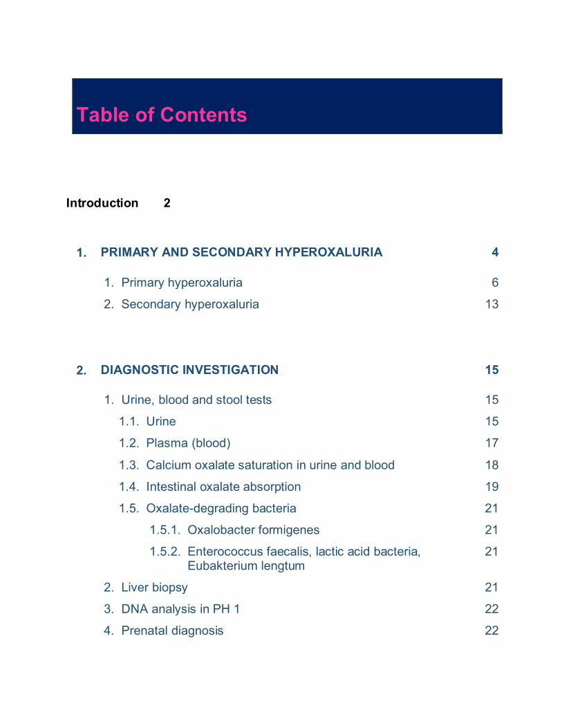
Table of Contents
Introduction 2
1. PRIMARY AND SECONDARY HYPEROXALURIA 4
1. Primary hyperoxaluria 6
2. Secondary hyperoxaluria 13
2. DIAGNOSTIC INVESTIGATION 15
1. Urine, blood and stool tests 15
1.1. Urine 15
1.2. Plasma (blood) 17
1.3. Calcium oxalate saturation in urine and blood 18
1.4. Intestinal oxalate absorption 19
1.5. Oxalate-degrading bacteria 21
1.5.1. Oxalobacter formigenes 21
1.5.2. Enterococcus faecalis, lactic acid bacteria, Eubakterium lengtum
21
2. Liver biopsy 21
3. DNA analysis in PH 1 22
4. Prenatal diagnosis 22
3. TREATMENT 24
1. Metaphylaxis 24
1.1. General 24
1.2. Pyridoxine = vitamin B6 26
1.3. Alkaline-citrate medication 27
1.4. Further inhibitors of calcium oxalate crystallization 28
1.5. Future therapies 28
2. Treatment of kidney stones passage 32
3. Dialysis 33
4. Transplantation 34
5. Conclusion 37
6. Self-help groups, centers 38
4. APPENDIX 39
5. GLOSSARY 40
1. PRIMARY AND SECONDARY HYPEROXALURIA
An elevated secretion of oxalic acid in urine is called hyperoxaluria. It is one of the main risk factors for a recurring urolithiasis (= kidney stone disease, Fig. 1a) or a progressing nephrocalcinosis (= calcification of the kidneys, that is deposit of calcium oxalate crystals in the renal tissue, Fig. 1b).
Hyperoxaluria = elevated risk of the formation of kidney stones
Oxalic acid is a final product of the metabolic processes, that is, it is usually no longer used in the human body for any further metabolic process, and thus needs to be mainly eliminated by the kidneys. With increased excretion, oxalic acid in urine is increasingly bound to calcium. This results in small calcium-oxalate (CaOx) crystals at first, which are actually excreted with a sufficient volume of urine without any problems. Ultimately, however, with a low volume of urine or with an excessively high oxalate excretion then result in the formation of bigger crystals as basis for kidney stones. Approximately 10-15% of the adult population have at least one kidney stone passage in their lives. Kidney stones are rarer in children and adolescents, but they may occur at any age, even in infancy, and the prevalence is increasing. Calcium-oxalate is the most common component of kidney stones (75%). However oftentimes patients do not specifically consult for an explanation of a kidney stone passage, but rather because of other dominant symptoms : ⬤ Blood (red, white blood cells) in urine ⬤ Pain while passing urine ⬤ In suspicion of urinary tract infection ⬤ Adverse ultrasound findings of kidneys upon explanation of abdominal pain
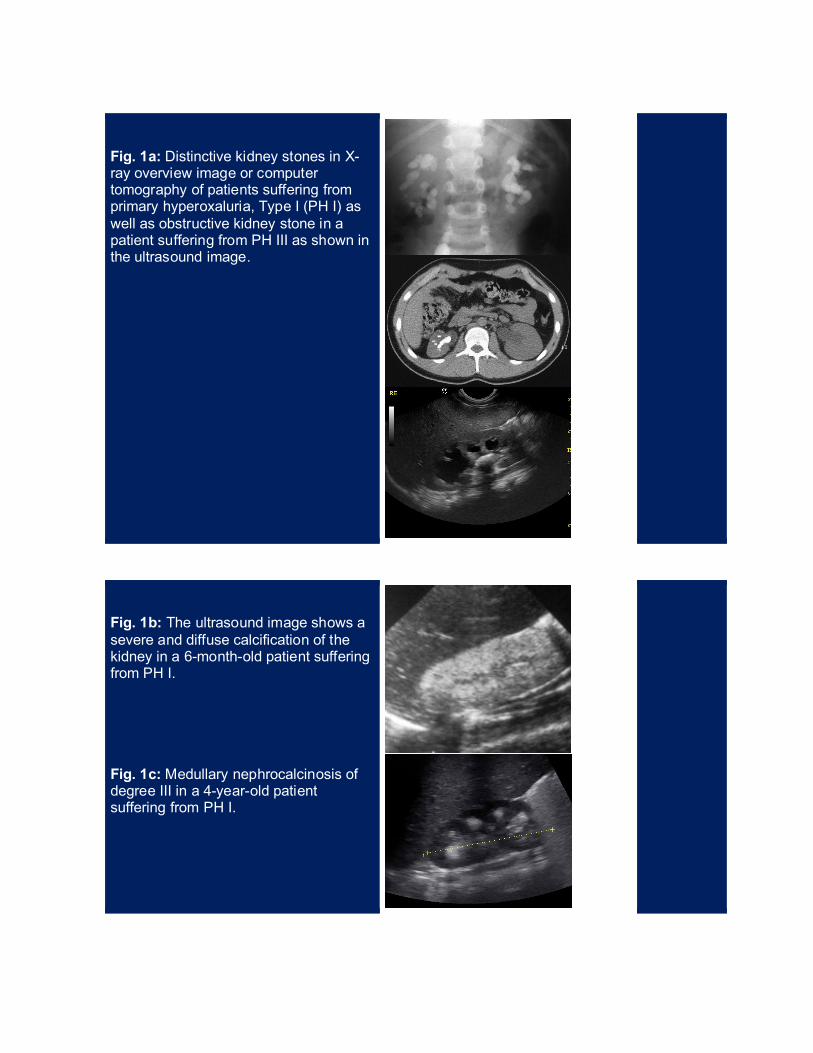
Fig. 1a: Distinctive kidney stones in X-ray overview image or computer tomography of patients suffering from primary hyperoxaluria, Type I (PH I) as well as obstructive kidney stone in a patient suffering from PH III as shown in the ultrasound image.
Fig. 1b: The ultrasound image shows a severe and diffuse calcification of the kidney in a 6-month-old patient suffering from PH I. Fig. 1c: Medullary nephrocalcinosis of degree III in a 4-year-old patient suffering from PH I.
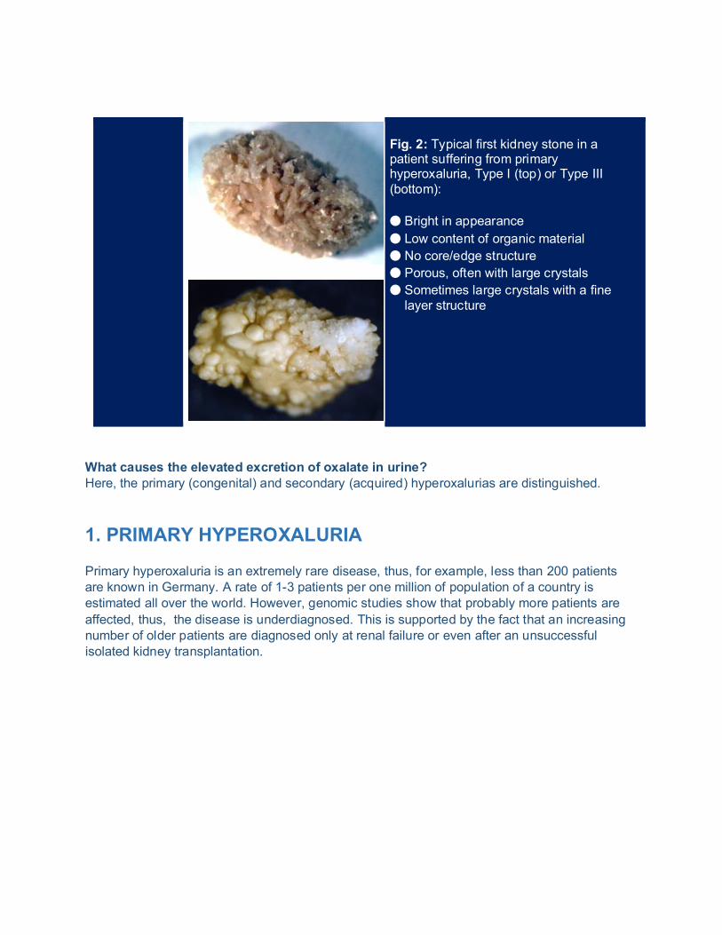
Fig. 2: Typical first kidney stone in a patient suffering from primary hyperoxaluria, Type I (top) or Type III (bottom): ⬤ Bright in appearance ⬤ Low content of organic material ⬤ No core/edge structure ⬤ Porous, often with large crystals ⬤ Sometimes large crystals with a fine
layer structure
What causes the elevated excretion of oxalate in urine? Here, the primary (congenital) and secondary (acquired) hyperoxalurias are distinguished. 1. PRIMARY HYPEROXALURIA Primary hyperoxaluria is an extremely rare disease, thus, for example, less than 200 patients are known in Germany. A rate of 1-3 patients per one million of population of a country is estimated all over the world. However, genomic studies show that probably more patients are affected, thus, the disease is underdiagnosed. This is supported by the fact that an increasing number of older patients are diagnosed only at renal failure or even after an unsuccessful isolated kidney transplantation.
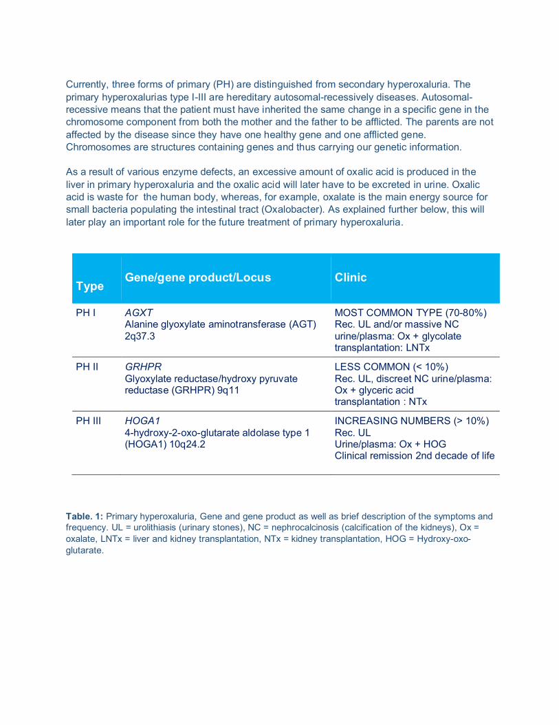
Currently, three forms of primary (PH) are distinguished from secondary hyperoxaluria. The primary hyperoxalurias type I-III are hereditary autosomal-recessively diseases. Autosomal-recessive means that the patient must have inherited the same change in a specific gene in the chromosome component from both the mother and the father to be afflicted. The parents are not affected by the disease since they have one healthy gene and one afflicted gene. Chromosomes are structures containing genes and thus carrying our genetic information. As a result of various enzyme defects, an excessive amount of oxalic acid is produced in the liver in primary hyperoxaluria and the oxalic acid will later have to be excreted in urine. Oxalic acid is waste for the human body, whereas, for example, oxalate is the main energy source for small bacteria populating the intestinal tract (Oxalobacter). As explained further below, this will later play an important role for the future treatment of primary hyperoxaluria.
Type
Gene/gene product/Locus
Clinic
PH I AGXT Alanine glyoxylate aminotransferase (AGT) 2q37.3
MOST COMMON TYPE (70-80%) Rec. UL and/or massive NC urine/plasma: Ox + glycolate transplantation: LNTx
PH II GRHPR Glyoxylate reductase/hydroxy pyruvate reductase (GRHPR) 9q11
LESS COMMON (< 10%) Rec. UL, discreet NC urine/plasma: Ox + glyceric acid transplantation : NTx
PH III HOGA1 4-hydroxy-2-oxo-glutarate aldolase type 1 (HOGA1) 10q24.2
INCREASING NUMBERS (> 10%) Rec. UL Urine/plasma: Ox + HOG Clinical remission 2nd decade of life
Table. 1: Primary hyperoxaluria, Gene and gene product as well as brief description of the symptoms and frequency. UL = urolithiasis (urinary stones), NC = nephrocalcinosis (calcification of the kidneys), Ox = oxalate, LNTx = liver and kidney transplantation, NTx = kidney transplantation, HOG = Hydroxy-oxo-glutarate.
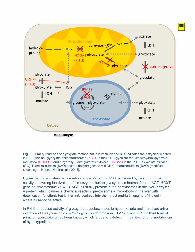
Fig. 3: Primary reactions of glyoxylate metabolism in human liver cells. X indicates the enzymeatic defect in PH I (alanine: glyoxylate aminotransferase (AGT), in the PH II (glycolate reductase/hydroxypyruvate reductase (GRHPR), and 4 hydroxy-2-oxo-glutarate aldolase (HOGA1) in the PH III. Glycolate oxidase (GO), D-amino-oxidase (DAO), lactate dehydrogenase A (LDHA), Diaminooxidase (DAO) [modified according to Hoppe, Nephrologist 2015]. Hyperoxaluria and elevated excretion of glycolic acid in PH I, is caused by lacking or missing activity or a wrong localization of the enzyme alanine glyoxylate aminotransferase (AGT, AGXT gene on chromosome 2q37.3). AGT is usually present in the peroxisomes in the liver (enzyme = protein, which causes a chemical reaction, peroxisome = micro-body in the liver with detoxication function), but is then mislocalized into the mitochondria (= engine of the cell), where it cannot be active. In PH II, a reduced activity of glyoxylate reductase leads to hyperoxaluria and increased urine excretion of L-Glyceric acid (GRHPR gene on chromosome 9p11). Since 2010, a third form of primary hyperoxaluria has been known, which is due to a defect in the mitochondrial metabolism of hydroxyproline.
glyoxylateglycine glyoxylateAGT
DAO
glyoxylate glyoxylate
pyruvate
HOGA1(PH 3)
Peroxisome
Mitochondrion
Cytosol
GRHPR
glycolate
glycolate
oxalateLDH
GRHPR (PH 2)
glycolate
LDH
oxalate
oxalate
LDH
GO
hydroxy-proline
HOG
HOGglyoxylate
oxalate
LDH(PH 1)
?
GRHPR(PH 3)
glycolate
Hepatocyte
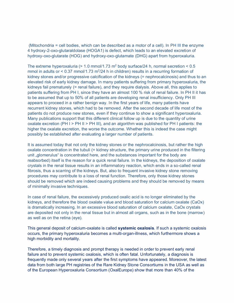
(Mitochondria = cell bodies, which can be described as a motor of a cell). In PH III the enzyme 4 hydroxy-2-oxo-glutaraldolase (HOGA1) is defect, which leads to an elevated excretion of hydroxy-oxo-glutarate (HOG) and hydroxy-oxo-glutamate (DHG) apart from hyperoxaluria. The extreme hyperoxaluria (> 1.0 mmol/1.73 m2 body surface/24 h, normal secretion < 0.5 mmol in adults or < 0.37 mmol/1.73 m2/24 h in children) results in a recurring formation of kidney stones and/or progressive calcification of the kidneys (= nephrocalcinosis) and thus to an elevated risk of early kidney damage. In many patients suffering from primary hyperoxaluria, the kidneys fail prematurely (= renal failure), and they require dialysis. Above all, this applies to patients suffering from PH I, since they have an almost 100 % risk of renal failure. In PH II it has to be assumed that up to 50% of all patients are developing renal insufficiency. Only PH III appears to proceed in a rather benign way. In the first years of life, many patients have recurrent kidney stones, which had to be removed. After the second decade of life most of the patients do not produce new stones, even if they continue to show a significant hyperoxaluria. Many publications support that this different clinical follow up is due to the quantity of urine oxalate excretion (PH I > PH II > PH III), and an algorithm was published for PH I patients: the higher the oxalate excretion, the worse the outcome. Whether this is indeed the case might possibly be established after evaluating a larger number of patients. It is assumed today that not only the kidney stones or the nephrocalcinosis, but rather the high oxalate concentration in the tubuli (= kidney structure, the primary urine produced in the filtering unit „glomerulus“ is concentrated here, and the substances important for the body are reabsorbed) itself is the reason for a quick renal failure. In the kidneys, the deposition of oxalate crystals in the renal tissue results in an inflammatory reaction, which ends in a so-called renal fibrosis, thus a scarring of the kidneys. But, also to frequent invasive kidney stone removing procedures may contribute to a loss of renal function. Therefore, only those kidney stones should be removed which are indeed causing problems and they should be removed by means of minimally invasive techniques. In case of renal failure, the excessively produced oxalic acid is no longer eliminated by the kidneys, and therefore the blood oxalate value and blood saturation for calcium oxalate (CaOx) is dramatically increasing. In an excessive blood saturation of calcium oxalate, CaOx crystals are deposited not only in the renal tissue but in almost all organs, such as in the bone (marrow) as well as on the retina (eye). This general deposit of calcium-oxalate is called systemic oxalosis. If such a systemic oxalosis occurs, the primary hyperoxaluria becomes a multi-organ-illness, which furthermore shows a high morbidity and mortality. Therefore, a timely diagnosis and prompt therapy is needed in order to prevent early renal failure and to prevent systemic oxalosis, which is often fatal. Unfortunately, a diagnosis is frequently made only several years after the first symptoms have appeared. Moreover, the latest data from both large PH registries of the Rare Kidney Stone Consortiums in the USA as well as of the European Hyperoxaluria Consortium (OxalEurope) show that more than 40% of the
sufferers are diagnosed only at renal failure and then on dialysis, or even only after an unsuccessful and isolated kidney transplantation.
Fig. 4: Systemic oxalosis = deposit of calcium-oxalate in all body parts, such as in the bone (here: shoulder joint as well as twice-refracting crystals in the bone marrow biopsy), the kidney (twice-refracting crystals in the renal tissue), such as calciphylaxia of the skin or maximal deposit in the dental root as well as on the retina and in the heart.
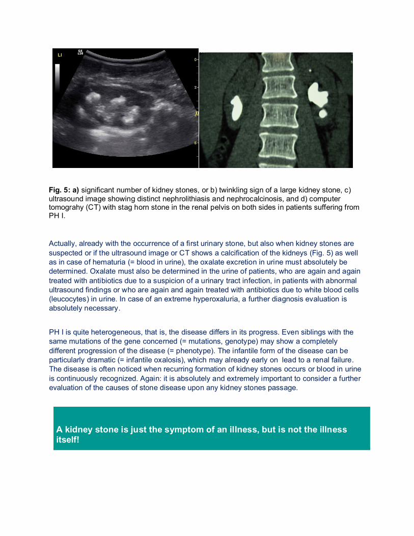
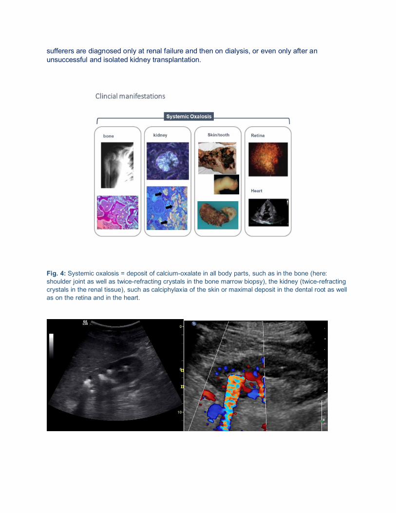
Fig. 5: a) significant number of kidney stones, or b) twinkling sign of a large kidney stone, c) ultrasound image showing distinct nephrolithiasis and nephrocalcinosis, and d) computer tomograhy (CT) with stag horn stone in the renal pelvis on both sides in patients suffering from PH I.
Actually, already with the occurrence of a first urinary stone, but also when kidney stones are suspected or if the ultrasound image or CT shows a calcification of the kidneys (Fig. 5) as well as in case of hematuria (= blood in urine), the oxalate excretion in urine must absolutely be determined. Oxalate must also be determined in the urine of patients, who are again and again treated with antibiotics due to a suspicion of a urinary tract infection, in patients with abnormal ultrasound findings or who are again and again treated with antibiotics due to white blood cells (leucocytes) in urine. In case of an extreme hyperoxaluria, a further diagnosis evaluation is absolutely necessary.
PH I is quite heterogeneous, that is, the disease differs in its progress. Even siblings with the same mutations of the gene concerned (= mutations, genotype) may show a completely different progression of the disease (= phenotype). The infantile form of the disease can be particularly dramatic (= infantile oxalosis), which may already early on lead to a renal failure. The disease is often noticed when recurring formation of kidney stones occurs or blood in urine is continuously recognized. Again: it is absolutely and extremely important to consider a further evaluation of the causes of stone disease upon any kidney stones passage.
A kidney stone is just the symptom of an illness, but is not the illness itself!
The removal of symptomatic kidney stones by the urologist is rather simple, but regardless, finding the cause is eventually more important to the patient than the assurance that the next stone can easily be removed as well. The possibility nowadays to simply remove the stones may also apply to the primary hyperoxaluria, but the kidney stone passages will eventually be increasingly frequent if the primary disease is not treated and thus become an agony. The kidneys will then be damaged both by the elevated excretion of oxalate in urine but also by the removal of stones. A further important mechanism of kidney damage is the permanent activation of an inflammation reaction (= inflammatory reaction) in the kidneys caused by the high oxalate concentration in the renal tubules and thus the absorption of oxalate into the renal tissue. If the disease is left untreated, it may quickly lead to renal failure. This especially applies to patients suffering from PH I and II. Simply a significant loss of fluid, such as from diarrhea, may lead to an obvious deterioration of renal function. This means for example, that patients suffering from PH should receive a permanent intravenous drip earlier than other patients in case of a fluid loss. When traveling abroad, the patient should have a certificate with him or her stating the diagnosis and which clearly describes how problems will be dealt with. But even early and adequate treatment does not mean that the patient is not on risk to develop renal failure. The problem with renal failure is that no form of renal replacement therapy (= dialysis) can remove adequate quantities of oxalate from the body, that increasingly more calcium oxalate crystals are deposited anywhere in the body and cause a multi-systemic disease called systemic oxalosis (= calcification in all possible parts of the body) (see Fig. 4). Consequences: a transplantation should be considered as early as possible. Above all, this should minimize the effect of systemic oxalosis. The longer the dialysis time prior to transplantation, the worse the course will be even after the transplantation (e.g. transplant renal failure when a calcification of the kidney is recurring in the transplant organ).
2. SECONDARY HYPEROXALURIA Less urine oxalate is excreted in the secondary hyperoxaluria but may reach values > 1.0 mmol/1.73 m2 body surface area/day and thus lead to a recurring formation of kidney stones or an increasing calcification of the kidneys. Secondary hyperoxaluria is caused by an increased intake of oxalate by the intestine (enteric, e.g. in case of chronic inflammatory bowel diseases) or by an excessive intake of oxalate from the food (dietary). It often occurs in chronic bowel diseases, especially frequently in patients suffering from Crohn’s disease or in patients after intestinal surgery (e.g. ileoceacal resection). Here, hyperoxaluria is explained by the bowel disease per se, as calcium is bound to fatty acids instead of oxalate, therefore there is a greater quantity of free oxalic acid which is then absorbed. A regular administration of antibiotics can also lead to a lack of oxalate-degrading intestinal bacteria (e.g. Oxalobacher formigenes), and thus may cause alterations of the intestinal flora with an increased absorption of oxalate. An oxalate absorption test by way of a stable isotope ([13C2] oxalate) as well as a stool test on oxalate-degrading bacteria may provide further insight into the nature of secondary hyperoxaluria. However, the repeated examination of 24-h urine samples
under different diets (ordinary food, food with a low oxalate content and oxalate-rich food), but with always the same and usual drinking quantity is even simpler. This allows to distinguish well and particularly simple between primary and secondary hyperoxaluria on an outpatient basis.
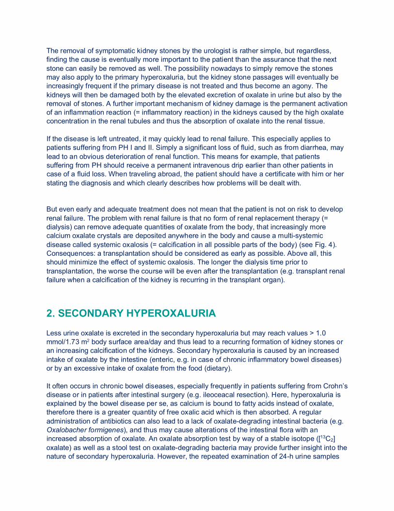
Fig. 6: Possible intake and elimination of oxalate in the intestinal tract. The unbound food oxalate can be well absorbed by the oxalate transporters into the blood circulation and then has to be eliminated by the kidneys. It can also degraded by oxalate-degrading bacteria in the intestinal tract, which can be basis for possible therapeutic options, such as the oral administration of bacteria with oxalate-degrading enzymes.
2. Diagnostic investigation
To start early with an adequate therapy, diagnosis of primary hyperoxaluria as timely as possibly is of utmost necessity. The diagnostic investigation particularly includes urine and blood tests:
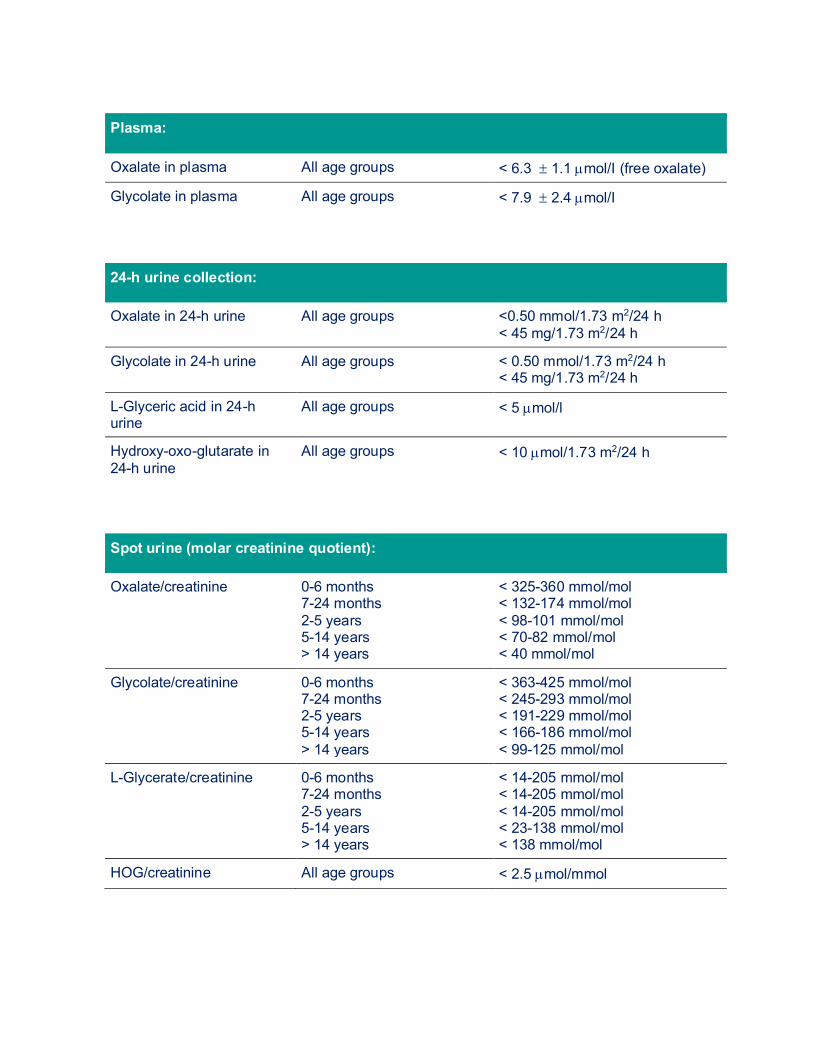
1. URINE, BLOOD AND STOOL TESTS 1.1. URINE The adequate diagnosis of a patient suffering from a primary hyperoxaluria requires the analysis of the urinary excretion of oxalic acid as well as glycolic acid in PH I, L-glyceric acid when PH II and hyperoxy-oxo-glutarate (HOG) or glutamate (DHG) when PH III is suspected. All this can be simultaneously determined routinely by means of ion chromatography/mass spectrometry or other methods. Oxalate can also be measured with an enzymatic method (oxalate oxidase, Sigma-Kit®). The urine or also plasma sample must have been conserved with hydrochloric acid prior to analysis. A demonstrable urine analysis will be possible only in this way—unfortunately however, the urine is often not preserved adequately and thus the urine results are not always usable. Spontaneous urine samples can be examined as well. In addition to the oxalate, the urine creatinine excretion is determined as well and a so-called oxalate/creatinine ratio is calculated and then compared to normal values according to age (see table 1). Premature infants but also infants born at term might show high normal and quite food-related values (the quotients are higher in formula than in breastfeeding). The analysis of at least two to three 24-h urine collections for PH related metabolites but also for other stone-forming and stone-preventing substances (under different diets, normal food, food with low oxalate content and oxalate-rich food) should follow subsequently. This is necessary in order to be able to distinguish primary from secondary hyperoxaluria, but also for follow up of oxalate excretion under therapy and to consider also other treatment options (low citrate excretion => citrate medication). If the oxalate and glycolate excretion is significantly higher than 0.5 mmol/24 h normalized to 1.73 m2 body surface (> 45 mg/24 h), there will usually be no doubt of the diagnosis of PH I especially with a typical clinical course. The same applies to PH II and PH III if high glyceric acid or HOG values apart from the elevated oxalate excretion are found. In approximately 25-30% of patients suffering from PH I, no elevated excretion of glycolate is found. Other sources additionally recommend the determination of the concentration of glycolate in the blood (plasma).
Plasma:
Oxalate in plasma All age groups < 6.3 ± 1.1 µmol/I (free oxalate)
Glycolate in plasma All age groups < 7.9 ± 2.4 µmol/I
24-h urine collection:
Oxalate in 24-h urine All age groups <0.50 mmol/1.73 m2/24 h < 45 mg/1.73 m2/24 h
Glycolate in 24-h urine All age groups < 0.50 mmol/1.73 m2/24 h < 45 mg/1.73 m2/24 h
L-Glyceric acid in 24-h urine
All age groups < 5 µmol/l
Hydroxy-oxo-glutarate in 24-h urine
All age groups < 10 µmol/1.73 m2/24 h
Spot urine (molar creatinine quotient):
Oxalate/creatinine 0-6 months 7-24 months 2-5 years 5-14 years > 14 years
< 325-360 mmol/mol < 132-174 mmol/mol < 98-101 mmol/mol < 70-82 mmol/mol < 40 mmol/mol
Glycolate/creatinine 0-6 months 7-24 months 2-5 years 5-14 years > 14 years
< 363-425 mmol/mol < 245-293 mmol/mol < 191-229 mmol/mol < 166-186 mmol/mol < 99-125 mmol/mol
L-Glycerate/creatinine 0-6 months 7-24 months 2-5 years 5-14 years > 14 years
< 14-205 mmol/mol < 14-205 mmol/mol < 14-205 mmol/mol < 23-138 mmol/mol < 138 mmol/mol
HOG/creatinine All age groups < 2.5 µmol/mmol
Tab. 1: Normal values for urine or plasma values. The urine parameters are expressed as excretion per 1.73 m2/24 h or molar creatinine ratios. Plasma values express the free oxalate or glycolate levels, total (free and protein bound) values are same for low levels, but increase more rapidly in renal failure. 1.2. PLASMA (BLOOD) Analysis of oxalate, glycolate, glyceric acid and HOG in blood (plasma) should always be performed for follow up, but especially in case of impaired renal function. In this instance, the sample preparation and preservation is very important, since new oxalate will be quickly generated, such as in the degradation of vitamin C, if the sample is incorrectly prepared. This results in incorrect measurements of high values. Hence, the blood sample taken must be cooled directly and then acidified, just like the urine sample, however, in a more complex procedure. Plasma oxalate can be measured by means of ion chromatography, gas chromatography or of an enzymatic method (oxalate oxidasis). The plasma glycolate, glyceric acid and HOG are principally determined by means of mass spectrometry. The normal values for plasma oxalate are between 1-6 µmol/l depending on the reference and laboratory method. In PH I, plasma oxalate values of > 10-20 µmol/l are measured in still good renal function. However, these values are already quickly increasing in the early stages of renal failure and soon reach a level which leads to an oversaturation of the blood for calcium oxalate (see below). Patients in the final stage of renal failure show plasma oxalate values of > 60-110 µmol/l (free oxalate), which gives evidence on how long and how often a renal replacement therapy (= dialysis) has to be performed. The total (free + protein bound oxalate) plasma oxalate values are mostly higher by 30%. In a patient suffering from renal failure requiring dialysis, the diagnosis of PH by means of urine analysis or by means of measuring plasma oxalate is not always safely usable. In all patients suffering from end stage renal failure, the plasma oxalate value is elevated. If the excretion of oxalate via the kidneys is significantly decreased in chronic but yet still compensated impairment of renal function, the urine examination can already no longer be classified as a valid parameter. That means that increasingly less oxalate is filtered by the kidneys, and the plasma oxalate value is hence increasing. In patients suffering from PH, the plasma oxalate value is disproportionately quickly elevated. Nevertheless, especially glycolate should be determined as well.
BRIEF DIAGNOSTIC ALGORITHM
Urine/plasma: oxalate (primary) ⬤ Glycolate ⬤ L-glyceric acid ⬤ Hydroxy-oxo-glutarate/glutamate
Urine/plasma: oxalate (secondary) ⬤ 13C2 oxalate absorption test ⬤ 3 x 24-h urine with different diet (normal, low oxalate, oxalate-rich)
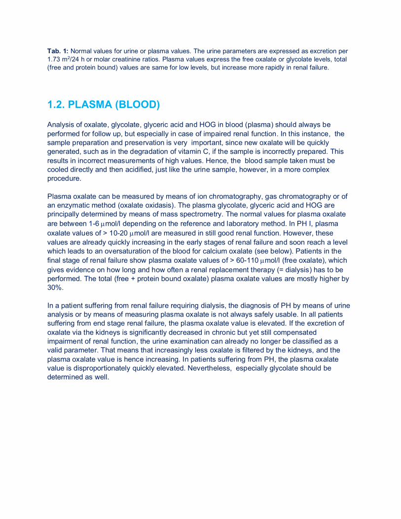
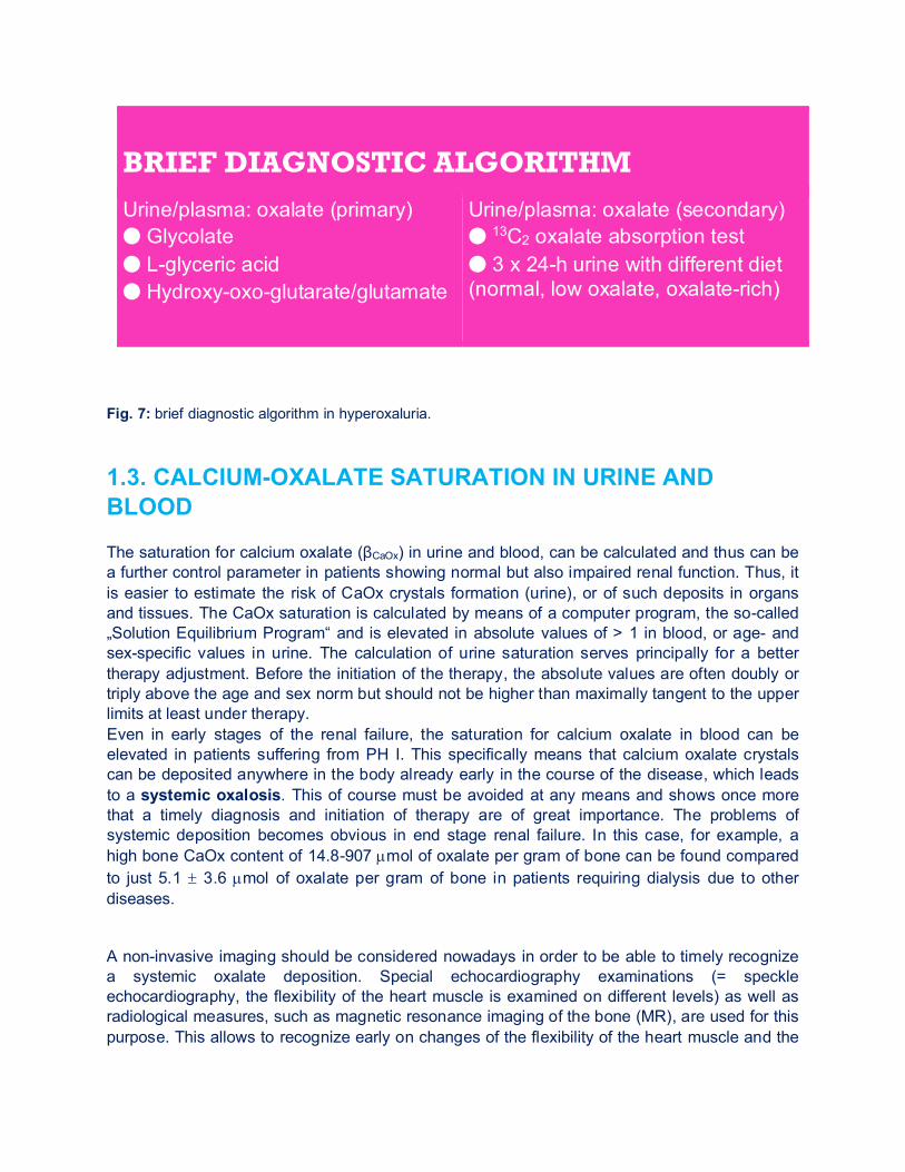
Fig. 7: brief diagnostic algorithm in hyperoxaluria. 1.3. CALCIUM-OXALATE SATURATION IN URINE AND BLOOD The saturation for calcium oxalate (βCaOx) in urine and blood, can be calculated and thus can be a further control parameter in patients showing normal but also impaired renal function. Thus, it is easier to estimate the risk of CaOx crystals formation (urine), or of such deposits in organs and tissues. The CaOx saturation is calculated by means of a computer program, the so-called „Solution Equilibrium Program“ and is elevated in absolute values of > 1 in blood, or age- and sex-specific values in urine. The calculation of urine saturation serves principally for a better therapy adjustment. Before the initiation of the therapy, the absolute values are often doubly or triply above the age and sex norm but should not be higher than maximally tangent to the upper limits at least under therapy. Even in early stages of the renal failure, the saturation for calcium oxalate in blood can be elevated in patients suffering from PH I. This specifically means that calcium oxalate crystals can be deposited anywhere in the body already early in the course of the disease, which leads to a systemic oxalosis. This of course must be avoided at any means and shows once more that a timely diagnosis and initiation of therapy are of great importance. The problems of systemic deposition becomes obvious in end stage renal failure. In this case, for example, a high bone CaOx content of 14.8-907 µmol of oxalate per gram of bone can be found compared to just 5.1 ± 3.6 µmol of oxalate per gram of bone in patients requiring dialysis due to other diseases. A non-invasive imaging should be considered nowadays in order to be able to timely recognize a systemic oxalate deposition. Special echocardiography examinations (= speckle echocardiography, the flexibility of the heart muscle is examined on different levels) as well as radiological measures, such as magnetic resonance imaging of the bone (MR), are used for this purpose. This allows to recognize early on changes of the flexibility of the heart muscle and the
trabecular bone structure. The latter changes can be recognized only later in conventional radiological examinations as well, which are mostly recorded in an X-ray picture of the hand. 1.4. INTESTINAL OXALATE ABSORPTION (OXALATE
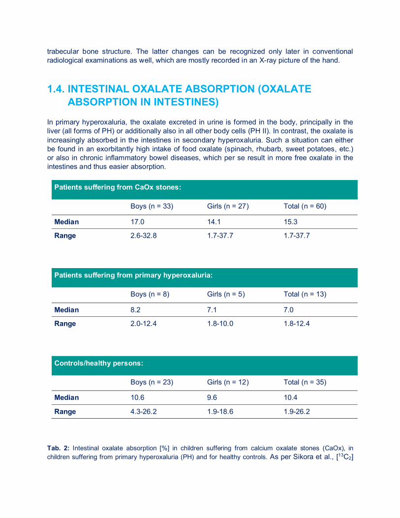
ABSORPTION IN INTESTINES) In primary hyperoxaluria, the oxalate excreted in urine is formed in the body, principally in the liver (all forms of PH) or additionally also in all other body cells (PH II). In contrast, the oxalate is increasingly absorbed in the intestines in secondary hyperoxaluria. Such a situation can either be found in an exorbitantly high intake of food oxalate (spinach, rhubarb, sweet potatoes, etc.) or also in chronic inflammatory bowel diseases, which per se result in more free oxalate in the intestines and thus easier absorption.
Patients suffering from CaOx stones:
Boys (n = 33) Girls (n = 27) Total (n = 60)
Median 17.0 14.1 15.3
Range 2.6-32.8 1.7-37.7 1.7-37.7
Patients suffering from primary hyperoxaluria:
Boys (n = 8) Girls (n = 5) Total (n = 13)
Median 8.2 7.1 7.0
Range 2.0-12.4 1.8-10.0 1.8-12.4
Controls/healthy persons:
Boys (n = 23) Girls (n = 12) Total (n = 35)
Median 10.6 9.6 10.4
Range 4.3-26.2 1.9-18.6 1.9-26.2 Tab. 2: Intestinal oxalate absorption [%] in children suffering from calcium oxalate stones (CaOx), in children suffering from primary hyperoxaluria (PH) and for healthy controls. As per Sikora et al., [13C2]
oxalate absorption in children with idiopathic, calcium oxalate urolithiasis or primary hyperoxaluria. Kidney Int. 2008 Mar 12. In order to better explain such a secondary hyperoxaluria, the [13C2] oxalate absorption test can be performed. This test is done on two consecutive days and mostly performed under inpatient conditions in children. A defined quantity of calcium and oxalate in food is given on both days and also a defined drinking quantity is adhered to. The 24-h collected urine on the first day is used for determining the baseline value. On the second day, the stable isotope of the oxalic acid is administered, which is precisely differing by exactly 2 mg in the mass from oxalate and thus can later also be well distinguished in the urine by way of mass spectrometry. Depending on the body weight, the patient is given between 25-50 mg of sodium oxalate, a 24-h urine is again collected and, as mentioned above, the percentage change of marked oxalic acid is measured apart from the oxalic acid normally occurring in urine. Table 2 shows the normal values and findings in individual patient groups. Due to the problematic performance especially in the pediatric patient (who has to be admitted to hospital), the „simple“ outpatient test with three consecutive 24-h urines collected under different oxalate feed (normal, low oxalate, high oxalate diet) is mostly used for children. Moreover, children are often unable to swallow the capsule with marked oxalate. 1.5. OXALATE-DEGRADING BACTERIA 1.5.1. OXALOBACTER FORMIGENES Oxalobacter formigenes (Oxf) is an obligate anaerobic, gram-negative bacterium, which is populating the intestinal tract in 70-80% of the population (Figure 6). It cannot only be found in humans, but also e.g. in ruminants, herbivores, in bird droppings, as well as in marine sediment. The normal colonialization rate is at 7.6 x 10⁶ to 2.3 x 10⁸ colony-forming units (KfE) per gram of stool. This allows for a degradation rate of 5-8 mmol/gram/h of oxalate in the intestine. In contrast to humans, Oxalobacter possesses two oxalate-degrading enzymes: Oxalyl-CoA decarboxylase and formyl-CoA transferase. This enables to degrade oxalate by Oxalobacter in the intestinal tract to CO2 and format, whereas the latter will further be metabolized or secreted later with stool. Both in patients suffering from primary but also from secondary hyperoxaluria, such as in patients suffering from cystic fibrosis (CF) or Crohn’s disease, Oxalobacter is only detected in a minority of patients. This would be one reason more to treat these patients with Oxalobacter. 1.5.2. ENTEROCOCCUS FAECALIS, LACTIC ACID BACTERIA,
EUBAKTERIUM LENTUM A more or less distinct oxalate-degrading ability is ascribed to all of these bacteria. Here, the enzymes known from Oxalobacter are playing a role. No adequately convincing studies on these bacteria species are either not known or have led to disappointing outcomes.
2. LIVER BIOPSY The diagnostic confirmation by means of liver biopsy has been paramount earlier in addition to urine and plasma analysis. Primarily a molecular genetic analysis is nowadays performed. In liver biopsy, the activity of AGT (PH I), GRHPR (PH II) or HOGA (PH III) in the liver cell is determined. The AGT activity can be determined according to various methods. Not more than 2 mg of tissue are respectively needed for an adequate determination. Even in patients suffering from chronic renal insufficiency, a PH can therefore be safely diagnosed. Due to the many different characteristics of primary hyperoxaluria Type I, it was not always possible to distinguish by means of the AGT activity between carriers of diseases and true patients. Especially in an only marginally lower AGT activity, the distribution of the enzyme in the individual components of the liver cell (peroxisom/mitochondrion) has also to be analyzed. This was important since the AGT activity can be significantly decreased in other liver diseases as well, such as in a liver cirrhosis. The immunoreactive AGT protein was eventually also be determined to the last specification of the diagnosis by means of western blot. 3. DNA ANALYSIS IN PH I The AGXT gene has been cloned and completely been analyzed. More than 200 mutations have been identified until now. The diagnosis can now be build up to the known mutation by means of so-called Exon-specific polymerase chain reaction (PCR) of genomic DNA, which has been obtained from e.g. white blood cells. In the meantime, this enables a molecular genetic analysis, this includes a complete analysis of the entire gene apart from the search of the three most frequently occurring mutations (c. 508A>C [earlier G630A], 33_34insC and 731T>C). This enables a safe diagnosis, which means, that a liver biopsy, e.g. prior to combined liver/kidney transplantation, will no longer be necessary. This method of course also applies to the other two PH genes, GRHPR (PH II) and HOGA1 (PH III). 4. PRENATAL DIAGNOSIS A prenatal (= before birth) diagnosis, when an index case is known within a family and with a serious infantile oxalosis will especially be important. A determination of oxalic acid in the amniotic fluid represents no adequate parameter for prenatal diagnosis. It is possible to measure all substances, which are also important in urine for diagnosis, but the maternal AGT for the metabolism of oxalic acid in the child plays an important role and thus wrongly low oxalate values in the amniotic fluid are measured. Since AGT (PH I) can be found only in liver cells and not e.g. in other tissue cells or white blood cells or leucocytes, a new liver biopsy would need to be performed to measure the AGT activity of the child. This signifies a significant risk for mother and child. A significant AGT activity can
also be determined only after the 14th week of pregnancy, whereas AGT could immunoelectronically be verified starting from the 9th week of pregnancy. The values remain clearly below the previously cited normal values in the progressing pregnancy, since the peroxisomes of the liver cells are still rather small. A liver biopsy of children inside the mother is limited due to „technical problems“. Therefore, it is only possibly as of the 16th week of pregnancy to obtain sufficient material in a low-risk manner. Even if the AGT activity after liver biopsy in children has successfully been determined and an immuno-electron microscopic diagnosis has been performed several times and also been published, such a procedure can lead to unwanted complications (e.g. a miscarriage) in the further course and thus should be an obsolete measure. A timely diagnosis is possible through a DNA analysis after chorionic villus sampling (extraction of membrane cavity cells), especially when known family members have previously been described. After a prenatal diagnosis, the genetic counselling of the parents based thereon must absolutely consider the heterogeneity of the disease progression. Even in identical mutations of the AGXT gene in siblings, completely different characteristic symptoms of the disease may occur. The follow up of siblings with an identical mutation are mentioned here as an example, where in the index case the diagnosis of PH I has been made based on typically higher excretion parameters as well as by a liver biopsy (before genetic testing was possible). Severe nephrocalcinosis (white kidneys) and a progressive impairment of the renal function was found. The patient’s sister remained clinically unremarkable with only slightly elevated oxalic acid excretion. However, both patients are genotypically identical, that is homozygous (= one mutation on both chromosomes each) both for the c. 508A>C mutation and for the C154T polymorphisms. The genetic counselling of numerous families may be rather difficult based on these findings since a precise prediction of the progression of the disease is certainly not possible. Therefore, the question arises whether a prenatal examination still makes sense, if no secure statement is possible on the progression of the disease after birth. Moreover, it should be noted that the disease is soon significantly better treatable. A case of PH within a family all other family members have to be examined, too. This does not only apply to the siblings, but also to the parents and grandparents. Sometimes surprising results are obtained, as in the case reported above, where a vertical (pseudo-dominant) inheritance was detected, so that the parent generation is also affected. The principle applies to all persons concerned, the earlier the diagnosis has been made, the better is the chance by an adequate treatment to prevent the fast deterioration of the disease.
3. Treatment
1. METAPHYLAXIS 1.1. GENERAL A daily fluid intake of > 2-3 L/m2 body surface area per day is a first important parameter to improve the solubility of calcium oxalate in urine through thinning of urine. Patients must be remembered frequently even for such a simple, but effective measure, since the majority of patient is not used to drink such a quantity of liquid throughout the day.
DRINKING A LOT IS IMPORTANT! There is no true success in any conservative therapeutic measure, if no adequate, regular fluid intake is ensured!
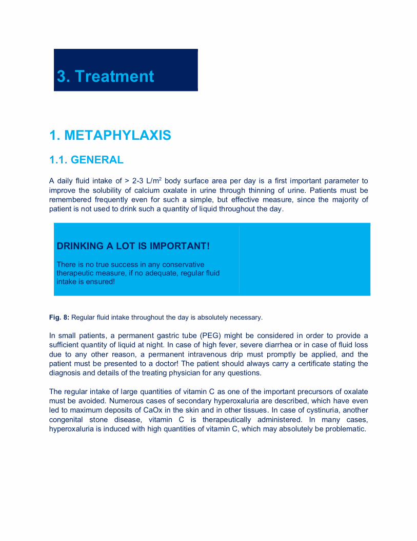
Fig. 8: Regular fluid intake throughout the day is absolutely necessary. In small patients, a permanent gastric tube (PEG) might be considered in order to provide a sufficient quantity of liquid at night. In case of high fever, severe diarrhea or in case of fluid loss due to any other reason, a permanent intravenous drip must promptly be applied, and the patient must be presented to a doctor! The patient should always carry a certificate stating the diagnosis and details of the treating physician for any questions. The regular intake of large quantities of vitamin C as one of the important precursors of oxalate must be avoided. Numerous cases of secondary hyperoxaluria are described, which have even led to maximum deposits of CaOx in the skin and in other tissues. In case of cystinuria, another congenital stone disease, vitamin C is therapeutically administered. In many cases, hyperoxaluria is induced with high quantities of vitamin C, which may absolutely be problematic.
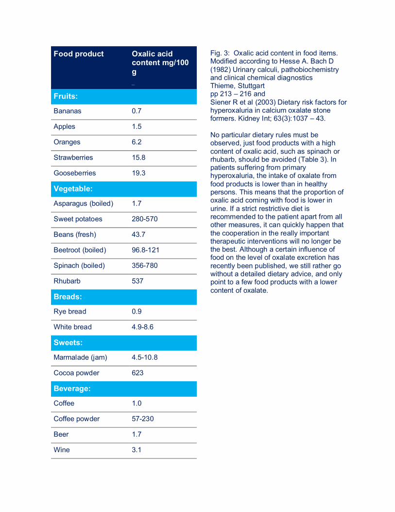
Food product Oxalic acid content mg/100 g M g/ 100 g
Fig. 3: Oxalic acid content in food items. Modified according to Hesse A. Bach D (1982) Urinary calculi, pathobiochemistry and clinical chemical diagnostics Thieme, Stuttgart pp 213 – 216 and Siener R et al (2003) Dietary risk factors for hyperoxaluria in calcium oxalate stone formers. Kidney Int; 63(3):1037 – 43. No particular dietary rules must be observed, just food products with a high content of oxalic acid, such as spinach or rhubarb, should be avoided (Table 3). In patients suffering from primary hyperoxaluria, the intake of oxalate from food products is lower than in healthy persons. This means that the proportion of oxalic acid coming with food is lower in urine. If a strict restrictive diet is recommended to the patient apart from all other measures, it can quickly happen that the cooperation in the really important therapeutic interventions will no longer be the best. Although a certain influence of food on the level of oxalate excretion has recently been published, we still rather go without a detailed dietary advice, and only point to a few food products with a lower content of oxalate.
Fruits:
Bananas 0.7
Apples 1.5
Oranges 6.2
Strawberries 15.8
Gooseberries 19.3
Vegetable:
Asparagus (boiled) 1.7
Sweet potatoes 280-570
Beans (fresh) 43.7
Beetroot (boiled) 96.8-121
Spinach (boiled) 356-780
Rhubarb 537
Breads:
Rye bread 0.9
White bread 4.9-8.6
Sweets:
Marmalade (jam) 4.5-10.8
Cocoa powder 623
Beverage:
Coffee 1.0
Coffee powder 57-230
Beer 1.7
Wine 3.1
Tea (2 min.) 7.0-10.8
Tealeaves 375-1450 Restricting the salt intake, can reduce the oxalate intake. The same is accomplished through a moderately increased intake of calcium, since calcium binds oxalic acid in the intestines, whereas these CaOx complexes will eventually be secreted with the stool and not absorbed into the blood circulation. And yet, no excessive calcium intake should be recommended, since oxalate is principally produced in the liver in primary hyperoxaluria and not increasingly absorbed in the intestines. A higher calcium intake may lead way to a higher calcium excretion and thus to even higher urinary saturation for calcium-oxalate. The drug therapy of primary hyperoxaluria is based on several cornerstones, which are linked depending on the treating center: 1.2. PYRIDOXINE = VITAMIN B6 Alanine:glyxolate aminotransferase (PH I) requires vitamin B6 as co-enzyme. In some patients, the daily administration of vitamin B6 (above all in patients where the AGT is in the wrong component of liver cell) leads to a reduction, sometimes even to a complete normalization of the excretion of oxalic acid. Since even just a quite low reduction of oxalate excretion represents a significant improvement, that is, a decrease of the relative saturation in urine for calcium oxalate, a therapeutic attempt in each patient with gradually increase of B6 dosages from 5-20 mg/kg of body weight per day should be started. In some patients, even just a small amount (20 mg) suffices to reach an effect on the oxalate production and thus excretion, while in others, a maximum dosage must be sought. A therapeutic attempt includes an initial dose of 5 mg/kg body weight/d in two individual doses, followed by a urine analysis approximately 3-4 weeks after initiating the medication. This enables to prove an eventual therapeutic success by measuring the excretion of oxalate, while the dosage can be adapted in steps of 5 mg/kg body weight/d if the decrease in oxalate excretion is not satisfactory. If no decrease of oxalate excretion has even been reached after 6-12 months, the therapy should be terminated in order to improve the patient’s cooperation with regard to further treatment measures. Known side effects of the high-dose B6 therapy are paresthesia (= prickling) in hands and feet, and a distinct touch sensitivity. An increased restlessness in children was reported as well. Serum vitamin B6 levels, which should be clearly above the normal range, can be determined for therapeutic control! 1.3. ALKALINE CITRATE MEDICATION The goal of the therapy with alkali citrates is to reduce the saturation in urine for calcium oxalate. Citrate forms soluble complexes with calcium, thus less calcium is available for binding to oxalate and urine shows a lower saturation for CaOx. In the liver, citrate is converted to
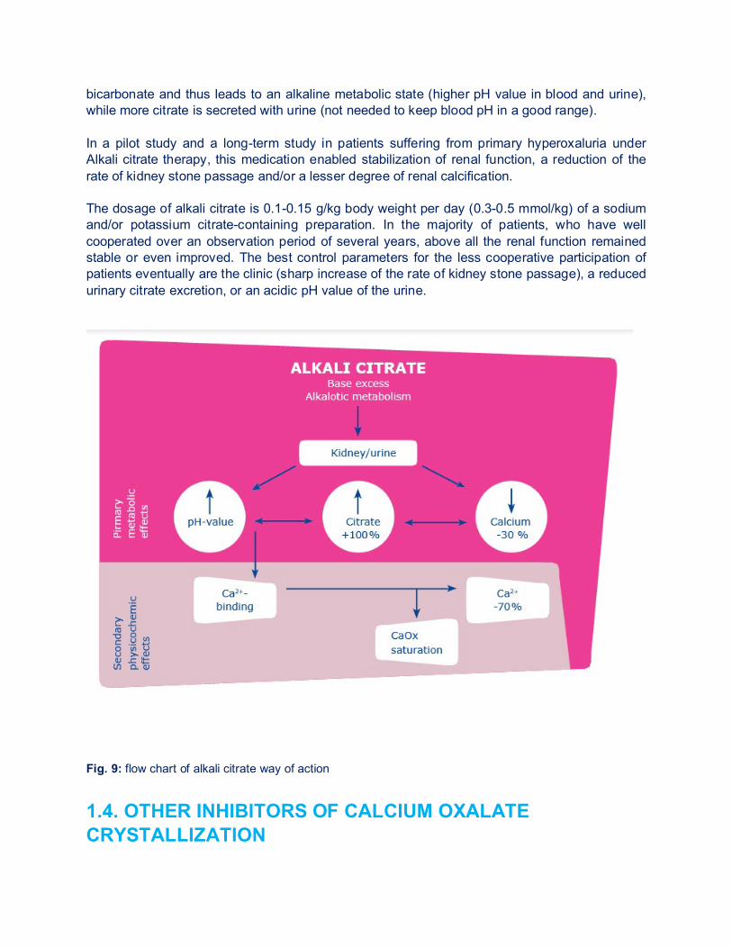
bicarbonate and thus leads to an alkaline metabolic state (higher pH value in blood and urine), while more citrate is secreted with urine (not needed to keep blood pH in a good range). In a pilot study and a long-term study in patients suffering from primary hyperoxaluria under Alkali citrate therapy, this medication enabled stabilization of renal function, a reduction of the rate of kidney stone passage and/or a lesser degree of renal calcification. The dosage of alkali citrate is 0.1-0.15 g/kg body weight per day (0.3-0.5 mmol/kg) of a sodium and/or potassium citrate-containing preparation. In the majority of patients, who have well cooperated over an observation period of several years, above all the renal function remained stable or even improved. The best control parameters for the less cooperative participation of patients eventually are the clinic (sharp increase of the rate of kidney stone passage), a reduced urinary citrate excretion, or an acidic pH value of the urine.
Fig. 9: flow chart of alkali citrate way of action
1.4. OTHER INHIBITORS OF CALCIUM OXALATE CRYSTALLIZATION
In its effectiveness, orthophosphate is also comparable to alkali citrate. Moreover, the administration of magnesium is recommended. Both substances lead to a good inhibition of the calcium oxalate crystallization. For example, in patients suffering from recurring kidney stone passages, a favorable effect of magnesium on urine saturation of CaOx has been noted. However, there were just a few reports until now about the long-term course in patients suffering from PH under orthophosphate and/or magnesium medication. 1.5. FUTURE THERAPIES Oxalobacter formigenes (Oxabact®, Oxthera AB, Sweden) is an anaerobic bacterium that can make use of oxalate as its only source of carbon. Orally administered, O. formigenes on the one hand degrades intestinal oxalate leading to a reduced oxalate absorption, while on the other hand, it activates the intestinal oxalate transporter (SLC26A6), which results in an active transport of oxalate from the blood (oxalate produced in the liver) into the intestinal lumen. Currently, O. formigenes is analyzed in a phase III placebo-controlled study (NCT02000219) in PH patients with still good renal function, but elevated plasma oxalate values (> 10 µmol/l). The results from earlier studies showed that O. formigenes is capable to lower the plasma oxalate value. However, it resulted in an elevated oxalate secretion in urine, although the other way around was expected. The elevated urinary oxalate excretion may have been caused by the dissolution of systemic oxalate deposits which would be a positive effect. The primary endpoint of the current study will therefore be the change in plasma oxalate. In a dialysis study it was shown in the course of several years that the plasma oxalate values were decreasing, the clinical situation stabilized and even improved, and thus the patients were in a better clinical condition when transplantation was performed. As described above, the normal course under dialysis rather shows an increase of plasma oxalate and a quick deterioration of systemic oxalate deposition! ALLN-177 (Allena Pharmaceuticals, USA) is a recombinant oxalate decarboxylase, thus an oxalate-degrading enzyme in the form of tablets, which is also able to degrade the oxalate in the intestinal tract. Even if ALLN-177 does not directly activate the oxalate transport in the intestine, the difference in concentration generated by the degradation possibly suffices to reach a shift of oxalate from the blood into the intestinal tract. In healthy persons, ALLN-177 could reduce the oxalate secretion in urine with an oxalate-rich diet and is currently analyzed in patients suffering from secondary hyperoxaluria as well as in patients suffering from PH II and PH III (NCT03391804). A further therapeutic approach are RNA-interference (RNAi-) therapeutics. Those are functioning on the level of RNA translation. Small double-strand RNA molecules (small interfering RNA, siRNA) in this case are binding to a cytoplasmic protein complex (RNA-induced silencing complex, RISC), which highly specifically degrades the targeted mRNA and thus the translation into the corresponding protein is prevented. Translated, this means that a wrong information is placed at the spot (in the liver), which normally produces an enzyme participating in the oxalate metabolism. If it is not produced, the oxalate production in the liver can be
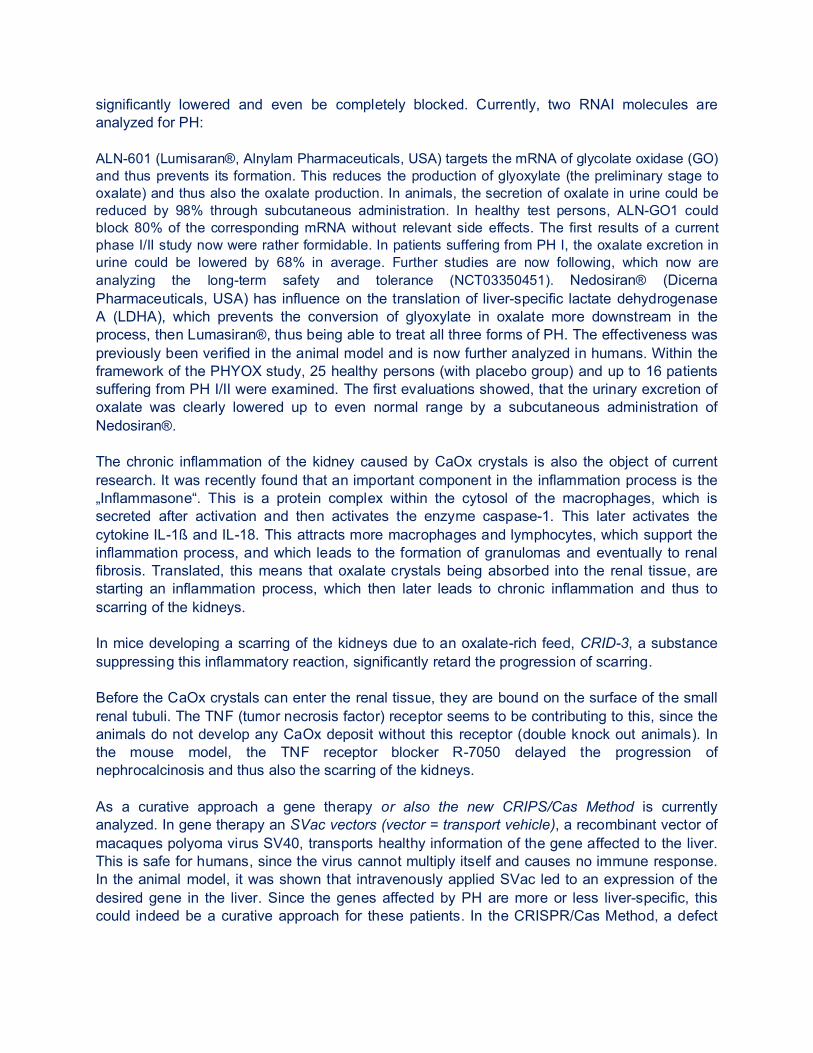
significantly lowered and even be completely blocked. Currently, two RNAI molecules are analyzed for PH: ALN-601 (Lumisaran®, Alnylam Pharmaceuticals, USA) targets the mRNA of glycolate oxidase (GO) and thus prevents its formation. This reduces the production of glyoxylate (the preliminary stage to oxalate) and thus also the oxalate production. In animals, the secretion of oxalate in urine could be reduced by 98% through subcutaneous administration. In healthy test persons, ALN-GO1 could block 80% of the corresponding mRNA without relevant side effects. The first results of a current phase I/II study now were rather formidable. In patients suffering from PH I, the oxalate excretion in urine could be lowered by 68% in average. Further studies are now following, which now are analyzing the long-term safety and tolerance (NCT03350451). Nedosiran® (Dicerna Pharmaceuticals, USA) has influence on the translation of liver-specific lactate dehydrogenase A (LDHA), which prevents the conversion of glyoxylate in oxalate more downstream in the process, then Lumasiran®, thus being able to treat all three forms of PH. The effectiveness was previously been verified in the animal model and is now further analyzed in humans. Within the framework of the PHYOX study, 25 healthy persons (with placebo group) and up to 16 patients suffering from PH I/II were examined. The first evaluations showed, that the urinary excretion of oxalate was clearly lowered up to even normal range by a subcutaneous administration of Nedosiran®. The chronic inflammation of the kidney caused by CaOx crystals is also the object of current research. It was recently found that an important component in the inflammation process is the „Inflammasone“. This is a protein complex within the cytosol of the macrophages, which is secreted after activation and then activates the enzyme caspase-1. This later activates the cytokine IL-1ß and IL-18. This attracts more macrophages and lymphocytes, which support the inflammation process, and which leads to the formation of granulomas and eventually to renal fibrosis. Translated, this means that oxalate crystals being absorbed into the renal tissue, are starting an inflammation process, which then later leads to chronic inflammation and thus to scarring of the kidneys. In mice developing a scarring of the kidneys due to an oxalate-rich feed, CRID-3, a substance suppressing this inflammatory reaction, significantly retard the progression of scarring. Before the CaOx crystals can enter the renal tissue, they are bound on the surface of the small renal tubuli. The TNF (tumor necrosis factor) receptor seems to be contributing to this, since the animals do not develop any CaOx deposit without this receptor (double knock out animals). In the mouse model, the TNF receptor blocker R-7050 delayed the progression of nephrocalcinosis and thus also the scarring of the kidneys. As a curative approach a gene therapy or also the new CRIPS/Cas Method is currently analyzed. In gene therapy an SVac vectors (vector = transport vehicle), a recombinant vector of macaques polyoma virus SV40, transports healthy information of the gene affected to the liver. This is safe for humans, since the virus cannot multiply itself and causes no immune response. In the animal model, it was shown that intravenously applied SVac led to an expression of the desired gene in the liver. Since the genes affected by PH are more or less liver-specific, this could indeed be a curative approach for these patients. In the CRISPR/Cas Method, a defect
information is replaced by a healthy one (Figure 10, modified from Weigert et al, Expert Opinion in Emerging Drugs, Volume 23, Issue 4, 2018).
Fig. 10: Overview of the underlying pathomechanismns of PH I-III and schematic representation of possible new therapeutics for the primary hyperoxalurias. PH I results from a mutation of the AGXT gene (encoding for alanine:glyoxylate aminotransferase, PH II from a mutation in the GRHPR gene (for glyoxylate reductase/hydroxypyruvate reductase), and PH III from a mutation in the HOGA1 gene (4-hydroxy-2-oxoglutarate aldolase 1). Every subtype leads to an accumulation of oxalate, which has to be eliminated by the kidney. The possible treatment alternatives (red: established therapy, here B6), orange: treatment in clinical study, green: future treatment option): 1) lumisaran, a RNAi medication, suppresses the glycolate oxidase (GO), less oxalate is being produced, 2) DCR-PHXC, a further RNAi medication, directed against the liver-specific lactate dehydrogenase A (LDHA), which also leads to a reduced oxalate production, 3) ALLN-177 is a recombinant microbial enzymatic oxalate decarboxylase, which degrades oxalate in the intestines, Reduction of the absorption of oxalate in the intestine, 4) O. formigenes is an anaerobic bacterium, where oxalate is used as the only one source of carbon; it is used to degrade oxalate in the intestine and to activate an intestinal oxalate transporter, which leads to an active secretion of plasma oxalate into the intestinal lumen, 5) CRID-3 inhibits the pathway of NLRP3-inflammasome, which prevents the development of renal fibrosis, 6) R-7050, a TNF receptor blocker, prevents the adhesion of calcium oxalate crystals in the proximal tubulus lumen, 7) DECA, amino oxy acetic acid and Emetine prevents the entering of AGT into mitochondria, 8) molecules derived from salicylic acid, which also inhibit the GO enzyme, 9) CRISPR/Cas-reducing glyoxylate production by GO gene editing, 10) AVV vectors, SVac vectors by functional gene expression into the liver.
2. TREATMENT OF KIDNEY STONE PASSAGE The recurrent passage of urinary stones represents a major problem of primary hyperoxaluria. A stone blocking the urinary tract, e.g. a stone in the ureter, makes a surgical procedure necessary, which should be as minimal as possible. However, a stone removal by surgery should only be considered for obstructive stones or in case of a massive stone burden in the kidney(s) and frequent painful kidney stone passage as well as in case of secondary infected stones. Kidney stones, which are not blocking or asymptomatic, can be left in situ.
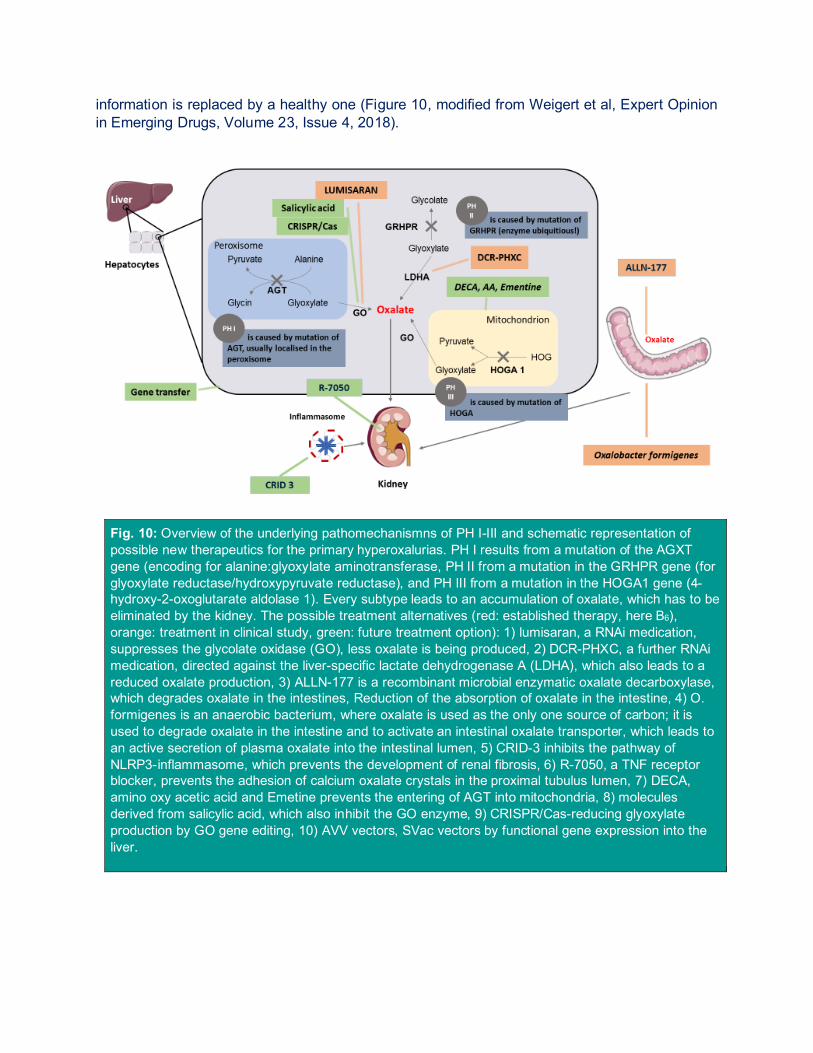
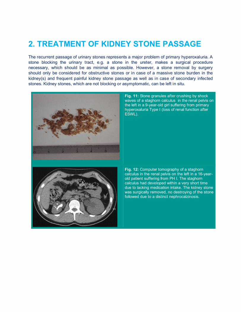
Fig. 11: Stone granules after crushing by shock waves of a staghorn calculus in the renal pelvis on the left in a 9-year-old girl suffering from primary hyperoxaluria Type I (loss of renal function after ESWL).
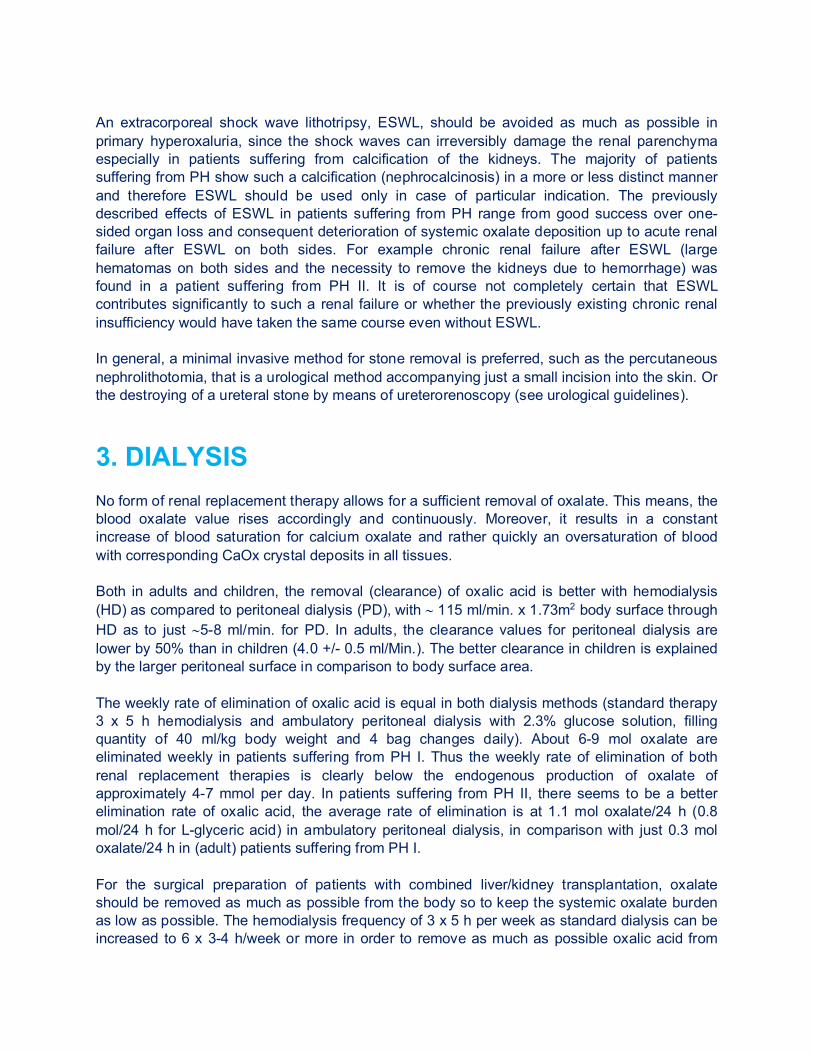
Fig. 12: Computer tomography of a staghorn calculus in the renal pelvis on the left in a 16-year-old patient suffering from PH I. The staghorn calculus had developed within a very short time due to lacking medication intake. The kidney stone was surgically removed, no destroying of the stone followed due to a distinct nephrocalzinosis.
An extracorporeal shock wave lithotripsy, ESWL, should be avoided as much as possible in primary hyperoxaluria, since the shock waves can irreversibly damage the renal parenchyma especially in patients suffering from calcification of the kidneys. The majority of patients suffering from PH show such a calcification (nephrocalcinosis) in a more or less distinct manner and therefore ESWL should be used only in case of particular indication. The previously described effects of ESWL in patients suffering from PH range from good success over one-sided organ loss and consequent deterioration of systemic oxalate deposition up to acute renal failure after ESWL on both sides. For example chronic renal failure after ESWL (large hematomas on both sides and the necessity to remove the kidneys due to hemorrhage) was found in a patient suffering from PH II. It is of course not completely certain that ESWL contributes significantly to such a renal failure or whether the previously existing chronic renal insufficiency would have taken the same course even without ESWL. In general, a minimal invasive method for stone removal is preferred, such as the percutaneous nephrolithotomia, that is a urological method accompanying just a small incision into the skin. Or the destroying of a ureteral stone by means of ureterorenoscopy (see urological guidelines).
3. DIALYSIS No form of renal replacement therapy allows for a sufficient removal of oxalate. This means, the blood oxalate value rises accordingly and continuously. Moreover, it results in a constant increase of blood saturation for calcium oxalate and rather quickly an oversaturation of blood with corresponding CaOx crystal deposits in all tissues. Both in adults and children, the removal (clearance) of oxalic acid is better with hemodialysis (HD) as compared to peritoneal dialysis (PD), with ∼ 115 ml/min. x 1.73m2 body surface through HD as to just ∼5-8 ml/min. for PD. In adults, the clearance values for peritoneal dialysis are lower by 50% than in children (4.0 +/- 0.5 ml/Min.). The better clearance in children is explained by the larger peritoneal surface in comparison to body surface area. The weekly rate of elimination of oxalic acid is equal in both dialysis methods (standard therapy 3 x 5 h hemodialysis and ambulatory peritoneal dialysis with 2.3% glucose solution, filling quantity of 40 ml/kg body weight and 4 bag changes daily). About 6-9 mol oxalate are eliminated weekly in patients suffering from PH I. Thus the weekly rate of elimination of both renal replacement therapies is clearly below the endogenous production of oxalate of approximately 4-7 mmol per day. In patients suffering from PH II, there seems to be a better elimination rate of oxalic acid, the average rate of elimination is at 1.1 mol oxalate/24 h (0.8 mol/24 h for L-glyceric acid) in ambulatory peritoneal dialysis, in comparison with just 0.3 mol oxalate/24 h in (adult) patients suffering from PH I. For the surgical preparation of patients with combined liver/kidney transplantation, oxalate should be removed as much as possible from the body so to keep the systemic oxalate burden as low as possible. The hemodialysis frequency of 3 x 5 h per week as standard dialysis can be increased to 6 x 3-4 h/week or more in order to remove as much as possible oxalic acid from
the body prior to transplantation. A combination of hemodialysis and peritoneal dialysis shall be considered at this time. However, only an insufficient amount of oxalic acid is removed with these measures, if there is a very large systemic oxalate deposit e.g. after a long dialysis period. It seems to be possible to prevent the plasma oxalate level to excessively rise further with a combination of dialysis methods but it remains continuously on a high level. Nevertheless, there will still be a shift of oxalate into the body tissues.
4. TRANSPLANTATION Time and mode of transplantation may be different between centers. Combined liver/Kidney transplantation for PH I and isolated kidney transplantation for PH II were state of the art. In the meantime, combined liver/kidney transplantation became necessary in some PH II patients, also. In PH I, the disease can only be „cured“ with liver transplantation. If at all, an isolated kidney transplantation should be made only in (older) patients, with good therapeutic success of vitamin B6. However, the experiences of the European Registers on Transplantations show rather poor survival rates with isolated kidney transplantations. A transplantation will not be taken into consideration until the filtration performance of the kidneys remains so good (> 40 ml/min. x 1.73 m2 body surface), that oxalate removal is accomplished adequately so that the effects of systemic oxalosis will be prevented. A further procedure should also be mentioned here: a liver transplantation is done at first to replace the lacking enzyme and to normalize the oxalate production, and ultimately a kidney is transplanted (KALT procedure, kidney after liver) after reduction of the body oxalate deposit through aggressive dialysis. The decision on the transplantation mode and the time of transplantation must be made in relation to the patient situation with regard to the very variable clinical picture of particularly the PH I. In chronic renal failure, the combined liver/kidney transplantation is nowadays still preferred for PH I, particularly if a transplantation can be performed prior to the occurrence of systemic oxalosis. In contrast, an isolated kidney transplantation always involves the high risk of a quick new renal failure again caused by calcium oxalate deposits. This even is the case, if aggressive dialysis measures for removing oxalic acid are applied before and after transplantation. If the renal function is not yet impaired and, therefore, the effects of systemic oxalosis have not yet occurred, an isolated liver transplantation could be taken into consideration. Meanwhile, several cases of such a procedure with good results have been described. The advantage of this procedure is with certainty the preservation of the own renal function, however, above all, the prevention of systemic oxalosis. Further positive experiences show that in selected cases it can result in lowering of blood oxalate and oxalate deposits in the body in long-term stable renal function, and thus a consecutive kidney transplantation is no longer necessary. It should be pointed out that it should always be an individual decision of the transplant center and the patient. The big disadvantage of this form of transplantation is timing, as the clinical follow up of the patient is not predictable and hence, transplantation might take place to early and immunosuppressants with all their risks are given for a long time, although not truly being necessary.
Good experiences (e.g. complication-free course over > 8 years after combined transplantation) also show that the effects of systemic oxalosis are generally reversible. Particularly, the massive calcium oxalate deposits in bones and bone marrow are completely dissolving over time, blood formation recovers again, the bone density increases. Even an extreme deposit of CaOx in the heart muscle should be no obstacle to a targeted transplantation. After a successful transplantation, all these patients showed a clear improvement of the heart function, based on a reduction of the CaOx deposits. Even successful pregnancies after combined liver/kidney transplantation have been reported in the meantime. Only eye deposits, which are extreme in patients with infantile oxalosis, did not really decrease. It is also worth mentioning that above all the liver transplantation may involve greater problems (e.g. permanent cholestasis). Such complications may even go as far as reducing the quality of life of the individual patient so dramatically, that they claim that even dialysis would be a preferred procedure and that they would not decide for transplant again, if they would have a choice.
A transplantation in a patient suffering from primary hyperoxaluria should only be performed in a center that really specializes in this disease.
With regard to the partly dramatic course of a primary hyperoxaluria, a transplantation should be planned as early as possible, so that it can be performed before the complications of a systemic oxalosis become apparent. In this event, attention should be paid to all therapeutical measures for preserving the renal function, since even a smallest residual function of the kidneys during elimination of oxalic acid is more efficient than any renal replacement therapy. A too large oxalate pool, such as in case of systemic oxalosis, will later definitely affect the success of a transplantation.
5. CONCLUSION The excretion of urinary oxalate must be analyzed in each patient with a kidney stone or a calcification of the kidneys. In a clearly elevated excretion of urinary oxalate, further evaluation is urgently recommended, since a timely diagnosis of a patient suffering from primary hyperoxaluria will decide the future progression of the disease: a long-term preservation of renal function versus an early renal failure. An aggressive therapeutic management is absolutely necessary, above all with regard to a sufficient daily fluid intake. In small children, a permanent gastric tube should be inserted in the stomach so that fluid can be supplied at night as well. Currently, there are rather few therapeutic options. In some patients suffering from PH I, treatment with vitamin B6 is successful. In general,
the patient suffering from hyperoxaluria should be given alkali citrate or orthophosphate for improving the solubility of oxalate in urine. There are not yet any new medications available, but a great number of new therapeutic options are tested in clinical studies (see Figure 10). The mentioned new medication will certainly be revolutionary for the therapy of PH and lead way that liver transplantation will no longer be necessary! No renal replacement therapy is capable to eliminate a sufficient quantity of oxalate. A quick transplantation should therefore be considered. In PH I, combined liver/kidney transplantation is he method of choice, in PH II isolated kidney transplantation is sufficient.
The early diagnosis of the primary hyperoxaluria is absolutely necessary!
6. SELF-SUPPORT GROUPS, CENTERS So far, well organized self-help groups have been in established in Germany (www.PH-Selbsthilfe.org), in the Netherlands, in Spain (https://asociacionaphes.wordpress.com) and in the USA (www.ohf.org). The European PH advocacy group was founded recently (www.ph-Europe.net). All these organizations are cooperating, and they make plans to create a higher-level structure. Contact persons in Germany, Europe and in the USA can be found on their websites.
and
In Europe, a group of scientists have joined the European Hyperoxaluria Consortium (www.oxalEurope.com). Apart from a European database, there are joint research projects.
O | X | A | L | E | U | R | O | P | E In the USA, above all the Hyperoxaluria Center of the rare kidney stone consortium is worth mentioning (http://www.mayoclinic.org/nephrology-rst/hyperoxaluriacenter.html). They also have a patient database, which is jointly organized (www.rarekidneystones.org).
4. APPENDIX
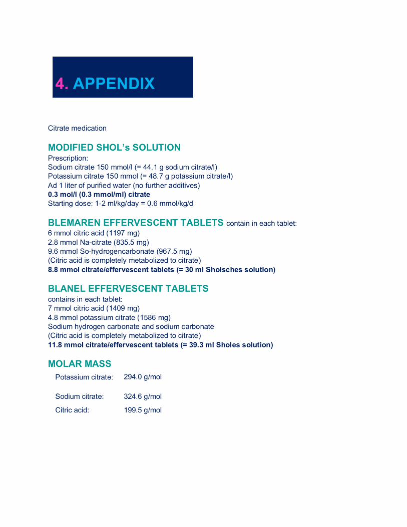
Citrate medication MODIFIED SHOL’s SOLUTION Prescription: Sodium citrate 150 mmol/l (= 44.1 g sodium citrate/l) Potassium citrate 150 mmol (= 48.7 g potassium citrate/l) Ad 1 liter of purified water (no further additives) 0.3 mol/l (0.3 mmol/ml) citrate Starting dose: 1-2 ml/kg/day = 0.6 mmol/kg/d BLEMAREN EFFERVESCENT TABLETS contain in each tablet: 6 mmol citric acid (1197 mg) 2.8 mmol Na-citrate (835.5 mg) 9.6 mmol So-hydrogencarbonate (967.5 mg) (Citric acid is completely metabolized to citrate) 8.8 mmol citrate/effervescent tablets (= 30 ml Sholsches solution) BLANEL EFFERVESCENT TABLETS contains in each tablet: 7 mmol citric acid (1409 mg) 4.8 mmol potassium citrate (1586 mg) Sodium hydrogen carbonate and sodium carbonate (Citric acid is completely metabolized to citrate) 11.8 mmol citrate/effervescent tablets (= 39.3 ml Sholes solution) MOLAR MASS
Potassium citrate: 294.0 g/mol
Sodium citrate: 324.6 g/mol
Citric acid: 199.5 g/mol
5. GLOSSARY
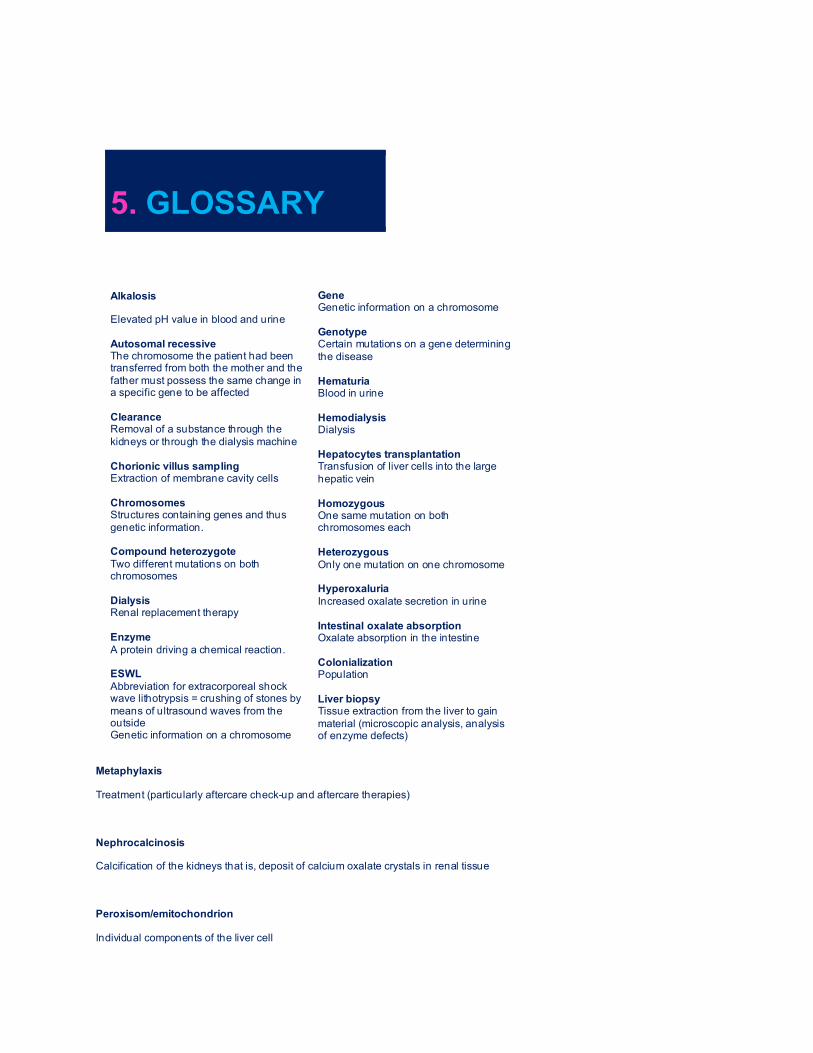
Alkalosis
Elevated pH value in blood and urine Autosomal recessive The chromosome the patient had been transferred from both the mother and the father must possess the same change in a specific gene to be affected Clearance Removal of a substance through the kidneys or through the dialysis machine Chorionic villus sampling Extraction of membrane cavity cells Chromosomes Structures containing genes and thus genetic information. Compound heterozygote Two different mutations on both chromosomes Dialysis Renal replacement therapy Enzyme A protein driving a chemical reaction. ESWL Abbreviation for extracorporeal shock wave lithotrypsis = crushing of stones by means of ultrasound waves from the outside Genetic information on a chromosome
Gene Genetic information on a chromosome Genotype Certain mutations on a gene determining the disease Hematuria Blood in urine Hemodialysis Dialysis Hepatocytes transplantation Transfusion of liver cells into the large hepatic vein Homozygous One same mutation on both chromosomes each Heterozygous Only one mutation on one chromosome Hyperoxaluria Increased oxalate secretion in urine Intestinal oxalate absorption Oxalate absorption in the intestine Colonialization Population Liver biopsy Tissue extraction from the liver to gain material (microscopic analysis, analysis of enzyme defects)
Metaphylaxis
Treatment (particularly aftercare check-up and aftercare therapies)
Nephrocalcinosis
Calcification of the kidneys that is, deposit of calcium oxalate crystals in renal tissue
Peroxisom/emitochondrion
Individual components of the liver cell
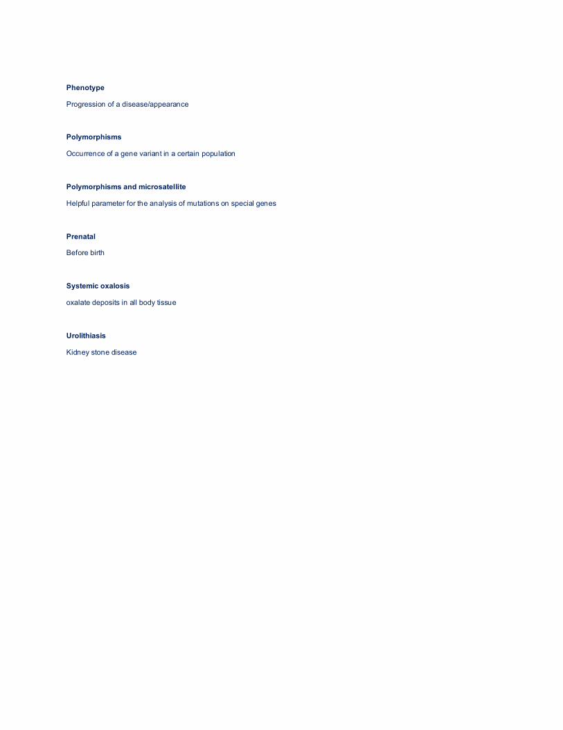
Phenotype
Progression of a disease/appearance
Polymorphisms
Occurrence of a gene variant in a certain population
Polymorphisms and microsatellite
Helpful parameter for the analysis of mutations on special genes
Prenatal
Before birth
Systemic oxalosis
oxalate deposits in all body tissue
Urolithiasis
Kidney stone disease

NOTES