Embed Size (px)
Citation preview
RESEARCH PAPER
Identification of aquatically available carbon from algaethrough solution-state NMR of whole 13C-labelled cells
Mohammad Akhter1,2 & Rudraksha Dutta Majumdar1,2 & Blythe Fortier-McGill1,2 &
Ronald Soong1,2 & Yalda Liaghati-Mobarhan1,2& Myrna Simpson1,2
&
George Arhonditsis2 & Sebastian Schmidt3 & Hermann Heumann3& André J. Simpson1,2
Received: 9 February 2016 /Revised: 24 March 2016 /Accepted: 30 March 2016 /Published online: 13 April 2016# Springer-Verlag Berlin Heidelberg 2016
Abstract Green algae and cyanobacteria are primary pro-ducers with profound impact on food web functioning. Bothrepresent key carbon sources and sinks in the aquatic environ-ment, helping modulate the dissolved organic matter balanceand representing a potential biofuel source. Underlying theimpact of algae and cyanobacteria on an ecosystem level istheir molecular composition. Herein, intact 13C-labelledwhole cell suspensions of Chlamydomonas reinhardtii,Chlorella vulgaris and Synechocystis were studied using avariety of 1D and 2D 1H/13C solution-state nuclear magneticresonance (NMR) spectroscopic experiments. Solution-stateNMR spectroscopy of whole cell suspensions is particularlyrelevant as it identifies species that are mobile (dissolved ordynamic gels), ‘aquatically available’ and directly contributeto the aquatic carbon pool upon lysis, death or become areadily available food source on consumption. In this study,a wide range of metabolites and structural components wereidentified within the whole cell suspensions. In addition, sig-nificant differences in the lipid/triacylglyceride (TAG) contentof green algae and cyanobacteria were confirmed. Mobilespecies in algae are quite different from those in abundance
in ‘classic’ dissolved organic matter (DOM) indicating that ifalgae are major contributors to DOM, considerable selectivepreservation of minor components (e.g. sterols) or biotransfor-mation would have to occur. Identifying the metabolites anddissolved components within algal cells by NMR permits fu-ture studies of carbon transfer between species and throughthe food chain, whilst providing a foundation to better under-stand the role of algae in the formation of DOM andthe sequestration/transformation of carbon in aquaticenvironments.
Keywords Solution-state NMR .HMQC . Algae .
Metabolomics . Dissolved organic matter
Introduction
Green algae are a diverse group of eukaryotic organisms foundin both freshwater and saltwater ecosystems. Blue-green algae(often termed cyanobacteria) are also prolifically found in wa-ter systems under certain nitrogen and phosphorus balances,outcompeting green algae at certain equilibria [1]. Both areautotrophic and carryout photosynthesis. As a result, both playa fundamental role in maintaining the energy balance in aquat-ic ecosystems as they belong to a lower trophic level andchanges in their populations are magnified through the foodweb. In addition, green algae and cyanobacteria may play arole in the formation of dissolved organic matter (DOM), atopic of interest to a diversity of water researchers, as well asform a primary source for the production of biofuels.
Importantly, green algae and cyanobacteria have differentmolecular compositions and homeostatic set points, specifi-cally concerning lipids and their metabolism [2]. This is ofparticular importance when cyanobacteria outcompete greenalgae in freshwater lakes, as this can have adverse effects on
Electronic supplementary material The online version of this article(doi:10.1007/s00216-016-9534-8) contains supplementary material,which is available to authorized users.
* André J. [email protected]
1 Environmental NMR Centre, University of Toronto Scarborough,1265 Military Trail, Toronto, Ontario M1C 1A4, Canada
2 Department of Physical and Environmental Sciences, University ofToronto Scarborough, 1265 Military Trail, Toronto, Ontario M1C1A4, Canada
3 Silantes GmbH, Gollierstr. 70 C, 80339 München, Germany
Anal Bioanal Chem (2016) 408:4357–4370DOI 10.1007/s00216-016-9534-8
micro-crustaceans such as Daphnia magna (a keystone spe-cies) [3]. For example, exposure to cyanobacteria has beenshown to increase the number of aborted eggs in D. magna,thus decreasing population size which directly impacts highertrophic levels [4]. Although the primary mechanisms bywhich these patterns are triggered is unclear, cyanobacteriaare known to be of poor food quality as a result of mechanicalinterference, toxicity and (or) inadequate biochemical nutri-tional value (e.g. low content in omega-3 highly unsaturatedfatty acids, such as docosahexaenoic and eicosapentaenoicacids, and sterols) for consumers. Consumers exclusivelyfed cyanobacteria typically exhibit near-starvation traits [5].
Furthermore, the amount and composition of DOM in wa-ter systems are of current interest, as DOM comprises a majorpercent of mobile organic carbon and plays a role in increasingthe solubility of environmental contaminants [6]. This workwill shed light on the metabolite profile of different algal spe-cies and the structural contributions that they can make toDOM when their cell walls are spliced. In particular, as lowpower solution-state nuclear magnetic resonance (NMR) tech-niques are employed (see ‘Conclusion’ section), soluble andmobile gel-like components (i.e. those readily dispersed intothe aqueous phase) are emphasised within the whole cells. Byunderstanding the potential flux of mobile metabolites andsoluble precursors from algae into DOM, researchers may beable to better predict changes in DOM in freshwater in relationto the amount of algae or cyanobacteria present.
Further interest in algae stems from their application in theproduction of biofuels. Biofuels are synthesised from lipid oilsin algae which may account for up to 50 % of the algae byweight in certain species [7]. Algae can be grown very rapidlyin bioreactors and will likely become an important source forrenewable energy in the future [7]. Current research on algaefocuses on identifying the best (greatest yield of biodiesel perunit mass algae) species out of an estimated 3×10⁵ species,where a number of factors must be considered including thegrowth rate and the lipid oil content [8, 9]. Underlying thesefactors is the molecular composition of algae and themetabolomic changes that occur in response to altering envi-ronmental conditions. Lipids in cells may exist in differentphysical forms; however, it is the extractable lipid componentthat makes up the primary building block for the synthesis ofbiofuels and identifying this extractable lipid component is achallenge that researchers face in identifying the most viablespecies [10]. The first step in biofuel production is the extrac-tion of lipids followed by esterification. Therefore, it is para-mount that researchers identify the percent of lipids that aremobile within whole cells and thus readily available for sim-ple extraction [10]. This work aims to identify and quantify arange of dissolved andmobile species within whole algae cellswhich includes the lipid fraction. The importance of algae foraquatic ecosystems, potential contribution to DOM formationand the production of biofuels drives a fundamental need to
better understand the metabolomic and structural profile ofalgae. This understanding will equip researchers with theknowledge necessary to precisely identify the impact of algaland cyanobacterial blooms at an ecosystem level as well as todevelop genetically modified algae strains that will make bio-fuel production more economically viable [11].
The metabolite profile of different species of algae andcyanobacteria has been studied using various analytical tech-niques including gas chromatographymass spectrometry-timeof flight–mass spectrometry (GC-MS TOF-MS) and high per-formance liquid chromatography inductively coupled plasmamass spectrometry (HPLC ICP-MS). Using GC-MS TOF-MS, the metabolomic changes in wild-type Chlamydomonasreinhardtii under nitrogen, phosphorus, sulphur, or iron-deprived conditions were examined [12]. Furthermore,HPLC ICP-MS was employed in identifying the speciationof arsenosugars in the brown alga Fucus serratus [13]. Suchstudies provide an excellent and sensitive targeted approach toevaluate specific metabolites in extracts but are often restrictedto small molecular components that can be easily extractedand derivatized. Additionally, MS techniques are selectiveand reliant on MS libraries and thus unknown compoundsmay be missed [14]. In contrast, NMR requires little samplepreparation and is a non-selective detector, with the main lim-itation being spectral overlap which can hamper assignmentand quantification in complex matrices [15].
Some limited NMR studies have also been conducted onalgae and cyanobacteria [16, 17]. Specifically, solution-stateNMR was used to build a limited metabolomic profile of thealgal species Chlorophyta and Rhosophyta with natural abun-dance 13C [16]. In addition, solid-state NMR has been used tostudy the light harvesting complexes (important for photosyn-thesis) in C. reinhardtii [17] and also to characterise the lipidand carbohydrate constituents of whole cells of marine(Pavlova lutheri and Nannochloropsis oculata) and freshwa-ter (C. reinhardtii) microalgae which have different cell wallcharacteristics [18]. High resolution–magic angle spinning(HR-MAS) 1H and 13C NMR spectroscopy have also beenshown to be useful for studying microalgal whole cells[19–21]. However, MAS tends to average anisotropic interac-tions and sharpens the signals from semi-solids and gels.Whilst this is often an advantage, if only dissolved metabolitesare of interest, then it has been noted that peak resolution at thebaseline tends to be poorer in the HR-MAS spectra comparedto corresponding solution-state spectra, and chemical shiftscan be slightly different [21]. To the best of our knowledge,a comparative study on the different species of 13C-enrichedgreen algae and cyanobacteria whole cell suspensions usingsolution-state NMR including a diversity of 1D and 2D ex-periments has yet to be employed in the establishment of amore complete metabolic profile. Solution-state NMR pro-vides high resolution molecular information concerning com-ponents in the sample that are dissolved or highly mobile in
4358 M. Akhter et al.
their natural state within whole algal cells [22] and representsmost common NMR technique available in the vast majorityof NMR facilities. Importantly, most biologically relevant me-tabolites are found and operate in the cytoplasm of cells andare therefore visible through solution-state NMR. This makessolution-state NMR ideal for metabolomic studies.Furthermore, a thorough understanding of mobile metaboliteswill shed light on the contributions of algae to the structuralcomponents of DOM as these components will contributedirectly to the aquatic environment on lysis.
In this study, the metabolomic profiles of two differentstrains of 13C isotopically labelled green algae and one cya-nobacterium were analysed using solution-state NMR spec-troscopy. Specifically, the strains C. reinhardtii (unicellulargreen algae with two characteristic flagella found in soilsand fresh water), Chlorella vulgaris (unicellular green algaewith no flagella found in fresh water) and Synechocystis(cyanobacteria found in fresh water) were examined. In a nat-ural environment, the presence of flagella allows the formerspecies to demonstrate phototactic behaviour and optimisetheir exposure to light for photosynthesis. The second speciesis considered a high quality food source for primary con-sumers with high protein content and other essential nutrients.The latter species is one of the most extensively studied typesof cyanobacteria as it has the capacity to demonstrate bothphototrophic growth by oxygenic photosynthesis in sunlightand heterotrophic growth by glycolysis and oxidative phos-phorylation during dark periods. The evaluation of thesolution-state NMR spectroscopic approach, in particular theability to identify key metabolites and major differences be-tween species, will facilitate future in vivo NMR-basedmetabolomic studies (for example feeding 13C-enriched algaeto D. magna and following real-time metabolism).Additionally, understanding the physical state in which differ-ent metabolites are found will shed light on their ability tocontribute to DOM as well as the potential extractability oflipid components for the production of biofuels.
Materials and methods
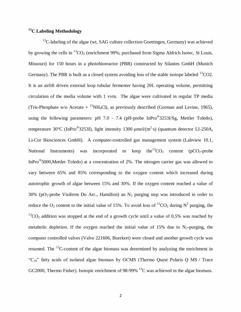
13C labelling of algae
The algae strains C. reinhardtii, C. vulgaris and thecyanobacteria Synechocystis were cultivated in a small scaleclosed loop system photobioreactor designed and built bySilantes GmbH. Each fermentation was carried out autotro-phically and entirely with 13CO2 (enrichment 99%; purchasedfrom Sigma-Aldrich, Isotec, St. Louis,MO). The environmen-tal conditions for growth including media, temperature, lightintensity and pH were optimised for maximum growth foreach species. The biomass was then harvested and freeze driedfor storage at −20 °C prior to use in the NMR experiments,
which is the standard practice for microalgal whole cell sam-ples [19, 20]. For further details on labelling methodology,refer to the Electronic Supplementary Material (ESM).
Sample preparation
Uniformly labelled 13C algae strains C. reinhardtii,C. vulgaris and the cyanobacteria Synechocystis were studiedusing solution-state NMR spectroscopy. Twenty milligrams(dry weight) of each was suspended in 100 % D2O(Cambridge Isotopes, Tewksbury, MA) and added to a high-precision 5-mm NMR tube (508-UP-7, Norell, Morganton,NC) for data collection.
1D NMR spectroscopy
The experiments were performed using a Bruker Avance IIINMR spectrometer with a 1H Larmor frequency of500.13 MHz equipped with a 5-mm four-channel (1H, 13C,15N, 19F) QXI NMR probe, fitted with an actively shielded z-gradient (Bruker BioSpin) at room temperature. A D2O Lockwas used for all experiments. Decoupling was used in all 1Dand 2D experiments to remove 1H-13C coupling from the 13C-enriched sample. The decoupling schemes GARP-4 andWALTZ16 were used for the proton and carbon observedexperiments, respectively. All NMR spectra were processedvia TopSpin 3.1.
All 1H NMR spectra were recorded using the SPR-W5WATERGATE water suppression sequence using a binomialdelay of 125 μs and GARP-4 13C decoupling during acquisi-tion [23]. A 90° pulse was calibrated for each sample. A spec-tral width of 20 ppm was collected, and 512 scans were ac-quired with 16 k time domain points. The 1H T1 was measuredat 1 s using an inverse recovery sequence, and the recycledelay set five times this. 1H NMR spectra were processedusing a zero filling factor of 2 and an exponential functioncorresponding to a line broadening of 0.3 Hz. For 1D 13CNMR spectra, a spectral width of 400 ppm was collected,4 k scans were acquired with 16 k time domain points and arecycle delay was 5 s. Note that the carbon spectra were usedfor relative but not absolute quantification. 13C NMR spectrawere processed using a zero filling factor of 2 and an expo-nential function corresponding to a line broadening of 5 Hz.
Spectral editing
Diffusion edited (DE) proton and carbon spectra were pro-duced using a bipolar pulse pair longitudinal encode-decode(BPLED) sequence with inverse gated decoupling [24].Spectra were collected using encoding/decoding gradients of2.5 ms at 55 gauss/cm and a diffusion time of 200 ms. DE 1Hspectra and 13C spectra were processed using a zero fillingfactor of 2 and an exponential function corresponding to a line
Identification of available carbon from algae 4359
broadening of 1.0 and 25 Hz, respectively. Inverse diffusionedited (IDE) and recovering relaxation losses from diffusionediting (RADE) were created via subtraction from the appro-priate controls as previously described [25]. The IDE protonspectra and carbon spectra were processed using a zero fillingfactor of 2 and an exponential function corresponding to a linebroadening of 1.0 and 5.0 Hz, respectively. The RADE protonspectra and carbon spectra were processed using a zero fillingfactor of 2 and an exponential function corresponding to a linebroadening of 10.0 and 25.0 Hz, respectively. For spectralediting, the spectra were scaled until the spectrum beingsubtracted was nulled leaving a difference spectrum contain-ing positive peaks [25]. The spectra were referenced to a D2Olock during acquisition and were then calibrated against aseries of known compounds in the Bruker BiofluidReference Compound Database (v 2.0.3).
2D NMR spectroscopy
2D (1H-13C) hetero-nuclear multiple-quantum correlation(HMQC) spectra were acquired using 2 k time domain points,64 scans and 128 increments in the indirect dimension and arecycle delay of 1 s (1×T1). HMQC data were used for rela-tive but not absolute quantification. HMQC spectra were proc-essed with an exponential function corresponding to a linebroadening of 15 Hz in F2 and using a qsine function in F1shifted by π/2 in both dimensions.
2D 1H-1H total correlation spectroscopy (TOCSY) spectrawere acquired in the phase-sensitive mode, using a mixingsequence with 300 μs 40 KHz WURST-2 pulses within anX_M16 mixing scheme [26]. Two thousand forty-eight datapoints (F2), 32 scans and a mixing time of 250 ms were usedfor each of the 128 increments in F1. F1 and F2 dimensionswere processed using sine-squared functions with a π/2 phaseshift and a zero filling factor of 2.
Compound identification
The spectra were calibrated against a series of known com-pounds in the Bruker Biofluid Reference CompoundDatabase(v 2.0.3). Pattern matching of both 1D/2D spectra was per-formed using Analysis of MIXtures (AMIX, v 3.9.3, BrukerBioSpin) against the Bruker Biofluid Reference CompoundDatabase (v 2.0.0 to v 2.0.3) using a procedure developedfor complex mixtures [27]. Compounds with a greater than80 % match (automated search) were selected for manual in-spection. The chemical shifts of the identified compoundswere compared with database values (r2=0.99, σ=0.01) toconfirm matching, and any compounds not meeting these re-quirements were removed. Major assignments were furtherchecked for consistency against TOCSY data where possibleand then further checked against the literature for consistency.Readers should note that error bars provided in the paper
represent the variance arising from processing and spectralanalysis but due lack of biomass (and long NMR acquisitiontimes) the samples could not be run in triplicate. Integrationwas performed using the ‘multi-integrate’ module withinAMIX (v 3.9.3, Bruker BioSpin). The threshold was set at1 % above the noise and ‘pattern files’ created for all metab-olites by selecting the extremities of the peaks at the 1 %threshold. These files were then used to calculate integralsacross the spectra in an automated fashion.
Results and discussion
1D 1H and 13C NMR spectra of 13C-enriched green algaeand cyanobacteria
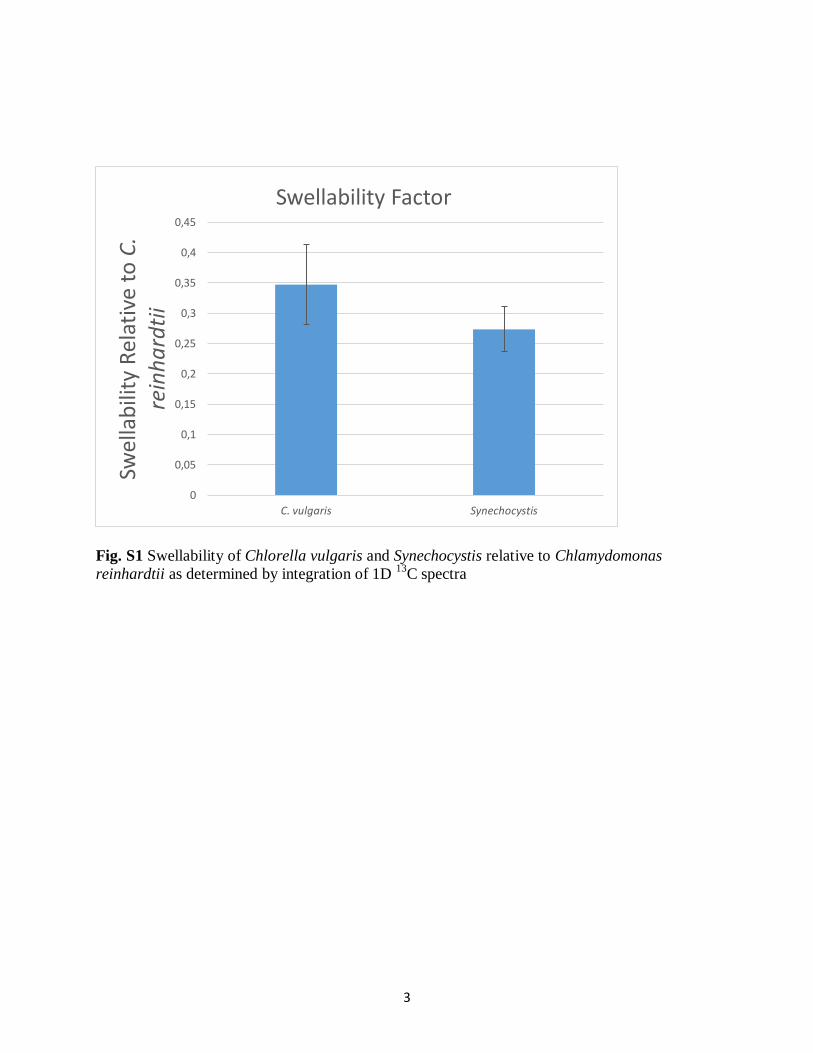
The 1D proton spectra of whole cell 13C-enriched green algaeand cyanobacteria suspended in D2O acquired using the SPR-W5 WATERGATE water suppression sequence are shown inFig. 1. It is important to note that solution-state NMR onlydetects the soluble and dynamic components (true solids willnot be observed using this technique). As such, throughoutthis paper, when for example the percentage difference of acomponent between the different species is compared, thecomparison is specifically for the fraction of material that inthe natural state (whole cells in water) is swollen/dissolved.This may not be the same as the percent fraction in totalbiomass. Due to its aquatic relevance, the swollen/dissolvedfraction of algae most readily dispersed on lysis is the focus ofthis work. The swellability of each algae type varies resultingin differing signal strength from lipid, carbohydrate and pro-tein components which may not be representative of the totalamount of each component present. Therefore, in order tocompare the differences more accurately, a relativeswellability factor was calculated by integrating the total areain the 13C spectra and normalising to C. reinhardtii (ESMFig. S1). C. reinhardtii was determined to be the mostswellable with C. vulgaris and Synechocystis exhibiting aswellability factor of 0.35 and 0.27, respectively, relative toC. reinhardtii. As a result, C. reinhardtii is ~3 times moreswellable than the other two species thus providing more bio-available carbon (dissolved phase) on ingestion by grazers andto the aquatic environment on lysis.
The 1D 1H NMR spectra show resonances that are consis-tent with signals for structural and metabolic components in-cluding lipids, carbohydrates and amino acids (Fig. 1). Thelipids are most clearly defined by the –CH3, –CH2 and doublebond resonances and are most abundant in the C. reinhardtii,followed by C. vulgaris and with the least in theSynechocystis. Conversely, C. vulgaris and Synechocystis ap-pear to contain more carbohydrates (3–4.2 ppm) thanC. reinhardtii (note the tall single peak that dominates theC. reinhardtii spectrum in this region is not carbohydrate).
4360 M. Akhter et al.
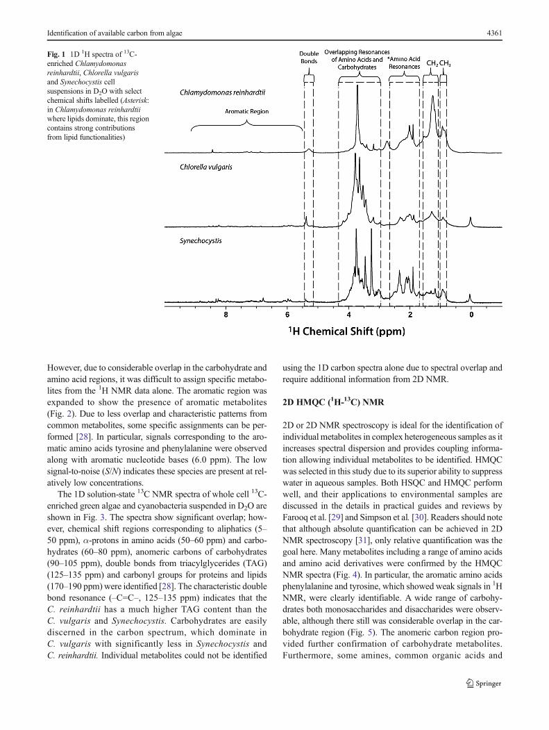
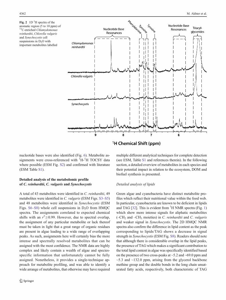
However, due to considerable overlap in the carbohydrate andamino acid regions, it was difficult to assign specific metabo-lites from the 1H NMR data alone. The aromatic region wasexpanded to show the presence of aromatic metabolites(Fig. 2). Due to less overlap and characteristic patterns fromcommon metabolites, some specific assignments can be per-formed [28]. In particular, signals corresponding to the aro-matic amino acids tyrosine and phenylalanine were observedalong with aromatic nucleotide bases (6.0 ppm). The lowsignal-to-noise (S/N) indicates these species are present at rel-atively low concentrations.
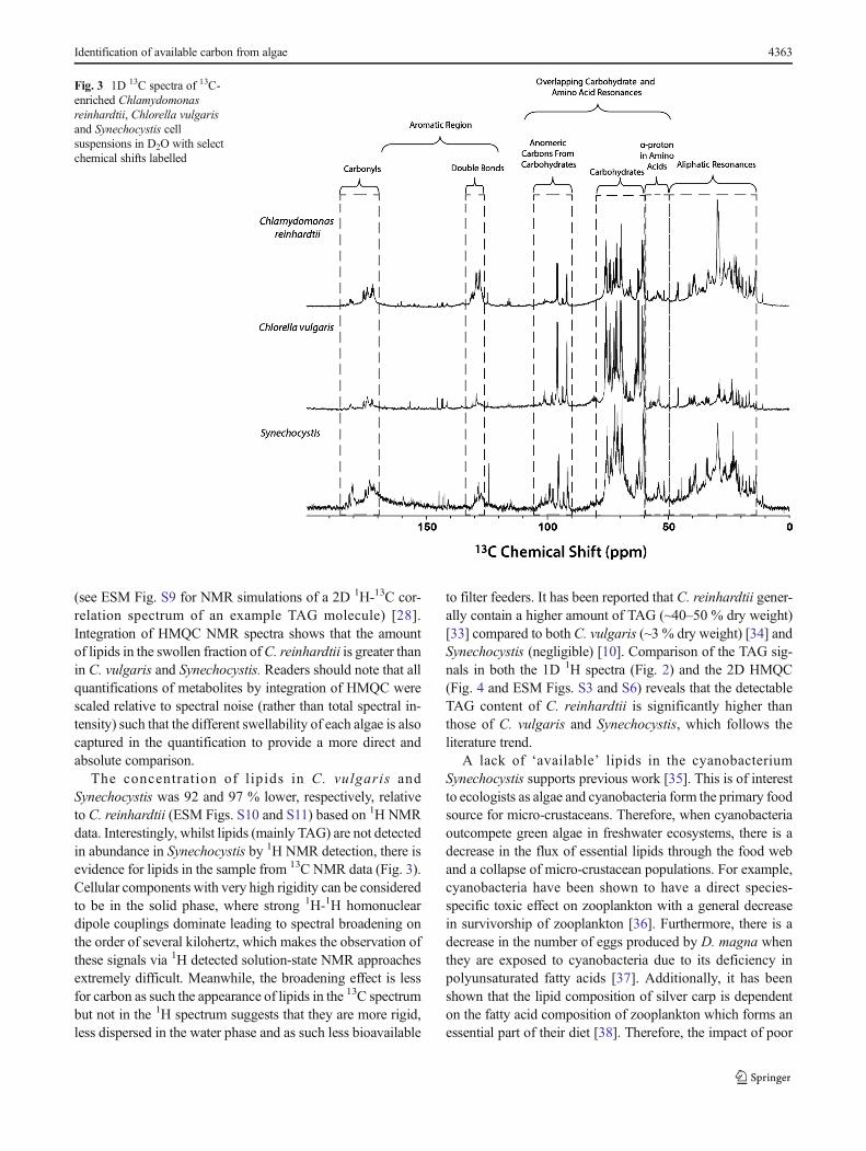
The 1D solution-state 13C NMR spectra of whole cell 13C-enriched green algae and cyanobacteria suspended in D2O areshown in Fig. 3. The spectra show significant overlap; how-ever, chemical shift regions corresponding to aliphatics (5–50 ppm), α-protons in amino acids (50–60 ppm) and carbo-hydrates (60–80 ppm), anomeric carbons of carbohydrates(90–105 ppm), double bonds from triacylglycerides (TAG)(125–135 ppm) and carbonyl groups for proteins and lipids(170–190 ppm) were identified [28]. The characteristic doublebond resonance (–C=C–, 125–135 ppm) indicates that theC. reinhardtii has a much higher TAG content than theC. vulgaris and Synechocystis. Carbohydrates are easilydiscerned in the carbon spectrum, which dominate inC. vulgaris with significantly less in Synechocystis andC. reinhardtii. Individual metabolites could not be identified
using the 1D carbon spectra alone due to spectral overlap andrequire additional information from 2D NMR.
2D HMQC (1H-13C) NMR
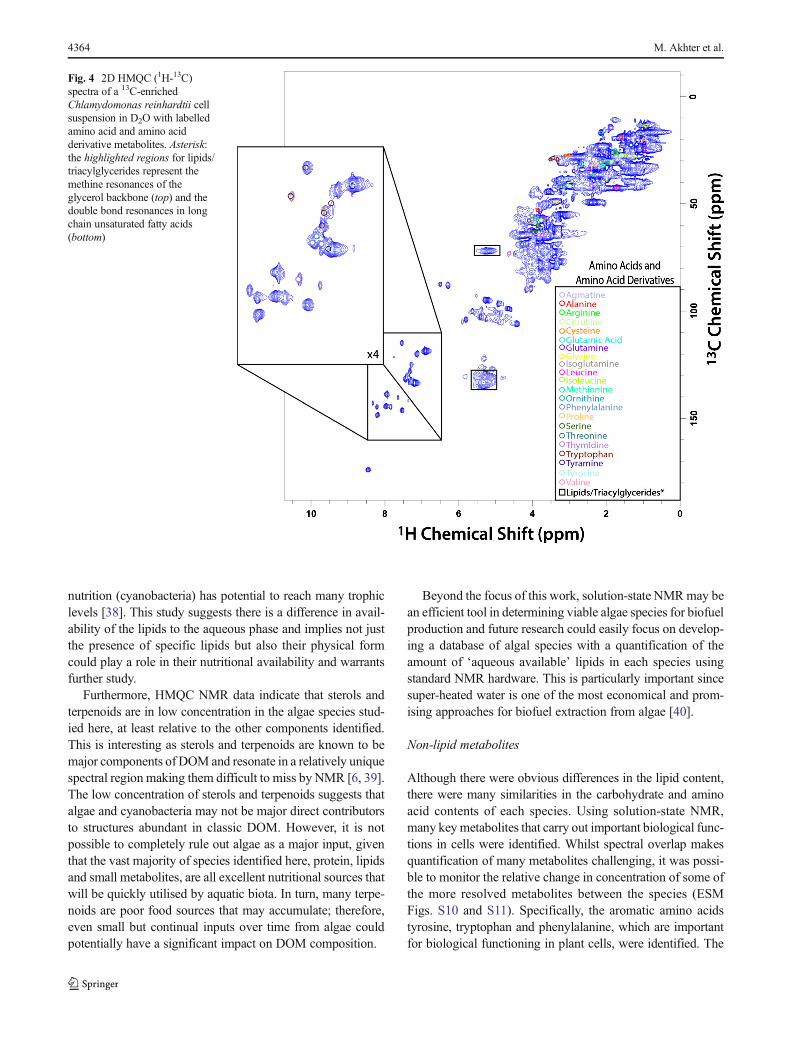
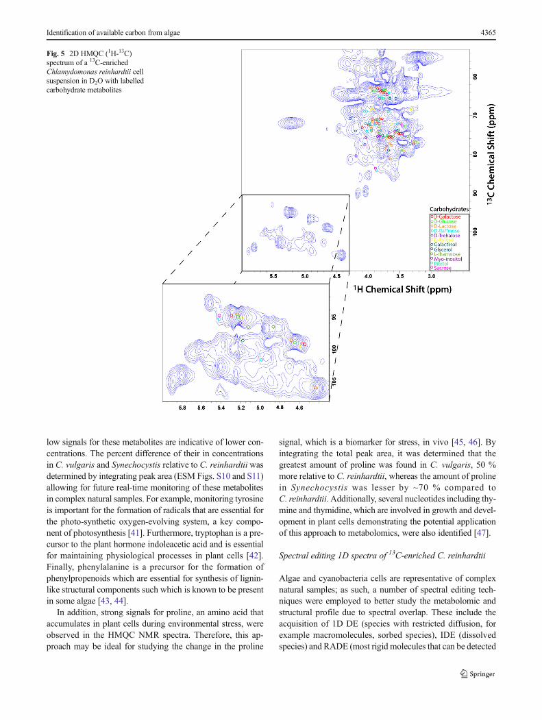
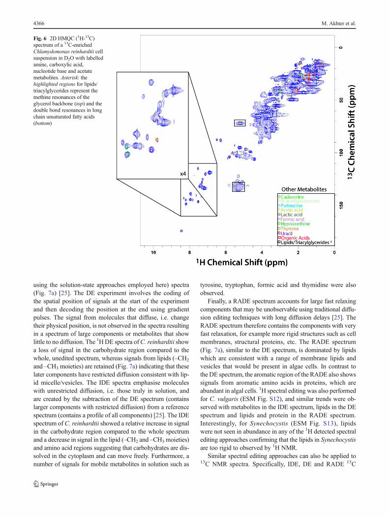
2D or 2D NMR spectroscopy is ideal for the identification ofindividual metabolites in complex heterogeneous samples as itincreases spectral dispersion and provides coupling informa-tion allowing individual metabolites to be identified. HMQCwas selected in this study due to its superior ability to suppresswater in aqueous samples. Both HSQC and HMQC performwell, and their applications to environmental samples arediscussed in the details in practical guides and reviews byFarooq et al. [29] and Simpson et al. [30]. Readers should notethat although absolute quantification can be achieved in 2DNMR spectroscopy [31], only relative quantification was thegoal here. Many metabolites including a range of amino acidsand amino acid derivatives were confirmed by the HMQCNMR spectra (Fig. 4). In particular, the aromatic amino acidsphenylalanine and tyrosine, which showed weak signals in 1HNMR, were clearly identifiable. A wide range of carbohy-drates both monosaccharides and disaccharides were observ-able, although there still was considerable overlap in the car-bohydrate region (Fig. 5). The anomeric carbon region pro-vided further confirmation of carbohydrate metabolites.Furthermore, some amines, common organic acids and
Fig. 1 1D 1H spectra of 13C-enriched Chlamydomonasreinhardtii, Chlorella vulgarisand Synechocystis cellsuspensions in D2O with selectchemical shifts labelled (Asterisk:in Chlamydomonas reinhardtiiwhere lipids dominate, this regioncontains strong contributionsfrom lipid functionalities)
Identification of available carbon from algae 4361
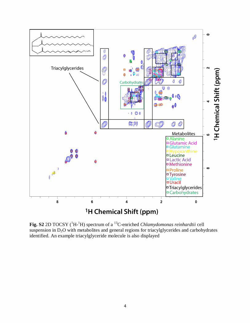
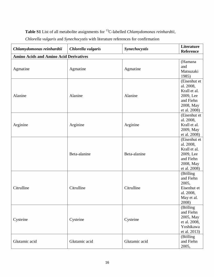
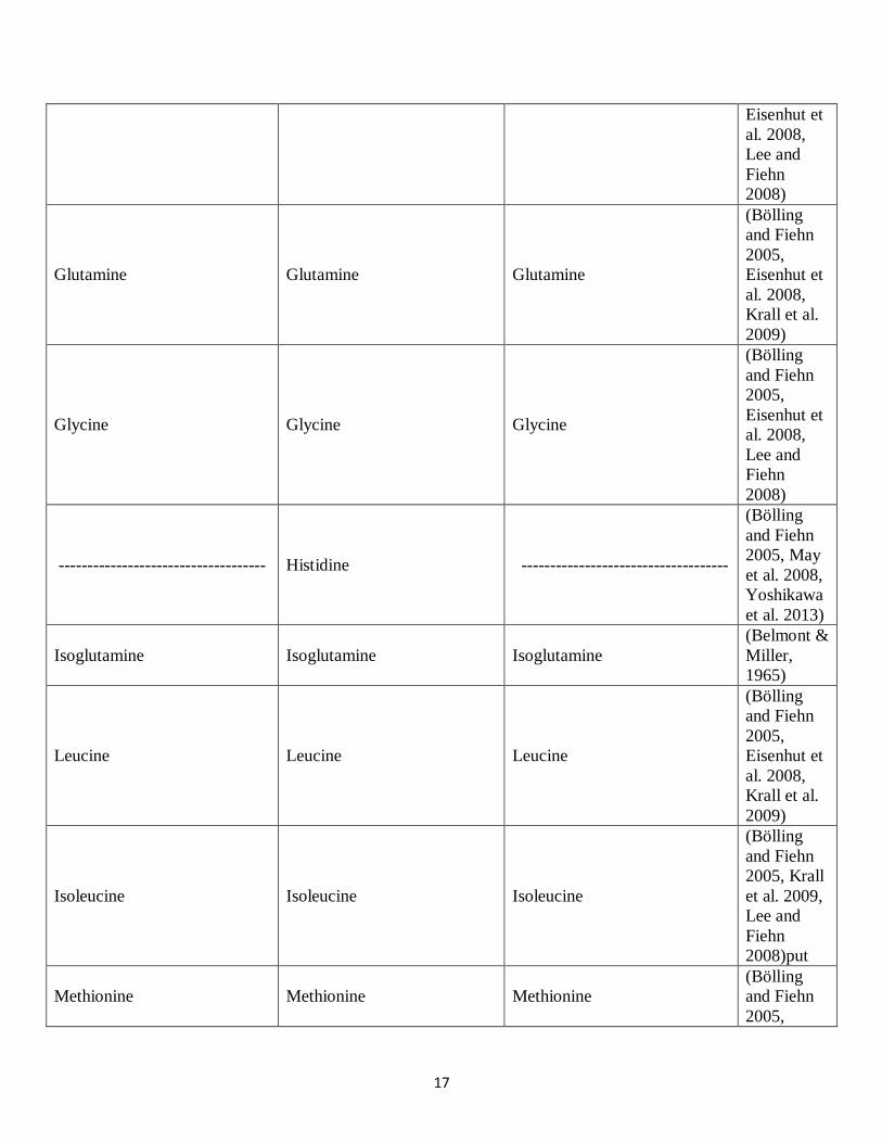
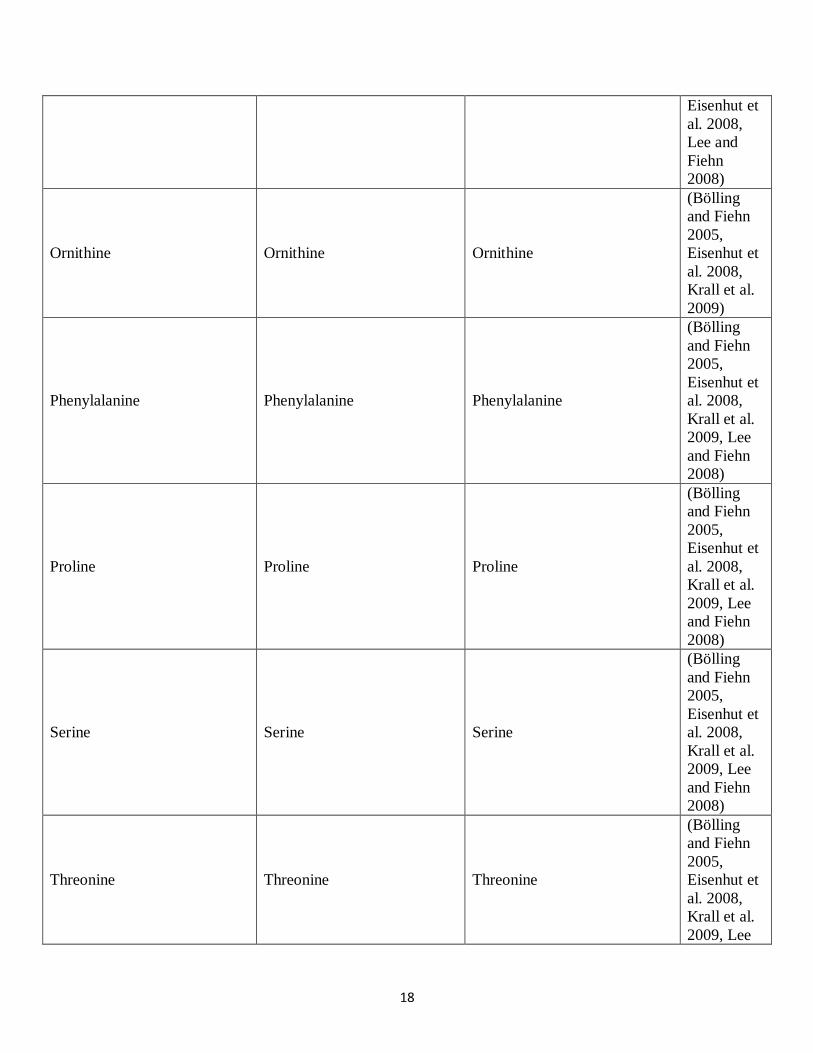
nucleotide bases were also identified (Fig. 6). Metabolite as-signments were cross-referenced with 1H-1H TOCSY datawhere possible (ESM Fig. S2) and confirmed with literature(ESM Table S1).
Detailed analysis of the metabolomic profileof C. reinhardtii, C. vulgaris and Synechocystis
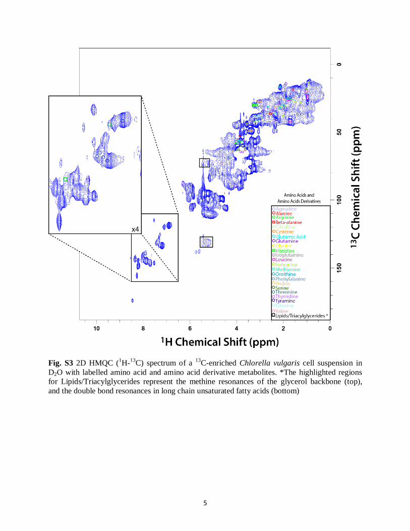
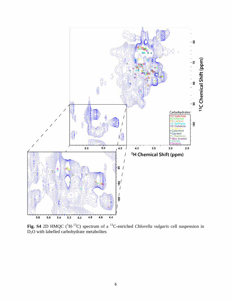
A total of 43 metabolites were identified in C. reinhardtii, 49metabolites were identified in C. vulgaris (ESM Figs. S3–S5)and 48 metabolites were identified in Synechocystis (ESMFigs. S6–S8) whole cell suspensions in D2O from HMQCspectra. The assignments correlated to expected chemicalshifts with an r2≥ 0.99. However, due to spectral overlap,the assignment of any particular metabolite or lack thereofmust be taken in light that a great range of organic residuesare present in algae leading to a wide range of overlappingpeaks. As such, assignments here will certainly bias the moreintense and spectrally resolved metabolites that can beassigned with the most confidence. The NMR data are highlycomplex and likely contain a wealth of algae and species-specific information that unfortunately cannot be fullyassigned. Nonetheless, it provides a single-technique ap-proach for metabolite profiling, and was able to identify awide arrange of metabolites, that otherwise may have required
multiple different analytical techniques for complete detection(see ESM, Table S1 and references therein). In the followingsection, a detailed overview of metabolites in each species andtheir potential impact in relation to the ecosystem, DOM andbiofuel synthesis is presented.
Detailed analysis of lipids
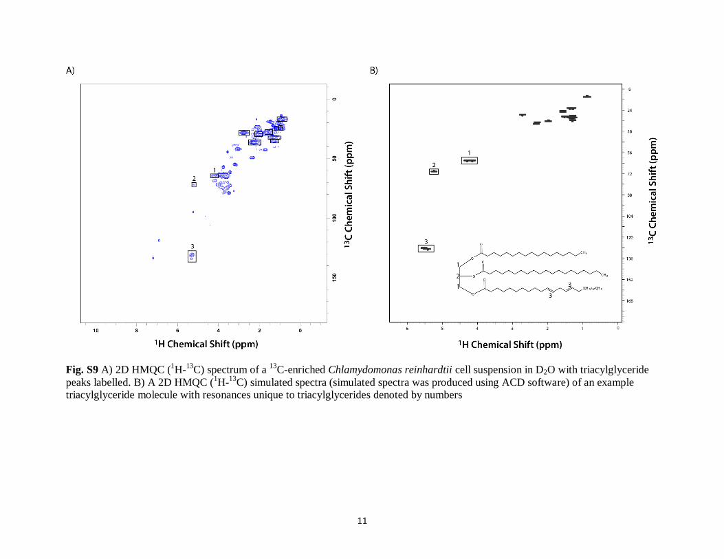
Green algae and cyanobacteria have distinct metabolite pro-files which reflect their nutritional value within the food web.In particular, cyanobacteria are known to be deficient in lipidsand TAG [32]. This is evident from 1H NMR spectra (Fig. 1)which show more intense signals for aliphatic metabolites(–CH2 and –CH3 moieties) in C. reinhardtii and C. vulgarisand weaker signal in Synechocystis. The 2D HMQC NMRspectra also confirm the difference in lipid content as the peakcorresponding to lipids/TAG shows a decrease in signalstrength in Synechocystis (ESM Fig. S8). Readers should notethat although there is considerable overlap in the lipid peaks,the presence of TAGwhichmakes a significant contribution tothe total lipid content in algae was specifically identified basedon the presence of two cross-peaks at ~5.2 and ~69.0 ppm and~5.3 and ~132.0 ppm, arising from the glycerol backbonemethine group and the double bonds in the long chain unsat-urated fatty acids, respectively, both characteristic of TAG
Fig. 2 1D 1H spectra of thearomatic region (5 to 10 ppm) of13C-enriched Chlamydomonasreinhardtii, Chlorella vulgarisand Synechocystis cellsuspensions in D2O withimportant metabolites labelled
4362 M. Akhter et al.
(see ESM Fig. S9 for NMR simulations of a 2D 1H-13C cor-relation spectrum of an example TAG molecule) [28].Integration of HMQC NMR spectra shows that the amountof lipids in the swollen fraction ofC. reinhardtii is greater thanin C. vulgaris and Synechocystis. Readers should note that allquantifications of metabolites by integration of HMQC werescaled relative to spectral noise (rather than total spectral in-tensity) such that the different swellability of each algae is alsocaptured in the quantification to provide a more direct andabsolute comparison.
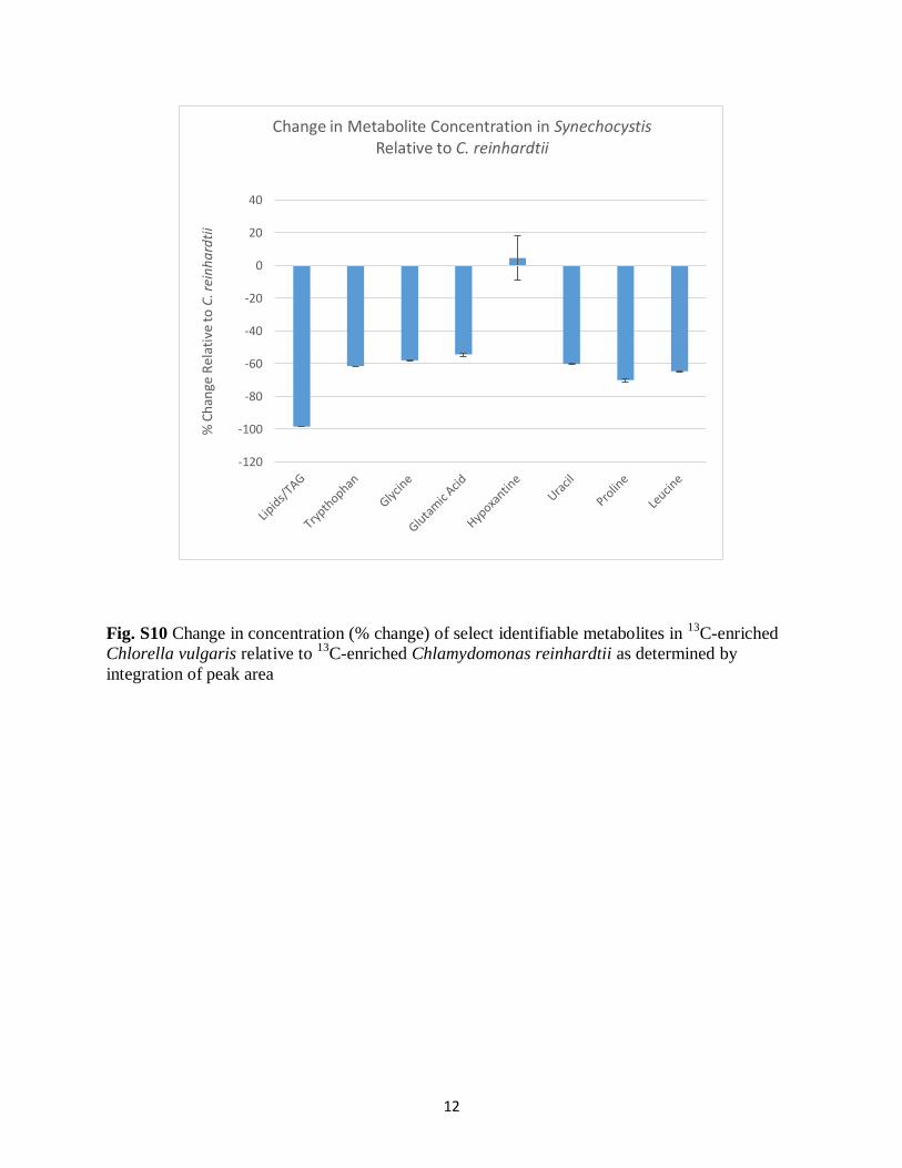
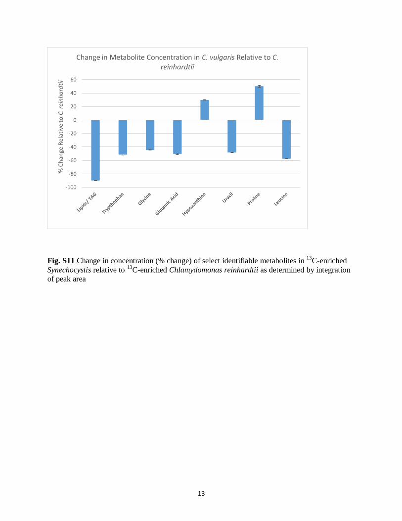
The concentration of lipids in C. vulgaris andSynechocystis was 92 and 97 % lower, respectively, relativeto C. reinhardtii (ESM Figs. S10 and S11) based on 1H NMRdata. Interestingly, whilst lipids (mainly TAG) are not detectedin abundance in Synechocystis by 1H NMR detection, there isevidence for lipids in the sample from 13C NMR data (Fig. 3).Cellular components with very high rigidity can be consideredto be in the solid phase, where strong 1H-1H homonucleardipole couplings dominate leading to spectral broadening onthe order of several kilohertz, which makes the observation ofthese signals via 1H detected solution-state NMR approachesextremely difficult. Meanwhile, the broadening effect is lessfor carbon as such the appearance of lipids in the 13C spectrumbut not in the 1H spectrum suggests that they are more rigid,less dispersed in the water phase and as such less bioavailable
to filter feeders. It has been reported that C. reinhardtii gener-ally contain a higher amount of TAG (~40–50 % dry weight)[33] compared to both C. vulgaris (~3 % dry weight) [34] andSynechocystis (negligible) [10]. Comparison of the TAG sig-nals in both the 1D 1H spectra (Fig. 2) and the 2D HMQC(Fig. 4 and ESM Figs. S3 and S6) reveals that the detectableTAG content of C. reinhardtii is significantly higher thanthose of C. vulgaris and Synechocystis, which follows theliterature trend.
A lack of ‘available’ lipids in the cyanobacteriumSynechocystis supports previous work [35]. This is of interestto ecologists as algae and cyanobacteria form the primary foodsource for micro-crustaceans. Therefore, when cyanobacteriaoutcompete green algae in freshwater ecosystems, there is adecrease in the flux of essential lipids through the food weband a collapse of micro-crustacean populations. For example,cyanobacteria have been shown to have a direct species-specific toxic effect on zooplankton with a general decreasein survivorship of zooplankton [36]. Furthermore, there is adecrease in the number of eggs produced by D. magna whenthey are exposed to cyanobacteria due to its deficiency inpolyunsaturated fatty acids [37]. Additionally, it has beenshown that the lipid composition of silver carp is dependenton the fatty acid composition of zooplankton which forms anessential part of their diet [38]. Therefore, the impact of poor
Fig. 3 1D 13C spectra of 13C-enriched Chlamydomonasreinhardtii, Chlorella vulgarisand Synechocystis cellsuspensions in D2O with selectchemical shifts labelled
Identification of available carbon from algae 4363
nutrition (cyanobacteria) has potential to reach many trophiclevels [38]. This study suggests there is a difference in avail-ability of the lipids to the aqueous phase and implies not justthe presence of specific lipids but also their physical formcould play a role in their nutritional availability and warrantsfurther study.
Furthermore, HMQC NMR data indicate that sterols andterpenoids are in low concentration in the algae species stud-ied here, at least relative to the other components identified.This is interesting as sterols and terpenoids are known to bemajor components of DOM and resonate in a relatively uniquespectral region making them difficult to miss by NMR [6, 39].The low concentration of sterols and terpenoids suggests thatalgae and cyanobacteria may not be major direct contributorsto structures abundant in classic DOM. However, it is notpossible to completely rule out algae as a major input, giventhat the vast majority of species identified here, protein, lipidsand small metabolites, are all excellent nutritional sources thatwill be quickly utilised by aquatic biota. In turn, many terpe-noids are poor food sources that may accumulate; therefore,even small but continual inputs over time from algae couldpotentially have a significant impact on DOM composition.
Beyond the focus of this work, solution-state NMRmay bean efficient tool in determining viable algae species for biofuelproduction and future research could easily focus on develop-ing a database of algal species with a quantification of theamount of ‘aqueous available’ lipids in each species usingstandard NMR hardware. This is particularly important sincesuper-heated water is one of the most economical and prom-ising approaches for biofuel extraction from algae [40].
Non-lipid metabolites
Although there were obvious differences in the lipid content,there were many similarities in the carbohydrate and aminoacid contents of each species. Using solution-state NMR,many keymetabolites that carry out important biological func-tions in cells were identified. Whilst spectral overlap makesquantification of many metabolites challenging, it was possi-ble to monitor the relative change in concentration of some ofthe more resolved metabolites between the species (ESMFigs. S10 and S11). Specifically, the aromatic amino acidstyrosine, tryptophan and phenylalanine, which are importantfor biological functioning in plant cells, were identified. The
Fig. 4 2D HMQC (1H-13C)spectra of a 13C-enrichedChlamydomonas reinhardtii cellsuspension in D2O with labelledamino acid and amino acidderivative metabolites. Asterisk:the highlighted regions for lipids/triacylglycerides represent themethine resonances of theglycerol backbone (top) and thedouble bond resonances in longchain unsaturated fatty acids(bottom)
4364 M. Akhter et al.
low signals for these metabolites are indicative of lower con-centrations. The percent difference of their in concentrationsin C. vulgaris and Synechocystis relative to C. reinhardtiiwasdetermined by integrating peak area (ESM Figs. S10 and S11)allowing for future real-time monitoring of these metabolitesin complex natural samples. For example, monitoring tyrosineis important for the formation of radicals that are essential forthe photo-synthetic oxygen-evolving system, a key compo-nent of photosynthesis [41]. Furthermore, tryptophan is a pre-cursor to the plant hormone indoleacetic acid and is essentialfor maintaining physiological processes in plant cells [42].Finally, phenylalanine is a precursor for the formation ofphenylpropenoids which are essential for synthesis of lignin-like structural components such which is known to be presentin some algae [43, 44].
In addition, strong signals for proline, an amino acid thataccumulates in plant cells during environmental stress, wereobserved in the HMQC NMR spectra. Therefore, this ap-proach may be ideal for studying the change in the proline
signal, which is a biomarker for stress, in vivo [45, 46]. Byintegrating the total peak area, it was determined that thegreatest amount of proline was found in C. vulgaris, 50 %more relative to C. reinhardtii, whereas the amount of prolinein Synechocystis was lesser by ~70 % compared toC. reinhardtii. Additionally, several nucleotides including thy-mine and thymidine, which are involved in growth and devel-opment in plant cells demonstrating the potential applicationof this approach to metabolomics, were also identified [47].
Spectral editing 1D spectra of 13C-enriched C. reinhardtii
Algae and cyanobacteria cells are representative of complexnatural samples; as such, a number of spectral editing tech-niques were employed to better study the metabolomic andstructural profile due to spectral overlap. These include theacquisition of 1D DE (species with restricted diffusion, forexample macromolecules, sorbed species), IDE (dissolvedspecies) and RADE (most rigid molecules that can be detected
Fig. 5 2D HMQC (1H-13C)spectrum of a 13C-enrichedChlamydomonas reinhardtii cellsuspension in D2O with labelledcarbohydrate metabolites
Identification of available carbon from algae 4365
using the solution-state approaches employed here) spectra(Fig. 7a) [25]. The DE experiment involves the coding ofthe spatial position of signals at the start of the experimentand then decoding the position at the end using gradientpulses. The signal from molecules that diffuse, i.e. changetheir physical position, is not observed in the spectra resultingin a spectrum of large components or metabolites that showlittle to no diffusion. The 1HDE spectra ofC. reinhardtii showa loss of signal in the carbohydrate region compared to thewhole, unedited spectrum, whereas signals from lipids (–CH2
and –CH3 moieties) are retained (Fig. 7a) indicating that theselater components have restricted diffusion consistent with lip-id micelle/vesicles. The IDE spectra emphasise moleculeswith unrestricted diffusion, i.e. those truly in solution, andare created by the subtraction of the DE spectrum (containslarger components with restricted diffusion) from a referencespectrum (contains a profile of all components) [25]. The IDEspectrum of C. reinhardtii showed a relative increase in signalin the carbohydrate region compared to the whole spectrumand a decrease in signal in the lipid (–CH2 and –CH3moieties)and amino acid regions suggesting that carbohydrates are dis-solved in the cytoplasm and can move freely. Furthermore, anumber of signals for mobile metabolites in solution such as
tyrosine, tryptophan, formic acid and thymidine were alsoobserved.
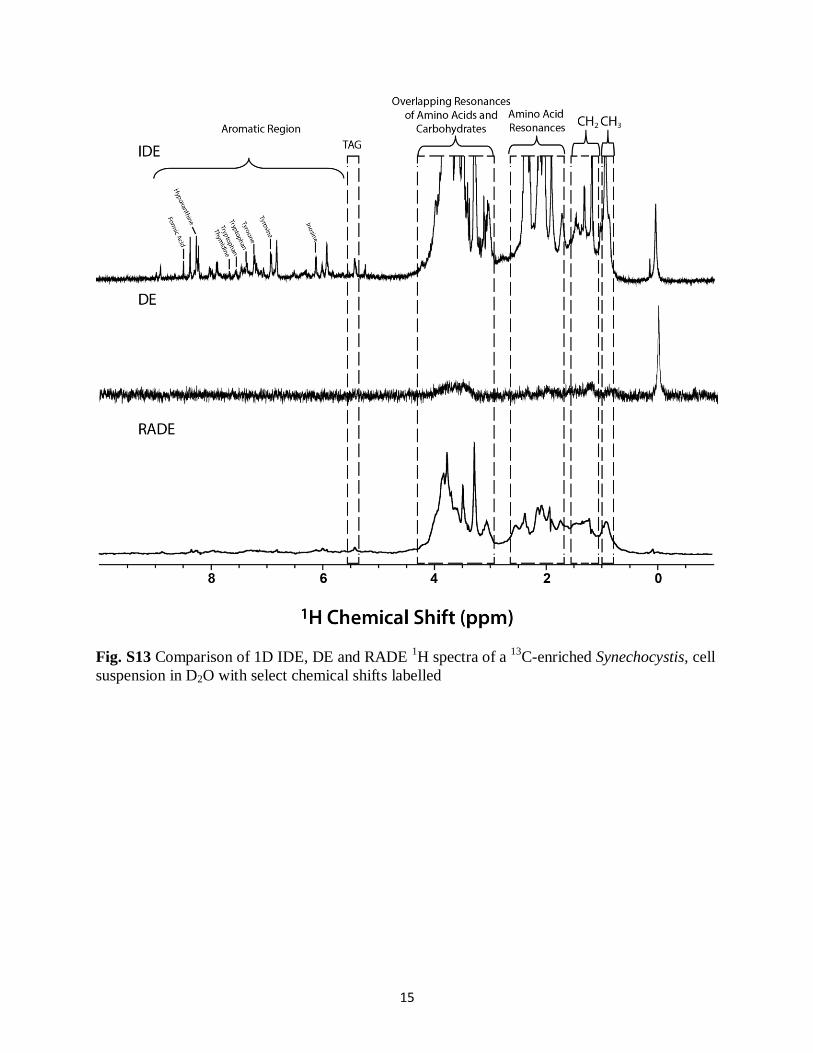
Finally, a RADE spectrum accounts for large fast relaxingcomponents that may be unobservable using traditional diffu-sion editing techniques with long diffusion delays [25]. TheRADE spectrum therefore contains the components with veryfast relaxation, for example more rigid structures such as cellmembranes, structural proteins, etc. The RADE spectrum(Fig. 7a), similar to the DE spectrum, is dominated by lipidswhich are consistent with a range of membrane lipids andvesicles that would be present in algae cells. In contrast tothe DE spectrum, the aromatic region of the RADE also showssignals from aromatic amino acids in proteins, which areabundant in algal cells. 1H spectral editing was also performedfor C. vulgaris (ESM Fig. S12), and similar trends were ob-served with metabolites in the IDE spectrum, lipids in the DEspectrum and lipids and protein in the RADE spectrum.Interestingly, for Synechocystis (ESM Fig. S13), lipidswere not seen in abundance in any of the 1H detected spectralediting approaches confirming that the lipids in Synechocystisare too rigid to observed by 1H NMR.
Similar spectral editing approaches can also be applied to13C NMR spectra. Specifically, IDE, DE and RADE 13C
Fig. 6 2D HMQC (1H-13C)spectrum of a 13C-enrichedChlamydomonas reinhardtii cellsuspension in D2O with labelledamine, carboxylic acid,nucleotide base and acetatemetabolites. Asterisk: thehighlighted regions for lipids/triacylglycerides represent themethine resonances of theglycerol backbone (top) and thedouble bond resonances in longchain unsaturated fatty acids(bottom)
4366 M. Akhter et al.
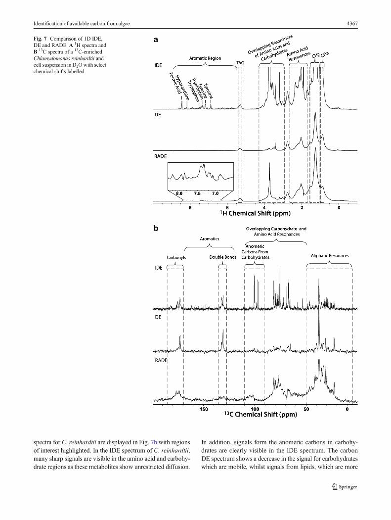
spectra for C. reinhardtii are displayed in Fig. 7b with regionsof interest highlighted. In the IDE spectrum of C. reinhardtii,many sharp signals are visible in the amino acid and carbohy-drate regions as these metabolites show unrestricted diffusion.
In addition, signals form the anomeric carbons in carbohy-drates are clearly visible in the IDE spectrum. The carbonDE spectrum shows a decrease in the signal for carbohydrateswhich are mobile, whilst signals from lipids, which are more
Fig. 7 Comparison of 1D IDE,DE and RADE. A 1H spectra andB 13C spectra of a 13C-enrichedChlamydomonas reinhardtii andcell suspension in D2Owith selectchemical shifts labelled
Identification of available carbon from algae 4367
restricted in their movement, are retained. Furthermore, theRADE carbon spectrum is dominated by lipids, particularlythe fraction of lipids in cells that have a more rigid character.As well, the presence of aromatic amino acid signals in theRADE spectra is indicative of large protein molecules whichexhibit restricted diffusion. The carbon IDE, DE and RADEspectra provide complimentary spectral information to theproton spectra. These diffusion edited NMR techniques areuseful as they separate signals of the free dissolved metabo-lites from structural components (lipids and proteins), de-crease overlap and provide some information as to the phys-ical form of the components within the cells. Editing based ondiffusivity has been previously applied to study complex mix-tures such as blood plasma, tissues and urine [48–52].However, the RADE experiment, which recovers fast relaxingcomponents that otherwise may be overlooked by diffusionbased editing, is relatively new [25]. When combined, inversediffusion (metabolites), diffusion editing (restricted diffusion)and RADE (semi-solids) provide a convenient and discreteapproach for the separation of major components in mixtures,including, as demonstrated here, application to whole cellsuspensions.
Conclusion
This study demonstrates that solution-state NMR can be usedto study intact algae cells allowing the observation of majordifferences between species and identify key metabolites.Specifically, the difference in the lipid profile between greenalgae and cyanobacteria was confirmed, shedding light on thecurrent understanding of the impact of cyanobacteria bloomson freshwater ecosystems. More importantly, a complete listof identifiable metabolites in each species was compiled,confirming and complimenting those previously reported.This work is important for future NMR-based metabolomicstudies as algae and cyanobacteria are often the primarysource for carbon labelling. For example, higher organismssuch as mice can be 13C-labelled by feeding them fish thathave eaten daphnia which have been grown on 13C-labelledalgae [53]. In order to understand the metabolomic changesoccurring in higher organisms, it is essential to understand themetabolite profile and flux from the primary source—algaeand cyanobacteria—to higher organisms.
However, providing absolute quantification was challeng-ing due to overlap in 1D spectra forcing identification andrelative quantification from 2DNMR for specific metabolites.Whilst 2D is generally considered less quantitative than 1DNMR, improvements in the field and the extension of an elec-tronic referencing method, Electronic REference To accessIn vivo Concentrations (ERETIC), have been shown to workfor 2D NMR and once integrated into commercially availablesoftware will make absolute quantification for 2D NMR
possible in the future [54]. This will allow the monitoring allthe metabolites using solution-state NMR to understand bio-logical processes in vivo. Studies that follow the consumptionof labelled algae through the food chain will also be possibleand could be extremely informative as the fate and conversionof the label at the molecular level may be monitored,explaining how and why organisms depend on each otheracross trophic levels.
The study also suggests that algae and cyanobacteria arelikely not major direct contributors to DOM composition asthey contain low concentrations of sterols and terpenoids inthe mobile state but still could be important sources after en-vironmental fractionation and preservation. Conversely, algalinputs may be highly significant to protein-rich DOM that hasbeen recently discovered in high-elevation lakes [55].
The ability to provide relative quantification as to theamount of lipids in each species may prove to be valuableto researchers trying to develop genetically modified al-gae strains with larger lipid fractions to increase the yieldof biofuel per unit mass algae. Of the three strains ofalgae studied here, it is clear that C. reinhardtii has byfar the highest proportion of aqueous available lipids, sug-gesting it as a potential strain for biofuel production. Eventhough valuable information can be gained using solution-state NMR, assignment of metabolites is challenging dueto significant spectral overlap. Future studies using 3DNMR should provide the additional spectral dispersionrequired for further spectral assignments [56], presentlynot feasible due to spectral overlap.
In addition, solution-state NMR provides the ability to se-lectively study dissolved/mobile species. This can be an ad-vantage where the goal of the study is to identify the aquati-cally available fraction as is the goal here but could be adrawback if all components in the cells (including true solids)need to be detected. The latter should be possible by the use ofa novel technique called comprehensive multiphase NMR(CMP-NMR) introduced in 2012, which allows researchersto access molecular information from all three phases simul-taneously [25]. This study demonstrated that lipids inSynechocystis were more ‘solid-like’ and less available tothe aqueous phase than in the other strains studied. TheCMP-NMR technique could be highly complementary pro-viding the additional information on the semi-solid and solidphases needed to explain their availability.
In summary, this study provides a structural and metabolicoverview as to the mobile/dissolved components in the naturalstate which are most likely to directly transfer into the aqueousphase on lysis. The study represents a key first step, permittingfuture studies of carbon transfer between species and throughthe food chain, as well as providing a foundation to betterunderstand the role of algae in the formation of DOM andsequestration/transformation of carbon in the aquatic environ-ment in general.
4368 M. Akhter et al.
Acknowledgments A. J. S. would like to thank Mark Krembil and theKrembil Foundation for both inspiring and funding this research. A. J. S.would like to thank the Natural Sciences and Engineering ResearchCouncil of Canada (NSERC) for financially supporting this research.
Compliance with ethical standards
Conflict of Interest The authors declare no conflicts of interest.
References
1. Paerl HW, Xu H, McCarthy MJ, Zhu G, Qin B, Li Y, et al.Controlling harmful cyanobacterial blooms in a hyper-eutrophiclake (Lake Taihu, China): the need for a dual nutrient (N & P)management strategy. Water Res. 2011;45:1973–83.
2. PiorreckM, Baasch K-H, Pohl P. Biomass production, total protein,chlorophylls, lipids and fatty acids of freshwater green and blue-green algae under different nitrogen regimes. Phytochemistry.1984;23:207–16.
3. Martin-Creuzburg D, Wacker A, von Elert E. Life history conse-quences of sterol availability in the aquatic keystone speciesDaphnia. Oecologia. 2005;144:362–72.
4. Bednarska A, Slusarczyk M. Effect of non-toxic, filamentouscyanobacteria on egg abortion in Daphnia under various thermalconditions. Hydrobiologia. 2013;715:151–7.
5. Perhar G, Arhonditsis GB, Brett MT. Modelling the role of highlyunsaturated fatty acids in planktonic food web processes: a mecha-nistic approach. Environ Rev. 2012;20:155–72.
6. Lam B, Baer A, Alaee M, Lefebvre B, Moser A, Williams A, et al.Major structural components in freshwater dissolved organic mat-ter. Environ Sci Technol. 2007;41:8240–7.
7. Demirbas A, Fatih DM. Importance of algae oil as a source ofbiodiesel. Energy Convers Manag. 2011;52:163–70.
8. Griffiths MJ, Harrison STL. Lipid productivity as a key character-istic for choosing algal species for biodiesel production. J ApplPhycol. 2009;21:493–507.
9. Scott SA, Davey MP, Dennis JS, Horst I, Howe CJ, Lea-Smith DJ,et al. Biodiesel from algae: challenges and prospects. Curr OpinBiotechnol. 2010;21:277–86.
10. Wahlen BD, Willis RM, Seefeldt LC. Biodiesel production by si-multaneous extraction and conversion of total lipids frommicroalgae, cyanobacteria, and wild mixed-cultures. BioresourTechnol. 2011;102:2724–30.
11. Radakovits R, Jinkerson RE, Darzins A, Posewitz MC. Geneticengineering of algae for enhanced biofuel production. EukaryotCell. 2010;9:486–501.
12. Bolling C, Fiehn O. Metabolite profiling of Chlamydomonasreinhardtii under nutrient deprivation. Plant Physiol. 2005;139:1995–2005.
13. Madsen AD, Goessler W, Pedersen SN, Francesconi KA.Characterization of an algal extract by HPLC-ICP-MS and LC-electrospray MS for use in arsenosugar speciation studies. J AnalAt Spectrom. 2000;15:657–62.
14. Halket JM,WatermanD, Przyborowska AM, Patel RKP, Fraser PD,Bramley PM. Chemical derivatization and mass spectral libraries inmetabolic profiling by GC/MS and LC/MS/MS. J Exp Bot.2004;56:219–43.
15. Aguilar JA, Cassani J, Delbianco M, Adams RW, Nilsson M,Morris GA. Minimising research bottlenecks by declutteringNMR spectra. Chem Eur J. 2015;21:6623–30.
16. Gupta V, Thakur RS, Reddy CRK, Jha B. Central metabolic pro-cesses of marine macrophytic algae revealed from NMR basedmetabolome analysis. RSC Adv. 2013;3:7037–47.
17. Pandit A, Morosinotto T, Reus M, Holzwarth AR, Bassi R, deGroot HJM. First solid-state NMR analysis of uniformly 13C-enriched major light-harvesting complexes from Chlamydomonasreinhardtii and identification of protein and cofactor spin clusters.Biochim Biophys Acta Bioenerg. 1807;2011:437–43.
18. Arnold AA, Genard B, Zito F, Tremblay R, Warschawski DE,Marcotte I. Identification of lipid and saccharide constituents ofwhole microalgal cells by 13C solid-state NMR. Biochim BiophysActa. 1848;2015:369–77.
19. Chauton MS, Optun OI, Bathen TF, Volent Z, Gribbestad IS. HRMAS 1H NMR spectroscopy analysis of marine microalgal wholecells. Mar Ecol Prog Ser. 2003;256:57–62.
20. Chauton MS, Røvik Størseth T, Johnsen G. High-resolution magicangle spinning 1H NMR analysis of whole cells of Thalassiosirapseudonana (Bacillariophyceae): broad range analysis of metaboliccomposition and nutritional value. J Appl Phycol. 2003;15:533–42.
21. Chauton MS, Størseth TR, Krane J. High-resolution magic anglespinning NMR analysis of whole cells of Chaetoceros muelleri(Bacillariophyceae) and comparison with 13C-NMR anddistortionless enhancement by polarization transfer 13C-NMR anal-ysis of lipophilic extracts. J Phycol. 2004;40:611–8.
22. Simpson AJ, Simpson MJ, Soong R. Nuclear magnetic resonancespectroscopy and its key role in environmental research. EnvironSci Technol. 2012;46:11488–96.
23. Lam B, Simpson AJ. Direct 1H NMR spectroscopy of dissolvedorganic matter in natural waters. Analyst. 2008;133:263–9.
24. Wu DH, Chen AD, Johnson CS. An improved diffusion-orderedspectroscopy experiment incorporating bipolar-gradient pulses. JMagn Reson Ser A. 1995;115:260–4.
25. Courtier-Murias D, Farooq H, Masoom H, Botana A, Soong R,Longstaffe JG, et al. Comprehensive multiphase NMR spectrosco-py: basic experimental approaches to differentiate phases in hetero-geneous samples. J Magn Reson. 2012;217:61–76.
26. Peti W, Griesinger C, Bermel W. Adiabatic TOCSY for C,C and H,H J-transfer. J Biomol NMR. 2000;18:199–205.
27. Woods GC, Simpson MJ, Koerner PJ, Napoli A, Simpson AJ.HILIC-NMR: toward the identification of individual molecularcomponents in dissolved organic matter. Environ Sci Technol.2011;45:3880–6.
28. Lam L, Soong R, Sutrisno A, de Visser R, Simpson MJ, WheelerHL, et al. Comprehensive multiphase NMR spectroscopy of intact13C-labeled seeds. J Agric Food Chem. 2014;62:107–15.
29. Farooq H, Courtier-Murias D, Soong R, Bermel W, Kingery WM,Simpson AJ. HR-MAS NMR spectroscopy: a practical guide fornatural samples. Curr Org Chem. 2013;17:3013–31.
30. Simpson AJ, McNally DJ, Simpson MJ. NMR spectroscopy inenvironmental research: from molecular interactions to global pro-cesses. Prog Nucl Magn Reson Spectrosc. 2011;58:97–175.
31. Giraudeau P. Quantitative 2D liquid-state NMR. Magn ResonChem. 2014;52:259–72.
32. Liu X, Sheng J, Curtiss R. Fatty acid production in geneticallymodified cyanobacteria. Proc Natl Acad Sci. 2011;108:6899–904.
33. Hu Q, Sommerfeld M, Jarvis E, Ghirardi M, Posewitz M, SeibertM, et al. Microalgal triacylglycerols as feedstocks for biofuel pro-duction: perspectives and advances. Plant J. 2008;54:621–39.
34. Stephenson AL, Dennis JS, Howe CJ, Scott SA, Smith AG.Influence of nitrogen-limitation regime on the production byChlorella vulgaris of lipids for biodiesel feedstocks. Biofuels.2010;1:47–58.
35. Cai T, Ge X, Park SY, Li Y. Comparison of Synechocystis sp.PCC6803 and Nannochloropsis salina for lipid production usingartificial seawater and nutrients from anaerobic digestion effluent.Bioresour Technol. 2013;144:255–60.
Identification of available carbon from algae 4369
36. DeMott WR, Zhang Q-X, Carmichael WW. Effects of toxiccyanobacteria and purified toxins on the survival and feeding of acopepod and three species ofDaphnia. Limnol Oceanogr. 1991;36:1346–57.
37. Wacker A, Martin-Creuzburg D. Allocation of essential lipids inDaphnia magna during exposure to poor food quality. FunctEcol. 2007;21:738–47.
38. Domaizon I, Desvilettes C, Debroas D, Bourdier G. Influence ofzooplankton and phytoplankton on the fatty acid composition ofdigesta and tissue lipids of silver carp: mesocosm experiment. JFish Biol. 2000;57:417–32.
39. Woods GC, Simpson MJ, Simpson AJ. Oxidized sterols as a sig-nificant component of dissolved organic matter: evidence from 2DHPLC in combination with 2D and 3D NMR spectroscopy. WaterRes. 2012;46:3398–408.
40. Savage PE, Levine R, Pinnarat T. Method of producing biodieselfrom a wet biomass. US Patent. 2014;8673028:B2.
41. Barry BA, Babcock GT. Tyrosine radicals are involved in the pho-tosynthetic oxygen-evolving system. Proc Natl Acad Sci. 1987;84:7099–103.
42. Bartel B. Auxin biosynthesis. Annu Rev Plant Physiol Plant MolBiol. 1997;48:51–66.
43. Delwiche CF, Graham LE, Thomson N. Lignin-like compoundsand Sporopollenin coleochaete, an algal model for land plant an-cestry. Science. 1989;245:399–401.
44. Ritter H, Schulz GE. Structural basis for the entrance into thephenylpropanoid metabolism catalyzed by phenylalanine ammo-nia-lyase. Plant Cell Online. 2004;16:3426–36.
45. Siripornadulsil S, Traina S, Verma DPS, Sayre RT. Molecularmechanisms of proline-mediated tolerance to toxic heavy metalsin transgenic microalgae. Plant Cell Online. 2002;14:2837–47.
46. Wu J, Chang S, Chou T. Intracellular proline accumulation in somealgae exposed to copper and cadmium. Bot Bull Acad Sin.1995;36:89–93.
47. Stasolla C, Katahira R, Thorpe TA, Ashihara H. Purine and pyrim-idine nucleotide metabolism in higher plants. J Plant Physiol.2003;160:1271–95.
48. Tang H, Wang Y, Nicholson JK, Lindon JC. Use of relaxation-edited one-dimensional and two dimensional nuclear magnetic res-onance spectroscopy to improve detection of small metabolites inblood plasma. Anal Biochem. 2004;325:260–72.
49. Wang Y, Bollard ME, Keun H, Antti H, Beckonert O,Ebbels TM, et al. Spectral editing and pattern recognitionmethods applied to high-resolution magic-angle spinning1H nuclear magnetic resonance spectroscopy of liver tissues.Anal Biochem. 2003;323:26–32.
50. Beckonert O, Keun HC, Ebbels TMD, Bundy J, Holmes E, LindonJC, et al. Metabolic profiling, metabolomic and metabonomic pro-cedures for NMR spectroscopy of urine, plasma, serum and tissueextracts. Nat Protoc. 2007;2:2692–703.
51. Smith LM, Maher AD, Cloarec O, Rantalainen M, Tang H, ElliottP, et al. Statistical correlation and projection methods for improvedinformation recovery from diffusion-edited NMR spectra of biolog-ical samples. Anal Chem. 2007;79:5682–9.
52. Liu M, Nicholson JK, Lindon JC. High-resolution diffusion andrelaxation edited one- and two-dimensional 1 H NMR spectroscopyof biological fluids. Anal Chem. 1996;68:3370–6.
53. Krüger M, Heumann H. Isotopic labeling of higher organisms. USPatent. 2014;20140328760:A1.
54. Michel N, Akoka S. The application of the ERETIC method to 2D-NMR. J Magn Reson. 2004;168:118–23.
55. Goldberg SJ, Ball GI, Allen BC, Schladow SG, Simpson AJ,Masoom H, et al. Refractory dissolved organic nitrogen accumula-tion in high-elevation lakes. Nat Commun. 2015;6:6347.
56. Simpson AJ, Kingery WL, Hatcher PG. The identification of plantderived structures in humic materials using three-dimensionalNMR spectroscopy. Environ Sci Technol. 2003;37:337–42.
4370 M. Akhter et al.
1
Analytical and Bioanalytical Chemistry
Electronic Supplementary Material
Identification of aquatically available carbon from algae through solution-
state NMR of whole 13
C-labelled cells
Mohammad Akhter, Rudraksha Dutta Majumdar, Blythe Fortier-McGill, Ronald Soong,
Yalda Liaghati Mobarhan, Myrna Simpson, George Arhonditsis, Sebastian Schmidt,
Hermann Heumann, André J. Simpson
2
13C Labeling Methodology
13
C-labeling of the algae (wt, SAG culture collection Goettingen, Germany) was achieved
by growing the cells in 13
CO2 (enrichment 99%; purchased from Sigma Aldrich Isotec, St Louis,
Missouri) for 150 hours in a photobioreactor (PBR) constructed by Silantes GmbH (Munich
Germany). The PBR is built as a closed system avoiding loss of the stable isotope labeled 13
CO2.
It is an airlift driven external loop tubular fermenter having 20L operating volume, permitting
circulation of the media volume with 1 vvm. The algae were cultivated in regular TP media
(Tris-Phosphate w/o Acetate + 15
NH4Cl), as previously described (Gorman and Levine, 1965),
using the following parameters: pH 7.0 - 7.4 (pH-probe InPro®3253I/Sg, Mettler Toledo),
temperature 30°C (InPro®3253I), light intensity 1300 μmol/(m
2∙s) (quantum detector LI-250A,
Li-Cor Biosciences GmbH). A computer-controlled gas management system (Labview 10.1,
National Instruments) was incorporated to keep the13
CO2 content (pCO2-probe
InPro®
5000,Mettler Toledo) at a concentration of 2%. The nitrogen carrier gas was allowed to
vary between 65% and 85% corresponding to the oxygen content which increased during
autotrophic growth of algae between 15% and 30%. If the oxygen content reached a value of
30% (pO2-probe Visiferm Do Arc., Hamilton) an N2 purging step was introduced in order to
reduce the O2 content to the initial value of 15%. To avoid loss of 13
CO2 during N2 purging, the
13CO2 addition was stopped at the end of a growth cycle until a value of 0,5% was reached by
metabolic depletion. If the oxygen reached the initial value of 15% due to N2-purging, the
computer controlled valves (Valve 221606, Buerkert) were closed and another growth cycle was
resumed. The 13
C-content of the algae biomass was determined by analyzing the enrichment in
“C18” fatty acids of isolated algae biomass by GCMS (Thermo Quest Polaris Q MS / Trace
GC2000, Thermo Fisher). Isotopic enrichment of 98-99% 13
C was achieved in the algae biomass.
3
Fig. S1 Swellability of Chlorella vulgaris and Synechocystis relative to Chlamydomonas
reinhardtii as determined by integration of 1D 13
C spectra
0
0,05
0,1
0,15
0,2
0,25
0,3
0,35
0,4
0,45
C. vulgaris Synechocystis
Swel
lab
ility
Rel
ativ
e to
C.
rein
ha
rdti
i
Swellability Factor
4
Fig. S2 2D TOCSY (1H-
1H) spectrum of a
13C-enriched Chlamydomonas reinhardtii cell
suspension in D2O with metabolites and general regions for triacylglycerides and carbohydrates
identified. An example triacylglyceride molecule is also displayed
5
Fig. S3 2D HMQC (1H-
13C) spectrum of a
13C-enriched Chlorella vulgaris cell suspension in
D2O with labelled amino acid and amino acid derivative metabolites. *The highlighted regions
for Lipids/Triacylglycerides represent the methine resonances of the glycerol backbone (top),
and the double bond resonances in long chain unsaturated fatty acids (bottom)
6
Fig. S4 2D HMQC (1H-
13C) spectrum of a
13C-enriched Chlorella vulgaris cell suspension in
D2O with labelled carbohydrate metabolites
7
Fig. S5 2D HMQC (1H-
13C) spectrum of a
13C-enriched Chlorella vulgaris cell suspension in
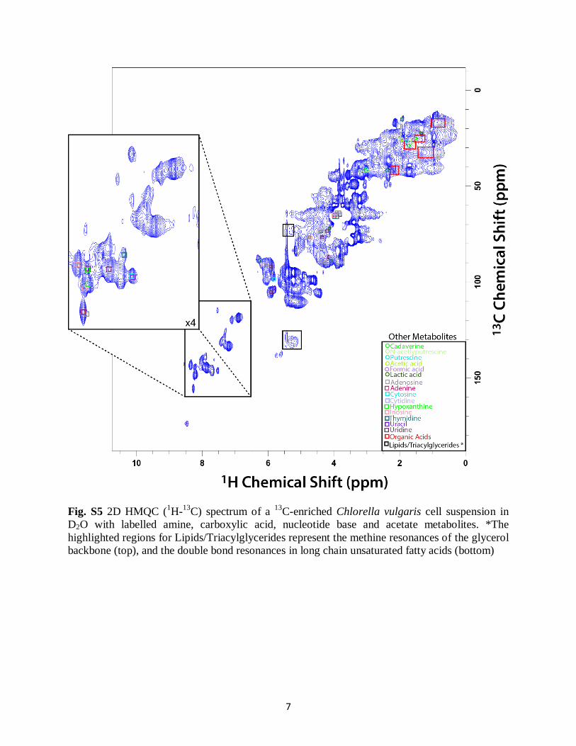
D2O with labelled amine, carboxylic acid, nucleotide base and acetate metabolites. *The
highlighted regions for Lipids/Triacylglycerides represent the methine resonances of the glycerol
backbone (top), and the double bond resonances in long chain unsaturated fatty acids (bottom)
8
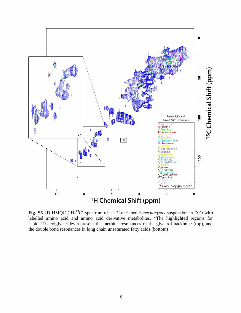
Fig. S6 2D HMQC (1H-
13C) spectrum of a
13C-enriched Synechocystis suspension in D2O with
labelled amino acid and amino acid derivative metabolites. *The highlighted regions for
Lipids/Triacylglycerides represent the methine resonances of the glycerol backbone (top), and
the double bond resonances in long chain unsaturated fatty acids (bottom)
9
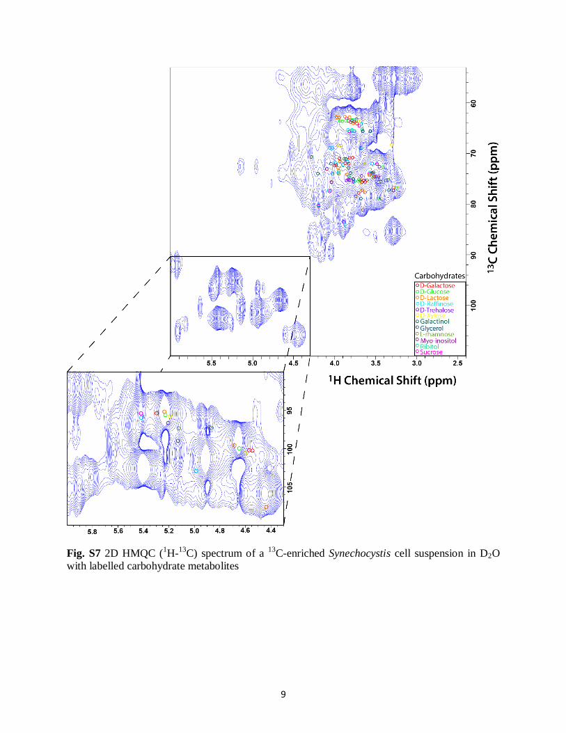
Fig. S7 2D HMQC (1H-
13C) spectrum of a
13C-enriched Synechocystis cell suspension in D2O
with labelled carbohydrate metabolites
10
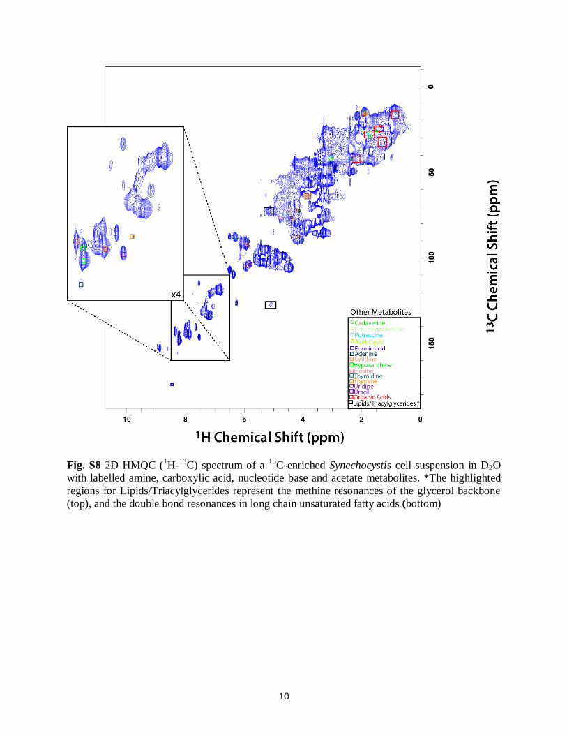
Fig. S8 2D HMQC (1H-
13C) spectrum of a
13C-enriched Synechocystis cell suspension in D2O
with labelled amine, carboxylic acid, nucleotide base and acetate metabolites. *The highlighted
regions for Lipids/Triacylglycerides represent the methine resonances of the glycerol backbone
(top), and the double bond resonances in long chain unsaturated fatty acids (bottom)
11
Fig. S9 A) 2D HMQC (1H-
13C) spectrum of a
13C-enriched Chlamydomonas reinhardtii cell suspension in D2O with triacylglyceride
peaks labelled. B) A 2D HMQC (1H-
13C) simulated spectra (simulated spectra was produced using ACD software) of an example
triacylglyceride molecule with resonances unique to triacylglycerides denoted by numbers
12
Fig. S10 Change in concentration (% change) of select identifiable metabolites in 13
C-enriched
Chlorella vulgaris relative to 13
C-enriched Chlamydomonas reinhardtii as determined by
integration of peak area
-120
-100
-80
-60
-40
-20
0
20
40 %
Ch
ange
Rel
ativ
e to
C. r
ein
ha
rdti
i
Change in Metabolite Concentration in Synechocystis Relative to C. reinhardtii
13
Fig. S11 Change in concentration (% change) of select identifiable metabolites in 13
C-enriched
Synechocystis relative to 13
C-enriched Chlamydomonas reinhardtii as determined by integration
of peak area
-100
-80
-60
-40
-20
0
20
40
60
% C
han
ge R
elat
ive
to C
. rei
nh
ard
tii
Change in Metabolite Concentration in C. vulgaris Relative to C. reinhardtii
14
Fig. S12 Comparison of 1D IDE, DE and RADE 1H spectra of a
13C-enriched Chlorella vulgaris,
cell suspension in D2O with select chemical shifts labelled
15
Fig. S13 Comparison of 1D IDE, DE and RADE 1H spectra of a
13C-enriched Synechocystis, cell
suspension in D2O with select chemical shifts labelled
16
Table S1 List of all metabolite assignments for 13
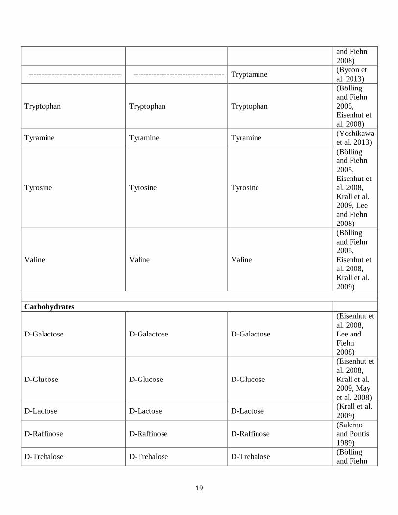
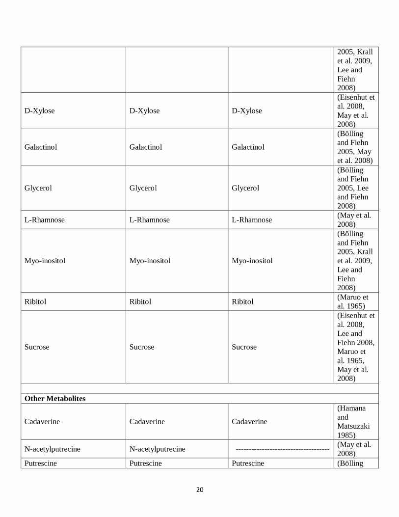
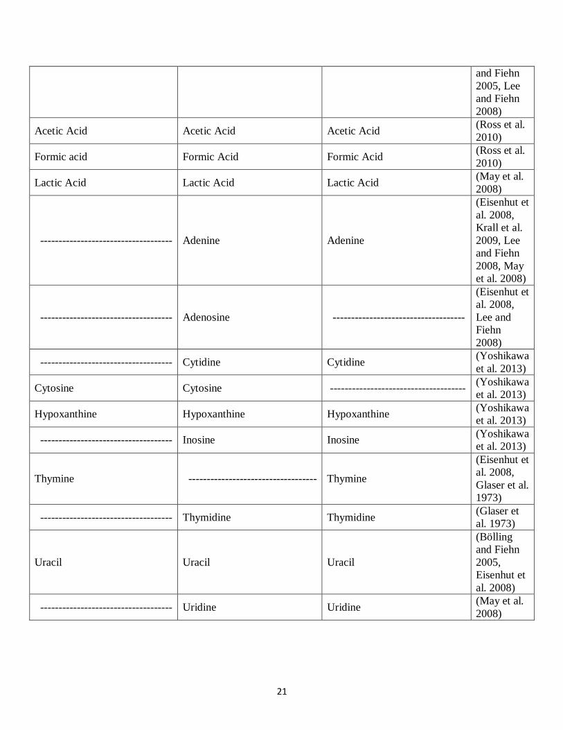
C-labelled Chlamydomonas reinhardtii,
Chlorella vulgaris and Synechocystis with literature references for confirmation
Chlamydomonas reinhardtii Chlorella vulgaris Synechocystis Literature
Reference
Amino Acids and Amino Acid Derivatives
Agmatine Agmatine Agmatine
(Hamana
and
Matsuzaki
1985)
Alanine Alanine Alanine
(Eisenhut et
al. 2008,
Krall et al.
2009, Lee
and Fiehn
2008, May
et al. 2008)
Arginine Arginine Arginine
(Eisenhut et
al. 2008,
Krall et al.
2009, May
et al. 2008)
Beta-alanine Beta-alanine
(Eisenhut et
al. 2008,
Krall et al.
2009, Lee
and Fiehn
2008, May
et al. 2008)
Citrulline Citrulline Citrulline
(Bölling
and Fiehn
2005,
Eisenhut et
al. 2008,
May et al.
2008)
Cysteine Cysteine Cysteine
(Bölling
and Fiehn
2005, May
et al. 2008,
Yoshikawa
et al. 2013)
Glutamic acid Glutamic acid Glutamic acid
(Bölling
and Fiehn
2005,
17
Eisenhut et
al. 2008,
Lee and
Fiehn
2008)
Glutamine Glutamine Glutamine
(Bölling
and Fiehn
2005,
Eisenhut et
al. 2008,
Krall et al.
2009)
Glycine Glycine Glycine
(Bölling
and Fiehn
2005,
Eisenhut et
al. 2008,
Lee and
Fiehn
2008)
------------------------------------ Histidine ------------------------------------
(Bölling
and Fiehn
2005, May
et al. 2008,
Yoshikawa
et al. 2013)
Isoglutamine Isoglutamine Isoglutamine
(Belmont &
Miller,
1965)
Leucine Leucine Leucine
(Bölling
and Fiehn
2005,
Eisenhut et
al. 2008,
Krall et al.
2009)
Isoleucine Isoleucine Isoleucine
(Bölling
and Fiehn
2005, Krall
et al. 2009,
Lee and
Fiehn
2008)put
Methionine Methionine Methionine
(Bölling
and Fiehn
2005,
18
Eisenhut et
al. 2008,
Lee and
Fiehn
2008)
Ornithine Ornithine Ornithine
(Bölling
and Fiehn
2005,
Eisenhut et
al. 2008,
Krall et al.
2009)
Phenylalanine Phenylalanine Phenylalanine
(Bölling
and Fiehn
2005,
Eisenhut et
al. 2008,
Krall et al.
2009, Lee
and Fiehn
2008)
Proline Proline Proline
(Bölling
and Fiehn
2005,
Eisenhut et
al. 2008,
Krall et al.
2009, Lee
and Fiehn
2008)
Serine Serine Serine
(Bölling
and Fiehn
2005,
Eisenhut et
al. 2008,
Krall et al.
2009, Lee
and Fiehn
2008)
Threonine Threonine Threonine
(Bölling
and Fiehn
2005,
Eisenhut et
al. 2008,
Krall et al.
2009, Lee
19
and Fiehn
2008)
------------------------------------ ----------------------------------- Tryptamine (Byeon et
al. 2013)
Tryptophan Tryptophan Tryptophan
(Bölling
and Fiehn
2005,
Eisenhut et
al. 2008)
Tyramine Tyramine Tyramine (Yoshikawa
et al. 2013)
Tyrosine Tyrosine Tyrosine
(Bölling
and Fiehn
2005,
Eisenhut et
al. 2008,
Krall et al.
2009, Lee
and Fiehn
2008)
Valine Valine Valine
(Bölling
and Fiehn
2005,
Eisenhut et
al. 2008,
Krall et al.
2009)
Carbohydrates
D-Galactose D-Galactose D-Galactose
(Eisenhut et
al. 2008,
Lee and
Fiehn
2008)
D-Glucose D-Glucose D-Glucose
(Eisenhut et
al. 2008,
Krall et al.
2009, May
et al. 2008)
D-Lactose D-Lactose D-Lactose (Krall et al.
2009)
D-Raffinose D-Raffinose D-Raffinose
(Salerno
and Pontis
1989)
D-Trehalose D-Trehalose D-Trehalose (Bölling
and Fiehn
20
2005, Krall
et al. 2009,
Lee and
Fiehn
2008)
D-Xylose D-Xylose D-Xylose
(Eisenhut et
al. 2008,
May et al.
2008)
Galactinol Galactinol Galactinol
(Bölling
and Fiehn
2005, May
et al. 2008)
Glycerol Glycerol Glycerol
(Bölling
and Fiehn
2005, Lee
and Fiehn
2008)
L-Rhamnose L-Rhamnose L-Rhamnose (May et al.
2008)
Myo-inositol Myo-inositol Myo-inositol
(Bölling
and Fiehn
2005, Krall
et al. 2009,
Lee and
Fiehn
2008)
Ribitol Ribitol Ribitol (Maruo et
al. 1965)
Sucrose Sucrose Sucrose
(Eisenhut et
al. 2008,
Lee and
Fiehn 2008,
Maruo et
al. 1965,
May et al.
2008)
Other Metabolites
Cadaverine Cadaverine Cadaverine
(Hamana
and
Matsuzaki
1985)
N-acetylputrecine N-acetylputrecine ------------------------------------ (May et al.
2008)
Putrescine Putrescine Putrescine (Bölling
21
and Fiehn
2005, Lee
and Fiehn
2008)
Acetic Acid Acetic Acid Acetic Acid (Ross et al.
2010)
Formic acid Formic Acid Formic Acid (Ross et al.
2010)
Lactic Acid Lactic Acid Lactic Acid (May et al.
2008)
------------------------------------ Adenine Adenine
(Eisenhut et
al. 2008,
Krall et al.
2009, Lee
and Fiehn
2008, May
et al. 2008)
------------------------------------ Adenosine ------------------------------------
(Eisenhut et
al. 2008,
Lee and
Fiehn
2008)
------------------------------------ Cytidine Cytidine (Yoshikawa
et al. 2013)
Cytosine Cytosine ------------------------------------- (Yoshikawa
et al. 2013)
Hypoxanthine Hypoxanthine Hypoxanthine (Yoshikawa
et al. 2013)
------------------------------------ Inosine Inosine (Yoshikawa
et al. 2013)
Thymine ----------------------------------- Thymine
(Eisenhut et
al. 2008,
Glaser et al.
1973)
------------------------------------ Thymidine Thymidine (Glaser et
al. 1973)
Uracil Uracil Uracil
(Bölling
and Fiehn
2005,
Eisenhut et
al. 2008)
------------------------------------ Uridine Uridine (May et al.
2008)
22
References
Belmont, L. and Miller, J. (1965) The utilization of glutamine by algae. Journal of Experimental
Botany 16(2), 318-324.
Bölling, C. and Fiehn, O. (2005) Metabolite profiling of Chlamydomonas reinhardtii under
nutrient deprivation. Plant physiology 139(4), 1995-2005.
Byeon, Y., Lee, K., Park, Y.I., Park, S. and Back, K. (2013) Molecular cloning and functional
analysis of serotonin N‐ acetyltransferase from the cyanobacterium Synechocystis sp. PCC 6803.
Journal of pineal research 55(4), 371-376.
Eisenhut, M., Huege, J., Schwarz, D., Bauwe, H., Kopka, J. and Hagemann, M. (2008)
Metabolome phenotyping of inorganic carbon limitation in cells of the wild type and
photorespiratory mutants of the cyanobacterium Synechocystis sp. strain PCC 6803. Plant
physiology 148(4), 2109-2120.
Glaser, V., Al-Nuri, M., Groshev, V. and Shestakov, S. (1973) The labelling of nucleic acids by
radioactive precursors in the blue-green algae Anacystis nidulans and Synechocystis aquatilis
Sanv. Archiv für Mikrobiologie 92(3), 217-226.
Gorman, D.S., Levine, R. P. (1965) Cytochrome f and plastocyanin: their sequence in the
photosynthetic electron transport chain of Chlamydomonas reinhardtii. Proc. Natl. Acad. Sci.
USA, 54(6),1665–1669.
Hamana, K. and Matsuzaki, S. (1985) Distribution of polyamines in prokaryotes, algae, plants
and fungi. Polyamines: basic and clinical aspects (Eds K. Imanoh, F. Suzuki, O. Suzuki, U.
Bachrach), VNU Science Press, Netherlands, 105-112.
Krall, L., Huege, J., Catchpole, G., Steinhauser, D. and Willmitzer, L. (2009) Assessment of
sampling strategies for gas chromatography–mass spectrometry (GC–MS) based metabolomics
of cyanobacteria. Journal of Chromatography B 877(27), 2952-2960.
Lee, D.Y. and Fiehn, O. (2008) High quality metabolomic data for Chlamydomonas reinhardtii.
Plant methods 4(1), 7.
Maruo, B., Hattori, T. and Takahashi, H. (1965) Excretion of ribitol and sucrose by green algae
into the culture medium. Agricultural and Biological Chemistry 29(12), 1084-1089.
May, P., Wienkoop, S., Kempa, S., Usadel, B., Christian, N., Rupprecht, J., Weiss, J., Recuenco-
Munoz, L., Ebenhöh, O. and Weckwerth, W. (2008) Metabolomics-and proteomics-assisted
genome annotation and analysis of the draft metabolic network of Chlamydomonas reinhardtii.
Genetics 179(1), 157-166.
23
Ross, A., Biller, P., Kubacki, M., Li, H., Lea-Langton, A. and Jones, J. (2010) Hydrothermal
processing of microalgae using alkali and organic acids. Fuel 89(9), 2234-2243.
Salerno, G.L. and Pontis, H.G. (1989) Raffinose synthesis in Chlorella vulgaris cultures after a
cold shock. Plant physiology 89(2), 648-651.
Yoshikawa, K., Hirasawa, T., Ogawa, K., Hidaka, Y., Nakajima, T., Furusawa, C. and Shimizu,
H. (2013) Integrated transcriptomic and metabolomic analysis of the central metabolism of
Synechocystis sp. PCC 6803 under different trophic conditions. Biotechnology journal 8(5), 571-
580.