Embed Size (px)
Citation preview
IMAGERIE DES LESIONS MUSCULAIRES
Dr. Guillaume MERCY Dr Jean-Louis BRASSEUR
INSEP et Hôpital Pitié-Salpêtrière
Colloque médical FFVB 26 / 03 / 2015
“ Présentation Sémiologie Principales lésions Pronostic Hématomes / traitement Cicatrisation
PLAN

PRESENTATION
QUELLES LESIONS?
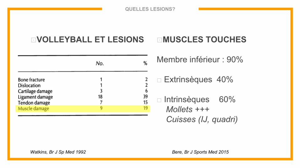
⊡ VOLLEYBALL ET LESIONS
⊡ MUSCLES TOUCHES Membre inférieur : 90% □ Extrinsèques 40%
□ Intrinsèques 60% Mollets +++ Cuisses (IJ, quadri)
Watkins, Br J Sp Med 1992 Bere, Br J Sports Med 2015
QUELLES LESIONS?
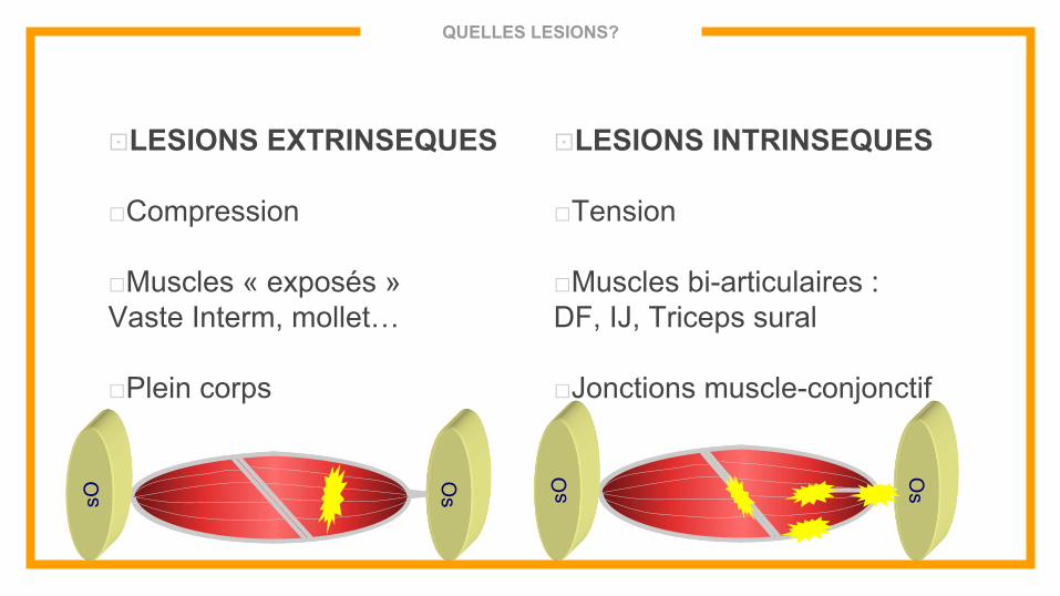
⊡ LESIONS EXTRINSEQUES
□ Compression
□ Muscles « exposés » Vaste Interm, mollet…
□ Plein corps
⊡ LESIONS INTRINSEQUES
□ Tension
□ Muscles bi-articulaires : DF, IJ, Triceps sural
□ Jonctions muscle-conjonctif
Os sO
Os sO
Os sO
Os sO
IMAGERIE : POUR OU CONTRE ?
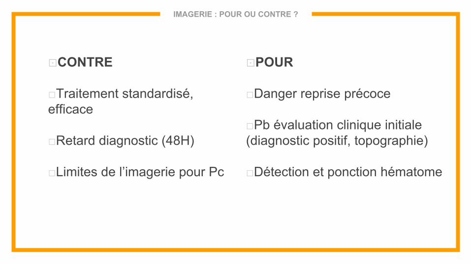
⊡ CONTRE
□ Traitement standardisé, efficace
□ Retard diagnostic (48H)
□ Limites de l’imagerie pour Pc
⊡ POUR
□ Danger reprise précoce
□ Pb évaluation clinique initiale (diagnostic positif, topographie)
□ Détection et ponction hématome
IMAGERIE : QUAND, COMMENT?
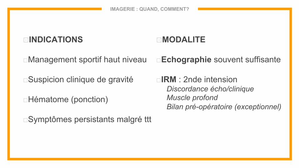
⊡ INDICATIONS
□ Management sportif haut niveau
□ Suspicion clinique de gravité
□ Hématome (ponction)
□ Symptômes persistants malgré ttt
⊡ MODALITE
□ Echographie souvent suffisante
□ IRM : 2nde intension Discordance écho/clinique Muscle profond Bilan pré-opératoire (exceptionnel)
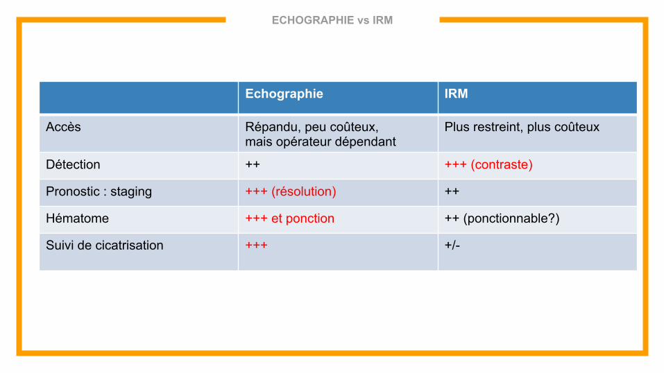
ECHOGRAPHIE vs IRM
Echographie IRM
Accès Répandu, peu coûteux, mais opérateur dépendant
Plus restreint, plus coûteux
Détection ++ +++ (contraste)
Pronostic : staging +++ (résolution) ++
Hématome +++ et ponction ++ (ponctionnable?)
Suivi de cicatrisation
+++ +/-
SEMIOLOGIE
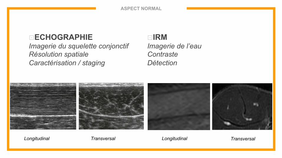
ASPECT NORMAL
⊡ ECHOGRAPHIE Imagerie du squelette conjonctif Résolution spatiale Caractérisation / staging
⊡ IRM Imagerie de l’eau Contraste Détection
Longitudinal Transversal Longitudinal Transversal
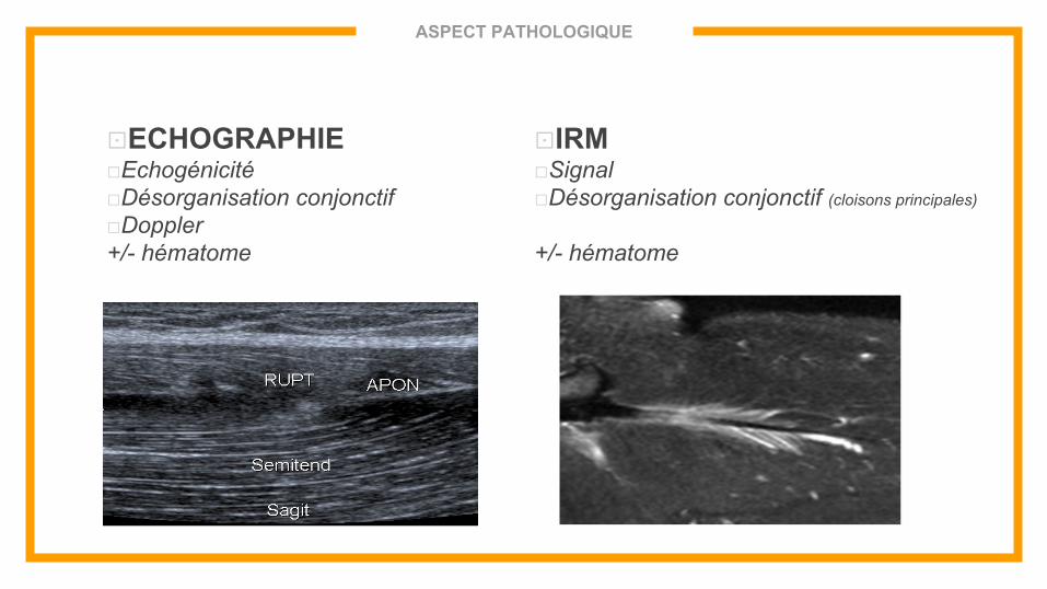
ASPECT PATHOLOGIQUE
⊡ ECHOGRAPHIE □ Echogénicité □ Désorganisation conjonctif □ Doppler +/- hématome
⊡ IRM □ Signal □ Désorganisation conjonctif (cloisons principales) +/- hématome
LESIONS PRINCIPALES
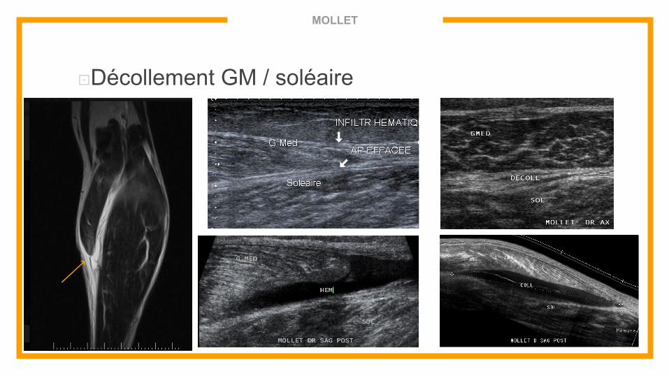
MOLLET
⊡ Décollement GM / soléaire
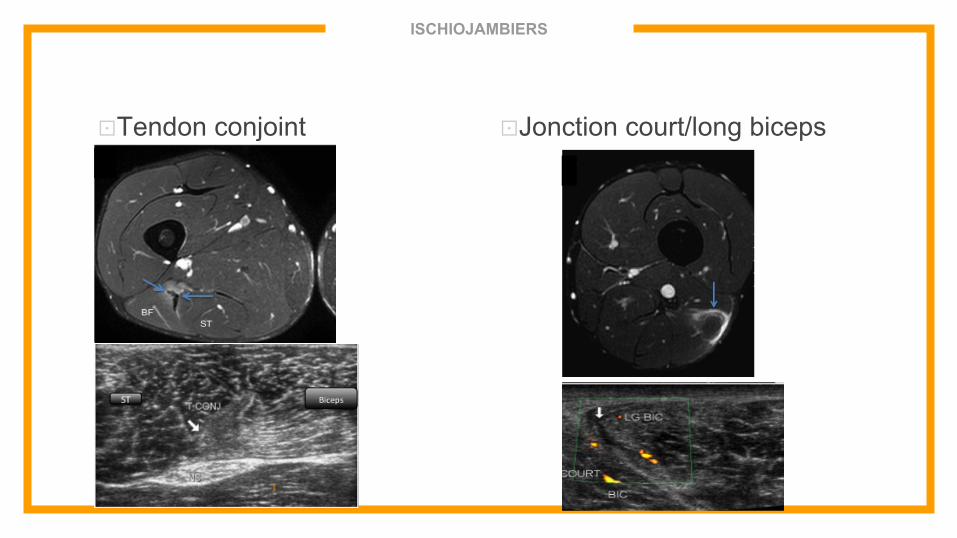
ISCHIOJAMBIERS
⊡ Tendon conjoint ⊡ Jonction court/long biceps
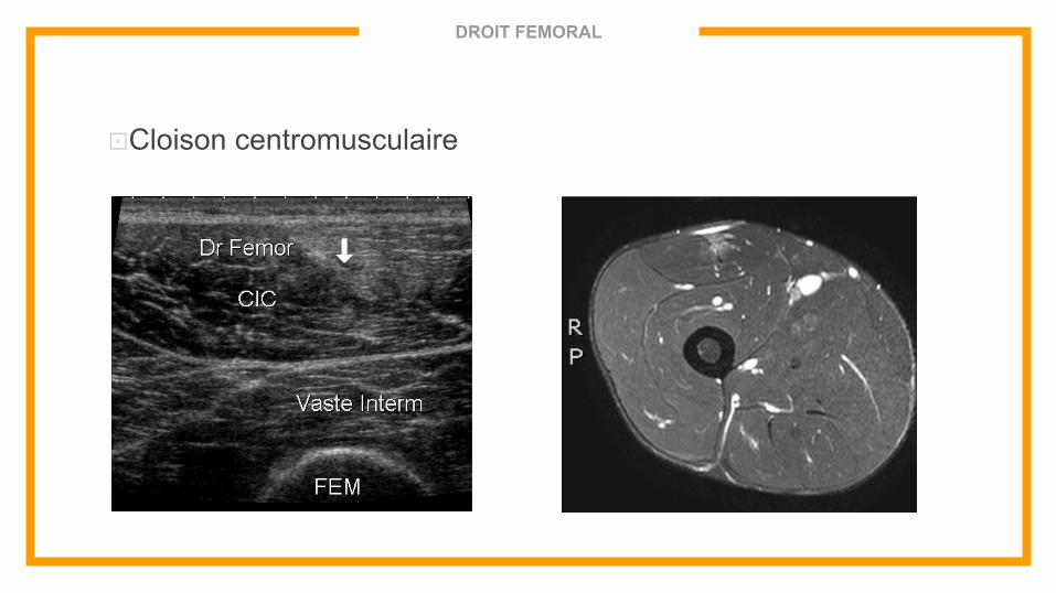
DROIT FEMORAL
⊡ Cloison centromusculaire
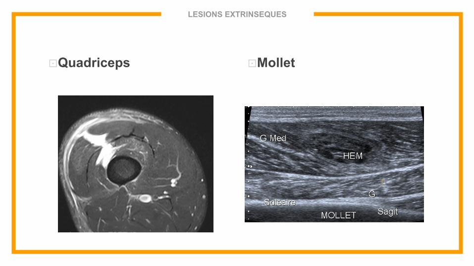
LESIONS EXTRINSEQUES
⊡ Quadriceps ⊡ Mollet
STAGING PRONOSTIQUE
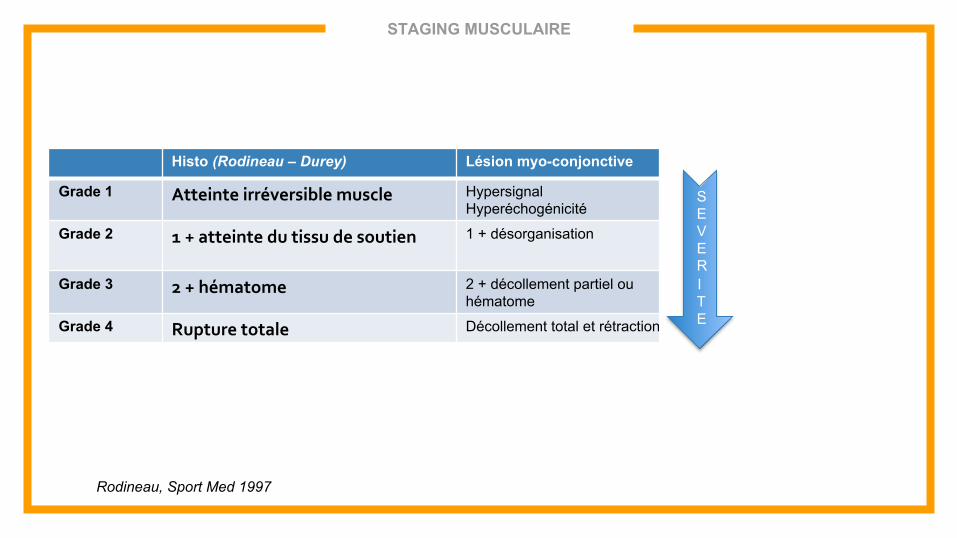
STAGING MUSCULAIRE
Histo (Rodineau – Durey) Lésion myo-conjonctive Conjonctif C
Grade 1 Atteinte irréversible muscle Hypersignal Hyperéchogénicité
-
Grade 2 1 + atteinte du tissu de soutien 1 + désorganisation Hypersignal Hyperéchogénicité
Grade 3 2 + hématome 2 + décollement partiel ou hématome
2 + rupture +/- hématome
Grade 4 Rupture totale Décollement total et rétraction
SEVERITE
Rodineau, Sport Med 1997
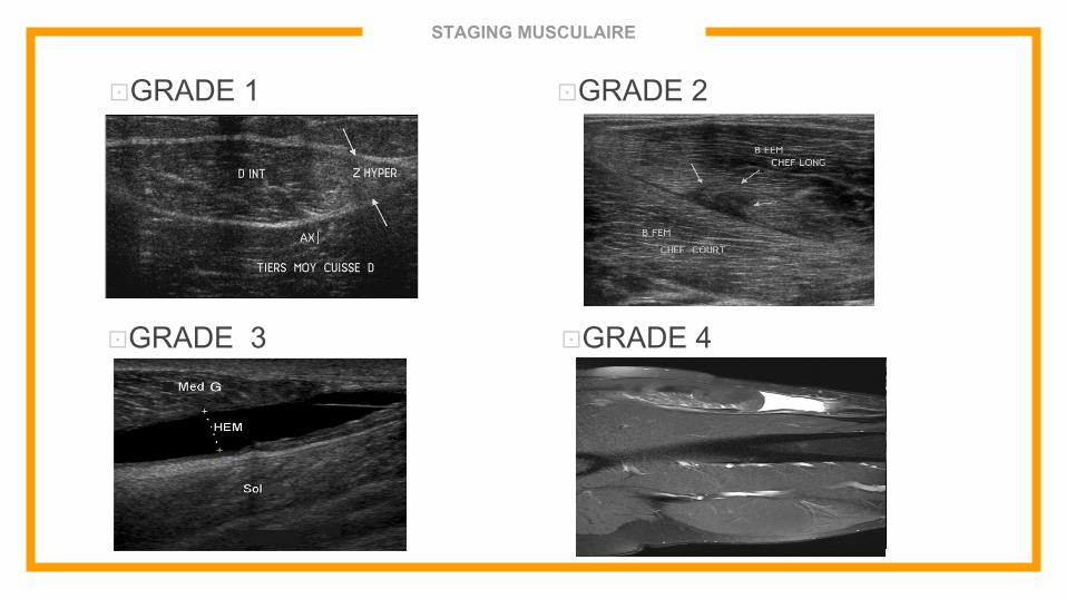
STAGING MUSCULAIRE
⊡ GRADE 1 ⊡ GRADE 2
⊡ GRADE 3 ⊡ GRADE 4
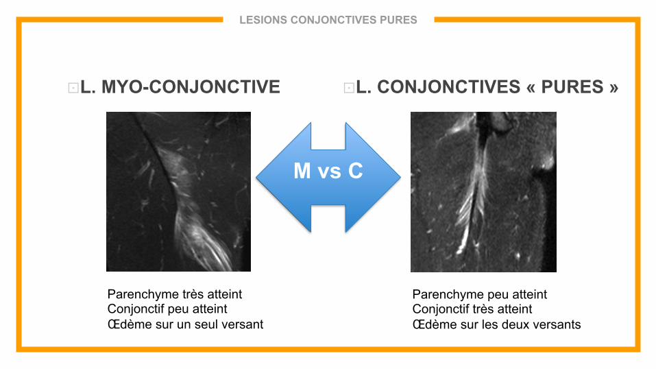
LESIONS CONJONCTIVES PURES
⊡ L. MYO-CONJONCTIVE ⊡ L. CONJONCTIVES « PURES »
M vs C
Parenchyme très atteint Conjonctif peu atteint Œdème sur un seul versant
Parenchyme peu atteint Conjonctif très atteint Œdème sur les deux versants
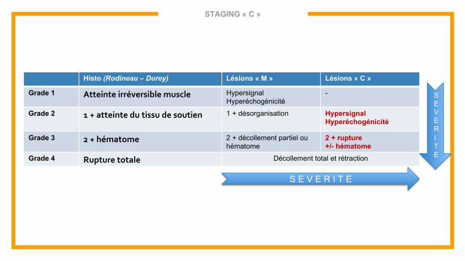
STAGING « C »
Histo (Rodineau – Durey) Lésions « M » Lésions « C »
Grade 1 Atteinte irréversible muscle Hypersignal Hyperéchogénicité
-
Grade 2 1 + atteinte du tissu de soutien 1 + désorganisation Hypersignal Hyperéchogénicité
Grade 3 2 + hématome 2 + décollement partiel ou hématome
2 + rupture +/- hématome
Grade 4 Rupture totale Décollement total et rétraction
S E V E R I T E
SEVERITE
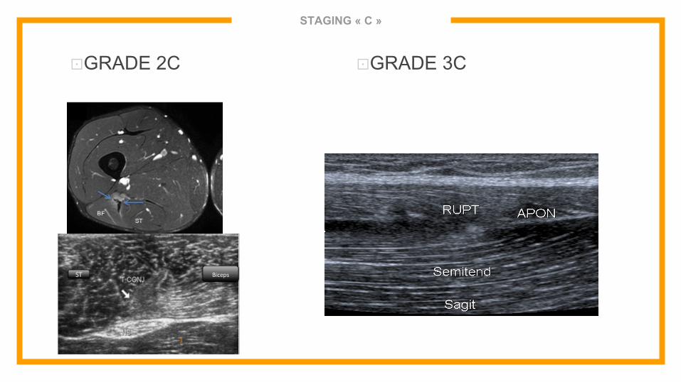
STAGING « C »
⊡ GRADE 2C ⊡ GRADE 3C
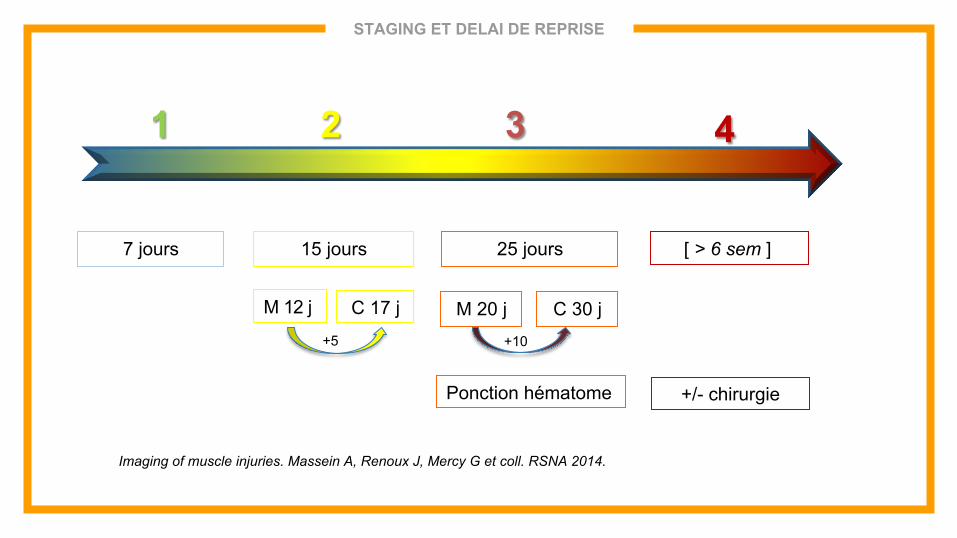
STAGING ET DELAI DE REPRISE
1 2 3 4
7 jours 15 jours 25 jours [ > 6 sem ]
Ponction hématome +/- chirurgie
M 12 j C 17 j M 20 j C 30 j +5 +10
Imaging of muscle injuries. Massein A, Renoux J, Mercy G et coll. RSNA 2014.
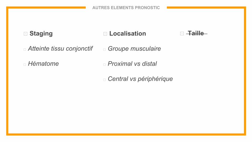
AUTRES ELEMENTS PRONOSTIC
⊡ Staging
□ Atteinte tissu conjonctif
□ Hématome
⊡ Localisation
□ Groupe musculaire
□ Proximal vs distal
□ Central vs périphérique
⊡ Taille .
TRAITEMENT DES HEMATOMES
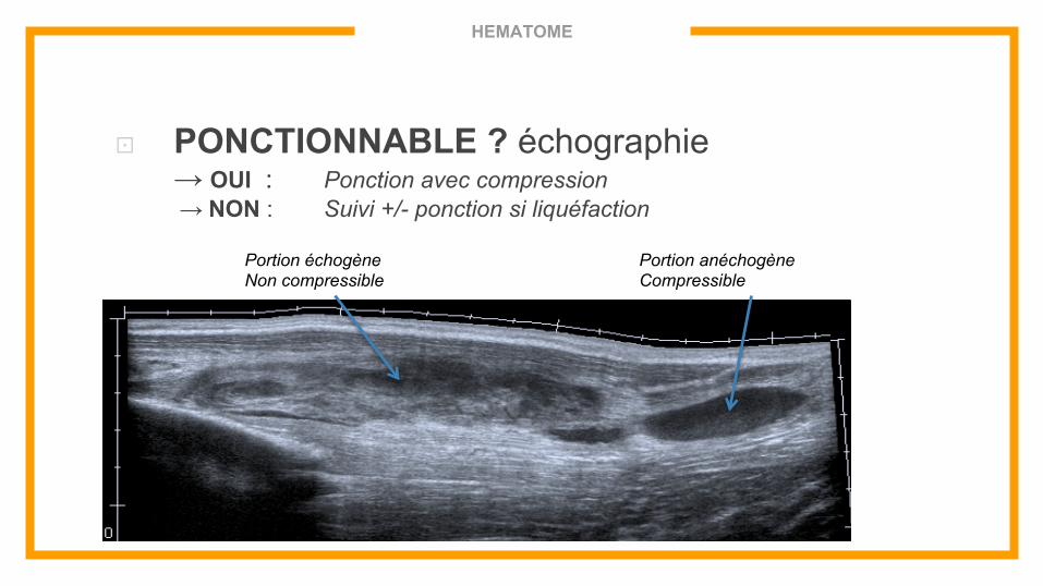
HEMATOME
⊡ PONCTIONNABLE ? échographie → OUI : Ponction avec compression → NON : Suivi +/- ponction si liquéfaction
Portion échogène Non compressible
Portion anéchogène Compressible
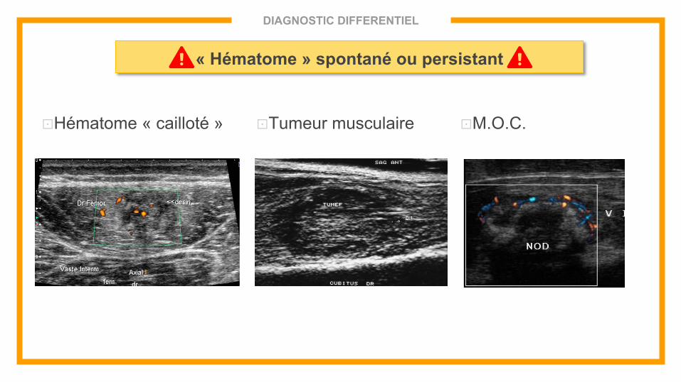
DIAGNOSTIC DIFFERENTIEL
⊡ Hématome « cailloté » ⊡ Tumeur musculaire ⊡ M.O.C.
« Hématome » spontané ou persistant

CICATRISATION
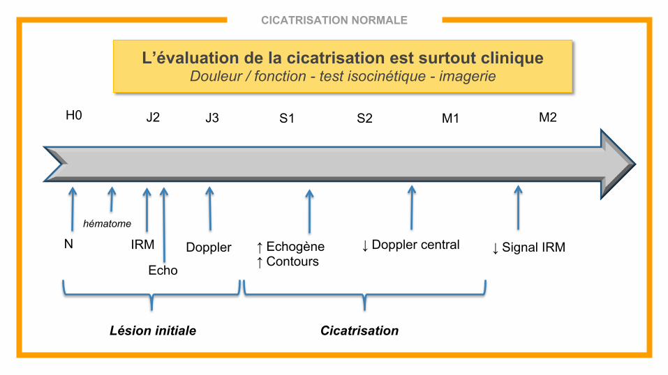
CICATRISATION NORMALE
N
Echo
IRM Doppler
H0
hématome
J2 J3 S1 S2 M2
↓ Doppler central
↑ Echogène ↑ Contours
M1
Cicatrisation
↓ Signal IRM
Lésion initiale
L’évaluation de la cicatrisation est surtout clinique Douleur / fonction - test isocinétique - imagerie
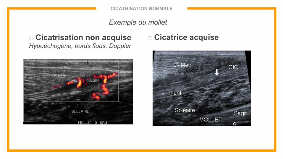
CICATRISATION NORMALE
Exemple du mollet
⊡ Cicatrisation non acquise Hypoéchogène, bords flous, Doppler
⊡ Cicatrice acquise
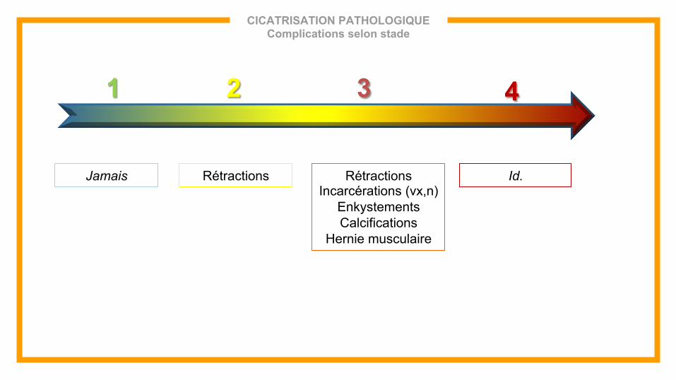
CICATRISATION PATHOLOGIQUE Complications selon stade
1 2 3 4
Jamais Rétractions Rétractions Incarcérations (vx,n)
Enkystements Calcifications
Hernie musculaire
Id.
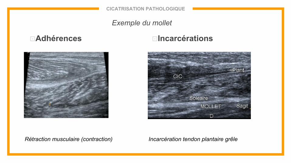
CICATRISATION PATHOLOGIQUE
⊡ Adhérences ⊡ Incarcérations
Rétraction musculaire (contraction) Incarcération tendon plantaire grêle
Exemple du mollet
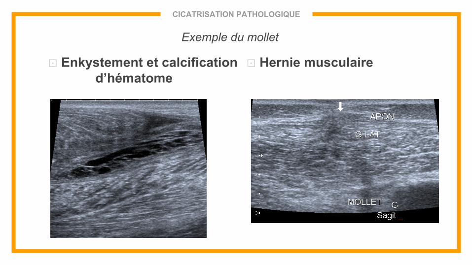
CICATRISATION PATHOLOGIQUE
⊡ Enkystement et calcification d’hématome
⊡ Hernie musculaire
Exemple du mollet

CONCLUSION
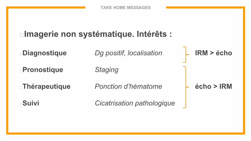
TAKE HOME MESSAGES
⊡ Imagerie non systématique. Intérêts :
□ Diagnostique Dg positif, localisation IRM > écho
□ Pronostique Staging
□ Thérapeutique Ponction d’hématome écho > IRM
□ Suivi Cicatrisation pathologique
“ MERCI DE VOTRE ATTENTION