Embed Size (px)
Citation preview
SummaryIntroduction: The masticatory muscles play an important partin determining the morphology of the facial skeleton. Skeletaltypology and the characteristics of the masticatory muscles areclosely linked. Several authors have studied muscle character-istics as related to facial typology. The aim of this work is tostudy the relationship between vertical and transverse skeletaldimensions and the dimensions (length, width and thickness) oftwo muscles of mastication, the masseter and the lateralpterygoid.
Materials and method: Our study was based on CT-scan exam-inations of a sample composed of patients consulting the X-raydepartment of the Rabat-Sal�e Teaching Hospital, and for whoma CT-scan had been requested. Forty CT examinations of the
R�esum�e
Introduction : Les muscles masticateurs jouent un role impor-tant dans la d�etermination de la morphologie du squelettefacial. La typologie squelettique et les caract�eristiques desmuscles masticateurs sont intimement li�ees. Plusieursauteurs ont �etudi�e les caract�eristiques musculaires en fonc-tion de la typologie squelettique. L’objectif de ce travail estd’�etudier la relation entre les dimensions squelettiques dansle sens vertical et transversal et les dimensions (longueur,largeur et �epaisseur) des muscles masticateurs mass�eter etpt�erygoıdien lat�eral.Mat�eriels et m�ethode : Notre �etude a �et�e men�ee a partird’examens tomodensitom�etriques d’un �echantillon constitu�ede patients venus consulter au service de radiologie del’hopital des sp�ecialit�es du CHU Rabat-Sal�e, pour lesquels
Original articleArticle original
� 2013 CEOPublished by / Edite par Elsevier Masson SAS
All rights reserved / Tous droits reserves
Relationship between dimensions ofmuscles of mastication (masseter andlateral pterygoid) and skeletal dimensions:Study of 40 cases
Relation entre dimensions des musclesmasticateurs (mass�eter et pt�erygoıdien lat�eral) etdimensions squelettiques : �etude sur 40 cas
Mohammed Faouzi AZAROUALa,*, Meriem FIKRIb, Redouan ABOUQALc,Hicham BENYAHYAd, Fatima ZAOUId
aService d’odontologie, hopital militaire d’instruction Mohamed V, avenue des FAR-Hay-Riad,10100 Rabat, MoroccobService de neuro-radiologie, hopital des sp�ecialit�es ONO, CHU de Rabat-Sal�e, BP, 6444 Rabat,MoroccocLaboratoire de biostatistique et de recherche clinique et�epid�emiologie, facult�e dem�edecine et depharmacie, avenue Med-Belarbi-El-Alaoui, BP 6203, Rabat, MoroccodService d’orthop�edie dento-faciale, facult�e de m�edecine dentaire de Rabat, universit�eMohamed-V Souissi, Rabat, Morocco
Available online: 21 October 2013 / Disponible en ligne : 21 octobre 2013
*Correspondence and reprints / Correspondance et tir�es a part :
M.F. AZAROUAL, BP 6609 Madinat Alirfane, Ryad, Rabat, Maroc.e-mail address / Adresse e-mail : [email protected](Mohammed Faouzi AZAROUAL)
International Orthodontics 2014 ; 12 : 111-124 111http://dx.doi.org/10.1016/j.ortho.2013.09.001

skull, performed in the context of sinus explorations or pre-surgical work-ups in the radiology department of the Rabat-Sal�e Teaching Hospital, were selected for this study. The samplecomprised 19 women and 21 men aged between 20 and 45, witha mean of 40.9 W 12.8. A Siemens 32-row 64-slice spiral CT-scan device was used for spiral acquisition of data around thefacial bones, with the mouth closed. The study was carried outin the parenchymal window for the muscle measurements, in theaxial and coronal planes. Bone measurements were performedafter 3D reconstruction in VRT mode.
Results: Our study showed that, for the masseter muscle, thick-ness is the dimension that correlates significantly with skeletaldimensions in the vertical, transverse and sagittal directions.For the lateral pterygoid muscle, length and width both presentsignificant correlations with transverse skeletal dimensions.Analysis of these results shows that the dimensional character-istics of the masticatory muscles vary according to the verticaland transverse skeletal typology of the subjects concerned.
� 2013 CEO. Published by Elsevier Masson SAS. All rightsreserved
Keywords
·Muscles of mastication.
·Masseter.
·Lateral pterygoid.·Skeletal dimension.Introduction
The masticatory muscles play an important part in determin-ing the morphology of the facial skeleton. Skeletal typologyand the characteristics of the masticatory muscles are closelylinked. Several authors have studied muscle characteristics asrelated to facial typology: Bakke et al. [1], Benington et al. [2],Gionhaku and Lowe [3], Kiliaridis et al. [4], Kubota et al. [5],Raadsheer et al. [6,7], Van Spronsen et al. [8–12], Weijs andHillen [13]. These studies have been made possible by con-tinuous improvements in imaging technology (particularly CT-scan), and the ability to reduce the doses of radiation receivedby the patient (Horger et al. [14], Francone et al. [15]). Thesedevelopments have led to a remarkable increase in the numberof CT-scans performed (from 5million to 20million in the USAbetween 1983 and 1995 [Mah et al. [16]], particularly inodontology). Computed tomography, with its numerous possi-bilities for reconstructions, particularly in three dimensions,offers patients a considerable gain in information and makes itpossible to evaluate the masticatory muscles (Katsumata et al.
un examen TDM a �et�e demand�e. Quarante examens tomo-densitom�etriques du crane, r�ealis�es dans le cadre d’explora-tions sinusiennes ou de bilans pr�echirurgicaux dans le servicede radiologie de l’hopital des sp�ecialit�es du CHU Rabat-Sal�e,ont �et�e s�electionn�es pour cette �etude. L’�echantillon �etaitcompos�e de 19 femmes et 21 hommes d’age compris entre20 et 45 ans et dont la moyenne �etait de 40,9W 12,8 ans.L’acquisition des donn�ees tomodensitom�etriques a �et�er�ealis�ee avec un scanner h�elicoıdal multibarettes de type« Siemens 32 barrettes 64 coupes », avec acquisitionspiral�ee sur le massif facial, en bouche ferm�ee. L’�etudes’est faite en fenetre parenchymateuse pour les mensurationsmusculaires, dans les plans axial et coronal. Les mesuresosseuses ont �et�e effectu�ees apr�es reconstructions tridimen-sionnelles en mode VRT.R�esultats : Notre �etude a r�ev�el�e que l’�epaisseur est la dimen-sion du muscle mass�eter qui pr�esente une corr�elation impor-tante avec les dimensions squelettiques dans les sens vertical,transversal et sagittal. Pour le muscle pt�erygoıdien lat�eral, lalongueur et la largeur pr�esentent une corr�elation significativeavec les dimensions squelettiques transversales. L’analysedes r�esultats montre que les caract�eristiques dimensionnellesdes muscles masticateurs varient en fonction de la typologiesquelettique verticale et transversale des sujets.� 2013 CEO. Edite par Elsevier Masson SAS. Tous droitsreserves
Mots cl�es
·Muscles masticateurs.
·Mass�eter.
·Pt�erygoıdien lat�eral.
·Dimension squelettique.
Introduction
Les muscles masticateurs jouent un role important dans lad�etermination de la morphologie du squelette facial. La typo-logie squelettique et les caract�eristiques des muscles masti-cateurs sont intimement li�ees. Plusieurs auteurs ont �etudi�e lescaract�eristiques musculaires en fonction de la typologiesquelettique : Bakke et al. [1], Benington et al. [2], Gionhakuet Lowe [3], Kiliaridis et al. [4], Kubota et al. [5], Raadsheer etal. [6,7], Van Spronsen et al. [8–12], Weijs et Hillen [13]. Ces�etudes ont �et�e rendues possibles grace a l’am�elioration cons-tante des techniques de radiologie (notamment de la tomo-densitom�etrie) et des possibilit�es de r�eduction des doses deradiations recues par le patient (Horger et al. [14], Francone etal. [15]). Ces �evolutions ont conduit a une multiplication con-sid�erable du nombre de scanners r�ealis�es (de 5 millionsa 20 millions entre 1983 et 1995 aux �Etats-Unis [Mah et al.[16]]), particuli�erement en odontologie. En effet, la tomoden-sitom�etrie informatis�ee et ses multiples possibilit�es de recon-struction, en particulier tridimensionnelle, apportent un gain
112 International Orthodontics 2014 ; 12 : 111-124
Mohammed Faouzi AZAROUAL et al.
[17], Raustia et al. [18]), while providing physicians withessential diagnostic information concerning the dimensionsof these same muscles of mastication. Technical progresshas thus made it possible to modify recording and measure-ment protocols, above all in CT where lowering of the doses ofradiation delivered has gone hand in hand with a reduction inslice thickness from 8 mm (Gionhaku and Lowe [3]) to 2 mm(Takashima et al. [19]) and in the spacing of slices, therebyimproving the precision of the images obtained. The develop-ment of spiral CT-scan now enables excellent 3D reconstruc-tions to be obtained in various different orientations(Katsumata et al. [17], Ueki et al. [20]).
Our study was based on CT-scan examinations of a samplecomposed of patients consulting the X-ray department of theRabat-Sal�e Teaching Hospital, and for whom a CT-scan hadbeen requested.The aim of this article is to study the relationship betweenvertical and transverse skeletal dimensions and the dimen-sions (length, width and thickness) of two muscles of masti-cation, the masseter and the lateral pterygoid.
Materials and method
Sample
Forty CT examinations of the skull, performed in the context ofsinus explorations or pre-surgical work-ups in the radiologydepartment of the Rabat-Sal�e Teaching Hospital, wereselected for this study.
The sample comprised 19 women and 21men aged between 20and 45, with a mean of 40.9 W 12.8.
A Siemens 32-row 64-slice spiral CT-scan device was used forspiral acquisition of data around the facial bones, with themouth closed:
– diameter of field of acquisition 250 mm;– thickness of reconstructed slices 0.6 mm;– matrix of reconstructed slices 512 � 512.The study was carried out in the parenchymal window for themuscle measurements, in the axial and coronal planes. Bonemeasurements were performed after 3D reconstruction in VRTmode.Inclusion criteria: the subjects selected were adult patientswith intact dentition and no significant skeletal asymmetry orantecedents of orthognathic surgery.
d’information consid�erable pour les patients et permettentd’�evaluer les muscles masticateurs (Katsumata et al. [17],Raustia et al. [18]) ainsi que d’apporter aux praticiens desinformations diagnostiques essentielles concernant lesdimensions des muscles masticateurs. Ces progr�es techni-ques ont permis aussi de modifier les protocoles d’enregistre-ment et de mesure, surtout en tomodensitom�etrie ou lar�eduction de la dose de radiation d�elivr�ee s’est accompagn�eed’une diminution de l’�epaisseur de 8 mm (Gionhaku et Lowe[3]) a 2 mm (Takashima et al. [19]) et de l’espacement descoupes, am�eliorant ainsi la pr�ecision des images. Led�eveloppement des scanners h�elicoıdaux autorise aujourd’huides reconstructions tridimensionnelles selon des orientationsdiff�erentes d’excellente qualit�e (Katsumata et al. [17], Ueki etal. [20]).Notre �etude a �et�e men�ee a partir d’examens tomodensitom�e-triques d’un �echantillon constitu�e de patients venus consulterau service de radiologie de l’hopital des sp�ecialit�es du CHURabat-Sal�e, pour lesquels un examen TDM a �et�e demand�e.L’objectif de cet article a �et�e d’�etudier la relation entre lesdimensions squelettiques dans le sens vertical et transversalet les dimensions (longueur, largeur et �epaisseur) desmusclesmasticateurs mass�eter et pt�erygoıdien lat�eral.
Mat�eriel et m�ethode
�Echantillon
Quarante examens tomodensitom�etriques du crane, r�ealis�esdans le cadre d’explorations sinusiennes ou de bilans pr�echir-urgicaux dans le service de radiologie de l’hopital dessp�ecialit�es duCHURabat-Sal�e ont �et�e s�electionn�es pour cette�etude.L’�echantillon �etait compos�e de 19 femmes et 21 hommes dontl’age �etait compris entre 20 et 45 ans et dont la moyenne �etaitde 40,9 W 12,8.L’acquisition des donn�ees tomodensitom�etriques a �et�er�ealis�ee avec un scanner h�elicoıdal multibarettes de type« Siemens 32 barrettes 64 coupes », avec acquisitionspiral�ee sur le massif facial, en bouche ferm�ee :– diam�etre du champ d’acquisition 250 mm ;– �epaisseur des coupes reconstruites 0,6 mm ;– matrice des coupes reconstruites 512 � 512.L’�etude s’est faite en fenetre parenchymateuse pour les men-surations musculaires, dans les plans axial et coronal. Lesmesures osseuses ont �et�e effectu�ees apr�es reconstructionstridimensionnelles en mode VRT.Crit�eres d’inclusion : les sujets retenus �etaient des patientsadultes ayant une denture intacte et ne pr�esentant pas dedissym�etrie squelettique importante ou d’ant�ec�edents de chir-urgie orthognathique.
International Orthodontics 2014 ; 12 : 111-124 113
Relationship between dimensions of muscles of mastication (masseter and lateral pterygoid) and skeletal dimensions: Study of 40cases
Relation entre dimensions des muscles masticateurs (mass�eter et pt�erygoıdien lat�eral) et dimensions squelettiques : �etude sur 40 cas
Measurement method
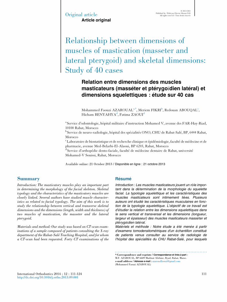
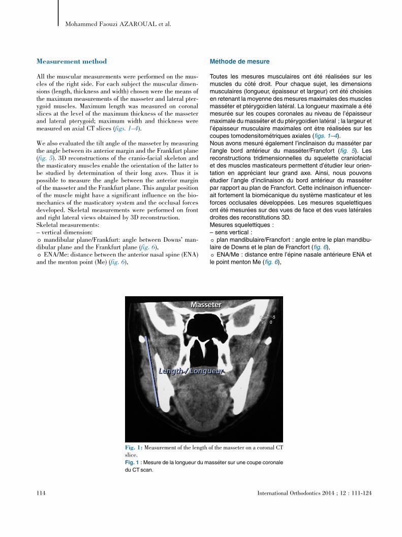
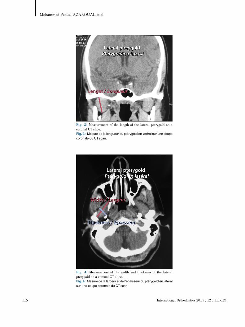
All the muscular measurements were performed on the mus-cles of the right side. For each subject the muscular dimen-sions (length, thickness and width) chosen were the means ofthe maximum measurements of the masseter and lateral pter-ygoid muscles. Maximum length was measured on coronalslices at the level of the maximum thickness of the masseterand lateral pterygoid; maximum width and thickness weremeasured on axial CT slices (figs. 1–4).
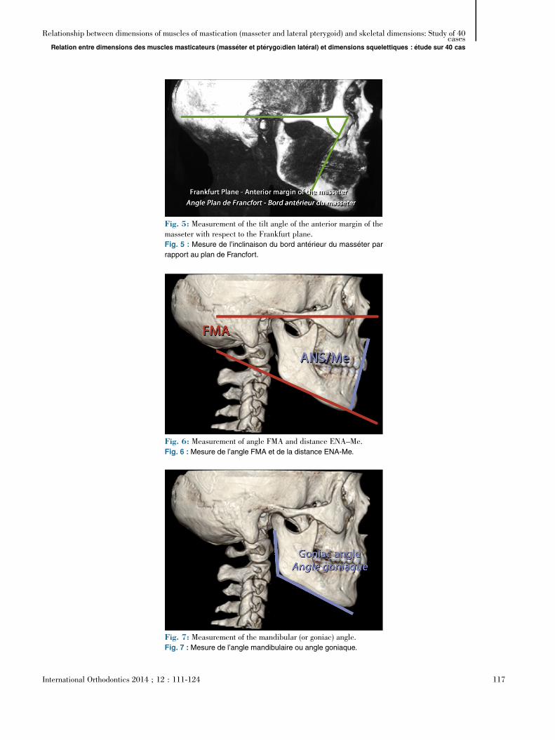
We also evaluated the tilt angle of the masseter by measuringthe angle between its anterior margin and the Frankfurt plane(fig. 5). 3D reconstructions of the cranio-facial skeleton andthe masticatory muscles enable the orientation of the latter tobe studied by determination of their long axes. Thus it ispossible to measure the angle between the anterior marginof the masseter and the Frankfurt plane. This angular positionof the muscle might have a significant influence on the bio-mechanics of the masticatory system and the occlusal forcesdeveloped. Skeletal measurements were performed on frontand right lateral views obtained by 3D reconstruction.Skeletal measurements:– vertical dimension:T mandibular plane/Frankfurt: angle between Downs’ man-dibular plane and the Frankfurt plane (fig. 6),T ENA/Me: distance between the anterior nasal spine (ENA)and the menton point (Me) (fig. 6),
M�ethode de mesure
Toutes les mesures musculaires ont �et�e r�ealis�ees sur lesmuscles du cot�e droit. Pour chaque sujet, les dimensionsmusculaires (longueur, �epaisseur et largeur) ont �et�e choisiesen retenant la moyenne des mesures maximales des musclesmass�eter et pt�erygoıdien lat�eral. La longueur maximale a �et�emesur�ee sur les coupes coronales au niveau de l’�epaisseurmaximale dumass�eter et du pt�erygoıdien lat�eral ; la largeur etl’�epaisseur musculaire maximales ont etre r�ealis�ees sur lescoupes tomodensitom�etriques axiales (figs. 1–4).Nous avons mesur�e �egalement l’inclinaison du mass�eter parl’angle bord ant�erieur du mass�eter/Francfort (fig. 5). Lesreconstructions tridimensionnelles du squelette craniofacialet des muscles masticateurs permettent d’�etudier leur orien-tation en appr�eciant leur grand axe. Ainsi, nous pouvons�etudier l’angle d’inclinaison du bord ant�erieur du mass�eterpar rapport au plan de Francfort. Cette inclinaison influencer-ait fortement la biom�ecanique du syst�eme masticateur et lesforces occlusales d�evelopp�ees. Les mesures squelettiquesont �et�e mesur�ees sur des vues de face et des vues lat�eralesdroites des reconstitutions 3D.Mesures squelettiques :– sens vertical :T plan mandibulaire/Francfort : angle entre le plan mandibu-laire de Downs et le plan de Francfort (fig. 6),T ENA/Me : distance entre l’�epine nasale ant�erieure ENA etle point menton Me (fig. 6),
[(Fig._1)TD$FIG]
Fig. 1: Measurement of the length of the masseter on a coronal CTslice.Fig. 1 :Mesure de la longueur du mass�eter sur une coupe coronale
du CTscan.
114 International Orthodontics 2014 ; 12 : 111-124
Mohammed Faouzi AZAROUAL et al.

T mandibular angle (goniac angle): angle between the man-dibular plane and the tangent to the mandibular ramus (fig. 7);
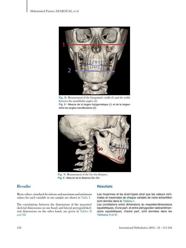
– transverse dimension:T bizygomatic width: maximum distance between the right
and left zygomatic arches,T mandibular angle width: maximum distance between the
right and left mandibular angles (fig. 8);– sagittal dimension:T distance Go–Gn: distance between the Gonion and
Gnathion (fig. 9).
Statistical method
The distribution of the various measurements was tested usingthe KS test; all the variables followed a normal distribution.The relationships between the various measurements wereevaluated using Spearman’s correlation coefficient.Reproducibility (intra-observer variability) was assessed byrepeating all the measurements on five randomly-chosen casesand was checked by calculating the intraclass correlationcoefficient, the standard value of which is 0.85.
T angle mandibulaire (angle goniaque) : angle entre le planmandibulaire et la tangente a la branche montante de la man-dibule (fig. 7) ;– sens transversal :T largeur bizygomatique : distance maximale ente les
arcades zygomatiques droite et gauche,T largeur angles mandibulaires : distance maximale ente
les angles mandibulaires droit et gauche (fig. 8) ;– sens sagittal :T distance Go–Gn : distance entre les points gonion et
gnathion (fig. 9).
M�ethode statistique
La distribution des diff�erentes mesures a �et�e test�ee par le testKS ; toutes les variables avaient une distribution gaussienne.Les relations entre les diff�erentes mesures ont �et�e �evalu�eesa l’aide du test de corr�elation de Spearman.La r�ep�etitivit�e (fiabilit�e intra-observateur) a �et�e �etudi�ee enreprenant toutes les mesures de cinq cas choisis au hasardet a �et�e v�erifi�ee en calculant le coefficient de corr�elation intra-classe dont la valeur standard est de 0,85.
[(Fig._2)TD$FIG]
Fig. 2: Measurement of the thickness and width of the mass-eter on an axial CT slice.Fig. 2 :Mesure de l’�epaisseur et de la largeur du mass�eter sur
une coupe axiale du CTscan.
International Orthodontics 2014 ; 12 : 111-124 115
Relationship between dimensions of muscles of mastication (masseter and lateral pterygoid) and skeletal dimensions: Study of 40cases
Relation entre dimensions des muscles masticateurs (mass�eter et pt�erygoıdien lat�eral) et dimensions squelettiques : �etude sur 40 cas
[(Fig._3)TD$FIG]
Fig. 3: Measurement of the length of the lateral pterygoid on acoronal CT slice.Fig. 3 :Mesure de la longueur du pt�erygoıdien lat�eral sur une coupe
coronale du CTscan.
[(Fig._4)TD$FIG]
Fig. 4: Measurement of the width and thickness of the lateralpterygoid on a coronal CT slice.Fig. 4 :Mesure de la largeur et de l’�epaisseur du pt�erygoıdien lat�eral
sur une coupe coronale du CTscan.
116 International Orthodontics 2014 ; 12 : 111-124
Mohammed Faouzi AZAROUAL et al.
[(Fig._5)TD$FIG]
Fig. 5: Measurement of the tilt angle of the anterior margin of themasseter with respect to the Frankfurt plane.Fig. 5 : Mesure de l’inclinaison du bord ant�erieur du mass�eter par
rapport au plan de Francfort.[(Fig._6)TD$FIG]
Fig. 6: Measurement of angle FMA and distance ENA–Me.Fig. 6 : Mesure de l’angle FMA et de la distance ENA-Me.[(Fig._7)TD$FIG]
Fig. 7: Measurement of the mandibular (or goniac) angle.Fig. 7 : Mesure de l’angle mandibulaire ou angle goniaque.
International Orthodontics 2014 ; 12 : 111-124 117
Relationship between dimensions of muscles of mastication (masseter and lateral pterygoid) and skeletal dimensions: Study of 40cases
Relation entre dimensions des muscles masticateurs (mass�eter et pt�erygoıdien lat�eral) et dimensions squelettiques : �etude sur 40 cas
Results
Mean values, standard deviations andmaximum andminimumvalues for each variable in our sample are shown in Table I.
The correlations between the dimensions of the masseter/skeletal dimensions on one hand, and lateral pterygoid/skel-etal dimensions on the other hand, are given in Tables IIand III.
R�esultats
Les moyennes et les �ecart-types ainsi que les valeurs mini-males et maximales de chaque variable de notre �echantillonsont donn�es dans le Tableau I.Les corr�elations entre dimensions du mass�eter/dimensionssquelettiques, d’une part, et entre pt�erygoıdien lat�eral/dimen-sions squelettiques, d’autre part, sont donn�ees dans lesTableaux II et III.
[(Fig._8)TD$FIG]
Fig. 8: Measurement of the bizygomatic width (1) and the widthbetween the mandibular angles (2).Fig. 8 : Mesure de la largeur bizygomatique (1) et de la largeur
entre les angles mandibulaires (2).[(Fig._9)TD$FIG]
Fig. 9: Measurement of the Go–Gn distance.Fig. 9 : Mesure de la distance Go–Gn.
118 International Orthodontics 2014 ; 12 : 111-124
Mohammed Faouzi AZAROUAL et al.
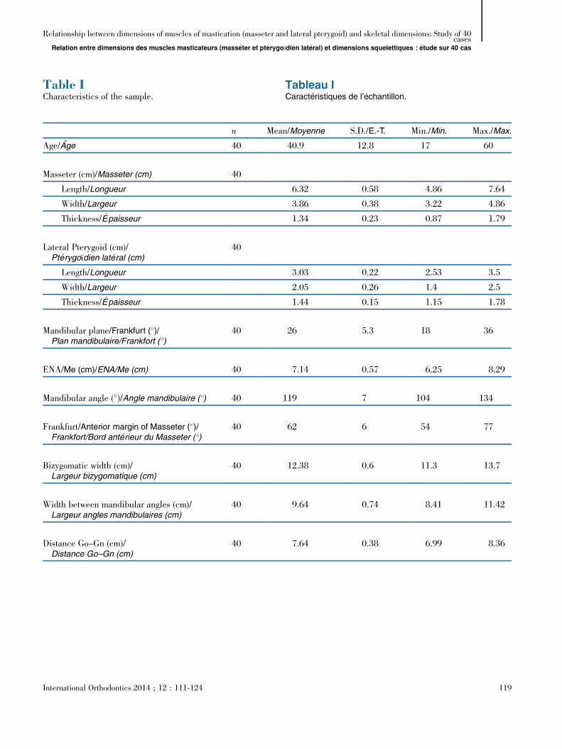
Table ICharacteristics of the sample.
Tableau ICaract�eristiques de l’�echantillon.
n Mean/Moyenne S.D./E.-T. Min./Min. Max./Max.
Age/Age 40 40.9 12.8 17 60
Masseter (cm)/Masseter (cm) 40
Length/Longueur 6.32 0.58 4.86 7.64
Width/Largeur 3.86 0.38 3.22 4.86
Thickness/ �Epaisseur 1.34 0.23 0.87 1.79
Lateral Pterygoid (cm)/Pt�erygoıdien lat�eral (cm)
40
Length/Longueur 3.03 0.22 2.53 3.5
Width/Largeur 2.05 0.26 1.4 2.5
Thickness/ �Epaisseur 1.44 0.15 1.15 1.78
Mandibular plane/Frankfurt (�)/Plan mandibulaire/Frankfort ( �)
40 26 5.3 18 36
ENA/Me (cm)/ENA/Me (cm) 40 7.14 0.57 6.25 8.29
Mandibular angle (�)/Angle mandibulaire ( �) 40 119 7 104 134
Frankfurt/Anterior margin of Masseter (�)/Frankfort/Bord ant�erieur du Masseter ( �)
40 62 6 54 77
Bizygomatic width (cm)/Largeur bizygomatique (cm)
40 12.38 0.6 11.3 13.7
Width between mandibular angles (cm)/Largeur angles mandibulaires (cm)
40 9.64 0.74 8.41 11.42
Distance Go–Gn (cm)/Distance Go–Gn (cm)
40 7.64 0.38 6.99 8.36
International Orthodontics 2014 ; 12 : 111-124 119
Relationship between dimensions of muscles of mastication (masseter and lateral pterygoid) and skeletal dimensions: Study of 40cases
Relation entre dimensions des muscles masticateurs (mass�eter et pt�erygoıdien lat�eral) et dimensions squelettiques : �etude sur 40 cas
Discussion
Our study shows that the thickness of the masseter muscle isthe dimension presenting the most significant correlationswith skeletal dimensions in the vertical, transverse and sagit-tal directions. For the lateral pterygoid muscle, length andwidth are significantly correlated with transverse skeletaldimensions. Analysis of the results shows that the dimensionalcharacteristics of the muscles of mastication vary according to
Discussion
Notre �etude a r�ev�el�e que l’�epaisseur du muscle mass�eter estla dimension qui pr�esente la corr�elation la plus importanteavec les dimensions squelettiques dans les sens vertical,transversal et sagittal. Pour le muscle pt�erygoıdien lat�eral, lalongueur et la largeur pr�esentent une corr�elation significativeavec les dimensions squelettiques transversales. L’analysedes r�esultats montre que les caract�eristiques dimensionnelles
Table IICorrelations between masseter dimensions and skeletaldimensions.
Tableau IICorr�elations dimensions Masseter - dimensionssquelettiques.
FMA/FMA Mandibularangle/Anglemandibulaire
ENA-Me/ENA-Me
Bizygomaticwidth/Largeurbizygomatique
Width betweenmandibular angles/Largeur Anglesmandibulaires
DistanceGo–Gn/DistanceGo–Gn
Thickness/ �Epaisseur NS r = �0.34P = 0.04
r = 0.42P = 0.013
r = 0.6P < 0.001
r = 0.62P < 0.001
r = 0.6P < 0.001
Length/Longueur NS r = �0.35P = 0.041
NS r = 0.41P = 0.016
NS NS
Width/Largeur NS NS NS r = 0.51P = 0.002
r = 0.41P = 0.017
NS
Angle Masseter anterior margin/Frankfurt/Angle BordAnt Masseter/Frankfort
r = �0.38P = 0.028
NS NS NS NS NS
Table IIICorrelations between lateral pterygoid dimensions andskeletal dimensions.
Tableau IIICorr�elations dimensions Pt�erygoıdien lat�eral - dimensionssquelettiques.
FMA Mandibularangle/Anglemandibulaire
ENA-Me/ENA-Pt menton
Bizygomaticwidth/Largeurbizygomatique
Width betweenmandibular angles/Largeur anglesmandibulaires
Distance(Go–Gn)/DistanceGo–Gn
Thickness/ �Epaisseur NS NS NS NS NS NS
Length/Longueur NS NS NS r = 0,51 NS NSp = 0,002
Width/Largeur NS NS NS r = 0,35 r = 0,36 NSp = 0,03 p = 0,033
120 International Orthodontics 2014 ; 12 : 111-124
Mohammed Faouzi AZAROUAL et al.
the vertical and transverse skeletal typologies of the subjectsconcerned.Various studies [1–3,21–25] have demonstrated that a closerelationship exists between the anatomical characteristics ofmuscles (insertion zones, volume, orientation) and skeletalmorphology, and that these characteristics help to explain inpart the functional differences observed between subjects ofdiffering skeletal types.
Muscle–skeleton relationship
The main aim of most imaging studies of the masticatorymuscles is to obtain a better understanding of the relationshipbetween these muscles and cranio-facial skeletal develop-ment. Computed tomography is an excellent method for obser-vation and quantitative analysis of the morphology of themuscles of mastication [4,5]. Although the muscular struc-tures are slightly less well visualized than with MRI, severalstudies have shown that the results obtained with CT-scan arecomparable to those of MRI [26].
There is general agreement between the results of these stud-ies, which reveal strong links between vertical craniofacialdevelopment (and to a lesser degree, transverse development)and masticatory muscle morphology. Few correlations havebeen identified in the sagittal direction.
Vertical direction
Several studies have shown that the dimensions and orienta-tions of the masseter muscle are closely correlated with ver-tical facial development: voluminous masseters are associatedwith a hyperdivergent skeletal morphology characterized bymarked vertical development of the anterior face and a narrowmandibular angle (Bakke et al. [1], Benington et al. [2],Gionhaku and Lowe [3], and Higashino [25]).
Masseter
In our study, only the angular position of the masseter,expressed as the angle between the masseter anterior marginand the Frankfurt plane, was correlated, in fact negatively,with facial divergence FMA (r = �0.38; P = 0.028). The mas-seter is more vertical in hypodivergent subjects, contrary towhat was found by Van Spronsen et al. [10] for whom themasseter was slightly more vertical in hyperdivergentsubjects.The results of our study show that the thickness, length andwidth of the masseter are not in any way correlated with FMA,in contradiction with Satiroglu et al. [27] who found a negativecorrelation between masseter thickness and divergence:hyperdivergent subjects had small masseter muscles. On theother hand, masseter thickness and length show a negativecorrelation, on average, with the mandibular angle; the figures
des muscles masticateurs varient en fonction des typologiessquelettiques verticale et transversale des sujets.Diverses �etudes [1–3,21–25] ont montr�e la relation �etroiteentre les caract�eristiques anatomiques musculaires (zonesd’insertion, volume, orientation) et les caract�eristiques mor-phologiques squelettiques et qu’elles contribuent a expliqueren partie les diff�erences fonctionnelles observ�ees chez lessujets de types squelettiques dissemblables.
Relations musculature–squelette
Une meilleure connaissance des relations entre la muscula-ture masticatrice et le d�eveloppement squelettique craniofa-cial est l’objectif principal de la majorit�e des �etudes de cessangles musculaires en imagerie. La tomodensitom�etrie estune excellente m�ethode d’observation et d’analyse quantita-tive de la morphologie des muscles masticateurs [4,5]. Malgr�eune visualisation des sangles musculaires l�eg�erementinf�erieure a celle de l’IRM, diff�erents travaux ont montr�e queles r�esultats obtenus avec le scanner �etaient comparablesa ceux de l’IRM [26].Les r�esultats de ces travaux sont concordants ; ils r�ev�elent defortes liaisons entre le d�eveloppement craniofacial vertical (aun degr�e moindre, le d�eveloppement transversal) et la mor-phologie desmusclesmasticateurs. Dans le sens sagittal, peude corr�elations ont �et�e mises en �evidence.
Sens vertical
Plusieurs �etudes ont trouv�e que les dimensions et les orienta-tions du mass�eter sont �etroitement corr�el�ees aud�eveloppement vertical de la face : desmass�eters volumineuxsont associ�es a la morphologie squelettique des hypodiver-gents caract�eris�ee par un fort d�eveloppement vertical de laface ant�erieure associ�e a un angle mandibulaire ferm�e :Bakke et al. [1], Benington et al. [2], Gionhaku et Lowe [3], etHigashino [25].
Mass�eter
Dans notre �etude, seule l’inclinaison du mass�eter exprim�eepar l’angle bord ant�erieur mass�eter/Francfort est corr�el�ee etce, de mani�ere n�egative, avec la divergence faciale FMA (r =�0,38 ; p = 0,028). Le mass�eter est plus vertical chez l’hypo-divergent contrairement aux r�esultats de Van Spronsen et al.[10] pour qui le mass�eter serait l�eg�erement plus vertical chezles sujets hyperdivergents.
Les r�esultats de notre �etude ont montr�e que l’�epaisseur, lalongueur et la largeur du mass�eter ne pr�esentent aucunecorr�elation avec le FMA contrairement a Satiroglu et al. [27]qui ont trouv�e une corr�elation n�egative entre l’�epaisseur dumass�eter et la divergence ; des mass�eters peu volumineuxsont retrouv�es chez les hyperdivergents. En revanche,l’�epaisseur et la longueur du mass�eter pr�esentent une
International Orthodontics 2014 ; 12 : 111-124 121
Relationship between dimensions of muscles of mastication (masseter and lateral pterygoid) and skeletal dimensions: Study of 40cases
Relation entre dimensions des muscles masticateurs (mass�eter et pt�erygoıdien lat�eral) et dimensions squelettiques : �etude sur 40 cas
are, respectively, (r = �0.34; P = 0.04) and (r = �0.35;P = 0.041).
These results corroborate those of Benington et al. [2] whofound a negative correlation between masseter thickness andthe goniac angle.Anterior facial height ENA-Me is strongly correlated withmasseter thickness (r = 0.42;P = 0.013), in contradiction withthe results of Killiaridis et al. [4], Killiaridis and Kalebo [26],and Bakke et al. [1] who noted a negative correlation betweenanterior facial height and masseter muscle thickness.Killiaridis found that women with more slender masseter mus-cles had longer faces.
Lateral pterygoid
No correlations exist between the thickness, length or width ofthe lateral pterygoid muscle and the FMA, the mandibularangle or the anterior facial length (ENA-Me).In conclusion, the dimensions of the lateral pterygoid muscleare not correlated with any vertical dimension.
Transverse direction
Several studies have shown a positive correlation between thedimensions of the pterygoid-masseter complex and transversefacial development (Hannam and Wood [24], Van Spronsen etal. [11], Kiliaridis and Kalebo [26]).
Masseter
The dimensions of the masseter muscle are closely related tobizygomatic width: thickness (r = 0.6; P < 0.001), length(r = 0.41; P = 0.016), width (r = 0.51; P = 0.002), and the sizeof the goniac angles: thickness (r = 0.62; P < 0.001), length(NS), width (r = 0.41; P = 0.017).The dimensions of the masseter muscle are strongly correlatedwith transverse skeletal dimensions. Thus, increasing masse-ter muscle development is accompanied by broader facialdimensions in the transverse direction.
Lateral pterygoid
The length of the lateral pterygoid muscle is strongly related tobizygomatic width (r = 0.51; P = 0.002). Width is mildly cor-related with transverse skeletal dimensions: bizygomaticwidth (r = 0.35 P = 0.03) and size of mandibular angle(r = 0.36; P = 0.033).
The length and width of the lateral pterygoid are stronglycorrelated with transverse skeletal dimensions. The thicknessof the muscle, on the contrary, is not correlated in any way withskeletal dimensions.
corr�elation n�egative moyenne avec l’angle mandibulairerespectivement (r = �0,34 ; p = 0,04) et (r = �0,35 ;p = 0,041).Ces r�esultats concordent avec ceux de Benington et al. [2] quiont trouv�e une corr�elation n�egative entre l’�epaisseur dumass�eter et l’angle goniaque.La hauteur faciale ant�erieure ENA–Me pr�esente unecorr�elation importante avec l’�epaisseur du mass�eter(r = 0,42 p = 0,013), contrairement aux r�esultats de Killiaridiset al. [4], Killiaridis et Kalebo [26] et Bakke [1] qui ont trouv�eune corr�elation n�egative entre la hauteur faciale ant�erieure etl’�epaisseur du mass�eter. Killiaridis et al. ont trouv�e que lesfemmes avec un mass�eter moins �epais avaient des visagesplus longs.
Pt�erygoıdien lat�eral
L’�epaisseur, la longueur et la largeur du pt�erygoıdien lat�eral nepr�esentent aucune corr�elation avec le FMA, l’angle mandibu-laire et la dimension faciale ant�erieure (ENA-Me).En conclusion, les dimensions du muscle pt�erygoıdien lat�eralne pr�esentent aucune corr�elation avec le sens vertical.
Sens transversal
Plusieurs �etudes ont �et�e men�ees montrant une corr�elationpositive entre les dimensions de la sangle pt�erygomass�et�erineet le d�eveloppement facial transversal (Hannam et Wood [24],Van Spronsen et al. [11], Kiliaridis et Kalebo [26]).
Mass�eter
Les dimensions du muscle mass�eter sont fortement li�eesa la largeur bizygomatique : �epaisseur (r = 0,6 ; p < 0,001),longueur (r = 0,41 ; p = 0,016), largeur (r = 0,51 ; p = 0,002),et a la largeur des angles goniaques : �epaisseur (r = 0,62 ;p < 0,001), longueur (NS), largeur (r = 0,41 ; p = 0,017).Les dimensions dumuscle mass�eter sont fortement corr�el�eesavec les dimensions squelettiques transversales. Ainsi, plusles sujets ont un mass�eter bien d�evelopp�e, plus ils pr�esententdes dimensions faciales importantes dans le sens transversal.
Pt�erygoıdien lat�eral
La longueur du muscle pt�erygoıdien lat�eral est fortement li�eea la largeur bizygomatique, (r = 0,51 ; p = 0,002). La largeurpr�esente des corr�elations moyennes avec les dimensionssquelettiques transversales : largeur bizygomatique(r = 0,35 p = 0,03) et largeur angle mandibulaire (r = 0,36 ;p = 0,033).La longueur et la largeur du muscle pt�erygoıdien lat�eral sontfortement corr�el�ees avec les dimensions squelettiques trans-versales. En revanche, l’�epaisseur du muscle pt�erygoıdienlat�eral ne pr�esente aucune corr�elation avec ces dimensionssquelettiques.
122 International Orthodontics 2014 ; 12 : 111-124
Mohammed Faouzi AZAROUAL et al.
Sagittal direction
Masseter
Only the thickness of the masseter muscle is correlated withthe Go–Gn distance (r = 0.6; P < 0.001).
Lateral pterygoid
The dimensions of the lateral pterygoid (thickness, length,width) display no significant correlations with the sagittaldirection as represented by the Go–Gn/SN distance.
Disclosure of interest
The authors declare that they have no conflicts of interestconcerning this article.
Sens sagittal
Mass�eter
Seule l’�epaisseur du muscle mass�eter est corr�el�ee a la dis-tance Go–Gn (r = 0,6 ; p < 0,001).
Pt�erygoıdien lat�eral
Les dimensions du pt�erygoıdien lat�eral (�epaisseur, longueur,largeur) ne pr�esentent aucune corr�elation significative avec lesens sagittal repr�esent�e par la distance Go–Gn/SN.
D�eclaration d’int�erets
Les auteurs d�eclarent ne pas avoir de conflits d’int�erets enrelation avec cet article.
References/R�ef�erences
1. Bakke M, Tuxen A, Vilmann P, Jensen BR, Vilmann A, Toft M. Ultrasound image of humanMasseter muscle related to bite force, electromyography, facial morphology and occlusalfactors. Scand J Dent Res 1992;100(3):164–71.
2. Benington PCM, Gardener JE, Hunt NP. Masseter muscle volume measured using ultra-sonography and its relationship with facial morphology. Eur J Orthod 1999;21(6):659–70.
3. Gionhaku N, Lowe AA. Relationship between jaw muscle volume and craniofacial form. JDent Res 1989;68(5):805–9.
4. Kiliaridis S, Mahboudi PH, Raadsheer MC, Katsaros C. Ultrasonographic thickness of themasseter muscle in growing individuals with unilateral crossbite. Angle Orthod 2007;77(4):607–11.
5. Kubota M, Nakano H, Sanjo I, et al. Maxillofacial morphology and masseter musclethickness in adults. Eur J Orthod 1998;20(5):535–42.
6. Raadsheer MC, Van Eidjen TMGJ, Van Ginkel FC, Prahl-Andersen B. Contribution of jawmuscle size and craniofacial morphology to human bite force magnitude. J Dent Res1999;78(1):31-42.
7. Raadsheer SA, Prabhu NT, Munshi AK. Electromyographic and ultrasonographic observa-tions of masseter and anterior temporalis muscles in children. J Clin Pediatr Dent 1996;20(2):127–32.
8. Van Spronsen PH, Weijs WA, Valk J, Prahl-Andersen B, Van Ginkel FC. Relationshipsbetween jaw muscle cross sections and craniofacial morphology in normal adults studiedwith magnetic resonance imaging. Eur J Orthod 1991;13(5):351–61.
9. Van Spronsen PH, Weijs WA, Van Ginkel FC, Prahl-andersen B. Jaw muscle orientationand moment arms of long-face and normal adults. J Dent Res 1996;75(6):1372–80.
10. Van Spronsen PH, Koolstra JH, Van Ginkel FC, Weijs WA, Valk J, Prahl-Andersen B.Relationships between the orientation and moment arms of the human jaw muscles andnormal craniofacial morphology. Eur J Orthod 1997;19(3):313–28.
11. Van Spronsen PH, Weijs WA, Valk J, Prahl-Andersen B, Van Ginkel FC. A comparison ofjaw muscle cross section of long-face and normal adults. J Dent Res 1992;71:1279–85.
12. Van Spronsen PH, Weijs WA, Valk J, Prahl-Andersen B, Van Ginkel FC. Comparison ofjaw-muscle bite force cross sections obtained by means of magnetic resonance imaging andhigh-resolution CT scanning. J Dent Res 1989;68(12):1765–70.
International Orthodontics 2014 ; 12 : 111-124 123
Relationship between dimensions of muscles of mastication (masseter and lateral pterygoid) and skeletal dimensions: Study of 40cases
Relation entre dimensions des muscles masticateurs (mass�eter et pt�erygoıdien lat�eral) et dimensions squelettiques : �etude sur 40 cas

13. Weijs WA, Hillen B. Correlation between the cross sectional area of the jaw muscles andcranio-facial size and shape. Am J Phys Anthropol 1986;70(4):423–31.
14. Horger M, Claussen CD, Bross-Bach U, et al. Wholebody low-dose multidetector row-CT inthe diagnosis of multiple myeloma: an alternative to conventional radiography. Eur J Radiol2005;54(2):289–97.
15. Francone M, Napoli A, Carbone I, et al. Noninvasive imaging of the coronary arteries usinga 64-row multidetector CT scanner: initial clinical experience and radiation dose concerns.Eur J Radiol 2005;54(2):289–97.
16. Mah JK, Danforth RA, Bumann A, Hatcher D. Radiation absorbed in maxillofacial imagingwith a new dental computed tomography device. Oral Surg Oral Med Oral Pathol OralRadiol Endod 2003;96:508–13.
17. Katsumata A, Fujishita M, Ariji Y, Ariji E, Langlais RP. 3D CT evaluation of massetermuscle morphology after setback osteotomy for mandibular prognathism. Oral Surg OralMed Oral Pathol Oral Radiol Endod 2004;98(4):461–70.
18. Raustia AM, Salonen MAM, Pyhtinen J. Evaluation of masticatory muscles of edentulouspatients by computed tomography and electromyography. J Oral Rehabil 1996;23(1):11–6.
19. Takashima M, Kitai N, Murakami S, Furukawa S, Kreiborg S, Takada K. Volume and shapeof masticatory muscles in patients with hemifacial microsomia. Cleft Palate Craniofac J2003;40(1):6-12.
20. Ueki K, Takazakura D, Marakuva K, Shimada M, Nakagawa K, Yamamoto E. Relationshipbetween the morphologies of the Masseter muscle and the ramus and occlusal force inpatients with mandibular prognathism. J Oral Maxillofac Surg 2006;64(10):1480–6.
21. Ariji Y, Kawamata A, Yoshida K, et al. Three-dimensional morphology of the Massetermuscle in patients with mandibular prognathism. Dentomaxillofac Radiol 2000;29(2):113–8.
22. Boileau MJ, Sampeur M, Radzkiewicz A. Apport de la tomodensitom�etrie dans l’�etude de lamusculature cranio-faciale. Revue de la litt�erature. Rev Orthop Dento Faciale 2003;37:75-92.
23. Chan HJ, Woods M, Stella D. Mandibular muscle morphology in children with differentvertical facial patterns: a 3-dimensional computed tomography study. Am J OrthodDentofacial Orthop 2008;133(1):10.e1-10.e13. doi: 10.1016/j.ajodo.2007.05.013.
24. Hannam AG, Wood WW. Relationship between the size and spatial morphology of humanMasseter and medial pterygoid muscles, the craniofacial skeleton and jaw biomechanics.Am J Phys Anthropol 1989;80(4):429–45.
25. Higashino R. Relationship between jaws and the masseter muscle by superimposing MRimages on the cephalogram. Kokubyo Gakkai Zasshi 2006;73(1):116–24.
26. Kiliaridis S, Kalebo P. Masseter muscle thickness measured by ultrasonography and itsrelation to facial morphology. J Dent Res 1991;70(9):1262–5.
27. Satiroglu F, Arun T, Isik F. Comparative data on facial morphology and muscle thicknessusing ultrasonography. Eur J Orthod 2005;27(6):562–7 [Epub 2005 Aug 31].
124 International Orthodontics 2014 ; 12 : 111-124
Mohammed Faouzi AZAROUAL et al.