Embed Size (px)
Citation preview
Basel • Freiburg • Hartford • Oxford • Bangkok • Dubai • Kuala Lumpur • Melbourne • Mexico City • Moscow • New Delhi • Paris • Shanghai • Tokyo
Vitamin D: From Gestation to Adolescence in Health and Disease
Editor
Carlos Lifschitz , Buenos Aires
Editorial Board
Jatinder Bhatia, Augusta, GAWeili Lin, Chapel Hill, NCCarlos Lifschitz, Buenos AiresAndrew Prentice, Banjul/LondonFrank M. Ruemmele, ParisHania Szajewska, Warsaw
Supported by
Vol. 77, Suppl. 3, 2019
https://www.nestlenutrition-institute.org
S. KargerMedical and Scientifi c PublishersBasel • Freiburg • Hartford • Oxford • Bangkok • Dubai • Kuala Lumpur • Melbourne • Mexico City • Moscow • New Delhi • Paris • Shanghai • Tokyo
DisclaimerTh e statements, opinions and data contained in this publica-tion are solely those of the individual authors and contributors and not of the publisher and the editor(s). Th e appearance of advertisements in the journal is not a warranty, endorsement, or approval of the products or services advertised or of their eff ectiveness, quality or safety. Th e publisher and the editor(s) disclaim responsibility for any injury to persons or property resulting from any ideas, methods, instructions or products referred to in the content or advertisements.
Drug DosageTh e authors and the publisher have exerted every eff ort to en-sure that drug selection and dosage set forth in this text are in accord with current recommendations and practice at the time of publication. However, in view of ongoing research, changes in government regulations, and the constant fl ow of informa-tion relating to drug therapy and drug reactions, the reader is urged to check the package insert for each drug for any change in indications and dosage and for added warnings and precau-tions. Th is is particularly important when the recommended agent is a new and/or infrequently employed drug.
All rights reserved.No part of this publication may be translated into other languages, reproduced or utilized in any form or by any means, electronic or mechanical, including photocopying, recording, microcopying, or by any information storage and retrieval system, without permission in writing from the publisher or, in the case of photocopying, direct payment of a specifi ed fee to the Copyright Clearance Center (see “General Information”).
© 2020 Nestlé Nutrition Institute, Switzerland/ S. Karger AG, BaselP.O. Box, CH–4009 Basel (Switzerland)e-ISBN 978–3–318–06796–5
Reprint of Annals of Nutrition and Metabolism Vol. 76, Suppl. 2, 2020
Sponsor Note
This publication was supported by an unrestricted educational grant by the Nestlé Nutrition Institute. The institute is a not-for-profi t association which was created to provide latest medical and scientifi c information to health profes-sionals in the fi eld of pediatric and adult nutrition and nutrition-related disorders (available at www.nestlenutrition-institute.org).Any liability of the sponsors for the content of the papers is hereby expressly excluded.
Disclosure Statement Editor
C.L. was a member of the Faculty Board of the Nestlé Nutrition Institute at the time of manuscript preparation and has received honoraria from Danone, Mead Johnson Nutrition, the Nestlé Nutrition Institute and Wyeth.
[email protected] www.karger.com/anm
Vol. 77, Suppl. 3, 2019
Contents
DOI: 10.1159/000510225
Vitamin D: From Gestation to Adolescence in Health and Disease– Infographic – Poster
11 Editorial Lifschitz, C. (Buenos Aires)
Vitamin D: From Gestation to Adolescence in Health and Disease
1 5 Focus on: Vitamin D in Preterm and Full-Term Infant s
16 Vitamin D in Preterm and Full-Term Infant s
Abrams, S.A. (Austin, TX)
15 Focus on: Early-Life Effects of Vitamin D: A Focus on Pregnancy and Lactation
16 Early-Life Effects of Vitamin D: A Focus on Pregnancy and
Lactation
Wagner, C.L.; Hollis, B.W. (Charlerston, SC)
29 Focus on: Vitamin D in Toddlers, Preschool Children, and Adolescents
30 Vitamin D in Toddlers, Preschool Children, and Adolescents
Taylor, S.N. (New Heaven, CT)
The above articles were originally published as a supplementary issue ofAnnals of Nutrition and Metabolism and are reprinted here with permission.
[email protected] © 2020 Nestlé Nutrition Institute, Switzerland/S. Karger AG, Basel
Policy Statement
The Nestlé Nutrition Institute was created to provide health professionals with up-to-date information on nutrition and nutrition-related disorders in order to enable them to continuously improve patient care based on the latest medical and scientific developments.
One of the key pillars of the Nestlé Nutrition Institute is Annales Nestlé, a pediatric journal that has been published on a regular basis since 1942. It contains review articles on clinical practice and research in all fields of pediatrics with focus on nutrition.
Annales Nestlé appears three times a year. Each article is supported by a Focus Page, and each issue by an Infographic illustrating the core topic. Published on www.nestlenutrition-institute.org as well as in print, Annales Nestlé is one of the most widely read pediatric journals in the world.
Annales Nestlé is edited by an independent editorial board of opinion leaders in pediatric research, thus guaranteeing the medical and scientific impartiality of the journal, and hence the high regard it enjoys in medical and scientific circles. The editorial board sets the editorial policy, identifies topics to be addressed, selects authors, and oversees the review process for each issue.
Every issue of Annales Nestlé initially appears as a supplement to Annals of Nutrition and Metabolism – a journal from Karger Publishers, Basel, Switzerland – and is listed in all major bibliographic services, such as Medline, PubMed, and Web of Science. This has been our practice since 2011.
We are pleased to offer you our innovative product, which results from a creative and effective cooperation with Karger Publishers, Switzerland.
Natalia Wagemans, MDGlobal HeadNestlé Nutrition InstituteVevey (Switzerland)
[email protected] © 2020 Nestlé Nutrition Institute, Switzerland/S. Karger AG, Basel
Editorial
Reprinted with permission from:Ann Nutr Metab 2020;76(suppl 2):1–4
Vitamin D
Carlos Lifschitz
Department of Pediatrics, Section of Gastroenterology, Hepatology and Transplantation, Hospital Italiano, Buenos Aires , Argentina
Carlos Lifschitz, MD Department of Pediatrics, Section of Gastroenterology, Hepatology and Transplantation Hospital Italiano, Juan D. Peron 4190 Buenos Aires, C1181ACH (Argentina) carlos.lifschitz @ hiba.org.ar
© 2020 Nestlé Nutrition Institute, Switzerland/S. Karger AG, Basel
DOI: 10.1159/000508423
The editorial role for this issue of Annales Nestlé was initially
assumed by Dr. Jatinder Bhatia. For unforeseen reasons, he
was not able to complete this work. His friends at the Nestlé
Nutrition Institute and the Institute’s Faculty members would
like to dedicate this issue to him. We look forward to his con-
tinued support and valuable contributions to the activities of
the Nestlé Nutrition Institute.
In the mid-1600s, two authors independently published in
Latin a description of rickets. In 1840, Sniadecki, a Polish phy-
sician, observed that cases of rickets occurred in children liv-
ing in the industrial center of Warsaw, while they did not occur
among those living in the country outside Warsaw. He hy-
pothesized that lack of exposure to sunlight in the cramped
city streets, with considerable pollution due to the burning of
wood and coal, was the cause of the disease. However, the
concept that the sun could have any useful benefit on the
skeleton was not well accepted at that time. Since then, much
has been learned about vitamin D. Undoubtedly, vitamin D
plays a fundamental role in the development and mainte-
nance of the musculoskeletal system. But its function goes
well beyond hormone-like regulation, as it can also be gener-
ated by simple unicellular organisms.
Vitamin D is a prohormone absorbed from food sources or
supplements and also synthesized in the skin following expo-
sure to ultraviolet light. The prohormone subsequently is con-
verted to the metabolically active form in the liver and then
the kidneys. Few foods naturally contain vitamin D. The prin-
cipal food sources of vitamin D are fish that are oil rich, such
as salmon, mackerel, and herring, as well as organ meats, liv-
er, and egg yolk. However, how much and how frequently do
most children consume these natural dietary sources of vita-
min D? In humans, dermal synthesis is the major natural
source of the vitamin. Individuals who do not have sufficient
sun exposure, especially infants, require supplemental vitamin
D from fortified foods or supplements. Vitamin D deficiency
is frequent in children and adults and leads to serious health
problems worldwide. In recent years, vitamin D has notably
attracted scholars’ attention because of its influences on cell
growth and differentiation, immune and cardiovascular func-
tion, as well as calcium and phosphate homeostasis [1] .
In this issue, firstly, Carol L. Wagner and Bruce W. Hollis
discuss the early-life effects of vitamin D in their article “Early-
Life Effects of Vitamin D: A Focus on Pregnancy and Lacta-
tion.” They describe that the active form of vitamin D –
1,25-dihydroxyvitamin D (1,25[OH] 2 D) – increases during
pregnancy and remains elevated throughout, and unlike at
other times during the lifecycle, it is directly affected by the
circulating 25-hydroxyvitamin D (25[OH]D) concentration.
When a mother is vitamin D deficient, her milk is deficient,
which can be remedied by direct infant supplementation;
however, this treats only the infant. A safe alternative during
lactation to infant supplementation is direct maternal vitamin
D supplementation at higher doses than usual (6,400 IU/day),
improving the vitamin D status of the mother and the content
Lifschitz Reprint with permission from:Ann Nutr Metab 2020;76(suppl 2):1–4
2
DOI: 10.1159/000508423
of the milk and, consequently, the vitamin D status of the in-
fant, effectively treating both mother and infant.
In the second article, Steven A. Abrams discusses vitamin
D needs in premature and full-term infants. He states that vi-
tamin D is necessary for the active (transcellular) absorption
of calcium and for skeletal health. Inadequate vitamin D in
infants leads to increased risks of poor bone mineralization
and ultimately rickets. Rickets is uncommon in full-term in-
fants with a much higher risk in very premature infants. How-
ever, the primary cause of rickets in premature infants is a
deficiency of calcium and phosphorus, not vitamin D. The
usual total dietary intake level should be approximately 400
IU daily in healthy infants.
In the last article, Sarah N. Taylor writes about vitamin D in
toddlers, preschool children, and adolescents. Childhood is a
period of significant bone development and, therefore, atten-
tion to the vitamin D needed to optimize bone health in child-
hood is imperative. Observational studies have pointed to a
vitamin D status, as indicated by a 25(OH)D concentration of
50 nmol/L, to ensure avoidance of rickets, and of 75 nmol/L
to optimize health. Ongoing research is directed to the estab-
lishment of the best method to measure vitamin D status, ex-
amination of genetic variation in vitamin D metabolism, and
consideration that vitamin D status is a marker of another vari-
able, such as physical activity, and its association with bone
health.
Aspects not addressed in the above-mentioned contribu-
tions will now be briefly discussed.
Vitamin D Deficiency in Obese Children
Another aspect of vitamin D are the requirements of obese
and chronically ill children. Excessive fat accumulation and
vitamin D insufficiency have negative effects on each other as
a result of excessive metabolic processes and enzymatic dis-
orders in a situation of decreased activity of the key enzyme
in the biotransformation of calciferol, α-hydroxylase, in a fat-
infiltrated liver [2] . This results in an accumulation of inactive
forms and decreased bioavailability of vitamin D [3] . Vitamin D
affects insulin secretion, tissue sensitivity to insulin, and sys-
temic inflammation in obesity. Insulin secretion and tissue in-
sulin sensitivity are Ca 2+ -dependent mechanisms, and vitamin
D regulates intracellular concentrations of Ca 2+ and its pas-
sage through membranes. In addition, vitamin D affects in a
positive manner the expression of insulin receptors in periph-
eral cells and counteracts the systemic immune response by
modulating the expression and activity of cytokines [4] . The
influence of adipose tissue on the metabolism of vitamin D,
on the one hand, and its pathogenic role in the obesity devel-
opment mechanisms, on the other, are closely interrelated
and represent mutually dependent processes [2] . A study con-
ducted in 58 obese adolescents demonstrated that a 1% in-
crease in fat weight was associated with a 1.15 ± 0.55 nmol/L
reduction in serum calcifediol (25[OH]D 3 ) [5] . There are sev-
eral theories regarding why calcifediol levels are decreased in
obese individuals. The first and most accepted is that adipose
tissue absorbs the fat-soluble vitamin D [6] . Another theory
explains the low 25(OH)D concentrations by the fact that
obese people lead a sedentary lifestyle and are less active
physically, which entails a decrease in exposure to sunlight
and in endogenous synthesis of vitamin D [7] . Also, vitamin D
metabolism and 25(OH)D synthesis could be impaired as a
result of hepatic steatosis developing in obesity [8] . The prev-
alence of vitamin D insufficiency in groups of overweight and
obese children is very high (36–93%) [1] . There is no conven-
tional dose universally recommended for the treatment of vi-
tamin D insufficiency in overweight and obese children. The
Committee on Nutrition of the French Society of Pediatrics
recommends administration of vitamin D in 80,000-IU single
doses and 100,000-IU single doses in the winter months for
obese children aged 5–10 years or uninterrupted supplemen-
tation over the age interval of 1–10 years [1] . The United States
Endocrine Society recommends a 2-fold increase in the ther-
apeutic dose of cholecalciferol for overweight and obese pa-
tients and setting the calcifediol target at 75 nmol/L (30 ng/
mL), with subsequent switching to a maintenance dose.
Vitamin D Deficiency in Children with Chronic Kidney Disease
Deficiency of vitamin D is prevalent and frequently severe in
children and adults with chronic kidney disease (CKD). 25(OH)
D deficiency is linked causally to rickets and fractures in
healthy children and more so in those with CKD, a contribut-
ing factor to the CKD–mineral and bone disorder complex [9] .
There are few studies to provide evidence for vitamin D ther-
apy or guidelines for its use in CKD. It has been suggested to
use native vitamin D supplements for the treatment of vitamin
D deficiency in children with CKD stages 2–5 who have serum
25(OH)D concentrations below 75 nmol/L. In children with
CKD stages 2–3, native vitamin D supplements may be used
for the prevention or treatment of secondary hyperparathy-
roidism. It has been suggested to use either vitamin D 2 (ergo-
calciferol) or vitamin D 3 (cholecalciferol) treatment in children
with CKD stages 2–5D to increase serum 25(OH)D levels to
the target range. Mega-dose vitamin D therapy is not recom-
mended.
Editorial 3Reprint with permission from:Ann Nutr Metab 2020;76(suppl 2):1–4DOI: 10.1159/000508423
Vitamin D Deficiency in Children with Chronic Liver Disease
The liver produces 25(OH)D (calcidiol), which is the imme-
diate precursor to the metabolically active 1,25(OH) 2 D (cal-
citriol). 25(OH)D is measured to assess vitamin D deficiency
[10] . Although in patients with liver failure levels of calcidiol
can be low due to impaired synthesis, liver function has to be
severely compromised for this impairment to occur. Liver dis-
ease could also lead to impaired absorption of vitamin D, like-
ly related to impaired bile acid production or intestinal edema
secondary to portal hypertension. Hypovitaminosis D and
bone disease are well-recognized complications of “choles-
tatic” liver disease, which impairs production or bile flow. Vi-
tamin D deficiency is frequent in biliary atresia patients [11] . In
their study, Dong et al. [11] also found that despite bile flow
restitution after surgery, vitamin D deficiency was confirmed
in the majority of biliary atresia patients.
Vitamin D in Children with Inflammatory Bowel Disease
Vitamin D deficiency is highly prevalent in children with
inflammatory bowel disease (IBD), which may contribute to
an increased risk of poor bone health as well as affect the
course of the illness. An optimal treatment strategy of vitamin
D therapy in children with IBD, however, has not yet been es-
tablished. A recent review article [12] identified that some pe-
diatric trials have shown that vitamin D deficiency may in part
contribute to an increased risk of poor bone health, but others
have reached contrasting conclusions. Recent studies have
also focused on the relationship between vitamin D deficien-
cy and disease severity in children with IBD. While some lim-
ited data suggest an association of vitamin D deficiency with
a more severe course of disease, other studies do not report
such a relationship. The authors of the above-cited review
identified 277 discrete articles, but only 10 met the require-
ments to be included in their review. The included trials fea-
tured diverse treatment regimens that were predominantly
insufficient in correcting vitamin D deficiency or maintaining
adequate levels in children with IBD. Better treatment regi-
mens are required for the management of vitamin D deficien-
cy in children with IBD. The Institute of Medicine sustains that
serum 25(OH)D levels above 30 ng/mL do not provide addi-
tional benefit [13] . However, other studies suggest that a level
of at least 32 ng/mL is required for optimal intestinal calcium
absorption. However, a level of 30 ng/mL has been found to
be sufficient to reduce parathormone activity [14] . Based on
the above, the goal for the serum 25(OH)D level should be at
least 30 ng/mL for children with IBD. In one of the included
studies, 2,000 IU vitamin D 3 daily raised serum 25(OH)D above
30 ng/mL in 74% of the participants after 6 months of treat-
ment [15] . Another study showed a similar response after
weekly dosing of 50,000 IU vitamin D 3 for 6 weeks [16] . In a
third study, 2,000 IU vitamin D 3 daily was able to achieve a
mean serum 25(OH)D above 30 ng/mL in the study popula-
tion throughout the course of treatment [17] . Both 5,000
IU/10 kg vitamin D 3 per week and 10,000 IU/10 kg vitamin D 3
per week for a period of 6 weeks were able to raise serum
25(OH)D above 30 ng/mL at week 8 in one of the trials; how-
ever, this effect was lost by week 12 [18] . We sincerely hope
that Dr. Bhatia will be satisfied with this issue of the Annals .
References
1 Christakos S, Dhawan P, Verstuyf A, Verlinden L, Carmeliet G. Vi-tamin D: metabolism, molecular mechanism of action, and pleio-tropic effects. Physiol Rev . 2016 Jan; 96(1): 365–408.
2 Zakharova I, Klimov L, Kuryaninova V, Nikitina I, Malyavskaya S, Dolbnya S, et al. Vitamin D Insufficiency in Overweight and Obese Children and Adolescents. Front Endocrinol (Lausanne) . 2019 Mar; 10: 103.
3 Hyppönen E, Power C. Vitamin D status and glucose homeostasis in the 1958 British birth cohort: the role of obesity. Diabetes Care . 2006 Oct; 29(10): 2244–6.
4 Wright DC, Hucker KA, Holloszy JO, Han DH. Ca2+ and AMPK both mediate stimulation of glucose transport by muscle contrac-tions. Diabetes . 2004 Feb; 53(2): 330–5.
5 Lenders CM, Feldman HA, Von Scheven E, Merewood A, Sweeney C, Wilson DM, et al.; Elizabeth Glaser Pediatric Research Network Obesity Study Group. Relation of body fat indexes to vitamin D status and deficiency among obese adolescents. Am J Clin Nutr . 2009 Sep; 90(3): 459–67.
6 Wortsman J, Matsuoka LY, Chen TC, Lu Z, Holick MF. Decreased bioavailability of vitamin D in obesity. Am J Clin Nutr . 2000 Sep; 72(3): 690–3.
7 Florez H, Martinez R, Chacra W, Strickman-Stein N, Levis S. Out-door exercise reduces the risk of hypovitaminosis D in the obese. J Steroid Biochem Mol Biol . 2007 Mar; 103(3-5): 679–81.
8 Targher G, Bertolini L, Scala L, Cigolini M, Zenari L, Falezza G, et al. Associations between serum 25-hydroxyvitamin D3 concen-trations and liver histology in patients with non-alcoholic fatty liver disease. Nutr Metab Cardiovasc Dis . 2007 Sep; 17(7): 517–24.
Lifschitz Reprint with permission from:Ann Nutr Metab 2020;76(suppl 2):1–4
4
DOI: 10.1159/000508423
9 Shroff R, Wan M, Nagler EV, Bakkaloglu S, Fischer DC, Bishop N, et al.; European Society for Paediatric Nephrology Chronic Kidney Disease Mineral and Bone Disorders and Dialysis Working Groups. Clinical practice recommendations for native vitamin D therapy in children with chronic kidney disease Stages 2-5 and on dialysis. Nephrol Dial Transplant . 2017 Jul; 32(7): 1098–113.
10 Nair S. Vitamin D deficiency and liver disease. Gastroenterol Hep-atol (N Y) . 2010 Aug; 6(8): 491–3.
11 Dong R, Sun S, Liu XZ, Shen Z, Chen G, Zheng S. Fat-soluble vita-min deficiency in pediatric patients with biliary atresia. Gastroen-terol Res Pract . 2017; 2017: 7496860.
12 Rigterink T, Appleton L, Day AS. Vitamin D therapy in children with inflammatory bowel disease: A systematic review. World J Clin Pediatr . 2019 Jan; 8(1): 1–14.
13 Ross AC, Manson JE, Abrams SA, Aloia JF, Brannon PM, Clinton SK, et al. The 2011 Dietary Reference Intakes for Calcium and Vi-tamin D: what dietetics practitioners need to know. J Am Diet As-soc . 2011 Apr; 111(4): 524–7.
14 Holick MF, Binkley NC, Bischoff-Ferrari HA, Gordon CM, Hanley DA, Heaney RP, et al.; Endocrine Society. Evaluation, treatment, and prevention of vitamin D deficiency: an Endocrine Society clin-ical practice guideline. J Clin Endocrinol Metab . 2011 Jul; 96(7): 1911–30.
15 Wingate KE, Jacobson K, Issenman R, Carroll M, Barker C, Israel D, et al. 25-Hydroxyvitamin D concentrations in children with Crohn’s disease supplemented with either 2000 or 400 IU daily for 6 months: a randomized controlled study. J Pediatr . 2014 Apr; 164(4): 860–5.
16 Pappa HM, Mitchell PD, Jiang H, Kassiff S, Filip-Dhima R, DiFabio D, et al. Treatment of vitamin D insufficiency in children and ado-lescents with inflammatory bowel disease: a randomized clinical trial comparing three regimens. J Clin Endocrinol Metab . 2012 Jun; 97(6): 2134–42.
17 Hradsky O, Soucek O, Maratova K, Matyskova J, Copova I, Za-rubova K, et al. Supplementation with 2000 IU of Cholecalciferol Is Associated with Improvement of Trabecular Bone Mineral Den-sity and Muscle Power in Pediatric Patients with IBD. Inflamm Bowel Dis . 2017 Apr; 23(4): 514–23.
18 Simek RZ, Prince J, Syed S, Sauer CG, Martineau B, Hofmekler T, et al. Pilot Study Evaluating Efficacy of 2 Regimens for Hypovita-minosis D Repletion in Pediatric Inflammatory Bowel Disease. J Pediatr Gastroenterol Nutr . 2016 Feb; 62(2): 252–8.
Focus
Reprinted with permission from: Ann Nutr Metab 2020;76(suppl 2):6–14
Vitamin D in Preterm and Full-Term InfantsSteven A. Abrams
© 2020 Nestlé Nutrition Institute, Switzerland/S. Karger AG, Basel
Key Insight
Vitamin D is essential for transcellular absorption of calcium and for skeletal health. Inadequate vitamin D in infants leads to poor bone mineralization and increased risk of rickets. Most guidelines recommend 400 IU daily of vitamin D to support bone health in preterm and full-term infants. Although cutaneous production of vitamin D occurs in infants, the use of sunblock and other factors limiting sun exposure make this an unreliable source. Therefore, recommendations for vitamin D intake are made assuming minimal or nonexistent cutaneous production of vitamin D. Not surprisingly, neonatal vitamin D status reflects maternal status. This knowledge has prompted current guidelines to recommend that vitamin D supplementation for infants is initiated as soon as possible.
Current knowledge
In the first weeks of life, calcium absorption occurs mainly via paracellular mechanisms that are not dependent on vitamin D. In preterm infants, absorption of vitamin D may be affected by various disease states, including malabsorptive disorders, such as cystic fibrosis. Cholestasis is another common prob-lem in high-risk neonates and is associated with long-term use of parenteral nutrition. These highlight the importance of identifying the populations of mothers and infants who are at risk in order to ensure adequate vitamin D intake. Caution should be taken to ensure that the appropriate dose is given and that accidental ingestion of high doses of vitamin D does not occur.
Practical implications
Currently, there is no clinical evidence to support the need for routine vitamin D supplementation for infants who are exclu-sively formula fed. In fully or partially breastfed infants, there are several methods for providing vitamin D. One is to admin-ister the drops to the infant using a dropper. Vitamin D drops can also be placed directly on the breast or given as dissolv-able film strips. Another approach is to have the lactating mother take a relatively high dose of vitamin D (6,400 IU dai-ly) to ensure an adequate level of vitamin D in the breast milk. However, adherence to guidelines varies widely between countries, highlighting the need for education for healthcare providers and families on the importance of providing suffi-cient vitamin D to infants.
Recommended reading
Roth DE, Abrams SA, Aloia J, Bergeron G, Bourassa MW, Brown KH, et al. Global prevalence and disease burden of vi-tamin D deficiency: A roadmap for action in low- and middle-income countries. Ann N Y Acad Sci. 2018 Oct;1430(1):44–79.
Vitamin D is a critical nutrient for bone health and needs to be provided to all infants whether via infant formula or as a supplement to breastfed infants or high-dose supplement to their mothers
Sufficient vitaminD intake, optimal bone health
Infant formulasupplemented with vitamin D
Use of drops,soluble strips,increase maternal intake of vitamin D
Formula-fed
Breastfed
Ensuring adequate vitamin D intake is essential for all infants, regard-less of whether they are formula fed or breastfed.
Vitamin D
Reprint with permission from:Ann Nutr Metab 2020;76(suppl 2):6–14
Vitamin D in Preterm and Full-Term Infants
Steven A. Abrams
Department of Pediatrics, Dell Medical School at the University of Texas, Austin , TX , USA
Steven A. Abrams Department of Pediatrics Dell Medical School at the University of Texas-Austin 1400 Barbara Jordan Blvd., Austin, TX 78723 (USA) sabrams @ austin.utexas.edu
© 2020 Nestlé Nutrition Institute, Switzerland/S. Karger AG, Basel
Key Messages
• Dietary vitamin D intake should be assured in all infants, preterm and full term, with emphasis on adequate supplementation of infants who are receiving human milk.
• Usual total dietary intake level should be approximately 400 IU daily in healthy infants.
• There are multiple methods for providing vitamin D to infants; these may be selected based on parental desires.
DOI: 10.1159/000508421
Keywords Bone health · Calcium absorption · Vitamins
Abstract Vitamin D is necessary for the active (transcellular) absorption
of calcium and for skeletal health. Inadequate vitamin D in
infants leads to increased risks of poor bone mineralization
and ultimately rickets. Rickets is uncommon in full-term in-
fants with a much higher risk in very premature infants. How-
ever, the primary cause of rickets in premature infants is a
deficiency of calcium and phosphorus, not vitamin D. Avail-
able research, as well as most guidelines, recommend an in-
take of 400 IU daily of vitamin D as adequate for bone health
in preterm and full-term infants. Higher doses have not been
consistently shown to have specific clinical benefits for
healthy infants. There are no strong data to support either
routine testing of serum 25-hydroxyvitamin D or targeting
high serum 25-hydroxyvitamin D levels (e.g., 30 ng/mL) in
healthy preterm or full-term infants. Vitamin D is commonly
provided to infants via drops for breastfed babies or via infant
formula, although alternative dosing approaches exist for
breastfed infants, which some families may prefer. These in-
clude the use of drops placed on the mother’s breast, dissolv-
able doses, and high maternal doses (approximately 6,400 IU
daily). Infant formula contains vitamin D, and most infants will
reach an intake from formula of about 400 IU daily within the
first 2 months of life if they are consuming routine cow milk-
based formula. Although vitamin D toxicity is very uncom-
mon, caution should be used to avoid extremely concentrat-
ed high doses found in some commercially available drops.
Infants with liver or kidney disease may need special attention
to vitamin D intake and status. Further research is needed to
define the role of vitamin D in non-bone health outcomes of
infants and to identify methods to enhance compliance with
current recommendations for vitamin D intake in infants.
© 2020 Nestlé Nutrition Institute, Switzerland/
S. Karger AG, Basel
Vitamin D in Infants 7Reprint with permission from:Ann Nutr Metab 2020;76(suppl 2):6–14DOI: 10.1159/000508421
Vitamin D Physiology and Bone Health in Infants
Vitamin D is an essential nutrient for bone health in all indi-
viduals, including infants regardless of size or gestational ma-
turity. Although other roles for vitamin D in health and disease
exist, this discussion will focus on bone health, especially
bone health in infants who do not have underlying endocrine
disorders or severe nutritional diseases.
Vitamin D is critical for the transcellular absorption of cal-
cium, via its active form, 1,25 dihydroxyvitamin D. Dietary vi-
tamin D or vitamin D formed via solar exposure is converted
in the liver to the circulating and primary storage, 25-hy-
droxyvitamin D (25[OH]D). The 25(OH)D is then transferred to
the kidney where it is converted to 1,25 dihydroxyvitamin D.
These physiological processes function normally in preterm
and full-term infants who are otherwise healthy. A detailed
review of vitamin D-related physiology can be found else-
where [1] .
Serum 25(OH)D in Infants
The role of serum 25(OH)D as a marker of vitamin D status has
been extensively reviewed and discussed in a 2011 Institute of
Medicine (IOM) report [2] . There are no recommendations ei-
ther in that report or in any official American Academy of Pe-
diatrics (AAP) statement for routine screening of 25(OH)D lev-
el in healthy preterm or full-term infants [2–7] . It is critical to
understand that 25(OH)D is not necessarily a marker of phys-
iological vitamin D function as it is not the primary active form
of vitamin D. Rather, its concentration in the serum is valuable
as a means of assessing individual and population vitamin D
status. Different values for serum 25(OH)D have been de-
scribed as “inadequate” or “deficient” in the literature. How-
ever, the adequate serum level indicated by the IOM and sub-
sequently affirmed by the AAP of at least 20 ng/mL is the val-
ue that may be used for infants, both preterm and full term
[2–6] , pending further information clearly documenting non-
bone health-related benefits to higher minimum levels. There
are no data reliably establishing a value of 25(OH)D that is
toxic, especially in infants. Values of > 100 ng/mL have been
used to indicate toxicity without good clinical correlation of
this or any specific toxic 25(OH)D level [7] . Nonetheless, un-
commonly, vitamin D toxicity associated with hypercalcemia
can exist in infants and may cause significant illness.
Values of serum 25(OH)D in the range often considered
“inadequate” (12–20 ng/mL) are not generally associated with
clinical evidence of vitamin D deficiency causing inadequate
calcium absorption or rickets in infants. Vitamin D-deficient
rickets is commonly seen with values of serum 25(OH)D be-
low 12 ng/mL, although this is dependent on calcium intake
as well as vitamin D status. In adults, data have suggested that
values of 12–20 ng/mL are associated with normal efficiency
of vitamin D-dependent calcium absorption, but data in in-
fants are very limited as such studies are difficult to perform
[2, 8] . In older children, values above about 12 ng/mL are as-
sociated with adequate calcium absorption, although there is
a small, likely clinically insignificant, benefit to calcium ab-
sorption associated with increasing values [9] .
In considering rickets, it is the relationship between vitamin
D and calcium intake and status, as well as the status of other
minerals, especially phosphorus and magnesium, which are
crucial for the development of rickets. Because of this central
role of mineral deficiency, rickets is not accurately described
as being entirely a disease of vitamin D deficiency in any group
of infants, especially preterm ones. Furthermore, some rare
disease states in which vitamin D function is not present are
relatively effectively treated with high doses of oral calcium
[10] .
Vitamin D Intake and Function
The relationship between dietary intake of vitamin D and se-
rum 25(OH)D levels has been evaluated both in preterm and
full-term infants for many years. There are far fewer data re-
lating 25(OH)D levels and bone mineral content or density in
preterm infants or even fracture rates in these infants. Some
data suggest a possible benefit for higher 25(OH)D levels on
bone mineralization but need confirmation in larger trials and
correlation with clinical events and outcomes [11–13] . There
are no data indicating that doses of vitamin D of 400 IU daily,
or serum 25(OH)D achieved with those doses, are associated
with an increased risk of rickets or fractures in any population
of preterm or full-term infants.
It is the relationship between
vitamin D and calcium intake
and status, as well as the
status of other minerals,
especially phosphorus and
magnesium, which are crucial
for the development of rickets
Abrams Reprint with permission from:utr Metab 2020;76(suppl 2):6–14
8
DOI: 10.1159/000508421
Most data in infants, both preterm and full term, do not
specifically allow for an understanding of the relationship be-
tween body weight and dose-response of vitamin D intake.
The IOM report considered these relationships related to age
but not specifically for infants [2] . Although cutaneous pro-
duction of vitamin D exists in infants, this too is generally min-
imally considered in most research as it is extremely hard to
quantify, and the use of sunblock as well as other factors lim-
iting sun exposure make this an unreliable source of vitamin
D for infants. Recommendations for vitamin D intake, includ-
ing those of the IOM [2] , are generally done on the assumption
that cutaneous conversion of pro-vitamin D to vitamin D in
infants is minimal or nonexistent.
Calcium absorption in all populations is both by transcel-
lular vitamin D-dependent and by paracellular vitamin D-in-
dependent mechanisms. There are very few data to indicate
the timing and relative role of these 2 mechanisms in new-
borns, whether preterm or full term. Numerous studies in pre-
term infants have shown a high level of calcium absorption,
about 50% (compared to adults of 10–25% typically), in pre-
term infants. This includes infants fed human milk with or
without fortification and those fed preterm formula across a
broad range of calcium intakes [14, 15] . It has been suggested
that these data indicate the likelihood that calcium absorption
is primarily paracellular, not vitamin D dependent, in the first
weeks of life in both preterm and possibly in full-term infants
[16] . Transition to a greater proportion of calcium absorption
by vitamin D-dependent active absorption may not occur for
1–2 months, but there are no data clearly defining this timing.
Such research is nearly impossible to conduct, and we may
never have a definitive answer to the timing and relative pro-
portion of active versus passive calcium absorption in small
infants and its relationship to dietary intake.
Preterm Infants
For preterm infants, it is generally found that a standard total
intake of 400 IU daily will achieve a value of serum 25(OH)D
above 20 ng/mL in most infants with averages well above 30
ng/mL [12] ( Fig. 1 ). Some infants, especially those who have
lower maternal vitamin D status at birth, may take longer to
reach this value, but there are no suggestions of any clinical
benefit to routinely giving higher doses [5] . A few infants re-
ceiving higher doses of vitamin D may have potentially toxic
levels exceeding 100 ng/mL, but more information is needed
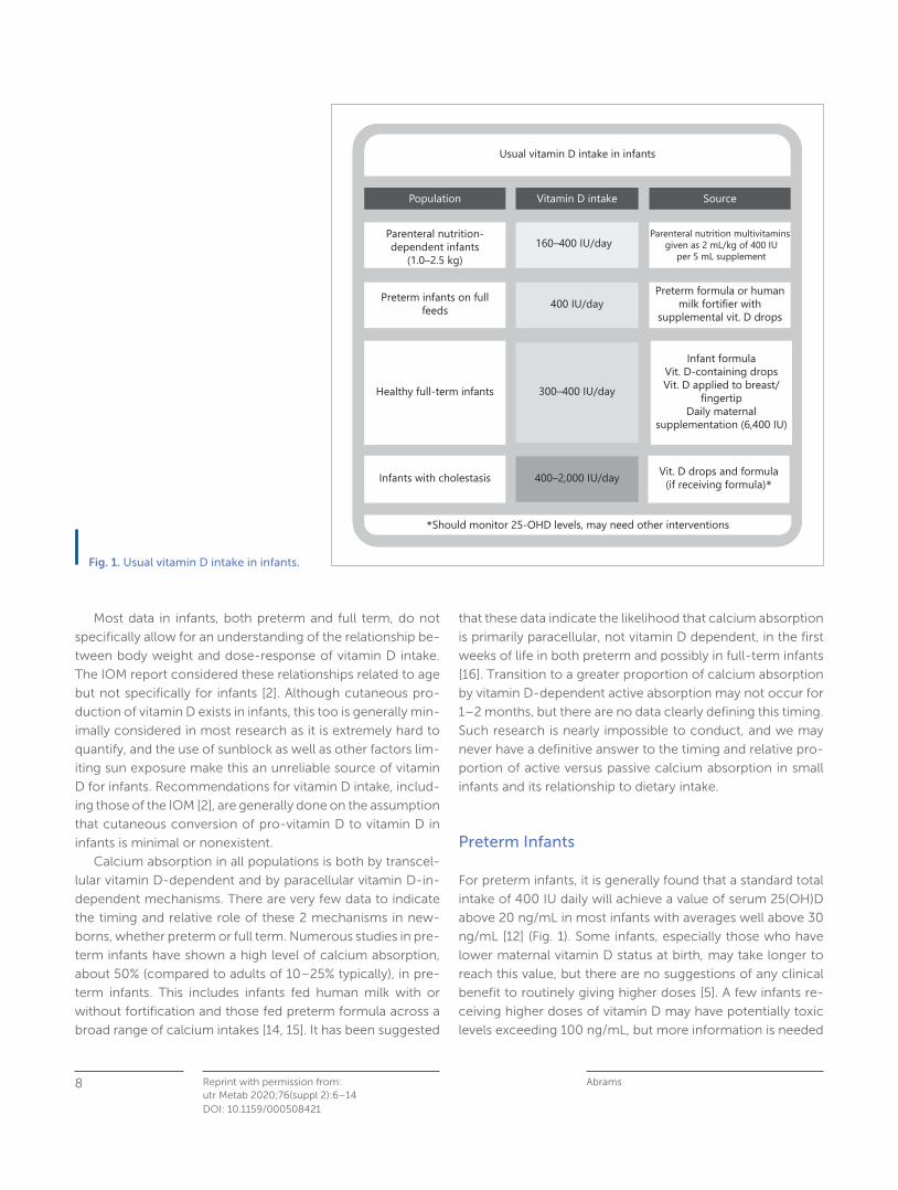
Usual vitamin D intake in infants
Parenteral nutrition-dependent infants
(1.0–2.5 kg)
Preterm infants on fullfeeds
Healthy full-term infants
Infants with cholestasis 400–2,000 IU/day Vit. D drops and formula(if receiving formula)*
*Should monitor 25-OHD levels, may need other interventions
160–400 IU/day
400 IU/day
300–400 IU/day
Parenteral nutrition multivitamins given as 2 mL/kg of 400 IU
per 5 mL supplement
Preterm formula or humanmilk fortifier with
supplemental vit. D drops
Infant formulaVit. D-containing dropsVit. D applied to breast/
fingertipDaily maternal
supplementation (6,400 IU)
Population Vitamin D intake Source
Fig. 1. Usual vitamin D intake in infants.
Vitamin D in Infants 9Reprint with permission from:Ann Nutr Metab 2020;76(suppl 2):6–14DOI: 10.1159/000508421
to evaluate this risk or any clinical correlates of relatively high
vitamin D status in preterm infants [12] .
However, in this regard, there are differences in recom-
mendations between those commonly given in the USA and
in Europe for vitamin D in preterm infants. European authori-
ties and authors have generally recommended a dose of
800–1,000 IU vitamin D daily, whereas in the USA, 400 IU
daily remains the standard recommendation [17] . This distinc-
tion is due to the perspective in European reviewers, based on
limited-balance studies, that a lower calcium intake can be
used with a higher vitamin D intake to increase total calcium
absorption to needed levels to support preterm infant bone
mineralization. In the USA, it has been preferred to maintain a
high calcium intake [4] , and there are no current reasons to
change recommendations or formulations of preterm infant
products in the USA as there is no evidence of any harmful
effects from calcium intake levels currently provided. None-
theless, those who supplement preterm infants to a total in-
take of 800–1,000 IU daily may likely do so without serious
concern for toxicity or need for close follow-up, given the
long history of use of higher doses up to 1,000 IU daily in many
countries in preterm infants.
Full-Term Infants
The requirements for vitamin D in full-term infants have been
extensively investigated. Research has shown that the dose
generally recommended for almost 100 years of 400 IU daily
meets the needs of nearly all full-term infants, and it remains
the recommendation for infants by the IOM and the AAP [2, 3]
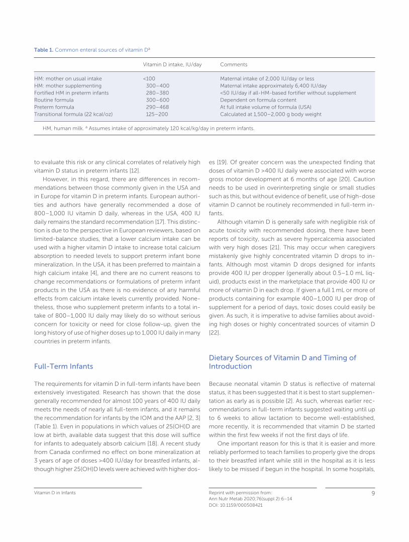
( Table 1 ). Even in populations in which values of 25(OH)D are
low at birth, available data suggest that this dose will suffice
for infants to adequately absorb calcium [18] . A recent study
from Canada confirmed no effect on bone mineralization at
3 years of age of doses > 400 IU/day for breastfed infants, al-
though higher 25(OH)D levels were achieved with higher dos-
es [19] . Of greater concern was the unexpected finding that
doses of vitamin D > 400 IU daily were associated with worse
gross motor development at 6 months of age [20] . Caution
needs to be used in overinterpreting single or small studies
such as this, but without evidence of benefit, use of high-dose
vitamin D cannot be routinely recommended in full-term in-
fants.
Although vitamin D is generally safe with negligible risk of
acute toxicity with recommended dosing, there have been
reports of toxicity, such as severe hypercalcemia associated
with very high doses [21] . This may occur when caregivers
mistakenly give highly concentrated vitamin D drops to in-
fants. Although most vitamin D drops designed for infants
provide 400 IU per dropper (generally about 0.5–1.0 mL liq-
uid), products exist in the marketplace that provide 400 IU or
more of vitamin D in each drop. If given a full 1 mL or more of
products containing for example 400–1,000 IU per drop of
supplement for a period of days, toxic doses could easily be
given. As such, it is imperative to advise families about avoid-
ing high doses or highly concentrated sources of vitamin D
[22] .
Dietary Sources of Vitamin D and Timing of Introduction
Because neonatal vitamin D status is reflective of maternal
status, it has been suggested that it is best to start supplemen-
tation as early as is possible [2] . As such, whereas earlier rec-
ommendations in full-term infants suggested waiting until up
to 6 weeks to allow lactation to become well-established,
more recently, it is recommended that vitamin D be started
within the first few weeks if not the first days of life.
One important reason for this is that it is easier and more
reliably performed to teach families to properly give the drops
to their breastfed infant while still in the hospital as it is less
likely to be missed if begun in the hospital. In some hospitals,
Table 1. Common enteral sources of vitamin Da
Vitamin D intake, IU/day Comments
HM: mother on usual intake <100 Maternal intake of 2,000 IU/day or lessHM: mother supplementing 300–400 Maternal intake approximately 6,400 IU/dayFortified HM in preterm infants 280–380 <50 IU/day if all-HM-based fortifier without supplementRoutine formula 300–600 Dependent on formula contentPreterm formula 290–468 At full intake volume of formula (USA)Transitional formula (22 kcal/oz) 125–200 Calculated at 1,500–2,000 g body weight
HM, human milk. a Assumes intake of approximately 120 kcal/kg/day in preterm infants.
Abrams Reprint with permission from:utr Metab 2020;76(suppl 2):6–14
10
DOI: 10.1159/000508421
the first bottle of the drops may be sent home with the fam-
ily. The opportunity to rapidly increase very low 25(OH)D lev-
els in infants born to mothers with very low levels is also a
reason to consider this. However, it should not be expected
that there will be specific clinical benefits to beginning vitamin
D in the first weeks of life, and if some families wish to delay
giving drops for 4–6 weeks until lactation is well-established
that should be considered as reasonable.
The situation for preterm infants is even less clear. Rickets
in preterm infants is primarily a disease of inadequate calcium
and phosphorus intake and absorption [5] . For those fed in-
travenously with parenteral nutrition, vitamin D is present in
the multivitamins given with the parenteral nutrition with the
standard intravenous multivitamin supplement containing
400 IU of vitamin D in 5 mL of the supplement. Typical dosing
of 2 mL/kg daily of the supplement in parenteral nutrition
would lead to doses from 160 to 400 IU daily for infants 1.0–
2.5 kg. It is important to provide vitamin D to infants who are
not taking enteral nutrition to prevent extremely low vitamin
D levels which can increase bone resorption and cause failure
to fully mineralize bone.
The timing of introduction of oral vitamin D in preterm in-
fants has not been studied in terms of relative risks and ben-
efits to any particular time point. The AAP recommended be-
ginning after full feeds are achieved at about 1,500 g, but it
was recognized that this specific time point is arbitrary and
chosen primarily to ensure the tolerance of the drops [5] . Oth-
ers might choose to begin supplementation somewhat ear-
lier in very-low-birth-weight infants, but it is common to en-
sure a nontrophic volume of feeds are being well tolerated
before doing so and waiting until after parenteral nutrition has
been discontinued.
Other Issues with Vitamin D Dosing in Infants
Some families are resistant to providing drops of vitamin D to
their breastfed infants or perceive them to be poorly tolerated,
especially when given with iron-containing multivitamins. In
these cases, there are several alternatives that may be consid-
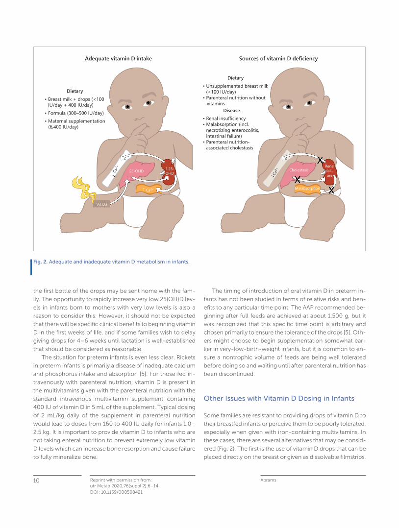
ered ( Fig. 2 ). The first is the use of vitamin D drops that can be
placed directly on the breast or given as dissolvable filmstrips.
Adequate vitamin D intake
Dietary• Breast milk + drops (<100 IU/day + 400 IU/day)• Formula (300–500 IU/day)• Maternal supplementation (6,400 IU/day)
25-OHD
MalabsorptionCa2+
Vit D3
1,25-OHDCa
2+
Dietary• Unsupplemented breast milk (<100 IU/day)• Parenteral nutrition without vitamins
Disease• Renal insufficiency• Malabsorption (incl. necrotizing enterocolitis, intestinal failure)• Parenteral nutrition- associated cholestasis
Renalfail-ure
Sources of vitamin D deficiency
Ca2+ Cholestasis
Fig. 2. Adequate and inadequate vitamin D metabolism in infants.
Vitamin D in Infants 11Reprint with permission from:Ann Nutr Metab 2020;76(suppl 2):6–14DOI: 10.1159/000508421
For some mothers, this is easier and more acceptable than
giving a dropper of vitamins directly to the infant or mixed in
their milk [23] .
Another approach is to have the lactating mother take a
relatively high dose of vitamin D. Studies have shown that a
maternal dose of 6,400 IU daily will provide an infant with
adequate vitamin D intake (usually about 300–400 IU daily)
from the mother’s milk if fully breastfed and if the mother
takes the dose every day. Of note is that lower maternal dos-
es, especially those of 400–2,000 IU daily, do not provide
adequate vitamin D in breast milk. The dose of 6,400 IU dai-
ly is slightly above the IOM upper limit of 5,000 IU/day but is
highly likely to be safe, and this should not be a concern in
recommending this approach if desired by breastfeeding
women [2, 24] .
It is frequently asked whether vitamin D should be given to
infants who are both breast and formula fed, and the general
answer is “yes.” An intake of 400 IU daily requires a full volume
of formula intake, and whereas going slightly below the 400
IU/day level of intake is not problematic, the mixed-fed baby
is best served by providing additional vitamin D as would be
done for fully breastfed infants. There is no risk of toxicity with
this approach, even if the infant switches entirely to infant for-
mula prior to stopping the vitamin D supplementation.
Another common clinical question is whether vitamin D
supplementation via drops is necessary for exclusively formu-
la-fed infants. Some have indicated that vitamin D should be
given until a volume of formula intake of 1,000 mL/day is
reached [7] . This is because, based on the formula label and
usual dilution of powdered infant formula, vitamin D was usu-
ally provided in infant formulas at 400 IU/L. Although there is
no harm in this practice, it is questionable if needed and if it is
best use of family and societal resources. The vitamin D re-
quirement of 400 IU daily from the IOM is an average require-
ment in the first 6 months of life and as noted, there is little
suggestion of a clinical concern with slightly lower doses un-
til full feeding volume is achieved [2] .
Also problematic with this recommendation is the per-
ception that 1,000 mL daily is the minimum volume of infant
formula an infant should receive and infants taking below that
need any supplements. The usual volume of breast milk in-
take is approximately 800 mL daily, and although formula
intakes are somewhat variable, an intake of 1,000 mL of for-
mula is higher than required for growth and development,
and not all infants will ever take this volume nor should they
be pushed to this volume [2] . Furthermore, although the label
claim for vitamin D content was commonly 400 IU/L, when
analyzed, many infant formula batches will have 10–20%
over this amount so as to meet the label claim at the end of
shelf life [25] . Overall, the IOM recommendation of 400 IU
daily for infants should be understood as an average intake,
not one needing to be met from label claim every day from
birth [2] .
Recently, many formulas have been marketed with vitamin
D intakes over 400 IU/L as this is permitted by the Food and
Drug Administration (FDA) and the European Food Safety Au-
thority (EFSA) [26] . Numerous routine cow milk-based and
other formulas marketed currently contain approximately
400 IU in 800 mL as prepared, a daily intake volume similar to
that ingested daily by many infants after the first 6–8 weeks
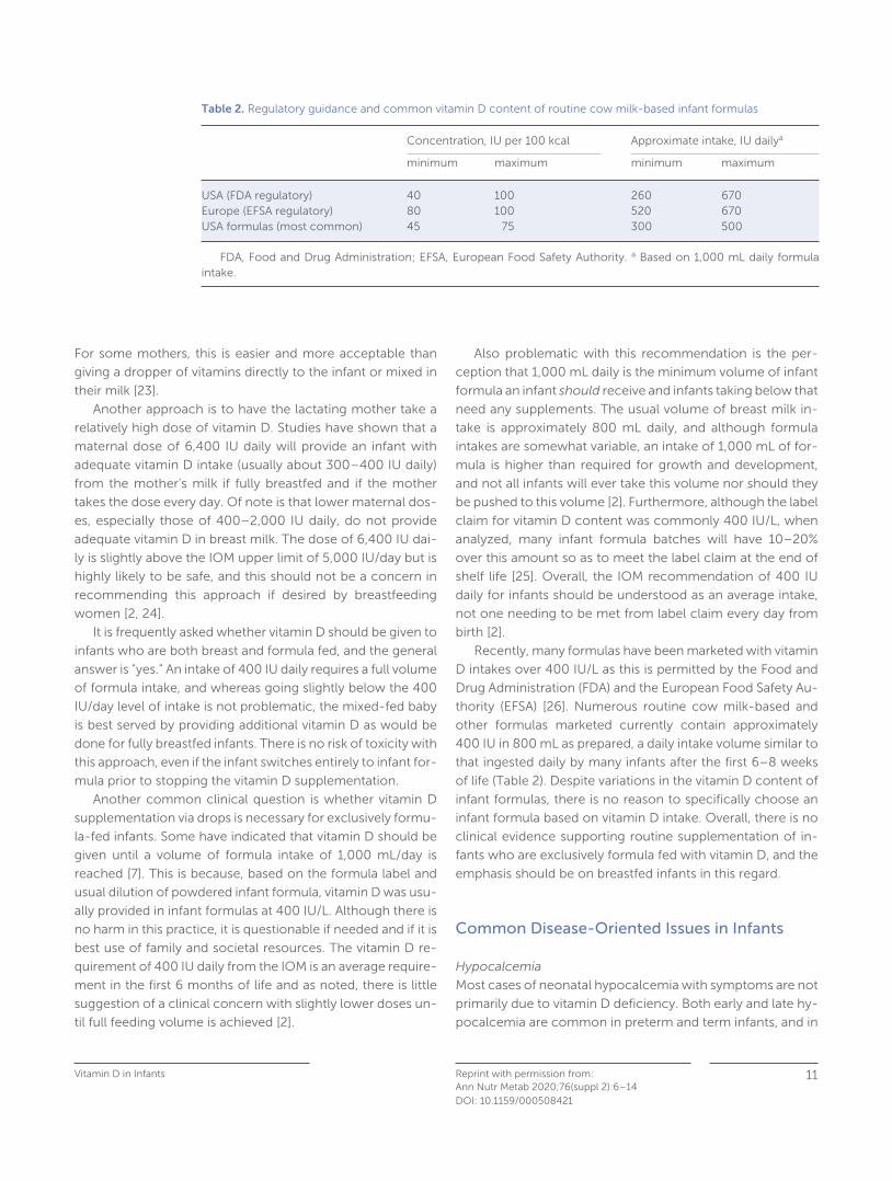
of life ( Table 2 ). Despite variations in the vitamin D content of
infant formulas, there is no reason to specifically choose an
infant formula based on vitamin D intake. Overall, there is no
clinical evidence supporting routine supplementation of in-
fants who are exclusively formula fed with vitamin D, and the
emphasis should be on breastfed infants in this regard.
Common Disease-Oriented Issues in Infants
Hypocalcemia
Most cases of neonatal hypocalcemia with symptoms are not
primarily due to vitamin D deficiency. Both early and late hy-
pocalcemia are common in preterm and term infants, and in
Table 2. Regulatory guidance and common vitamin D content of routine cow milk-based infant formulas
Concentration, IU per 100 kcal Approximate intake, IU dailya
minimum maximum mi nimum maximum
USA (FDA regulatory) 40 100 260 670Europe (EFSA regulatory) 80 100 520 670USA formulas (most common) 45 75 300 500
FDA, Food and Drug Administration; EFSA, European Food Safety Authority. a Based on 1,000 mL daily formula intake.
Abrams Reprint with permission from:utr Metab 2020;76(suppl 2):6–14
12
DOI: 10.1159/000508421
the USA, early hypocalcemia in preterm infants (first 2–3 days
of life) is primarily related to hormonal factors [27, 28] . In full-
term infants, hypocalcemia is commonly seen in infants of
diabetic mothers or associated with severe birth depression
and neonatal asphyxial disorders among other causes. In late
hypocalcemic tetany (usually 4–7 days of age), vitamin D lev-
els may be low, but the primary cause of hypocalcemia is the
use of high phosphorus intakes associated with whole cow
milk or use of infant formula [29, 30] . In late neonatal hypo-
calcemia, treatment with 1,25-dihydroxyvitamin D may
shorten the time to resolution likely due to direct effects of
vitamin D on the bone rather than a calcium absorptive effect
[31] .
An important situation in which vitamin D deficiency is
more central to the etiology of hypocalcemia are cases com-
monly reported in full-term infants during the second week
of life. Reports of this problem have primarily come from Mid-
dle Eastern countries and are associated with extremely low
maternal and, thus, infant vitamin D levels [32, 33] . Although
the etiology of the hypocalcemia is not clearly defined, likely
it is due both to lack of effects of vitamin D at the bone and in
the intestine. This highlights the importance of identifying at-
risk maternal populations and providing them with adequate
vitamin D intake during pregnancy.
Cholestasis
Common conditions related to the vitamin D requirement in
preterm infants are those that affect either the enteral absorp-
tion of nutrients or those which affect the formation of 25(OH)
D in the liver or 1,25-dihydroxyvitamin D in the kidneys. Ab-
sorption of fat-soluble vitamins, such as vitamin D, may be
affected by a variety of disease states in preterm infants, in-
cluding those with the loss of the terminal ileum surgically and
malabsorptive diseases, such as cystic fibrosis. Management
of these conditions is beyond the scope of this review, but
these would be an indication for closely monitoring the serum
25(OH)D concentration and potentially providing higher dos-
es of vitamin D or vitamin D metabolites as described below
[34] .
A second relatively common problem in high-risk neo-
nates is cholestasis, especially secondary to long-term paren-
teral nutrition use. Although relatively little is known specifi-
cally about relating the level of conjugated bilirubin to 25(OH)
D values or calcium absorption in infants, this may become a
clinical problem in which it is difficult to maintain adequate
vitamin D status using usual dietary approaches. In this case,
if careful monitoring and higher vitamin D (e.g., 1,000–2,000
IU daily) intakes show a persistent level of serum 25(OH)D
< 20 ng/mL, then supplementation with very-high-dose vita-
min D or use of vitamin D analogues, such as calcitriol, while
continuing vitamin D may be considered [35] . This should
generally be done in the context of consultation or manage-
ment by a pediatric endocrinologist, nephrologist, or other
expert in the use of vitamin D metabolites. Of note is that the
enteral medication 25(OH)D, called calcidiol (also referred to
as calcifediol), has recently become available in the USA but
does not have a FDA-approved indication for use in infants
and children.
Compliance with Vitamin D Intake Recommendations
As noted, many families of breastfed infants do not provide
vitamin D supplements per recommendations. Recent data
suggest that only about 20% of US infants who are breastfed
are receiving vitamin D supplements to meet the recommen-
dations [36] . Of note is that this is much lower than the rate in
a study in Canada which found over 70% adherence, perhaps
due to greater awareness of this issue in Canada among pe-
diatricians and families [37] . Discharge from the hospital with
vitamin D can markedly increase this rate as suggested in
these preliminary results [38] . Education is needed for both
providers and families related to the risks of rickets and the
importance of providing vitamin D for infants. Providers
should be prepared to answer concerns related to the use of
drops in breastfed infants and provide alternatives as de-
scribed above for those families unwilling to use drops. The
option of delaying the drops for 6–8 weeks after birth can also
be given, especially for families intending to offer a bottle of
mother’s milk at that time into which the drops could be add-
ed.
Future Research
Further studies are needed focusing on the risks associated
with very low vitamin D status in infants, in particular, identify-
ing the risks and best management approaches for infants
who are at risk of hypocalcemia from extremely low maternal
vitamin D status. Although this problem has not been identi-
fied commonly in the USA, it may not be identified when it
occurs, and population studies of high-risk maternal infant
pairs are needed.
Although vitamin D is largely safe, the increasing use of
high-dose supplements in infants should be evaluated and
practitioners encouraged to report cases to understand this
problem and the clinical consequence of high-dose inges-
tion, whether intentional or accidental.
Vitamin D in Infants 13Reprint with permission from:Ann Nutr Metab 2020;76(suppl 2):6–14DOI: 10.1159/000508421
Summary of Recommendations
Vitamin D is a critical nutrient for bone health and needs to be
provided to all infants whether via infant formula or as a sup-
plement to breastfed infants or high-dose supplement to their
mothers. Solar conversion and cutaneous formation of vita-
min D cannot be ensured in any population. In most healthy
infants, preterm as well as full term, who are on full enteral
nutrition and have a normal intestine and normal liver and re-
nal function, provision of approximately 400 IU daily is neces-
sary and sufficient for bone health, and routine monitoring of
serum 25(OH)D levels is not needed. Caution should be used
to ensure that the appropriate dose is provided and that ac-
cidental ingestion of high doses of vitamin D does not occur.
Acknowledgment
The author would like to acknowledge Hannah R. Abrams for assis-tance with the Figures used in the manuscript and Keli M. Hawthorne for editorial review.
Disclosure Statement
Dr. Steven A. Abrams is a member of the scientific advisory board of MilkPep, the education program of the Milk Processor Education Pro-gram. The writing of this article was supported by Nestlé Nutrition Institute.
References
1 Carpenter TO, Shaw NJ, Portale AA, Ward LM, Abrams SA, Pettifor JM. Rickets. Nat Rev Dis Primers . 2017 Dec; 3(1): 17101.
2 Institute of Medicine Committee to Review Dietary Reference In-takes for Vitamin D. Calcium. The National Academies collection: reports funded by National Institutes of Health. In: Ross AC, Taylor CL, Yaktine AL, Del Valle HB, editors. Dietary Reference Intakes for Calcium and Vitamin D. Washington (DC): National Academies Press; 2011.
3 Abrams SA. Dietary guidelines for calcium and vitamin D: a new era. Pediatrics . 2011 Mar; 127(3): 566–8.
4 Golden NH, Abrams SA; Committee on Nutrition. Optimizing bone health in children and adolescents. Pediatrics . 2014 Oct; 134(4):e1229–43.
5 Abrams SA; Committee on Nutrition. Calcium and vitamin D re-quirements of enterally fed preterm infants. Pediatrics . 2013 May; 131(5):e1676–83.
6 Roth DE, Abrams SA, Aloia J, Bergeron G, Bourassa MW, Brown KH, et al. Global prevalence and disease burden of vitamin D de-ficiency: A roadmap for action in low- and middle-income coun-tries. Ann N Y Acad Sci . 2018 Oct; 1430(1): 44–79.
7 Misra M, Pacaud D, Petryk A, Collett-Solberg PF, Kappy M; Drug and Therapeutics Committee of the Lawson Wilkins Pediatric En-docrine Society. Vitamin D deficiency in children and its manage-ment: review of current knowledge and recommendations. Pedi-atrics . 2008 Aug; 122(2): 398–417.
8 Aloia JF, Dhaliwal R, Shieh A, Mikhail M, Fazzari M, Ragolia L, et al. Vitamin D supplementation increases calcium absorption without a threshold effect. Am J Clin Nutr . 2014 Mar; 99(3): 624–31.
9 Abrams SA, Hicks PD, Hawthorne KM. Higher serum 25-hydroxyvi-tamin D levels in school-age children are inconsistently associ-ated with increased calcium absorption. J Clin Endocrinol Metab . 2009 Jul; 94(7): 2421–7.
10 Tiosano D, Hadad S, Chen Z, Nimerovsky A, Gepstein V, Militiano D, et al. Calcium absorption, kinetics, bone density, and bone structure in patients with hereditary vitamin D-resistant rickets. J Clin Endocrinol Metab . 2011; 96: 3701–9.
11 Anderson-Berry A, Thoene M, Wagner J, Lyden E, Jones G, Kaufmann M, et al. Randomized trial of two doses of vitamin D3 in preterm infants < 32 weeks: dose impact on achieving desired serum 25(OH)D3 in a NICU population. PLoS One . 2017 Oct; 12(10):e0185950.
12 Fort P, Salas AA, Nicola T, Craig CM, Carlo WA, Ambalavanan N. A Comparison of 3 Vitamin D Dosing Regimens in Extremely Pre-term Infants: A Randomized Controlled Trial. J Pediatr . 2016 Jul; 174: 132–138.e1.
13 Backström MC, Mäki R, Kuusela AL, Sievänen H, Koivisto AM, Ikonen RS, et al. Randomised controlled trial of vitamin D supple-mentation on bone density and biochemical indices in preterm infants. Arch Dis Child Fetal Neonatal Ed . 1999 May; 80(3):F161–6.
14 Abrams SA, Esteban NV, Vieira NE, Yergey AL. Dual tracer stable isotopic assessment of calcium absorption and endogenous fecal excretion in low birth weight infants. Pediatr Res . 1991 Jun; 29(6): 615–8.
15 Schanler RJ, Abrams SA, Garza C. Mineral balance studies in very low birth weight infants fed human milk. J Pediatr . 1988 Jul; 113(1 Pt 2): 230–8.
16 Bronner F, Salle BL, Putet G, Rigo J, Senterre J. Net calcium ab-sorption in premature infants: results of 103 metabolic balance studies. Am J Clin Nutr . 1992 Dec; 56(6): 1037–44.
17 Agostoni C, Buonocore G, Carnielli VP, De Curtis M, Darmaun D, Decsi T, et al.; ESPGHAN Committee on Nutrition. Enteral nutrient supply for preterm infants: commentary from the European Soci-ety of Paediatric Gastroenterology, Hepatology and Nutrition Committee on Nutrition. J Pediatr Gastroenterol Nutr . 2010 Jan; 50(1): 85–91.
18 Abrams SA, Hawthorne KM, Rogers SP, Hicks PD, Carpenter TO. Effects of ethnicity and vitamin D supplementation on vitamin D status and changes in bone mineral content in infants. BMC Pedi-atr . 2012 Jan; 12(1): 6.
19 Gallo S, Hazell T, Vanstone CA, Agellon S, Jones G, L’Abbé M, et al. Vitamin D supplementation in breastfed infants from Montréal, Can-ada: 25-hydroxyvitamin D and bone health effects from a follow-up study at 3 years of age. Osteoporos Int . 2016 Aug; 27(8): 2459–66.
Abrams Reprint with permission from:utr Metab 2020;76(suppl 2):6–14
14
DOI: 10.1159/000508421
20 Wicklow B, Gallo S, Majnemer A, Vanstone C, Comeau K, Jones G, et al. Impact of Vitamin D Supplementation on Gross Motor De-velopment of Healthy Term Infants: A Randomized Dose-Re-sponse Trial. Phys Occup Ther Pediatr . 2016 Aug; 36(3): 330–42.
21 Vanstone MB, Oberfield SE, Shader L, Ardeshirpour L, Carpenter TO. Hypercalcemia in children receiving pharmacologic doses of vitamin D. Pediatrics . 2012; 129(4):e1060–3.
22 https://www.fda.gov/consumers/consumer-updates/infant-overdose-risk-liquid-vitamin-d.
23 Rodd C, Jean-Philippe S, Vanstone C, Weiler H. Comparison of 2 vitamin D supplementation modalities in newborns: adherence and preference. Appl Physiol Nutr Metab . 2011 Jun; 36(3): 414–8.
24 Hollis BW, Wagner CL, Howard CR, Ebeling M, Shary JR, Smith PG, et al. Maternal Versus Infant Vitamin D Supplementation During Lactation: A Randomized Controlled Trial. Pediatrics . 2015 Oct; 136(4): 625–34.
25 Verkaik-Kloosterman J, Seves SM, Ocké MC. Vitamin D concen-trations in fortified foods and dietary supplements intended for infants: implications for vitamin D intake. Food Chem . 2017 Apr; 221: 629–35.
26 European Food Safety Authority. Outcome of a public consulta-tion on the Draft Scientific Opinion of the EFSA Panel on Dietetic Products, Nutrition and Allergies (NDA) on the update of the toler-able upper intake level for vitamin D for infants. EFSA J . 2018; 16: 5365.
27 Thomas TC, Smith JM, White PC, Adhikari S. Transient neonatal hypocalcemia: presentation and outcomes. Pediatrics . 2012 Jun; 129(6):e1461–7.
28 Venkataraman PS, Tsang RC, Steichen JJ, Grey I, Neylan M, Fleischman AR. Early neonatal hypocalcemia in extremely preterm infants. High incidence, early onset, and refractoriness to supra-physiologic doses of calcitriol. Am J Dis Child . 1986 Oct; 140(10): 1004–8.
29 Yılmaz B, Aygün C, Çetinoğlu E. Vitamin D levels in newborns and association with neonatal hypocalcemia. J Matern Fetal Neonatal Med . 2018 Jul; 31(14): 1889–93.
30 Venkataraman PS, Tsang RC, Greer FR, Noguchi A, Laskarzewski P, Steichen JJ. Late infantile tetany and secondary hyperparathy-roidism in infants fed humanized cow milk formula. Longitudinal follow-up. Am J Dis Child . 1985 Jul; 139(7): 664–8.
31 Amaral JM, Abrams S, Karaviti L, McKay SV. Effects of 1,25-dihy-droxycholecalciferol on recovery and resolution of late transient neonatal hypocalcemia. Int J Pediatr Endocrinol . 2010; 2010(1): 409670.
32 Teaema FH, Al Ansari K. Nineteen cases of symptomatic neonatal hypocalcemia secondary to vitamin D deficiency: a 2-year study. J Trop Pediatr . 2010 Apr; 56(2): 108–10.
33 Balasubramanian S, Shivbalan S, Kumar PS. Hypocalcemia due to vitamin D deficiency in exclusively breastfed infants. Indian Pedi-atr . 2006 Mar; 43(3): 247–51.
34 Mutanen A, Mäkitie O, Pakarinen MP. Risk of metabolic bone dis-ease is increased both during and after weaning off parenteral nutrition in pediatric intestinal failure. Horm Res Paediatr . 2013; 79(4): 227–35.
35 Jensen M, Abu-El-Haija M, Bishop W, Rahhal RM. Difficulty achiev-ing Vitamin D sufficiency with high-dose oral repletion therapy in infants with cholestasis. J Pediatr Gastroenterol Nutr . 2015 Aug; 61(2): 187–9.
36 Ahrens KA, Rossen LM, Simon AE. Adherence to Vitamin D Recom-mendations Among US Infants Aged 0 to 11 Months, NHANES, 2009 to 2012. Clin Pediatr (Phila) . 2016 Jun; 55(6): 555–6.
37 Gallo S, Jean-Philippe S, Rodd C, Weiler HA. Vitamin D supple-mentation of Canadian infants: practices of Montreal mothers. Appl Physiol Nutr Metab . 2010 Jun; 35(3): 303–9.
38 Drake A, Lustik M, Sampert C. Improving vitamin D supplementa-tion rates in the neonate. Pediatrics . 2018; 142: 603.
Focus
Reprinted with permission from: Ann Nutr Metab 2020;76(suppl 2):16–28
Early- Life Effects of Vitamin D: A Focus on Pregnancy and LactationCarol L. Wagner and Bruce W. Hollis
© 2020 Nestlé Nutrition Institute, Switzerland/S. Karger AG, Basel
Key Insight
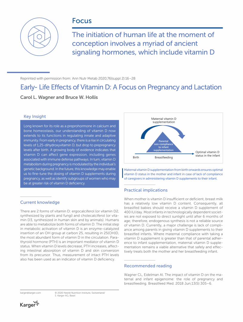
Long known for its role as a preprohormone in calcium and bone homeostasis, our understanding of vitamin D now extends to its functions in regulating innate and adaptive immunity. From early in pregnancy, there is a rise in circulating levels of 1,25-dihydroxyvitamin D, but drop to prepregnancy levels after birth. A growing body of evidence indicates that vitamin D can affect gene expression, including genes associated with immune defense pathways. In turn, vitamin D metabolism during pregnancy is modulated by the individual’s genetic background. In the future, this knowledge may enable us to fine-tune the dosing of vitamin D supplements during pregnancy, as well as identify subgroups of women who may be at greater risk of vitamin D deficiency.
Current knowledge
There are 2 forms of vitamin D: ergocalciferol (or vitamin D2, synthesized by plants and fungi) and cholecalciferol (or vita-min D3, synthesized in human skin and by animals). Humans are able to metabolize both forms of vitamin D. The initial step in metabolic activation of vitamin D is an enzyme-catalyzed insertion of an OH group at carbon 25, resulting in 25(OH)D, the most abundant form of vitamin D in the circulation. Para-thyroid hormone (PTH) is an important mediator of vitamin D status. When vitamin D levels decrease, PTH increases, affect-ing intestinal absorption of vitamin D and skin conversion from its precursor. Thus, measurement of intact PTH levels also has been used as an indicator of vitamin D deficiency.
Practical implications
When mother is vitamin D insufficient or deficient, breast milk has a relatively low vitamin D content. Consequently, all breastfed babies should receive a vitamin D supplement of 400 IU/day. Most infants in technologically dependent societ-ies are not exposed to direct sunlight until after 6 months of age; therefore, endogenous synthesis is not a reliable source of vitamin D. Currently, a major challenge is lack of compli-ance among parents in giving vitamin D supplements to their breastfed infants. Where maternal compliance with taking a vitamin D supplement is greater than that of parental adher-ence to infant supplementation, maternal vitamin D supple-mentation remains a viable alternative that safely and effec-tively treats both the mother and her breastfeeding infant.
Recommended reading
Wagner CL, Eidelman AI. The impact of vitamin D on the ma-ternal and infant epigenome: the role of pregnancy and breastfeeding. Breastfeed Med. 2018 Jun;13(5):305–6.
The initiation of human life at the moment of conception involves a myriad of ancient signaling hormones, which include vitamin D
Optimal vitamin D status in the infant
Maternal vitamin Dsupplementation
Parentalnon-compliance
to infantsupplementation
Birth Breastfeeding
Maternal vitamin D supplementation from birth onwards ensures optimal vitamin D status in the mother and infant in case of lack of compliance of caregivers in administering vitamin D supplements to their infant.
Vitamin D
Reprinted with permission from:Ann Nutr Metab 2020;76(suppl 2):16–28
Early-Life Effects of Vitamin D: A Focus on Pregnancy and Lactation
Carol L. Wagner Bruce W. Hollis
Division of Neonatology, Department of Pediatrics, Medical University of South Carolina, Charleston , SC , USA
Carol L. Wagner Medical University of South Carolina 10 McClennan Banks Drive, MSC 915 Charleston, SC 29425 (USA) wagnercl @ musc.edu
© 2020 Nestlé Nutrition Institute, Switzerland/S. Karger AG, Basel
Key Messages
• The active form of vitamin D – 1,25-dihydroxyvitamin D (1,25[OH] 2 D) – increases during pregnancy, remains elevated throughout, and, unlike at other times during the lifecycle, is directly affected by circulating 25-hydroxyvitamin D (25[OH]D) concentration with the optimal point of conversion of 25(OH)D to 1,25(OH) 2 D at 100 nmol/L (40 ng/mL).
• Lactation has increased demands on the mother regarding nutrient intake delivered through her breast milk to her recipient infant: when a mother is vitamin D deficient, her milk is deficient, which can be remedied by direct infant supplementation; however, this treats only the infant.
• A safe alternative during lactation to infant supplementation is direct maternal vitamin D supplementation at higher doses than usual (6,400 IU/day), improving the vitamin D status of the mother, the content of the milk, and, consequently, the vitamin D status of the infant, effectively treating both the mother and the infant.
DOI: 10.1159/000508422
Keywords 1,25-dihydroxyvitamin D · 25-hydroxyvitamin D ·
Cholecalciferol · Calcidiol · Clinical nutrition · Human
nutrition · Infancy and childhood · Lactation · Pregnancy
Abstract Vitamin D is an endocrine regulator of calcium and bone me-
tabolism. Yet, its effects include other systems, such as innate
and adaptive immunity. Unique to pregnancy, circulating
1,25-dihydroxyvitamin D (1,25[OH] 2 D) increases early on to
concentrations that are 2–3 times prepregnant values. At no
other time during the lifecycle is the conversion of 25-hy-
droxyvitamin D (25[OH]D) to 1,25(OH) 2 D directly related and
optimized at ≥ 100 nmol/L. Vitamin D deficiency appears to
affect pregnancy outcomes, yet randomized controlled trials
of vitamin D supplementation achieve mixed results depend-
ing on when supplementation is initiated during pregnancy,
the dose and dosing interval, and the degree of deficiency at
the onset of pregnancy. Analysis of trials on an intention-to-
treat basis as opposed to the use of 25(OH)D as the interme-
diary biomarker of vitamin D metabolism yields differing re-
sults, with treatment effects often noted only in the most
deficient women. Immediately after delivery, maternal circu-
lating 1,25(OH) 2 D concentrations return to prepregnancy
baseline, at a time when a breastfeeding woman has in-
creased demands of calcium, beyond what was needed dur-
ing the last trimester of pregnancy, making one question why
1,25(OH) 2 D increases so significantly during pregnancy. Is it
to serve as an immune modulator? The vitamin D content of
mother’s milk is directly related to maternal vitamin D status,
and if a woman was deficient during pregnancy, her milk will
be deficient unless she is taking higher doses of vitamin D.
Because of this relative “deficiency,” there is a recommenda-
tion that all breastfed infants receive 400 IU vitamin D 3 /day
starting a few days after birth. The alternative – maternal sup-
plementation with 6,400 IU vitamin D 3 /day, effective in safe-
ly raising maternal circulating vitamin D, that of her breast
milk, and effective in achieving sufficiency in her recipient
Vitamin D during Pregnancy and Lactation 17Reprinted with permission from:Ann Nutr Metab 2020;76(suppl 2):16–28DOI: 10.1159/000508422
breastfeeding infant – remains a viable option. Additional re-
search is needed to understand vitamin D’s influence on
pregnancy health and the effect of maternal supplementa-
tion on breast milk’s immune signaling.
© 2020 Nestlé Nutrition Institute, Switzerland/
S. Karger AG, Basel
Conception Onward
From the moment of conception, there are tremendous
changes that must occur for growth and shaping of a single-
cell organism to billions of cells as the construct of diverse
systems, which function in concert to form a living human
being. It is in the context of this timing, this concert of matter
and energy transfer across cells, that we can appreciate what
is happening surrounding conception. Conception does not
occur in a hostile or nonnurturing environment, yet the very
invasion of extravillous cytotrophoblasts into the uterine wall
is an invasive and inflammatory process [1–3] . Pregnancy is a
state of change and flux that must balance between negen-
tropy—organization of tissue—and cellular death and apop-
tosis – necessary for refinement of tissue and organ structure.
The very event of conception is dependent upon a functional
neuroendocrine system in both the mother and father, a
functioning uterus with a rich lining to allow for invasion of
the extravillous cytotrophoblasts into the uterine wall, and a
dynamic synchrony of cell division and cell death. The initia-
tion of human life at the moment of conception involves a
myriad of ancient signaling hormones, which include vitamin
D [4, 5] .
Vitamin D as Preprohormone
Long known as an endocrine facilitator in its role as a prepro-
hormone affecting calcium and bone metabolism and ho-
meostasis, vitamin D is something more as well. Our under-
standing of vitamin D has expanded in the decades since its
discovery in the early 20th century. There are provocative ex-
perimental models in animals that extend to observational
and some clinical trials in humans, which suggest that vitamin
D plays a role in both innate and adaptive immunity, affecting
our ability to survive infectious insults as well as long-latency
diseases, such as autoimmune diseases and cancers, all of
which depend on a balanced and functional immune system
[6] .
There are 2 forms of vitamin D: ergocalciferol (or vitamin
D 2 , which is synthesized by plants and fungi) and cholecalcif-
erol (or vitamin D 3 , which is synthesized in the skin of humans
and animals). Humans can metabolize both forms of vitamin
D. Pre-vitamin D 3 is synthesized in the epidermal layer of the
skin by keratinocytes mainly in the stratum basale and stra-
tum spinosum when 7-dehyrocholesterol is exposed to ul-
traviolet B light in the wavelength of 290–320 nm [7] . Through
this photolytic energy transfer, pre-vitamin D is formed, and
with further thermally induced isomerization in the skin, the
parent compound vitamin D 3 is produced. Vitamin D 3 is car-
ried into the bloodstream bound to vitamin D-binding pro-
tein (VDBP) or, less frequently, to albumin. Once vitamin D
(either form D 2 or D 3 ) enters the circulation, either through
epidermal transfer or intestinal absorption, it associates with
VDBP, a 58-kD globular protein that binds vitamin D and its
metabolites with various affinities based on the number and
position of polar functional groups and/or methyl groups [8] .
The initial step in the metabolic activation of vitamin D is the
enzyme-catalyzed insertion of an OH group at carbon 25;
this oxidation process is primarily a hepatic microsomal func-
tion mostly by CYP2R1, a 25-hydroxylase [9] , producing
25-hydroxyvitamin D (25[OH]D), the most abundant circulat-
ing form of vitamin D [10] .
As shown in Figure 1 (from Hollis and Wagner [11] , with
permission), following formation in the liver, 25(OH)D appears
in the circulation – bound primarily to VDBP. The half-life of
the parent compound is 12–24 h, while that of its first me-
tabolite 25(OH)D is 2–3 weeks. The conversion of 25(OH)D to
the active hormone 1,25-dihydroxyvitamin D (1,25[OH] 2 D)
through the CYP27B1 enzyme mainly occurs in the proximal
tubules of the kidney, and then it is carried throughout the
body also bound to VDBP.
Unlike 25(OH)D, 1,25(OH) 2 D has a much shorter half-life
of 4–8 h. VDBP preferentially binds 25(OH)D with higher af-
finity than 1,25(OH) 2 D or the parent compound [12] . The high
affinity of VDBP for the vitamin D and its metabolites, coupled
with the excessive binding capacity, keeps “free” or unbound
concentrations of vitamin D and its metabolites at quite low
relative concentrations [13, 14] . This is important because only
the “free” concentrations of the vitamin and its metabolites
have transmembrane diffusion capabilities, thus exerting their
Vitamin D plays a role in both
innate and adaptive immunity,
affecting our ability to survive
infectious insults as well as
long-latency diseases
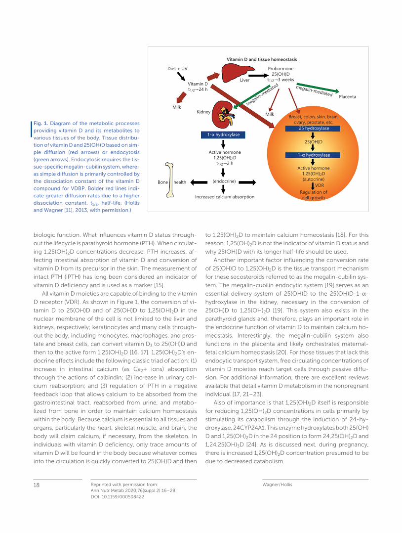
Wagner/Hollis Reprinted with permission from:Ann Nutr Metab 2020;76(suppl 2):16–28
18
DOI: 10.1159/000508422
biologic function. What influences vitamin D status through-
out the lifecycle is parathyroid hormone (PTH). When circulat-
ing 1,25(OH) 2 D concentrations decrease, PTH increases, af-
fecting intestinal absorption of vitamin D and conversion of
vitamin D from its precursor in the skin. The measurement of
intact PTH (iPTH) has long been considered an indicator of
vitamin D deficiency and is used as a marker [15] .
All vitamin D moieties are capable of binding to the vitamin
D receptor (VDR). As shown in Figure 1 , the conversion of vi-
tamin D to 25(OH)D and of 25(OH)D to 1,25(OH) 2 D in the
nuclear membrane of the cell is not limited to the liver and
kidneys, respectively; keratinocytes and many cells through-
out the body, including monocytes, macrophages, and pros-
tate and breast cells, can convert vitamin D 3 to 25(OH)D and
then to the active form 1,25(OH) 2 D [16, 17] . 1,25(OH) 2 D’s en-
docrine effects include the following classic triad of action: (1)
increase in intestinal calcium (as Ca 2 + ions) absorption
through the actions of calbindin; (2) increase in urinary cal-
cium reabsorption; and (3) regulation of PTH in a negative
feedback loop that allows calcium to be absorbed from the
gastrointestinal tract, reabsorbed from urine, and metabo-
lized from bone in order to maintain calcium homeostasis
within the body. Because calcium is essential to all tissues and
organs, particularly the heart, skeletal muscle, and brain, the
body will claim calcium, if necessary, from the skeleton. In
individuals with vitamin D deficiency, only trace amounts of
vitamin D will be found in the body because whatever comes
into the circulation is quickly converted to 25(OH)D and then
to 1,25(OH) 2 D to maintain calcium homeostasis [18] . For this
reason, 1,25(OH) 2 D is not the indicator of vitamin D status and
why 25(OH)D with its longer half-life should be used.
Another important factor influencing the conversion rate
of 25(OH)D to 1,25(OH) 2 D is the tissue transport mechanism
for these secosteroids referred to as the megalin-cubilin sys-
tem. The megalin-cubilin endocytic system [19] serves as an
essential delivery system of 25(OH)D to the 25(OH)D-1-α-
hydroxylase in the kidney, necessary in the conversion of
25(OH)D to 1,25(OH) 2 D [19] . This system also exists in the
parathyroid glands and, therefore, plays an important role in
the endocrine function of vitamin D to maintain calcium ho-
meostasis. Interestingly, the megalin-cubilin system also
functions in the placenta and likely orchestrates maternal-
fetal calcium homeostasis [20] . For those tissues that lack this
endocytic transport system, free circulating concentrations of
vitamin D moieties reach target cells through passive diffu-
sion. For additional information, there are excellent reviews
available that detail vitamin D metabolism in the nonpregnant
individual [17, 21–23] .
Also of importance is that 1,25(OH) 2 D itself is responsible
for reducing 1,25(OH) 2 D concentrations in cells primarily by
stimulating its catabolism through the induction of 24-hy-
droxylase, 24CYP24A1. This enzyme hydroxylates both 25(OH)
D and 1,25(OH) 2 D in the 24 position to form 24,25(OH) 2 D and
1,24,25(OH) 3 D [24] . As is discussed next, during pregnancy,
there is increased 1,25(OH) 2 D concentration presumed to be
due to decreased catabolism.
Vitamin D and tissue homeostasisDiet + UV
MilkMilk
megalin-mediated megalin-mediated
Kidney
1- hydroxylase
1- hydroxylase
Liver
Placenta
VDR
25 hydroxylase
25(OH)D
Regulation ofcell growth
Breast, colon, skin, brain,ovary, prostate, etc.
Active hormone1,25(OH)2D(autocrine)
Vitamin Dt1/2 24 h
Prohormone25(OH)D
t1/2 3 weeks
Active hormone1,25(OH)2D
t1/2 2 h
Bone health (endocrine)
Increased calcium absorption
Fig. 1. Diagram of the metabolic processes providing vitamin D and its metabolites to various tissues of the body. Tissue distribu-tion of vitamin D and 25(OH)D based on sim-ple diffusion (red arrows) or endocytosis (green arrows). Endocytosis requires the tis-sue-specific megalin-cubilin system, where-as simple diffusion is primarily controlled by the dissociation constant of the vitamin D compound for VDBP. Bolder red lines indi-cate greater diffusion rates due to a higher dissociation constant. t 1/2 , half-life. (Hollis and Wagner [11] , 2013, with permission.)
Vitamin D during Pregnancy and Lactation 19Reprinted with permission from:Ann Nutr Metab 2020;76(suppl 2):16–28DOI: 10.1159/000508422
Differences in Vitamin D Metabolism during Pregnancy
From early on in pregnancy, circulating 1,25(OH) 2 D concen-
trations increase without the predicted surge in PTH that
causes a rise in calcitriol in nonpregnant individuals. While
calcitriol is synthesized by the placenta, during pregnancy it
is mainly synthesized by the kidneys [25] . There appears to be
a slower rate of catabolism of 1,25(OH) 2 D to 24,25(OH) 2 D
[26] . What purpose does this early and sustained rise in
1,25(OH) 2 D serve? There has been much speculation about
this. It has been theorized for decades that this increase dur-
ing pregnancy was due to increased fetal calcium require-
ments, most notable during the last trimester [27] . Elevated
circulating 1,25(OH) 2 D was also thought to continue during
lactation [28] , but later, with more sensitive assay methodol-
ogy surrounding the measurement of 1,25(OH) 2 D, this was
shown not to be the case [29, 30] . The return to prepreg-
nancy circulating concentrations of 1,25(OH) 2 D during lacta-
tion is poorly understood and suggests that the role of
1,25(OH) 2 D during pregnancy may be for reasons that extend
beyond calcium metabolism and which surround vitamin D’s
role in immune function [25] . The above occurs in the pres-
ence of a continued high calcium requirement of the breast-
feeding infant of at least 200–350 mg/day for growth that is
comparable to fetal requirements during the last trimester of
pregnancy.
Specific to pregnancy, there are changes in states of in-
flammation: early in pregnancy, there is inflammation – to
allow the conceptus to invade the uterine milieu and for a
network of channels between maternal and embryo to de-
velop that give rise to the placenta, following a time of relative
quiescence of those inflammatory processes that facilitate
fetal growth beginning in the middle of the first trimester to-
ward the end of pregnancy, with a return to a relatively in-
flammatory state with the onset of labor and the expulsion of
the placenta [3] . Pregnancy represents tremendous change
in numerous systems with most notable increases in estro-
gen, progesterone, human placental growth factor, the inter-
leukins, as well as 1,25(OH) 2 D. Each has its purpose, but with
any system, various growth factors and cytokines do not op-
erate in isolation, but there is much interaction.
There is evidence that maternal vitamin D deficiency –
however this is defined – affects maternal and fetal out-
comes. Although scientific inquiry on the topic with pub-
lished observational and clinical vitamin D supplementation
trials did not consistently appear in the literature until the late
1970s/early 1980s [31] , there is historical information as early
as the 1940s with halibut liver oil – rich in both vitamins A and
D and other vitamins – given as a supplement to pregnant
women that showed benefit [32] . Specifically, a study con-
ducted by the People’s League of Health in 1938–1939 in-
volving over 5,000 pregnant women who were randomized
to receive a cocktail of vitamins and halibut liver oil (a source
of both vitamins A and D) or placebo was rediscovered by
Olsen and Secher [32] and the results published in 1990. This
nutritional supplement was superior compared to control in
achieving reductions in preterm birth and preeclampsia.
Since that time, studies that have focused on one nutrient
instead of a combined nutritional supplement, with the ex-
ception of higher-dose vitamin D studies in the most defi-
cient women, and more recently in systematic reviews and
meta-analyses, have failed to demonstrate this effect. Much
research has occurred with far more studies published each
year on the topic. With those trials, there have been mixed
results, with some studies showing a positive effect and oth-
ers showing a minimal or no effect. There are, however, in-
disputable findings surrounding gene expression on the basis
of maternal vitamin D status.
Focusing on vitamin D, the metabolism of this important
preprohormone during pregnancy is vastly different when
compared to the nonpregnant state. As noted earlier,
1,25(OH) 2 D increases 2- to 3-fold within days of conception,
while 25(OH)D remains relatively stable within a certain range
[33–35] . It is 25(OH)D which crosses the placenta to the fetus
and, thus, is the main pool of vitamin D in the fetus, not
1,25(OH) 2 D; the fetus must synthesize 1,25(OH) 2 D from that
pool. While the main source of the increased 1,25(OH) 2 D dur-
ing pregnancy comes from the kidney, its other source is the
placenta, with VDR and regulatory metabolic enzymes syn-
thesized in the placenta and decidua. This is considered a
potential critical point in the immunomodulation at the ma-
ternal-fetal interface and raises the question if maternal hy-
povitaminosis D during pregnancy leads to pregnancy-relat-
ed disorders [36, 37] .
There is historical information
as early as the 1940s with
halibut liver oil – rich in both
vitamins A and D and other
vitamins – given as a
supplement to pregnant
women that showed benefit
Wagner/Hollis Reprinted with permission from:Ann Nutr Metab 2020;76(suppl 2):16–28
20
DOI: 10.1159/000508422
Genetic Studies and Vitamin D Status
There is an increasing number of genetic studies to evaluate
vitamin D’s effect on gene expression. One of the first was a
study by Al-Garawi et al. [38] who, in their post hoc analysis
of a randomized clinical trial of maternal vitamin D supple-
mentation in women who themselves or of whom a first-
degree relative had allergy or asthma, sought to explore vita-
min D’s effect on genomic changes during pregnancy, which
is one of the first reports of its kind. Women were randomized
at 10–18 weeks of gestation to 400 and 4,400 IU vitamin D 3 /
day [39] with the primary outcome wheezing in the offspring
at 3 and 6 years. An analysis of a subset of blood samples for
RNA gene expression changes between the first and third tri-
mesters was conducted. Using significance of analysis of mi-
croarrays (SAM) and clustered weighted gene co-expression
network analysis (WGCNA) to identify major biological tran-
scriptional profiles between those time points, 5,839 signifi-
cantly differentially expressed genes were studied. Tran-
scripts from these genes clustered into 14 co-expression
modules, of which 2 (associated with immune defense path-
ways, extracellular matrix reorganization, and Notch signal-
ing and transcription factor networks) showed significant
correlation with maternal 25(OH)D concentrations. The find-
ings show that maternal gene expression changes during
pregnancy are affected by maternal vitamin D status, which,
in turn, is a direct reflection of maternal vitamin D supple-
mentation.
Additional evidence of vitamin D’s effect on gene expres-
sion comes from Baca et al. [40] and another from Barchitta
et al. [41] in their focus on vitamin D-related genes. Baca et al.
[40] conducted a meta-analysis of 2 large cohorts – the Epi-
demiology of Vitamin D Study (EVITA) and the Collaborative
Perinatal Project (CPP) – where the combined analysis of
more than 4,000 randomly selected samples showed that the
maternal genotypes of 7 SNPs in VDR, 3 SNPs in GC (VDBP),
and 1 SNP in the flanking regions of Cyp27B1 were associated
with maternal vitamin D status as expressed as the log25(OH)
D concentration. Adjusting for multiple comparisons, 1 SNP in
VDR and 2 SNPs in GC remained significant. The investigators
theorized that SNPs in VDR may influence circulating 25(OH)
D by changing the rate at which 25(OH)D is hydroxylated ei-
ther directly or indirectly through a negative feedback loop.
The 2 SNPs in GC are likely related to the response of an in-
dividual to vitamin D supplementation, with certain GC poly-
morphisms associated with an attenuated or refractory re-
sponse to supplementation compared to other genotypes,
such as 1S or 2 [42] .
Barchitta et al. [41] conducted a study to examine the as-
sociation of VDR polymorphisms and preterm birth and neo-
natal anthropometric measures. Utilizing the Italian “Mamma
and Bambino” cohort ( n = 187), they studied the most com-
mon polymorphisms – BsmI, ApaI, FokI and TaqI. The inves-
tigators found that for the FokI polymorphism, gestational du-
ration (age) and birth weight (that are clearly linked) were sta-
tistically significantly lower with increasing number of the A
allele. In addition, when compared to mothers with the GG or
GA genotype, those mothers who carried the AA genotype
had a higher risk of preterm birth (OR 12.049, 95% CI 2.606–
55.709, p = 0.001). Further, the BsmI polymorphism appeared
to be protective against preterm birth, both allelic (A vs. G: OR
0.74, 95% CI 0.59–0.93) and recessive (AA vs. GG + AG: OR
0.62, 95% CI 0.43–0.89, p = 0.0001). Mothers with the AA
genotype exhibited a 12-fold increased risk of preterm birth
that was independent of sociodemographic characteristics,
lifestyle, vitamin D intake/use of supplements, type of delivery,
and parity. The results of this study were combined with ear-
lier reported studies, which strengthened the robustness of
these findings.
These genetic studies collectively suggest that genotyp-
ing of common allelic variants and polymorphisms may play
an important role in vitamin D metabolism during pregnancy.
The findings further suggest that certain functional genetic
variants may contribute to vulnerability or risk of vitamin D
deficiency. The findings suggest that there may be subgroups
of women based on their genotype profile for relevant vita-
min D-related genes who would benefit from certain dosing
regimens while others would not. The changes in gene ex-
pression from the first trimester compared to the third may
also suggest that the prescription of one vitamin D supple-
ment dose throughout pregnancy does not meet the physi-
ological needs of the pregnant woman and might be based
more on convenience than what is needed for optimal vita-
min D status.
Maternal gene expression
changes during pregnancy are
affected by maternal vitamin
D status, which, in turn, is a
direct reflection of maternal
vitamin D supplementation
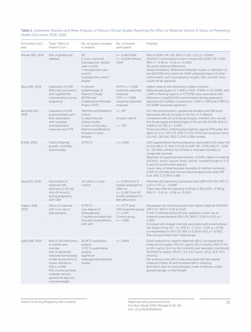
Vitamin D during Pregnancy and Lactation 21Reprinted with permission from:Ann Nutr Metab 2020;76(suppl 2):16–28DOI: 10.1159/000508422
Table 1. Systematic Reviews and Meta-Analyses of Various Clinical Studies Reporting the Effect of Maternal Vitamin D Status on Pregnancy Health Outcomes, 2018–2019
First author [ref.], year
Topic: Effect of Vitamin D on …
No. of studies included in analysis
No. of pooled participants
Findings
Amraei [65], 2018 Risk of gestational diabetes
26: 8 cross-sectional6 prospective, nested case-control7 retrospective case-control5 prospective cohort studies
n = 5,464 GDMn = 15,039 without GDM
Risk of GDM, OR 1.18, 95% CI 1.01–1.35, p < 0.0001;25(OH)D concentration lower in those with GDM, OR –0.26, 95% CI –0.39 to –0.14, p < 0.0001No world regional differencesStated limitations: differences between studies in definition of low 25(OH)D and criteria for GDM; potential impact of other confounders, such as pregnancy wt gain, SES, and skin color could not be explored
Baca [40], 2018 Expression of VDR SNPs and association with log25(OH)D concentration during pregnancy
2 cohorts:Epidemiology of Vitamin D Study (EVITA) and Collaborative Perinatal Project (CPP)
EVITA: n = 1,958 randomly selected/ analyzedCPP: n = 4,285 randomly selected/ analyzed
Higher rates of vitD deficiency in black mothersMaternal genotypes of 7 SNPs in VDR, 3 SNPs in GC/VDBP, and 1 SNP in flanking regions of CYP27B1 were associated with difference in log25(OH)D concentration during pregnancyAdjusting for multiple comparisons, 1 SNP in VDR and 2 SNPs in GC/VDBP remained significant
Barchitta [41], 2018
Evaluation of VDR polymorphisms and their association with neonatal anthropometric measures and PTB
“Mamma and Bambino” cohort11 observational clinical studies3 case-control plus Mamma and Bambino included in meta-analysis
n = 187
Unclear total N
n = 763
For FokI polymorphism, gestational duration and BW were decreased with an increase in the No. of A allele(s)Compared with GG and GA genotypes, mothers who carried the AA genotype exhibited higher PTB risk (OR 12.049, 95% CI 2.606 to 55.709, p = 0.001)Protective effect of BsmI polymorphism against PTB under the allelic (A vs G: OR 0.74, 95% CI 0.59–0.93) and recessive (AA vs GG+AG: OR 0.62, 95% CI 0.43–0.89) models
Bi [66], 2018 Infant/offspring growth, morbidity and mortality
24 RCTs n = 5,405 VitD supplemented during pregnancy associated with lower risk of SGA (RR 0.72, 95% CI 0.52 to 0.99; RD –5.6%, 95% CI –0.86 to –10.34%), without risk of fetal or neonatal mortality or congenital anomalyNeonates of supplemented mothers: at birth, higher circulating 25(OH)D, serum calcium levels, and wt, carried through to 3, 6, 9, and 12 months after delivery Lower rates of fetal/neonatal mortality of mothers receiving 2,000 IU vitD/day and risk not reduced above that dose (RR 0.35, 95% CI 0.59 to 0.80)
Fang [67], 2019 Association of maternal vitD deficiency (<20 ng/mL/<50 nmol/L) during pregnancy with LBW
16 cohort or case-control
n = 8,403 from 8 studies analyzed for LBW riskn = 11,867 from 10 studies analyzed for BW differences
Maternal vitD deficiency associated with LBW (OR 2.39, 95% CI 1.25 to 4.57, p = 0.008)Total mean BW decreased by 0.08 kg or 80 g (OR –0.08 kg, 95% CI –0.10 to –0.06, p < 0.001)
Fogacci [68], 2019
Effect of maternal vitD-S on risk of preeclampsia
27 RCTsLow degree of heterogeneity3 studies excluded that included multivitamins with vitD
n = 4,777 totalVitD treatment group n = 2,487Control groupn = 2,290
Decreased risk of preeclampsia with higher maternal 25(OH)D (OR 0.37, 95% CI 0.26 to 0.52)If vitD-S initiated before 20 wks’ gestation, lower risk of maternal preeclampsia (OR 0.35, 95% CI 0.26 to 0.52, p < 0.001)Increased vitD dosage inversely associated with preeclampsia risk (slope of log OR –1.1, 95% CI –1.73 to –0.46, p < 0.001 corresponding to OR 0.33, 95% CI 0.18 to 0.63, p < 0.001)Risk not associated with maternal age
Gallo [69], 2019 Risk of vitD deficiency in mother and neonateRisk of abnormal maternal homeostatic model assessment of insulin resistanceEffect on BWRisk of preeclampsia, cesarean section, gestational age and neonatal length
20 RCTs qualitative analysis17 RCTs quantitative analysisSignificant heterogeneity between studies
n = 2,844 Good evidence to support maternal vitD-S, increased both maternal (13 studies, MD 14.1 ng/mL [35.2 nmol/L]; 95% CI 9.6 to 18.6 ng/mL [24.0 to 46.4 nmol/L]) and neonatal (cord blood) 25(OH)D (9 studies, MD 9.7, 5.2, 14.2 ng/mL [24.2, 12.9, 35.5 nmol/L])Fair evidence that vitD-S was associated with decreased maternal HOMA-IR and increased BW in offspringNull effect seen for preeclampsia, mode of delivery, infant gestational age, or birth length
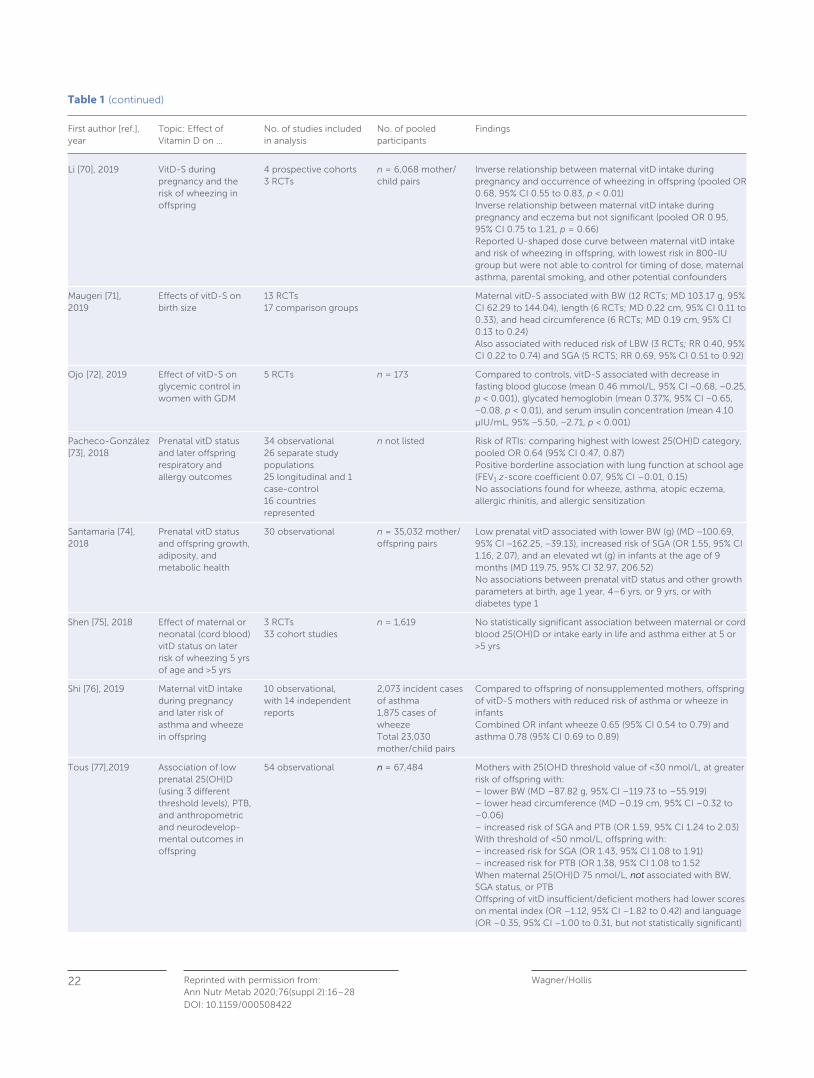
Wagner/Hollis Reprinted with permission from:Ann Nutr Metab 2020;76(suppl 2):16–28
22
DOI: 10.1159/000508422
Table 1 (continued)
First author [ref.], year
Topic: Effect of Vitamin D on …
No. of studies included in analysis
No. of pooled participants
Findings
Li [70], 2019 VitD-S during pregnancy and the risk of wheezing in offspring
4 prospective cohorts3 RCTs
n = 6,068 mother/child pairs
Inverse relationship between maternal vitD intake during pregnancy and occurrence of wheezing in offspring (pooled OR 0.68, 95% CI 0.55 to 0.83, p < 0.01)Inverse relationship between maternal vitD intake during pregnancy and eczema but not significant (pooled OR 0.95, 95% CI 0.75 to 1.21, p = 0.66)Reported U-shaped dose curve between maternal vitD intake and risk of wheezing in offspring, with lowest risk in 800-IU group but were not able to control for timing of dose, maternal asthma, parental smoking, and other potential confounders
Maugeri [71], 2019
Effects of vitD-S on birth size
13 RCTs17 comparison groups
Maternal vitD-S associated with BW (12 RCTs; MD 103.17 g, 95% CI 62.29 to 144.04), length (6 RCTs; MD 0.22 cm, 95% CI 0.11 to 0.33), and head circumference (6 RCTs; MD 0.19 cm, 95% CI 0.13 to 0.24)Also associated with reduced risk of LBW (3 RCTs; RR 0.40, 95% CI 0.22 to 0.74) and SGA (5 RCTS; RR 0.69, 95% CI 0.51 to 0.92)
Ojo [72], 2019 Effect of vitD-S on glycemic control in women with GDM
5 RCTs n = 173 Compared to controls, vitD-S associated with decrease in fasting blood glucose (mean 0.46 mmol/L, 95% CI −0.68, −0.25, p < 0.001), glycated hemoglobin (mean 0.37%, 95% CI −0.65, −0.08, p < 0.01), and serum insulin concentration (mean 4.10 μIU/mL, 95% −5.50, −2.71, p < 0.001)
Pacheco-González [73], 2018
Prenatal vitD status and later offspring respiratory and allergy outcomes
34 observational26 separate study populations25 longitudinal and 1 case-control16 countries represented
n not listed Risk of RTIs: comparing highest with lowest 25(OH)D category, pooled OR 0.64 (95% CI 0.47, 0.87)Positive borderline association with lung function at school age (FEV1 z-score coefficient 0.07, 95% CI –0.01, 0.15) No associations found for wheeze, asthma, atopic eczema, allergic rhinitis, and allergic sensitization
Santamaria [74], 2018
Prenatal vitD status and offspring growth, adiposity, and metabolic health
30 observational n = 35,032 mother/offspring pairs
Low prenatal vitD associated with lower BW (g) (MD −100.69, 95% CI −162.25, −39.13), increased risk of SGA (OR 1.55, 95% CI 1.16, 2.07), and an elevated wt (g) in infants at the age of 9 months (MD 119.75, 95% CI 32.97, 206.52)No associations between prenatal vitD status and other growth parameters at birth, age 1 year, 4–6 yrs, or 9 yrs, or with diabetes type 1
Shen [75], 2018 Effect of maternal or neonatal (cord blood) vitD status on later risk of wheezing 5 yrs of age and >5 yrs
3 RCTs33 cohort studies
n = 1,619 No statistically significant association between maternal or cord blood 25(OH)D or intake early in life and asthma either at 5 or >5 yrs
Shi [76], 2019 Maternal vitD intake during pregnancy and later risk of asthma and wheeze in offspring
10 observational, with 14 independent reports
2,073 incident cases of asthma1,875 cases of wheezeTotal 23,030 mother/child pairs
Compared to offspring of nonsupplemented mothers, offspring of vitD-S mothers with reduced risk of asthma or wheeze in infants Combined OR infant wheeze 0.65 (95% CI 0.54 to 0.79) and asthma 0.78 (95% CI 0.69 to 0.89)
Tous [77],2019 Association of low prenatal 25(OH)D (using 3 different threshold levels), PTB, and anthropometric and neurodevelop-mental outcomes in offspring
54 observational n = 67,484 Mothers with 25(OHD threshold value of <30 nmol/L, at greater risk of offspring with:– lower BW (MD –87.82 g, 95% CI –119.73 to –55.919)– lower head circumference (MD –0.19 cm, 95% CI –0.32 to –0.06)– increased risk of SGA and PTB (OR 1.59, 95% CI 1.24 to 2.03)With threshold of <50 nmol/L, offspring with: – increased risk for SGA (OR 1.43, 95% CI 1.08 to 1.91)– increased risk for PTB (OR 1.38, 95% CI 1.08 to 1.52When maternal 25(OH)D 75 nmol/L, not associated with BW, SGA status, or PTBOffspring of vitD insufficient/deficient mothers had lower scores on mental index (OR –1.12, 95% CI –1.82 to 0.42) and language (OR –0.35, 95% CI –1.00 to 0.31, but not statistically significant)
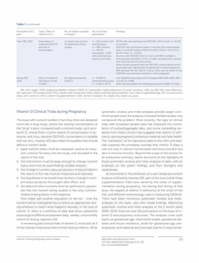
Vitamin D during Pregnancy and Lactation 23Reprinted with permission from:Ann Nutr Metab 2020;76(suppl 2):16–28DOI: 10.1159/000508422
Vitamin D Clinical Trials during Pregnancy
The issue with nutrient studies is that they often are designed
more like a drug study, where the starting concentration of
the “drug” is zero, compared with a nutrient study, such as vi-
tamin D, where there is some vitamin D concentration in ev-
eryone, and, thus, baseline 25(OH)D concentration is variable
and not zero. Heaney [43] described the qualities that should
define a nutrient study:
1 basal nutrient status must be measured, used as an inclu-
sion criterion for entry into the study, and recorded in the
report of the trial;
2 the intervention must be large enough to change nutrient
status and must be quantified by suitable analysis;
3 the change in nutrient status produced in those enrolled in
the report of the trial must be measured and reported;
4 the hypothesis to be tested must be that a change in nutri-
ent status produces the sought-after effect; and
5 the status of other nutrients must be optimized to guaran-
tee that the nutrient being studied is the only nutrition-
related limiting factor in the response.
One might add another stipulation to the list – that the
nutrient being investigated has to follow an appropriate dos-
ing schedule to match what happens naturally. In the case of
vitamin D, there is a plethora of data that show substantial
physiological differences between daily, weekly, and monthly
vitamin D dosing regimens [11] .
In reviewing past clinical trials of vitamin D, most lack all 5
of the Heaney criteria and often the 6th dosing criterion. While
systematic reviews and meta-analyses provide larger com-
bined sample sizes, the analyses of several limited studies only
compound the problem. More recently, the rigor of clinical
trials with increased sample sizes has improved the consis-
tency of pooled/aggregate data, and some compelling evi-
dence from these clinical trials suggests that vitamin D suffi-
ciency during pregnancy enhances maternal and fetal health.
The “translation” of the laboratory data to the clinic and bed-
side supports this emerging concept that vitamin D plays a
role not only in calcium homeostasis and bone function but
also in immune function. Beyond the scope of this review for
an exhaustive summary, below are some of the highlights of
those systematic reviews and meta-analyses to date, with an
emphasis on the salient findings, and their strengths and
weaknesses.
As mentioned in the attributes of a well-designed nutrient
study as outlined by Heaney [43] , part of the issue is that these
supplementation trials have varied by the onset of supple-
mentation during pregnancy, the dosing and timing of that
dose, the degree of vitamin D deficiency at the onset of the
trial, and different methodology used in measuring 25(OH)D.
There have been numerous systematic reviews and meta-
analyses on the topic, also with mixed findings. Restricting
systematic reviews and meta-analyses to the 2 recent years
2018–2019, there are over 30 publications on the topic of vi-
tamin D and pregnancy outcomes. The analyses cover such
topics as gestational age, infant birth weight, gestational dia-
betes and insulin resistance, small-for-gestational age, pre-
eclampsia, and maternal and neonatal vitamin D status at de-
Table 1 (continued)
First author [ref.], year
Topic: Effect of Vitamin D on …
No. of studies included in analysis
No. of pooled participants
Findings
Yuan [78], 2019 Association of maternal 25(OH)D and risk of preeclampsia
1 nested case-control20 additional clinical studies
n = 122 women with preeclampsian = 480 controlsn = 39,031 participants: 3,305 with preeclampsia, various ethnicities
65.6% with preeclampsia had 25(OH)D <50.0 nmol/L vs. 55.3% of controls25(OH)D was significantly lower in women with preeclampsia than in controls (median [IQR] 43.3 [35.5, 55.2] vs. 47.5 [37.6, 60.4] nmol/L, p = 0.014) Women with 25(OH)D <50.0 nmol/L with 65% increase in preeclampsia risk (95% CI 1.02 to 2.69) compared with women with 25(OH)D 50.0 to 74.9 nmol/LMeta-analysis showed that low 25(OH)D concentrations were associated with significantly increased risk of preeclampsia by 62% (pooled OR 1.62, 95% CI 1.36 to 1.94), and risk effect of low 25(OH)D concentrations existed in most subgroups
Zhang [79], 2018
Effect of maternal vitD status on risk of GDM
87 observational and 25 RCTs
n = 55,859 in observational studiesn = 2,445 in RCTs
Low 25(OH)D associated with increased GDM (OR 1.850, 95% CI 1.471 to 2.328)25(OH)D associated with fasting glucose and HOMA-IR index
BW, birth weight; GDM, gestational diabetes mellitus; HOMA-IR, homeostatic model assessment of insulin resistance; LBW, low BW; MD, mean difference; OR, odds ratio; PTB, preterm birth; RTCs, randomized clinical trials; SNPs, single nucleotide polymorphisms; SGA, small for gestational age; SES, socioeconomic status; vitD, vitamin D; vitD-S; vitamin D supplementation; VDR, vitamin D receptor; wt, weight; wks, weeks; yrs, years.
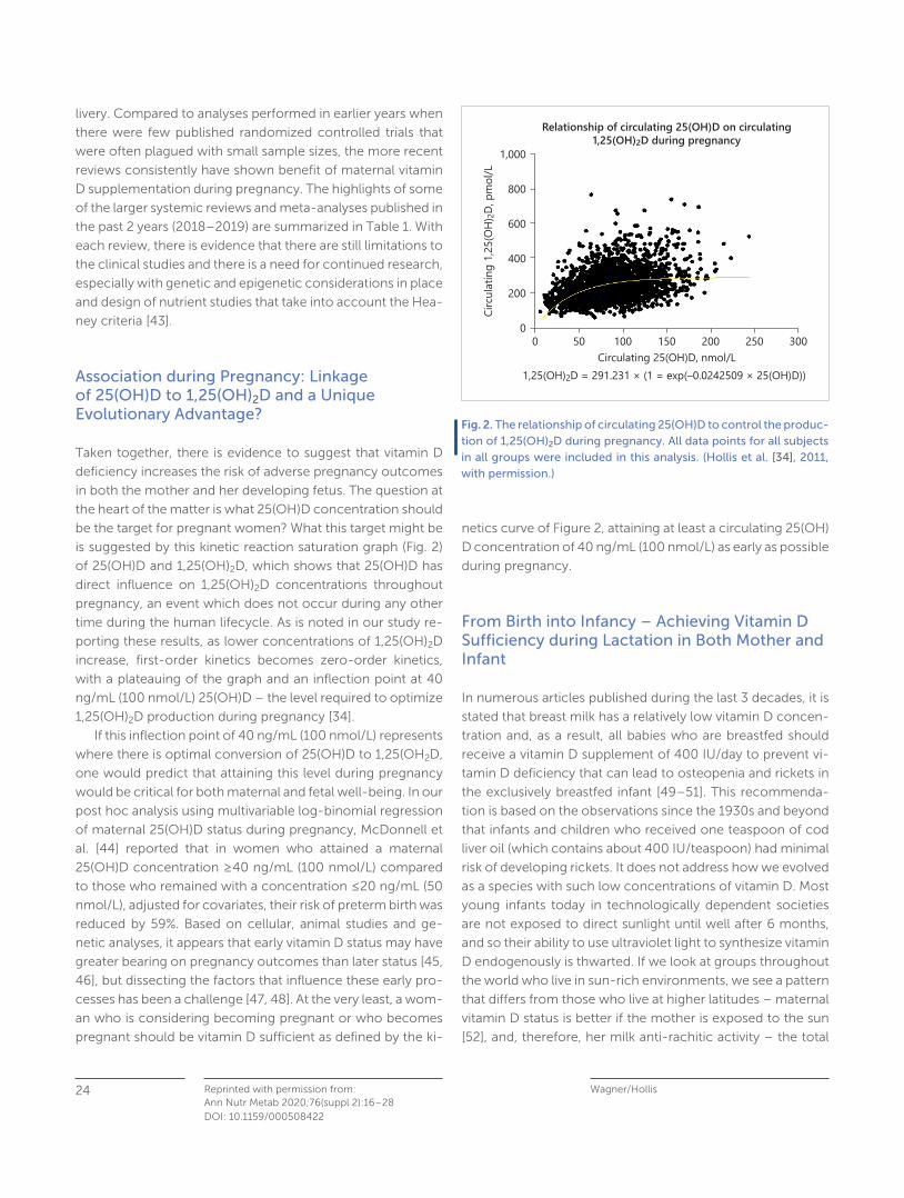
Wagner/Hollis Reprinted with permission from:Ann Nutr Metab 2020;76(suppl 2):16–28
24
DOI: 10.1159/000508422
livery. Compared to analyses performed in earlier years when
there were few published randomized controlled trials that
were often plagued with small sample sizes, the more recent
reviews consistently have shown benefit of maternal vitamin
D supplementation during pregnancy. The highlights of some
of the larger systemic reviews and meta-analyses published in
the past 2 years (2018–2019) are summarized in Table 1 . With
each review, there is evidence that there are still limitations to
the clinical studies and there is a need for continued research,
especially with genetic and epigenetic considerations in place
and design of nutrient studies that take into account the Hea-
ney criteria [43] .
Association during Pregnancy: Linkage of 25(OH)D to 1,25(OH) 2 D and a Unique Evolutionary Advantage?
Taken together, there is evidence to suggest that vitamin D
deficiency increases the risk of adverse pregnancy outcomes
in both the mother and her developing fetus. The question at
the heart of the matter is what 25(OH)D concentration should
be the target for pregnant women? What this target might be
is suggested by this kinetic reaction saturation graph ( Fig. 2 )
of 25(OH)D and 1,25(OH) 2 D, which shows that 25(OH)D has
direct influence on 1,25(OH) 2 D concentrations throughout
pregnancy, an event which does not occur during any other
time during the human lifecycle. As is noted in our study re-
porting these results, as lower concentrations of 1,25(OH) 2 D
increase, first-order kinetics becomes zero-order kinetics,
with a plateauing of the graph and an inflection point at 40
ng/mL (100 nmol/L) 25(OH)D – the level required to optimize
1,25(OH) 2 D production during pregnancy [34] .
If this inflection point of 40 ng/mL (100 nmol/L) represents
where there is optimal conversion of 25(OH)D to 1,25(OH 2 D,
one would predict that attaining this level during pregnancy
would be critical for both maternal and fetal well-being. In our
post hoc analysis using multivariable log-binomial regression
of maternal 25(OH)D status during pregnancy, McDonnell et
al. [44] reported that in women who attained a maternal
25(OH)D concentration ≥ 40 ng/mL (100 nmol/L) compared
to those who remained with a concentration ≤ 20 ng/mL (50
nmol/L), adjusted for covariates, their risk of preterm birth was
reduced by 59%. Based on cellular, animal studies and ge-
netic analyses, it appears that early vitamin D status may have
greater bearing on pregnancy outcomes than later status [45,
46] , but dissecting the factors that influence these early pro-
cesses has been a challenge [47, 48] . At the very least, a wom-
an who is considering becoming pregnant or who becomes
pregnant should be vitamin D sufficient as defined by the ki-
netics curve of Figure 2 , attaining at least a circulating 25(OH)
D concentration of 40 ng/mL (100 nmol/L) as early as possible
during pregnancy.
From Birth into Infancy – Achieving Vitamin D Sufficiency during Lactation in Both Mother and Infant
In numerous articles published during the last 3 decades, it is
stated that breast milk has a relatively low vitamin D concen-
tration and, as a result, all babies who are breastfed should
receive a vitamin D supplement of 400 IU/day to prevent vi-
tamin D deficiency that can lead to osteopenia and rickets in
the exclusively breastfed infant [49–51] . This recommenda-
tion is based on the observations since the 1930s and beyond
that infants and children who received one teaspoon of cod
liver oil (which contains about 400 IU/teaspoon) had minimal
risk of developing rickets. It does not address how we evolved
as a species with such low concentrations of vitamin D. Most
young infants today in technologically dependent societies
are not exposed to direct sunlight until well after 6 months,
and so their ability to use ultraviolet light to synthesize vitamin
D endogenously is thwarted. If we look at groups throughout
the world who live in sun-rich environments, we see a pattern
that differs from those who live at higher latitudes – maternal
vitamin D status is better if the mother is exposed to the sun
[52] , and, therefore, her milk anti-rachitic activity – the total
Circu
lating
1,25
(OH)
2D, p
mol/
L
0 50 100 150Circulating 25(OH)D, nmol/L
1,25(OH)2D = 291.231 × (1 = exp(–0.0242509 × 25(OH)D))
200 250 300
1,000
800
600
200
0
400
Relationship of circulating 25(OH)D on circulating1,25(OH)2D during pregnancy
Fig. 2. The relationship of circulating 25(OH)D to control the produc-tion of 1,25(OH) 2 D during pregnancy. All data points for all subjects in all groups were included in this analysis. (Hollis et al. [34] , 2011, with permission.)
Vitamin D during Pregnancy and Lactation 25Reprinted with permission from:Ann Nutr Metab 2020;76(suppl 2):16–28DOI: 10.1159/000508422
amount of vitamin D moieties in human milk – is better. It is
critical to understand that human milk is deficient in vitamin
D only when the mother herself is deficient [53] . We know that
during pregnancy, maternal vitamin D status is closely linked
with fetal and neonatal vitamin D status. That connection and
relationship continues during lactation.
It was hypothesized by Hollis and Wagner [54] more than
2 decades ago that if a mother had an improved vitamin D
status, then her milk anti-rachitic activity would be improved
and that of her recipient infant, obviating the need for infant
vitamin D supplementation. Maternal vitamin D supplementa-
tion would effectively treat both the mother and her breast-
feeding infant. This was studied in 2 pilot studies by our group
[54, 55] and then in a larger trial sponsored by the National
Institutes of Health [56] that has since been repeated by Da-
wodu et al. [57] in another region of the world – the Middle
East – where there is profound vitamin D deficiency. In the
various trials, mothers at 1 month postpartum were random-
ized to receive 1 of 3 treatments: 400, 2,400, or 6,400 IU vi-
tamin D/day. Infants of mothers in the 400-IU group received
the standard of care of 400 IU/day, while infants of mothers
in the 2,400- and 6,400-IU group received 0 IU/day (place-
bo). Maternal supplementation with 2,400 IU vitamin D/day
with infants on placebo resulted in higher rates of infant insuf-
ficiency and that arm of the study was stopped early-on in the
study. Mothers in the 6,400-IU group had improved vitamin
D status, milk anti-rachitic activity, and their infants had cir-
culating 25(OH)D concentrations that were comparable to in-
fants receiving 400 IU/day direct supplementation [55, 56] .
There were no safety issues noted in these studies except with
the 2,400 IU arm and the higher rates of infant deficiency, but
no issues with toxicity from vitamin D. Similar results were re-
ported by Dawodu et al. [57] .
Oberhelman et al. [58] studied 40 exclusively breastfeed-
ing mothers and infants who were randomized to receive ei-
ther daily maternal vitamin D supplementation of 5,000 IU/
day versus a single large bolus of 150,000 IU once as a higher
bolus vitamin D, with the primary outcome at 28 days being
maternal and infant vitamin D status. The daily versus single
bolus were comparable at 28 days; however, the mother and
infant pair who received the single bolus had a large increase
in their circulating 25(OH)D that rapidly declined but was still
improved compared to baseline.
A systematic review and meta-analysis by O’Callaghan et
al. [59] , reviewing relevant studies on the topic of alternatives
to daily infant vitamin D supplementation through September
2018, identified 28 relevant papers of which 5 were random-
ized clinical trials that met inclusion criteria for the analysis.
The meta-analysis suggests that the results are promising,
with the need for larger studies in diverse groups of women
necessary to be carried out before policy changes can be
made [59] .
While application of alternatives to infant supplementation
are being discussed, a major issue complicating recommen-
dations is that compliance by parents to give their breastfeed-
ing infants daily vitamin D drops is low in many regions of the
world [60, 61] . In the USA, reports of compliance with the
recommendation of infant vitamin D supplementation range
from 9 to 20%, leaving most breastfeeding infants in the USA
deficient, dependent on their mothers who are often them-
selves deficient [62–64] . These are less than satisfying statis-
tics. At the end of the day, where maternal compliance with
taking a vitamin D supplement is much greater than that of
parental adherence with infant supplementation, maternal vi-
tamin D supplementation alone remains as a viable alternative
to infant vitamin D supplementation that safely and effective-
ly treats both the mother and her breastfeeding infant.
Conflict of Interest Statement
The writing of this article was supported by Nestlé Nutrition Institute, and the author declares no other conflicts of interest.
Funding Sources
Funded in part by NIH UL1 TR000062 and Stone Foundation, Chil-dren’s Hospital Fund, Medical University of South Carolina.
That if a mother had an
improved vitamin D status,
then her milk anti-rachitic
activity would be improved
and that of her recipient
infant, obviating the need
for infant vitamin D
supplementation
Wagner/Hollis Reprinted with permission from:Ann Nutr Metab 2020;76(suppl 2):16–28
26
DOI: 10.1159/000508422
References
1 Racicot K, Kwon JY, Aldo P, Silasi M, Mor G. Understanding the complexity of the immune system during pregnancy. Am J Reprod Immunol . 2014 Aug; 72(2): 107–16.
2 Mor G, Cardenas I. The immune system in pregnancy: a unique complexity. Am J Reprod Immunol . 2010 Jun; 63(6): 425–33.
3 Mor G, Cardenas I, Abrahams V, Guller S. Inflammation and preg-nancy: the role of the immune system at the implantation site. Ann N Y Acad Sci . 2011 Mar; 1221(1): 80–7.
4 Bikle DD. Vitamin D: an ancient hormone. Exp Dermatol . 2011 Jan; 20(1): 7–13.
5 Wagner CL, Eidelman AI. The impact of vitamin D on the maternal and infant epigenome: the role of pregnancy and breastfeeding. Breastfeed Med . 2018 Jun; 13(5): 305–6.
6 Bikle DD. Vitamin D regulation of immune function. Vitam Horm . 2011; 86: 1–21.
7 Bikle DD. Vitamin D metabolism and function in the skin. Mol Cell Endocrinol . 2011 Dec; 347(1-2): 80–9.
8 Hollis BW. Comparison of equilibrium and disequilibrium assay conditions for ergocalciferol, cholecalciferol and their major me-tabolites. J Steroid Biochem . 1984 Jul; 21(1): 81–6.
9 Bhattacharyya MH, DeLuca HF. Subcellular location of rat liver calciferol-25-hydroxylase. Arch Biochem Biophys . 1974 Jan; 160(1): 58–62.
10 Hollis BW, Pittard WB 3rd. Evaluation of the total fetomaternal vi-tamin D relationships at term: evidence for racial differences. J Clin Endocrinol Metab . 1984 Oct; 59(4): 652–7.
11 Hollis BW, Wagner CL. Clinical review: The role of the parent com-pound vitamin D with respect to metabolism and function: Why clinical dose intervals can affect clinical outcomes. J Clin Endo-crinol Metab . 2013 Dec; 98(12): 4619–28.
12 Hollis BW, Pittard WB 3rd, Reinhardt TA. Relationships among vi-tamin D, 25-hydroxyvitamin D, and vitamin D-binding protein concentrations in the plasma and milk of human subjects. J Clin Endocrinol Metab . 1986 Jan; 62(1): 41–4.
13 Bikle DD, Gee E, Halloran B, Kowalski MA, Ryzen E, Haddad JG. Assessment of the free fraction of 25-hydroxyvitamin D in serum and its regulation by albumin and the vitamin D-binding protein. J Clin Endocrinol Metab . 1986 Oct; 63(4): 954–9.
14 Bikle DD, Halloran BP, Gee E, Ryzen E, Haddad JG. Free 25-hy-droxyvitamin D levels are normal in subjects with liver disease and reduced total 25-hydroxyvitamin D levels. J Clin Invest . 1986 Sep; 78(3): 748–52.
15 Kumar R, Thompson JR. The regulation of parathyroid hormone secretion and synthesis. J Am Soc Nephrol . 2011 Feb; 22(2): 216–24.
16 Bouillon R, Bikle D. Vitamin D Metabolism Revised: fall of Dogmas. J Bone Miner Res . 2019 Nov; 34(11): 1985–92.
17 Bouillon R, Marcocci C, Carmeliet G, Bikle D, White JH, Dawson-Hughes B, et al. Skeletal and Extraskeletal Actions of Vitamin D: Current Evidence and Outstanding Questions. Endocr Rev . 2019 Aug; 40(4): 1109–51.
18 Hollis BW. Circulating 25-hydroxyvitamin D levels indicative of vi-tamin D sufficiency: implications for establishing a new effective dietary intake recommendation for vitamin D. J Nutr . 2005 Feb; 135(2): 317–22.
19 Nykjaer A, Dragun D, Walther D, Vorum H, Jacobsen C, Herz J, et al. An endocytic pathway essential for renal uptake and activation of the steroid 25-(OH) vitamin D3. Cell . 1999 Feb; 96(4): 507–15.
20 Marzolo MP, Farfán P. New insights into the roles of megalin/LRP2 and the regulation of its functional expression. Biol Res . 2011; 44(1): 89–105.
21 Holick MF. Vitamin D deficiency. N Engl J Med . 2007 Jul; 357(3): 266–81.
22 Bikle DD. Vitamin D metabolism, mechanism of action, and clini-cal applications. Chem Biol . 2014 Mar; 21(3): 319–29.
23 Holick MF. Vitamin D: evolutionary, physiological and health per-spectives. Curr Drug Targets . 2011 Jan; 12(1): 4–18.
24 Gallagher JC, Bikle DD. Vitamin D: Mechanisms of Action and Clinical Applications. Endocrinol Metab Clin North Am . 2017 Dec; 46(4):xvii–xviii.
25 Hollis BW, Wagner CL. New insights into the vitamin D require-ments during pregnancy. Bone Res . 2017 Aug; 5(1): 17030.
26 Bikle DD, Schwartz J. Vitamin D Binding Protein, Total and Free Vitamin D Levels in Different Physiological and Pathophysiological Conditions. Front Endocrinol (Lausanne) . 2019 May; 10: 317.
27 Hacker-Thompson A, Schloetter M, Sellmeyer DE. Validation of a dietary vitamin D questionnaire using multiple diet records and the block 98 health habits and history questionnaire in healthy post-menopausal women in northern California. J Acad Nutr Diet . 2012 Mar; 112(3): 419–23.
28 Kumar R, Cohen WR, Silva P, Epstein FH. Elevated 1,25-dihy-droxyvitamin D plasma levels in normal human pregnancy and lactation. J Clin Invest . 1979 Feb; 63(2): 342–4.
29 Carneiro RM, Prebehalla L, Tedesco MB, Sereika SM, Hugo M, Hol-lis BW, et al. Lactation and bone turnover: a conundrum of marked bone loss in the setting of coupled bone turnover. J Clin Endocri-nol Metab . 2010 Apr; 95(4): 1767–76.
30 Ritchie LD, Fung EB, Halloran BP, Turnlund JR, Van Loan MD, Cann CE, et al. A longitudinal study of calcium homeostasis during hu-man pregnancy and lactation and after resumption of menses. Am J Clin Nutr . 1998 Apr; 67(4): 693–701.
31 Hollis BW, Wagner CL. Assessment of dietary vitamin D require-ments during pregnancy and lactation. Am J Clin Nutr . 2004 May; 79(5): 717–26.
32 Olsen SF, Secher NJ. A possible preventive effect of low-dose fish oil on early delivery and pre-eclampsia: indications from a 50-year-old controlled trial. Br J Nutr . 1990 Nov; 64(3): 599–609.
33 Bikle DD, Gee E, Halloran B, Haddad JG. Free 1,25-dihydroxyvita-min D levels in serum from normal subjects, pregnant subjects, and subjects with liver disease. J Clin Invest . 1984 Dec; 74(6): 1966–71.
34 Hollis BW, Johnson D, Hulsey TC, Ebeling M, Wagner CL. Vitamin D supplementation during pregnancy: double-blind, randomized clinical trial of safety and effectiveness. J Bone Miner Res . 2011 Oct; 26(10): 2341–57.
Vitamin D during Pregnancy and Lactation 27Reprinted with permission from:Ann Nutr Metab 2020;76(suppl 2):16–28DOI: 10.1159/000508422
35 Hollis BW, Wagner CL. Vitamin D and pregnancy: skeletal effects, nonskeletal effects, and birth outcomes. Calcif Tissue Int . 2013 Feb; 92(2): 128–39.
36 Karras SN, Wagner CL, Angeloudi E, Kotsa K. Maternal vitamin D status during pregnancy in Europe: the two sides of the story. Eur J Nutr . 2017 Sep; 56(6): 2207–8.
37 Karras SN, Wagner CL, Castracane VD. Understanding vitamin D metabolism in pregnancy: from physiology to pathophysiology and clinical outcomes. Metabolism . 2018 Sep; 86: 112–23.
38 Al-Garawi A, Carey VJ, Chhabra D, Mirzakhani H, Morrow J, Lasky-Su J, et al. The Role of Vitamin D in the Transcriptional Program of Human Pregnancy. PLoS One . 2016 Oct; 11(10):e0163832.
39 Litonjua AA, Carey VJ, Laranjo N, Harshfield BJ, McElrath TF, O’Connor GT, et al. Effect of Prenatal Supplementation with Vita-min D on Asthma or Recurrent Wheezing in Offspring by Age 3 Years: The VDAART Randomized Clinical Trial. JAMA . 2016 Jan; 315(4): 362–70.
40 Baca KM, Govil M, Zmuda JM, Simhan HN, Marazita ML, Bodnar LM. Vitamin D metabolic loci and vitamin D status in Black and White pregnant women. Eur J Obstet Gynecol Reprod Biol . 2018 Jan; 220: 61–8.
41 Barchitta M, Maugeri A, La Rosa MC, Magnano San Lio R, Favara G, Panella M, et al. Single Nucleotide Polymorphisms in Vitamin D Receptor Gene Affect Birth Weight and the Risk of Preterm Birth: Results From the “Mamma & Bambino” Cohort and A Meta-Anal-ysis. Nutrients . 2018 Aug; 10(9): 10.
42 Newton DA, Baatz JE, Kindy MS, Gattoni-Celli S, Shary JR, Hollis BW, et al. Vitamin D binding protein polymorphisms significantly impact vitamin D status in children. Pediatr Res . 2019 Nov; 86(5): 662–9.
43 Heaney RP. Guidelines for optimizing design and analysis of clin-ical studies of nutrient effects. Nutr Rev . 2014 Jan; 72(1): 48–54.
44 McDonnell SL, Baggerly KA, Baggerly CA, Aliano JL, French CB, Baggerly LL, et al. Maternal 25(OH)D concentrations ≥ 40 ng/mL associated with 60% lower preterm birth risk among general ob-stetrical patients at an urban medical center. PLoS One . 2017 Jul; 12(7):e0180483.
45 Mirzakhani H, Litonjua AA, McElrath TF, O’Connor G, Lee-Parritz A, Iverson R, et al. Early pregnancy vitamin D status and risk of preeclampsia. J Clin Invest . 2016 Dec; 126(12): 4702–15.
46 Weiss ST, Litonjua AA. Vitamin D in Host Defense: Implications for Future Research. Am J Respir Cell Mol Biol . 2017 Jun; 56(6): 692–3.
47 Hewison M. The earlier the better: preconception vitamin D and protection against pregnancy loss. Lancet Diabetes Endocrinol . 2018 Sep; 6(9): 680–1.
48 Hewison M, Wagner CL, Hollis BW. Vitamin D Supplementation in Pregnancy and Lactation and Infant Growth. N Engl J Med . 2018 Nov; 379(19): 1880–1.
49 Hollis BW, Roos BA, Lambert PW. Vitamin D compounds in human and bovine milk. Adv Nutr Res . 1982; 4: 59–75.
50 Wagner CL, Greer FR; American Academy of Pediatrics Section on Breastfeeding; American Academy of Pediatrics Committee on Nutrition. Prevention of rickets and vitamin D deficiency in infants, children, and adolescents. Pediatrics . 2008 Nov; 122(5): 1142–52.
51 Dawodu A, Tsang RC. Maternal vitamin D status: effect on milk vitamin D content and vitamin D status of breastfeeding infants. Adv Nutr . 2012 May; 3(3): 353–61.
52 Luxwolda MF, Kuipers RS, Kema IP, van der Veer E, Dijck-Brouwer DA, Muskiet FA. Vitamin D status indicators in indigenous popula-tions in East Africa. Eur J Nutr . 2013 Apr; 52(3): 1115–25.
53 Wagner C. Current concepts in vitamin D requirements for moth-er and her breastfeeding infant. In: Lawrence R, editor. Interna-tional Society for Research in Human Milk and Lactation. Breast-feeding Medicine. Trieste, Italy; 2013. pp. 561–562.
54 Hollis BW, Wagner CL. Vitamin D requirements during lactation: high-dose maternal supplementation as therapy to prevent hypo-vitaminosis D for both the mother and the nursing infant. Am J Clin Nutr . 2004 Dec; 80(6 Suppl): 1752S–8S.
55 Wagner CL, Hulsey TC, Fanning D, Ebeling M, Hollis BW. High-dose vitamin D3 supplementation in a cohort of breastfeeding mothers and their infants: a 6-month follow-up pilot study. Breastfeed Med . 2006; 1(2): 59–70.
56 Hollis BW, Wagner CL, Howard CR, Ebeling M, Shary JR, Smith PG, et al. Maternal Versus Infant Vitamin D Supplementation During Lactation: A Randomized Controlled Trial. Pediatrics . 2015 Oct; 136(4): 625–34.
57 Dawodu A, Salameh KM, Al-Janahi NS, Bener A, Elkum N. The Ef-fect of High-Dose Postpartum Maternal Vitamin D Supplementa-tion Alone Compared with Maternal Plus Infant Vitamin D Supple-mentation in Breastfeeding Infants in a High-Risk Population. A Randomized Controlled Trial. Nutrients . 2019 Jul; 11(7): 11.
58 Oberhelman SS, Meekins ME, Fischer PR, Lee BR, Singh RJ, Cha SS, et al. Maternal vitamin D supplementation to improve the vita-min D status of breast-fed infants: a randomized controlled trial. Mayo Clin Proc . 2013 Dec; 88(12): 1378–87.
59 O’Callaghan KM, Taghivand M, Zuchniak A, Onoyovwi A, Korsiak J, Leung M, et al. Vitamin D in Breastfed Infants: Systematic Review of Alternatives to Daily Supplementation. Adv Nutr . 2020 Jan; 11(1): 144–59.
60 Oberhelman S, Cozine E, Umaretiya P, Maxson J, Thacher T. Vita-min D and the breastfeeding infant: family medicine clinicians’ knowledge, attitudes, and practices. J Hum Lact . 2018 May; 34(2): 331–6.
61 Wagner CL, Hollis BW. Commentary on “Vitamin D and the Breast-feeding Infant: Family Medicine Clinicians’ Knowledge, Attitudes, and Practices” by Oberhelman et al. J Hum Lact . 2018 May; 34(2): 337–9.
62 Perrine CG, Sharma AJ, Jefferds ME, Serdula MK, Scanlon KS. Ad-herence to vitamin D recommendations among US infants. Pedi-atrics . 2010 Apr; 125(4): 627–32.
63 Weernink MG, van Wijk RM, Groothuis-Oudshoorn CG, Lanting CI, Grant CC, van Vlimmeren LA, et al. Insufficient vitamin D supple-ment use during pregnancy and early childhood: a risk factor for positional skull deformation. Matern Child Nutr . 2016 Jan; 12(1): 177–88.
64 Ahrens K, Rossen L, Simon A. Adherence to vitamin D recommen-dations among US infants aged 0 to 11 Months, NHANES, 2009 to 2012. Clin Pediatr (Phila) . 2016 Jun; 55(6): 555–6.
Wagner/Hollis Reprinted with permission from:Ann Nutr Metab 2020;76(suppl 2):16–28
28
DOI: 10.1159/000508422
65 Amraei M, Mohamadpour S, Sayehmiri K, Mousavi SF, Shirzadpour E, Moayeri A. Effects of Vitamin D Deficiency on Incidence Risk of Gestational Diabetes Mellitus: A Systematic Review and Meta-analysis. Front Endocrinol (Lausanne). 2018 Feb;9:7.
66 Bi WG, Nuyt AM, Weiler H, Leduc L, Santamaria C, Wei SQ. Asso-ciation Between Vitamin D Supplementation During Pregnancy and Offspring Growth, Morbidity, and Mortality: A Systematic Re-view and Meta-analysis. JAMA Pediatr. 2018 Jul;172(7):635–45.
67 Fang K, He Y, Mu M, Liu K. Maternal vitamin D deficiency during pregnancy and low birth weight: a systematic review and meta-analysis. J Matern Fetal Neonatal Med. 2019 Jul 8;1–7.
68 Fogacci S, Fogacci F, Banach M, Michos ED, Hernandez AV, Lip GYH, Blaha MJ, Toth PP, Borghi C, Cicero AFG; Lipid and Blood Pressure Meta-analysis Collaboration (LBPMC) Group. Vitamin D supplementation and incident preeclampsia: A systematic review and meta-analysis of randomized clinical trials. Clin Nutr. 2020 Jun;39(6):1742–52.
69 Gallo S, McDermid JM, Al-Nimr RI, Hakeem R, Moreschi JM, Pari-Keener M, et al. Supplementation during Pregnancy: An Evidence Analysis Center Systematic Review and Meta-Analysis. J Acad Nutr Diet. 2020 May;120(5):898–924.e4.
70 Li W, Qin Z, Gao J, Jiang Z, Chai Y, Guan L, et al. Vitamin D supple-mentation during pregnancy and the risk of wheezing in offspring: a systematic review and dose-response meta-analysis. J Asthma. 2019 Dec;56(12):1266–73.
71 Maugeri A, Barchitta M, Blanco I, Agodi A. Effects of Vitamin D Supplementation During Pregnancy on Birth Size: A Systematic Review and Meta-Analysis of Randomized Controlled Trials. Nu-trients. 2019 Feb;11(2):E442.
72 Ojo O, Weldon SM, Thompson T, Vargo EJ. The Effect of Vitamin D Supplementation on Glycaemic Control in Women with Gesta-tional Diabetes Mellitus: A Systematic Review and Meta-Analysis of Randomised Controlled Trials. Int J Environ Res Public Health. 2019 May;16(10):E1716.
73 Pacheco-González RM, García-Marcos L, Morales E. Prenatal vi-tamin D status and respiratory and allergic outcomes in child-hood: A meta-analysis of observational studies. Pediatr Allergy Immunol. 2018 May;29(3):243–53.
74 Santamaria C, Bi WG, Leduc L, Tabatabaei N, Jantchou P, Luo ZC, et al. Prenatal vitamin D status and offspring’s growth, adiposity and metabolic health: a systematic review and meta-analysis. Br J Nutr. 2018 Feb;119(3):310–9.
75 Shen SY, Xiao WQ, Lu JH, Yuan MY, He JR, Xia HM, et al. Early life vitamin D status and asthma and wheeze: a systematic review and meta-analysis. BMC Pulm Med. 2018 Jul;18(1):120.
76 Shi D, Wang D, Meng Y, Chen J, Mu G, Chen W. Maternal vitamin D intake during pregnancy and risk of asthma and wheeze in chil-dren: a systematic review and meta-analysis of observational studies. J Matern Fetal Neonatal Med. 2019 May 7:1–7.
77 Tous M, Villalobos M, Iglesias L, Fernández-Barrés S, Arija V. Vita-min D status during pregnancy and offspring outcomes: a system-atic review and meta-analysis of observational studies. Eur J Clin Nutr. 2020 Jan;74(1):36-53.
78 Yuan Y, Tai W, Xu P, Fu Z, Wang X, Long W, Guo X, Ji C, Zhang L, Zhang Y, Wen J. Association of maternal serum 25-hydroxyvita-min D concentrations with risk of preeclampsia: a nested case-control study and meta-analysis. J Matern Fetal Neonatal Med. 2019 Jul 17:1–10.
79 Zhang Y, Gong Y, Xue H, Xiong J, Cheng G. Vitamin D and gesta-tional diabetes mellitus: a systematic review based on data free of Hawthorne effect. BJOG. 2018 Jun;125(7):784–93.
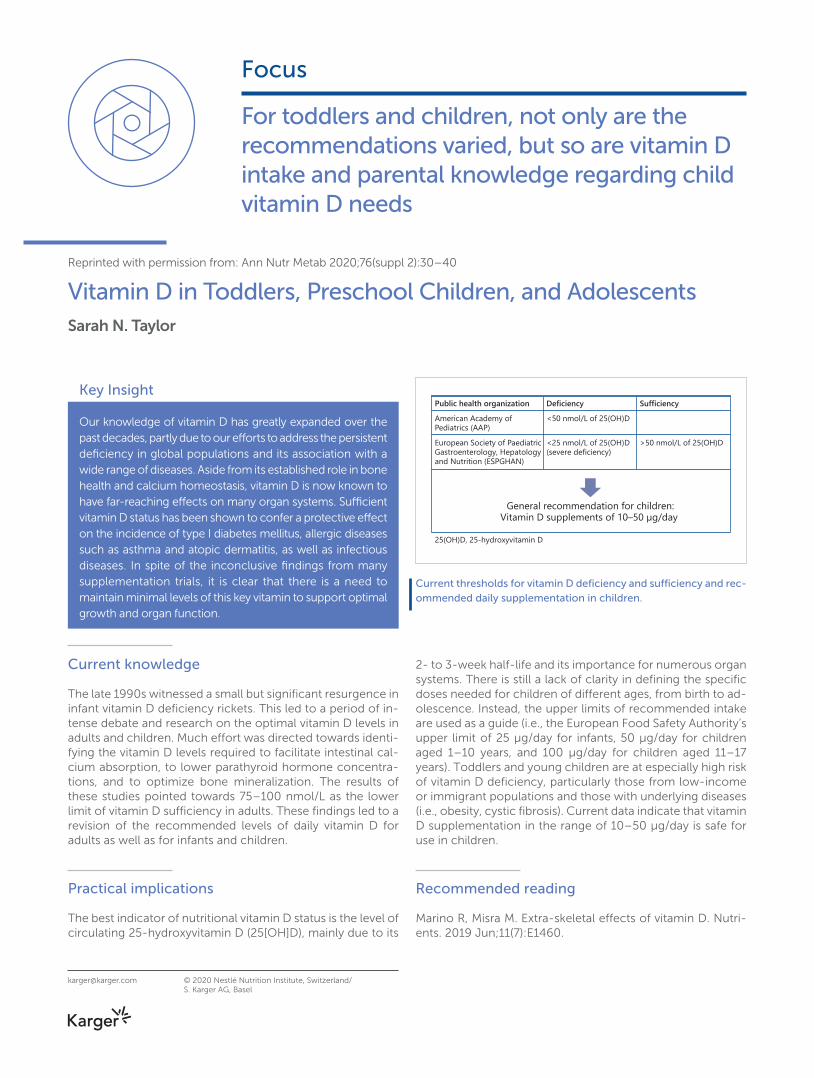
Focus
Reprinted with permission from: Ann Nutr Metab 2020;76(suppl 2):30–40
Vitamin D in Toddlers, Preschool Children, and AdolescentsSarah N. Taylor
© 2020 Nestlé Nutrition Institute, Switzerland/S. Karger AG, Basel
Key Insight
Our knowledge of vitamin D has greatly expanded over the past decades, partly due to our efforts to address the persistent deficiency in global populations and its association with a wide range of diseases. Aside from its established role in bone health and calcium homeostasis, vitamin D is now known to have far-reaching effects on many organ systems. Sufficient vitamin D status has been shown to confer a protective effect on the incidence of type I diabetes mellitus, allergic diseases such as asthma and atopic dermatitis, as well as infectious diseases. In spite of the inconclusive findings from many supplementation trials, it is clear that there is a need to maintain minimal levels of this key vitamin to support optimal growth and organ function.
Current knowledge
The late 1990s witnessed a small but significant resurgence in infant vitamin D deficiency rickets. This led to a period of in-tense debate and research on the optimal vitamin D levels in adults and children. Much effort was directed towards identi-fying the vitamin D levels required to facilitate intestinal cal-cium absorption, to lower parathyroid hormone concentra-tions, and to optimize bone mineralization. The results of these studies pointed towards 75–100 nmol/L as the lower limit of vitamin D sufficiency in adults. These findings led to a revision of the recommended levels of daily vitamin D for adults as well as for infants and children.
Practical implications
The best indicator of nutritional vitamin D status is the level of circulating 25-hydroxyvitamin D (25[OH]D), mainly due to its
2- to 3-week half-life and its importance for numerous organ systems. There is still a lack of clarity in defining the specific doses needed for children of different ages, from birth to ad-olescence. Instead, the upper limits of recommended intake are used as a guide (i.e., the European Food Safety Authority’s upper limit of 25 μg/day for infants, 50 μg/day for children aged 1–10 years, and 100 μg/day for children aged 11–17 years). Toddlers and young children are at especially high risk of vitamin D deficiency, particularly those from low-income or immigrant populations and those with underlying diseases (i.e., obesity, cystic fibrosis). Current data indicate that vitamin D supplementation in the range of 10–50 μg/day is safe for use in children.
Recommended reading
Marino R, Misra M. Extra-skeletal effects of vitamin D. Nutri-ents. 2019 Jun;11(7):E1460.
For toddlers and children, not only are the recommendations varied, but so are vitamin D intake and parental knowledge regarding child vitamin D needs
Public health organization Deficiency SufficiencyAmerican Academy ofPediatrics (AAP)
<50 nmol/L of 25(OH)D
European Society of PaediatricGastroenterology, Hepatologyand Nutrition (ESPGHAN)
<25 nmol/L of 25(OH)D(severe deficiency)
>50 nmol/L of 25(OH)D
General recommendation for children:Vitamin D supplements of 10–50 μg/day
25(OH)D, 25-hydroxyvitamin D
Current thresholds for vitamin D deficiency and sufficiency and rec-ommended daily supplementation in children.
Vitamin D
Reprinted with permission from:Ann Nutr Metab 2020;76(suppl 2):30–40
Vitamin D in Toddlers, Preschool Children, and Adolescents
Sarah N. Taylor
Department of Pediatrics, Yale School of Medicine, New Haven , CT , USA
Sarah N. Taylor Department of Pediatrics Yale School of Medicine, PO Box 208064 New Haven, CT 06520-8064 (USA) sarah.n.taylor @ yale.edu
© 2020 Nestlé Nutrition Institute, Switzerland/S. Karger AG, Basel
Key Messages
• Vitamin D status is associated with avoidance of rickets and various autoimmune, infectious, and allergic diseases.
• Randomized, controlled trials of vitamin D supplementation for pediatric bone health are limited and equivocal in their results.
• The specific vitamin D supplementation to optimize toddler, child, and adolescent outcomes is unknown, but doses 10–50 μg/day are safe and may be beneficial.
DOI: 10.1159/000505635
Keywords Vitamins · Pediatrics · Bone disease · Calcium homeostasis ·
Fractures · Vitamin D
Abstract Background: Vitamin D supplementation is known to both
prevent and treat rickets, a disease of hypomineralized bone.
Childhood is a period of great bone development and, there-
fore, attention to the vitamin D needed to optimize bone
health in childhood is imperative. Summary: Observational
studies have pointed to a vitamin D status, as indicated by a
25-hydroxyvitamin D concentration, of 50 nmol/L to ensure
avoidance of rickets and of 75 nmol/L to optimize health.
However, the benefits of achieving these levels of vitamin D
status are less evident when pediatric randomized, controlled
trials are performed. In fact, no specific pediatric vitamin D
supplementation has been established by the existing evi-
dence. Yet, study of vitamin D physiology continues to un-
cover further potential benefits to vitamin D sufficiency. This
disconnection between vitamin D function and trials of sup-
plementation has led to new paths of investigation, including
establishment of the best method to measure vitamin D sta-
tus, examination of genetic variation in vitamin D metabolism,
and consideration that vitamin D status is a marker of anoth-
er variable, such as physical activity, and its association with
bone health. Nevertheless, vitamin D supplementation in the
range of 10–50 μg/day appears to be safe for children and
remains a promising intervention that may yet be supported
by clinical trials as a method to optimize pediatric health. Key
Message: Pediatric vitamin D status is associated with avoid-
ance of rickets. Randomized, controlled trials of vitamin D
supplementation for pediatric bone health are limited and
equivocal in their results. Beyond bone, decreased risk for
autoimmune, infectious, and allergic diseases has been as-
sociated with higher vitamin D status. The specific vitamin D
supplementation to optimize toddler, child, and adolescent
outcomes is unknown, but doses 10–50 μg/day are safe and
may be beneficial. © 2020 Nestlé Nutrition Institute, Switzerland/
S. Karger AG, Basel
Vitamin D in Children 31Reprinted with permission from:Ann Nutr Metab 2020;76(suppl 2):30–40DOI: 10.1159/000505635
Introduction
Vitamin D deficiency rickets was a disease pervasive in chil-
dren during the Industrial Revolution and prevented with one
spoonful of cod liver oil. The disease dissipated in the pediat-
ric population with improved living conditions, including sun-
light exposure, and the addition of vitamin D fortification to
food products. Through the 20th century, only a small num-
ber of studies investigated the vitamin D supplementation re-
quired to maintain health in general populations. Lack of
attention to vitamin D health status is evident by the 1989
United States Institute of Medicine (IOM) report which recom-
mended 5 μg/day (200 IU/day) for adults because this repre-
sented half the dose recommended to infants and was con-
sidered a “generous allowance” of supplementation [1] . The
10 μg/day dose recommended to infants was based on the
amount of vitamin D in a spoonful of cod liver oil that pre-
vented rickets and demonstrated how the scientific under-
standing of vitamin D physiology remained limited even in
1989.
However, in the 1990s, a resurgence in infant vitamin D
deficiency rickets was described worldwide. These cases oc-
curred in various populations but concentrated in those with
less exposure to sunlight (i.e., high latitude especially in winter
months), those with darker skin pigmentation, or those prac-
ticing complete covering of women and were often associ-
ated with breastfeeding. Despite this small but significant rise
in prevalence, the IOM in 1997 decreased their recommenda-
tion of vitamin D supplementation from 10 to 5 μg/day due to
studies demonstrating that this dose provided adequate vita-
min D to achieve a 25-hydroxyvitamin D (25[OH]D) status of
27.5 nmol/L (11 ng/mL), which was thought to prevent rickets
in “most” populations [1] . Remarkably, despite increasing re-
ports of rickets, the American Academy of Pediatrics (AAP)
chose to uphold the 1997 IOM recommendation and de-
creased their recommendation from 400 to 200 IU/day for all
infants, children, and adolescents [2] .
Therefore, the early 21st century marked a period of dis-
cord in vitamin D public health; as reports of disease esca-
lated, public policy paradoxically decreased the recom-
mended supplementation, and, finally, research in adult pop-
ulations launched the identification of the vitamin D status,
25(OH)D status, associated with optimal health outcomes.
These investigations into the vitamin D status needed to op-
timize vitamin D function were greatly needed because the
previous recommended 25(OH)D concentration of 27.5
nmol/L (11 ng/mL) was based on an observational study of
25(OH)D status in 3 adult cohorts. The vitamin D status in a
cohort of “healthy” adult volunteers was compared to status
in a cohort of lifeguards (high sunlight exposure) and to status
in a cohort of subjects with biliary cirrhosis who were as-
sumed to have difficulty with conversion of vitamin D to
25(OH)D in the liver. From these measurements, a bell curve
of 25(OH)D status was developed with the status of lifeguards
deemed the highest of “normal,” the healthy volunteers
deemed “normal,” and the biliary cirrhosis patients deemed
“low” [3] . This study and others ignored the potential issue
that “healthy volunteers” were exhibiting insufficient or defi-
cient vitamin D.
In the early 21st century, several investigators challenged
this existing definition of normal when they performed studies
to identify the vitamin D status required to optimize intestinal
calcium absorption, to appropriately lower parathyroid hor-
mone (PTH) concentration, and to optimize bone mineraliza-
tion [4–6] . In these studies of vitamin D function in the adult
population, results pointed to 75–100 nmol/L as the lower
limit of vitamin D sufficiency for adult calcium homeostasis
and bone health. These studies and others led to new public
health recommendations that include not only a definition of
vitamin D deficiency but also a definition of vitamin D suffi-
ciency ( Table 1 ) [7–11] . In 2008, the AAP reevaluated its 2003
statement and chose to increase to a recommendation of at
least 400 IU/day for all infants, children, and adolescents [9] .
Additionally, in the early 21st century, there were reports
of vitamin D’s potential role in disease processes not related
to calcium homeostasis and bone health. Many organs have
25(OH)D receptors which bind 25(OH)D. Instead of relying on
the renal production of 1,25-dihydroxyvitamin D (1,25[OH] 2 D)
from 25(OH)D, these organs form 1,25(OH) 2 D locally in a
paracrine fashion ( Fig. 1 ). Therefore, these organs depend on
the availability of vitamin D and its transformation to 25(OH)D
by the liver to provide circulating 25(OH)D. Disease processes
in adults found to have an association with vitamin D status
include cancer, specifically breast, colon, and prostate; heart
The early 21st century marked
a period of discord in vitamin
D public health; as reports of
disease escalated, public
policy paradoxically
decreased the recommended
supplementation
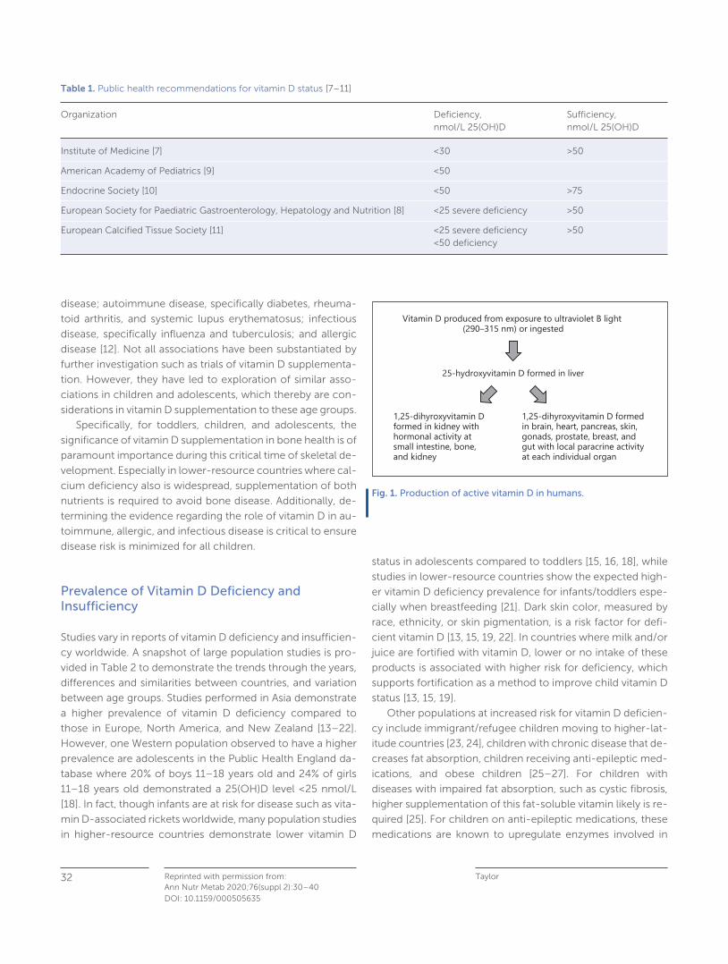
Taylor Reprinted with permission from:Ann Nutr Metab 2020;76(suppl 2):30–40
32
DOI: 10.1159/000505635
disease; autoimmune disease, specifically diabetes, rheuma-
toid arthritis, and systemic lupus erythematosus; infectious
disease, specifically influenza and tuberculosis; and allergic
disease [12] . Not all associations have been substantiated by
further investigation such as trials of vitamin D supplementa-
tion. However, they have led to exploration of similar asso-
ciations in children and adolescents, which thereby are con-
siderations in vitamin D supplementation to these age groups.
Specifically, for toddlers, children, and adolescents, the
significance of vitamin D supplementation in bone health is of
paramount importance during this critical time of skeletal de-
velopment. Especially in lower-resource countries where cal-
cium deficiency also is widespread, supplementation of both
nutrients is required to avoid bone disease. Additionally, de-
termining the evidence regarding the role of vitamin D in au-
toimmune, allergic, and infectious disease is critical to ensure
disease risk is minimized for all children.
Prevalence of Vitamin D Deficiency and Insufficiency
Studies vary in reports of vitamin D deficiency and insufficien-
cy worldwide. A snapshot of large population studies is pro-
vided in Table 2 to demonstrate the trends through the years,
differences and similarities between countries, and variation
between age groups. Studies performed in Asia demonstrate
a higher prevalence of vitamin D deficiency compared to
those in Europe, North America, and New Zealand [13–22] .
However, one Western population observed to have a higher
prevalence are adolescents in the Public Health England da-
tabase where 20% of boys 11–18 years old and 24% of girls
11–18 years old demonstrated a 25(OH)D level < 25 nmol/L
[18] . In fact, though infants are at risk for disease such as vita-
min D-associated rickets worldwide, many population studies
in higher-resource countries demonstrate lower vitamin D
status in adolescents compared to toddlers [15, 16, 18] , while
studies in lower-resource countries show the expected high-
er vitamin D deficiency prevalence for infants/toddlers espe-
cially when breastfeeding [21] . Dark skin color, measured by
race, ethnicity, or skin pigmentation, is a risk factor for defi-
cient vitamin D [13, 15, 19, 22] . In countries where milk and/or
juice are fortified with vitamin D, lower or no intake of these
products is associated with higher risk for deficiency, which
supports fortification as a method to improve child vitamin D
status [13, 15, 19] .
Other populations at increased risk for vitamin D deficien-
cy include immigrant/refugee children moving to higher-lat-
itude countries [23, 24] , children with chronic disease that de-
creases fat absorption, children receiving anti-epileptic med-
ications, and obese children [25–27] . For children with
diseases with impaired fat absorption, such as cystic fibrosis,
higher supplementation of this fat-soluble vitamin likely is re-
quired [25] . For children on anti-epileptic medications, these
medications are known to upregulate enzymes involved in
Table 1. Public health recommendations for vitamin D status [7–11]
Organization Deficiency, nmol/L 25(OH)D
Sufficiency, nmol/L 25(OH)D
Institute of Medicine [7] <30 >50
American Academy of Pediatrics [9] <50
Endocrine Society [10] <50 >75
European Society for Paediatric Gastroenterology, Hepatology and Nutrition [8] <25 severe deficiency >50
European Calcified Tissue Society [11] <25 severe deficiency<50 deficiency
>50
25-hydroxyvitamin D formed in liver
Vitamin D produced from exposure to ultraviolet B light (290–315 nm) or ingested
1,25-dihyroxyvitamin Dformed in kidney withhormonal activity atsmall intestine, bone,and kidney
1,25-dihyroxyvitamin D formed in brain, heart, pancreas, skin, gonads, prostate, breast, and gut with local paracrine activity at each individual organ
Fig. 1. Production of active vitamin D in humans.
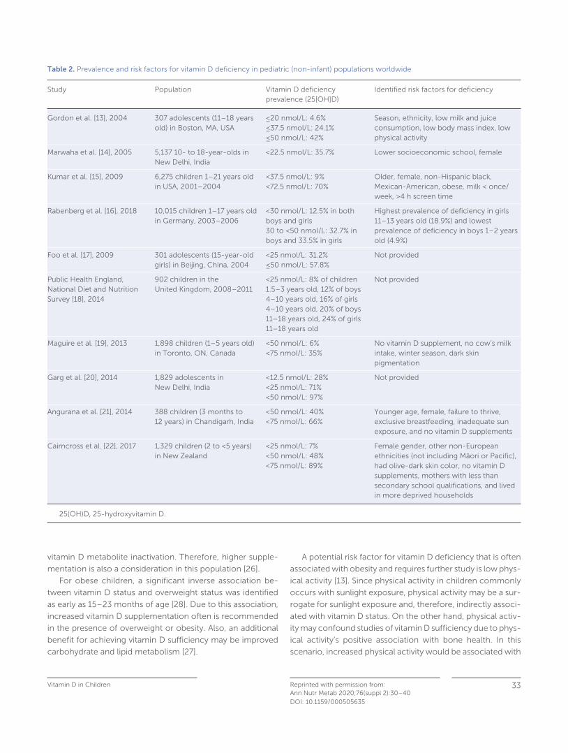
Vitamin D in Children 33Reprinted with permission from:Ann Nutr Metab 2020;76(suppl 2):30–40DOI: 10.1159/000505635
vitamin D metabolite inactivation. Therefore, higher supple-
mentation is also a consideration in this population [26] .
For obese children, a significant inverse association be-
tween vitamin D status and overweight status was identified
as early as 15–23 months of age [28] . Due to this association,
increased vitamin D supplementation often is recommended
in the presence of overweight or obesity. Also, an additional
benefit for achieving vitamin D sufficiency may be improved
carbohydrate and lipid metabolism [27] .
A potential risk factor for vitamin D deficiency that is often
associated with obesity and requires further study is low phys-
ical activity [13] . Since physical activity in children commonly
occurs with sunlight exposure, physical activity may be a sur-
rogate for sunlight exposure and, therefore, indirectly associ-
ated with vitamin D status. On the other hand, physical activ-
ity may confound studies of vitamin D sufficiency due to phys-
ical activity’s positive association with bone health. In this
scenario, increased physical activity would be associated with
Table 2. Prevalence and risk factors for vitamin D deficiency in pediatric (non-infant) populations worldwide
Study Population Vitamin D deficiencyprevalence (25[OH]D)
Identified risk factors for deficiency
Gordon et al. [13], 2004 307 adolescents (11–18 years old) in Boston, MA, USA
≤20 nmol/L: 4.6%≤37.5 nmol/L: 24.1%≤50 nmol/L: 42%
Season, ethnicity, low milk and juice consumption, low body mass index, low physical activity
Marwaha et al. [14], 2005 5,137 10- to 18-year-olds in New Delhi, India
<22.5 nmol/L: 35.7% Lower socioeconomic school, female
Kumar et al. [15], 2009 6,275 children 1–21 years old in USA, 2001–2004
<37.5 nmol/L: 9%<72.5 nmol/L: 70%
Older, female, non-Hispanic black, Mexican-American, obese, milk < once/week, >4 h screen time
Rabenberg et al. [16], 2018 10,015 children 1–17 years old in Germany, 2003–2006
<30 nmol/L: 12.5% in both boys and girls30 to <50 nmol/L: 32.7% in boys and 33.5% in girls
Highest prevalence of deficiency in girls 11–13 years old (18.9%) and lowest prevalence of deficiency in boys 1–2 years old (4.9%)
Foo et al. [17], 2009 301 adolescents (15-year-old girls) in Beijing, China, 2004
<25 nmol/L: 31.2%≤50 nmol/L: 57.8%
Not provided
Public Health England, National Diet and Nutrition Survey [18], 2014
902 children in the United Kingdom, 2008–2011
<25 nmol/L: 8% of children 1.5–3 years old, 12% of boys 4–10 years old, 16% of girls 4–10 years old , 20% of boys 11–18 years old, 24% of girls 11–18 years old
Not provided
Maguire et al. [19], 2013 1,898 children (1–5 years old) in Toronto, ON, Canada
<50 nmol/L: 6%<75 nmol/L: 35%
No vitamin D supplement, no cow’s milk intake, winter season, dark skin pigmentation
Garg et al. [20], 2014 1,829 adolescents in New Delhi, India
<12.5 nmol/L: 28%<25 nmol/L: 71%<50 nmol/L: 97%
Not provided
Angurana et al. [21], 2014 388 children (3 months to 12 years) in Chandigarh, India
<50 nmol/L: 40%<75 nmol/L: 66%
Younger age, female, failure to thrive, exclusive breastfeeding, inadequate sun exposure, and no vitamin D supplements
Cairncross et al. [22], 2017 1,329 children (2 to <5 years) in New Zealand
<25 nmol/L: 7%<50 nmol/L: 48%<75 nmol/L: 89%
Female gender, other non-European ethnicities (not including Māori or Pacific), had olive-dark skin color, no vitamin D supplements, mothers with less than secondary school qualifications, and lived in more deprived households
25(OH)D, 25-hydroxyvitamin D.
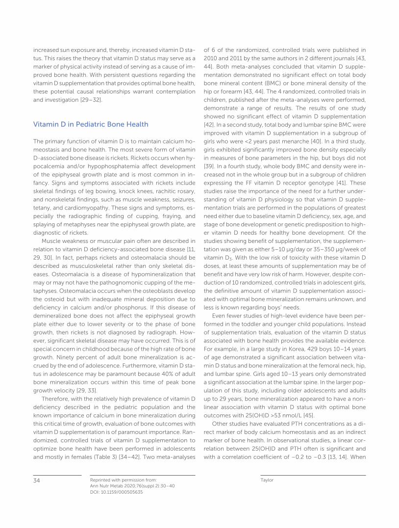
Taylor Reprinted with permission from:Ann Nutr Metab 2020;76(suppl 2):30–40
34
DOI: 10.1159/000505635
increased sun exposure and, thereby, increased vitamin D sta-
tus. This raises the theory that vitamin D status may serve as a
marker of physical activity instead of serving as a cause of im-
proved bone health. With persistent questions regarding the
vitamin D supplementation that provides optimal bone health,
these potential causal relationships warrant contemplation
and investigation [29–32] .
Vitamin D in Pediatric Bone Health
The primary function of vitamin D is to maintain calcium ho-
meostasis and bone health. The most severe form of vitamin
D-associated bone disease is rickets. Rickets occurs when hy-
pocalcemia and/or hypophosphatemia affect development
of the epiphyseal growth plate and is most common in in-
fancy. Signs and symptoms associated with rickets include
skeletal findings of leg bowing, knock knees, rachitic rosary,
and nonskeletal findings, such as muscle weakness, seizures,
tetany, and cardiomyopathy. These signs and symptoms, es-
pecially the radiographic finding of cupping, fraying, and
splaying of metaphyses near the epiphyseal growth plate, are
diagnostic of rickets.
Muscle weakness or muscular pain often are described in
relation to vitamin D deficiency-associated bone disease [11,
29, 30] . In fact, perhaps rickets and osteomalacia should be
described as musculoskeletal rather than only skeletal dis-
eases. Osteomalacia is a disease of hypomineralization that
may or may not have the pathognomonic cupping of the me-
taphyses. Osteomalacia occurs when the osteoblasts develop
the osteoid but with inadequate mineral deposition due to
deficiency in calcium and/or phosphorus. If this disease of
demineralized bone does not affect the epiphyseal growth
plate either due to lower severity or to the phase of bone
growth, then rickets is not diagnosed by radiograph. How-
ever, significant skeletal disease may have occurred. This is of
special concern in childhood because of the high rate of bone
growth. Ninety percent of adult bone mineralization is ac-
crued by the end of adolescence. Furthermore, vitamin D sta-
tus in adolescence may be paramount because 40% of adult
bone mineralization occurs within this time of peak bone
growth velocity [29, 33] .
Therefore, with the relatively high prevalence of vitamin D
deficiency described in the pediatric population and the
known importance of calcium in bone mineralization during
this critical time of growth, evaluation of bone outcomes with
vitamin D supplementation is of paramount importance. Ran-
domized, controlled trials of vitamin D supplementation to
optimize bone health have been performed in adolescents
and mostly in females ( Table 3 ) [34–42] . Two meta-analyses
of 6 of the randomized, controlled trials were published in
2010 and 2011 by the same authors in 2 different journals [43,
44] . Both meta-analyses concluded that vitamin D supple-
mentation demonstrated no significant effect on total body
bone mineral content (BMC) or bone mineral density of the
hip or forearm [43, 44] . The 4 randomized, controlled trials in
children, published after the meta-analyses were performed,
demonstrate a range of results. The results of one study
showed no significant effect of vitamin D supplementation
[42] . In a second study, total body and lumbar spine BMC were
improved with vitamin D supplementation in a subgroup of
girls who were < 2 years past menarche [40] . In a third study,
girls exhibited significantly improved bone density especially
in measures of bone parameters in the hip, but boys did not
[39] . In a fourth study, whole body BMC and density were in-
creased not in the whole group but in a subgroup of children
expressing the FF vitamin D receptor genotype [41] . These
studies raise the importance of the need for a further under-
standing of vitamin D physiology so that vitamin D supple-
mentation trials are performed in the populations of greatest
need either due to baseline vitamin D deficiency, sex, age, and
stage of bone development or genetic predisposition to high-
er vitamin D needs for healthy bone development. Of the
studies showing benefit of supplementation, the supplemen-
tation was given as either 5–10 μg/day or 35–350 μg/week of
vitamin D 3 . With the low risk of toxicity with these vitamin D
doses, at least these amounts of supplementation may be of
benefit and have very low risk of harm. However, despite con-
duction of 10 randomized, controlled trials in adolescent girls,
the definitive amount of vitamin D supplementation associ-
ated with optimal bone mineralization remains unknown, and
less is known regarding boys’ needs.
Even fewer studies of high-level evidence have been per-
formed in the toddler and younger child populations. Instead
of supplementation trials, evaluation of the vitamin D status
associated with bone health provides the available evidence.
For example, in a large study in Korea, 429 boys 10–14 years
of age demonstrated a significant association between vita-
min D status and bone mineralization at the femoral neck, hip,
and lumbar spine. Girls aged 10–13 years only demonstrated
a significant association at the lumbar spine. In the larger pop-
ulation of this study, including older adolescents and adults
up to 29 years, bone mineralization appeared to have a non-
linear association with vitamin D status with optimal bone
outcomes with 25(OH)D > 53 nmol/L [45] .
Other studies have evaluated PTH concentrations as a di-
rect marker of body calcium homeostasis and as an indirect
marker of bone health. In observational studies, a linear cor-
relation between 25(OH)D and PTH often is significant and
with a correlation coefficient of –0.2 to –0.3 [13, 14] . When
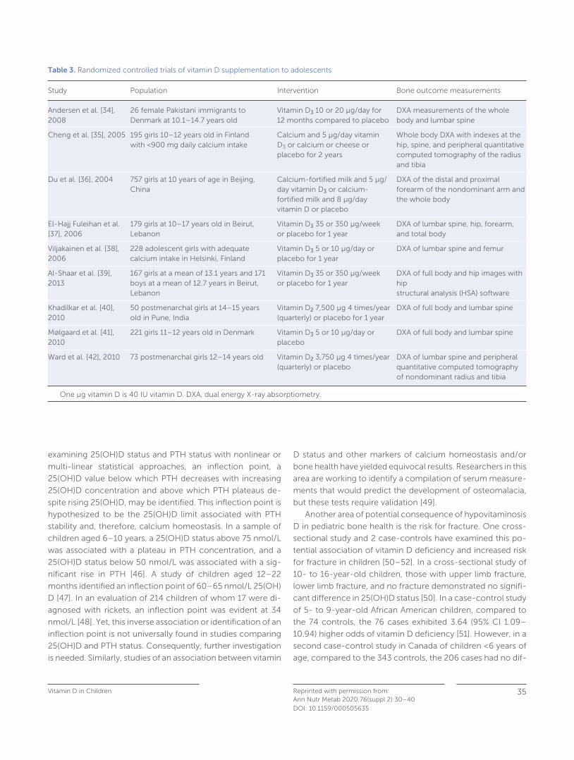
Vitamin D in Children 35Reprinted with permission from:Ann Nutr Metab 2020;76(suppl 2):30–40DOI: 10.1159/000505635
examining 25(OH)D status and PTH status with nonlinear or
multi-linear statistical approaches, an inflection point, a
25(OH)D value below which PTH decreases with increasing
25(OH)D concentration and above which PTH plateaus de-
spite rising 25(OH)D, may be identified. This inflection point is
hypothesized to be the 25(OH)D limit associated with PTH
stability and, therefore, calcium homeostasis. In a sample of
children aged 6–10 years, a 25(OH)D status above 75 nmol/L
was associated with a plateau in PTH concentration, and a
25(OH)D status below 50 nmol/L was associated with a sig-
nificant rise in PTH [46] . A study of children aged 12–22
months identified an inflection point of 60–65 nmol/L 25(OH)
D [47] . In an evaluation of 214 children of whom 17 were di-
agnosed with rickets, an inflection point was evident at 34
nmol/L [48] . Yet, this inverse association or identification of an
inflection point is not universally found in studies comparing
25(OH)D and PTH status. Consequently, further investigation
is needed. Similarly, studies of an association between vitamin
D status and other markers of calcium homeostasis and/or
bone health have yielded equivocal results. Researchers in this
area are working to identify a compilation of serum measure-
ments that would predict the development of osteomalacia,
but these tests require validation [49] .
Another area of potential consequence of hypovitaminosis
D in pediatric bone health is the risk for fracture. One cross-
sectional study and 2 case-controls have examined this po-
tential association of vitamin D deficiency and increased risk
for fracture in children [50–52] . In a cross-sectional study of
10- to 16-year-old children, those with upper limb fracture,
lower limb fracture, and no fracture demonstrated no signifi-
cant difference in 25(OH)D status [50] . In a case-control study
of 5- to 9-year-old African American children, compared to
the 74 controls, the 76 cases exhibited 3.64 (95% CI 1.09–
10.94) higher odds of vitamin D deficiency [51] . However, in a
second case-control study in Canada of children < 6 years of
age, compared to the 343 controls, the 206 cases had no dif-
Table 3. Randomized controlled trials of vitamin D supplementation to adolescents
Study Population Intervention Bone outcome measurements
Andersen et al. [34], 2008
26 female Pakistani immigrants to Denmark at 10.1–14.7 years old
Vitamin D3 10 or 20 μg/day for 12 months compared to placebo
DXA measurements of the whole body and lumbar spine
Cheng et al. [35], 2005 195 girls 10–12 years old in Finland with <900 mg daily calcium intake
Calcium and 5 μg/day vitamin D3 or calcium or cheese or placebo for 2 years
Whole body DXA with indexes at the hip, spine, and peripheral quantitative computed tomography of the radius and tibia
Du et al. [36], 2004 757 girls at 10 years of age in Beijing, China
Calcium-fortified milk and 5 μg/day vitamin D3 or calcium-fortified milk and 8 μg/day vitamin D or placebo
DXA of the distal and proximal forearm of the nondominant arm and the whole body
El-Hajj Fuleihan et al. [37], 2006
179 girls at 10–17 years old in Beirut, Lebanon
Vitamin D3 35 or 350 μg/week or placebo for 1 year
DXA of lumbar spine, hip, forearm, and total body
Viljakainen et al. [38], 2006
228 adolescent girls with adequate calcium intake in Helsinki, Finland
Vitamin D3 5 or 10 μg/day or placebo for 1 year
DXA of lumbar spine and femur
Al-Shaar et al. [39], 2013
167 girls at a mean of 13.1 years and 171 boys at a mean of 12.7 years in Beirut, Lebanon
Vitamin D3 35 or 350 μg/week or placebo for 1 year
DXA of full body and hip images with hipstructural analysis (HSA) software
Khadilkar et al. [40], 2010
50 postmenarchal girls at 14–15 years old in Pune, India
Vitamin D2 7,500 μg 4 times/year (quarterly) or placebo for 1 year
DXA of full body and lumbar spine
Mølgaard et al. [41], 2010
221 girls 11–12 years old in Denmark Vitamin D3 5 or 10 μg/day or placebo
DXA of full body and lumbar spine
Ward et al. [42], 2010 73 postmenarchal girls 12–14 years old Vitamin D2 3,750 μg 4 times/year (quarterly) or placebo
DXA of lumbar spine and peripheral quantitative computed tomography of nondominant radius and tibia
One μg vitamin D is 40 IU vitamin D. DXA, dual energy X-ray absorptiometry.
Taylor Reprinted with permission from:Ann Nutr Metab 2020;76(suppl 2):30–40
36
DOI: 10.1159/000505635
ferences in vitamin D status or intake of cow’s milk. Yet, use
of vitamin D supplementation was associated with decreased
odds of fracture (adjusted odds ratio of 0.42 [95% CI 0.25–
0.69]) [52] .
Another approach to evaluate the relationship between vi-
tamin D deficiency and fractures was published recently. In
this study of children under 2 years of age who were admitted
with fractures, 11 of 79 demonstrated hypomineralization on
skeletal survey. For every 10-point increase in vitamin D sta-
tus, the adjusted odds of hypomineralization were reduced
0.3 (95% CI 0.17–0.82) [53] . This limited data of the associa-
tion of vitamin D, osteomalacia, and fracture risk requires fur-
ther exploration especially for populations at higher risk for
vitamin D deficiency.
Expedient exploration of this relationship is crucial due to
considerable debate in the literature as to whether fractures
due to osteomalacia/rickets can be differentiated from frac-
tures due to nonaccidental trauma by X-ray or laboratory
markers [54–57] . Currently, published literature points to the
consideration of bone biopsy as a method to detect hypomin-
eralization disease versus child abuse. Further investigation to
reliably differentiate the cause of fractures will provide further
knowledge regarding osteomalacia and how this disease af-
fects bone strength and development.
Vitamin D in Non-Bone-Related Disease
As in the adult population, in children, vitamin D has actions
in health beyond calcium homeostasis and bone develop-
ment. Greater detail regarding non-bone-related effects of
vitamin D is provided in a separate article in this issue, but
these potential effects are important to mention as they do
affect recommendations for vitamin D supplementation. The
role of vitamin D in both protection from development of type
I diabetes mellitus and in improved glucose tolerance has
been described with meta-analyses of vitamin D supplemen-
tation of observational trials [58] . Further randomized, con-
trolled trials and investigation as to whether vitamin D im-
proves β-cell function directly or through its beneficial effect
on immune function are warranted. Children with allergic dis-
ease, especially asthma and atopic dermatitis, experience de-
creased exacerbations with vitamin D supplementation [31,
59, 60] . Infectious diseases with high incidence in childhood,
such as otitis media, urinary tract infection, pneumonia, influ-
enza, and other acute respiratory infections, all have a number
of investigations demonstrating decreased incidence with
higher vitamin D status [12] . Several randomized, controlled
trials of vitamin D supplementation to prevent infections have
been performed with equivocal results [12, 61, 62] . Therefore,
these roles of vitamin D in health promotion warrant consid-
eration but require further study before definitive supplemen-
tation recommendations.
Recommendations for Vitamin D Status
Several international and national guidelines for categoriza-
tion of vitamin D status are presented in Table 1 . Of note, only
2 recommendations are specifically for children. Circulating
25(OH)D is the best indicator of nutritional vitamin D status
due to its half-life of 2–3 weeks and to its mechanism of ac-
tion in numerous organ systems. Nonetheless, 25(OH)D sta-
tus does not consistently increase as expected with vitamin D
dosing, which has raised the question as to what further con-
siderations should be taken in the identification of healthy or
unhealthy vitamin D status. For example, antibody-based
methods of 25(OH)D that are readily available for use in vari-
ous settings demonstrate less reproducibility when compared
to liquid chromatography-mass spectroscopy (LCMS). On the
other hand, LCMS is not universally available, and excellent
antibody-based methods do exist [63] . Other issues in the
analysis of 25(OH)D status include the potential that free
25(OH)D is a more important measurement than total 25(OH)
D due to population variation in vitamin D-binding protein af-
finities. Lastly, individual variation in response to vitamin D
supplementation may be due to genetic differences in the vi-
tamin D receptor [64] . Until these issues are elucidated, the
interpretation of vitamin D study results remains with some
uncertainty. In fact, the European Calcified Tissue Society has
called for standardization of testing for all research of vitamin
D status to improve consistency [11] . In the clinical setting, no
guidelines are available to recommend specific vitamin D sta-
tus screening in routine pediatric care partly because of this
ambiguity in 25(OH)D test results [7, 65, 66] . Therefore, em-
phasis should be placed on providing the vitamin D supple-
mentation to optimize health and to avoid toxicity for all chil-
dren without the need for individual screening.
Children with allergic disease,
especially asthma and atopic
dermatitis, experience
decreased exacerbations with
vitamin D supplementation
Vitamin D in Children 37Reprinted with permission from:Ann Nutr Metab 2020;76(suppl 2):30–40DOI: 10.1159/000505635
Recommended Vitamin D Intake for Toddlers, Children, and Adolescents
In 2008, in response to the growing evidence of vitamin D
deficiency in children, the AAP recommended at least 10 μg/
day vitamin D for all children [9] . In 2010, the IOM recom-
mended at least 15 μg/day for children over 1 year of age [7] .
In Europe, ESPGHAN recommended 10 μg/day for infants
but chose not to provide recommendations for older chil-
dren. Instead, they endorsed the European Food Safety Au-
thority’s upper limit of recommended intake of 25 μg/day
for infants, 50 μg/day for children 1–10 years, and 100 μg/
day for children 11–17 years [8] . Recently, new recommen-
dations in Europe, and specifically in Germany, have contin-
ued to emphasize the need for vitamin D supplementation
to infants, have added recommendations for pregnant
women, but have decreased the recommendations for old-
er children and adolescents due to the equivocal results of
randomized, controlled trials of vitamin D supplementation
and due to the difficulties in interpretation of vitamin D sta-
tus [11, 31] . Of note, the higher risk for vitamin D deficiency
in non-Western or immigrant populations of Europe re-
mains a concern with recommendation to consider 10 μg/
day supplementation [11] .
Recommendation for supplementation to children aged
1–3 years is quite varied in these new recommendations.
These children, especially if not receiving fortified food prod-
ucts, remain at risk for inadequate bone mineralization. They
also are a population found to have persistently low vitamin D
intake despite existing recommendations. In France, in chil-
dren who received vitamin D from food sources and supple-
mentation, 10% of infants 30–35 months of age still received
less than the recommended intake [67] . In the USA, despite
attention to vitamin D supplementation in the last 2 decades,
a greater number of children 0–47.9 months of age received
less than the recommended supplementation in 2016 com-
pared to 2002 [68] . In the United Kingdom, just over half of
parents reported receiving information regarding vitamin D
supplementation for their infant and 80% described that they
found the information lacking adequate details [69] . There-
fore, for toddlers and children, not only are the recommenda-
tions varied, but so are vitamin D intake and parental knowl-
edge regarding child vitamin D needs.
When contemplating the appropriate intake of vitamin D,
the potential for toxicity must be considered. Though the
25(OH)D concentration at which harm occurs is likely well
above the therapeutic range, intoxication due to misadminis-
tration or accidental ingestion is reported to occur with doses
of 6,000–112,500 μg and result in severe hypercalcemia. The
literature contains one case report of vitamin D intoxication
in a 7-month-old receiving 35–40 μg/day [70] . Therefore,
overdose is a rare but potential risk. Parental education re-
garding safe administration is required.
Conclusion
As the amount of research investigating the vitamin D needs of
toddlers, children, and adolescents has grown, unfortunately, the
answer has become less clear. Vitamin D-deficient rickets is a dis-
ease with severe morbidity that responds well to vitamin D reple-
tion. This disease is most common in infants but can be observed
in children especially in resource-limited countries. Osteomala-
cia, or bone hypomineralization not of the magnitude of rickets,
is more difficult to diagnose and, therefore, study of its response
to vitamin D supplementation is challenging. Observational stud-
ies in pediatrics point to at least 10 μg/day vitamin D supplemen-
tation to achieve optimal bone health, but results of randomized,
controlled trials have been ambiguous. Vitamin D has been found
to play a significant role in immune function and especially in au-
toimmune, infectious, and allergic disease, but again trials of vita-
min D supplementation have been equivocal. Due to these study
results and other issues, national and international guidelines are
being modified to reflect this uncertainty and provide less directive
regarding vitamin D supplementation after infancy. Attention to
standard 25(OH)D concentration measurement and investigation
of genetic or other individual variations in vitamin D metabolism
hopefully will identify the cause of these discrepancies in research
results. Until then, with the potential benefits and low risk of vita-
min D supplementation of 10–50 μg/day for children, some phy-
sicians and public health leaders may elect to recommend these
doses until further information is known.
Disclosure Statement
The writing of this article was supported by Nestlé Nutrition Institute and the author declares no other conflict of interest.
Funding Sources
Dr. Sarah N. Taylor received National Institute of Health funding for research in vitamin D 2007–2012 and has served as a consultant for Alcresta Therapeutics. She has no funding source related to this man-uscript.
Author Contributions
Dr. Sarah N. Taylor performed the literature review and wrote all as-pects of this manuscript.
Taylor Reprinted with permission from:Ann Nutr Metab 2020;76(suppl 2):30–40
38
DOI: 10.1159/000505635
References
1 Institute of Medicine (US) Standing Committee on the Scientific Evaluation of Dietary Reference Intakes. Dietary Reference Intakes for Calcium, Phosphorus, Magnesium, Vitamin D, and Fluoride. Washington (DC): National Academies Press (US); 1997. Available from: https://www.ncbi.nlm.nih.gov/books/NBK109825/
2 Gartner LM, Greer FR; Section on Breastfeeding and Committee on Nutrition. American Academy of Pediatrics. Prevention of rick-ets and vitamin D deficiency: new guidelines for vitamin D intake. Pediatrics . 2003 Apr; 111(4 Pt 1): 908–10.
3 Haddad JG, Chyu KJ. Competitive protein-binding radioassay for 25-hydroxycholecalciferol. J Clin Endocrinol Metab . 1971 Dec; 33(6): 992–5.
4 Heaney RP, Dowell MS, Hale CA, Bendich A. Calcium absorption varies within the reference range for serum 25-hydroxyvitamin D. J Am Coll Nutr . 2003 Apr; 22(2): 142–6.
5 Vieth R, Ladak Y, Walfish PG. Age-related changes in the 25-hy-droxyvitamin D versus parathyroid hormone relationship suggest a different reason why older adults require more vitamin D. J Clin Endocrinol Metab . 2003 Jan; 88(1): 185–91.
6 Bischoff-Ferrari HA, Dietrich T, Orav EJ, Dawson-Hughes B. Pos-itive association between 25-hydroxy vitamin D levels and bone mineral density: a population-based study of younger and older adults. Am J Med . 2004 May; 116(9): 634–9.
7 Dietary Reference Intakes for Calcium and Vitamin D. Washington (DC): National Academy of Sciences; 2011.
8 Braegger C, Campoy C, Colomb V, Decsi T, Domellof M, Fewtrell M, et al.; ESPGHAN Committee on Nutrition. Vitamin D in the healthy European paediatric population. J Pediatr Gastroenterol Nutr . 2013 Jun; 56(6): 692–701.
9 Wagner CL, Greer FR; American Academy of Pediatrics Section on Breastfeeding; American Academy of Pediatrics Committee on Nutrition. Prevention of rickets and vitamin D deficiency in infants, children, and adolescents. Pediatrics . 2008 Nov; 122(5): 1142–52.
10 Holick MF, Binkley NC, Bischoff-Ferrari HA, Gordon CM, Hanley DA, Heaney RP, et al.; Endocrine Society. Evaluation, treatment, and prevention of vitamin D deficiency: an Endocrine Society clin-ical practice guideline. J Clin Endocrinol Metab . 2011 Jul; 96(7): 1911–30.
11 Lips P, Cashman KD, Lamberg-Allardt C, Bischoff-Ferrari HA, Obermayer-Pietsch B, Bianchi ML, et al. Current vitamin D status in European and Middle East countries and strategies to prevent vitamin D deficiency; a position statement of the European Calci-fied Tissue Society. Eur J Endocrinol . 2019 Apr; 180(4):P23–54.
12 Marino R, Misra M. Extra-skeletal effects of vitamin D. Nutrients . 2019 Jun; 11(7):E1460.
13 Gordon CM, DePeter KC, Feldman HA, Grace E, Emans SJ. Preva-lence of vitamin D deficiency among healthy adolescents. Arch Pediatr Adolesc Med . 2004 Jun; 158(6): 531–7.
14 Marwaha RK, Tandon N, Reddy DR, Aggarwal R, Singh R, Sawhney RC, et al. Vitamin D and bone mineral density status of healthy schoolchildren in northern India. Am J Clin Nutr . 2005 Aug; 82(2): 477–82.
15 Kumar J, Muntner P, Kaskel FJ, Hailpern SM, Melamed ML. Preva-lence and associations of 25-hydroxyvitamin D deficiency in US children: NHANES 2001-2004. Pediatrics . 2009 Sep; 124(3):e362–70.
16 Rabenberg M, Scheidt-Nave C, Busch MA, Thamm M, Rieckmann N, Durazo-Arvizu RA, et al. Implications of standardization of se-rum 25-hydroxyvitamin D data for the evaluation of vitamin D sta-tus in Germany, including a temporal analysis. BMC Public Health . 2018 Jul; 18(1): 845.
17 Foo LH, Zhang Q, Zhu K, Ma G, Trube A, Greenfield H, et al. Re-lationship between vitamin D status, body composition and phys-ical exercise of adolescent girls in Beijing. Osteoporos Int . 2009 Mar; 20(3): 417–25.
18 Public Health England, Food Standards Agency. National Diet and Nutrition Survey: Results from Years 1, 2, 3 and 4 (combined) of the Rolling Programme (2008/2009–2011/2012). Secondary Na-tional Diet and Nutrition Survey Results from Years 1, 2, 3 and 4 (combined) of the Rolling Programme (2008/2009–2011/2012). London: Public Health England; 2014.
19 Maguire JL, Birken CS, Khovratovich M, Degroot J, Carsley S, Thorpe KE, et al.; TARGet Kids! Collaboration. Modifiable determi-nants of serum 25-hydroxyvitamin D status in early childhood: opportunities for prevention. JAMA Pediatr . 2013 Mar; 167(3): 230–5.
20 Garg MK, Tandon N, Marwaha RK, Menon AS, Mahalle N. The re-lationship between serum 25-hydroxy vitamin D, parathormone and bone mineral density in Indian population. Clin Endocrinol (Oxf) . 2014 Jan; 80(1): 41–6.
21 Angurana SK, Angurana RS, Mahajan G, Kumar N, Mahajan V. Prev-alence of vitamin D deficiency in apparently healthy children in north India. J Pediatr Endocrinol Metab . 2014 Nov; 27(11-12): 1151–6.
22 Cairncross CT, Stonehouse W, Conlon CA, Grant CC, McDonald B, Houghton LA, et al. Predictors of vitamin D status in New Zea-land preschool children. Matern Child Nutr . 2017 Jul; 13(3):e12340.
23 Lane G, Nisbet C, Whiting SJ, Vatanparast H. Canadian newcomer children’s bone health and vitamin D status. Appl Physiol Nutr Metab . 2019 Jul; 44(7): 796–803.
24 Omand JA, Darling PB, Parkin PC, Birken CS, Khovratovich M, Thorpe KE, et al.; TARGet Kids! Collaboration. Non-Western im-migrant children have lower 25-hydroxyvitamin D than children from Western families. Public Health Nutr . 2014 Jul; 17(7): 1547–54.
25 Ferguson JH, Chang AB. Vitamin D supplementation for cystic fi-brosis. Cochrane Database Syst Rev . 2014 May;(5):CD007298.
26 Viraraghavan VR, Seth A, Aneja S, Singh R, Dhanwal D. Effect of high dose vitamin d supplementation on vitamin d nutrition status of pre-pubertal children on anti-epileptic drugs – A randomized controlled trial. Clin Nutr ESPEN . 2019 Feb; 29: 36–40.
27 Zakharova I, Klimov L, Kuryaninova V, Nikitina I, Malyavskaya S, Dolbnya S, et al. Vitamin D Insufficiency in Overweight and Obese Children and Adolescents. Front Endocrinol (Lausanne) . 2019 Mar; 10: 103.
Vitamin D in Children 39Reprinted with permission from:Ann Nutr Metab 2020;76(suppl 2):30–40DOI: 10.1159/000505635
28 Sharif Y, Sadeghi O, Dorosty A, Siassi F, Jalali M, Djazayery A, et al. Serum Levels of Vitamin D, Retinol and Zinc in Relation to over-weight among Toddlers: Findings from a National Study in Iran. Arch Iran Med . 2019 Apr; 22(4): 174–81.
29 Lewis RD, Laing EM. Conflicting reports on vitamin D supplemen-tation: evidence from randomized controlled trials. Mol Cell En-docrinol . 2015 Jul; 410: 11–8.
30 Moon RJ, Davies JH, Cooper C, Harvey NC. Vitamin D, and Mater-nal and Child Health. Calcif Tissue Int . 2020 Jan; 106(1): 30–46.
31 Reinehr T, Schnabel D, Wabitsch M, Bechtold-Dalla Pozza S, Büh-rer C, Heidtmann B, et al. Vitamin D supplementation after the second year of life: joint position of the Committee on Nutrition, German Society for Pediatric and Adolescent Medicine (DGKJ e.V.), and the German Society for Pediatric Endocrinology and Di-abetology (DGKED e.V.). Mol Cell Pediatr . 2019 May; 6(1): 3.
32 Hill TR, Cotter AA, Mitchell S, Boreham CA, Dubitzky W, Murray L, et al. Vitamin D status and its determinants in adolescents from the Northern Ireland Young Hearts 2000 cohort. Br J Nutr . 2008 May; 99(5): 1061–7.
33 Bailey DA, Martin AD, McKay HA, Whiting S, Mirwald R. Calcium accretion in girls and boys during puberty: a longitudinal analysis. J Bone Miner Res . 2000 Nov; 15(11): 2245–50.
34 Andersen R, Mølgaard C, Skovgaard LT, Brot C, Cashman KD, Ja-kobsen J, et al. Effect of vitamin D supplementation on bone and vitamin D status among Pakistani immigrants in Denmark: a ran-domised double-blinded placebo-controlled intervention study. Br J Nutr . 2008 Jul; 100(1): 197–207.
35 Cheng S, Lyytikäinen A, Kröger H, Lamberg-Allardt C, Alén M, Koistinen A, et al. Effects of calcium, dairy product, and vitamin D supplementation on bone mass accrual and body composition in 10-12-y-old girls: a 2-y randomized trial. Am J Clin Nutr . 2005 Nov; 82(5): 1115–26.
36 Du X, Zhu K, Trube A, Zhang Q, Ma G, Hu X, et al. School-milk in-tervention trial enhances growth and bone mineral accretion in Chinese girls aged 10-12 years in Beijing. Br J Nutr . 2004 Jul; 92(1): 159–68.
37 El-Hajj Fuleihan G, Nabulsi M, Tamim H, Maalouf J, Salamoun M, Khalife H, et al. Effect of vitamin D replacement on musculoskel-etal parameters in school children: a randomized controlled trial. J Clin Endocrinol Metab . 2006 Feb; 91(2): 405–12.
38 Viljakainen HT, Natri AM, Kärkkäinen M, Huttunen MM, Palssa A, Jakobsen J, et al. A positive dose-response effect of vitamin D supplementation on site-specific bone mineral augmentation in adolescent girls: a double-blinded randomized placebo-con-trolled 1-year intervention. J Bone Miner Res . 2006 Jun; 21(6): 836–44.
39 Al-Shaar L, Nabulsi M, Maalouf J, El-Rassi R, Vieth R, Beck TJ, et al. Effect of vitamin D replacement on hip structural geometry in adolescents: a randomized controlled trial. Bone . 2013 Oct; 56(2): 296–303.
40 Khadilkar AV, Sayyad MG, Sanwalka NJ, Bhandari DR, Naik S, Khad-ilkar VV, et al. Vitamin D supplementation and bone mass accrual in underprivileged adolescent Indian girls. Asia Pac J Clin Nutr . 2010; 19(4): 465–72.
41 Mølgaard C, Larnkjaer A, Cashman KD, Lamberg-Allardt C, Ja-kobsen J, Michaelsen KF. Does vitamin D supplementation of healthy Danish Caucasian girls affect bone turnover and bone mineralization? Bone . 2010 Feb; 46(2): 432–9.
42 Ward KA, Das G, Roberts SA, Berry JL, Adams JE, Rawer R, et al. A randomized, controlled trial of vitamin D supplementation upon musculoskeletal health in postmenarchal females. J Clin Endocri-nol Metab . 2010 Oct; 95(10): 4643–51.
43 Winzenberg TM, Powell S, Shaw KA, Jones G. Vitamin D supple-mentation for improving bone mineral density in children. Co-chrane Database Syst Rev . 2010 Oct;(10):CD006944.
44 Winzenberg T, Powell S, Shaw KA, Jones G. Effects of vitamin D supplementation on bone density in healthy children: systematic review and meta-analysis. BMJ . 2011 Jan; 342:c7254.
45 Joo NS, Dawson-Hughes B, Yeum KJ. 25-Hydroxyvitamin D, cal-cium intake, and bone mineral content in adolescents and young adults: analysis of the fourth and fifth Korea National Health and Nutrition Examination Survey (KNHANES IV-2, 3, 2008-2009 and V-1, 2010). J Clin Endocrinol Metab . 2013 Sep; 98(9): 3627–36.
46 Rajakumar K, Fernstrom JD, Janosky JE, Greenspan SL. Vitamin D insufficiency in preadolescent African-American children. Clin Pe-diatr (Phila) . 2005 Oct; 44(8): 683–92.
47 Houghton LA, Szymlek-Gay EA, Gray AR, Ferguson EL, Deng X, Heath AL. Predictors of vitamin D status and its association with parathyroid hormone in young New Zealand children. Am J Clin Nutr . 2010 Jul; 92(1): 69–76.
48 Atapattu N, Shaw N, Högler W. Relationship between serum 25-hydroxyvitamin D and parathyroid hormone in the search for a biochemical definition of vitamin D deficiency in children. Pedi-atr Res . 2013 Nov; 74(5): 552–6.
49 Uday S, Högler W. Spot the silent sufferers: A call for clinical diag-nostic criteria for solar and nutritional osteomalacia. J Steroid Bio-chem Mol Biol . 2019 Apr; 188: 141–6.
50 Ceroni D, Anderson de la Llana R, Martin X, Lamah L, De Coulon G, Turcot K, et al. Prevalence of vitamin D insufficiency in Swiss teenagers with appendicular fractures: a prospective study of 100 cases. J Child Orthop . 2012 Dec; 6(6): 497–503.
51 Ryan LM, Teach SJ, Singer SA, Wood R, Freishtat R, Wright JL, et al. Bone mineral density and vitamin D status among African American children with forearm fractures. Pediatrics . 2012 Sep; 130(3):e553–60.
52 Anderson LN, Heong SW, Chen Y, Thorpe KE, Adeli K, Howard A, et al.; TARGet Kids Collaboration. Vitamin D and Fracture Risk in Early Childhood: A Case-Control Study. Am J Epidemiol . 2017 Jun; 185(12): 1255–62.
53 Servaes S, States L, Wood J, Schilling S, Christian CW. Rachitic change and vitamin D status in young children with fractures. Skeletal Radiol . 2020 Jan; 49(1): 85–91.
54 Chapman T, Sugar N, Done S, Marasigan J, Wambold N, Feldman K. Fractures in infants and toddlers with rickets. Pediatr Radiol . 2010 Jul; 40(7): 1184–9.
55 Kepron C, Pollanen MS. Rickets or abuse? A histologic comparison of rickets and child abuse-related fractures. Forensic Sci Med Pathol . 2015 Mar; 11(1): 78–87.
Taylor Reprinted with permission from:Ann Nutr Metab 2020;76(suppl 2):30–40
40
DOI: 10.1159/000505635
56 Cannell JJ, Holick MF. Multiple unexplained fractures in infants and child physical abuse. J Steroid Biochem Mol Biol . 2018 Jan; 175: 18–22.
57 Miller M, Stolfi A, Ayoub D. Findings of metabolic bone disease in infants with unexplained fractures in contested child abuse inves-tigations: a case series of 75 infants. J Pediatr Endocrinol Metab . 2019 Oct; 32(10): 1103–20.
58 Zipitis CS, Akobeng AK. Vitamin D supplementation in early child-hood and risk of type 1 diabetes: a systematic review and meta-analysis. Arch Dis Child . 2008 Jun; 93(6): 512–7.
59 Jolliffe DA, Greenberg L, Hooper RL, Griffiths CJ, Camargo CA Jr, Kerley CP, et al. Vitamin D supplementation to prevent asthma exacerbations: a systematic review and meta-analysis of individ-ual participant data. Lancet Respir Med . 2017 Nov; 5(11): 881–90.
60 Hattangdi-Haridas SR, Lanham-New SA, Wong WH, Ho MH, Dar-ling AL. Vitamin D Deficiency and Effects of Vitamin D Supplemen-tation on Disease Severity in Patients with Atopic Dermatitis: A Systematic Review and Meta-Analysis in Adults and Children. Nu-trients . 2019 Aug; 11(8):E1854.
61 Martineau AR, Jolliffe DA, Greenberg L, Aloia JF, Bergman P, Dub-nov-Raz G, et al. Vitamin D supplementation to prevent acute re-spiratory infections: individual participant data meta-analysis. Health Technol Assess . 2019 Jan; 23(2): 1–44.
62 Singh N, Kamble D, Mahantshetti NS. Effect of Vitamin D Supple-mentation in the Prevention of Recurrent Pneumonia in Under-Five Children. Indian J Pediatr . 2019 Dec; 86(12): 1105–11.
63 de la Hunty A, Wallace AM, Gibson S, Viljakainen H, Lamberg-Al-lardt C, Ashwell M. UK Food Standards Agency Workshop Consen-sus Report: the choice of method for measuring 25-hydroxyvita-min D to estimate vitamin D status for the UK National Diet and Nutrition Survey. Br J Nutr . 2010 Aug; 104(4): 612–9.
64 Newton DA, Baatz JE, Kindy MS, Gattoni-Celli S, Shary JR, Hollis BW, et al. Vitamin D binding protein polymorphisms significantly impact vitamin D status in children. Pediatr Res . 2019; 86(5): 662–9.
65 Davis RL, Aksornsri A, Papachrisanthou MM, Vitamin D. Screening Variations in Children and Adolescents: who should be Screened? J Pediatr Nurs . 2019 Mar-Apr; 45: 57–61.
66 LeFevre ML; U.S. Preventive Services Task Force. Screening for vi-tamin D deficiency in adults: U.S. Preventive Services Task Force recommendation statement. Ann Intern Med . 2015 Jan; 162(2): 133–40.
67 Chouraqui JP, Tavoularis G, Turck D, Ferry C, Feillet F. Mineral and vitamin intake of infants and young children: the Nutri-Bébé 2013 survey. Eur J Nutr . 2019, Epub ahead of print.
68 Eldridge AL, Catellier DJ, Hampton JC, Dwyer JT, Bailey RL. Trends in Mean Nutrient Intakes of US Infants, Toddlers, and Young Chil-dren from 3 Feeding Infants and Toddlers Studies (FITS). J Nutr . 2019 Jul; 149(7): 1230–7.
69 Day RE, Krishnarao R, Sahota P, Christian MS. We still don’t know that our children need vitamin D daily: a study of parents’ under-standing of vitamin D requirements in children aged 0-2 years. BMC Public Health . 2019 Aug; 19(1): 1119.
70 Waheed N, Cheema HA, Saeed A, Alvi MA, Fayyaz Z, Anjum N, et al. Vitamin D Intoxication In 7-Month-Old Infant With Recom-mended Daily Intake Of Vitamin D. J Ayub Med Coll Abbottabad . 2018 Oct-Dec; 30(Suppl 1)(4):S673–5.