Embed Size (px)
Citation preview
Université de Liège Collège de doctorat de Biochimie, Biologie moléculaire et cellulaire,
Bioinformatique et modélisation
Perturbations of interactome networks in acute lymphoblastic leukemia: identification of EXT-1 tumor suppressor as a Notch pathway regulator
Sarah Daakour
A thesis submitted for the degree of Doctor of Philosophy in Science
2016
Supervisor: Prof. Jean-Claude Twizere
Co-supervisor: Prof. Jacques Piette


EXAMINATIONCOMMITTEEProf.FranckDEQUIEDT;UniversitédeLiège(CommitteeChair)
Prof.Jean-ClaudeTWIZERE;UniversitédeLiège(Supervisor)
Prof.JacquesPIETTE;UniversitédeLiège(Co-supervisor)
Dr.IsabelleMANFROID;UniversitédeLiège
Prof.KristelVANSTEEN;UniversitédeLiège
Prof.RenéREZSOHAZY;UniversitéCatholiquedeLouvain
Dr.NicolasSIMONIS;InstitutdePathologieetdeGénétique,Gosselies
Prof.KouroshSALEHI-ASHTIANI;NewYorkUniversityAbuDhabi
SUMMARY(ENGLISH)
Wholegenomesequencingtechnologieshaveenabledtheidentificationofmutations
implicated in diseases including cancer. Recently, research efforts to compare and
categorize mutations, genes expression and genomic characteristics helped
generating literature-curation databases. A large number of databases were
developedtoaddressdataintegrationandstandardizationforhumancancers,suchas
Catalogue Of Somatic Mutations In Cancer (COSMIC), The Cancer Genome Atlas
(TCGA), International Cancer Genome Consortium, Integrative Onco Genomics
(IntOGen).Althoughtheidentificationofthesemutationshighlights“cancercausative
genes”,itdoesnotgiveadetailedexplanationofmolecularmechanismsleadingtothe
development of cancer. Though, understanding mechanisms leading to cancer
development and progression remains a challenge that requires further
investigations.
Thegreatmajorityofmutatedgenesarefoundinliquidtumorssuchasleukemiaand
lymphomas. In the first part of this study, we reasoned that leukemia associated
genes could be extended to additional candidates identified using interactomic
approaches.Weusedprotein-proteininteraction(PPI)mappingstrategiestoexplore
information on cancer genes frequently mutated in Acute lymphoblastic leukemia
(ALL).We first extractedmutational data associated to ALL, and used interactome
mapping analysis for literature-curated interactions and yeast two-hybrid
experimental data in order to identify potential novel target genes associatedwith
ALL. We highlighted mutated hub proteins interconnected in an ALL-cancer gene
productsnetworkand identifiednovel interactingpartnersthat linkkeyALL-cancer
drivergeneproducts.WeidentifiedEXT1tumorsuppressorgeneasanovelcommon
interactorforNOTCH1andFBXW7.
Inthesecondpartofthisstudy,weexperimentallyvalidatedEXT1,asanovelplayer
intheregulationoftheNotchpathway.
Our study thus provides a proof-of-concept on how systematic interactome
approaches could allow identification of novel targeted genes and pathways
associatedtohumancancer.
RÉSUMÉ(FRANÇAIS)
Les technologies de séquençage de génomes entiers ont permis l'identification de
mutationsimpliquéesdansdesmaladiescommelecancer.Récemment,deseffortsde
recherchedanslebutdecompareretdeclasserdesmutations,l'expressiondesgènes
etlescaractéristiquesgénomiquesontaidéàconstruiredesrépertoiresdedonnéesse
basantsurlalittérature.Ungrandnombredebasesdedonnéesontétédéveloppées
afind'intégrerdesdonnéessurlescancershumains,tellesqueCOSMIC;lecatalogue
des mutations somatiques dans le cancer, “The Cancer Genome Atlas” (TCGA),
International Cancer Genome Consortium, Integrative Genomics Onco (IntOGen).
L'identificationdecesmutationspermetuneclassificationdesgènesresponsablesdes
cancers,parcontreellenepermetpasunecompréhensiondétailléedesmécanismes
moléculaires conduisant au développement du cancer. Ainsi, la comprehension des
mécanismesmenantaudéveloppementducanceretsaprogressionresteundéfiqui
nécessitedesinvestigationscomplémentaires.
La majorité des gènes mutés se trouvent dans les tumeurs liquides tels que la
leucémieetleslymphomes.Danslapremièrepartiedecetteetude,nousavonssoumis
l’hypothèsequel’ensembledesgènesassociésàlaleucémiepourraitêtreétendupour
inclured’autrecandidats identifiésenutilisantuneapproches interactomique.Nous
avons utilisé des stratégies de cartographie des réseaux d'interaction proteines-
protéines(IPP)afind’explorerdesinformationssurlesgènesducancerfréquemment
mutésdanslaleucémielymphoblastiqueaiguë(LLA).Nousavonsextraitlesdonnées
de mutations associées à la LLA, par la suite nous avons analysé les réseaux des
interactions extraites de la littérature ainsi que des données expérimentales
provenant du double hybride en levure, ceci afin d'identifier de nouveaux gènes
potentiellement associés à la LLA. Nous avons souligné des “hubs” de protéines
mutées et interconnectées dans le réseau de produits des gènes de cancer liés à la
LLA, et de nouveaux partenaires d'interaction qui relient les produits des gènes
associésàlaLLA.NousavonsidentifiéEXT1,ungènesuppresseurdetumeur,comme
étantunnouvelinteractantcommunpourNOTCH1etFBXW7.
Dans ladeuxièmepartiedecetteétude,nousavonsvalidéEXT1,entantquenouvel
acteurdanslarégulationdelavoieNotch.

Notre etude fournit ainsi une preuve de concept démontrant que l’approche
interactomiquepourraitpermettrel’identificationdenouveauxgènesetdesvoiesde
signalisationassociésauxcancers.
Copyright.Auxtermesdelaloibelgedu30juin1994,surledroitd’auteuretlesdroitsvoisins,seul l’auteurà ledroitdereproduirepartiellementoucomplètementcetouvragedequelque façon et forme que ce soit ou d’en autoriser la reproduction partielle oucomplètedequelquemanièreetsousquelqueformequecesoit.Toutephotocopieoureproduction sous autre forme est donc faite en violation de la dite loi et de sesmodificationultérieures.

Acknowledgements
It has been six years since I entered the “research world” as
a PhD student, I knew from the beginning that to survive
and succeed this “experiment” I needed motivation, hard
work and ambition but most important of all, none of this
would have been possible if it weren’t for the support and
the help of a number of people without whom this thesis
might not have been written, and to whom I am greatly
indebted.
I would like to thank the “Télévie” and the fund Léon
Frédéricq for providing the financial support that allowed
the realization of this project. I would also like to thank
the members of my jury for their time and consideration
throughout the phase of the review process.
I would like to express my sincere gratitude to Jean-Claude
for granting me the opportunity to be part of his team and
introducing me to the world of interatomics. Thank you for
believing in me from the beginning and giving me the
chance to go further in this project, for providing me with
guidance, for your support and enthusiasm for this project.
My sincere thanks also go to Franck for his support, his
insightful comments and encouragement throughout my
thesis, for creating a friendly environment with the
instructive “PSI meetings”. Being part of the PSI team
helped me grow not only on the professional level but also
on the personal level. I enjoyed our delightful group
gatherings at the ski weekends and the summer activities
filled with fun and amusement.
I would like to thank all the members of the PSI team.
Aurélie, thank you for your enthusiasm and your valuable
help at the start of my thesis in Gembloux. I would like to
thank Jean-François for spreading humor in our lab in
Gembloux.
Karim, you were like a big brother. Your advices, your
organized small “team meetings” and our long
conversations were very valuable and helpful during my
thesis. Despoina, I was happy to have you as a master
trainee who also became a friend to whom I passed the
torch of “Notch1 - EXT1”, and I believe that you’ll do a
great job on carrying out this project. I would also like to
thank my colleagues Michel, Majid, and charlotte.
My special thanks go to the members of the “PSI-Franck
team”; John, Maud, Anouk, Cécile, Xavier, Alex and
Thomas for being friendly and welcoming me into their
team, for their support on many levels in conducting my
experimental research during my PhD.
I wish to thank the members of the virology lab “our
neighbors” in GIGA.
I wish to thank Nicolas for his guidance and valuable
comments that were essential to initiate and improve this
project. A special thanks go to Léon, for all his valuable
efforts in the bioinformatics part of this study.
My sincere thanks go to the great people I met in the GIGA
lab, friends who made this experience valuable and

beautiful. Sathya, thank you for inspirational and spiritual
talks, which gave me motivation to carry on with my work.
Mariam, thank you for your encouragement and our nice
“coffee break” chats. Ayman my “compatriot” friend, thank
you for your support and helping me understand the
“GIGA” world and of course the enjoyable meals at “au
cedre”. I wish to thank the lovely “Al Ansary” family, for
making my stay in Liège pleasant with your kindness and
humor. My warm thanks also go to Sonya, Alex.C and
Katia.
During these six years, I was lucky to have amazing
supporting and loving friends: Zainab, Affef and Sabrine
thank you for everything, for all the beautiful time we
spend together moving from Tilff, to Louvain la neuve, to
Brussels and Antwerp. You made this experience
exceptional and unforgettable.
I would like to express my deep gratitude to my uncle
Hussein and Assia who were there from the beginning,
providing me with their love and support, creating a warm
family environment throughout these years. A special
thanks to Amar, my 8-year-old adorable cousin, one of my
biggest supporters, thank you for your love and affection.
Your interest and enthusiasm about my work had always
given me the motivation to achieve my goal.
My special thanks to my family and friends back home. My
friends; Amal, Hanan, Rola and Shadia, thank you for your
love and care, and all the fun and humour in our endless
group chats in “ ” . My warm thanks go to Batoul

(abla) for your affection and positive energy.
Special thanks go to my brothers; Ahmad for being always
positive and cheerful, and Ihab for your loving support
especially when writing this manuscript.
Nadine, my dear friend and sister, though far in distance
but always close in mind and heart, thank you for your
love and valuable advices, for believing in me and cheering
me up when I was feeling down.
My dear sister Lara, I can’t thank you enough for your
constant support and encouragement, our long videocalls,
your unconditional love and especially the love of my 3
little musketeers: Hammoudi, Lea and Celine, who
enlightened my world even through distance.
My precious Sarah, I was blessed to have you as a friend,
and sister. I can’t thank you enough for everything you’ve
done; you were my home and family away from home. I am
grateful for all the countless unforgettable memories and
beautiful moments we lived together, for encouraging me,
for being there for me “when the rain start to fall”! For the
countless things you’ve done, thank you! I wish also to
thank your husband Mouhammed, for welcoming me into
your sweet loving home in Antwerp, for our interesting
discussions and late night movies.
Last but not least, I wish to express my deepest gratitude to
my parents. What I become today is only because of you;
you are the reason behind every success in my life. Your
unconditional love and precious support and Dua’a have
lightened my way and brought me this far in this journey.

There are no words to describe my appreciation and my
love to you.
It is to you, Mama & Baba that I dedicate this dissertation.
TABLEOFCONTENTS
LISTOFABBREVIATIONS
LISTOFFIGURES
LISTOFTABLES
INTRODUCTION1
1.Analyzingnetworksincancer1
1.1.Typesofpathwaysandnetworkanalysistechniques1
1.2.Protein-proteininteractiondetectionmethods3
1.2.1.Invitromethods3
1.2.2.Invivomethods4
1.2.3.Insilicomethods6
1.3.Proteininteractionsdatabases7
1.4.Protein-proteininteractionnetworkmanagement8
2.AnalyzingnetworksinAcuteLymphoblasticleukemia9
2.1.Bcellacutelymphoblasticleukemia12
2.2.Tcellacutelymphoblasticleukemia13
2.2.1CrosstalkbetweenNotch1,PI3K-AKT-mTOR
signallingpathwarysinT-ALL15
2.2.1.1RegulationofPI3K-AKTsignallingbyNotch115
2.2.1.2.Notch1affectsp53,cMYCandPIK3-AKTpathways
inT-ALL18
2.3.TherapeuticapproachinALL20
3.NOTCH1networkandsignaling21
3.1.TheNotchprotein22
3.2.Notchligands25
3.2.1.Canonicalligands25
3.2.2.Non-canonicalligands28
3.2.2.1.Membrane-boundnon-canonicalligands28
3.2.2.2.Membrane–boundGPI-linked
non-canonicalligands29
3.2.2.3.Secretednon-canonicalligands29
3.3.Notchtranscriptionalregulation30
3.3.1.NICD-CSL-MAMLternarycomplex30
3.3.2.NOTCH1targetgenes35
3.3.2.1.HESandHERPgenes35
3.3.2.3.OtherNotchtargetgenes36
3.4.Post-translationalmodificationsinNotchsignalling37
3.4.1.Glycosylation37
3.4.2.Phosphorylation40
3.4.3.Ubiquitination41
3.4.3.1.Ubiquitinationmechanism41
3.4.3.2.UbiquitinationintheNotchsignallingpathway43
3.4.3.2.1.UbiquitinationandNICDstability44
3.4.3.2.2.UbiquitinationofNotchatthecellsurface45
3.4.3.2.3.Ligandubiquitination46
3.4.3.2.4.E3ubiquitinligases48
3.4.3.2.5.Fboxproteins50
3.4.3.2.6.FBXW7E3ubiquitinligase51
3.4.3.2.7.FBXW7substrates51
3.4.3.2.8.FBXW7atumorsuppressorprotein53
3.4.3.2.9.NOTCH1FBXW7-dependentdegradation56
3.5.NOTCH1protein-proteininteractions57
AIMOFTHEWORK59
RESULTS
PARTI.Interactomemappingofacutelymphoblastic
leukemiageneproducts60
1. MutationsassociatedtoALLincancergenecensus60
2.InterconnectionsbetweenALL-geneproducts64
3.Co-occurrenceofmutationsinALL-genes65
4.FunctionalassociationsbetweenALL-geneproductsand
theirpartnersinthehumanproteome67
DISCUSSIONPARTI71
PARTII.EXT1isfunctionallyassociatedwiththeNotch
pathwaythroughitsinteractionwithNOTCH1andFBXW782
1.ValidationofNICDinteractions83
2.EXT1affectsNOTCH1transactivationandproteinlevel85
3.EXT1regulatesNOTCH1degradationthroughFBXW791
4.GenescoregulatedbyEXT1andFBXW795
DISCUSSIONPARTII98
CONCLUSIONANDPERSPECTIVES105
MATERIALSANDMETHODS110
1.Plasmids110
2.Gatewaycloning110
3.Cellculture,DNAandsiRNAtransfection,andtreatments112
4.Immunofluorescenceandconfocalmicroscopy114
5.Luciferasereporterassay114
6.qRT-PCR115
7.FACSanalysesandlabeling115
8.High-throughputyeast-twohybrid116
9.DatabasesandliteraturePPIcuration119
9.1.Mutationaldatasets119
9.2.Protein-proteininteractionsdatasets120
10.Networkdataanalysesandvisualization121
11.Proteincomplementationassay(PCA)121
12.RNAsequencing121
13.EXT1silencinginzebrafish122
14.Statisticalanalysis123
BIBLIOGRAPHY124
PUBLICATIONS142
ANNEXES143
LISTOFABBREVIATIONSAGM:aorta-gonad-mesophros
AGS:Alagillesyndrome
ALL:acutelymphoblasticleukemia
AML:acutemyeloidleukemia
Ank:Ankyrinrepeats
AP-MS:affinitypurification–massspectrometry
B-ALL:Bcellacutelymphoblasticleukemia
BD:basicdomain
bHLH:basichelix-loop-helix
BiFC:biomolecularfluorescencecomplementation
BM:bonemarrow
BRET:bioluminescenceresonanceenergytransfer
CBF1:C-promoterbindingfactor1
cDNA:complementaryDNA
CDK:syclindependentkinase
CDL:Cullindependentligasesfamily
Chip:chromatinimmunoprecipitation
CHX:cycloheximide
CIC:cancerinitiatingcell
CML:chronicmyeloidleukemia
DLL:deltalikeligand
CNS:centralnervoussystem
CoR:co-repressor
CPD:Cdc4phosphodegrons
DNA:desoxyribonucleicacid
DNER:Delta/NotchlikeEGFrelatedreceptor
DN:doublenegative
DOS:DeltaandOSM-11likeproteins
DP:doublepositive
DSL:delta-serrate-lag2typeligands
ECM:extracellularmatrix
ELR:epidermalgrowthfactorrepeat
ER:endoplasmicreticulum
ESC:embryonicstemcells
ETP:earlyTcellprogenitor
FBW:F-boxandWD40repeats
FRET:fluorescenceresonanceenergytransfer
GlcNAc:N-acetylglucosamine
GO:geneontology
GPI:Glycosylphosphatidilinositol
GSI:gamma-sectretaseinhibitor
HDAC1:histonedeacethylase1
HD:heterodimerizationdomain
HECT:homologtotheE6APcarboxyterminusdomainfamily
HT:highthroughput
HSC:hematopoieticstemcells
IP:immunoprecipitation
JAK:janusekinase
JM:juxtamembrane
Luc:luciferase
Mib:minbomb
miRNA:microRNA
Mo:morpholino
mRNA:messengerRNA
NICD:Notch1intracellulardomain
LNR:lin12-notchrepeats
MSC:mesenchymalstromalcell
N-coR:nuclearco-repressor
NEC:notchextracellularsubunit
Neur:neuralized
NLR:LIN-12-Notchrepeats
NLS:nuclearlocalizationsignal
NRARP:Notch-regulatingANKyrinrepeatsprotein
NRR:negativeregulatoryregion
NTMnotchtransmembranesubunit
NKT:naturalkillerTcell
nTR:naturalregulatoryTcell
ORF:openreadingframe
PCA:proteincomplementationassay
PCR:polymerasechainereaction
PDZL:PSD-95/Dlg/ZO-1-ligand
PEST:proline,glutamicacid,serineandthreonine-richdomain
PPI:protein-proteininteraction
PSM:presomiticmesoderm
qPCR:quantitativePCR
RAM:RBP-Jkassociatedmodule
RBR:ring-between-Ringfamily
Rluc:Renillaluciferase
RNA:ribonucleicacid
RT:reversetranscription
siRNA:smallinterferingRNA
SMRT:silencingmediatorretinoidandthyroidreceptors
TAD:transactivationdomain
T-ALL:Tcellacutelymphoblasticleukemia
TAN1:truncatedformofNotch1
TAP:TandemAffinityPurification
TK:tyrosinekinase
WB:westernblot
WD:tryptophan-asparticacid
Y2H:yeasttwohybrid
LISTOFFIGURESFigure1:Majorapproachestopathwayandnetworkanalysisofcancerdata
Figure2:SchematicrepresentationofPPIdetectionmethods
Figure3:SpectrumofrecurringchromosomalrearrangementsinchildhoodALL
Figure4:NotchandT-celldevelopment
Figure5:ThelandscapeofgeneticalterationsinT-ALL
Figure6:RegulationofPIK-AKTsignallinginT-ALLbyNotch1
Figure7:Notch1orchestratescrosstalkbetweenp53,cMycandPI3K-AKTpathways
inT-ALLcells
Figure8:StructureofNotchproteins
Figure9:CanonicalNotchsignallingpathway
Figure10:Structuraldomainsofcanonicalligands
Figure11:ActivationandrepressioncomplexesregulatingtranscriptionofNotch
targetgenes
Figure12:TheCSL–NICD–MastermindternarycomplexboundtoDNA
Figure13:ModelofassemblyofNotchactivationcomplex
Figure14:Ubiquitinationmechanism
Figure15:UbiquitinationofNotchreceptorandligandsatdifferentstagesofNotch
signalling
Figure16:StructureofE3ubiquitinligaseforNotchreceptorandligands
Figure17:FBXW7isrequiredformaintenanceofnormalstemcells
Figure18:Notch1proteininteractionnetworkextractedfromSTRINGdatabase
Figure19:ALLcensusgenesranking
Figure20:Top20ALLcensusgenes
Figure21:OccurrenceofmutationspergeneinALLsamples
Figure22:DistributionofmutationsinALLpatientsforNOTCH1,FLT3andFBXW7
Figure23:InteractionsbetweenproteinsmutatedinALLsamples
Figure24:Co-occurrencesofmutationsinALLsamples
Figure25:Literaturecurationofinteractionsbetweenthe116proteinsmutatedin
ALLandtheirhumanpartners
Figure26:InteractionsidentifiedbyhighthroughputY2Hscreen
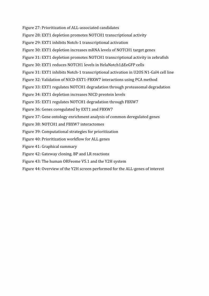
Figure27:PrioritizationofALL-associatedcandidates
Figure28:EXT1depletionpromotesNOTCH1transcriptionalactivity
Figure29:EXT1inhibitsNotch-1transcriptionalactivation
Figure30:EXT1depletionincreasesmRNAlevelsofNOTCH1targetgenes
Figure31:EXT1depletionpromotesNOTCH1transcriptionalactivityinzebrafish
Figure30:EXT1reducesNOTCH1levelsinHelaNotch1∆EeGFPcells
Figure31:EXT1inhibitsNotch-1transcriptionalactivationinU2OSN1-Gal4cellline
Figure32:ValidationofNICD-EXT1-FBXW7interactionsusingPCAmethod
Figure33:EXT1regulatesNOTCH1degradationthroughproteasomaldegradation
Figure34:EXT1depletionincreasesNICDpreoteinlevels
Figure35:EXT1regulatesNOTCH1degradationthroughFBXW7
Figure36:GenescoregulatedbyEXT1andFBXW7
Figure37:Geneontologyenrichmentanalysisofcommonderegulatedgenes
Figure38:NOTCH1andFBXW7interactomes
Figure39:Computationalstrategiesforprioritization
Figure40:PrioritizationworkflowforALLgenes
Figure41:Graphicalsummary
Figure42:Gatewaycloning.BPandLRreactions
Figure43:ThehumanORFeomeV5.1andtheY2Hsystem
Figure44:OverviewoftheY2HscreenperformedfortheALL-genesofinterest

LISTOFTABLESTable1:FeaturesofhumanPPIdatabasesTable2:MammalianglycosyltransferasesthatpreferentiallymodifyEGFrepeatsTable3:NormalizedluciferasevaluecalculatedforNotch1interactorstestedinPCATable4:NICDinteractingproteinpartnersidentifiedintheY2Hscreen.Table5:qRT-PCRprimersequences


INTRODUCTION
1
INTRODUCTION1.AnalyzingnetworksincancerCancerisacomplexdiseaseinwhichvariouscellularprocesses,signallingpathways
and environmental influences contribute to the development and the expression of
cancerphenotypes.Understandingthemechanismsleadingtocancer,isnotfulfilled
by studying individual components in isolation, but requires systems biology
approachtoestablishinteractionsbetweengenes,proteinsandcellularcomponents,
and the associations ofmutations and deregulations to the perturbation of cellular
processes and pathways implicated in cancer 1. It is thus important to analyze
networks in cancer, where biological systems are represented and described as
networks such as protein-protein interactions networks (PPIs), cell signalling
pathways networks, transcriptional regulatory networks and other functional
associationnetworks.
The development of high-throughput interaction assays such as yeast two-hybrid
(Y2H)andaffinitypurificationcoupledtomassspectrometry(AP-MS),andofcurated
databases has led to the generation of large-scale interaction networks for a
considerablenumberoforganisms2,3.Constructingsuchnetworksnotonlyshedsthe
light on the complexity of cellular mechanisms and processes, but also helps
generatinghypothesesabouttherapeutictargetsorderegulatedpathwaysincancer4.
1.1.Typesofpathwaysandnetworkanalysistechniques
Thefirststeptoestablishingnetworkandpathwayanalyses forcancermutations is
definedbysettingthedatabaseresources,representedbyalistofgeneticalterations
inadditiontodatabasesforpathwaysandnetworkinteractions.Analysistechniques
can be divided into three major approaches 4. The first is the “fixed-gene set
enrichmentanalysis”approach,consistinginanalyzinggenesetswithoutconsidering
their interactions. Gene lists are gathered from literature-curated databases or
experimental sources, and using different tools of enrichment analysis leads to
determining pathways and cellular processes for filtered gene sets. The second
INTRODUCTION
approach“denovonetworkconstructionandclustering”consistsinanalyzingalistof
mutated or altered genes taking in consideration their molecular and functional
interactions provided by interactomic databases such as the Biological General
Repository for Interaction Datasets BioGrid 5,6, theMolecular Interaction Database
IntAct7andtheHumanProteinReferenceDatabaseHPRD8.Aninterestingadvantage
of this approach is that networks are expanded due to the “guilt by association”
concept, which increases the complexity of interactomes and helps providing
potential cancer candidate genes. The third approach is “network-basedmodeling”,
that have been applied tomap signalling pathways and functional networks,which
helps to predict the influences of deregulation and perturbations in cancer. An
example of this approach is comparative analysis of regulatorynetworks in normal
and disease states 9, 10. A graphical summary of the three major approaches is
representedinfigure1indicatingthegoalsanddifferenttoolsusedineachmethod4.
Figure 1: Major approaches to pathway and network analysis of cancer data. In thenetworkdiagramsdesignatedby“output”,rednodesrepresentgeneswhoseactivitiesareincreased(firstandthirdcolumns)oralteredbymutations(centercolumn).Greennodesaregeneswhoseactivitiesaredecreased.Adaptedfrom4.
INTRODUCTION
3
1.2.Protein-proteininteractiondetectionmethodsProtein-protein interaction (PPI) is one of the key topics for the development and
progressofmodernsystemsbiology.Inthispartwewillintroduceessentialprotein-
proteininteractiondetectionmethods.
PPIsdetectionmethodscanbeclassified intothreemajorcategories: invitro,invivo
andinsilicomethods.
1.2.1.InvitromethodsTandem affinity purification (TAP) method coupled with mass spectrometry. This
method is basedon taggingproteins andpurifyingprotein complexes associated to
theproteinofinterest.Whenassociatedwithmassspectrometryanalysis,itgenerates
high throughputdata forprotein interactions.AnalyzingAP-MSdatasets inorder to
derive biologically meaningful information from protein interactions remains
challenging.Avarietyofstatisticalmodelsweredevelopedtoassessscoringmethods
fordatasettesting11.
Protein microarrays technology has also been developed to study biochemical
activities of proteins and their interactions in vitro. Three types of protein
microarrays are used: analytical microarrays, functional microarrays and reverse
phasemicroarrays12.Analyticalmicroarraysmostlyuseantibodymicroarrayswhile
functionalmicroarraysarecomposedof full-lengthorproteindomainschips.As for
the third type of microarrays, it enables the protein expression of hundreds of
samples, printed on nitrocellulose slides to be interrogated simultaneously, using
labeled antibodies (with fluorescent detection for example). Reverse phase protein
microarrays have been developed to generate a functional patient-specific circuit
“map” of the cell signallingnetworksbaseddirectly on cellular analysis of a biopsy
specimen13.Inotherwords,differentialproteinexpressionacrosssamplesinahigh
throughputmanner,generatingprotein interactionandactivationmaps that lead to
the identificationofcriticalnodes for individualizedorcombinatorial targettherapy14,15.
Usingtheseprotein-basedmicroarraysenablestheglobalobservationofbiochemical
activities, where thousands of proteins can be screened for different types of
interactions. These methods have important applications in disease marker
identificationandpharmaceuticaltargetscreening16.
INTRODUCTION
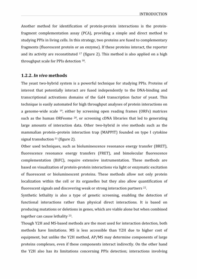
Another method for identification of protein-protein interactions is the protein-
fragment complementation assay (PCA), providing a simple and direct method to
studyingPPIsinlivingcells.Inthisstrategy,twoproteinsarefusedtocomplementary
fragments(fluorescentproteinoranenzyme).Iftheseproteinsinteract,thereporter
and itsactivityarereconstituted17(figure2).Thismethod isalsoappliedonahigh
throughputscaleforPPIsdetection18.
1.2.2.InvivomethodsThe yeast two-hybrid system is a powerful technique for studyingPPIs. Proteins of
interest that potentially interact are fused independently to the DNA-binding and
transcriptional activations domains of the Gal4 transcription factor of yeast. This
techniqueiseasilyautomatedforhighthroughputanalysesofproteininteractionson
a genome-wide scale 19, either by screening open reading frames (ORFs) matrices
such as the humanORFeome 20, or screening cDNA libraries that led to generating
large amounts of interaction data. Other two-hybrid in vivomethods such as the
mammalian protein–protein interaction trap (MAPPIT) founded on type I cytokine
signaltransduction21(figure2).
Other used techniques, such as bioluminescence resonance energy transfer (BRET),
fluorescence resonance energy transfers (FRET), and bimolecular fluorescence
complementation (BiFC), require extensive instrumentation. These methods are
basedonvisualizationofprotein-proteininteractionsvialightorenzymaticexcitation
of fluorescent or bioluminescent proteins. These methods allow not only protein
localization within the cell or its organelles but they also allow quantification of
fluorescentsignalsanddiscoveringweakorstronginteractionpartners22.
Synthetic lethality is also a type of genetic screening, enabling the detection of
functional interactions rather than physical direct interactions. It is based on
producingmutationsordeletionsingenes,whichareviablealonebutwhencombined
togethercancauselethality23.
ThoughY2HandMS-basedmethodsarethemostusedforinteractiondetection,both
methods have limitations. MS is less accessible than Y2H due to higher cost of
equipment,butunlike theY2Hmethod,AP/MSmaydeterminecomponentsof large
proteinscomplexes,even if thesecomponents interact indirectly.Ontheotherhand
the Y2H also has its limitations concerning PPIs detection; interactions involving
INTRODUCTION
5
membrane proteins or proteins that require post-translational modifications are
missed.Thusbothmethodsareconsideredcomplementaryinthetypeofinteractions
they detect. Recently, advance made in AP-MS technology has helped increase its
sensitivityandrobustness24.
Figure 2: schematic representation of PPI detection methods. Y2H: the yeast two-hybridsystem(Y2H)consistingofbaitandpreyproteins(representedbyXandY)arefusedtotheGal4DNAbindingdomain(DB)andtheGal4activatingdomain(AD).Whenfusion proteins are produced andbait part of the first interactwith prey part of thesecond,transcriptionfactorofthereportergeneoccurs.MAPPIT:Thebaitproteinisafusionwithaleptinreceptor(LR),whichcontainsthreeY-to-Fmutationssoitisunableto activate STATs spontaneously. The prey fusion contains a domain of gp130whichcan recruit STATs. After interaction of the bait with the prey, Janus kinases (JAKs)phosphorylate gp130,which stimulates binding of gp130with the STATs. The STATsthemselvesarephosphorylatedby the JAKs,whichresults in the formationofaSTATcomplex. The STAT complex binds the rat PAP1 promoter (rPAP1p) and activatesluciferase transcription. The leptin receptor is further fused with the extracellulardomain of EpoR, a receptor of erythropoietin (Epo), and therefore LR complexformation, which is necessary to make the association with the JAKs, is induced byadditionofEpo 21.PCA: schematicof theproteincomplementationassay.XandYarebait and prey proteins, fused to inactive fragments luciferase or YFP proteins.InteractionbetweenbaitandpreyresultsinthereconstitutionofanactiveformoftheproteinanddetectionofluciferaseactivityofYFPsignal25.
INTRODUCTION
1.2.3.InsilicomethodsAvariety of insilicomethods have beendeveloped to support the interactions that
have been detected by experimental approach. The computational methods for in
silico prediction include sequence-based approaches, structure-based approaches,
chromosome proximity, gene fusion, in silico two-hybrid, mirror tree, phylogenetic
tree,geneontology,andgeneexpression-basedapproaches.
Structure-based method aims to predict protein-protein interaction based on
homologymodelingmethods.Differentalgorithmsweredeveloped,suchasprocesses
thatinvolvepredictionofthebindinginterface,evaluationofthecompatibilityofthe
interfacewithaninterfacecoevolutionbasedmodel,andevaluationoftheconfidence
score for the interaction 26. As for sequence-basedmethod, it depends on primary
structurehomologyinordertoclassifypotentialinteracting27.Genefusionsapproach
reliesoncompletegenomesequencestoidentifyfusionevents.Inthismethodcertain
protein families in given species consist of fuseddomains that usually are found as
singlefull-lengthproteinsinotherspecies.Thesefusionscanpredicteitherdirector
indirect functional interactions.Thismethod canbeused topredictprotein-protein
interactionbyusinginformationofdomainarrangementsindifferentgenomes28.
Insilico two-hybridanalysis isbasedonpreviousstudiesof sequencecorrelation in
multiplesequencealignmentsleadingtothepredictionofphysicalclosenessbetween
residuepairsofpairsindividualproteins.Theresultfromthismethodautomatically
indicatesthepossiblephysicalinteractionbetweentheproteins29.
Similarity of phylogenetic tree method is based on the analyzing the relationship
between protein interactions and co-evolution histories that are represented by
phylogenetic trees. Themirror tree approach has been used to determine potential
interactionpartnersinlargedatasetsofproteinsandalsotobetterunderstandtheco-
evolutionandinteractionsinspecificpairsofproteinfamilies30,31.
The gene expression approach predicts interactions based on the relationship
between gene co-expression and protein interactions. It consists of grouping genes
according to their expression in different experimental conditions, and evaluating
similarities between expression profiles 32. The concept behind this method is
explainedbythefactthatgenesbelongingtocommonexpressionprofilesmorelikely
interactwitheachotherthanproteinsencodedbygenefromdifferentclusters33.
INTRODUCTION
7
Recent technological advances have allowed the development of high throughput
interaction detectionmethods. Despite progress made in this field, each of these
experimentaltechniqueshasitsownadvantagesandlimitations.Onlyacombination
ofdifferentapproachesthatnecessarilyincludesbioinformaticstools,willeventually
lead to a complete characterization of physiologically relevant protein-protein
interactionsinagivencellororganism.
1.3.ProteininteractionsdatabasesAnumberofpubliclyavailabledatabasescollectandstoreprotein-proteininteraction
dataprovidingresearcheswithaccesstothesecurateddatasets.Inordertoavoidthe
duplicationofthecurationdataandenabledataexchange,theInternationalMolecular
Exchange(IMEx)consortiumwasformed.InadditionProteomicsStandardsInitiative
- Molecular Interaction (PSI-MI) format sets data standards in order to specify a
unifiedstructureforsharingPPIs34.TherearetwotypesofPPIsdatabasesbasedon
theircontent;thosecontainingdatasupportedbyexperimentalvalidationandthose
derivedfrom insilicopredictions35.WecannameseveralPPIsdatabasessuchasthe
Biological General Repository for Interaction Datasets (BIOGRID) 6, the IntAct
molecular Interaction Network database (IntAct) 36, the Human Protein Reference
Database(HPRD)8,theMolecularINTeractiondatabase(MINT)37,whichonlyreport
experimentallyverifiedinteractions.SomeofthefeaturesofthesePPIdatabasesare
representedtable1.
INTRODUCTION
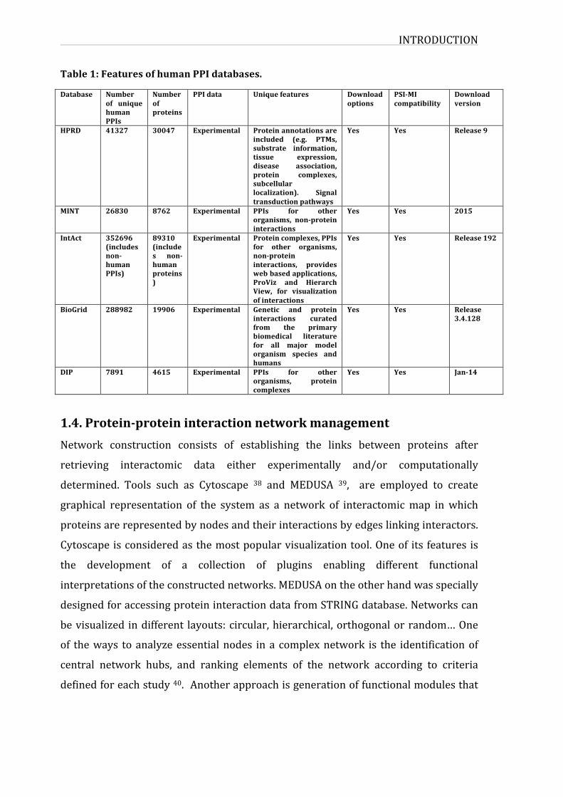
Table1:FeaturesofhumanPPIdatabases.
Database Numberof uniquehumanPPIs
Numberofproteins
PPIdata Uniquefeatures Downloadoptions
PSI-MIcompatibility
Downloadversion
HPRD 41327 30047 Experimental Proteinannotationsareincluded (e.g. PTMs,substrate information,tissue expression,disease association,protein complexes,subcellularlocalization). Signaltransductionpathways
Yes Yes Release9
MINT 26830 8762 Experimental PPIs for otherorganisms, non-proteininteractions
Yes Yes 2015
IntAct 352696(includesnon-humanPPIs)
89310(includes non-humanproteins)
Experimental Proteincomplexes,PPIsfor other organisms,non-proteininteractions, provideswebbasedapplications,ProViz and HierarchView, for visualizationofinteractions
Yes Yes Release192
BioGrid 288982 19906 Experimental Genetic and proteininteractions curatedfrom the primarybiomedical literaturefor all major modelorganism species andhumans
Yes Yes Release3.4.128
DIP 7891 4615 Experimental PPIs for otherorganisms, proteincomplexes
Yes Yes Jan-14
1.4.Protein-proteininteractionnetworkmanagementNetwork construction consists of establishing the links between proteins after
retrieving interactomic data either experimentally and/or computationally
determined. Tools such as Cytoscape 38 and MEDUSA 39, are employed to create
graphical representation of the system as a network of interactomicmap inwhich
proteinsarerepresentedbynodesandtheirinteractionsbyedgeslinkinginteractors.
Cytoscapeisconsideredasthemostpopularvisualizationtool.Oneof its features is
the development of a collection of plugins enabling different functional
interpretationsoftheconstructednetworks.MEDUSAontheotherhandwasspecially
designedforaccessingproteininteractiondatafromSTRINGdatabase.Networkscan
bevisualized indifferent layouts:circular,hierarchical,orthogonalorrandom…One
of theways toanalyzeessentialnodes ina complexnetwork is the identificationof
central network hubs, and ranking elements of the network according to criteria
definedforeachstudy40.Anotherapproachisgenerationoffunctionalmodulesthat
INTRODUCTION
9
can be established by functional annotation for representing biological networks,
accordingtogeneontologies,commonpathways,anddiseaseimplication41…
Theavailabilityofhigh-throughputexperimentaldataandcomputationalinteraction
prediction datasets has allowed construction of increasingly comprehensive and
accurate protein-protein interaction networks. As we have seen, each method or
approach has its strengths andweaknesses; thereforewe cannot define a “perfect”
approach. Accordingly it is essential to integrate different techniques and define
criteriadependingonthespecificityandtheaimofeachstudy.
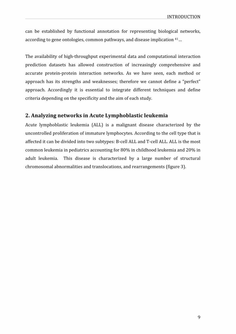
2.AnalyzingnetworksinAcuteLymphoblasticleukemiaAcute lymphoblastic leukemia (ALL) is a malignant disease characterized by the
uncontrolledproliferationofimmaturelymphocytes.Accordingtothecelltypethatis
affecteditcanbedividedintotwosubtypes:B-cellALLandT-cellALL.ALListhemost
commonleukemiainpediatricsaccountingfor80%inchildhoodleukemiaand20%in
adult leukemia. This disease is characterized by a large number of structural
chromosomalabnormalitiesandtranslocations,andrearrangements(figure3).
INTRODUCTION
Figure 3: Spectrum of recurring chromosomal rearrangements in childhood ALL.Representationof commentrecurringnumericalandstructuralgeneticalterations inchildhood B-progenitor and T-lineage ALL, including approximate frequencies.Alterations specific to T-lineage ALL are shown at the bottom of the pie chart inmagenta.ReviewedinMulligan201142.
The Notch signalling pathway plays a vital role in determination of the fate of
hematopoieticcells,itisessentialforthegenerationofembryonichematopoieticstem
cellsandalsoincontrollingTcelldifferentiation.Numerousstudieshaveshownthat
in mammals, the 4 Notch receptors are expressed in hematopoietic cells but at
different stages and in different contexts of differentiation 43. Hematopoietic
developmentstartsintwodistinctphases;thefirstistheprimitivehematopoiesisat
extra-embryonic sites initiated in the yolk sac and the second is the definitive
hematopoiesisintheembryoitself.Thefirsthematopoieticstemcells(HSCs)appear
inthedorsalaorta inaregioncalledAGM(aorta-gonad-mesonephros) inadultmice
anditisthoughtthatthesecellsmightoriginatefromendothelialcellseveniftheyare
first detected in the yolk sac. Notch signalling promotes expansion of HSCs by
activatingRunx1expression,andalsoarterialspecificationthroughGata2regulation44.
INTRODUCTION
11
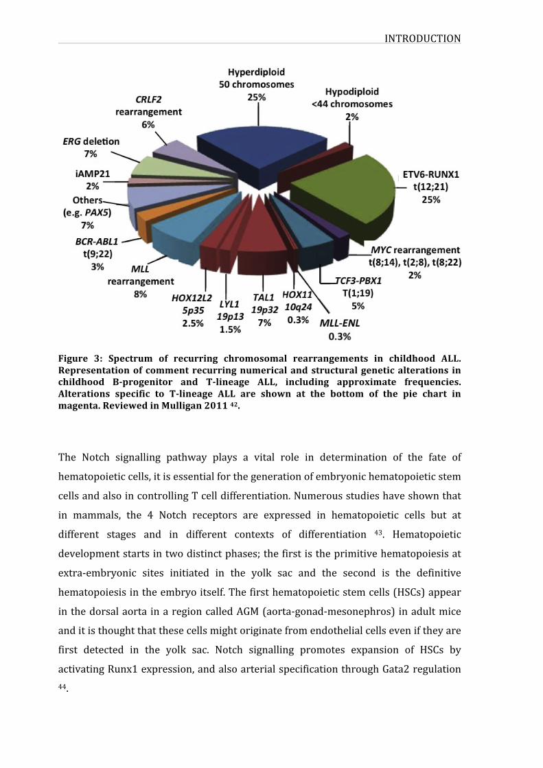
OneofthemostcharacterizedfunctionsofNotchsignallingisitsroleinpromotingT
cell differentiation in the bone marrow (BM). Active Notch signalling involving
NOTCH1receptorandDll4 ligand inearlystagesofT-celldevelopment isnecessary
for inhibiting the differentiation of B-cell and myeloid lineages. Notch1 is also
requiredtopromotetransformationofearlyTlineageprogenitorsintoprogressively
matureTlymphocytes(figure4).InfactlymphoidprecursorslackingNOTCH1result
inT-celldefectinthethymus.
Figure4:NotchandT-celldevelopment.Notch1andDll4interactionsinthethymusareabsolutelyrequiredduringearlystagesofT-celldevelopment.ETPs,doublenegative2(DN2) and DN3a cells experience a high intensity of Notch signalling. Active Notchsignallingduring early stagesofT-cell development leads to inhibitionof bothB-cellandmyeloidlineages.Attheβ-selectioncheckpoint,Notchsignallingisrapidlyturnedoff as a consequence of preTCR signalling. Hence, double positive (DP) T-cellsexperiencea very low intensityofNotch signalling.TSLP:Thymus-SeedingLymphoidProgenitors;ETP:EarlyTlineageProgenitor;DN:DoubleNegative;DP:DoublePositive;NKT:NKT-cell;nTR:naturalregulatoryT-cell.Extractedfrom45.
Recently, advancesmade in genomic techniques such aswhole genome sequencing,
genome wide profiling and cytogenetic methods enabled the identification of
mutationsandaberrationsinALLpatients,whichledtoabetterunderstandingofthis
INTRODUCTION
disease.
It became certain that Notch signalling regulates hematopoiesis at different levels,
through raging fromHSC formation to their differentiation and fate decision,while
mutations in the receptor and aberrant Notch signalling were also linked to
leukemogenesis andhematopoieticdisorders suchasacute lymphoblastic leukemia.
Therefore it has been the subject of intensive research to better understand the
mechanisms underlying tumorigenesis and studies showed increased interest in
therapeuticmodulationoftheNotchpathwayinthisfield.
2.1Bcellacutelymphoblasticleukemia
Chromosomal rearrangements were highly associated to B-cell ALL, with
hyperdiploidy and ETV6-RUNX1 fusion representing 25% incidence for each
aberration among other types of genetic alterations described in acute leukemias.
Other fusions were also foundwith a less frequency such asMLL rearrangements,
TCF3-PBX1, BCR-ABL1 translocations. Observations show that these genetic
alterations modify the normal lymphoid maturation process through disrupting
hematopoietic transcription factors or activate oncogenes 42. Several studies have
shownthatIKFZalterationsisahallmarkofBCR-ABL1B-ALLsubtype,andusuallyit
is associatedwith apooroutcome, itwas alsodemonstrated that in almost50%of
BCR-ABL1ALLpatients,CRLF2(encodingcytokinereceptorlikefactor2)expression
isdisrupted.CRLF2wasfoundtobeoverexpressed inALLpatientsaccompaniedby
otheractivatingmutationsinIKAROSgene,JAK1andJAK2(januskinases),IL7R(IL7
receptor), with high rates of relapse 46. As previously mentioned, Notch signalling
mightplayaroleasatumorsuppressororanoncogene,accordingtocellcontextand
microenviromentalconditions.InB-cellALLNotchsignallinginductioninB-ALLcells
lines leads to cell cycle arrest and apoptosis. Notch receptors and their ligands are
expressedonB-ALLcellsurface.Levelsofexpressionofligandsandreceptorsinbone
marrowmesenshymal stromal cells (MSCs) and their interactions are important in
leukomegenesis ofB-cellALL andwas also linked to chemoresistance to therapy in
thesepatients47.
INTRODUCTION
13
2.2.T-cellacutelymphoblasticleukemia
T-cell ALL accounts for 10 to 15% of pediatric ALL and 25% of adult ALL 48. It is
characterizedbydiffusionofimmatureT-cellsthroughthebonemarrow.Incontrast
to B-lineage ALL, where malignant cells often have additional specific genetic
abnormalities (chromosomal rearrangements and genetic fusions), which have
significant impact on the clinical outcome of the disease, in T-lineage ALL few
molecular abnormalities have been detected. A chromosomal translocation
t(7;9)(q34;q34.3) involving Notch gene was found in T-ALL patients. This
translocation fuses the 3’ portion of the truncated form of Notch1 TAN1 on
chromosome9totheTCRβlocusonchromosome7,butwasonlyfoundinlessthan
1% of T-ALL patients 49. In murine model, transplantation of bone marrow
progenitors expressing TAN1, develop T-cell neoplasm within two weeks, proving
thatthistranslocationcanbecausativeforT-ALL50.
Gene expression profiles using oligonucleotide microarrays was applied to T-ALL
samples and cell lines in order to characterize immunologic markers as well as
cytogenetic and molecular abnormalities. A study carried out on T-ALL patient
samples, showed that applying hierarchical clustering on a set of differentially
expressed genes (313 genes) between T-ALL patients reflect the degree of
differentiation of leukemic cells. In addition gene expression profiling was also
associated with response to treatment and long-term outcome of the disease51.
Consistentwiththese findings,A.FerrandoandT.Lookestablishedgeneexpression
profiles analysis in T-ALL showing that different oncogenic transcription factors
define molecularly distinct groups of T-ALL, which are characterized by
transcriptionalpatterns that involve regulatorsof cell growth, apoptosis, thymocyte
development, and responsiveness to therapy. Using gene expression analysis in T-
ALL,theyidentifiedHOX11expressionasanindicatoroffavorableprognosiscategory
whileTAL1 (T-cell acute lymphocytic leukemia1protein) andLYL1 (lymphoblastic
leukemia associated hematopoiesis regulator 1) showed poorer outcome 52. These
examples alongwith other studies show how development ofmicroarrays, making
possibletheanalysisofT-ALLonagenomicscale,hashelpedtodefinetheoncogenic
pathwaysresponsibleforleukemictransformationinthisdisease.
INTRODUCTION
Ontheotherhand,mutationsindifferentoncogenesandtumorsuppressorgenesthat
are initially known to be involved in the deregulation of mechanisms of T-cell
proliferation, differentiation and thymopoiesis have been linked to T-ALL
pathogenesis53.Amongthesegenesandmutations,activatingmutationsofNOTCH1
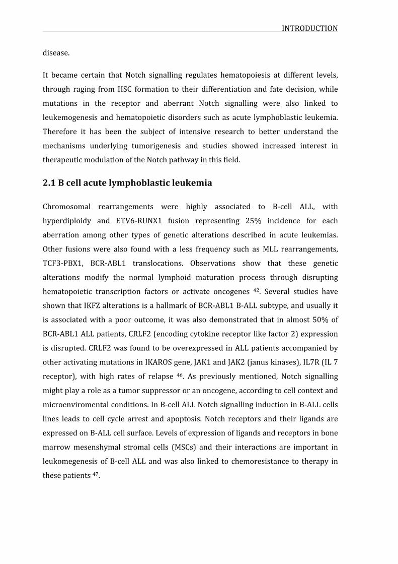
werefoundinmorethan50%ofT-ALLpatients54.AlargenumberofNotchmutations
were limited to specific regions of the protein, involving the heterodimerization
domain and leading to a ligand-independent constitutive activation of the receptor,
andalsointheN-terminalPESTdomainaffectingNOTCH1stabilityanddegradation.
Mutational Notch and aberrant Notch signalling in T-ALL are also accompanied by
deregulation of other oncogenes such as cMYC, E2A-PBX, Ikaros and tumor
suppressorslikeFBXW7,PTENandPIK3CA.ThereforeNotchmutationsalonearenot
sufficient for disease development but they rather highly contribute to
leukomogenesis and resistance to chemotherapeutic treatments. Gain of function
mutationsofNotchaccompaniedwithmutational lossofPTEN induce resistance to
Notch1 inhibition in T-cell leukemia. NOTCH1 downregulates PTEN expression
throughcMYCandHES1,whichcouldmediateanupregulationofPI3K-AKTsignalling
pathwayinbothnormalandleukemicT-celllines55.cMYCisaNotchtargetgeneinT-
ALL. It has been shown thatwhen overexpressed, cMYC is able to induce T-ALL in
animalmodels. In additionNotch blockade usingϒ-secratase inhibitorsGSI, lead to
downregulationofcMYCexpressionindifferentT-ALLcelllines56.Assomedatashow
thatcMYCisadownstreamtargetofNotchinT-ALL,otherstudiesdemonstratethat
cMYCandNotch1canact through independentbutyetcomplementarypathways to
promotepre-T-celltransformationandthusexpandingapoolof“highriskoncogenic”
pre-T-cells 54. A study carried out on T-ALL patients, showed that the presence of
Notch activating mutations in the heteodimerization and PEST domains, lead to
upregulation of HES1, cMYC, Deltex downstream Notch genes. In addition some of
these patients also presentmutations in the FBXW7 gene, associatedwith a higher
transcriptionalactivationforNOTCH1genetargets,andchemotherapyrelatedgenes
such as Bcl-2 andMDR1 57. Another study also showed that in some cases FBXW7
mutationsinleukemiccellsmediateNotchpathwayactivation,andthatmutantforms
of FBXW7cannotbind to the intracellular formofNOTCH1 (NICD) leading toNICD
andMYC stabilization 58.Mutations in theFBXW7 gene are found in 15% of T-ALL
cases, interfering alsowithNOTCH1 proteasomal degradation 58. The essential role
INTRODUCTION
15
that Notch plays in T-ALL, shown by several studies, invitro, in vivoand driven by
analyses made on samples from leukemic patients, highlighted the importance of
Notch signalling and its effect in the regulation of downstream Notch targets and
othersignallingpathway,anditspotentialintargetedtherapyagainstALL.
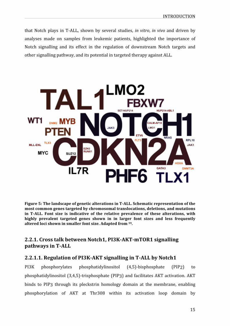
Figure5:ThelandscapeofgeneticalterationsinT-ALL.Schematicrepresentationofthemostcommongenestargetedbychromosomaltranslocations,deletions,andmutationsin T-ALL. Font size is indicative of the relative prevalence of these alterations, withhighly prevalent targeted genes shown in in larger font sizes and less frequentlyalteredlocishowninsmallerfontsize.Adaptedfrom59.
2.2.1.CrosstalkbetweenNotch1,PI3K-AKT-mTOR1signallingpathwaysinT-ALL2.2.1.1.RegulationofPI3K-AKTsignallinginT-ALLbyNotch1PI3K phosphorylates phosphatidylinositol (4,5)-bisphosphate (PIP2) to
phosphatidylinositol(3,4,5)-trisphosphate(PIP3)andfacilitatesAKTactivation.AKT
binds to PIP3 through its pleckstrin homology domain at the membrane, enabling
phosphorylation of AKT at Thr308 within its activation loop domain by
INTRODUCTION
phosphoinositide-dependentkinase1 (PDK1).AdditionalphosphorylationofAKTat
Ser473within its hydrophobicmotif bymammalian target of rapamycin complex 2
(mTOR2 or PDK2) results in full activation of AKT, and phosphorylation of its
substrates(i.e.,glycogensynthasekinase3(GSK3), theFOXO-familyof transcription
factors, BAD, MDM2, and TSC2, thereby promoting cell growth, survival, and
proliferation60.
Notch1playsan importantrole inregulatingPI3K-AKTsignalling.The firststudy to
provethatNotch1canactivatePI3K-AKTpathwayshowedthatNotchsignalsviathe
Delta-like1 ligand (Dll1) interactionspromoting the survival of preT-cells through
maintenance of cell size, glucose uptake andmetabolism. Furthermore, the trophic
effectsofNotchsignalingweremediatedbythepathwayofphosphatidylinositol-3-OH
kinase and the kinase Akt, such that expression of active Akt overcame the
requirementforNotchinβ-selection61.
T-ALL cells are also dependent on Notch1 and AKT signalling for proliferation and
survival. The link between Notch1 and activation of PI3K-AKT signalling was
establishedbyPalomeroetal.,theyshowedthatNOTCH1regulatestheexpressionof
PTENandtheactivityof thePI3K-AKTsignalingpathway innormaland leukemicT
cells55.Hes1gene,oneofNotch1transcriptionaltargets,bindstoPtenpromoterand
represses its activity, decreasing PTEN protein levels and increasing AKT
phosphorylation and downstream signalling (figure 6). In addition, alterations of
PI3K, PTEN, and AKT were reported in 47,7% of T-ALL cases from children, with
PTEN mutants being most common 62. Several studies showed that Notch could
regulate PI3K-AKT-mTOR1 signalling at multiple levels, through both PTEN-
dependentandindependentroutes.AnexampleisthematurationofILR7leadingto
activation of JAK-STAT5 and PI3K pathways,which play important roles in normal
hematopoiesisandleukemia63.Notch1canalsoregulateIGF1RlevelsandPI3K-AKT
activity in T-ALL, and Notch signaling is required tomaintain IGF1R expression at
highlevelsinT-ALLcells64.
INTRODUCTION
17
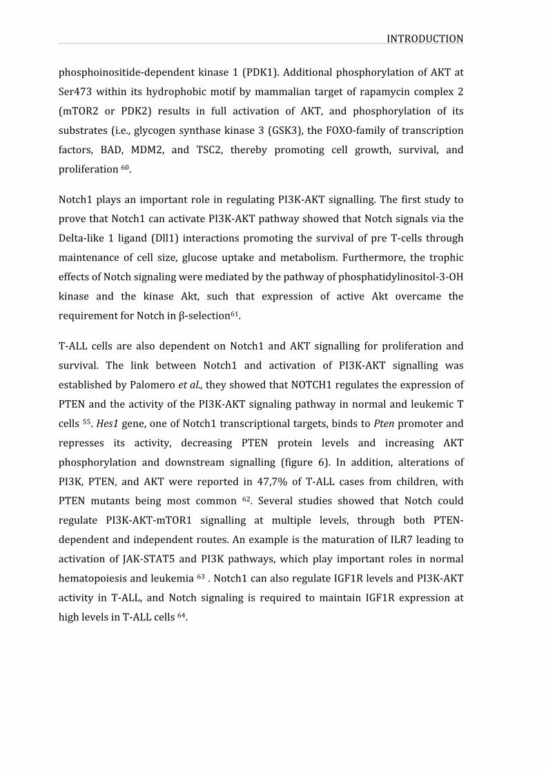
Figure 6: Regulation of PI3K–AKT signalling in T-ALL cells by Notch1. The signallingpathways downstream of Notch1 that converge on PI3K–AKT signaling in T-ALL areshown.Theactivated formofNotch1 (NICD) is shownandresults innetactivationofPI3K–AKT signaling by increasing levels of growth factor receptors [IGF1R and IL7R(IL7Rαsubunit)]thatrecruitPI3K,orbyreducingPTENlevelsthroughtranscriptionalrepression of thePten gene by Hes1. Although cMyc can activatePten transcription,Hes1 repression dominates. PI3K phosphorylates PIP2(4,5) to generate PIP3(3,4,5)thatrecruitsAKTforphosphorylationatThr308byPDK1andSer473bymTOR2.Thisresults in phosphorylation of AKT substrates (GSK3, FOXO, BAD, MDM2, TSC2, andmTOR), promoting glucose metabolism, proliferation, growth, and translation, butimpairing apoptosis and autophagy. PTEN dephosphorylates PIP3(3,4,5) back toPIP2(4,5)toblockAKTactivation.NICDwasreportedtodecreasePKCθandROSlevelsthrough induction of the RUNX3 transcription factor. ROS promotes PTEN oxidationand inactivation.NICDmight also contribute to regulationofPTEN inactivation sinceGSItreatmentsfurtherincreasedPTENphosphorylation,stabilization,andinactivationbyCK2,whichsuggeststhatNICDmayplayamoreactiveroleinregulatingCK2activity,although NICD- independent pathways are also likely to contribute. Either of thesepathwayscouldinfluencePTENactivitythroughpost-translationalmodifications.Lossof PTEN results in constitutive AKT activation and contributes to GSI-resistance.FBXW7 regulates NICD stability and signal duration but has also been suggested todecreaseAKTandmTOR1levels.FBXW7issubjectedto frequentmutations inT-ALL.Notch1 pathway activation and/or loss of PTEN result in net activation of PI3K–AKTsignalinginT-ALLcells(lineweightsareproportionaltotheneteffectsonsignaling).Arrows denote activation and blunt arrows repression. Dashed lines denoteuncharacterizedmechanisms.Transcription factors(orange),growth factorreceptors(white), kinases (blue), phosphatases (purple), E3-ubiquitin ligases (MDM2 andFBXW7; yellow), BAD (bgreen), TSC1/2 (white/gray), and unknown negativeregulatory factors (red dashed ovals) are depicted. AKT activating phosphorylations
INTRODUCTION
are indicated (green stars with “P”) while phosphorylations of other proteins havebeenomittedforsimplicity.Extractedfrom65.
2.2.1.2.Notch1affectsp53,cMYCandPI3K-AKTpathwaysinT-ALLRecent studies suggest that Notch1 regulates p53 levels and activation in T-ALL.
Activation ofmutantNotch1 in someT-ALLwith constitutively high levels of PI3K-
AKT signalling, secondary to lossor inactivationofPTEN,may contribute to lossof
p53viaMDM266.P53mutationsarefrequentinT-ALLrelapse,butrarelymutatedin
primaryT-ALL.InductionofNICDinaninduciblemurinelymphomamodeldecreased
ARFandp53,whichisakeymechanismunderlyingtheinitiationofT-celllymphoma67. cMYC, a well-characterized Notch1 target gene, can induce T-ALL in mice and
zebrafish. However Notch1 is oncogenic dominant over cMyc in T-ALL68. Recent
studiessuggest thatactivationofPI3K-AKTpathwaydownstreamofNotch1maybe
sufficienttodriveT-ALL.PI3K-AKTcanfunctionallyreplaceNotch1duringβ-seletion.
MAPsignallingpathwayandGSK3βphosphorylatecMycleadingtoitsubiquitination
byFBXW7anditssubsequentdegradation(figure7).Recentevidenceshowthatpost-
transcriptionalderegulationofcMycviaPTENisamajoralternativepathwayofMYC
activationinT-ALL69.
INTRODUCTION
19
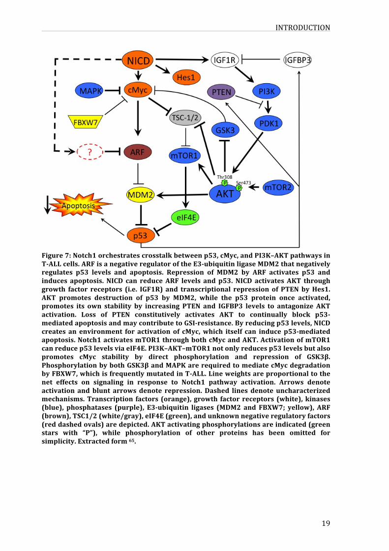
Figure7:Notch1orchestratescrosstalkbetweenp53,cMyc,andPI3K–AKTpathwaysinT-ALLcells.ARFisanegativeregulatoroftheE3-ubiquitinligaseMDM2thatnegativelyregulates p53 levels and apoptosis. Repression of MDM2 by ARF activates p53 andinduces apoptosis. NICD can reduceARF levels and p53. NICD activates AKT throughgrowth factor receptors (i.e. IGF1R) and transcriptional repression of PTENbyHes1.AKT promotes destruction of p53 by MDM2, while the p53 protein once activated,promotes its own stability by increasing PTEN and IGFBP3 levels to antagonize AKTactivation. Loss of PTEN constitutively activates AKT to continually block p53-mediatedapoptosisandmaycontributetoGSI-resistance.Byreducingp53levels,NICDcreates an environment for activation of cMyc,which itself can induce p53-mediatedapoptosis.Notch1activatesmTOR1throughbothcMycandAKT.ActivationofmTOR1canreducep53levelsviaeIF4E.PI3K–AKT–mTOR1notonlyreducesp53levelsbutalsopromotes cMyc stability by direct phosphorylation and repression of GSK3β.PhosphorylationbybothGSK3βandMAPKarerequiredtomediatecMycdegradationbyFBXW7,whichisfrequentlymutatedinT-ALL.Lineweightsareproportionaltothenet effects on signaling in response to Notch1 pathway activation. Arrows denoteactivation andblunt arrowsdenote repression.Dashed lines denote uncharacterizedmechanisms.Transcription factors (orange),growth factorreceptors (white),kinases(blue), phosphatases (purple), E3-ubiquitin ligases (MDM2 and FBXW7; yellow), ARF(brown),TSC1/2(white/gray),eIF4E(green),andunknownnegativeregulatoryfactors(reddashedovals)aredepicted.AKTactivatingphosphorylationsareindicated(greenstars with “P”), while phosphorylation of other proteins has been omitted forsimplicity.Extractedform65.
INTRODUCTION
2.3.TherapeuticapproachinALLGlucocorticoids were among the first drugs used in the treatment of acute
lymphoblastic leukemia, and have remained essential components of therapy.
Primary genetic abnormalities of leukemic cells have important prognostic
significance and in some cases they are associatedwithdrug resistant. The current
chemotherapeutic approaches in ALL are adapted according to patient’s age,
cytogeneticsandbonemarrowresponse.Due to thedifferences indrug tolerability,
adult and paediatric ALL treatments vary considerably between these groups.
Prednisolone, vincristine, asparaginase, and daunorubicin are widely used
medicationsinALLtreatment70.
The gene expression profiles of leukemia cells have been used to identify genes
related to the intracellular disposition of anti-leukemic agent in vivo and to reveal
differentsetsofgenesassociated todrugresistance.An interestinggeneexpression
patternsstudy identified172gene-probesetsasdifferentiallyexpressed inprimary
B-lineage leukemia cells. These geneswere also associatedwith resistance to drug
treatments(Prednisolone,vincristine,asparaginase,anddaunorubicin).Theyshowed
thatresistancetomechanisticallydistinctanti-leukemicagentsisassociatedwiththe
expressionofdifferentfunctionalgroupsofgenesandsupporttheuseofcombination
chemotherapy for cancer treatment 71.A similar analysis of gene expression inALL
was carried out on over 9600 genes before and after in vivo treatment with
methoxtrexate and mercaptopurine alone or in combination. They identified 124
genes differentially expressed among these treatments. The identified set included
genesrelatedtoapoptosis,mismatchrepair,cellcyclecontrolandstressresponse72.
Comparisons of gene expression levels through time in B- ALL patients showed
consistentdifferencesamongasetof23genesatleastattwoofthethreetimepoints
evaluatedand thedifferences in theexpression levelsof IL2RA,SORT1,DEFA1,and
FLT3genesinatleastoneofthetimesstudiedwereassociatedwithrelapseand/orB-
ALL-relateddeath73.
Exploring acute treatment-induced changes in gene expression in leukemia cells
offers new insight into thedifferences in cellular response to individual agents and
drug combinations. Identification of treatment-induced changes in gene expression
can serve as a new tool for assessing the interaction of anticancer agents andmay
provideabasisforoptimizingcombinationofchemotherapy.
INTRODUCTION
21
Otheragentsintheearlyphaseofclinicaltestingarebeingdeveloped;includingFLT3
inhibitors 74, γ-secretase inhibitors (GSI) 54, proteasome inhibitors and short
interfering RNAs 75. ALL treatment has advanced significantly. In addition gene
profiling performed in T-ALL cell lines showed that GSI treatment induce gene
expressionchangesin239genesincludingdirectNotch1targetssuchasDELTEXand
HES1.Importantly,thisanalysisalsoidentifiedc-MYC,amasterregulatorofmultiple
biosynthesisandmetabolicpathways76.Recent findings ingeneexpressionprofiles,
mutations and molecular characterization of ALL led to the development of novel
targeted therapies. However, cure is often challenging and toxic. If we take for
example γ-secretase inhibitors though effective in some cases but they still present
high gastrointestinal toxicity 77. Another example is bortezomib a proteasome
inhibitordrug thatwas testedonALLpatients in bothpediatric and adult patients,
andshowedinfectioustoxicity insomepatients78.Thechallengegoingforwardwill
be to find safe and effective combinations and determine where in the treatment
schema these agents will be most effective in ALL therapy. Studies in the field of
therapy focus on the effect of drugs on prognosis and relapse rather than gene
expression profiling. Therefore, comparing gene expression profiling for leukemic
patient samples undergoing treatment can serve as a powerful tool to better
understandtheeffectofthesedrugs.
The following chapters focus on Notch1 and FBXW7, two connected proteins
frequentlymutatedinT-ALL.
3.NOTCH1networkandsignallingNOTCH1 was described for the first time in 1917, when Thomas Hunt Morgan an
American geneticist and embryologist, described a strain of fruit flies Drosophila
melanogaster,withnotchesatthemarginoftheirwingblades79.TheNotchhomolog
in human was described in the mid 1980’s, as a transmembrane receptor that is
essential in a highly conserved signalling pathway involved in the regulation of
different processes during development and tissue homeostasis 80. The first time
NOTCH1 was linked to human cancer when the t(7; 9)(q34; q34.3) chromosomal
translocation found in T-cell acute lymphoblastic leukemia (T-ALL) was sequenced
INTRODUCTION
and cloned 81. This chromosomal translocation resulted in anN-terminal truncated
dominant active ligand - independent human NOTCH1 receptor (TAN1). It wasn’t
untilyearslaterthatstudiesshowedthatTAN1iscausativefordiseasedevelopment
inmousemodels50,andlateranotherstudyshowedthatapproximately50%ofallT-
ALLpatientshadactivatingmutationsinthehumanNOTCH1gene54.
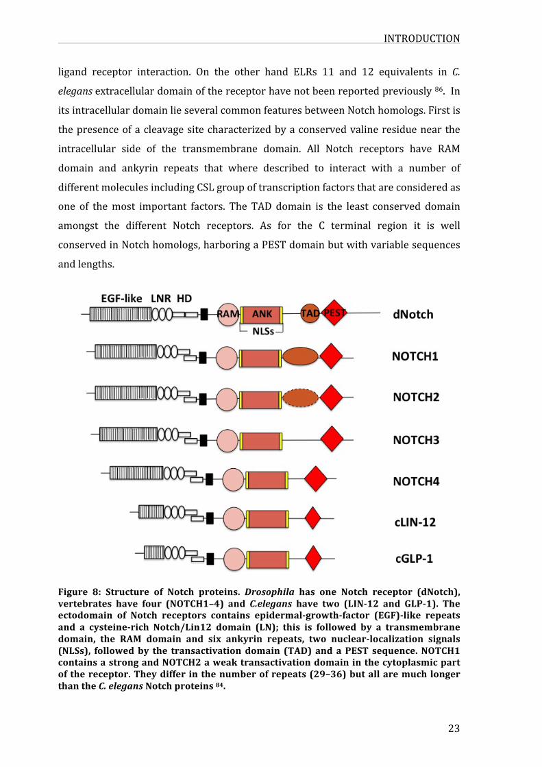
3.1.TheNotchprotein
NotchsignallingisanevolutionaryconservedmechanismfromDrosophilatohumans.
Notchreceptorsarelargesingle-passproteins,inDrosophila,thereisonlyoneNotch-
encodinggene82,inC.elegans,therearetwogenesencodingforNotch(lin-12andglp-
1) 83.Whilst inmammals, there are four Notch genes encoding different receptors
(NOTCH1-4) 84. TheNotch receptors are composed of two functional domains, the
extracellular (NEC) and transmembrane (NTM) domains. The extracellular domain
containsbetween29 to36 epidermal growth factor-like repeats (ELRs) involved in
ligandbindingfollowedbythreemodulesofLIN-12-Notchrepeats(NLR)linkednon-
covalently by an heterodimerization domain to NTM 85. These three cysteine-rich
Lin12-Notch repeats (LNR) and a heterodimerization domain represent a region
calledNRRfornegativeregulatoryregionplayingacentralroleinpreventingreceptor
activation in the absence of ligands. The NTM contains a RAM domain for RBP-Jk
associatedmodule,linkedbyanuclearlocalizationsequencetosevenAnkyrinrepeats
domain. An additional nuclear localization sequence links the ANK domain to a
transactivation domain (TAD) known to be different and evolutionarily divergent
amongNotchorthologs.TAD is followedbyaPESTdomainrich inproline,glutamic
acid,serine,andthreonineintheC-terminusoftheNTM,whichharborsdegradation
signals(degrons)regulatingNICDstability.
AlthoughtherearebroadvariationsinsizeamongstNotchfamilyorthologs,especially
relativetotheC.elegansmembers,lin-12andglp-1,severalmajorstructuralfeatures
are conserved amongst all members (figure 8). As previously mentioned, the
extracellular domain is characterized by the large number of EGF repeats, which
number varies from 10 in glp-1 to 36 in Drosophila and some vertebrate Notch
receptors.Severaldatasuggestedthatthe11thand12thELRsinbothDrosophilaand
vertebrateNotchreceptorsplayacrucialroleforbeingconsideredasprimarysitesof
INTRODUCTION
23
ligand receptor interaction. On the other hand ELRs 11 and 12 equivalents in C.
elegansextracellulardomainofthereceptorhavenotbeenreportedpreviously86.In
itsintracellulardomainlieseveralcommonfeaturesbetweenNotchhomologs.Firstis
thepresenceofacleavagesitecharacterizedbyaconservedvalineresiduenearthe
intracellular side of the transmembrane domain. All Notch receptors have RAM
domain and ankyrin repeats that where described to interact with a number of
differentmoleculesincludingCSLgroupoftranscriptionfactorsthatareconsideredas
one of themost important factors. The TAD domain is the least conserved domain
amongst the different Notch receptors. As for the C terminal region it is well
conservedinNotchhomologs,harboringaPESTdomainbutwithvariablesequences
andlengths.
Figure 8: Structure of Notch proteins. Drosophila has one Notch receptor (dNotch),vertebrates have four (NOTCH1–4) and C.elegans have two (LIN-12 and GLP-1). Theectodomain of Notch receptors contains epidermal-growth-factor (EGF)-like repeatsand a cysteine-rich Notch/Lin12 domain (LN); this is followed by a transmembranedomain, the RAM domain and six ankyrin repeats, two nuclear-localization signals(NLSs), followedby the transactivationdomain (TAD)andaPEST sequence.NOTCH1containsastrongandNOTCH2aweaktransactivationdomaininthecytoplasmicpartof thereceptor.Theydiffer inthenumberofrepeats(29–36)butallaremuchlongerthantheC.elegansNotchproteins84.
INTRODUCTION
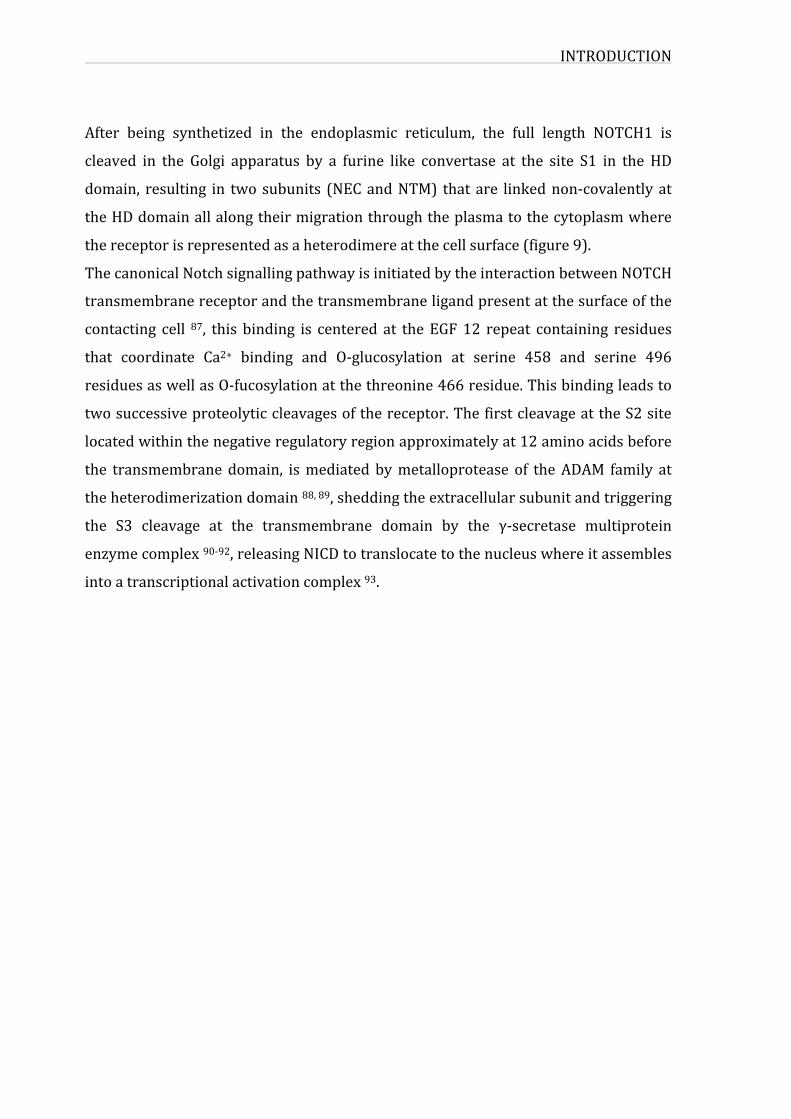
After being synthetized in the endoplasmic reticulum, the full length NOTCH1 is
cleaved in the Golgi apparatus by a furine like convertase at the site S1 in the HD
domain, resulting in twosubunits (NECandNTM) that are linkednon-covalentlyat
theHDdomainallalongtheirmigrationthroughtheplasmatothecytoplasmwhere
thereceptorisrepresentedasaheterodimereatthecellsurface(figure9).
ThecanonicalNotchsignallingpathwayisinitiatedbytheinteractionbetweenNOTCH
transmembranereceptorandthetransmembraneligandpresentatthesurfaceofthe
contacting cell 87, this binding is centered at theEGF12 repeat containing residues
that coordinate Ca2+ binding and O-glucosylation at serine 458 and serine 496
residuesaswellasO-fucosylationatthethreonine466residue.Thisbindingleadsto
twosuccessiveproteolyticcleavagesofthereceptor.ThefirstcleavageattheS2site
locatedwithinthenegativeregulatoryregionapproximatelyat12aminoacidsbefore
the transmembranedomain, ismediatedbymetalloprotease of theADAM family at
theheterodimerizationdomain88,89,sheddingtheextracellularsubunitandtriggering
the S3 cleavage at the transmembrane domain by the γ-secretase multiprotein
enzymecomplex90-92,releasingNICDtotranslocatetothenucleuswhereitassembles
intoatranscriptionalactivationcomplex93.
INTRODUCTION
25
Figure 9: Canonical Notch signalling pathway. The Notch protein is syntheized as aprecursor for that is cleaved by furin-like convertase (S1 cleavage) to generate themature receptor, which is composed of two subunits that are held together by non-covalent interactions. On binding to the Notch receptor, the ligand induces aconformational change, exposing the S2 cleavage site in the extracellular domain ofNotchtothemetalloproteinasetumornecrosisfactor-α-convertingenzyme(TACEalsoknownasADAM).FollowingS2cleavage,Notchundergoesathirdcleavage(S3)thatismediatedbythepresenilin-γ-secratasecomplex.TheS3cleavageresultsinthereleaseoftheactiveNICDfromtheplasmamembraneadthesubsequenttranslocationintothenucleusandactivationoftranscriptionoftargetgenes.Adaptedfrom84.
3.2.Notchligands
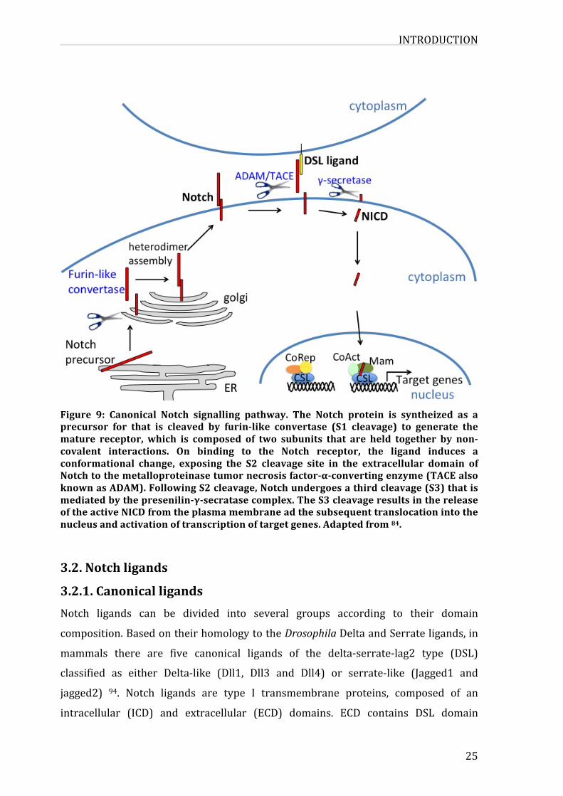
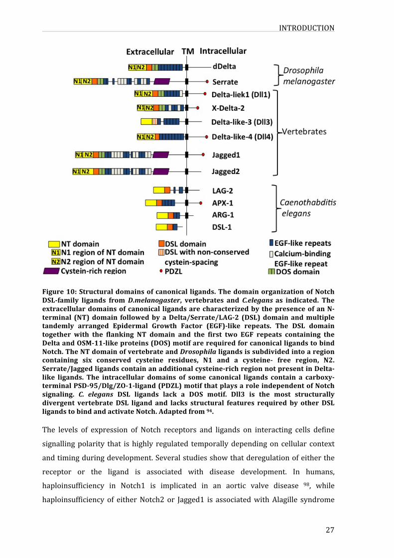
3.2.1.CanonicalligandsNotch ligands can be divided into several groups according to their domain
composition.BasedontheirhomologytotheDrosophilaDeltaandSerrateligands,in
mammals there are five canonical ligands of the delta-serrate-lag2 type (DSL)
classified as either Delta-like (Dll1, Dll3 and Dll4) or serrate-like (Jagged1 and
jagged2) 94. Notch ligands are type I transmembrane proteins, composed of an
intracellular (ICD) and extracellular (ECD) domains. ECD contains DSL domain
INTRODUCTION
followedbyEGFrepeatsbothcalciumandnon-calciumbinding.TheDSLdomainwith
thefirsttwoEGFrepeatsandtheDeltaandOSM-11-likeproteins(DOS)areessential
sitesforligandbindingtoNotchreceptor.Whiletheseligandshaveseveralconserved
domains features in their extracellular domain (according to their sequence
alignmentsandfunction),theintracellulardomainlacksnotablesequencehomology.
Some of DSL ligands contain a carboxy-terminal PSD-95/Dlg/ZO-1-ligand (PDZL)
motif, the role of this motif have been linked to interaction with the cytoskeleton
rather than to the notch signalling (figure 10). It has also been shown that the
cytoplasmic tail of these ligands contains several lysine residues representing
potentialubiquitinationsitestargetedbytheE3ubiquitinligase(Mind-Bomb1and2
andNeuralized 1 and 2 inmammals) leading to the subsequent endocytosis of the
ligand95.CanonicalligandshavebeenwelldefinedforactivatingtheNotchsignalling
by cell-to-cell contact in trans as described previously. On the other hand these
ligandshavebeendescribedfortheirroleincis inhibitioninNotchsignalling. Some
dataindicatethattransactivationorcisinhibitionimpliescompetitivemechanismsat
thereceptor-ligandinteractionlevel,withahighthresholdrequiredforcisinhibition96,andthattheciseffectismorelikelytopreventthesheddingofNotchecto-domain
byaninteractionmediatedbytheEGFrepeats10-12ofthereceptor97.
INTRODUCTION
27
Figure10:Structuraldomainsofcanonical ligands.ThedomainorganizationofNotchDSL-family ligands fromD.melanogaster, vertebrates and C.elegansas indicated. Theextracellulardomainsofcanonical ligandsarecharacterizedbythepresenceofanN-terminal (NT)domain followedbyaDelta/Serrate/LAG-2 (DSL)domainandmultipletandemly arranged Epidermal Growth Factor (EGF)-like repeats. The DSL domaintogether with the flanking NT domain and the first two EGF repeats containing theDeltaandOSM-11-likeproteins(DOS)motifarerequiredforcanonicalligandstobindNotch.TheNTdomainofvertebrateandDrosophilaligandsissubdividedintoaregioncontaining six conserved cysteine residues, N1 and a cysteine- free region, N2.Serrate/Jaggedligandscontainanadditionalcysteine-richregionnotpresentinDelta-like ligands. The intracellular domains of some canonical ligands contain a carboxy-terminalPSD-95/Dlg/ZO-1-ligand(PDZL)motifthatplaysaroleindependentofNotchsignaling. C. elegans DSL ligands lack a DOS motif. Dll3 is the most structurallydivergent vertebrate DSL ligand and lacks structural features required by other DSLligandstobindandactivateNotch.Adaptedfrom94.
The levels of expression of Notch receptors and ligands on interacting cells define
signallingpolarity that ishighly regulated temporallydependingon cellular context
andtimingduringdevelopment.Severalstudiesshowthatderegulationofeitherthe
receptor or the ligand is associated with disease development. In humans,
haploinsufficiency in Notch1 is implicated in an aortic valve disease 98, while
haploinsufficiency of eitherNotch2or Jagged1 is associatedwithAlagille syndrome
INTRODUCTION
(AGS) which is a dominant, multisystem disorder defined clinically by hepatic bile
ductpaucityandcholestasisinassociationwithcardiac,skeletal,andophthalmologic
manifestations. Ninety four percent of patients clinically diagnosed of AGS have
mutations in Jagged1 ligand 99. In addition to trans interaction betweenNotch and
ligands that activates the signallingmechanism, cis interaction between ligand and
receptor within the same cell also occur and limits the levels of activation by
inhibiting Notch signalling through the process defined as cis-inhibition thus
restricting Notch activation to signal-receiving cells 100. Therefore it became
important to understand the mechanism of cis-inhibition and the factors that
contribute to the regulationofcis or trans interactionbetween ligandand receptor.
Two different hypotheses have been proposed for this mechanism, Cordle et al.
assumethatinteractionsitesforbothcisandtransinhibitionoverlap101,whileother
define specific EGF repeats for each type of interaction. Though cis and trans-
interactionbindingsiteswithNotchmightoverlap,butonlytrans-ligandinteractions
activateNotchandinduceproteolyticcleavageduetoconformationalchangesofthe
receptor at the cell surface 102. Several data support that ligand inhibition can take
placeasacis-inhibitoryeffectactingbypreventingastepbeforeNotchecto-domain
sheddingandinvolvesaninteractionmediatedbyNotchEGFrepeats10-1297,101.
3.2.2.Non-canonicalligands
In addition to DSL ligands known as the canonical ligands of Notch, non-canonical
ligandslackingtheDSLdomainhavebeenidentified.ThiscategoryofNotchligandsis
divided into 3 subclasses: integral membrane-bound, GPI-linked membrane bound
andsecretedligands.
3.2.2.1.Membrane-boundnon-canonicalligands
The first non-canonical ligand identified was delta like 1 (DLK1). This ligand
representssimilaritiesinstructurewithdeltalikeligands;DLK1iscleavedbyADAM
metalloprotease and negatively regulated by Notch. The role of DLK1 was more
evidentincis-inhibitionofNotchsignalling,anditwasdescribedasanantagonistof
DSLligandsforNotchbindingandalsodecreasesexpressionofHes1103.
INTRODUCTION
29
Delta/Notch likeEGF related receptor (DNER) is another integralmembranebound
NotchligandlackingDSLdomain,andsimilartoDLK1itcontainsEGFrepeats.DNER
binds the Notch receptor in trans, and activates γ-secretase and Deltex-dependent
Notch signalling thus promoting neuron-glial interaction and leading to
morphologicaldifferentiationinthecentralnervoussystem(CNS)104.
In2007Krictsovetal.identifiedanovelDSLligandlikeproteincalledJedi(forJagged
and Delta protein (Jedi)), coding for a transmembrane protein containing multiple
EGF repeats, and expressed in early hematopoietic cells. They demonstrated that
soluble formof Jedi inhibitsNotch signalling, in a similarmanner to that of soluble
Jagged1, but there has been no proof that demonstrates the direct interaction
betweenJediandNotchreceptors105.
3.2.2.2.Membrane–boundGPI-linkednon-canonicalligandsThe identified Glycosylphosphatidilinositol linked neural cell recognitionmolecules
are the F3/contactin1 and NB3/contactin6. F3/contactin1 interacts with Notch
receptorandinducesitscleavageandnucleartranslocationpromotingmaturationof
oligodendrocytes. NB3/contactin6 promotes neural progenitor cell differentiation
into oligodendroctes by activating the Notch signalling pathway. It has been
demonstratedthatbothGPI-linked ligandspromoteNotch/DTX1signallingpathway106,107.
3.2.2.3.Secretednon-canonicalligandsInDrosophilamelanogaster twosecretedligandswereidentified:Scabrous(Sca)and
Wingless(Wn)theflyorthologofmammalianWntproteins.ScaandWnbothactivate
Notch signalling by trans-binding to the receptor. InC.Elegans five secreted ligands
lackingDSLdomainhavebeenidentified:OSM11,OSM7,DOS1,DOS2andDOS3.They
allcontainDOSmotif(DeltaandOSM-11)thatisconservedacrossspeciesandfound
incanonicalNotchligandsandoverlappingtheEGFmotifs.InteractionbetweenLin12
and OSM11 was detected in yeast two-hybrid but no other evidence showed that
Notchdirectly interactswithOSM.Ontheotherhand, theeffectofOSM11onNotch
signallingwasdemonstratedinC.elegans,duringvulvaldevelopment108.
INTRODUCTION
In vertebrate five putative secreted non-canonical ligands have been identified:
Connective Tissue Growth Factor/cysteine rich 61/Nephroblastoma Overexpressed
Gene familymember, CCN3/NOV 109, themicrofibril associated glycoprotein family
(MAGP1andMAGP2),thrombospondin2(TSP2),Yboxprotein1(YB1)andfinallythe
EGFlikedomain7(EGFL7).TheseligandsinteractwithNotchreceptors,inducingthe
activationofNotchsignallingindifferentcellularcontexts.CCN3,MAPG1,MAPG2and
TSP2 enhance Notch signalling induced by DSL ligands when exposed to Notch
receptorsorco-expressed inthesamecell 110-112.YB1,acoldshockproteinbindsto
Notch3, activates its nuclear translocation followed by an upregulation of its target
genes113.EGFL7wasfoundtobeexpressedinneuralstemcells(NSCs)whereitbinds
toaregioninNotchinvolvedinligand-mediatedreceptoractivation,thusactingasan
antagonist of Notch signalling and regulating their proliferation and differentiation114. It has been also demonstrated for the majority of these ligands, that the
interaction with the receptor activates CSL dependent reporter constructs in a γ-
secretasedependentmanner.
3.3.Notchtranscriptionalregulation
3.3.1.NICD-CSL-MAMLternarycomplexFollowingNotchactivationandcleavages,NICDtranslocatestothenucleus,whereit
activatesthetranscriptionofseveraltargetgenes.NICDcannotbinddirectlytoDNA;
it acts throughCSL transcription factor, enablingNotch to regulate geneexpression
andtranscription.CSLisforCBF1(C-promoterbindingfactor1),RBP-jk/Su(H)/Lag-1
inmammals/Drosophila/C.elegans),itiscomposedofthreedomains:N-terminalRel
homology domain (NTD), a central beta-trefoil domain (BTD) and a C-terminal Rel
homologydomain (CTD) 115. CSLproteinbinds to theDNA target gene regions that
was identified5’-CGTGGGAA-3’ 116. In theabsenceofNICD,RBP-jk formsa complex
with co-repressor proteins such as SMRT (silencing mediator retinoid and thyroid
receptors), N-coR (nuclear co-repressor), CBF1-interacting co-repressor associated
with histone deacethylase 1 (HDAC1), which prevents chromatin transcriptional
activation. SHARP (SMRTandHDACassociated repressorprotein) interactsdirectly
withRBP-jkandwithotherco-repressorssuchasSMRT/N-coRviaahighlyconserved
sequence in its SPOC-domain also called SHARP repression domain (SHARP-RD) at
theC-terminusendoftheprotein117.SHARPplaysacentralrolebyinteractingwith
INTRODUCTION
31
two different co-repressing complexes. It recruits CtIP/CtBP (C-terminal binding
protein/C-terminalinteractingprotein)orETOinadditiontootherco-repressorsand
histonemodifyingenzymes(HDAC)117,118(figure11). Itwasalsodemonstratedthat
SHARP recruits HDAC/SMART/SKIP containing complex 119. SKIP (Ski-interacting
protein) that was originally identified in a yeast two hybrid screen and CIR (CBF1
interacting co-repressor) are both direct RBP-j binding proteins, they bridge
interactionsbetweenthetranscriptionfactorandotherco-repressors.Somedataalso
demonstrated their role in mediating the interaction between Notch and CBF1
suggesting that they might also play a role as co-ativators 120, 121. When Notch is
activated,NICDrecruitsco-activators,displacingco-repressorsandformingaternary
proteincomplexSBF-NICD-MAMboundtoDNApromoterregion122,wheretheBTD
domainofCSLinteractswiththeRAMdomainofNICDbutonlyweaklywiththeANK
domainviaitsCTD123,andMAMinteractsdirectlywithNICDviatheANKyrinrepeats
andalsowithotherco-activators,likeCBP/p300(histoneacetyltranferase)througha
transactivation domain TAD1 in the C-term of Mastermind. P/CAF and GCN5, are
conservedhistoneacetyltransferases,thatweredescribedfortheirroleintheRBP-jk-
mediated transactivation by NICD, interact through their N-terminal regions with
activatedNotch124.OtherfactorsarerecruitedbythisternarycomplexsuchasSKIP
which is thought to displace co-repressors, and recruits kinases that specifically
phosphorylate NICD in order to be later targeted by FBXW7 for subsequent
proteosomaldegradation121.
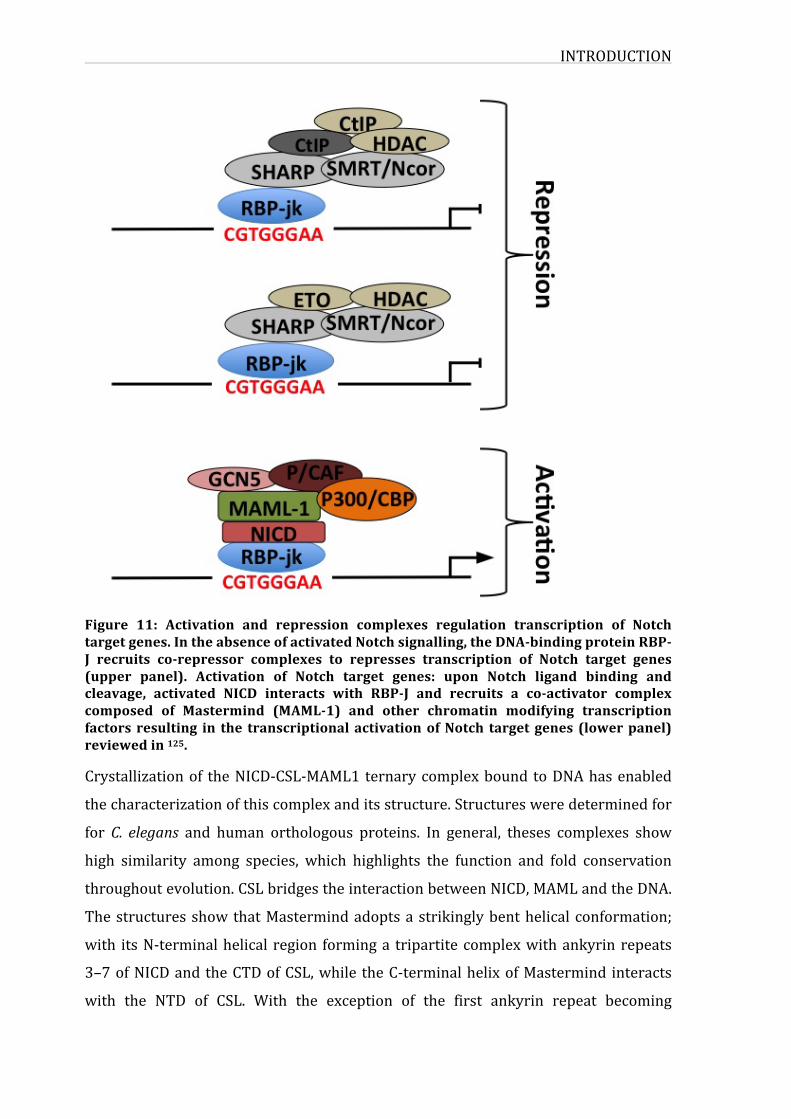
INTRODUCTION
Figure 11: Activation and repression complexes regulation transcription of Notchtargetgenes.IntheabsenceofactivatedNotchsignalling,theDNA-bindingproteinRBP-J recruits co-repressor complexes to represses transcription of Notch target genes(upper panel). Activation of Notch target genes: upon Notch ligand binding andcleavage, activated NICD interacts with RBP-J and recruits a co-activator complexcomposed of Mastermind (MAML-1) and other chromatin modifying transcriptionfactors resulting in the transcriptionalactivationofNotch targetgenes (lowerpanel)reviewedin125.
Crystallizationof theNICD-CSL-MAML1ternarycomplexboundtoDNAhasenabled
thecharacterizationofthiscomplexanditsstructure.Structuresweredeterminedfor
for C. elegans and human orthologous proteins. In general, theses complexes show
high similarity among species, which highlights the function and fold conservation
throughoutevolution.CSLbridgestheinteractionbetweenNICD,MAMLandtheDNA.
ThestructuresshowthatMastermindadoptsastrikinglybenthelicalconformation;
with itsN-terminalhelical region forminga tripartitecomplexwithankyrinrepeats
3–7ofNICDandtheCTDofCSL,while theC-terminalhelixofMastermind interacts
with the NTD of CSL. With the exception of the first ankyrin repeat becoming
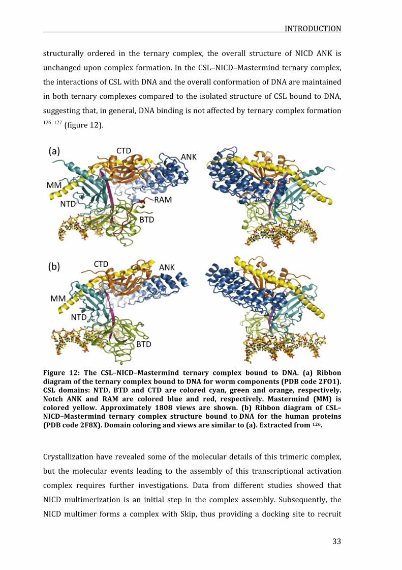
INTRODUCTION
33
structurally ordered in the ternary complex, the overall structure of NICD ANK is
unchangeduponcomplexformation. IntheCSL–NICD–Mastermindternarycomplex,
theinteractionsofCSLwithDNAandtheoverallconformationofDNAaremaintained
inbothternarycomplexescomparedtothe isolatedstructureofCSLboundtoDNA,
suggestingthat,ingeneral,DNAbindingisnotaffectedbyternarycomplexformation126, 127 (figure12).
Figure 12: The CSL–NICD–Mastermind ternary complex bound to DNA. (a) RibbondiagramoftheternarycomplexboundtoDNAforwormcomponents(PDBcode2FO1).CSL domains: NTD, BTD and CTD are colored cyan, green and orange, respectively.Notch ANK and RAM are colored blue and red, respectively. Mastermind (MM) iscolored yellow. Approximately 1808 views are shown. (b) Ribbon diagram of CSL–NICD–Mastermind ternary complex structure bound toDNA for the human proteins(PDBcode2F8X).Domaincoloringandviewsaresimilarto(a).Extractedfrom126.
Crystallizationhaverevealedsomeof themoleculardetailsof this trimericcomplex,
but the molecular events leading to the assembly of this transcriptional activation
complex requires further investigations. Data from different studies showed that
NICDmultimerization is an initial step in the complex assembly. Subsequently, the
NICDmultimer forms a complexwith Skip, thus providing a docking site to recruit
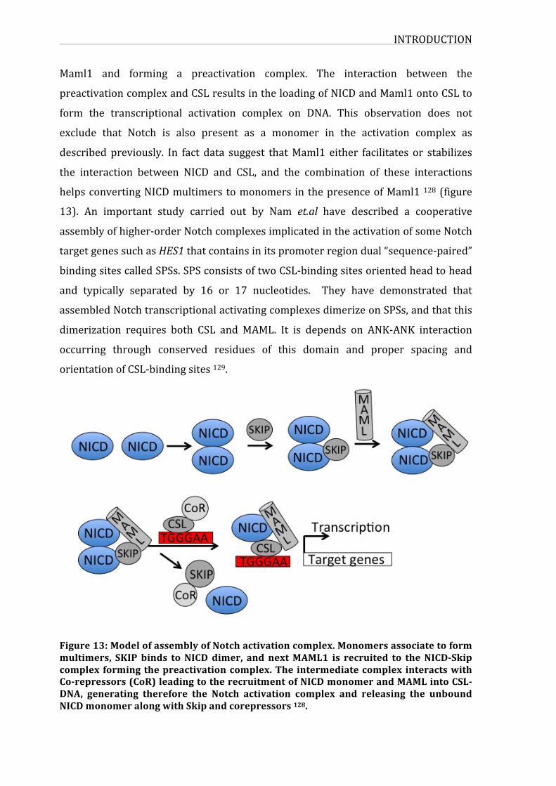
INTRODUCTION
Maml1 and forming a preactivation complex. The interaction between the
preactivationcomplexandCSLresultsintheloadingofNICDandMaml1ontoCSLto
form the transcriptional activation complex on DNA. This observation does not
exclude that Notch is also present as a monomer in the activation complex as
described previously. In fact data suggest thatMaml1 either facilitates or stabilizes
the interaction between NICD and CSL, and the combination of these interactions
helpsconvertingNICDmultimerstomonomers in thepresenceofMaml1128(figure
13). An important study carried out by Nam et.al have described a cooperative
assemblyofhigher-orderNotchcomplexesimplicatedintheactivationofsomeNotch
targetgenessuchasHES1thatcontainsinitspromoterregiondual“sequence-paired”
bindingsitescalledSPSs.SPSconsistsoftwoCSL-bindingsitesorientedheadtohead
and typically separated by 16 or 17 nucleotides. They have demonstrated that
assembledNotchtranscriptionalactivatingcomplexesdimerizeonSPSs,andthatthis
dimerization requires both CSL and MAML. It is depends on ANK-ANK interaction
occurring through conserved residues of this domain and proper spacing and
orientationofCSL-bindingsites129.
Figure13:ModelofassemblyofNotchactivationcomplex.Monomersassociatetoformmultimers, SKIPbinds toNICDdimer, andnextMAML1 is recruited to theNICD-Skipcomplex formingthepreactivationcomplex.The intermediatecomplex interactswithCo-repressors(CoR)leadingtotherecruitmentofNICDmonomerandMAMLintoCSL-DNA, generating therefore the Notch activation complex and releasing the unboundNICDmonomeralongwithSkipandcorepressors128.
INTRODUCTION
35
3.3.2.NOTCH1targetgenes
3.3.2.1.HESandHERPgenesNotch regulates the transcription of several target genes, activation ofNotch at the
surface followed by the formation of the NICD-RBPj-k complex leads to an
upregulation of the expression of primary target genes. The most known are the
HES/E(spl) (for Enhancer of split in Drosophila). Several studies have provided
evidence showing that these genes are directNotch targets, includingpromoters of
Hes1,Hes5andHes7aswellasHey1,Hey2andHeyL(subfamilyofHes,relatedwith
YRPW motif). Hes and Hey proteins are helix-loop-helix transcription factors that
functionas transcriptionalrepressors 130.Othereffectorsof theNotchsignallingare
bHLH family that has been isolated and named as
Hey/Hesr/HRT/CHF/gridlock/HERP (hereafterHERPorHES relatedproteins).This
familyalso functionsas transcriptionalrepressors,butemploysdifferentrepression
mechanisms than HES 131. bHLH family are classified into several groups. Class A
proteinsthataretranscriptionalactivatorssuchasMyoDandMash1,classBproteins
are leucinzipperproteinssuchasMycandMax.ClassCproteinsaretranscriptional
repressorsHESandHERPproteins.HESproteinsarecharacterizedbythepresenceof
a conservedprolineresidueata specific site in theirbasicdomain,as for theHERP
familyhasaconservedglycineatthecorrespondingposition.AllHESfamilymembers
shareaWRPWmotif in theC-terminuswhereasHERPfamilyhasYRPWmotif.Both
HES and HERP families contain bHLH domain, and another domain, termed the
Orange (or helix3- helix4) in the corresponding regions carboxy-terminus to bHLH
region.Althoughbothfamilieshaveclosely-relatedmotifsanddomains,theyseemto
employ different mechanisms for transcriptional repression. Three different
mechanismsofrepressionweredescribedforHESproteins;(1)anactiverepression
whereHEShomodimerbindsDNAandinteractswithproteinsrepressors(Grouchoin
Drosophila TLE ortholog in mammals) then recruits histone deacethylase proteins
that can alter chromatin structure and repress gene transcription. (2) Passive
repression where HES protein can form heterodimer with other HES activating
proteins (suchasMyoD)disrupting the formationof functionalheterodimers. (3)A
mechanismmediatedbytheorangedomain/helix3-helix4thatrecruitsco-repressors
orstabilizestheWRPW-mediatedrepression132.HERPproteinsassociatewithN-CoR,
they can recruit other repressor complexes such as Sin3/HDAC. In additionpassive
INTRODUCTION
repressionmechanismhavealsobeensuggestedfortheHERPfamily133.
Regulation of HERP and HES by Notch signalling is essential for different
developmental processes including angiogenesis, somitogenesis myogenesis and
gliogenesis134.
3.3.2.2.OtherNotchtargetgenes
MembersofthebHLHfamilywereidentifiedasprimarytargetsofNotch,butthisfact
does not exclude that Notch targets a large number of effectors as it has been
demonstrated by several studies. Notch1 exerts its multiple effects by specifically
regulating the expression of genes depending on cellular context. As an example,
genome-widetranscriptomeanalysesofNotch1-inducedgenes inmurineESCunder
different cell extrinsic cues and inmesodermal cells revealed that Notch signalling
regulates theexpressionofnumerousgenesplayingkeyroles incelldifferentiation,
cell cycle control and apoptosis. Meier-Stiegen et.al identified specific Notch target
genes using genomewide gene expression arrays,with 465 transcripts that can be
differentially expressed due toNotch1 induction in different conditions. Theywere
abletovalidatetranscriptionfactorSox9,Pax6,Runx1,Myf5andIdproteinsasdirect
Notch1 targets that are critically involved in lineage decisions in the absence of
proteinsynthesis135.
Notchpromotesproliferationvia regulating the transcriptionofCyclin-DandCDK5
and can directly control apoptosis effector genes such as bcl2. It was also
demonstrated that Notch regulates the expression of Deltex, a ubiquitin ligase that
regulates Notch1 trafficking and TCR in T cells 136. NRARP (for Notch-regulating
ANKyrin repeatsprotein) anegative regulatorofNotch is alsooneofNotch targets
regulated through DLL4/Notch signalling. NRARP is implicated in the cross talk
between Notch and Wnt signalling in endothelial cells to control stability of new
vesselconnectionsinmouseandzebrafish137.
Notchalsoregulatesitsownexpression;itcandirectlybindautoregulatoryCSLsites
inNotchlocusincreasingitsownexpression138.
ChIPonchipanalysiscombinedwithgeneprofilingdata,enabledtheidentificationof
NOTCH1directtargetgenesregulatingcellgrowthgenesandTcelltransformation.In
INTRODUCTION
37
thisstudyc-MYCwasidentifiedasadirecttargetgeneregulatedbyNOTCH1,andthis
interactionispartofafeed-forward-looptranscriptionalregulatorymotifimplicated
in the leukemic cell growth where some of c-MYC and NOTCH1-regulated genes
overlap56,76.
Now it became evident that Notch directly regulates expression of genes encoding
proteinsthatimplementvariablecellularfunctionsdependingoncellularcontext. In
factaspreviouslydescribed,systematicstudiesofNotchtargetshaverevealedseveral
regulatorymotifscontrolledbynotchandlinkedtoothersignallingpathways.
3.4.Post-translationalmodificationsinNotchsignalling
Notchsignallingishighlydependentoftheligand-receptorinteractionrelatedtothe
expressionofthesetwocomponentsatthecellsurface.Bothligandandreceptorare
subjecttopost-translationalmodificationssuchasglycosylationandubiquitination.
3.4.1.GlycosylationNotch receptors and ligands both undergo glycosylation, their correspondent
extracellulardomainsaremodifiedbyavarietyofglycansthatcanregulateaspectsof
Notchsignallingandactivation.Severalmutationsreportedinenzymesthatregulate
the glycosylation mechanisms are associated with Notch signalling disruption and
deregulation.
After being translated in the Golgi, Notch protein is fucosylated by a GDP fucose
protein O-fucosyltransferase (Ofut-1 in Drosophila and Pofut1 inmammals) within
EGFrepeatsoftheECDtoaserineorthreonineresidue,facilitatingtheproperfolding
of theprotein in theendoplasmic reticulum (ER). Subsequent to theO-fucosylation,
theEGFO-linked fucosechain iselongatedbytheglycosyltransferase,Fringe,which
attaches N-acetylglucosamine (GlcNAc) in a β1,3 linkage 139. Fringe is a
glycosyltransferase which transfers N-acetylglucosamine (GlcNAc) to fucose on the
EGFrepeatsofNotch,itwasfirstdiscoveredinDrosophila140,whereitcanmodulate
dorsal-ventralcellinteractionsinthedevelopingwingbymodulatingtheresponseof
Notch to its ligands and later onmammalian homologues were identified: Lunatic,
ManicanRadicalFringe141.MammalianFringemighthavedifferentcatalyticactivities,
INTRODUCTION
it has been shown in different animal models (mice, zebrafish…) that they are
differentiallyexpressedaccordingtocellularcontext141,142.Modificationsinducedby
Fringe contribute to the receptor-ligand binding interaction 143, it has also been
demonstratedthatreducedlevelsofO-linkedfucosedecreaseNotchactivation,which
highlightstheirroleinmodulatingNotchactivation.Ontheotherhandsomestudies
showed that a normal Notch activation is also possible in the absence of Fringe
proteins,supportingthehypothesisthatfucosylationandglycosylationareimportant
inmodulatingthesignalstrengthbutnotobligatoryinthenotchpathwayactivation.
Transfer of fucose residues to the extracellular domain of Notch is catalyzed byO-
fucosyltransferaseencodedbyOfut1gene inDrosophilaand thePofut1homologous
gene inmammals. This enzyme transfers fucose to serine or threonine amino acid
residues in the EGF repeats 144.O-fucosyltransferase and Fringe alsomodify Notch
ligands,DeltaandSerrateinDrosophilaandmammals145.
In addition to O-fucose glycans, O-glucose glycans were also discovered. O-glucose
glycansarecatalyzedbyO-glucosyltransferases,thisenzymeinencodedbyrumigene146, and elongation ofO-glucose residues is possible by addition of xylose residues
throughthefunctionspecificxylostransferases,forexamplebothNotch1andNotch2
are extended by the addition of one or two xylose residues. In fact it has been
demonstrated that Rumi is required for both Drosophila and mammalian Notch
signalingatastepdownstreamofligandbinding142.
Another modification that has been identified is the presence of O-GLcNAc in the
intracellulardomainofNotch,andsurprisinglyforthefirsttimeasamodificationin
the extracellular of Notch receptors 147. Thesemodifications by glycosylation occur
duringtransitthroughtheendoplasmicreticulum(ER)andGolgiapparatuswherethe
glycosylation machinery adds glycans progressively by the corresponding
glycosyltransferases.
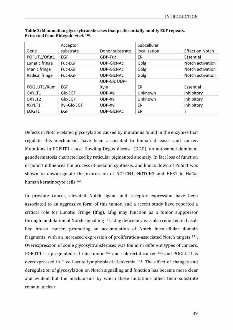
INTRODUCTION
39
Table2:MammalianglycosyltransferasesthatpreferentiallymodifyEGFrepeats.ExtractedfromHideyukietal.148.
GeneAcceptorsubstrate Donorsubstrate
Subcellularlocalization EffectonNotch
POFUT1/Ofut1 EGF GDP-Fuc ER EssentialLunaticfringe Fuc-EGF UDP-GlcNAc Golgi NotchactivationManicfringe Fuc-EGF UDP-GlcNAc Golgi NotchactivationRadicalfringe Fuc-EGF UDP-GlcNAc Golgi Notchactivation
POGLUT1/Rumi EGFUDP-GlcUDP-Xyla ER Essential
GXYLT1 Glc-EGF UDP-Xyl Unknown InhibitoryGXYLT2 Glc-EGF UDP-Xyl Unknown InhibitoryXXYLT1 Xyl-Glc-EGF UDP-Xyl ER InhibitoryEOGT1 EGF UDP-GlcNAc ER ?
DefectsinNotch-relatedglycosylationcausedbymutationsfoundintheenzymesthat
regulate this mechanism, have been associated to human diseases and cancer.
Mutations in POFUT1 cause Dowling-Degos disease (DDD), an autosomal-dominant
genodermatosischaracterizedbyreticularpigmentedanomaly.Infactlossoffunction
ofpofut1influencestheprocessofmelaninsynthesis,andknockdownofPofut1was
shown to downregulate the expression of NOTCH1, NOTCH2 and HES1 in HaCat
humankeratinocytecells149.
In prostate cancer, elevated Notch ligand and receptor expression have been
associated to an aggressive formof this tumor, and a recent study have reported a
critical role for Lunatic Fringe (lfng). Lfng may function as a tumor suppressor
throughmodulationofNotchsignalling150.Lfngdeficiencywasalsoreportedinbasal-
like breast cancer, promoting an accumulation of Notch intracellular domain
fragments,withanincreasedexpressionofproliferation-associatedNotchtargets151.
Overexpressionofsomeglycosyltransferaseswasfoundindifferenttypesofcancers.
POFUT1 isupregulated isbrain tumor 152andcolorectal cancer 153andPOGLUT1 is
overexpressed inT cell acute lymphoblastic leukemia 154.Theeffect of changes and
deregulationofglycosylationonNotchsignallingandfunctionhasbecamemoreclear
and evident but the mechanisms by which these mutations affect their substrate
remainunclear.
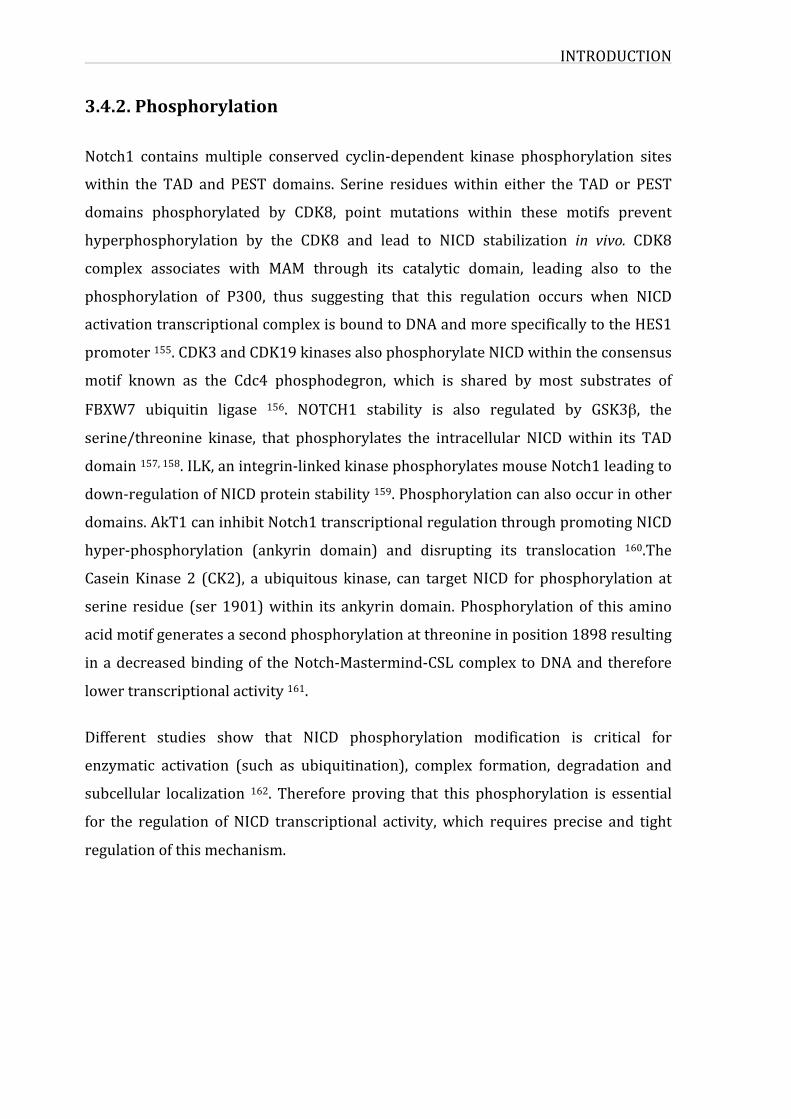
INTRODUCTION
3.4.2.Phosphorylation
Notch1 contains multiple conserved cyclin-dependent kinase phosphorylation sites
within the TAD and PEST domains. Serine residueswithin either the TAD or PEST
domains phosphorylated by CDK8, point mutations within these motifs prevent
hyperphosphorylation by the CDK8 and lead to NICD stabilization in vivo. CDK8
complex associates with MAM through its catalytic domain, leading also to the
phosphorylation of P300, thus suggesting that this regulation occurs when NICD
activationtranscriptionalcomplexisboundtoDNAandmorespecificallytotheHES1
promoter155.CDK3andCDK19kinasesalsophosphorylateNICDwithintheconsensus
motif known as the Cdc4 phosphodegron, which is shared by most substrates of
FBXW7 ubiquitin ligase 156. NOTCH1 stability is also regulated by GSK3β, the
serine/threonine kinase, that phosphorylates the intracellular NICDwithin its TAD
domain157,158.ILK,anintegrin-linkedkinasephosphorylatesmouseNotch1leadingto
down-regulationofNICDproteinstability159.Phosphorylationcanalsooccurinother
domains.AkT1caninhibitNotch1transcriptionalregulationthroughpromotingNICD
hyper-phosphorylation (ankyrin domain) and disrupting its translocation 160.The
CaseinKinase 2 (CK2), a ubiquitous kinase, can targetNICD for phosphorylation at
serine residue (ser1901)within its ankyrindomain.Phosphorylationof this amino
acidmotifgeneratesasecondphosphorylationatthreonineinposition1898resulting
inadecreasedbindingof theNotch-Mastermind-CSLcomplex toDNAandtherefore
lowertranscriptionalactivity161.
Different studies show that NICD phosphorylation modification is critical for
enzymatic activation (such as ubiquitination), complex formation, degradation and
subcellular localization 162. Therefore proving that this phosphorylation is essential
for the regulation ofNICD transcriptional activity,which requires precise and tight
regulationofthismechanism.
INTRODUCTION
41
3.4.3.Ubiquitination
3.4.3.1.UbiquitinationmechanismUbiquitination is a post-translational modification essential for proteasomal
degradation, which is the main degradation path in eukaryotes. Ubiquitination
controlsseveralcellularprocessesviadegradation.Ubiquitinationiscarriedoutbya
three-stepprocessthatstartsbyanactivationstep,followedbyconjugationandends
by the ligation step. Ubiquitin is a small protein consisting of only 76 amino acid
polypeptideof8,5Kda,itishighlyconservedfromyeasttohuman.Thismodification
was initially linked to protein half-life since ubiquitinated proteins undergo
proteosomalATP-dependentdegradation.UbiquitinisaddedfromitsC-terminustoa
lysineresidueofasubstrate,duetothecontributionofasetofthreeenzymes;anE1
ubiquitinactivatingenzymethatbindstheubiquitinviaathioestherbondinanATP-
dependentmanner,thancarriestheubiquitintoanE3ubiquitin-conjugatingenzyme.
ThefinalstepisperformedbytheE3ubiquitinligasethatbindsboththechargedE2
enzymeandthesubstrate facilitatingtheirbindingtotheubiquitin(figure14).Only
twoE1enzymesarefoundinhumans,and37E2enzymeswhileawiderangeofE3
enzymes were identified accounting for more than 600 encoded by the human
genome. Ubiquitination is a reversible process therefore deubiquitinaseswere also
identifiedwithanumberof≈85enzymesinhumans163.
Different forms of ubiquitinationwere described, according to the number and the
typeofubiquitindisplayonaminoacidswithinthesubstrate.Monoubiquitinationis
addingoneubiquitintoasubstrate,whilemulti-monoubiquitinationistheadditionof
severalubiquitinmoietieseachonasinglelysineresidueofthetargetedprotein.An
importantcharacteristicofubiquitinisthatitcontainssevenlysineresiduesthatare
also subjected for subsequent ubiquitination, serving as substrates for
polyubiquitination. Several studies were conducted on ubiquitin structure and
complexity, more specifically on the role of lysine residues in determining the
specificity towards substrate recognition and protein processing. Ubiquitination
throughlys48isessentialforpolyubiquitinationtargetedbyproteosomaldegradation
(lys63canalsoactasaproteasomalsignal),whilemonoubiquitinationthroughlys6is
involved in DNA repair. Lysosomal degradation and protein recycling also require
INTRODUCTION
ubiquitinationandmorepreciselymulti-monoubiquitinationthroughlys29,allowing
endosomalsortingoftargetedproteinsthroughESCRTmachinery(endosomalsorting
complex required for transport). Interestingly, proteins involved in these processes
employ highly specific ubiquitin-binding domains, which recognize specific Lysine
residuesandubiquitinchaintypesspecificallyregulatingdifferentaspectsofcellular
biology164.
Figure 14: Ubiquitination mechanism. The ubiquitination cascade begins with ATP-dependent charging of the E1 enzyme and results in formation of a thioester bondbetween the ubiquitin C terminus and the E1 active site cysteine. Ubiquitin istransferred to the E2 active site cysteine in a transthioesterification reaction. An E3ligase catalyzes transfer of the ubiquitin from the active site cysteine of the E2 to aprimaryamineonalysinesidechainorproteinNterminus.TherearethreeclassesofE3 ligases:RINGE3s,whichbind tobothE2~Ub thioester and substrate and catalyzeattackofthesubstratelysineonthethioester,andHECTandRBRE3s,whichbothhaveactive site cysteines and catalyze substrate ubiquitination in a two-step reactioninvolving formationofa thioesterwith theHECTorRBRE3 followedbyattackof thesubstrate lysine or N terminus on the E3~Ub thioester to form an isopeptide (orpeptide) linkage between the ubiquitin C terminus and lysine (or the protein Nterminus).Adaptedfrom165.
INTRODUCTION
43
3.4.3.2.UbiquitinationintheNotchsignallingpathwayUbiquitinationofNotchandits ligandsplaysanimportantrole inmodulating ligand
endocytosis, their level of expression at the cell surface and in regulating Notch
stabilityanddegradation.
MultiplelysineresiduesserveaspotentialubiquitinationsitesinDSLdomainofNotch
ligands,andtheyaretargetedbyE3ubiquitinligasesMinbomb(Mb)andNeuralized
(Neur) in both human and Drosophila. Ubiquitination is important for generating
active ligand via endocytosis where several mechanisms have been described. The
ubiquitinited ligand can be internalized by endocytosis in sorting or recycling
endosomestobelaterexpressedatcellsurfaceasactiveligands.Ubiquitinationmay
alsooccurafterligandbindingtoNotch,leadingtoendocytosisofboththeligandand
Notch extracellular domain in a Clathrin-coated endocytic structure 166. The pulling
force resulting from this mechanism helps exposing the S2 site tometalloprotease
complex,andsubsequentcleavagereleasesNICDinthecell85.
Ubiquitination of the receptor occurs in two different contexts; the first is during
receptorendocytosisandthesecondinNotchproteosomaldegradation.
Unliganded Notch receptor in endocytosed into rab5 and endocytic syntaxin
avalanche containing compartiments, where Notch is either recycled back to cell
surfaceordirectedforlysosomaldegradation.Inearlyendosomes,fulllengthNotchis
targetedbytheRINGfingerE3ubiquitin ligases:Deltex(Dx)andNedd4and/orCbl.
Ubiquitinated Notch is sorted into multivascular bodies or lysosomes by ESCRT
components (Endosomal Sorting Complex Required for Transport) for subsequent
degradation.Throughout thisphase, anactivationofNotch canoccur inendosomes
whereV-ATpasearethoughttoplayaroleinendosomalacidificationrequiredforγ-
secretase dependent cleavage 166. Another E3 ubiquitin ligase target Notch, it is
FBXW7 (WD40-repeat containing F-Box protein component of an Skp1/Cul1/F-box
protein- Rbx1-type ubiquitin ligase) promoting its proteosomal degradation (figure
15).WhilepreviouslyreportedE3ligaseDxandNedd4targetNECD,FBXW7targets
NICD. It hasbeenalsodemonstrated that Itch, anotherE3 ligase thatubiquitinates
NICD,negativelyregulatesNotchmediatedsignalling167.
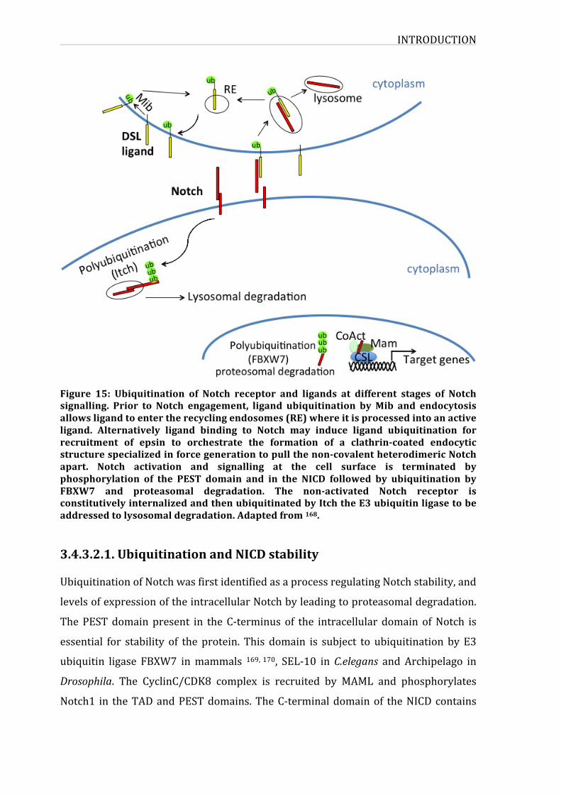
INTRODUCTION
Figure 15: Ubiquitination of Notch receptor and ligands at different stages of Notchsignalling. Prior to Notch engagement, ligand ubiquitination byMib and endocytosisallowsligandtoentertherecyclingendosomes(RE)whereitisprocessedintoanactiveligand. Alternatively ligand binding to Notch may induce ligand ubiquitination forrecruitment of epsin to orchestrate the formation of a clathrin-coated endocyticstructurespecializedinforcegenerationtopullthenon-covalentheterodimericNotchapart. Notch activation and signalling at the cell surface is terminated byphosphorylation of the PEST domain and in the NICD followed by ubiquitination byFBXW7 and proteasomal degradation. The non-activated Notch receptor isconstitutivelyinternalizedandthenubiquitinatedbyItchtheE3ubiquitinligasetobeaddressedtolysosomaldegradation.Adaptedfrom168.
3.4.3.2.1.UbiquitinationandNICDstabilityUbiquitinationofNotchwasfirstidentifiedasaprocessregulatingNotchstability,and
levelsofexpressionoftheintracellularNotchbyleadingtoproteasomaldegradation.
ThePESTdomainpresent in theC-terminus of the intracellular domain ofNotch is
essential for stability of the protein. This domain is subject to ubiquitination by E3
ubiquitin ligase FBXW7 inmammals 169,170, SEL-10 inC.elegans and Archipelago in
Drosophila. The CyclinC/CDK8 complex is recruited by MAML and phosphorylates
Notch1 in theTADandPESTdomains.TheC-terminaldomainof theNICDcontains
INTRODUCTION
45
multiple conserved cyclin-dependent kinase phosphorylation sites that can be
phosphorylated by CycC:CDK8 in vitro. A study showed that mutation of Serine
residues within either the TAD or PEST domains of the NICD can stabilize the
protein155.NuclearNotchcanalsobephosphorylatedbyGSK3β.Itwasdemonstrated
that Notch2 interacts with GSK3β in vitro and in vivo, and phosphorylates specific
threonine and serine residues in the serine threonine rich region located in the C-
terminaloftheankyrinrepeats,andregulatesnegativelyNICDtranscriptionalactivity.
ThoughphosphorylatedformsofNotchinteractwithFBXW7fordegradation,GSK3β
mediated phosphorylation seems to regulate Notch by a different mechanism and
doesnot leadto itssubsequentdegradation157.GSK3βisalsoabletophosphorylate
Notch1 and enhances NICD1 stability 158, and could also up-regulate NICD
transcriptionalactivitybyenhancing itsnuclear localization171.WhenFBXW7binds
thephosphorylatedresidues inthePESTdomainofNotch,NICDisdegradedviathe
proteasome.
3.4.3.2.2.UbiquitinationofNotchatthecellsurface
AtthecellsurfaceNotchisactivatedbyligandsandthereceptorwillundergoaseries
ofproteolyticcleavages.Notchquantityishighlyregulatedatthecellsurface;ithasa
limitedhalf-lifeandisconstantlyinternalized,recycledordegradedviathelysosome.
Notchreceptoranditsligandcanalsobeinternalizedintoendocyticvesicles.Itcanbe
cleavedbyγ-secretase inthecytoplasmreleasingNICDtothenucleus.Butnormally
after internalization, Notch is recycled back to the membrane or degraded.
Ubiquitinationplaysan importantrole in theregulationofNotchat thecell surface.
Non-activatedNotchreceptorisconstitutivelyinternalized;Itch/AIP4themammalian
E3 ubiquitin ligase targets Notch for ubiquitination and promotes its lysosomal
degradation172.InDrosophilaSu(dx)performsthesamefunctionasmammalianItch
and regulates endosomal sorting of Notch 173. Itch seems to bind Notch indirectly
through interactingwithotherprotein factorssuchasNumbthat interactswith the
cytosolicHECTdomaincontainingofthisE3ligaseandactscooperativelywithItchto
promoteubiquitinationofmembrane-tetheredNotch1174.TheRINGfingercontaining
E3ligasec-Cblalsoregulatestheubiquitinationofmembrane-boundNotch1andhas
beenreportedtoresultinlysosomaldegradationofNotch1 175.
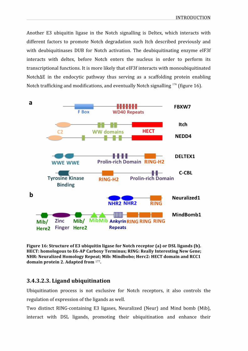
INTRODUCTION
Another E3 ubiquitin ligase in the Notch signalling is Deltex, which interacts with
different factors to promote Notch degradation such Itch described previously and
with deubiquitinases DUB for Notch activation. The deubiquitinating enzyme eIF3f
interacts with deltex, before Notch enters the nucleus in order to perform its
transcriptionalfunctions.ItismorelikelythateIF3finteractswithmonoubiquitinated
NotchΔE in the endocytic pathway thus serving as a scaffolding protein enabling
Notchtraffickingandmodifications,andeventuallyNotchsignalling 176 (figure16).
Figure16:StructureofE3ubiquitinligaseforNotchreceptor(a)orDSLligands(b).HECT:homologoustoE6-APCarboxyTerminus;RING:ReallyInterestingNewGene;NHR:NeuralizedHomologyRepeat;Mib:Mindbobo;Herc2:HECTdomainandRCC1domainprotein2.Adaptedfrom177.
3.4.3.2.3.LigandubiquitinationUbiquitination process is not exclusive for Notch receptors, it also controls the
regulationofexpressionoftheligandsaswell.
Two distinct RING-containing E3 ligases, Neuralized (Neur) andMind bomb (Mib),
interact with DSL ligands, promoting their ubiquitination and enhance their
INTRODUCTION
47
endocytosis. Ubiquitination of ligands is required for transendocytosis and ligand
recyclingatthecellsurface;twophenomenanecessaryforNotchactivationthrough
itsligands.
Transendocytosisandligandrecycling
Upon receptor-ligand interaction, a proteolytic cleavage is produced and Notch is
released in the cytoplasmof the signal-receiving cell,while the ligandexpressedon
the surface of the signal-sending cell is internalized. In fact, in the absence of
endocytocis, DSL ligands accumulate at the cell surface, unable to activate Notch.
Moreover mechanical forces generated during DSL ligand endocytosis function to
physically dissociate Notch, and that dissociation is a necessary step in Notch
activation166.Insomeoftheendocyticevents,monoubiquitinatedDSLligandsrequire
thepresenceofEpsinaconservedclathrin-interactingendocyticadaptor.Epsinbinds
phosphatidylinositol4,5-biphosphate (PIP2), inserts into theplasmamembraneand
induces membrane curvature. Then the epsin C-terminus recruits clathrin directly
and also AP-2 and/or Eps15 to eventually promote clathrin-dependent ligand
endocytosis178,179.OneofthehypothesesproposedforDSLligandintheregulationof
Notch signalling is that internalization of the ligand and their entry into coated
vesicles in the signal-sending cell is an essential process leading to shedding of the
extracellular Notch, thus enabling its cleavage and release in the cell 180. Another
hypothesis focusesmoreonevents that takeplaceafter the ligandsare internalized
whereseveralenzymaticprocessesoccurinordertorecycleactiveligandstothecell
surface181.
IntheabsenceofNotch,DSL ligandsundergoendocytosis, itwasdemonstratedthat
delta ligand was concentrated in recycling endosomes during specific steps of cell
determination in Drosophila 182. Rab5 and Rab11 colocalize with internalized DSL
ligands.RabGTPasesareassociatedwithdistinctcellularcompartmentsandfunction
as specific regulatorsof intracellular transport, in theendocyticpathway.Moreover
lossofRab11inducederegulationofandlossofDSLligandactivity182.
Recycling of the ligands before they interact with Notch is an important step for
propersignalling.Aftertheirendocytosis,ligandssuchasDll1andDll4undergomono
ormulti-ubiquitination. Itwasdemonstrated forDll1 thatubiquitination isessential
INTRODUCTION
forDll1 recycling and that recycling is required to acquire affinity for the receptor.
Dll1 mutant (Dll1K17R) is endocytosed but is unable to signal, this observation is
explained by the lack or absence of ubiquitination of this mutant. However the
chimeric ligand Dll1–3, although not ubiquitinated, was able to recycle to the cell
surface,indicatingthatanunknownsignalpresentintheintracellulardomainofDll3
canavoidthenecessityforubiquitination.Ontheotherhand,thismoleculeisunable
to signal. These data prove that ubiquitinationprocess is indeednecessary forDll1
recyclingandNotchactivation183.
Notch1 signalling and activation by the Dll4 ligand is crucial in intrathymic T cell
development.Dll4functiondependsonacombinationofseveralfactorsthatinvolve
interaction with the ubiquitin ligase Mib1 for ubiquitination, internalization and
recycling back to the cell surface for effective Notch signalling and therefore
supporting T cell development and inhibiting B cell development. In contrast with
Dll1,Dll4areexcludedfromlipidraftsinordertoberecycled184.Thoughtheymight
differ in their specific localization or the factors contributing to their subsequent
recyclingandmaturation,alldatapointatthenecessityofubiquitinationduringthis
process.
UbiquitinationeventsoccuratdifferentlevelsofNotchsignalling.Recentstudieshave
identified several new regulators in the ubiquitination and deubiquitination
mechanisms, raging from degradation to regulation of membrane trafficking and
affectingthedevelopmentalcontrolofthesignallingactivitiesofbothNotchreceptors
andtheirligands.
3.4.3.2.4.E3ubiquitinligasesInthispart,wewilldiscussmoreindetailE3ubiquitinligasesandmorespecifically
FBXW7 protein. As previouslymentioned, E3 ligases carry out the final step in the
ubiquitinationcascade,catalyzingtransferofubiquitinfromanE2enzymetoforma
covalentbondwithasubstrate lysine.ThreedistinctclassesofE3 ligaseshavebeen
identified the family of RING finger E3s, homolog to the E6AP carboxy terminus
(HECT)domain familyand theRing-between-Ring (RBR) family.Thegreatdiversity
found among these ligases is due to their specificity toward their targets, therefore
INTRODUCTION
49
each class is also characterizedby their conserveddomains and themechanismsof
ubiquitintransferfromE2enzymestothesubstrate185.
Ring E3 enzymes catalyze ubiquitin transfer process, the E2 binding domain of the
Ring domain binds the E2 enzyme through its N-terminus, and it also contains
additional domains required for substrate recognition and its subsequent
ubiquitination. Ring E3s can act as monomers, dimers or even multisubunit
complexes. An interesting superfamily of the Ring E3 ligases, that function as
complexesaretheCullinRingligasessuperfamily,consistingofmulti-subunitproteins
containing the cullinprotein, aRing-boxprotein, andanF-Boxproteinessential for
binding the substrate. The RING E3 act as scaffold that brings E2 and substrate
together, on the other hand it was suggested that RING finger domains can also
allosterically modify and activate E2 enzymes 186. HECT E3 ligases catalyze the
transferreaction ina two-stepprocessofwhat iscalledadirectubiquitination.The
firststepisthetransferoftheubiquitinfromtheE2conjugatingenzymetotheHECT-
domainE3 ligase,andnext thisubiquitinresidue is transferredfromthecysteine in
theE2activesitetothelysineresidueofthesubstrate187.TheHECTdomainconsists
of two lobes; one in the N-terminal lobe interacting with the E2 enzyme and the
second C-terminal lobe that contains the cysteine residue that forms a catalytic
intermediatewith theubiquitinmoiety.Duringubiquitin transfer theHECTdomain
undergoes conformational changes due to the flexible region separating the lobes,
thusallowingtheubiquitintransferfromtheE2enzymetothesubstrate188.TheRBR
(Ring-between-Ring)-domainE3ligasescontaintwoRingdomains(Ring1andRing2)
separatedbyaconserveddomaincalledtheIBRdomain(InBetweenRing),theyalso
share features with HECT domain ubiquitination mechanism directly catalyzing
ubiquitin transfer an intrinsic cysteine present in the Ring 2 domain, while Ring1
domain helps recruiting thioester-bound E2 enzymes. This class of E3 ligases is
characterizedbyhavinganauto-inhibitionmechanismthatmodulatesubiquitination
activity,andbycombiningaspectsofbothRingandHECTE3ligasesfunction189.The
resemblancewithHECTreliesinthemechanism,bywhichRing1domainbindstoE2-
ubiquitinviaacysteinyl-thiolwithinthesameE3protein.
INTRODUCTION
3.4.3.2.5.FboxproteinsTheSCF(SKP1-CUL1-FBP)familybelongstotheCullindependentligases(CDL)family
that has beenwell characterized. In this complex, SKP1 (S-phase-kinase associated
protein1)interactswiththeCul1proteinsfunctioningasascaffoldproteinthatsorts
the proteins destined for degradation. Cul1 is also bond to the RING-finger protein
(Rbx1),andtotheubiquitinconjugatingenzyme(UBC)andtooneoftheFBPs.FBPs
confer to the targeted substrates. Originally Skip1was found to be essential to for
interactionwith several proteins such asCdc4 and cyclin F via their 40 amino acid
domain that was later found to be a conserved domain characterizing the F-box
proteins190.
Mammalian F-box proteins are classified into three groups according to their
substrate-bindingdomain.WerecognizetheFBW(F-boxandWD40repeats),theFBL
(F-boxandLeucine-richrepeat)andtheFBXthatdonothaveeitheraWDdomainora
Leucine rich repeat. FBX class rather contains protein-protein interaction domains
suchaszinc-finger,proline–richdomain…FBPsrecognitionof theirsubstrateoften
requires specific post-translational modifications such as phosphorylation,
glycosylation,methylation,acetylationandsoon.In2004Cardozoetal.reviewedthe
SCF ubiquitin ligases, the mechanism of this protein complex was described and
compared to the mechanism known for typical enzymes. Therefore in the SCF
complex,theubiquitinistransferredinsteadofwhatisusuallyasmallchemicalgroup
forotherprotein enzymes.The activatedE2 conjugating enzyme canbe considered
likethekeycatalyticresidueoftheenzyme,whereastheentireFBPplaystheroleof
thesubstratebindingdomainintheenzymaticcomplex191.
As previously mentioned, the F-box proteins class consists of a wide variety of
proteins, due to their role as the substrate-recognizing subunits of the SCF E3
complex.Inthehumangenome,theF-boxproteinsaccountfor≈70proteins,themost
wellknownamongthemareFBXW7,Skp2andβ-TrCP.
A single F-box protein can target several proteins, for exampleNOTCH1, cMYC and
cJUN are targeted by FBXW7 for ubiquitination and subsequent proteasomal
degradation,ontheotherhandaproteincanbeatargetofdifferentF-boxproteins,
forinstanceCyclinEcanberecognizedbyFBXW7andSkp2192.
INTRODUCTION
51
3.4.3.2.6.FBXW7E3ubiquitinligaseFBXW7is thesubstraterecognitionunitof theconservedSCFE3ubiquitin ligaseas
previouslydescribed.FBXW7genewasoriginallyfoundinyeast,itiscalledCdc4,its
rolewasdescribedinthecontrolofthecell–cycle-regulateddisposalofSic1protein;
an inhibitor the cyclin-dependent kinase (CDK). Cdc4 binds specifically to
phosphorylatedresiduesoftheirsubstrates,alsoknownasphospho-degrons.FBXW7
is thehumanCdc4orthologue, itwasalsodemonstrated that inhuman, it catalyzes
thephosphorylation-dependentubiquitinationofcyclinE193.Inaddition,theflyCdc4
orthologueAGOalsoregulatescyclinEinDrosophilamelanogaster194.
In the human genome, FBWX7 gene encodes for three protein isoforms: FBW7α,
FBXW7βandFBXW7γ, thatareproducedbyalternativesplicingandthat localize to
thenucleoplasm,cytoplasm,andnucleolus,respectively.Severalstudieshavefocused
on defining exon-specific localization characteristics, and they demonstrated that
FBXW7 proteins comprise a common region containing three basic domains (BDs).
FBXW7α and FBXW7β both contain in their common region, localized at the N-
terminus of the protein, a nuclear localization signal (NLS). FBXW7α contains a
secondNLS, and itwas revealed that both NLSs are required for its nucleoplasmic
localization. Whereas FBXW7β contains a putative hydrophobic transmembrane
domain,within itsexon-specific region in theN-terminus, thatwasassociated to its
cytoplasmic localization195.Theα isoformofFBXW7constitutes themostabundant
and major isoform of cellular FBXW7, and data also suggest that FBXW7α is
responsible for cMYC and Cyclin E degradationwhereasNotch is targeted by the β
isoform196.
3.4.3.2.7.FBXW7substratesFBXW7 has pivotal roles in cell division, growth, and differentiation by targeting
several proteins. Approximately 20 substrates are known to be targets of FBXW7;
amongst them themost studied are the oncoproteins c-Myc, Notch1, Notch4, c-Jun,
andcyclinEthataretargetedbyFBXW7fordegradation.FBXW7bindssubstratesat
specificconservedphosphorylatedmotifs,knownastheCdc4phosphodegrons(CPD).
It has also been demonstrated that FBXW7 acts as a dimer, and that dimerization
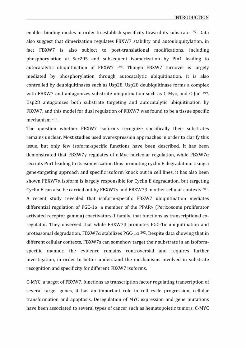
INTRODUCTION
enablesbindingmodesinordertoestablishspecificitytowarditssubstrate197.Data
also suggest that dimerization regulates FBXW7 stability and autoubiquitylation, in
fact FBXW7 is also subject to post-translational modifications, including
phosphorylation at Ser205 and subsequent isomerization by Pin1 leading to
autocatalytic ubiquitination of FBXW7 198. Though FBXW7 turnover is largely
mediated by phosphorylation through autocatalytic ubiquitination, it is also
controlledbydeubiquitinasessuchasUsp28.Usp28deubiquitinaseformsacomplex
withFBXW7andantagonizes substrateubiquitination suchasC-Myc, andC-Jun 199.
Usp28 antagonizes both substrate targeting and autocatalytic ubiquitination by
FBXW7,andthismodelfordualregulationofFBXW7wasfoundtobeatissuespecific
mechanism200.
The question whether FBXW7 isoforms recognize specifically their substrates
remainsunclear.Moststudiesusedoverexpressionapproachesinordertoclarifythis
issue, but only few isoform-specific functions have been described. It has been
demonstrated that FBXW7γ regulates of c-Myc nucleolar regulation,while FBXW7α
recruitsPin1leadingtoitsisomerizationthuspromotingcyclinEdegradation.Usinga
gene-targetingapproachandspecificisoformknockoutincell lines, ithasalsobeen
shownFBXW7αisoformislargelyresponsibleforCyclinEdegradation,buttargeting
CyclinEcanalsobecarriedoutbyFBXW7γandFBXW7βinothercellularcontexts201.
A recent study revealed that isoform-specific FBXW7 ubiquitination mediates
differential regulation of PGC-1α; amember of the PPARγ (Perixosome proliferator
activatedreceptorgamma)coactivators-1family,thatfunctionsastranscriptionalco-
regulator. They observed that while FBXW7β promotes PGC-1α ubiquitination and
proteasomaldegradation,FBXW7αstabilizesPGC-1α202.Despitedatashowingthatin
differentcellularcontexts,FBXW7scansomehowtargettheirsubstrateinanisoform-
specific manner, the evidence remains controversial and requires further
investigation, in order to better understand the mechanisms involved in substrate
recognitionandspecificityfordifferentFBXW7isoforms.
C-MYC,atargetofFBXW7,functionsastranscriptionfactorregulatingtranscriptionof
several target genes, it has an important role in cell cycle progression, cellular
transformation and apoptosis. Deregulation ofMYC expression and genemutations
havebeenassociatedtoseveraltypesofcancersuchashematopoietictumors.C-MYC
INTRODUCTION
53
protein’s abundance are regulated by levels of degradation of the protein, and
phosphorylation of c-Myc on threonine-58byGSK3 regulates its binding to FBXW7
(like Cyclin E phosphorylation at T380), as well as FBXW7-mediated c-MYC
degradationandubiquitination203,204.
Another target isC-jun, a transcription factorandanoncoprotein, implicated in the
regulation ofmany cellular processes such as proliferation, transformation and cell
death.Itisacomponentoftheactivatorprotein-1(AP1)transcriptionfactorcomplex.
DeregulationofC-junhasbeenreportedinmanytypesofcancer.Itisknowntobea
highlyunstableoncoproteinthatneedstobetightlyregulatedonbothtranscriptional
andpost-translationallevels.C-juncanbestabilizedonceit isphosphorylatedatthe
N-terminus by C-jun N-terminal kinases (JNKs) or other protein kinases. This
phosphorylation decreases C-jun ubiquitination and degradation205. C-jun
ubiquitination is carried out by FBXW7 in a GSK3 phosphorylation-dependent
manner.Phosphorylationofaminoacidresiduesatpositions239and243isrequired
forFBXW7recognition206.Ubiquitindependentdegradationcanalsobepromotedby
CSK-dependentphosphorylation(CSKaproteintyrosinekinase)207.C-juncanalsobe
recognized by other ubiquitin ligases such as Itch and COP1, in a phosphorylation-
independentmanner,inducingitsubiquitinationanddegradation208.
CyclinE is alsoamong theFBXW7 targets.CyclinEbindsCDK2 the cyclin-dependent
kinase subunit that is implicated in the regulation of cell cycle processes and cell
division.TheroleofCyclinEisnotlimitedtoCDK2activation;itisalsoimplicatedin
CDK2-independentregulation.InaCDK2dependentcontext,degradationofCyclinE
ismediatedbytheSCFFBXW7complexthatrecognizephosphorylateddegronsupon
thesubstrate.CyclinEcontainstwoCDPs:aC-terminaldegronatthreonine380and
anN-terminal degron at threonine 62. These residues are phosphorylated byGSK3
andCDK2leadingtosubsequentbindingandubiquitinationbyFBXW7209.
3.4.3.2.8.FBXW7atumorsuppressorproteinFBXW7 is considered as a tumor suppressor protein due to the fact that it targets
several oncoproteins, therefore regulates their levels of expression and stability via
ubiquitinationandsubsequentdegradation.Manystudieshavereportedmutationsin
FBXW7 in different types of cancer. The majority of these mutations are point
INTRODUCTION
mutations resulting in amino-acid substitutions within the WD40 repeats and
consequentdisruptionofsubstratebinding.Nonsensemutationswerealsoidentified
resulting in theproductionof truncated formsofFBXW7.Thesemutations leadtoa
deregulation or loss of function of FBXW7 promoting an accumulation of FBXW7
substrates210.
FBXW7isrequiredfornormalstemcellsmaintenance,forhematopoietic,neuraland
intestinalstemcells(HSCs,NSCsandISCs),andfortheproperregulationofcellcycle
progressionandbalancebetweendifferentiationandself-renewal.AbsenceofFBXW7
inHSCsleadstoanaccumulationofcMYCinducingthere-entryofHSCsinthecycle.In
a P53-downregulated context, this accumulation leads to the development of T-cell
malignancies.Ontheotherhand,NSCsdeficientforFBXW7accumulateNotch1,c-JUN
butnotcMYCandrepresenthigherproliferationratethannormalHSCsinadditionto
morphological abnormality in the brain 211(figure 17). FBXW7 deregulation is also
implicated in promoting proliferation andmaintenance of cancer stem cells (CSCs)
alsoknownascancerinitiatingcells(CICs).CICsarecharacterizedbytheirabilityto
self-renew and proliferate and their ability to initiate cancer as indicated by their
name.Aftercancertherapies,CICsmaybelatentbutcanreemergeatinterruptionof
treatment gaining also resistance to therapy 212. FBXW7 plays a pivotal role in
maintenance of quiescence in leukemia-initiating cells (LICs) of chronic myeloid
leukemia. It has been demonstrated that FBXW7 ablation in LICs results in
deregulatedactivationofc-Mycandimpairedmaintenanceofquiescencefollowedby
p53-dependentapoptosis.LICswerefoundtobemoresensitivetoFBXW7deficiency
thanareHSCs,suggestingtheirpotentialroleastherapeutictargets213,214.
AnextensivegeneticanalysisofFBXW7wascarriedoutinprimaryhumantumorsof
diverse tissueorigin inorder todetermineFBXW7’s role as a tumor suppressor. In
thisstudytheygenerateddatafrom534primarytumorspecimenstestedinaddition
todatapreviouslyreportedonFBXW7(totalof1554specimens)andtheyfoundthat
FBXW7 is mutated in a variety of human tumor types with an overall mutation
frequencyof 6%.Thehighest frequencies ofmutationswereobserved in tumorsof
thebileduct(cholangio-carcinomas,35%),blood(T-cellacutelymphocyticleukemia,
31%), colon (9%), endometrium (9%), and stomach (6%). Furthermore, they show
that expressionof anFBXW7mutant corresponding tooneof themajormutational
hotspots inprimary tumors interfereswithwild-typeFBXW7 function, suggestinga
INTRODUCTION
55
potentialdominantnegativemechanismofFBXW7inactivation215.
Figure17:FBXW7isrequiredformaintenanceofnormalstemcells.Fbxw7ablationinHSCsresultsintheaccumulationofc-Mycandconsequentre-entryofthecellsintothecellcycle.ThecyclingHSCsarethensubjecttoapoptosisasaresultofp53activation,leadingtothedevelopmentofpancytopenia.Intheabsenceofp53induction,theFbxw7-deficientHSCseventuallygiverisetoleukemia.Ontheotherhand,lossofFbxw7inNSCsresultsinNotchaccumulation,withtheconsequentimbalancebetweenself-renewalanddifferentiationinthesecells,leadingtoaberrantbraindevelopment.ReviewedinTakeishietal.211.
Inactivating FBXW7 mutations were identified in a large fraction of human T-ALL
lines andprimary leukemias. FBXW7mutationwere identified in40%ofhumanT-
ALLcelllineand16%ofprimaryleukemiaswhereFBXW7abilitytointeractwithits
targets (NOTCH1, cMYC and cyclin E) was abolished. The majority of the FBXW7
mutationswerepresentduringrelapse,and theywereassociatedwithNOTCH1HD
mutations. Interestingly,most of theT-ALL lines harboring FBXW7mutationswere
resistanttoγ-secretaseinhibitortreatmentandthisresistanceappearedtoberelated
tothestabilizationofthec-Mycprotein216.
Hotspot mutation sites were identified and associated to cancer phenotypes. The
catalogue of somatic mutations in cancer database (COSMIC) have reported 496
mutation in FBXW7, where half of these mutations were identified as missense
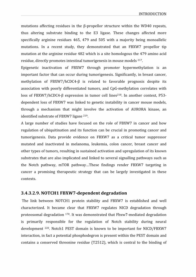
INTRODUCTION
mutations affecting residues in the β-propellor structurewithin theWD40 repeats,
thus altering substrate binding to the E3 ligase. These changes affected more
specifically arginine residues 465, 479 and 505 with a majority being monoallelic
mutations. In a recent study, they demonstrated that an FBXW7 propellor tip
mutationatthearginineresidue482whichisasitehomologousthe479aminoacid
residue,directlypromotesintestinaltumorigenesisinmousemodels217.
Epigenetic inactivation of FBXW7 through promoter hypermethylation is an
importantfactorthatcanoccurduringtumorigenesis.Significantly, inbreastcancer,
methylation of FBXW7/hCDC4-β is related to favorable prognosis despite its
associationwith poorly differentiated tumors, and CpG-methylation correlateswith
loss of FBXW7/hCDC4-β expression in tumor cell lines218. In another context, P53-
dependent lossofFBXW7was linked togenetic instability in cancermousemodels,
through a mechanism that might involve the activation of AURORA kinase, an
identifiedsubstrateofFBXW7ligase219.
A large number of studies have focused on the role of FBXW7 in cancer and how
regulationofubiquitinationand its functioncanbecrucial inpromotingcancerand
tumorigenesis. Data provide evidence on FBXW7 as a critical tumor suppressor
mutated and inactivated in melanoma, leukemia, colon cancer, breast cancer and
othertypesoftumors,resultinginsustainedactivationandupregulationofitsknown
substratesthatarealsoimplicatedandlinkedtoseveralsignallingpathwayssuchas
the Notch pathway, mTOR pathway…These findings render FBXW7 targeting in
cancer a promising therapeutic strategy that can be largely investigated in these
contexts.
3.4.3.2.9.NOTCH1FBXW7-dependentdegradationThe link between NOTCH1 protein stability and FBXW7 is established and well
characterized. It became clear that FBXW7 regulates NICD degradation through
proteosomaldegradation170.ItwasdemonstratedthatFbxw7-mediateddegradation
is primarily responsible for the regulation of Notch stability during neural
development 220. Notch1 PEST domain is known to be important for NICD/FBXW7
interaction,infactapotentialphosphodegronispresentwithinthePESTdomainand
contains a conserved threonine residue (T2512),which is central to the binding of
INTRODUCTION
57
FBXW7 to NOTCH1 221, a study showed that NOTCH T2512A mutant represents
increasedstabilityanddecreasedbindingtoFBXW758.
The C-terminal domain of the NICD contains multiple conserved cyclin-dependent
kinase phosphorylation sites that can be phosphorylated by CycC:CDK8. MAM
interacts directly with the catalytic subunit of the CycC:CDK8 complex, and CDK8
directly targets serine residues in the PEST motif that control NICD
hyperphosphorylation and turnover, mediated by FBXW7 PEST-dependent
degradation 155. Moreover NICD is phosphorylated by GSK3β that regulates its
proteasomal-dependentdegradation155.
3.5.NOTCH1protein-proteininteractions
WhenwelookthroughSTRINGdatabaseforNOTCH1protein-proteininteractions,we
identifyteninterconnectedproteinsintheNOTCH1network(figure18).Amongthese
proteins RBPJ and MAML1 that are part of NICD transcriptional complex in the
nucleus. ADAM17, a metalloprotease responsible for NOTCH1 cleavage at the cell
surface independentof ligandinducedactivation.WecanalsoaddADAM10another
enzymerequiredforreceptoractivationbutinanotherligand–dependentcontext222.
PSEN1, the catalytic subunit of γ-secretase also interacts with notch and has been
showntoplayanimportantroleintheproteolyticprocessingofNOTCH1223.STRING
list includes FBXW7 and DTX1 ubiquitin ligases that are both known for direct
physicalinteractionwithNOTCH1andinducingitsproteasome-mediateddegradation221, 224. NUMB promotes NOTCH1 ubiquitination and proteasomal degradation of
NICD, described also as an inhibitor of Notch pathway 225, 226.The polyubiquitin C
protein encoded by UBC gene, is among the reported interactors. As we have
explainedpreviously,ubiquitinationisanessentialpost-translationalmodificationin
the regulation of the Notch signalling, and NOTCH1 can undergo ubiquitination at
differentstagesofitssignallingprocessthroughavarietyofubiquitinenzymes169,227,
228. The last two proteins listed are HEY1 and HEY2 from Hey family of bHLH
transcription factors that are direct targets of Notch signaling. Hey1 and Hey2 are
essential mediators of Notch functions in blood vessel morphogenesis 229. The list
provided by STRING database is limited, we have discussed in the previous parts
differentaspectsoftheNotchsignallingpathwayandthusawidevarietyofproteins
INTRODUCTION
interactwithNOTCH1.Among them ligands of delta and serratewhich activate the
receptor at the cell surface, different enzymes involved in post-translational
modifications:POFUT1,POGLUTglycosyltransferases thatpreferentiallymodifyEGF
repeats of NOTCH1; kinases (CDK2, CDK19, CDK8…) and ubiquitin ligases (FBXW7,
DELTEX, Itch…).Yatimetal. established theNOTCH1nuclear interactome inT-ALL,
and showed that beyond the NICD-CSL-MAML1 activation complex, NOTCH1
associates a multifunctional complex containing DNA-binding partners,
transcriptional coactivators and corepressors acting at different stages of the
transcriptionactivationprocess230.
Figure 18: Notch1 protein interaction network extracted from STRING database.Colorednodes representproteins thataredirectly linked toNotch1.Edges representpredicted functional links. Edgewidth is proportional to the number of evidence fortheseinteractions(consistingof:databases,experiments,textminingandhomology).
AIMOFTHEWORK
59
AIMOFTHEWORKThe first aim of this thesis was to establish a comprehensive interactomic map of
proteins that are associated to acute lymphoblastic leukemia (ALL) using high-
throughput experimental techniques and bioinformatics methods, to identify
unexploredaxestobetterunderstandALL.
Thisinteractomicapproachenabledustodefineimportantproteinshubsforproteins
andpathwaysknowntobelinkedtoALL,butalsonewmoleculardeterminantsthat
canbepotentiallylinkedtoALL.
Then, the second aim of the project was to characterize the functional interplay
between EXT1, NOTCH1 and FBXW7 as a proof of our concept for combining
interactomeanalysisandbioinformaticstools.
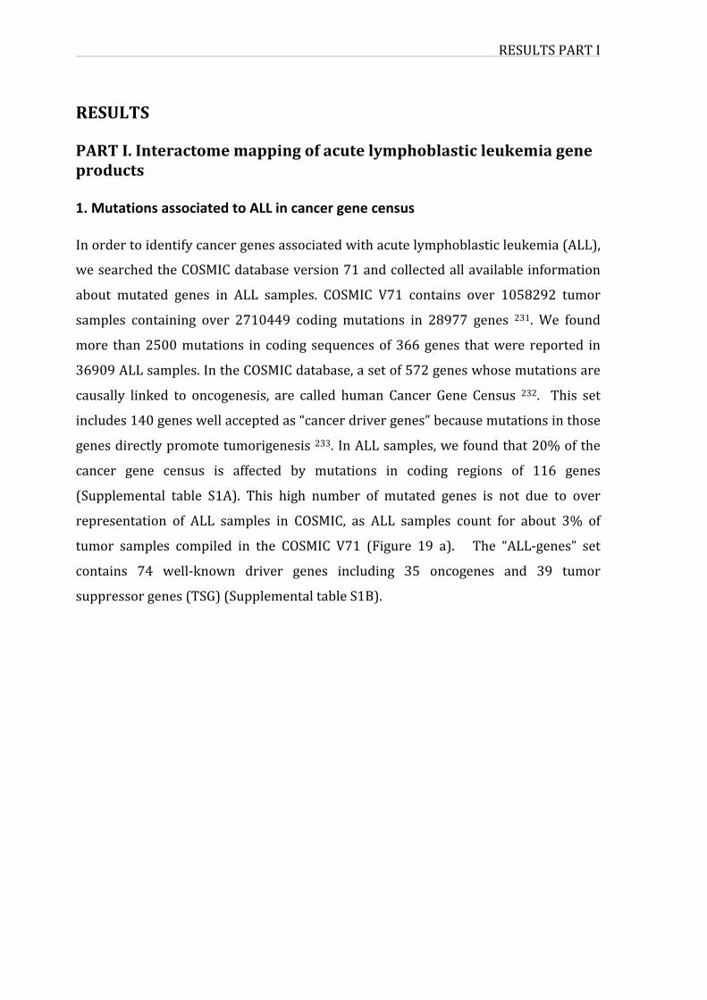
RESULTSPARTI
RESULTSPARTI.Interactomemappingofacutelymphoblasticleukemiageneproducts1.MutationsassociatedtoALLincancergenecensusInordertoidentifycancergenesassociatedwithacutelymphoblasticleukemia(ALL),
wesearchedtheCOSMICdatabaseversion71andcollectedallavailableinformation
about mutated genes in ALL samples. COSMIC V71 contains over 1058292 tumor
samples containing over 2710449 coding mutations in 28977 genes 231. We found
more than2500mutations in coding sequencesof366genes thatwere reported in
36909ALLsamples.IntheCOSMICdatabase,asetof572geneswhosemutationsare
causally linked to oncogenesis, are called human Cancer Gene Census 232. This set
includes140geneswellacceptedas“cancerdrivergenes”becausemutationsinthose
genesdirectlypromotetumorigenesis233.InALLsamples,wefoundthat20%ofthe
cancer gene census is affected by mutations in coding regions of 116 genes
(Supplemental table S1A). This high number of mutated genes is not due to over
representation of ALL samples in COSMIC, as ALL samples count for about 3% of
tumor samples compiled in the COSMIC V71 (Figure 19 a). The “ALL-genes” set
contains 74 well-known driver genes including 35 oncogenes and 39 tumor
suppressorgenes(TSG)(SupplementaltableS1B).
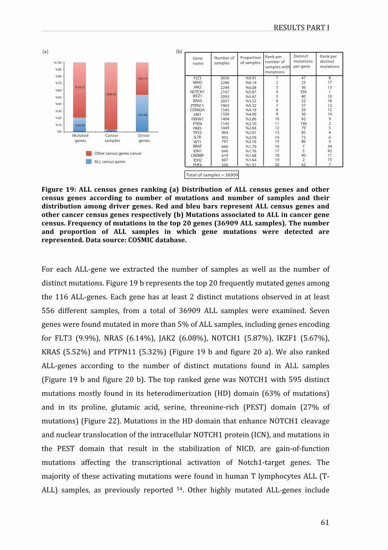
RESULTSPARTI
61
Figure 19: ALL census genes ranking(a) Distribution of ALL census genes and othercensus genes according to number of mutations and number of samples and theirdistribution amongdriver genes. Red andbleubars representALL census genes andothercancercensusgenesrespectively(b)MutationsassociatedtoALLincancergenecensus.Frequencyofmutationsinthetop20genes(36909ALLsamples).Thenumberand proportion of ALL samples in which gene mutations were detected arerepresented.Datasource:COSMICdatabase.
For each ALL-gene we extracted the number of samples as well as the number of
distinctmutations.Figure19brepresentsthetop20frequentlymutatedgenesamong
the116ALL-genes. Each genehas at least 2distinctmutationsobserved in at least
556 different samples, from a total of 36909 ALL samples were examined. Seven
geneswerefoundmutatedinmorethan5%ofALLsamples,includinggenesencoding
for FLT3 (9.9%), NRAS (6.14%), JAK2 (6.08%), NOTCH1 (5.87%), IKZF1 (5.67%),
KRAS (5.52%) andPTPN11 (5.32%) (Figure 19b and figure 20 a).We also ranked
ALL-genes according to the number of distinct mutations found in ALL samples
(Figure19b and figure20b). The top ranked genewasNOTCH1with595distinct
mutationsmostly found in its heterodimerization (HD)domain (63%ofmutations)
and in its proline, glutamic acid, serine, threonine-rich (PEST) domain (27% of
mutations)(Figure22).MutationsintheHDdomainthatenhanceNOTCH1cleavage
andnucleartranslocationoftheintracellularNOTCH1protein(ICN),andmutationsin
the PEST domain that result in the stabilization of NICD, are gain-of-function
mutations affecting the transcriptional activation of Notch1-target genes. The
majorityof theseactivatingmutationswere found inhumanT lymphocytesALL(T-
ALL) samples, as previously reported 54. Other highly mutated ALL-genes include
(a)
FLT3
PTENFBXW7JAK1
CDKN2APTPN11KRASIKFZ1
NOTCH1JAK2NRAS
PHF6IDH2
CREBBPIDH1BRAFWT1IL7RTP53PAX5
Genename
Number ofsamples
Proportion of samples
Rank pernumber ofsamples withmutations
Distinct mutationsper gene
Rank per distinctmutations
1
201918171615141312111098765432
%9.91
%1.51%1.64%1.68%1.76%1,79%2.16%2.59%2.61%2.84%3.10%3.80%4.09%4.19%5.32%5.52%5.67%5.87%6.08%6.14
47
622
4057
86738579
140423629372240
5953623
8
773114334364529
14151218101
1317
3656
556607619649660797955964
10491145140415091545196320372093216722442266
Total of samples = 36909
(b)
%0
%10
%20
%30
%40
%50
%60
%70
%80
%90
%100
Mutated genes
Cancer samples
Driver genes
Other census genes cancer
ALL census genes
%79.72
%20.28
%96.39
%3.61
%47.14
%52.86
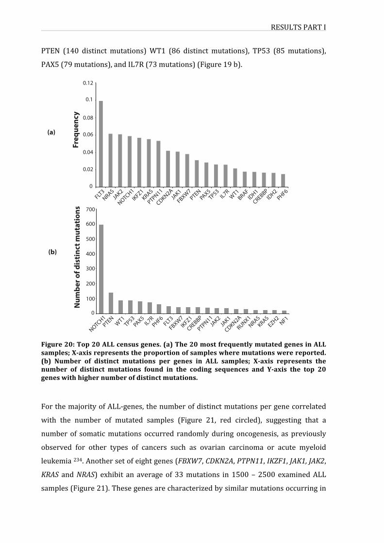
RESULTSPARTI
PTEN (140 distinct mutations)WT1 (86 distinct mutations), TP53 (85mutations),
PAX5(79mutations),andIL7R(73mutations)(Figure19b).
Figure20:Top20ALLcensusgenes.(a)The20mostfrequentlymutatedgenesinALLsamples;X-axisrepresentstheproportionofsampleswheremutationswerereported.(b) Number of distinct mutations per genes in ALL samples; X-axis represents thenumber of distinct mutations found in the coding sequences and Y-axis the top 20geneswithhighernumberofdistinctmutations.
ForthemajorityofALL-genes,thenumberofdistinctmutationspergenecorrelated
with the number of mutated samples (Figure 21, red circled), suggesting that a
numberof somaticmutationsoccurred randomlyduringoncogenesis, as previously
observed for other types of cancers such as ovarian carcinoma or acute myeloid
leukemia234.Anothersetofeightgenes(FBXW7,CDKN2A,PTPN11,IKZF1,JAK1,JAK2,
KRAS andNRAS) exhibit an averageof 33mutations in1500–2500 examinedALL
samples(Figure21).Thesegenesarecharacterizedbysimilarmutationsoccurringin
FLT3
PTEN
FBXW
7JAK1
CDKN2A
PTPN11
KRAS
IKFZ1
NOTCH1
JAK2
NRAS
PHF6
IDH2
CREBBP
IDH1
BRAFWT
1IL7R
TP53
PAX5
0
0.12
0.1
0.08
0.06
0.04
0.02
(a)
Freq
uenc
y
FLT3
PTEN
FBXW
7JAK1
CDKN2A
PTPN11
KRAS
IKFZ1
NOTCH1
JAK2
NRAS
PHF6 NF
1
CREBBP
EZH2
RUNX1
WT1
IL7R
TP53PAX5
100
0
700
600
500
400
300
200
(b)
Num
ber o
f dis
tinct
mut
atio
ns
RESULTSPARTI
63
distinctALLsamples,suggestingtheirpotentialrolesinclonalexpansionofALL.Two
genes are outliers, and displaymanymoremutations (NOTCH1) or aremutated in
manymoresamplesthanaverage(FLT3).Theselargernumbersreflectthehighrate
ofNOTCH1mutations specifically in T-ALL samples (99,9% ofNOTCH1mutations);
and the involvement of FLT3 in childhood ALL, as previously described 235.
Interestingly, NOTCH1 and FLT3 mutations, mostly localized in two functional
domains(HDandPESTforNOTCH1,juxtamembrane(JM)andtyrosinekinase(TKD)
forFLT3),arefoundrespectivelyin1897and723differentpatients,andareexclusive
inexaminedALLsamples(Figure22andFigure19b).
Figure 21: Occurrence of mutations per gene in ALL samples. X-axis represents thenumberofdistinctmutationsfoundinthecodingsequencesandY-axisthenumberofALLsamples.Eachdotrepresentsasinglegene.
NOTCH1600
100
200
300
400
500
00 500 1000 20001500 2500 35003000 4000
Number of samples
Num
ber o
f dis
tinct
mut
atio
ns
FLT3
NRASJAK2IKZF1
KRASPTPN11CDKN2A
JAK1FBXW7
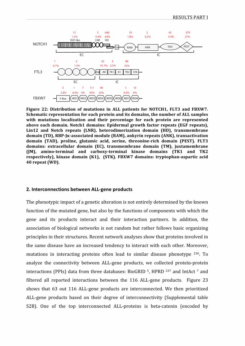
RESULTSPARTI
Figure 22: Distribution of mutations in ALL patients for NOTCH1, FLT3 and FBXW7.Schematicrepresentationforeachproteinanditsdomains,thenumberofALLsampleswith mutations localization and their percentage for each protein are representedaboveeachdomain.Notch1domains:Epidermalgrowth factorrepeats (EGFrepeats),Lin12 and Notch repeats (LNR), heterodimerization domain (HD), transmembranedomain(TD),RBP-Jκ-associatedmodule(RAM),ankyrinrepeats(ANK),transactivationdomain (TAD), proline, glutamic acid, serine, threonine-rich domain (PEST). FLT3domains: extracellular domain (EC), transmembrane domain (TM), juxtamembrane(JM), amino-terminal and carboxy-terminal kinase domains (TK1 and TK2respectively), kinase domain (K1), (STK). FBXW7domains: tryptophan-aspartic acid40repeat(WD).
2.InterconnectionsbetweenALL-geneproductsThephenotypicimpactofageneticalterationisnotentirelydeterminedbytheknown
functionofthemutatedgene,butalsobythefunctionsofcomponentswithwhichthe
gene and its products interact and their interaction partners. In addition, the
associationofbiologicalnetworks isnotrandombutrather followsbasicorganizing
principlesintheirstructures.Recentnetworkanalysesshowthatproteinsinvolvedin
thesamediseasehavean increasedtendencyto interactwitheachother.Moreover,
mutations in interacting proteins often lead to similar disease phenotype 236. To
analyze the connectivity between ALL-gene products, we collected protein-protein
interactions(PPIs)data fromthreedatabases:BioGRID5,HPRD237andIntAct 7and
filtered all reported interactions between the 116 ALL-gene products. Figure 23
shows that 63 out 116 ALL-gene products are interconnected.We then prioritized
ALL-gene products based on their degree of interconnectivity (Supplemental table
S2B). One of the top interconnected ALL-proteins is beta-catenin (encoded by
NOTCH1
FTL3
131.2%
EGF-like LNR HD
RAM ANK TAD PESTTM
ICEC
5 6480.4% 63% 1.8% 0.2% 4.3% 27%
27919 2 45
1 2 63 5 800.7% 1.3% 41.7% 3.3% 53%
EC IC
TM JM TK1 K1 TK2 STK
5 1 7 111 40 1 10
2.8% 0.6% 4% 63% 23% 0.6% 6%
FBXW7 F Box WD1 WD2 WD3 WD4 WD5 WD6 WD7 WD8
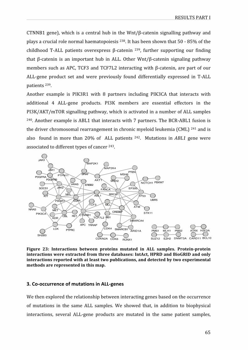
RESULTSPARTI
65
CTNNB1gene),which is a centralhub in theWnt/β-catenin signallingpathwayand
playsacrucialrolenormalhaematopoiesis238.Ithasbeenshownthat50-85%ofthe
childhood T-ALL patients overexpress β-catenin 239, further supporting our finding
that β-catenin is an important hub in ALL. OtherWnt/β-catenin signaling pathway
memberssuchasAPC,TCF3andTCF7L2 interactingwithβ-catenin,arepartofour
ALL-gene product set and were previously found differentially expressed in T-ALL
patients239.
Another example is PIK3R1 with 8 partners including PIK3CA that interacts with
additional 4 ALL-gene products. PI3K members are essential effectors in the
PI3K/AKT/mTORsignallingpathway,whichisactivatedinanumberofALLsamples240.AnotherexampleisABL1thatinteractswith7partners.TheBCR-ABL1fusionis
thedriverchromosomalrearrangementinchronicmyeloidleukemia(CML)241andis
also found inmore than 20% of ALL patients 242. Mutations inABL1 genewere
associatedtodifferenttypesofcancer243.
Figure 23: Interactions between proteins mutated in ALL samples. Protein-proteininteractionswereextractedfromthreedatabases:IntAct,HPRDandBioGRIDandonlyinteractionsreportedwithatleasttwopublications,anddetectedbytwoexperimentalmethodsarerepresentedinthismap.
3.Co-occurrenceofmutationsinALL-genesWethenexploredtherelationshipbetweeninteractinggenesbasedontheoccurrence
ofmutations in the same ALL samples.We showed that, in addition to biophysical
interactions, several ALL-gene products are mutated in the same patient samples,
KIT
JAK1
PDGFRB
SOCS1
JAK3
PDGFRA
ABL1UBR5
TP53
CREBBP
BRCA1
MLLTCF3ATM
STK11
MAP2K1
BRAF
ERBB2
AKT1
MAP2K4
MSH2
FANCA
NOTCH1
MSH6
EP300
PTEN
FBXW7
CDK6
TRRAPSMARCA4
CDK4
RB1
CDKN2A RUNX1
SMARCB1APC
ARID1A
TCF7L2
CARD11EZH2SUZ12 BCL10DNMT3A
KDR
MET
CTNNB1PIK3CANRAS
CBL RET
PIK3R1
PTPN11
JAK2
MPL
HRAS
EGFR
SH2B3
H3F3APTPRC MLH1LCK PMS2 ATRXPMS1
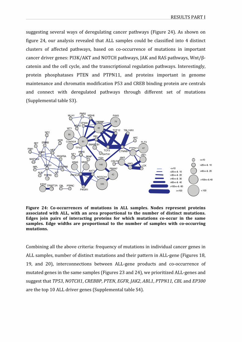
RESULTSPARTI
suggesting severalways of deregulating cancer pathways (Figure24). As shownon
figure24, our analysis revealed thatALL samples couldbe classified into4distinct
clusters of affected pathways, based on co-occurrence of mutations in important
cancerdrivergenes:PI3K/AKTandNOTCHpathways,JAKandRASpathways,Wnt/β-
cateninandthecellcycle,andthetranscriptionalregulationpathways.Interestingly,
protein phosphatases PTEN and PTPN11, and proteins important in genome
maintenanceandchromatinmodificationP53andCREBbindingproteinarecentrals
and connect with deregulated pathways through different set of mutations
(SupplementaltableS3).
Figure 24: Co-occurrences of mutations in ALL samples. Nodes represent proteinsassociatedwith ALL, with an area proportional to the number of distinctmutations.Edges join pairs of interacting proteins for which mutations co-occur in the samesamples. Edge widths are proportional to the number of samples with co-occurringmutations.
Combiningalltheabovecriteria:frequencyofmutationsinindividualcancergenesin
ALLsamples,numberofdistinctmutationsandtheirpatterninALL-gene(Figures18,
19, and 20), interconnections between ALL-gene products and co-occurrence of
mutatedgenesinthesamesamples(Figures23and24),weprioritizedALL-genesand
suggestthatTP53,NOTCH1,CREBBP,PTEN,EGFR,JAK2,ABL1,PTPN11,CBLandEP300
arethetop10ALLdrivergenes(SupplementaltableS4).
<=10>20=> & 10>30=> & 20>40=> & 30>60=> & 40>100=> & 60
>=100
<=10
>20=> & 10
>40=> & 20
>100=> & 40
CRLF2
FBXW7
TCF3
PAX5
TCF12
PTENBRAFGATA1
PIK3R1
NOTCH1
HRAS
AKT1
NRAS
PIK3CA
PTPRC
NF2
KDRERBB2
FGFR3
MET
SOCS1
KIT
MSH6
RB1
CREBBPEP300
MLH1
SMARCB1TP53
CDKN2A
PMS2
WT1MSH2
MAP2K4
RET
EGFR
MPL
PIM1
ALK
TBL1XR1
LCKJAK3
CTNNB1
FLT3APC
PTPN11
JAK2JAK1
CBLDNMT3A
KRAS
PDGFRB
NF1PDGFRA
EZH2
< 100
595
140
42
47
79
40
85
86
RESULTSPARTI
67
4.FunctionalassociationsbetweenALL-geneproductsandtheirpartnersinthehumanproteomeWe hypothesized that the ALL-genes set is not limited to mutated genes in ALL
samples, but could be extended to functional related genes and their products. In
order to identify ALL-gene products interactors, we filtered from 3 different PPI
databases(BioGRID,HPRDandIntAct),proteinsthatinteractwithatleast2ofthe116
ALL-geneproducts(SupplementaltableS5A).Theobtainedinteractomemap(Figure
25) shows that interconnected ALL-gene products have also several common
partners, prioritized according to the number of interacting ALL-gene products
(SupplementaltableS5B).PPIstoredindatabasesarecuratedfromtheliteratureand
some proteins such as P53, BRCA1 or ATM heavily studied with hundreds of
publications, have more PPI reported than others that are not studied with equal
intensity. Previous studies suggested that unbiased PPI mapping allow
characterization of overlooked PPI and identification of unknown diseases-related
candidates244.
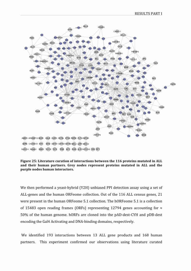
RESULTSPARTI
Figure25:Literaturecurationofinteractionsbetweenthe116proteinsmutatedinALLand their human partners. Grey nodes represent proteins mutated in ALL and thepurplenodeshumaninteractors.
Wethenperformedayeast-hybrid(Y2H)unbiasedPPIdetectionassayusingasetof
ALL-genesandthehumanORFeomecollection.Outofthe116ALLcensusgenes,21
werepresentinthehumanORFeome5.1collection.ThehORFeome5.1isacollection
of 15483 open reading frames (ORFs) representing 12794 genes accounting for ≈
50%of thehumangenome.hORFs are cloned into thepAD-dest-CYHandpDB-dest
encodingtheGal4ActivatingandDNA-bindingdomains,respectively.
We identified 193 interactions between 13 ALL gene products and 168 human
partners. This experiment confirmed our observations using literature curated
NUP214 NUMA1
CACNA1D
AMER1 ARHGEF12
GOPC
TPRTET2 NF1NOTCH2TCF12 TFEB PHF6WT1SRSF2 IL7R
MKL1IDH2IDH1 MED12GATA3GATA2GATA1 LHFP
ECT2L FGFR2ETV6 KMT2DCLRF2CALRBCOR FLT3
DNMT3A
RPA1
DAXX
UBE2I
ATRX
PPP1CA
MRE11A TP53BP1
BRCA1
HIST2H3C
PIAS1
RAD51
MDM4 CHEK2
MLL3
FANCA
CHEK1
SUZ12
EZH2
USP7TWIST1 MAPK1
UBR5
EED
RBBP5
HDAC6
RBBP4
TP53 TCF3
DNMT1 MLL PTENYWHAG
AKT1
ATM
CREBBPYY1RAF1
SFN
HSPA5AR BCR
UBE2L3VDRPIAS2 RB1
FOXO4
PHB HIST1H3A
SMAD2 MYOD1
CSNK2A1TRRAP HSPB1 HSPA8
KDM6A
DNMT3B
ASH2LWDR5 CHUK
ZNF278
JAK3
CDKN2D PDGFRAJAK1
ROS1
IRS1
TYK2 NRAS
INSR
SOCS3
PDGFB
PDGFRB
PIK3CA
KAT2A
YWHAZGSK3B
UBC
TCF7L2
KRAS
PSEN1
BRAF
CTNNB1NEDD4
AXIN1 TSC2
APC
YWHAB
YWHAH
YWHAQ
TNFAIP3
CYLD
IKBKG
MAP2K1
TRAF2
BCL10
CARD11
MAP3K1
MAP2K4
CTNNA1PTPRJ
NF2MUC1
GTF2ICALM1
CDH1HRAS
LGALS1KITCBL
RASA1
RIN1VAV1
EGFR
SOS1
ITSN1
FGFR2
METLYN
PIK3R2
HGSFGF1
FGFR1
SH3KBP1
KAT2BESR1
NR3C1
SIRT1
TBL1XR1 HUWE1 STUB1
UBE2D2
SKP1
STK11
HSP90AB1
HSP90AA1
ABL1
SRCHCK
EZR
GRB2CRK
PTPRC
PIK3R1
HSPA9
MYC
RELA
CDKN1ANCOA3
MDM2
TP73PSMD10
HDAC3 JUN EP300FOS
KLF4
NPM1
RUNX1 PTPN1ERBB2
CDC37STAT5B
PIM1KHDRBS1 SHC1
INPPL1
JAK2NFKB1ETS1 SREBF1
HIF1ACDKN2A
NOTCH1
CDKN2B
CCND1
CDKN2C
CCND3PAX5CDK6
STAT3CDK4
FBXW7STAT5A SOCS1
RNF43
FGFR3
STAT1 GAB1ALKFRS2
ZAP70
FYN
DNM2
SH2B3
PLCG1
EPOR
MPL
CRKL
PTPN6
PTPN11
LCK
NCK1
CSF1RRET
KDR
ERBB3
IKZF1
PMS1
WHSC1
PMS2
FAN1MLH1
MSH6
SMARCC2
ARID1A
SMARCE1
SMARCB1
PCNA
BUB1B
EXO1
NCOA2
HDAC1
CREB1PML
SMAD3
ATF2HDAC2
SP1
NCOR1
SMARCA4
CDC27
MSH3
SMARCC1
POLR2A
MSH2
ANAPC7
BLM
HIST1H4A
RBBP8
PLK1
PARP1
H3F3A
SUMO1
NBN
MDC1
H2AFX
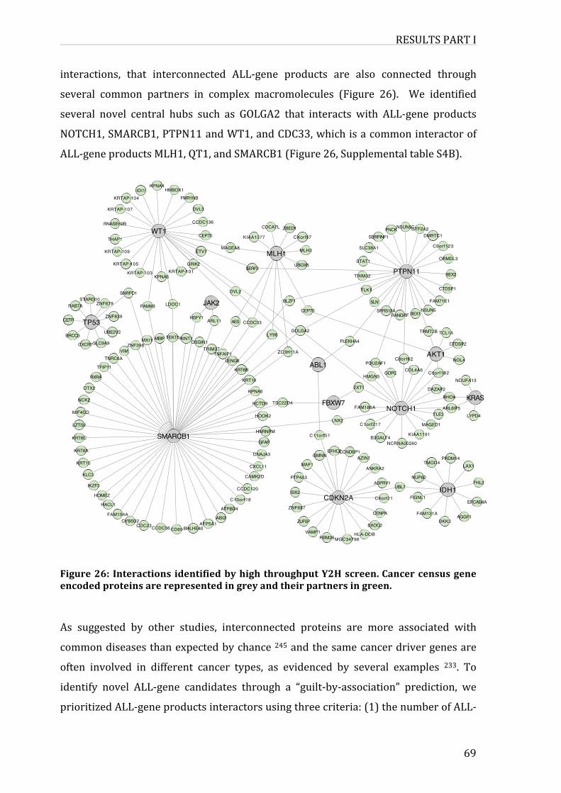
RESULTSPARTI
69
interactions, that interconnected ALL-gene products are also connected through
several common partners in complex macromolecules (Figure 26). We identified
several novel central hubs such as GOLGA2 that interacts with ALL-gene products
NOTCH1,SMARCB1,PTPN11andWT1,andCDC33,whichisacommoninteractorof
ALL-geneproductsMLH1,QT1,andSMARCB1(Figure26,SupplementaltableS4B).
Figure26: Interactions identifiedbyhighthroughputY2Hscreen.Cancercensusgeneencodedproteinsarerepresentedingreyandtheirpartnersingreen.
As suggested by other studies, interconnected proteins are more associated with
commondiseasesthanexpectedbychance245andthesamecancerdrivergenesare
often involved in different cancer types, as evidenced by several examples 233. To
identify novel ALL-gene candidates through a “guilt-by-association” prediction, we
prioritizedALL-geneproductsinteractorsusingthreecriteria:(1)thenumberofALL-
UBE2V2
ZNF679STARD10
CETP
BRCC3
RAB7A
ZNF839TP53
RNASEH2B
HMBOX1
THAP1
KRTAP-104
KRTAP-109
IDI1
KRTAP-107
KPNA4
WT1
ZNF398TEKT5MBIPMXI1 RINT1
VIM
OSGIN1SLC9A9CXCR7
TFIP11
NCK2
MIF4GD
TNRC6A
RXRA
OTX2
LZTS2
FAM154A
BHLHE40CYB5D2
CDC23CD69CCDC36
ATP5A1
HACL1
KRT6A
HOMEZ
IKZF3
KRT15
KLC3
KRT6C SMARCB1
CEP70
GRIK2
HSFY1
FMR1NB
DVL3
ETV7
CCDC136
SNRPD1
KRTAP-105
FAM9B
KRTAP-101KPNA5
KRTAP-103
LDOC1
HLA-DOB
EXOC2
AGGF1
MGC34796
DKK3ZUFSP
RBM26VAMP1
FAM131ACENPA
FHL2
C4orf21EFCAB4A
FIGNL1
UBL7
NUP62
IDH1ASPRV1
LAX1PRDM14
TMOD4AZIN1
ANKRA2
KCTD9
HOOK2
HNRNPM
GFAP
DNAJA3
CAMK2D
KPNA6
CXCL11
TSC22D4
LYPD4TLE3
RHOA
ARL6IP5
MAGED1
DAZAP2
KRASFBXW7
C1orf217
C11orf51
NOTCH1FAM185A
B3GALT4 KIAA1191
EXT1
LNX2
NCRNA00260
PTP4A3
EFHC2
MAF1
CCNDBP1GMNN
CDKN2AATPBD4
C10orf18ZNF687
ABI2
CCDC120SIX2
SERF2
MAGEA8 MLH3
ZBED1
CXorf57
CDCA7L
KIAA1377
UBOX5
MLH1
CEP76
ZC3H11A
BLZF1
LY96GOLGA2
ABL1 POU2AF1
PLEKHA4
BEX1
TLK1
C9orf62
SFRS13A
GOPC
SLN
RANGRF
HMGN5COL4A6
TRIM32
STAT1
SLC38A1
GTF2A2SERPINF1
NSUN5CPNCK
PTPN11
CTDSP1
NSUN5
DMRTC1
C6orf123
BEX2
FAM71E1
ORMDL3
JAK2
TRIM27
AES
KRT6B
KRT19
CCDC33
LENG8
ARL11
DVL2
TNFAIP1
NDUFA13
NOL4
CTDSP2
TCL1A
C6orf182
TRMT2B
AKT1
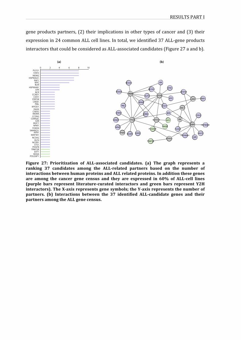
RESULTSPARTI
geneproductspartners, (2) their implications inother typesofcancerand(3) their
expressionin24commonALLcelllines.Intotal,weidentified37ALL-geneproducts
interactorsthatcouldbeconsideredasALL-associatedcandidates(Figure27aandb).
Figure 27: Prioritization of ALL-associated candidates. (a) The graph represents aranking 37 candidates among the ALL-related partners based on the number ofinteractionsbetweenhumanproteinsandALLrelatedproteins.Inadditionthesegenesare among the cancer gene census and they are expressed in 60% of ALL-cell lines(purple bars represent literature-curated interactors and green bars represent Y2Hinteractors).TheX-axisrepresentsgenesymbols;theY-axisrepresentsthenumberofpartners. (b) Interactions between the 37 identified ALL-candidate genes and theirpartnersamongtheALLgenecensus.
(a)
CREBBPEP300
JUN
KLF4
FOXO4
MDM2
MYOD1
PML
NCOA2
ETS1
MYC
EXT1
BCR
PTPN11
RHOA
STAT3
NCOR1 DAXX
NOTCH1TP53
CHEK2
PTEN
NPM1
CREB1
POU2AF1
TRMT2B
MDM4
JAK2
CDH1
EGFR HSP90AB1
MUC1
EZR
RAF1
HSP90AA1
PLCG1
CBL
ABL1
STAT5B
0 102 64 8PLCG1STAT3MDM2
HSP90AA1RAF1MYC
HSP90AB1JUNBCR
CCND1CDH1
STAT5BCREB1PML
MYOD1DAXX
BLM
CHEK2MDM4CCDN3
CDKN2CEZR
MUC1NPM1FOXO4
SMARCE1AXN1
MAP3K1NCOA2
KLF4NCOR1ETS1
PDGFBTRMT2B
EXT1RHOA
POU2AF1
(b)
DISCUSSIONPARTI
71
DISCUSSIONPARTIAmajorandpowerfulapproachtoelucidatingthemolecularmechanismsunderlying
the complex diseases such as cancer is represented by the investigation of
interactionsbetweenproteinsencodedbydiseasegenesinthehumanprotein-protein
interaction network 246. Thus, a systematic examination of the proteins encoded by
ALL cancer genes in the human interactome may help us identify new candidate
genes, improve candidate gene prioritization methods, and have a deeper
understandingofthegeneticlandscapeofALL.
Deregulatedsignalingisconsideredamajorcontributingfactorinleukomogenesisof
T-ALL, it is thus important to analyze interaction hubs illustrated by different
examples in our network. Proteins interactwith each other andwith other cellular
components forming complex networks. These networks share some features and
they are part of commonpathways.Diseases are not independent from each other,
potential cellular network-based dependencies between human diseases has led to
thegenerationofvariousglobaldiseasenetworkmaps,whichlinkdiseasephenotypes
together if somemolecular or phenotypic relationships exist between them 247. An
interestingexampleelucidating“diseaseinterdependency”isgivenbystudythatused
highthroughputdatasetssuchasmRNAexpressionandPPInetworks.Theyrevealed
significant pathological similarities between 54 human diseases, in addition to
common pathways and processes implicating many proteins that are mutated and
knowntobetherapeutictargets248.StudyingtheinterconnectivitybetweenALLgene
products enabled the identificationof severalALL-gene candidates. Inourmapwe
showedthat54%oftheALLrelatedproteinsareinterconnectedformingonecomplex
network. Some of these genes are highly connected;we take as an exampleBRCA1
gene interacting with 12 proteins in the interactome of ALL proteins. Among its
partners; the family ofDNA repairmismatchproteins:MSH2,MSH6andMLH1, the
transcription factor TP53 and ATM (ataxia telangiectasia mutated serine-protein
kinase),thatareinvolvedintheDNA-damagerepairpathwayalongwithBCRA1and
BCRA2proteins249,250.TheinteractionslinkingtheseproteinstoBCRA1indicatethat,
inadditiontoPPIs,thisproteinhubalsorepresentsacommonsignallingpathwayin
whichtheseproteins interact functionallyandphysically.BRCA1andBRCA2arethe
DISCUSSIONPARTI
most well-known genes linked to breast cancer risk 251. In addition to being
implicated in breast cancer, it was demonstrated that defects in BRCA pathway
increase the risks of a subset for lymphomas and leukemias that are probably
associated with gene rearrangements among them ALL 252. Their connections to
Fanconi anemia proteins (FANC), further implicate BRCA1 and BRCA2 deficits in
hematological cancers 253. Faconi anemia is a genetically and phenotypically
heterogenous autosomaldisorderdefinedby congenitalmalformations, progression
marrowfailure,andmarkedpredispositiontomalignancy254.
Aspreviouslymentioned,theCTNNB1(theβ-cateningene)interactionhubalsodrew
ourattention,thisproteininlinkedto9partnersamongtheALLrelatedproteins.The
β-cateninproteinencodedbyCTNNB1isessentialintheWNT-signallingpathway;it
bindstoseveralproteinsinthecytoplasm(suchasAPC)triggeringitstranslocationto
the nucleus. It interacts with other nuclear proteins (TCF, p300) leading to the
transcriptionalactivationoftargetgenesregulatingdifferentcellularprocessessuch
asadhesion,proliferationanddevelopment 255.CTNNB1mutationsandderegulation
are linked to many epithelial-originating malignancies such as breast, ovarian,
endometrial,colorectalandskincancers256.WeobservehowCTNNB1geneproductis
a central protein linked to proteins that are implicated inWNT signalling pathway
(Figure 23). It is known that activation of the Wnt/β-catenin signalling pathway
through loss-of-functionmutations in the APC protein and axin, or gain-of-function
mutations in β-catenin are linked to various hematological malignancies including
ALL.Infactβ-CateninplaysacrucialroleasaneffectoroftheWnt/β-cateninpathway
innormalhaematopoiesis 238. In theabsenceofWNTsignals,acomplexofproteins,
including APC, axin and GSK3β, controls phosphorylation and subsequent
proteasomal degradation of β-catenin.WhenWNT signalling is activated, β–catenin
accumulates inthenucleuswhereit interactswithT-cell factortranscriptionfactors
(TCF)leadingtothetranscriptionalactivationoftargetgenes.Mutationsinβ-catenin
orAPCcanleadtotheformationofconstitutivelyactiveβ-catenin/TCFcomplexesand
alteredexpressionoftargetgenes,thatareassociatedtocancer257.Morethan85%of
thechildhoodT-ALLpatientsshowedupregulatedβ-cateninexpressionattheprotein
level compared with normal human thymocytes. A recent study using microarray
analysiscarriedoutonT-ALLpatientsshowedthat50%ofT-ALLpatientsrepresenta
two-fold change inβ-catenin expression. In addition, otherWNT signallingmember
DISCUSSIONPARTI
73
suchasTCF3andTCF7L2,partnersofCTNNB1shownintheinteractomemap,were
differentiallyexpressedinT-ALLpatients,andinsomecasesassociatedtoβ-catenin
upregulation239.
AnotherexampleofproteinhubsinourmapisPIK3R1with8partnersamongthem
wefindPIK3CAthatinteractswith4proteins.PI3Kproteinsareessentialeffectorsin
the PI3K/AKT/mTOR signalling pathway. This signalling cascade is activated by
receptor tyrosine kinases, integrins, B and T-cell receptors and other stimuli that
induce the production of phosphatidylinositol triphosphate (PIP3) by
phosphoinositide 3-kinase (PI3K). This step induces the activation of AKT due to
phosphorylationby thePDK1andmTORC2 258,259.AKT is aproto-oncoproteinwith
many substrates and effects such as apoptosis and protein synthesis. The tumor
suppressor phosphate and tensin homolog PTEN, inhibits AKT activity by
dephosphorylatingPIP3,whileinactivatingmutationsordeletionofPTENleadingto
AktactivationoccurinALL240.
ABL1isconnectedto7partners,thisgeneconstitutesthesubjectofalargenumberof
studies, andmutations in this genewere associated todifferent typesof cancer.An
important gene fusion that implicates ABL1 and BCR genes has been described in
leukemias(chronicmyeloid leukemia;CML), the fusionproteinresulting isessential
and sufficient for the malignant transformation of CML 241. In addition, BCR-ABL1
fusionisalsofoundinothercancerssuchasbreastandprostatecancers.
Theseexamplesofproteinhubsprovethatinteractionsbetweenasetofproteinsor
interactionslinkedtoacentralproteinarenotrandomlyorganized.Moreoverinthe
context of mutations and genes associated to ALL, we show that proteins with
mutationsinALLarehighlyinterconnectedtoeachother,theyarealsoimplicatedin
commonsignallingpathwayswherederegulationinoneorseveralcomponentsshow
highincidenceinALLdevelopment.Mutationsinoneormoreinterconnectedproteins
may lead to disruption of these complexes and development of ALL, and drug
resistancemechanisms.Inaddition,theseproteinswerelinkedtoothercancerssuch
as breast cancer and other types of leukemias. Other studies have highlighted the
importanceofevaluatinggenemutationsfromaglobalpointofview;andwentdeeper
to assess the interconnectivity between diseases. Goh et al. showed that genes
associatedwithsimilardisordersshowbothhigherlikelihoodofphysicalinteractions
between their products and higher expression profiling similarity for their
DISCUSSIONPARTI
transcripts, supporting the existence of distinct disease-specific functionalmodules.
They established the human disorder network, showing that cancer in among the
most connected disorders. This is due in part to the fact that cancer subtypes are
tightlyconnectedwitheachotherthroughtumorsuppressorssuchasTP53andPTEN.
Anotherinterestingfindingwasthatleukemia,incontrasttomostdisorders,relateto
a large number of disease genes 260. This observation was also confirmed in our
interactome. Disease annotation analysis using DAVID tool revealed for the ALL-
relatedgenes that theyare implicated incommoncancers suchascolorectal cancer
(EP300, APC, TP53, CTNNB1, NRAS, FGFR3, AKT1, BRAF and PIK3CA), ovarian and
breast cancer (BRCA1, CTNNB1, MSH2, KRAS, AKT1 and PIK3CA), acute myeloid
leukemia(FLT3,ARHGEF12,NUP214,RUNX1,andKIT).
Co-occurrenceofmutationsExploring the relationship between the ALL-gene candidates based on the co-
occurrenceofmutationsinthesameALLsamples,provideduswithvaluabledatafor
potentialALL-genecandidates.
The network in figure 24 shows that in addition to the physical interactions these
proteinsarealsofoundtobemutatedinthesameALLsamples.PIK3CA,PIK3R1and
AKT1,proteinsofthePIK3-AKTpathway,areinterconnectedandareco-mutatedina
high number of ALL samples. Mutations at the PI3K/AKT/PTEN pathway can
contribute to leukemogenesis by elevating the expression of the pleiotropic anti-
apoptotic AKT protein, which can stimulate antiapoptotic proteins and inhibit pro-
apoptotic proteins. Inactivating PTENmutations are known to occur in ALL and to
disturbsignallingofAKT/PI3Kpathway.Co-occurrenceofmutationsinthesameALL
samplescanhaveanimportantsignificancewhenitcomestotheclinicaloutcomeof
ALLpatients.InactivationofPTENcanbeassociatedwithpoortreatmentresponsein
ALL.A studywas carriedouton300ALLpatients, analyzing the incidenceofPTEN
point mutations, they show that inactivating PTEN mutations associated with
activating NOTCH1 mutations in patients have better response to therapy 261.
ImportantlythemajorityofPTENidentifiedmutationslocalizeinexon7“hotspot”in
the C2 domain of the protein, accounting for 82% of ALL samples with PTEN
mutations. Thesemutations lead to the truncation of the C2 domain responsible of
DISCUSSIONPARTI
75
bindingtothemembraneforsubsequentdephosphorylationofthemembrane-bound
PIP3 262.Moreover, in T-ALL, activation of AKT has been described downstream of
NOTCH1andAKTmaybeactivatedupon transcriptional repressionofPTENby the
NOTCH1-activatedtranscriptionalrepressorHES155.Notch1thetopmutatedgenein
ALL (Figure 20 b) interacts with both AKT1 and FBXW7 and several ALL samples
exhibitmutationsingenesencodingthoseproteins.Forexample,NOTCH1andFBXW7
mutations co-occur in ≈ 40 samples, most of these mutations result in a higher
expression and activation of NOCTH1, which is highly linked to T-cell ALL cancer
development. PTEN, PI3K and AKT abnormalities showed high incidence in T cell
acutelymphoblasticleukemia62.InfactthePTEN/PI3K/AKTpathwayconstitutesan
important pathway that regulates the signalling of multiple processes such as
apoptosis,cellproliferationandgrowth.Deregulationofthispathwayisimplicatedin
a number of human diseases, where activating mutations in one of these proteins
werereported263.ThissubnetworkalsoincludesNOTCH1andFBXW7.Mutationsin
theHD/PESTdomainsofNOTCH1andFBXW7,which leads toanover-activationof
theNotchpathway,frequentlyoccurinT-ALLpatients58.InadditionPTENmutations
alsooccurredwithFBXW7mutationsinthesameT-ALLsamples.PTENwasdescribed
as an indirectNOTCH1 target (throughHES1 and cMYC), resulting in constitutively
activePI3K-AKTsignalling57.Infigure24weshowthatFBXW7mutationsco-occur
with NOTCH1 mutations and with PTEN mutations in 30 and 37 ALL samples
respectively.Infact,severalstudieshavereportedbetteroutcomeinT-ALLharboring
NOTCH1and/orFBXW7mutations,converselywhenPTENmutationsco-occurredin
thesepatients,itassociateswithpoorprognostic264.
The same observation is valid for JAK proteins, a family of large tyrosine kinases
(JAK1, JAK2 and JAK3). This family of genes along with STAT family (signal
transducers and activators of transcription) and the CIS/SOCS family constitute the
effector genes of the JAK/STAT pathway. JAKs mediate signals from a variety of
cytokines and growth factors. In general, receptor dimerization or oligomerization
due to ligand binding results in the juxtapositioning of the JAKs, either through
homodimericorheterodimeric interactions.Therecruitmentof JAKsresults in their
phosphorylation, either via autophosphorylation and/or transphosphorylation by
other JAKsorother familiesof tyrosinekinases.Thisactivation results in increased
DISCUSSIONPARTI
JAK kinase activity. Activated JAKs then phosphorylate receptors on target tyrosine
residues that serve as docking sites allowing the binding of STATs and their
subsequentphosphorylation.ActivatedSTATsdimerizeandtranslocatetothenucleus
leading to theregulationof targetgenepromoters, including thebcl-2 familygenes,
MYC, CyclinD…265. Since activating mutations in JAK2 have been linked to
leukemogenesis, several studies have been focusing on the development of small
molecule JAK2 inhibitiors, such as AG-490 a specific tyrosine kinase blocker.
InhibitionofJak2withAG-490,selectivelyblocksleukemiccellgrowthinvitroandin
vivo by inducing programmed cell death, with no deleterious effect on normal
haematopoiesis 265, 266. Sequencing analyses and gene expression profiles for ALL
patients revealed high frequency of recurrent somatic alterations in Janus kinase
signalling and also other key signalling pathways including the TP53/RB tumor
suppressorpathwayandRassignalling267.
Exploring mutations co-occurring in several different genes for the same patients
enablesthecharacterizationofdrivergenesthatarespecificallymutatedinALL.The
interactingproteinsencodedbygeneswithmutations,orproteinsthathavecommon
interactors that are implicated in common signalling pathways such as NOTCH1,
FBXW7andPTENrepresentinterestingcandidatesfortargetedtherapydevelopment.
FunctionalassociationsbetweenALL-geneproductsandtheirpartnersinthehumanproteomeTheidentificationofnovelproteinslinkedtotheproteinsthataremutatedinALL,can
also be associated to this disease and therefore represent novel targets or ALL
candidates.Applyingthe“guilt-by-association”concept,withspecificcriterialinkedto
ALL such as the number of ALL-gene products partners, their implications in other
typesofcancerandtheirexpressionALLcelllines,providedavaluablecluetotherole
ofnovelgeneproductspotentiallyassociatedwithALL.InfactwefindthatALL-gene
productsare interconnectedbythemeanoftheseproteinsthatwerenotpreviously
linked to ALL in the human proteome (Figure 25). In figure 25, the interactions
between the ALL proteins and their partners from literature are represented. The
interactionswereextracted from3differentdatabases(BioGRID,HPRDand IntAct).
Proteins that interact with at least 2 of the ALL related proteins, and interactions
reported in two scientific papers and detected with two different experimental
DISCUSSIONPARTI
77
methodsarerepresented.Thiscomplexmapshowsthatinadditiontobeingdirectly
linked to each other, the ALL related proteins are interconnected by the mean of
common partners. The degree of connectivity for the common interactors is
representedbythegraphthatshowsthenumberofALLproteinsinteractingwiththe
identified partners from the human interactome. The proteins in grey were not
previouslyassociatedwithALL,butcanbeconsideredasinterestingcandidatesthat
canbestudied inALL,duetotheirconnectivitywithgenesmutated inALL.Thetop
interconnectedproteinistheubiquitinC,apolyubiquitinprecursorencodedbyUBC
gene,which interactswith 13ALL-related proteins (node in the center of CTNNB1,
PTEN, AKT1 andBRAF on the upper right zone of themap in figure 25). Ubiquitin
exists in the cell either covalently attached to another protein of free. Ubiquitin-
conjugatedproteinsaretargetedfordegradationviatheproteasome,lysosomeorthe
endoplasmic reticulum,dependingon theubiquitinatedresidues.Therefore it isnot
surprising to find thatUBC is highly connected to several ALL proteins. In fact, the
ubiquitin systemplaysan important role inmaintaining thehomeostaticbalanceof
cellularprocesses;thereforeitsderegulationcanpromotethedevelopmentofcancer268. GRB2 interacts with 10 of the ALL related proteins. GRB2 is a ubiquitously
expressed adaptor protein that facilitates the assembly of multiprotein signalling
complexesatactivatedreceptorsandsignallingproteins.GRB2containsasingleSH2
domainthatallowsittobindtyrosine-phosphorylatedreceptors,thisdomainbindsto
thehematopoietic specificadaptorprotein linker foractivationofT cells (LAT)and
otheradaptorproteins.InadditiontotwoSH3domainswhichbringsvariousligands
tothesitesofactivatingsignals269.Duetoitsroleinpropagatingsignallingpathways
downstream of several receptors, deregulation of GRB2-mediated signalling
complexeshasbeenlinkedtooncogenesis.TargetingtheGRB2hasbeenexploredas
ananti-cancerdrug,usingcandidatesthatcanassociatewithGRB2andantagonizeits
signalling 270.GRB2 was not previously linked to ALL, but in a recent study they
investigated the role of GRB2 in ETV6/FLT3mediated leukemogenesis, they found
that FLT3 mutations at specific tyrosine residues alter the GRB2 binding to
FLT3/ETV6andresult inanimpairedactivationofSTAT5,Erk1/2andAktinBa/F3
cells.Theirdata indicatethatGRB2isapotential therapeutic target inpatientswith
EXT6/FLT3-positivemyeloid/lymphoidneoplasmwitheosinophilia-patients271.Other
examples of highly interconnected proteins are STAT1 and 5 of the JAK/STAT
DISCUSSIONPARTI
pathway.MDM2 the negative regulator of p53 protein interactswith 8 ALL-related
proteins,MDM2isknowntobindtop53andinhibit itstranscriptionalactivity, thus
favoring its nuclear transport and degradation. Overexpression of MDM2 was
describedtofavoruncontrolledcellproliferationthroughinhibitionofp53invarious
cancers. Several compounds that inhibit the p53-MDM2 pathway interaction show
anti-proliferative effect in tumour cells overexpressingMDM2 272. TargetingMDM2
with a natural product called Triptolide, induces apoptosis in a subgroup of acute
lymphoblastic leukemiacells273.Anotherexample isNilotinib the selectiveBCR-ABL
tyrosinekinaseinhibitor,widelyusedtotreatPhiladelphia-positiveandPhiladelphia-
negativeALL,studiesalsodemonstratedthatnilotinibinhibitsMDM2byinducingits
self-ubiquitination and degradation, leading to p53-independent apoptosis pathway
and helping treating ALL 274. These interconnected proteins constitute interesting
example showing that even proteins that are not mutated in leukemic patients
constitute potential candidates for targeted anti-cancer therapy. Thus interactomic
approachandestablishingtheconnectionsbetweenproteinsthataremutatedinALL
andtheirpartnersinthehumaninteractomeenablestheidentificationofkeyproteins
thatcanopendoorsfornoveltargetedtherapies.
Databasesgatheringdataaboutinteractionsfromscientificreports,attempttocreate
acompletelistingofliterature-curateddata.Itshouldbenotedthatinteractomicdata
forgenesarebiased,duetothefactthatsomegenesorproteinsaremorefrequently
studiedthanothers.AnexampleillustratingthistheoryisBRCA1thatwefoundinour
interactome,asthemost interconnectedproteinamongtheALL-geneproducts.This
protein was frequently studied in ovarian and breast cancer, in addition to recent
findings that relate BRCA proteins to leukemias as we previously mentioned.
Furthermore experimentalmethodsused to detect interactions, largely vary among
different studies. Using a high throughput PPI screening technique limits the bias
resulting from the irregularity of applied methods and focusing on “well-studied”
genes.
Highthroughputyeasttwo-hybridscreeningDespitetheseextensivecurationefforts,theexistingmapsareconsideredincomplete,
and the literature-based datasets, while richer in interactions, are prone to
DISCUSSIONPARTI
79
investigative biases, containing more interactions for the more explored disease
proteins236.
High throughput Y2H screening enabled the identification of novel interactions
between ALL gene products and proteins from the human interactome. The
constructed network confirmed that ALL-related proteins are interconnected; they
represent several common partners such as GOLGA2 interacting with NOTCH1,
SMARCB1, PTPN11 and WT1, and CDC33, which interacts with MLH1, QT1, and
SMARCB1. We classified the identified partners according to the degree of
connectivity,which is represented by the number ofALL gene products interacting
witheachprotein.Theadvantageofidentifyingthesenovelinteractionsistohighlight
proteinsthathavenotyetbeenassociatedwithALL,theseproteinsarenotknownto
beinvolvedinALLandmayprovidenewinsightsforresearchinthisdomain.GOLGA2
isacis-GolgimatrixproteinthatplaysamajorroleinthestackingofGolgicisternae
and maintenance of Golgi structure. It also participates in the glycosylation and
transportofproteinsandlipidsinthesecretorypathway275.TargetingGOLGA2using
specificshRNAshowedtoinhibitangiogenesisandinduceautophagy-dependentcell
death in lungcancercells 276,but ithasnotbeenpreviously linkedtoALL.ABI2the
abl-interactor2(encodedbytheAblassociatedgeneARG)isanon-receptortyrosine
kinase, togetherwithc-Abl theyconstitute themembersof themammalianAbelson
family of kinases that were first identified as oncogenes associated to human
leukemias.ArgisactivatedbyfusionwithTeltranscriptionfactorincasesofAML277.
AmongthecommoninteractorsinourY2Hmapsomewerepreviouslyassociatedto
cancerandhaveservedas therapeutic targets,butnot in theALLcontext. It is thus
important to exploit thesenovel partners and analyze theirpotential implication in
ALL.
The advantage of using the high throughput approach is in the reduction of
interactomic hubs constituted by proteins that are most studied. As we have
previously mentioned, information represented in different literature–curation
databasesarebiasedduetothefactthatrepeatedlystudiedproteins,aremorelikely
to havemore interactomic data than less studiedproteins. Thus using a systematic
highthroughputinteractiondetectiontechniquesuchastheY2Hhelpsminimizethe
observed bias. In our Y2H map we can highlight novel candidates that can be
DISCUSSIONPARTI
associated to ALL, such GOLGA2 and CDC33. Another example that grabbed our
attention is EXT1 that interact with 2 ALL related proteins, NOTCH1 and FBXW7
(Figure26).EXT1wasdetected inourY2Hscreenasapartner forbothNotch1and
FBXW7.Notchwasthesubjectoftheanalysisandinvestigationsinahugenumberof
publicationsinvolvingitsmechanismofaction,regulationofthesignallingpathwayto
itsimplicationindiseasesanditsroleasimportantandpromisingtherapeutictarget278.
WeinvestigatedtheexpressionofinteractorsinT-ALLcelllinesandfoundthat79%
of genes these are expressed in 60%of T-ALL cell lines. Applying selection criteria
such as the degree of connectivity and gene expression in ALL cell lines to the
interaction partners identified from both literature curated data and our Y2H
interactome enabled the prioritization of 37ALL-gene products interactors asALL-
candidates (Figure 26). Using similar approach that integrates expression profiles,
mutationeffectsandinteractomedatahasbeenrecentlyusedasananalyticaltoolfor
identifyingnoveldrivergenesincancer246,279.
In summary, we extracted mutations found in ALL patients that were reported in
COSMICdatabase, and identified 116ALL-related genes among cancer gene census.
Networkanalysesofprotein-proteininteractionsshowthat54%amongtheALL-gene
products are interconnected and represent central protein hubs such as the beta-
cateninproteinandPIK3R1.Inaddition,wefoundthatseveralmutationsintheALL-
genesproductsco-occurinthesamesamples,whichcanbeclassifiedinto4clustersof
affected pathways. ALL-related gene products are also interconnected through
common partners from the human interactome. Our experimental data from Y2H
screening enabled the identification of novel partners for the ALL-related proteins.
AmongtheY2Handliterature-curatedidentifiedinteractorsweprioritized37genes
asALL-associatedcandidates.
Thefieldofsystemsbiologyhasevolvedenormouslyduringtherecentyears,dueto
the availability of a huge amount of genomic and proteomic data and the progress
made in technologies allowing high-throughput screening and the development
bioinformaticstools.Despitethegreatadvantageofapplyingthisapproachinthefirst
partofouranalysis,representedbytheidentificationofseveralnovelpotentialcancer
genes, it should be noted that interactomic and bioinformatics analyses have their
DISCUSSIONPARTI
81
limitations. Their advantage resides inhighlighting interesting candidatesbut these
hypotheses need to be validated, in order to provide biological and molecular
explanationfortheroleandtheimpactofthesecancergeneproductsinthedisease
developmentortherapy.
Further analyses are required to clarify this point; interactions can be tested in
conditions where ALL-related proteins are mutated (reported mutations in ALL
samples).Weexpectthatmutationswilldisruptthenetworkorganization,leadingto
loss of some interactions, which can provide a more detailed explanation of the
molecular basis of ALL pathogenesis and development. On another level ALL-
candidategenes canbe tested for theirpotential role inALL, firstby characterizing
the links between these candidates and theirALL-relatedproteinpartners and in a
secondstep investigating theireffect inALLusingaT-ALLcell linesorALL-specific
animalmodels.
Inthesecondpartofouranalysesweprovideaproof-of-conceptonhowsystematic
interactome approaches could allow identification of novel targeted genes, by
investigatingEXT1-NOTCH1novelinteraction.

RESULTSPARTII
PART II. EXT1 is functionally associated with the Notch pathwaythroughitsinteractionwithNOTCH1andFBXW7The following example illustrates the validity of combining interactomeapproaches
and gene mutations characterization to identify specific cancer type-related genes.
Exostosinglycosyltransferase1(EXT1)isanendoplasmicreticulumtransmembrane
proteinfrequentlymutatedinmultipleosteochondromas280-282. WeidentifiedEXT1
as a common interactor of twoALL-geneproductsNOTCH1andFBXW7 (Figure26
and26a).Wetheninvestigatedthepotential functional interplaybetweenEXT1and
theNOTCHpathway.
Theexostin (EXT) familyofgenesencodesglycosyltransferases involved inheparan
sulfate (HS) biosynthesis. Five humanmembers of this family have been cloned to
date: EXT1, EXT2, EXTL1 (EXT-like 1), EXTL2, and EXTL3. EXT1 and EXT2 form a
golgi-located complex that catalyzes the chain elongation step in heparan sulfate
biosynthesis. HS are sulfated glycosaminoglycans (GAGs) distributed on the cell
surfaces and in the extracellular matrices of most tissues. EXT1 and EXT2 localize
predominantly to theendoplasmicreticulum(ER).Together they formacomplex in
thegolgiapparatus.EXT1/EXT2complexisresponsibleforchainelongation,andthe
levelsoftheindividualproteinsaffectthepolymerizationprocess.ThelevelofEXTL3
also affects chain elongation, but the changes indicate that or EXTL3 must be an
initiatorofHSchains283.HSproteoglycanshavebeenimplicatedindiversenumberof
biologicalprocesseslinkedtointracellularsignalling,cell-cellinteractions,andtissue
morphogenesis 284. Mutations in EXT1 and/or EXT2 were essentially linked to
hereditary multiple exostoses (HME). Hereditary multiple exostoses (HME) is an
autosomal dominant hereditary disorder with a prevalence of 1/100 000
characterized by the formation of cartilage-capped tumors, known as
osteochondromasorexostoses,whichdevelopprimarilyonthelongbonesofaffected
individualsfromearlychildhooduntilpuberty285.MutationsineitherEXT1orEXT2
andtheresultingreductionorabsenceofHS in theexostosiscartilagecaphasbeen
implicated in disturbed signalling response in exostosis chondrocytes 286, 287.
Moreover, individuals with HME have a significantly higher risk than the general
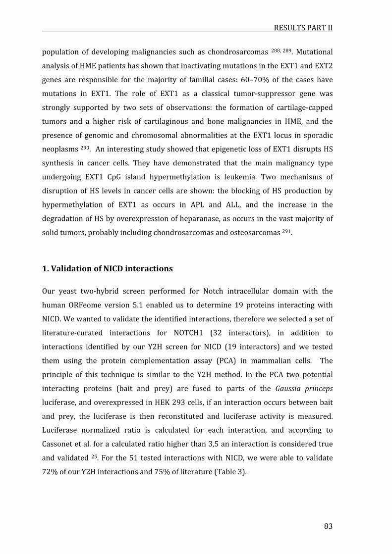
RESULTSPARTII
83
population of developingmalignancies such as chondrosarcomas 288,289.Mutational
analysisofHMEpatientshasshownthatinactivatingmutationsintheEXT1andEXT2
genes are responsible for themajority of familial cases: 60–70% of the cases have
mutations in EXT1. The role of EXT1 as a classical tumor-suppressor gene was
strongly supported by two sets of observations: the formation of cartilage-capped
tumors and a higher risk of cartilaginous and bone malignancies in HME, and the
presenceof genomic and chromosomal abnormalities at theEXT1 locus in sporadic
neoplasms290.AninterestingstudyshowedthatepigeneticlossofEXT1disruptsHS
synthesis in cancer cells. They have demonstrated that the main malignancy type
undergoing EXT1 CpG island hypermethylation is leukemia. Two mechanisms of
disruptionofHS levels in cancer cells are shown: theblockingofHSproductionby
hypermethylation of EXT1 as occurs in APL and ALL, and the increase in the
degradationofHSbyoverexpressionofheparanase,asoccursinthevastmajorityof
solidtumors,probablyincludingchondrosarcomasandosteosarcomas291.
1.ValidationofNICDinteractions
Our yeast two-hybrid screen performed for Notch intracellular domain with the
humanORFeome version 5.1 enabled us to determine 19 proteins interactingwith
NICD.Wewantedtovalidatetheidentifiedinteractions,thereforeweselectedasetof
literature-curated interactions for NOTCH1 (32 interactors), in addition to
interactions identified by our Y2H screen for NICD (19 interactors) and we tested
them using the protein complementation assay (PCA) in mammalian cells. The
principle of this technique is similar to the Y2Hmethod. In the PCA two potential
interacting proteins (bait and prey) are fused to parts of the Gaussia princeps
luciferase,andoverexpressedinHEK293cells,ifaninteractionoccursbetweenbait
and prey, the luciferase is then reconstituted and luciferase activity is measured.
Luciferase normalized ratio is calculated for each interaction, and according to
Cassonetetal.foracalculatedratiohigherthan3,5aninteractionisconsideredtrue
andvalidated 25.For the51 tested interactionswithNICD,wewereable tovalidate
72%ofourY2Hinteractionsand75%ofliterature(Table3).
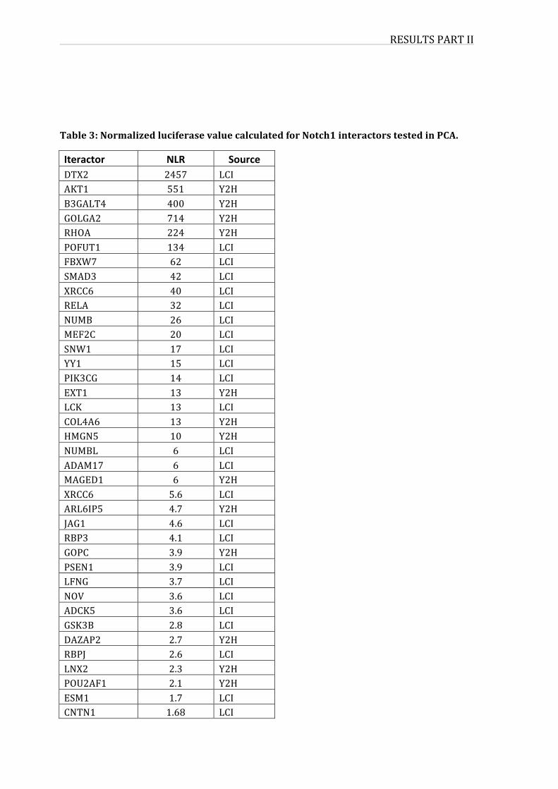
RESULTSPARTII
Table3:NormalizedluciferasevaluecalculatedforNotch1interactorstestedinPCA.
Iteractor NLR SourceDTX2 2457 LCIAKT1 551 Y2HB3GALT4 400 Y2HGOLGA2 714 Y2HRHOA 224 Y2HPOFUT1 134 LCIFBXW7 62 LCISMAD3 42 LCIXRCC6 40 LCIRELA 32 LCINUMB 26 LCIMEF2C 20 LCISNW1 17 LCIYY1 15 LCIPIK3CG 14 LCIEXT1 13 Y2HLCK 13 LCICOL4A6 13 Y2HHMGN5 10 Y2HNUMBL 6 LCIADAM17 6 LCIMAGED1 6 Y2HXRCC6 5.6 LCIARL6IP5 4.7 Y2HJAG1 4.6 LCIRBP3 4.1 LCIGOPC 3.9 Y2HPSEN1 3.9 LCILFNG 3.7 LCINOV 3.6 LCIADCK5 3.6 LCIGSK3B 2.8 LCIDAZAP2 2.7 Y2HRBPJ 2.6 LCILNX2 2.3 Y2HPOU2AF1 2.1 Y2HESM1 1.7 LCICNTN1 1.68 LCI
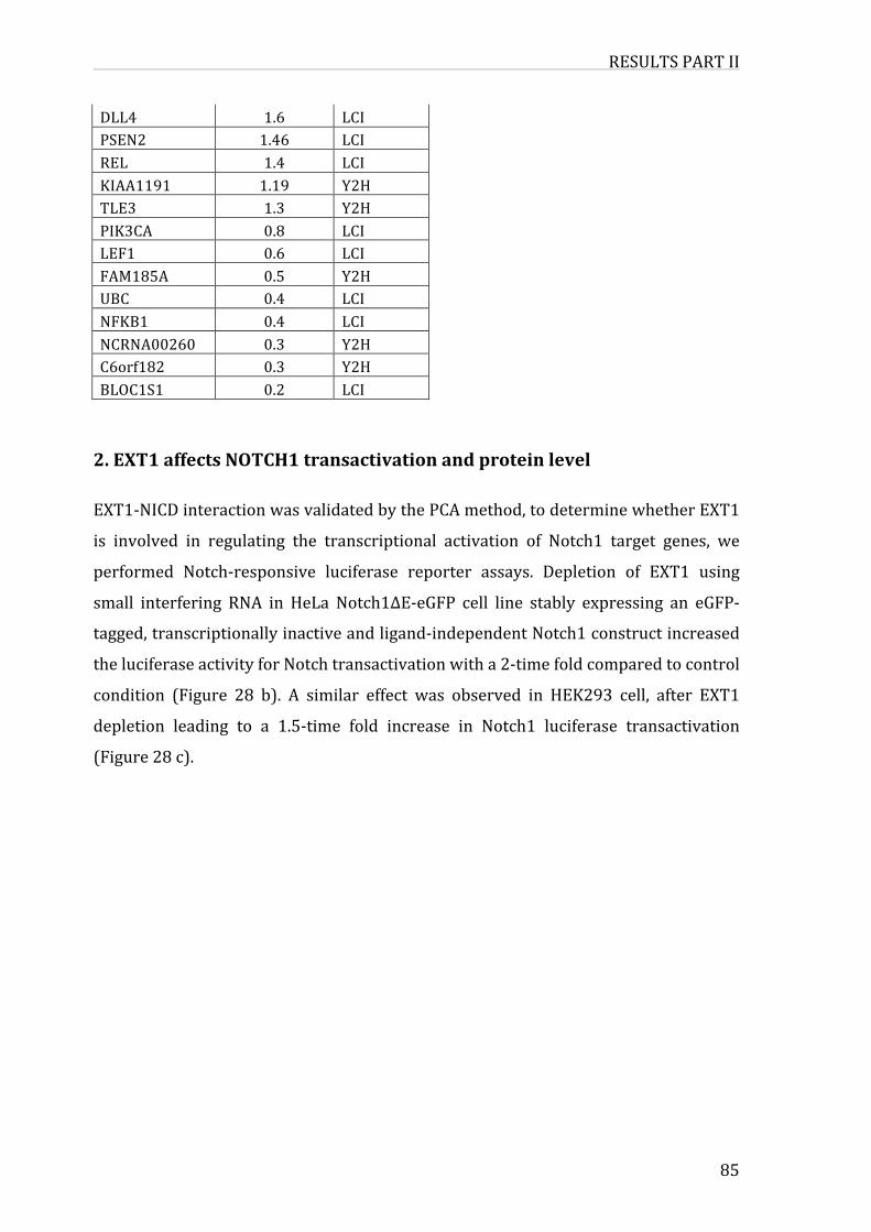
RESULTSPARTII
85
DLL4 1.6 LCIPSEN2 1.46 LCIREL 1.4 LCIKIAA1191 1.19 Y2HTLE3 1.3 Y2HPIK3CA 0.8 LCILEF1 0.6 LCIFAM185A 0.5 Y2HUBC 0.4 LCINFKB1 0.4 LCINCRNA00260 0.3 Y2HC6orf182 0.3 Y2HBLOC1S1 0.2 LCI
2.EXT1affectsNOTCH1transactivationandproteinlevel
EXT1-NICDinteractionwasvalidatedbythePCAmethod,todeterminewhetherEXT1
is involved in regulating the transcriptional activation of Notch1 target genes, we
performed Notch-responsive luciferase reporter assays. Depletion of EXT1 using
small interfering RNA in HeLa Notch1∆E-eGFP cell line stably expressing an eGFP-
tagged,transcriptionallyinactiveandligand-independentNotch1constructincreased
theluciferaseactivityforNotchtransactivationwitha2-timefoldcomparedtocontrol
condition (Figure 28 b). A similar effect was observed in HEK293 cell, after EXT1
depletion leading to a 1.5-time fold increase in Notch1 luciferase transactivation
(Figure28c).
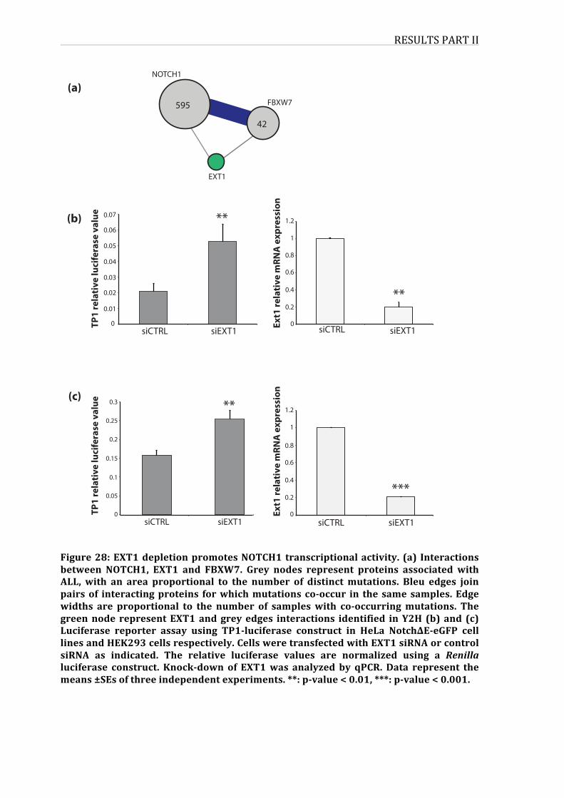
RESULTSPARTII
Figure28:EXT1depletionpromotesNOTCH1transcriptionalactivity. (a) Interactionsbetween NOTCH1, EXT1 and FBXW7. Grey nodes represent proteins associated withALL, with an area proportional to the number of distinctmutations. Bleu edges joinpairsof interactingproteins forwhichmutationsco-occur in thesamesamples.Edgewidths are proportional to the number of sampleswith co-occurringmutations. Thegreennode representEXT1andgreyedges interactions identified inY2H (b)and (c)Luciferase reporter assay using TP1-luciferase construct in HeLa Notch∆E-eGFP celllinesandHEK293cellsrespectively.CellsweretransfectedwithEXT1siRNAorcontrolsiRNA as indicated. The relative luciferase values are normalized using a Renillaluciferase construct. Knock-downof EXT1was analyzed by qPCR.Data represent themeans±SEsofthreeindependentexperiments.**:p-value<0.01,***:p-value<0.001.
siEXT1siCTRL
**
0
0.01
0.07
0.06
0.05
0.04
0.03
0.02
0
1.2
1
0.8
0.6
0.4
0.2
siCTRL siEXT1TP1
rela
tive
luci
fera
se v
alue
Ext1
rela
tive
mRN
A e
xpre
ssio
n
**
(b)
(a)NOTCH1
FBXW7
EXT1
595
42
0
0.05
0.3
0.25
0.2
0.15
0.1
0
1.2
1
0.8
0.6
0.4
0.2
siCTRL siEXT1 siCTRL siEXT1
(c)**
***
TP1
rela
tive
luci
fera
se v
alue
Ext1
rela
tive
mRN
A e
xpre
ssio
n
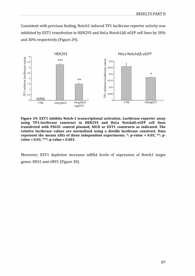
RESULTSPARTII
87
Consistentwithpreviousfinding,Notch1inducedTP1luciferasereporteractivitywas
inhibitedbyEXT1transfectioninHEK293andHeLaNotch1∆E-eGFPcelllinesby50%
and30%respectively(Figure29).
Figure29:EXT1 inhibitsNotch-1 transcriptionalactivation.Luciferasereporterassayusing TP1-luciferase construct in HEK293 and HeLa Notch∆E-eGFP cell linestransfected with PSG5C control plasmid, NICD or EXT1 constructs as indicated. Therelative luciferase values are normalized using a Renilla luciferase construct. Datarepresent themeans±SEsof three independent experiments. *: p-value<0.05, **: p-value<0.01,***:p-value<0.001.
Moreover, EXT1 depletion increases mRNA levels of expression of Notch1 target
genes:HES1andcMYC(Figure30).
CTRL 10ng NICD 10ngNICD+µgEXT1
0
0.5
1
1.5
2
2.5
3
3.5
4
TP1
rela
tive
luci
fera
se v
alue ***
**
0
0.005
0.01
0.015
0.02
0.025
0.03
CTRL 100ngEXT1TP
1 re
lati
ve lu
cife
rase
val
ue
*
HEK293 HeLa Notch∆E-eGFP
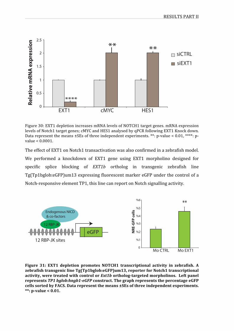
RESULTSPARTII
Figure30:EXT1depletionincreasesmRNAlevelsofNOTCH1targetgenes.mRNAexpressionlevelsofNotch1targetgenes;cMYCandHES1analysedbyqPCRfollowingEXT1Knockdown.Datarepresentthemeans±SEsofthreeindependentexperiments.**:p-value<0.01,****:p-value<0.0001.
TheeffectofEXT1onNotch1transactivationwasalsoconfirmedinazebrafishmodel.
We performed a knockdown of EXT1 gene using EXT1 morpholino designed for
specific splice blocking of EXT1b ortholog in transgenic zebrafish line
Tg(Tp1bglob:eGFP)um13expressingfluorescentmarkereGFPunderthecontrolofa
Notch-responsiveelementTP1,thislinecanreportonNotchsignallingactivity.
Figure 31: EXT1 depletion promotes NOTCH1 transcriptional activity in zebrafish. AzebrafishtransgeniclineTg(Tp1bglob:eGFP)um13,reporterforNotch1transcriptionalactivity,weretreatedwithcontrolorExt1bortholog-targetedmorpholinos.LeftpanelrepresentsTP1bglob:hngb1-eGFPconstruct.ThegraphrepresentsthepercentageeGFPcellssortedbyFACS.Datarepresentthemeans±SEsofthreeindependentexperiments.**:p-value<0.01.
EXT1 cMYC HES10
0.5
1
1.5
2
2.5
****
** **siEXT1
siCTRL
Rela
tive
mRN
A e
xpre
ssio
n
Mo CTRL Mo EXT10
%1
%2
%3
%4
%5
%6
NRE
-GFP
cells
**
eGFP12 RBP-JK sites
Endogenous NICD & co-factors
12 RBP-JK
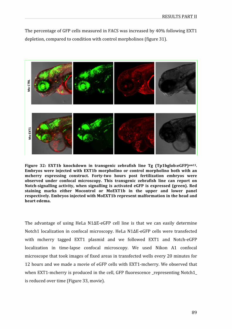
RESULTSPARTII
89
ThepercentageofGFPcellsmeasuredinFACSwasincreasedby40%followingEXT1
depletion,comparedtoconditionwithcontrolmorpholinos(figure31).
Figure 32: EXT1b knockdown in transgenic zebrafish line Tg (Tp1bglob:eGFP)um13.Embryoswere injectedwith EXT1bmorpholino or controlmorpholino bothwith anmcherry expressing construct. Forty-two hours post fertilization embryos wereobserved under confocal microscopy. This transgenic zebrafish line can report onNotch-signalling activity,when signalling is activated eGFP is expressed (green). Redstaining marks either Mocontrol or MoEXT1b in the upper and lower panelrespectively.EmbryosinjectedwithMoEXT1brepresentmalformationintheheadandheartedema.
The advantage of using HeLa N1ΔE-eGFP cell line is that we can easily determine
Notch1 localization in confocalmicroscopy.HeLaN1ΔE-eGFP cellswere transfected
with mcherry tagged EXT1 plasmid and we followed EXT1 and Notch-eGFP
localization in time-lapse confocal microscopy. We used Nikon A1 confocal
microscopethattookimagesoffixedareasintransfectedwellsevery20minutesfor
12hoursandwemadeamovieofeGFPcellswithEXT1-mcherry.Weobservedthat
whenEXT1-mcherryisproducedinthecell,GFPfluorescence_representingNotch1_
isreducedovertime(Figure33,movie).
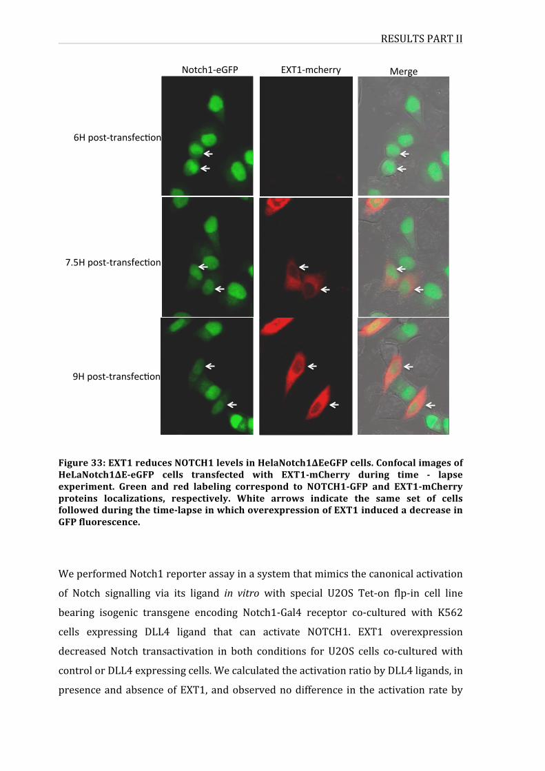
RESULTSPARTII
Figure33:EXT1reducesNOTCH1levelsinHelaNotch1∆EeGFPcells.ConfocalimagesofHeLaNotch1∆E-eGFP cells transfected with EXT1-mCherry during time - lapseexperiment. Green and red labeling correspond to NOTCH1-GFP and EXT1-mCherryproteins localizations, respectively. White arrows indicate the same set of cellsfollowedduringthetime-lapseinwhichoverexpressionofEXT1inducedadecreaseinGFPfluorescence.
WeperformedNotch1reporterassayinasystemthatmimicsthecanonicalactivation
of Notch signalling via its ligand in vitro with special U2OS Tet-on flp-in cell line
bearing isogenic transgene encoding Notch1-Gal4 receptor co-cultured with K562
cells expressing DLL4 ligand that can activate NOTCH1. EXT1 overexpression
decreasedNotch transactivation in both conditions for U2OS cells co-culturedwith
controlorDLL4expressingcells.WecalculatedtheactivationratiobyDLL4ligands,in
presenceandabsenceofEXT1,andobservednodifference in theactivationrateby
Notch1-eGFP EXT1-mcherry Merge
ϲ,�ƉŽƐƚͲƚƌĂŶƐĨĞĐƟŽŶ
ϳ͘ϱ,�ƉŽƐƚͲƚƌĂŶƐĨĞĐƟŽŶ
ϵ,�ƉŽƐƚͲƚƌĂŶƐĨĞĐƟŽŶ
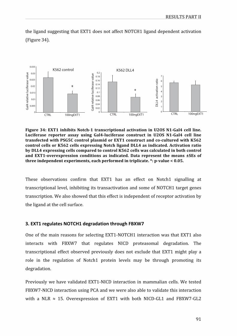
RESULTSPARTII
91
theligandsuggestingthatEXT1doesnotaffectNOTCH1liganddependentactivation
(Figure34).
Figure34: EXT1 inhibitsNotch-1 transcriptional activation inU2OSN1-Gal4 cell line.Luciferase reporter assay using Gal4-luciferase construct in U2OS N1-Gal4 cell linetransfectedwithPSG5CcontrolplasmidorEXT1constructandco-culturedwithK562controlcellsorK562cellsexpressingNotchligandDLL4asindicated.ActivationratiobyDLL4expressingcellscomparedtocontrolK562cellswascalculatedinbothcontroland EXT1-overexpression conditions as indicated. Data represent themeans ±SEs ofthreeindependentexperiments,eachperformedintriplicate.*:p-value<0.05.
These observations confirm that EXT1 has an effect on Notch1 signalling at
transcriptionallevel, inhibitingitstransactivationandsomeofNOTCH1targetgenes
transcription.Wealsoshowedthatthiseffectisindependentofreceptoractivationby
theligandatthecellsurface.
3.EXT1regulatesNOTCH1degradationthroughFBXW7Oneof themainreasons forselectingEXT1-NOTCH1interactionwasthatEXT1also
interacts with FBXW7 that regulates NICD proteasomal degradation. The
transcriptional effect observed previously does not exclude that EXT1might play a
role in the regulation of Notch1 protein levels may be through promoting its
degradation.
PreviouslywehavevalidatedEXT1-NICD interaction inmammaliancells.We tested
FBXW7-NICDinteractionusingPCAandwewerealsoabletovalidatethisinteraction
with a NLR ≈ 15. Overexpression of EXT1 with both NICD-GL1 and FBXW7-GL2
0
0.005
0.01
0.015
0.02
0.025
0.03
0.035
*
Gal
4 re
lativ
e lu
cife
rase
val
ue
CTRL 100ngEXT1
*
0
0.02
0.04
0.06
0.08
0.1
0.12
0.14
0.16
0.18
0.2
Gal
4 re
lativ
e lu
cife
rase
val
ue
CTRL 100ngEXT1 CTRL 100ngEXT10
1
2
3
4
5
6
7
K562 control
DLL
4 a
ctiv
atio
n ra
tio
K562 DLL4
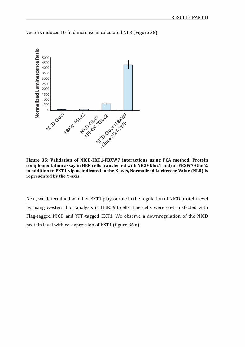
RESULTSPARTII
vectorsinduces10-foldincreaseincalculatedNLR(Figure35).
Figure 35: Validation of NICD-EXT1-FBXW7 interactions using PCA method. ProteincomplementationassayinHEKcellstransfectedwithNICD-Gluc1and/orFBXW7-Gluc2,inadditiontoEXT1-yfpasindicatedintheX-axis,NormalizedLuciferaseValue(NLR)isrepresentedbytheY-axis.
Next,wedeterminedwhetherEXT1playsaroleintheregulationofNICDproteinlevel
by usingwestern blot analysis in HEK393 cells. The cellswere co-transfectedwith
Flag-taggedNICD and YFP-tagged EXT1.We observe a downregulation of theNICD
proteinlevelwithco-expressionofEXT1(figure36a).
500045004000
3500300025002000
15001000500
0
NICD-Gluc1
FBXW
-7Gluc2
NICD-Gluc1
+FBXW-7Gluc2
NICD-Gluc+1FBXW
7
-Gluc+2EXT-1YFP
Nor
mal
ized
Lum
ines
cenc
e Ra
tio
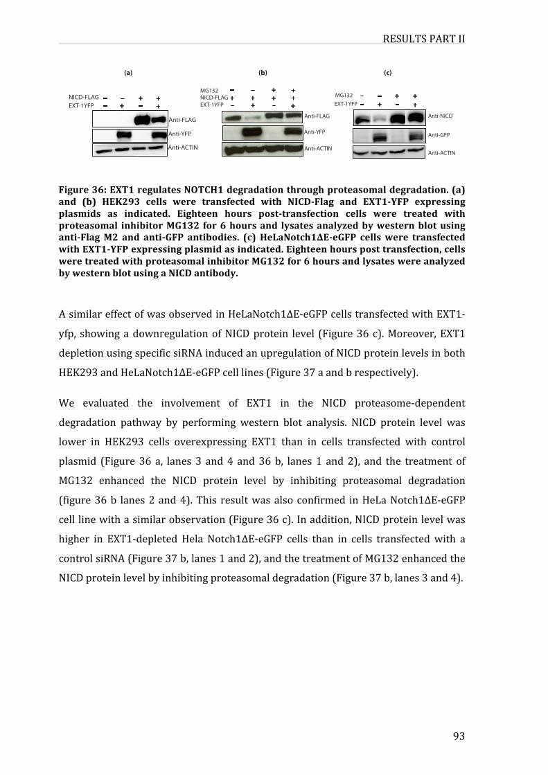
RESULTSPARTII
93
Figure36:EXT1regulatesNOTCH1degradationthroughproteasomaldegradation.(a)and (b) HEK293 cells were transfected with NICD-Flag and EXT1-YFP expressingplasmids as indicated. Eighteen hours post-transfection cells were treated withproteasomal inhibitorMG132 for6hoursand lysatesanalyzedbywesternblotusinganti-FlagM2 and anti-GFP antibodies. (c) HeLaNotch1∆E-eGFP cellswere transfectedwithEXT1-YFPexpressingplasmidasindicated.Eighteenhoursposttransfection,cellsweretreatedwithproteasomalinhibitorMG132for6hoursandlysateswereanalyzedbywesternblotusingaNICDantibody.
AsimilareffectofwasobservedinHeLaNotch1ΔE-eGFPcellstransfectedwithEXT1-
yfp, showingadownregulationofNICDprotein level (Figure36c).Moreover,EXT1
depletionusingspecificsiRNAinducedanupregulationofNICDproteinlevelsinboth
HEK293andHeLaNotch1∆E-eGFPcelllines(Figure37aandbrespectively).
We evaluated the involvement of EXT1 in the NICD proteasome-dependent
degradation pathway by performing western blot analysis. NICD protein level was
lower in HEK293 cells overexpressing EXT1 than in cells transfected with control
plasmid (Figure 36 a, lanes 3 and4 and36b, lanes 1 and2), and the treatment of
MG132 enhanced the NICD protein level by inhibiting proteasomal degradation
(figure36b lanes2and4).This resultwasalsoconfirmed inHeLaNotch1∆E-eGFP
celllinewithasimilarobservation(Figure36c).Inaddition,NICDproteinlevelwas
higher in EXT1-depleted Hela Notch1∆E-eGFP cells than in cells transfectedwith a
controlsiRNA(Figure37b,lanes1and2),andthetreatmentofMG132enhancedthe
NICDproteinlevelbyinhibitingproteasomaldegradation(Figure37b,lanes3and4).
(a) (c)
_ _ + +_ _+ +
NICD-FLAGEXT-1YFP
Anti-FLAG
Anti-YFP
Anti-ACTIN
_ _ + +_ _+ +
MG132
Anti-NICD
Anti-GFP
EXT-1YFP
Anti-ACTIN
(b)
_ _ + ++ +++
_ _+ +
MG132NICD-FLAGEXT-1YFP
Anti-FLAG
Anti-YFP
Anti-ACTIN
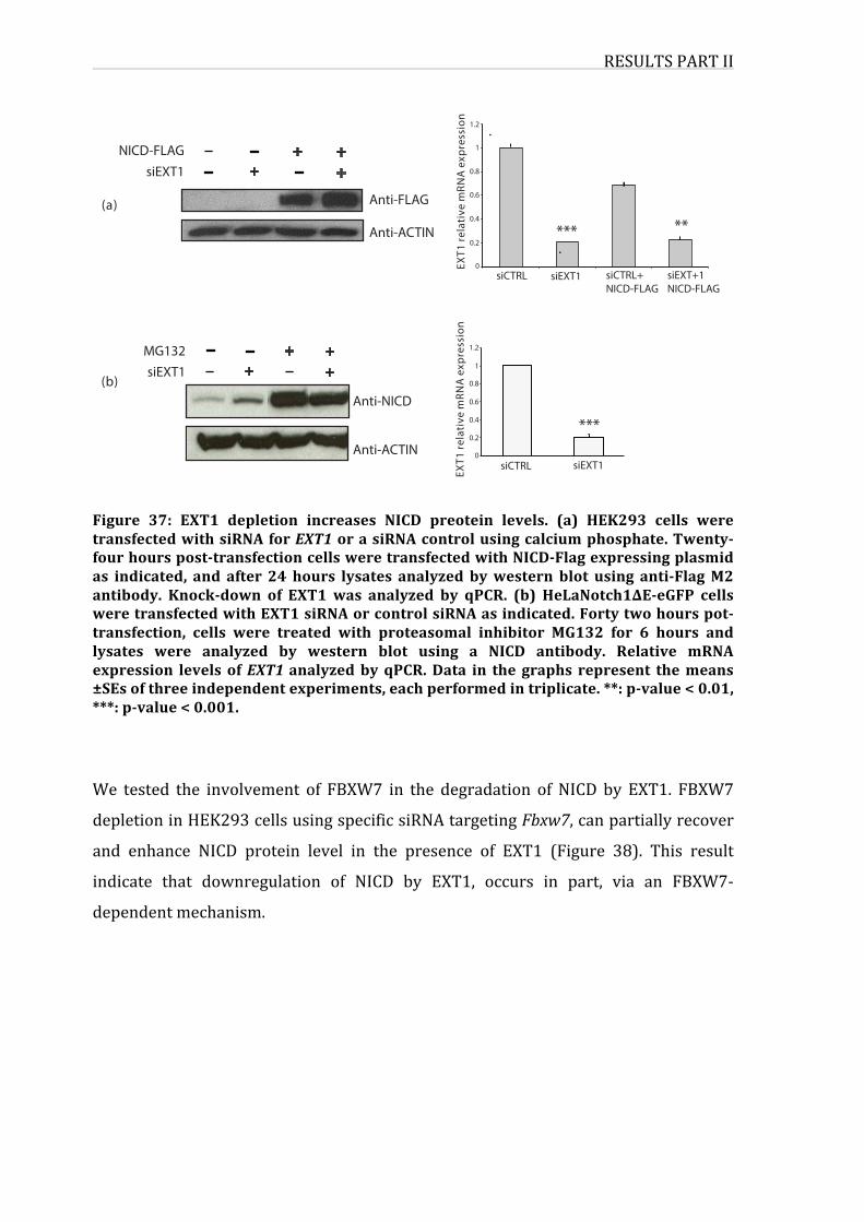
RESULTSPARTII
Figure 37: EXT1 depletion increases NICD preotein levels. (a) HEK293 cells weretransfectedwithsiRNAforEXT1orasiRNAcontrolusingcalciumphosphate.Twenty-fourhourspost-transfectioncellsweretransfectedwithNICD-Flagexpressingplasmidas indicated, and after 24 hours lysates analyzed bywestern blot using anti-FlagM2antibody. Knock-down of EXT1 was analyzed by qPCR. (b) HeLaNotch1∆E-eGFP cellsweretransfectedwithEXT1siRNAorcontrolsiRNAasindicated.Fortytwohourspot-transfection, cells were treated with proteasomal inhibitor MG132 for 6 hours andlysates were analyzed by western blot using a NICD antibody. Relative mRNAexpression levels ofEXT1analyzedbyqPCR.Data in the graphs represent themeans±SEsofthreeindependentexperiments,eachperformedintriplicate.**:p-value<0.01,***:p-value<0.001.
We tested the involvement of FBXW7 in the degradation of NICD by EXT1. FBXW7
depletioninHEK293cellsusingspecificsiRNAtargetingFbxw7,canpartiallyrecover
and enhance NICD protein level in the presence of EXT1 (Figure 38). This result
indicate that downregulation of NICD by EXT1, occurs in part, via an FBXW7-
dependentmechanism.
0
1
0.8
0.6
0.4
0.2
1.2
***
_ _ + +_ _+ +
(b)
_ _ + +_ _+ +
MG132
0
0.2
0.4
0.6
0.8
1
1.2
*** **(a)
NICD-FLAGsiEXT1
Anti-FLAG
Anti-ACTIN
EXT1
rela
tive
mRN
A e
xpre
ssio
n
siCTRL siEXT1 siCTRL+NICD-FLAG
siEXT+1NICD-FLAG
siEXT1
Anti-ACTIN
Anti-NICD
EXT1
rela
tive
mRN
A e
xpre
ssio
nsiCTRL siEXT1
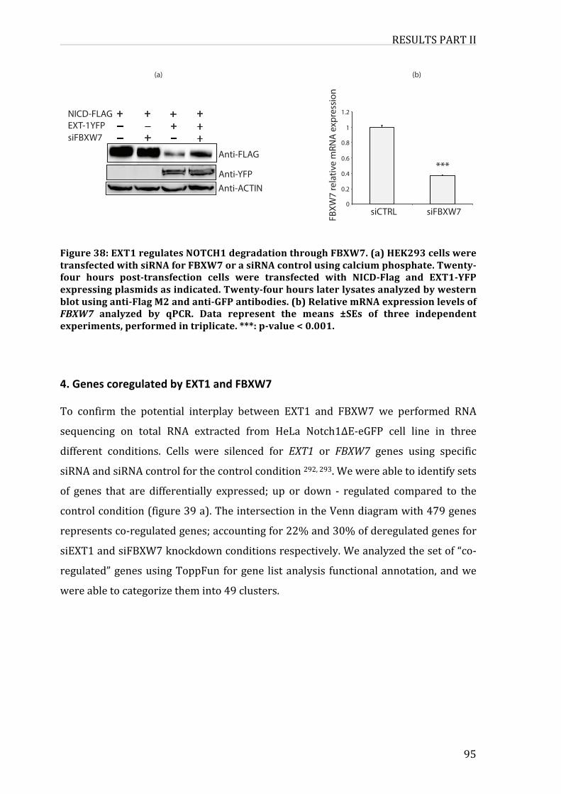
RESULTSPARTII
95
Figure38:EXT1regulatesNOTCH1degradationthroughFBXW7.(a)HEK293cellsweretransfectedwithsiRNAforFBXW7orasiRNAcontrolusingcalciumphosphate.Twenty-four hours post-transfection cells were transfected with NICD-Flag and EXT1-YFPexpressingplasmidsasindicated.Twenty-fourhourslaterlysatesanalyzedbywesternblotusinganti-FlagM2andanti-GFPantibodies.(b)RelativemRNAexpressionlevelsofFBXW7 analyzed by qPCR. Data represent the means ±SEs of three independentexperiments,performedintriplicate.***:p-value<0.001.
4.GenescoregulatedbyEXT1andFBXW7To confirm the potential interplay between EXT1 and FBXW7 we performed RNA
sequencing on total RNA extracted from HeLa Notch1∆E-eGFP cell line in three
different conditions. Cells were silenced for EXT1 or FBXW7 genes using specific
siRNAandsiRNAcontrolforthecontrolcondition292,293.Wewereabletoidentifysets
of genes that are differentially expressed; up or down - regulated compared to the
controlcondition(figure39a).TheintersectionintheVenndiagramwith479genes
representsco-regulatedgenes;accountingfor22%and30%ofderegulatedgenesfor
siEXT1andsiFBXW7knockdownconditionsrespectively.Weanalyzedthesetof“co-
regulated”genesusingToppFun forgene listanalysis functionalannotation,andwe
wereabletocategorizetheminto49clusters.
(a)
+ +++_ _ + +_ _+ +
NICD-FLAGEXT-1YFPsiFBXW7
Anti-FLAG
Anti-ACTINAnti-YFP
siCTRL 0
1
0.8
0.6
0.4
0.2
1.2
siFBXW7
FBXW
7 re
lativ
e m
RNA
expr
essio
n
(b)
***
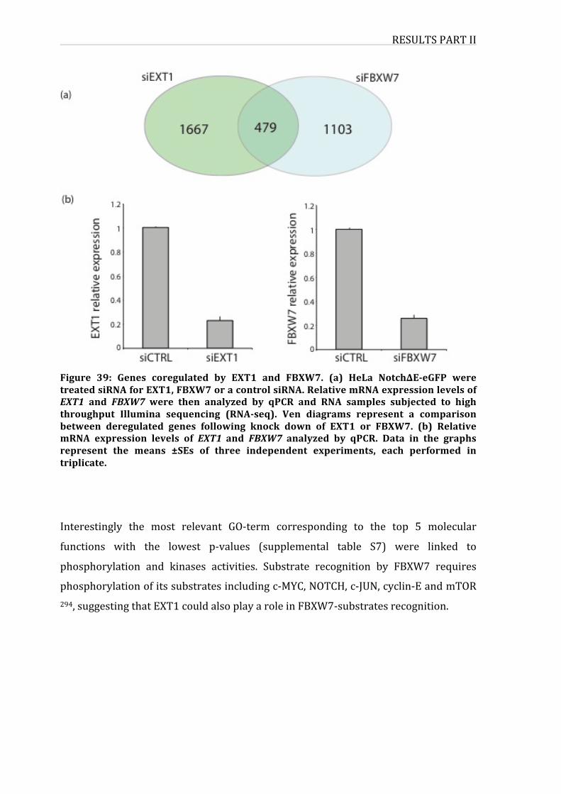
RESULTSPARTII
Figure 39: Genes coregulated by EXT1 and FBXW7. (a) HeLa Notch∆E-eGFP weretreatedsiRNAforEXT1,FBXW7oracontrolsiRNA.RelativemRNAexpressionlevelsofEXT1 and FBXW7were then analyzed by qPCR and RNA samples subjected to highthroughput Illumina sequencing (RNA-seq). Ven diagrams represent a comparisonbetween deregulated genes following knock down of EXT1 or FBXW7. (b) RelativemRNA expression levels of EXT1 and FBXW7 analyzed by qPCR. Data in the graphsrepresent the means ±SEs of three independent experiments, each performed intriplicate.
Interestingly the most relevant GO-term corresponding to the top 5 molecular
functions with the lowest p-values (supplemental table S7) were linked to
phosphorylation and kinases activities. Substrate recognition by FBXW7 requires
phosphorylationofitssubstratesincludingc-MYC,NOTCH,c-JUN,cyclin-EandmTOR294,suggestingthatEXT1couldalsoplayaroleinFBXW7-substratesrecognition.
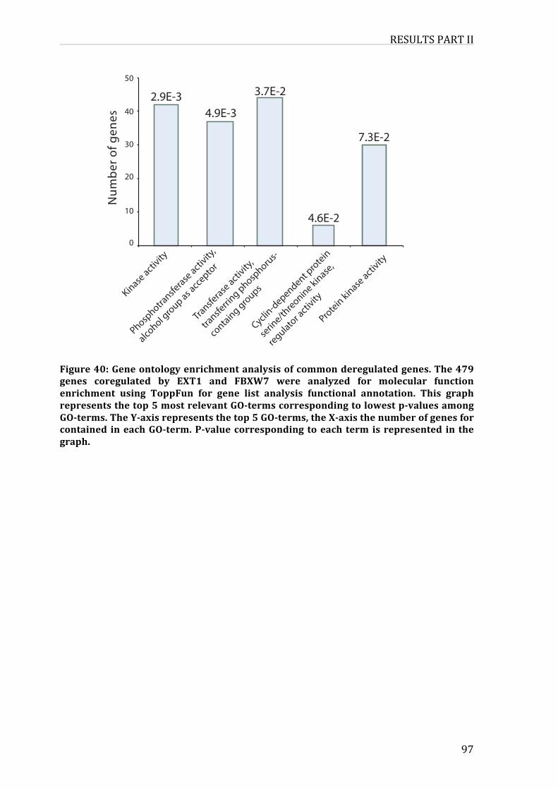
RESULTSPARTII
97
Figure40:Geneontologyenrichmentanalysisofcommonderegulatedgenes.The479genes coregulated by EXT1 and FBXW7 were analyzed for molecular functionenrichment using ToppFun for gene list analysis functional annotation. This graphrepresentsthetop5mostrelevantGO-termscorrespondingtolowestp-valuesamongGO-terms.TheY-axisrepresentsthetop5GO-terms,theX-axisthenumberofgenesforcontained ineachGO-term.P-valuecorresponding toeach term isrepresented in thegraph. .
0
40
30
20
10
50
Num
ber o
f gen
es
Kinase activ
ity
Phosphotra
nsferase
activity
,
alcohol g
roup as acce
ptor
Transferase
activity
,
transfe
rring phosp
horus-
containg groups
Cyclin-dependent p
rotein
serin
e/threonine kinase,
regulator activ
ity
Protein kinase activ
ity
4.9E-32.9E-3 3.7E-2
4.6E-2
7.3E-2
DISCUSSIONPARTII
DISCUSSIONPARTIISeveralexamplescanillustratetheeffectivenessofusingthe“interactomes”approach
fortheidentificationofnovelcancerrelatedgenes.AswehaveprioritizedALL-gene
products, the exostin glysosyltransferase 1 (EXT1) gene grabbed our attention for
several reasons; first for being a novel interaction linked to two of the ALL-gene
products:NOTCH1andFBXW7,secondforbeingalreadyimplicatedinanothertype
ofcancer(osteosarcoma)viainactivatingmutations.
ValidationofinteractioninNOTCH1interactome
BeforeexploringtherelationshipbetweenEXT1andNOTCH1weanalysedtheNotch1
interactomeandproceededtothevalidationoftheidentifiedinteractions.
Our yeast two-hybrid screen performed for Notch intracellular domain with the
humanORFeome version 5.1 enabled us to determine 19 proteins interactingwith
NICD.AmongtheseNICDpartners,AKT1hasbeenreportedpreviouslyforinteracting
withNotchsignallingandbeingessentialforneuraldevelopmentthroughregulation
of this signallingpathway. Itwasdemonstrated thatbothAKTandNotch signalling
pathways interact through complex molecular interactions in development 295, 296.
SeveraldatashowedthatNotchcanaffectAKTsignallingandAKT1alsoaffectsNotch
signallingbuttheirdirectinteractionwasnotpreviouslydemonstrated.
Among the identifiedNICD partners, AKT1, RhoA, LNX2 andTLE3 can be linked to
Notchsignallingaccordingtodatapublishedinliterature.
Recently it was demonstrated that RhoA/Rho kinase is regulated by Notch in
endothelial cells, promoting senescence phenotype associated with barrier
dysfunction297.RhoAisamemberofRasfamilyofsmallGTPasesthatweidentifiedin
our screen as a partner of Notch1. In fact, Notch and Rho GTPase signalling were
previously shown to control dendritic development 298. Activation of RhoA in a
numberofdifferentcelltypesappearstolimitdendriticgrowth,whereasactivationof
Notch appears to restrict dendritic growth and promote dendritic branching by
regulatingtheactivationofmembersofRhoGTPasefamily.AsshownforAKT1,RhoA
DISCUSSIONPARTII
99
might be affected byNotch signalling.We are the first to show a direct interaction
betweenNotch1andRhoA.
LNX2 in the ligand of Numb X 2, LNX is a RING finger and PDZ domain containing
proteinthatinteractswiththecellfatedeterminantNumb.Numbwasidentifiedasa
substrate ubiquitinated by LNX which is considered as the first example of an E3
ligasethatappearstoinvolvePDZdomainsforsubstraterecognition299.It isknown
that proteins with PDZ domains often interact with the cytoplasmic tails of
transmembrane proteins and localize to discrete sub-membranous sites. Therefore,
the LNX PDZ domains may be involved in the recognition of transmembrane
receptors, which may in turn be substrates for ubiquitylation by LNX. This
explanationcouldbevalidforLNX2andNOTCH1,ontheotherhandNumbpromotes
Notchubiquitinationanddegradation.Numbcan interactwithother factorssuchas
ItchtopromoteNotchdegradation.KnowingthatNumbcanbindLNX2,wespeculate
that LNX2 might be involved in Notch1 ubiquitination via Numb also. These
hypothesisneedtobeinvestigated,andarenotpartofouranalysis.
TLEfamilyhasbeenimplicatedintumorigenesisandhasbeenshowntointeractwith
andmodulate theNotchpathway,viaphosphorylationbymitogen-activatedprotein
kinase(MAPK)inresponsetoepidermalgrowthfactorreceptor(EGFR)signalling300.
TLE3havebeenshowntointeractwiththeNotchpathwayforthecontrolofepithelial
differentiation 301,302. TLE family also act as important effectors ofNotch signalling
throughtheirinteractionswithHesproteins,andthederegulationofNotchsignalling
hasbeenimplicatedinthepathogenesisofsomecancers.
Werepresentthe19proteins identifiedintheY2Hintable4,withknownfunctions
describedforeachprotein.
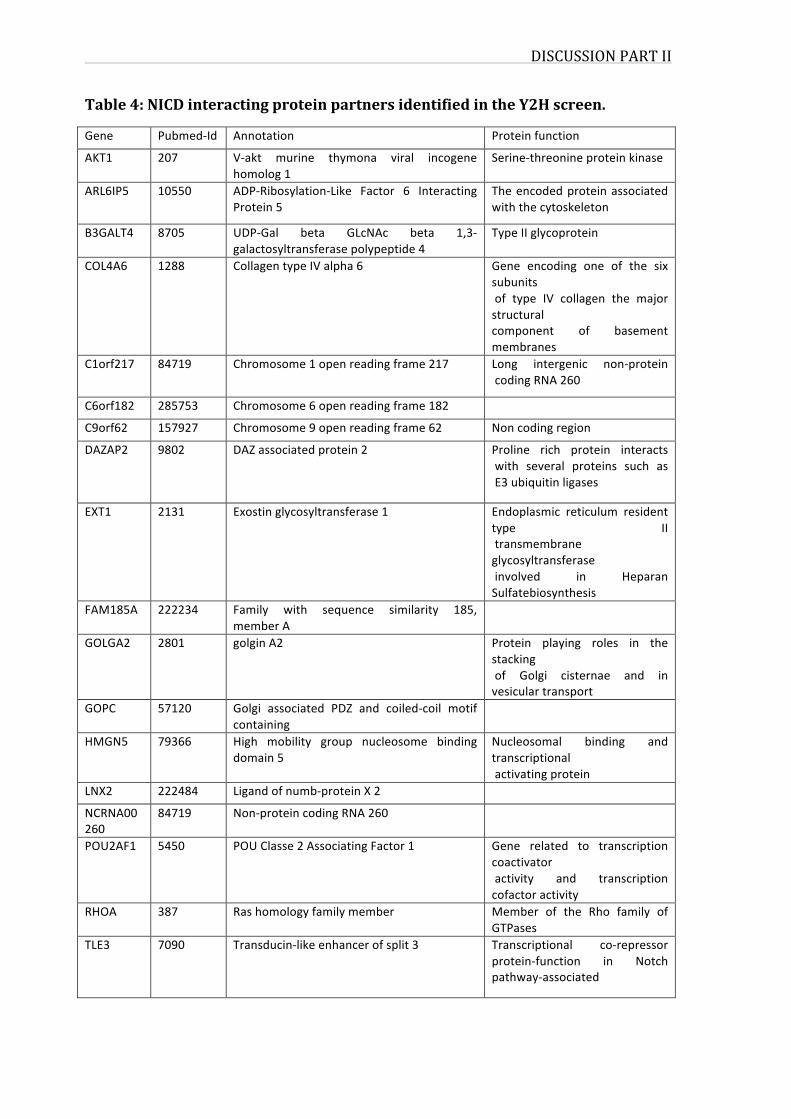
DISCUSSIONPARTII
Table4:NICDinteractingproteinpartnersidentifiedintheY2Hscreen.
Gene Pubmed-Id Annotation Proteinfunction
AKT1 207 V-akt murine thymona viral incogenehomolog1
Serine-threonineproteinkinase
ARL6IP5 10550 ADP-Ribosylation-Like Factor 6 InteractingProtein5
Theencodedproteinassociatedwiththecytoskeleton
B3GALT4 8705 UDP-Gal beta GLcNAc beta 1,3-galactosyltransferasepolypeptide4
TypeIIglycoprotein
COL4A6 1288 CollagentypeIValpha6 Gene encoding one of the sixsubunitsof type IV collagen the majorstructuralcomponent of basementmembranes
C1orf217 84719 Chromosome1openreadingframe217 Long intergenic non-proteincodingRNA260
C6orf182 285753 Chromosome6openreadingframe182
C9orf62 157927 Chromosome9openreadingframe62 Noncodingregion
DAZAP2 9802 DAZassociatedprotein2 Proline rich protein interactswith several proteins such asE3ubiquitinligases
EXT1 2131 Exostinglycosyltransferase1 Endoplasmic reticulum residenttype IItransmembraneglycosyltransferaseinvolved in HeparanSulfatebiosynthesis
FAM185A 222234 Family with sequence similarity 185,memberA
GOLGA2 2801 golginA2 Protein playing roles in thestackingof Golgi cisternae and invesiculartransport
GOPC 57120 Golgi associated PDZ and coiled-coil motifcontaining
HMGN5 79366 High mobility group nucleosome bindingdomain5
Nucleosomal binding andtranscriptionalactivatingprotein
LNX2 222484 Ligandofnumb-proteinX2
NCRNA00260
84719 Non-proteincodingRNA260
POU2AF1 5450 POUClasse2AssociatingFactor1 Gene related to transcriptioncoactivatoractivity and transcriptioncofactoractivity
RHOA 387 Rashomologyfamilymember Member of the Rho family ofGTPases
TLE3 7090 Transducin-likeenhancerofsplit3 Transcriptional co-repressorprotein-function in Notchpathway-associated
DISCUSSIONPARTII
101
Though the Y2H is a stringent method for identifying protein-protein interactions
(PPIs), it requires cross-validation to increase coverage and accuracy, which is the
case for any other information provided by a certain screening methods for PPIs.
ThereforeitwasimportanttotestasetofNOTCH1interactionsfrombothliterature-
curated interactions and Y2H interactions. The result obtained using the protein
complementationassayisverypromisingforthesetofinteractionsthatweextracted
from our Y2H screen, with a rate comparable to the one obtained for literature
interactions(table3).
Notch1-EXT1anovelinteraction
Ifwe take a closer look to theNOTCH1 andFBXW7 interactomesmerged together,
withinteractionsextractedfromliterature(BioGRID,HPRDandIntActdatabases)and
from our Y2H screen we identify 2 common interactors: PSEN1 and EXT1 as it is
showninfigure41.TheinteractomicmaphighlightsnovelY2Hinteractionsforboth
Notch1 and FBXW7 represented in red, as for literature-curated interactions (LCI)
theyarerepresentedingrey.Apartfrombeingknowntodirectlyinteract,Notch1and
its E3 ubiquitin ligase FBXW7 have a common interactor PSEN1, reported in
literature. PSEN1 belongs to the family of presenilin basically known for being
mutated in patients with Alzheimer’s disease. Presenilins are implicated in the
regulation of Notch signalling through affecting the receptors cleavage at the cell
surface. PSEN1,with PSEN2 form the central components of γ-secretase complexes
that are required for intra-membrane cleavage of over 70 different substrates
includingNotch1.Psen1isacriticalfacilitatorofNotchsignallingandothersignalling
pathways303.OntheotherhandPSEN1isasubstrateofFBXW7.
AnothercommoninteractorlinkingNOTCH1andFBXW7interactomeswasEXT1.
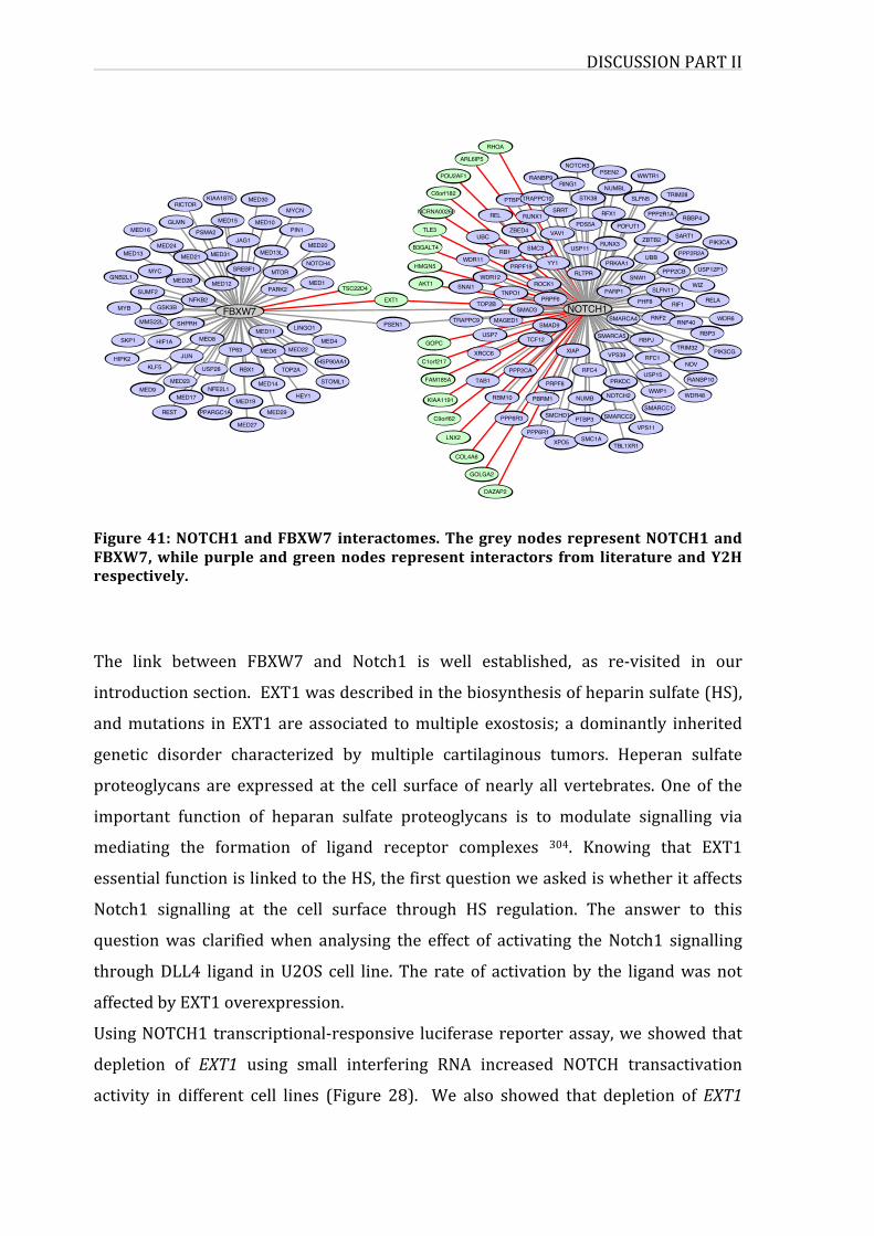
DISCUSSIONPARTII
Figure41:NOTCH1andFBXW7interactomes.ThegreynodesrepresentNOTCH1andFBXW7,whilepurpleandgreennodesrepresent interactors from literatureandY2Hrespectively.
The link between FBXW7 and Notch1 is well established, as re-visited in our
introductionsection.EXT1wasdescribedinthebiosynthesisofheparinsulfate(HS),
andmutations inEXT1are associated tomultiple exostosis; a dominantly inherited
genetic disorder characterized by multiple cartilaginous tumors. Heperan sulfate
proteoglycans are expressed at the cell surface of nearly all vertebrates.Oneof the
important function of heparan sulfate proteoglycans is to modulate signalling via
mediating the formation of ligand receptor complexes 304. Knowing that EXT1
essentialfunctionislinkedtotheHS,thefirstquestionweaskediswhetheritaffects
Notch1 signalling at the cell surface through HS regulation. The answer to this
questionwas clarifiedwhen analysing the effect of activating theNotch1 signalling
throughDLL4 ligand inU2OS cell line. The rate of activationby the ligandwasnot
affectedbyEXT1overexpression.
UsingNOTCH1transcriptional-responsive luciferasereporterassay,weshowedthat
depletion of EXT1 using small interfering RNA increased NOTCH transactivation
activity in different cell lines (Figure 28). We also showed that depletion ofEXT1
C1orf217
FAM185A
C9orf62
GOPC
KIAA1191
GOLGA2
DAZAP2
RBM10
LNX2
TAB1PPP2CA
PPP6R3
COL4A6
PSEN1
TLE3
AKT1
HMGN5
EXT1
B3GALT4
PTBP1
NCRNA00260
RHOA
POU2AF1
ARL6IP5
REL
C6orf182
RB1
ZBED4
WDR11
UBC
PRPF19
USP7
TNPO1
TOP2B
MAGED1
SNAI1
XRCC6
TRAPPC9
WDR12
RBBP4
SLFN5
PPP2R2A
SART1
TRIM28
ZBTB2 PIK3CA
UBB
PPP2R1A
POFUT1
RUNX3
RLTPR
USP11
YY1 PRKAA1
SMC3
VAV1
STK38
RFX1PDS5A
RUNX1SRRT
TRAPPC10
RNF2
WIZ
RELA
SNW1
RNF40
RIF1PHF8
WDR6
USP12P1
SLFN11
PPP2CB
USP15
RBPJ
SMARCC1
WWP1
RFC1
NOTCH3
RANBP9RING1
PSEN2 WWTR1
NUMBL
WDR48
TRIM32
NOV
RANBP10
RBP3
PIK3CG
XPO5
SMCHD1 SMARCC2
PPP6R1SMC1A
PTBP3
TBL1XR1
VPS11
SMARCA4
PARP1
SMARCA5
PRPF6
VPS39
ROCK1
XIAP
SMAD3
TCF12
SMAD9
NOTCH1
NOTCH2NUMBPBRM1
RFC4
PRKDCPRPF8
JAG1
SREBF1
MED15
MED12
MED30KIAA1875
MED31MED20
MED1MTOR
PARK2
NOTCH4
MED13L
MYCN
MED10PIN1
TSC22D4
MED13
MYC
SKP1
MMS22L
SUMF2
HIPK2
MYB
GNB2L1
MED16
MED21
PSMA2GLMN
RICTOR
MED24
MED28
GSK3B
JUN
MED8HIF1A
NFKB2
SHPRH
TP63
FBXW7
MED27
USP28
MED23
KLF5
NFE2L1MED9
RBX1
PPARGC1AREST
MED17 MED19
MED14
LINGO1
MED29
TOP2A
MED22MED6HSP90AA1
MED11MED4
STOML1
HEY1
DISCUSSIONPARTII
103
increasesmRNAexpressionlevelsoftwoimportantNOTCH1-targetgenes:HES1and
MYC (Figure 30). Consistent with this finding, over-expression of EXT1 inhibits
NOTCH1-transactivationindifferentcelllines(Figures29and34)andcorrelateswith
areductionofNICDproteinlevels(Figure33andtime-lapsevideo).Weconfirmedthe
effect of EXT1 on NOTCH1 pathway using a zebrafish in vivo model. We treated
transgenic zebrafish line Tg(Tp1bglob:eGFP)um13 expressing fluorescent marker
eGFP under the control of a Notch-responsive element TP1, with morpholino
targeting EXT1 b zebrafish orholog. As shown on Figure 32, we observed a 40%
increase cells expressing Notch activity following depletion of EXT1 zebrafish
orthologs.
FBXW7isanE3ubiquitinligaseregulatingNOTCH1proteasomaldegradation305.To
determine whether EXT1 interferes with FBXW7 – NOTCH1 association, we first
showedthattheinteractionbetweenNOTCH1andFBXW7wasdramaticallyenhanced
inthepresenceofEXT1(Figure35).Then,weshowedthat,inthepresenceofEXT1,
the level of NOTCH1 intracellular domain (NICD) is reduced in a proteasome-
dependent manner (Figure 36 b and c: compare presence and absence of MG132
proteasome inhibitor). Interestingly,wealso showed that reduced levelsofNICD in
thepresenceofEXT1areFBXW7-dependent(Figure38).Tomoredeeplyanalyzethe
functionalrelationshipbetweenEXT1andFBXW7inregulatingcellularhomeostasis,
a functionwell known forFBXW7 305,weperformedagenome-wideanalysisof the
transcriptome of HeLa-NICDdeltae-GFP 306 depleted for EXT1 or FBXW7. We
identified479mRNAsco-regulatedbybothEXT1andFBXW7,whichrepresentmore
than30%ofFBXW7targets(Figure39). Functionalannotationenrichmentanalysis
oftheseEXT1/FBXW7co-regulatedgenesfinallyindicatedasignificantenrichmentin
genesencodingforkinasesincludingcyclin-dependentandMAPkinases(Figure40).
Kinases play an essential role in cell cycle regulation, and protein phosphorylation
regulates most aspects of cell life 307. Deregulation of kinase mechanism leads to
disease and cancer development, therefore Kinases are intensively studied as anti-
cancer drugs through the developing inhibitor candidates that are able to arrest
proliferation and induce apoptosis 308. FBXW7 is known to regulate proteasomal
degradation of a large number of proteins, in a mechanism dependent of
phosphorylation activity that ismediated by kinases and cyclin-dependent kinases.
DISCUSSIONPARTII
Recent studies demonstrated that GATA3 is a novel target of FBXW7, and its
degradation depends on CDK2 309 and CDK1 310 kinases. GSK3B is also one of the
kinasesthatarecrucialforFBXW7-dependentdegradation,forMYC,JUN,cyclinEand
NOTCH1proteins195.ItisnotsurprisingtofindthatFBXW7depletioncanaffectaset
ofgenesencodingproteinsimplicatedinthekinaseandphosphorylationpathway.As
forEXT1,thelinkwithkinaseswasdescribedthroughtheroleofheparansulfatefor
proper binding of growth factor to its kinase receptor. Among growth factors,
fibroblast growth factors (FGFs) depend on heparan sulfate proteoglycans (HSPGs)
for cell signalling activity, and FGFs act through tyrosine-kinase receptors. These
components form a complex at cell surface leading to the phosphorylation of the
receptor and therefore triggering the activation of signalling cascades. This role of
EXT1rolewasdescribed ingrowthfactorsignallingandfibroblast interactionswith
theextracellularmatrix311,312. HoweverEXT1associationwithkinasesactivitywas
notpreviouslyestablished.
Our data suggest a novel role of EXT1. Performing RNA-seq transciptome analysis
combined with enrichment analysis confirmed that EXT1 is functionally linked to
FBXW7, probably through priming kinases and substrates such as Notch1 towards
proteasomaldegradation.
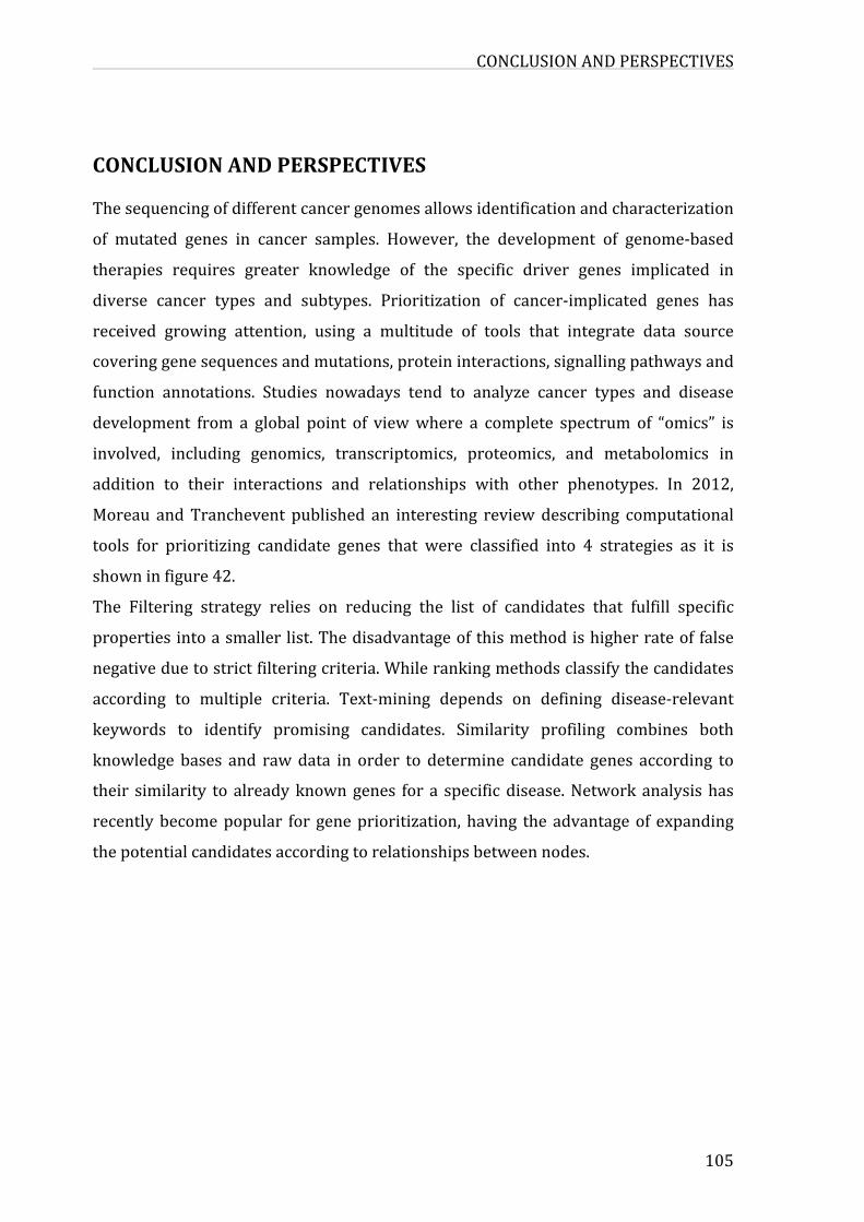
CONCLUSIONANDPERSPECTIVES
105
CONCLUSIONANDPERSPECTIVESThesequencingofdifferentcancergenomesallowsidentificationandcharacterization
of mutated genes in cancer samples. However, the development of genome-based
therapies requires greater knowledge of the specific driver genes implicated in
diverse cancer types and subtypes. Prioritization of cancer-implicated genes has
received growing attention, using a multitude of tools that integrate data source
coveringgenesequencesandmutations,proteininteractions,signallingpathwaysand
function annotations. Studies nowadays tend to analyze cancer types and disease
development from a global point of viewwhere a complete spectrum of “omics” is
involved, including genomics, transcriptomics, proteomics, and metabolomics in
addition to their interactions and relationships with other phenotypes. In 2012,
Moreau andTranchevent published an interesting reviewdescribing computational
tools for prioritizing candidate genes that were classified into 4 strategies as it is
showninfigure42.
The Filtering strategy relies on reducing the list of candidates that fulfill specific
propertiesintoasmaller list.Thedisadvantageofthismethodishigherrateoffalse
negativeduetostrictfilteringcriteria.Whilerankingmethodsclassifythecandidates
according to multiple criteria. Text-mining depends on defining disease-relevant
keywords to identify promising candidates. Similarity profiling combines both
knowledge bases and rawdata in order to determine candidate genes according to
their similarity to alreadyknowngenes for a specificdisease.Networkanalysishas
recentlybecomepopular for geneprioritization,having theadvantageof expanding
thepotentialcandidatesaccordingtorelationshipsbetweennodes.
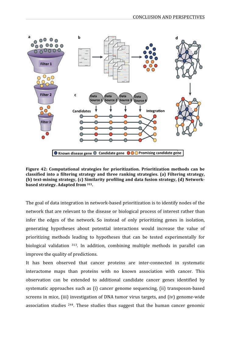
CONCLUSIONANDPERSPECTIVES
Figure 42: Computational strategies for prioritization. Prioritizationmethods can beclassified into a filtering strategy and three ranking strategies. (a) Filtering strategy,(b)text-miningstrategy,(c)Similarityprofilinganddatafusionstrategy,(d)Network-basedstrategy.Adaptedfrom313.
Thegoalofdataintegrationinnetwork-basedprioritizationistoidentifynodesofthe
networkthatarerelevanttothediseaseorbiologicalprocessofinterestratherthan
infer the edges of the network. So instead of only prioritizing genes in isolation,
generating hypotheses about potential interactions would increase the value of
prioritizing methods leading to hypotheses that can be tested experimentally for
biological validation 313. In addition, combining multiple methods in parallel can
improvethequalityofpredictions.
It has been observed that cancer proteins are inter-connected in systematic
interactome maps than proteins with no known association with cancer. This
observation can be extended to additional candidate cancer genes identified by
systematic approaches suchas (i) cancergenomesequencing, (ii) transposon-based
screensinmice,(iii)investigationofDNAtumorvirustargets,and(iv)genome-wide
association studies 244. These studies thus suggest that the human cancer genomic
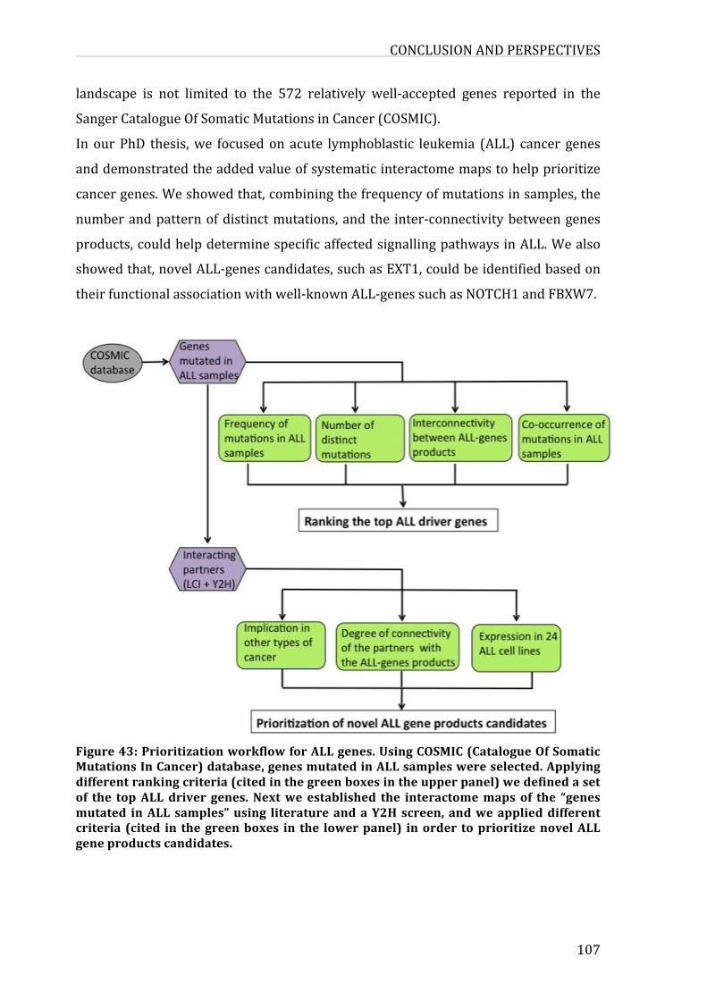
CONCLUSIONANDPERSPECTIVES
107
landscape is not limited to the 572 relatively well-accepted genes reported in the
SangerCatalogueOfSomaticMutationsinCancer(COSMIC).
In ourPhD thesis,we focusedon acute lymphoblastic leukemia (ALL) cancer genes
anddemonstratedtheaddedvalueofsystematicinteractomemapstohelpprioritize
cancergenes.Weshowedthat,combiningthefrequencyofmutationsinsamples,the
numberandpatternofdistinctmutations,andthe inter-connectivitybetweengenes
products,couldhelpdeterminespecificaffectedsignallingpathwaysinALL.Wealso
showedthat,novelALL-genescandidates,suchasEXT1,couldbeidentifiedbasedon
theirfunctionalassociationwithwell-knownALL-genessuchasNOTCH1andFBXW7.
Figure43:PrioritizationworkflowforALLgenes.UsingCOSMIC(CatalogueOfSomaticMutationsInCancer)database,genesmutatedinALLsampleswereselected.Applyingdifferentrankingcriteria(citedinthegreenboxesintheupperpanel)wedefinedasetof the topALLdriver genes.Nextwe established the interactomemapsof the “genesmutated inALLsamples”using literatureandaY2Hscreen,andweapplieddifferentcriteria (cited in thegreenboxes in the lowerpanel) inorder toprioritizenovelALLgeneproductscandidates.
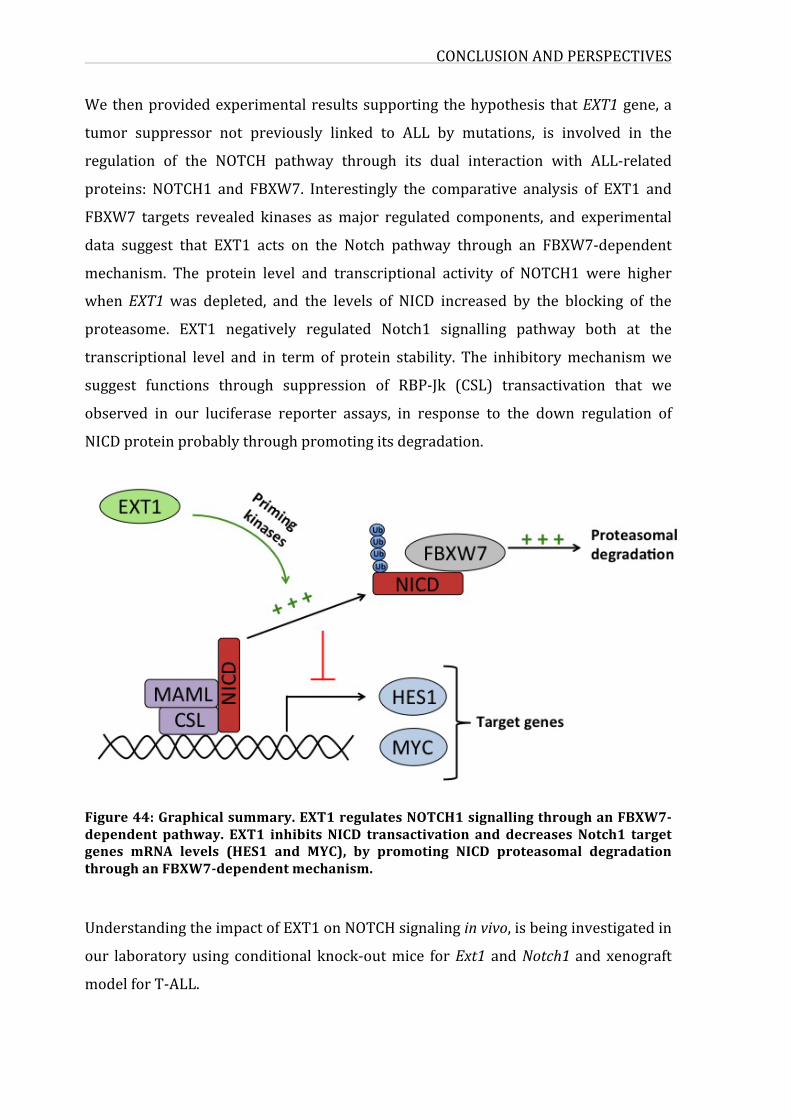
CONCLUSIONANDPERSPECTIVES
Wethenprovidedexperimentalresultssupporting thehypothesis thatEXT1gene,a
tumor suppressor not previously linked to ALL by mutations, is involved in the
regulation of the NOTCH pathway through its dual interaction with ALL-related
proteins: NOTCH1 and FBXW7. Interestingly the comparative analysis of EXT1 and
FBXW7 targets revealed kinases asmajor regulated components, and experimental
data suggest that EXT1 acts on the Notch pathway through an FBXW7-dependent
mechanism. The protein level and transcriptional activity of NOTCH1 were higher
when EXT1was depleted, and the levels of NICD increased by the blocking of the
proteasome. EXT1 negatively regulated Notch1 signalling pathway both at the
transcriptional level and in term of protein stability. The inhibitorymechanismwe
suggest functions through suppression of RBP-Jk (CSL) transactivation that we
observed in our luciferase reporter assays, in response to the down regulation of
NICDproteinprobablythroughpromotingitsdegradation.
Figure44:Graphicalsummary.EXT1regulatesNOTCH1signallingthroughanFBXW7-dependent pathway. EXT1 inhibits NICD transactivation and decreasesNotch1 targetgenes mRNA levels (HES1 and MYC), by promoting NICD proteasomal degradationthroughanFBXW7-dependentmechanism.
UnderstandingtheimpactofEXT1onNOTCHsignalinginvivo,isbeinginvestigatedin
our laboratoryusing conditional knock-outmice forExt1 andNotch1andxenograft
modelforT-ALL.
CONCLUSIONANDPERSPECTIVES
109
In conclusion, we believe that this work presents conceptual advances of broad
interestforcancerbiologyandapplicationofsystemsbiologyintheidentificationof
noveltargetedgenesandpathwaysassociatedtohumancancer.
MATERIALSANDMETHODS
MATERIALSANDMETHODS
1.PlasmidsOpen reading frames (ORF) encoding Notch1 partners (tested for Protein
complementationassay)wereobtainedfromhumanORFeomev5.1(centerofcancer
systems biology: CCSB) as pDONR223 vectors. As for human NICD plasmid, it was
purchased from addgene. FBXW7 α was obtained from Dr. E. Dejardin from the
laboratory of molecular immunology and signal transduction – Groupe
Interdisciplinaire de Génoprotéomique Appliquée (GIGA). ORFs that were not
availablefromthehORFV5.1(BRAF,HRAS,ABL1, JAK2andSMARCB1andNOTCH1
genes), and Notch1 seperate intacellular domains were cloned by Gateway
recombination technology (Invitrogen) using specific primers flanked with the
following AttB1 and AttB2 Gateway sites: 5’-
GGGGACAACTTTGTACAAAAAAGTTGGCATG-3’ (AttB1) and 5’-
GGGGACAACTTTGTACAAGAAAGTTGA-3’ (AttB2). These constructions were verified
byPCRandsequencing.
Inserts from pDONR223were transferred by LR cloning (Invitrogen) into different
destination vectors: pAD-destCYH and pDB-dest the Y2H expression vectors, and
pDEST1899(flagtag),pDEST491(YFP-tag)andpDEST-mcherry.
2.GatewaycloningCloningwas carriedoutusing theGateway recombination technologyasmentioned
previously.Thissystemisauniversalcloningmethodthattakesadvantageofthesite-
specific recombination properties of bacteriophage lambda to provide a rapid and
highly efficientway tomoveDNA sequences intomultiple vector systems.Genesof
interest were cloned in the donor vector pDNR223 and in different expression
vectors: pAD and pDB yeast expression vectors, and pN1GLuc and pN2GLuc for
GaussialuciferasecomplementationassayandothervectorssuchYFP,mCherry,Flag
expressionvectors. Inorder to cloneNICD,BRAF,HRAS,ABL1, JAK2andSMARCB1
thatwerenotpresentinthehORFeomecollection5.1,weperformedPCRtoamplify
the gene of interestwith specific primers flanked by attB Gateway sequences. PCR
MATERIALSANDMETHODS
111
product was then cloned into the donor vector in BP reaction, using Gateway BP
clonase (Invitrogen) (figure45).This reaction inperformedbyadding100ngof the
PCRproductwith150ngofthedonorvectorwith1µloftheBPclonaseIIenzymemix
inatotalvolumeof10µl ina1,5mleppendorfat25oCforanhour.Toterminatethe
reaction1µlofproteinaseKsolutionwasaddedtothemixtureandincubatedfor10
minutes at 37oC. 2µl of each BP reaction were transformed into 40µl of Hb101
electrocompetent bacterial cells, then transferred into 1ml of LB medium and
incubatedat37oCfor1hourwithshaking.100µlofeachtransformationwereplated
ontoLBagarplatescomplementedwithampicillinantibiotic(1µg/ml)andincubated
at37oCovernight.Positivecolonieswereselectedandtransferredinto5mlofliquid
LB+ampicillinmediumforDNAextractionusingNucleoSpinPlasmidkit(Marcherey-
Nagel) according tomanufacturer instructions. ExtractedDNAwas verified by PCR,
subsequent electrophoresis and sequencing using specific primers in the donor
vectors.
Next, in order to clone genes into expression vectors, LR reaction was carried out
usingGatewayLRclonase(Invitrogen)(Figure45).Thisreactionwasperformedby
adding150ngoftheentryclone(containingthegeneofinterestflankedbyattLsites)
and150ngofadestinationvector(containingattRsites)with1µloftheLRclonaseII
enzymemixinatotalvolumeof10µlina1,5mleppendorfat25oCforanhour.Next
we follow the same steps mentioned above for the BP clonase reaction but
spectinomycin antibiotic is used instead ampicillin antibiotic in bacterial culture.
Cloned plasmids were verified by PCR and gel subsequent electrophoresis and
sequencing.
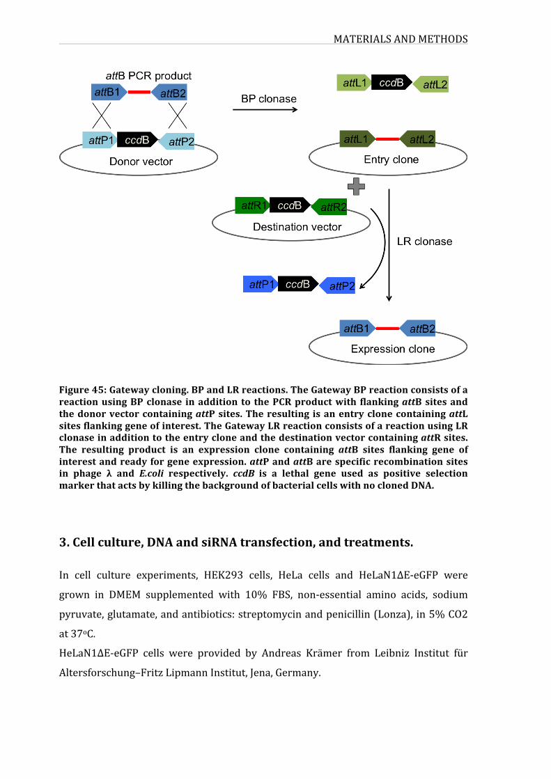
MATERIALSANDMETHODS
Figure45:Gatewaycloning.BPandLRreactions.TheGatewayBPreactionconsistsofareactionusingBPclonase inaddition to thePCRproductwith flankingattBsitesandthedonorvectorcontainingattPsites.Theresulting isanentryclonecontainingattLsitesflankinggeneofinterest.TheGatewayLRreactionconsistsofareactionusingLRclonaseinadditiontotheentrycloneandthedestinationvectorcontainingattRsites.The resulting product is an expression clone containing attB sites flanking gene ofinterestandreadyforgeneexpression.attPandattBarespecificrecombinationsitesin phage λ and E.coli respectively. ccdB is a lethal gene used as positive selectionmarkerthatactsbykillingthebackgroundofbacterialcellswithnoclonedDNA.
3.Cellculture,DNAandsiRNAtransfection,andtreatments.
In cell culture experiments, HEK293 cells, HeLa cells and HeLaN1ΔE-eGFP were
grown in DMEM supplemented with 10% FBS, non-essential amino acids, sodium
pyruvate,glutamate,andantibiotics:streptomycinandpenicillin(Lonza),in5%CO2
at37oC.
HeLaN1ΔE-eGFP cells were provided by Andreas Krämer from Leibniz Institut für
Altersforschung–FritzLipmannInstitut,Jena,Germany.
MATERIALSANDMETHODS
113
U2OSTet-onflp-incellsbearingisogenictransgenesencodingNotch1-Gal4andK562-
Dll4cellswereprovidedbyIngeVanDeWallefromDepartmentofClinicalChemistry,
Microbiology and Immunology, Faculty of Medicine and Health Sciences, Ghent
University.K562weregrownandmaintainedinRPMI(Gibco)containing15%FBSin
a5%C02incubatorat37°C.
13T-ALLcelllinesweregrownandmaintainedinRMPIcontaining10-20%FBSina5
%C02incubatorat37°C.
T-ALL cell lines: ALL-SIL, CTV-1, JURKAT, Karpas 45, KOPT-K1, LOUCY, MOLT-13,
MOLT-4,PEER,SUP-T11,SUP-T13,SUP-T7,TALL-1.
HEK293 cells were DNA–transfected with polyethylenimine (PEI) purchased from
Sigma, reagent was dissolved in water at 1mg/ml and preserved at -80oC.
Transfection with PEI was performed on HEK cells cultured in DMEM at 80%
confluence. Medium was changed before transfection and cells were collected 24
hourspost-transfection.
HeLacellsandHeLaN1ΔE-eGFPcellswereDNA-transfectedwithlipofectamine2000
reagent(Invitrogen)accordingtomanufacturer’sinstructionsandcollected24hours
post-transfection.
SiRNA transfection was performed with Calcium Phosphate using ProFection
MammalianTransfectionkit fromPromegaaccordingtomanufacturer’s instructions
oncellsculturedinDMEMat40-50%confluence.Mediumwaschanged24hourslater
andcellswerecollected48hpost-transfection.
As forexperiments involvingbothDNAandsiRNA transfections, siRNA-transfection
wasperformedaswepreviouslydescribedand24hourslaterafterchangingmedium
cells were transfected with DNA using lipofectamin 2000 reagent (Invitrogen) and
cellswerecollected24hpostDNA-transfection.
Forproteasomaldegradation inhibition, cellswere treatedof10µg/mlMG132 for6
hoursbeforebeingcollected.
Forinhibitionofγ-secretaseweusedDAPTat10µm/mlfor6hours.
siRNAsequences:
siEXT1:5′-GGAUUCCAGCGUGCACAUUtt-3′
siFBXW7:5’-GCAUAGAUUUUAUGGUAAtt-3’
siCtrl:5’-GGCUGCUUCUAUGAUUAUGtt-3’
MATERIALSANDMETHODS
4.Immunofluorescenceandconfocalmicroscopy
Cellsweregrownonglass slides in24-wellplates.Theywerewashed inwarmPBS
three timesand fixedwithPBS-paraformaldehyde4%for15minandwashedtwice
withPBS.CellswerepermeabilizedinPBS-TritonX-1000,5%for20minandblocked
inblockingsolutionPBS-FBS20%for30minandwashedtwicewithPBS.Cellswere
thenincubatedwithcorrespondingprimaryantibodydilutedinthePBS-TritonX-100
0,5%for2hours,washed3timeswithPBS,incubated1hourwiththecorresponding
Alexa-conjugated secondary antibodies (Invitrogen) diluted 1/1000 in PBS-Triton
solution,washed 3 timeswith PBS andmounted on glass coverslips using ProLonf
GoldAntifademontantwithDAPI(lifetechnologies).
Slides were examined by confocal microscopy with Leica TCS SP2 or Nikon A1R
confocal microscope with one 60X oil-objective and pictures were taken with a
1024x1024pixelsresolution.PictureswereprocessedandassembledwithLeicaLAS
AFLitesoftware.
5.Luciferasereporterassay
Cells were seeded in 24-well plates and transfected with 300ng of either TP1
luciferasereporterplasmid(TP1-luc)orCBF1reporterplasmid(CBF1-luc)and30ng
ofrenillaLuciferase(R-Luc).TP1promoterconsistsof8multimerizedRBP-Jκbinding
motifs upstream of luciferase. The TP1 promoter is transactivated in a RBP-Jκ
dependentmannerbytheactivatedformsofthefourmammalianNotchreceptorand
thus, has been often used to read out Notch pathway activity. CBF1 reporter also
contains a firefly luciferase gene under the control ofmultimerized CSL responsive
element upstream of a minimal promoter. 24 hours post-transfection luciferase
activitywasmeasuredincelllysates.
AsforU2OSN1-Gal4cellstheyweretransfected300ngofGal4-fireflyluciferaseand
30ng R-Luc reporter plasmid. After 24 hours, K562 cells expressing Notch ligands
DLL4 or K562 control cells were added to the transfected cells in the presence of
tetracycline(2μg/mL).After24-hourco-culture,luciferaseactivitywasmeasuredin
celllysates.
MATERIALSANDMETHODS
115
Cell lysis and luciferase assays were performed in triplicate using Dual-luciferase
reporterassaysystemfromPromega.Luciferasemeasurementswereperformedin96
wellplatesusingDLRautomatedmachine.Firefly luciferasevalueswerenormalized
toR-lucvaluesandcalculatedratiorepresentluciferaseactivity.
6.qRT-PCR
Total RNA was extracted using GeneJET RNA Purification Kit (Thermo scientific),
DNaseI-treated on the column (Thermo Scientific) and reverse-transcribed with
random primers (Thermo scientific). qPCR was performed using SYBER Green
detectionfromRocheandrunonLightcycler480(Roche).mRNAquantificationwas
ensured by normalization onGAPDHhousekeeping gene. Relative expression levels
werecalculatedforeachgeneusingtheΔCtorΔΔCtmethod.PrimersusedinRT-qPCR
arerepresentedintable5.
Table5:qRT-PCRprimersequences
Target sequencereference Forwardprimer ReverseprimermNotch1 NM_008714 TGCAGAACAACAAGGAGGAG AGTGGTCCAGCAACACTTTGeGFP pEGFP-N1ORFframe1 TGACCCTGAAGTTCATCTGC GAAGTCGTGCTGCTTCATGTGAPDH 2597 TTGCCATCAATGACCCCTTCA CGCCCCACTTGATTTTGGAhNotch1 CAACTGCCAGAACCTTGTGC GCACTCGTCGATCTCCTCAGFBXW7 CCCGGAGCTGTGCAGCAA CAGATGTAATTCGGCGTCGTTHES1 TAGCTCGCGGCATTCCAAGC GTGCTCAGCGCAGCCGTCATCTHES5 GAAAAACCGACTGCGGAAGC GGAAGTGGTACAGCAGCTTCHEY1 CGAGGTGGAGAAGGAGAGTG CTGGGTACCAGCCTTCTCAGEXT1 GCTCTTGTCTCGCCCTTTTGT TGGTGCAAGCCATTCCTACCcMYC CTCCTACGTTGCGGTCACAC CCGGGTCGCAGATGAAACTC
7.FACSanalysesandlabeling
Heparan sulfate staining: about 2.105 cells were trypsinized, washed twice and
resuspended inPBS.Cellswere incubated for1hourwithHeparansulfateantibody
(10E4 anti-HS fromUSBio) diluted1/50 in PBS,washed three times and incubated
MATERIALSANDMETHODS
withAlexa-conjugatedsecondaryantibodies(Invitrogen)diluted1/1000inPBS.Cells
wereanalysisaFACSCaliburflowcytometer(BDBiosciences).
Apoptosis assay:2.105cellswerewashedtwicewithcoldPBSthenresuspendedin
100µlof1Xbindingbuffer,5µlofV450Annexin(BDBiosciences)wasadded,thecells
were gently vortexed and incubated for 15 min in the dark, 400µl of 1X biding
solutionswasaddedandcellswereanalyzedbyflowcytometry.
8.High-throughputyeast-twohybrid
The yeast two-hybrid assay is a regular and well-established interaction screening
method allowing binary interactions detection. In regular molecular biology
laboratorysettings,itispossibletoeitherscreeninteractionsone-by-one,ortoscreen
librariesofGal4-ADfusionproteinsrepresentingentireproteomes.Weperformeda
large-scale pairwise interaction screen using specific equipment and robotics to
performmost of the screenings steps in an automatedway. Themethod described
here below corresponds to a standard yeast two-hybrid interaction assay. In our
screenweusedthehORFeomeversion5.1acollectionlibraryofhumanORFscloned
from the Mammalian Gene Collection (MGC) resource, representing a resource of
ORFs that canbe transferred easily to anyGateway compatible destination vectors.
Thiscollectioncontains15483ORFsrepresentingalmosthalfofthehumangenome,
that they are cloned into the pAD-dest-CYH and pDB-dest encoding the Gal4
Activating and DNA-binding domains, respectively. The resulting individual clones
weretransferredinMATaY8800(pAD)andMATαY8930(pDB)S.cerevisiaestrains
(figure 46). As for the ALL-genes ORFs that were not available in the hORF V5.1
collection, they were purchased from addgene and cloned into pAD-dest-CYH and
pDB-dest.
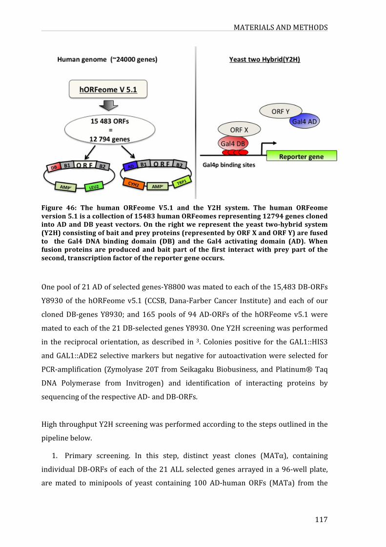
MATERIALSANDMETHODS
117
Figure 46: The human ORFeome V5.1 and the Y2H system. The human ORFeomeversion5.1isacollectionof15483humanORFeomesrepresenting12794genesclonedintoADandDByeastvectors.Ontherightwerepresent theyeast two-hybridsystem(Y2H)consistingofbaitandpreyproteins(representedbyORFXandORFY)arefusedto the Gal4 DNA binding domain (DB) and the Gal4 activating domain (AD). Whenfusion proteins are produced andbait part of the first interactwith prey part of thesecond,transcriptionfactorofthereportergeneoccurs.
Onepoolof21ADofselectedgenes-Y8800wasmatedtoeachofthe15,483DB-ORFs
Y8930of thehORFeomev5.1 (CCSB,Dana-FarberCancer Institute) andeachof our
clonedDB-genesY8930; and165pools of 94AD-ORFsof thehORFeomev5.1were
matedtoeachofthe21DB-selectedgenesY8930.OneY2Hscreeningwasperformed
in the reciprocalorientation, asdescribed in 3. Coloniespositive for theGAL1::HIS3
andGAL1::ADE2selectivemarkersbutnegative forautoactivationwereselected for
PCR-amplification (Zymolyase20T fromSeikagakuBiobusiness, andPlatinum®Taq
DNA Polymerase from Invitrogen) and identification of interacting proteins by
sequencingoftherespectiveAD-andDB-ORFs.
HighthroughputY2Hscreeningwasperformedaccordingtothestepsoutlinedinthe
pipelinebelow.
1. Primary screening. In this step, distinct yeast clones (MATα), containing
individualDB-ORFsofeachof the21ALLselectedgenesarrayed ina96-wellplate,
aremated tominipools of yeast containing 100 AD-human ORFs (MATa) from the
MATERIALSANDMETHODS
ORFeomecollectiononYEPDplatesmedia.Alternatively, eachDB-humanORF from
the ORFeome collection is mated to a pool of the AD-ORFs of 21 ALL-genes. After
matingyeastarereplicatedtoSc-Leu-Trp-HisselectionplatesandSc-Leu-His+3-AT+
1mg/mlofcycloheximide(CYH).Threetofivedayspostselection,primarypositives
diploidcellsarepickedandusedforsecondaryphenotyping.
2. Secondaryphenotyping.Primarypositivesaregrowninliquidselectingmedia
(Sc-Leu-Trp-His) to saturation (at least 24 hours) and spotted on selection and
controlplates:Sc-Leu-Trp-His+1mMof3-AT;Sc-Leu-His+1mMof3-AT+1mg/mlof
CYH; Sc-Leu-Trp-Ade+1mMof 3-AT and Sc-Leu-Ade+1mMof 3-AT+1mg/mlof
CYH for the Y strains. Replica cleaning is necessary to decrease the background
growthandtoinsurethatyoustartoutwithcomparableamountsofcellsineachspot.
Todeterminecandidatepositivecloneswesystematicallyaddtoselectionandcontrol
plates,acollectionof sixdiploidcontrol strains.Weusuallyscorepositivescolonies
showinga reporter transcriptionalactivity levelequalorhigher to thatof control2
(pDEST-AD-E2F1+pDEST-DB-CYH2-pRB).
3. Identificationof interactingpairs:Positivesyeastclonesaregrownovernight
on YPD plates. A small amount of yeast cells is lysed using zymolase, an enzyme
preparationfromArthrobacterluteus,whicheffectivelylysesliveyeastwall.Foreach
positive diploid, two PCRs are then performed to individually amplify DB-ORF
(primersDBandTerm)andAD-ORF(primersADandTerm).PCRproductsarethen
verified on E-Gel 96 agarose 1%, and used as template in sequencing reactions to
obtainidentitiesofinteractingpairs.
5. Retestinginteractingpairs.CandidateY2Hpairsshouldwereverifiedbytwo-
foldindependentexperiments.Tothisend,thelocationofeachcloneis identifiedin
theORFeomedatabaseandcorrespondingfresharchivalyeaststocksculturedinSc-
Leu (DB-ORFs) or Sc-Trp (AD-ORFs) liquidmedia,mated on YPDmedia plates and
transferred two-fold, on two phenotyping selection (His3 and Ade2) and control
media(+CYH).
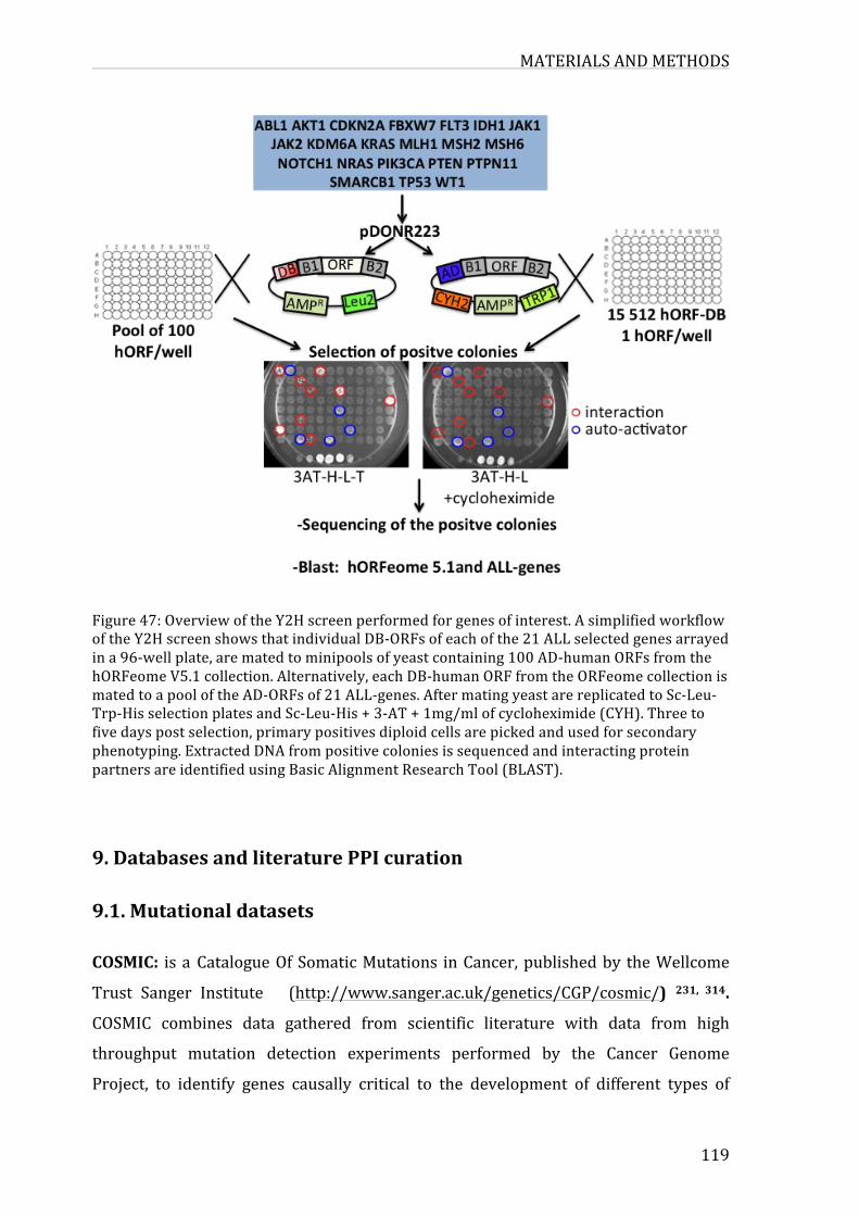
MATERIALSANDMETHODS
119
Figure47:OverviewoftheY2Hscreenperformedforgenesofinterest.AsimplifiedworkflowoftheY2HscreenshowsthatindividualDB-ORFsofeachofthe21ALLselectedgenesarrayedina96-wellplate,arematedtominipoolsofyeastcontaining100AD-humanORFsfromthehORFeomeV5.1collection.Alternatively,eachDB-humanORFfromtheORFeomecollectionismatedtoapooloftheAD-ORFsof21ALL-genes.AftermatingyeastarereplicatedtoSc-Leu-Trp-HisselectionplatesandSc-Leu-His+3-AT+1mg/mlofcycloheximide(CYH).Threetofivedayspostselection,primarypositivesdiploidcellsarepickedandusedforsecondaryphenotyping.ExtractedDNAfrompositivecoloniesissequencedandinteractingproteinpartnersareidentifiedusingBasicAlignmentResearchTool(BLAST).
9.DatabasesandliteraturePPIcuration
9.1.Mutationaldatasets
COSMIC: isaCatalogueOfSomaticMutations inCancer,publishedby theWellcome
Trust Sanger Institute (http://www.sanger.ac.uk/genetics/CGP/cosmic/) 231, 314.
COSMIC combines data gathered from scientific literature with data from high
throughput mutation detection experiments performed by the Cancer Genome
Project, to identify genes causally critical to the development of different types of
MATERIALSANDMETHODS
humancancers.ThecurrentversionofCOSMICdatabase(v75)ofsomaticmutations
in cancer contains information referenced in 22621 scientific reports and 23223
human genes implicated via mutation in cancer, 3702312 coding mutations and
1177397samples.Itincludesprecisedefinitionsofmutationscomparedtoreference
sequencesfromGenBank,publishedarticles,typeofmutation,numberofpatientsand
samplesaffectedbythesemutations,etc.
HumanORFeomeV5.1: (http://horfdb.dfci.harvard.edu/index.php?page=home):It
is a collection of 18,414 cloned human Open Reading Frames (ORFs) representing
more than 15,000 human genes. We have access to this collection through our
collaborationwiththeCenterforCancerSystemsBiology(CCSB)attheDanaFarber
CancerInstitute(DFCI),HarvardMedicalSchool,Boston,USA.
9.2.Protein-proteininteractionsdatasetsHumanPPIswerecollectedandverified from thedifferent interactomicsdatabases
BioGRID 5, HPRD 237 and IntAct 7. Only physical PPIs validated at least in two
independent references or by two methods were considered as confident and
maintainedfortheanalysis.
Information about genes containing mutations in their coding regions is retrieved
fromtheCOSMICdatabase,evaluated;organizedandselectedgenesweresubmitted
forexperimentalanalysis. Toestablisha catalogofgenesandmutationsassociated
withALL,weusedtheversion71ofCOSMIC,previouslydownloadedtoalocalserver
andweextracteddata related toALL.Wedevelopedand implementedaprocedure
thatautomaticallycollectsinformationandchecktheconsistencyofchangeswiththe
coding sequences and find the corresponding positions on clones from the human
ORFeome.
The retrieved information include details provided at either nucleotide or protein
level (mutation syntax), sample id (portion of a tumour being examined for
mutations),tissuefromwhichthesampleoriginated,histologicalclassificationofthe
sampleandthePubmedidofthearticlethatpublishedthestudy.
MATERIALSANDMETHODS
121
10.Networkdataanalysesandvisualization
For each interaction in IntAct, HPRD and BioGRID databases, we retrieved
information corresponding toofficial symbols of genes in interaction (genenames),
experimental system and the pubmed id. To this information,we added the entrez
gene ids fromEntrez gene (NCBI's repository for gene-specific information) 315.We
removedduplicateinteractions(interactionsfoundinmorethanonedatabases)and
wegeneratedalistofall interactionspresentinallthesethreedatabases.Fromthis
list,webuiltanetworkofinteractions,inwhichweextractedasubnetworksofgenes
associatedwithALL.ThisprocedurewasperformedusingRgraphpackages.
Networkanalysesandvisualizationofprotein-protein interactionswerecarriedout
withCytoscapesoftware38.
11.Proteincomplementationassay(PCA)
NICD, FBXW7 and selected interactants were cloned in Gaussia luciferase 1 and 2
(GL1 for NICD and its domains, GL2 for FBXW7 and the interactants) using the
Gateway cloning technology. HEK293 cells were seeded in 24 well-plates at a
concentration5.104cell/well,thentransfectedwithGL1or/andGL2plasmidsand24
hourspost-transfection, luciferaseactivitywasmeasuredon lysatestransferred into
96-well plate using and automated machine DLR with Renilla luciferase substrate.
Normalized luciferase ratio was calculated as follows: NLR= luciferase value
GL1+GL2/ (luciferase valueGL1+luciferase valueGL2).An interaction is considered
positiveorvalidatedwhenNLR≥3.5.
Celllysisandluciferaseassayswereperformedintriplicateforeachcondition.
12.RNAsequencing
TotalRNAwas extracted fromHeLaN1ΔE-eGFP cells (siCTRL, siEXT1, siFBXW7and
combinedsiEXT1+siFBXW7),quantifiedandtestedforRNAqualitycontrolledusing
Agilent 2100 bioanalyzer using the Eukaryote Total RNA Nano assay. Total RNA
strandsfromwereusedtogeneratelibrariesandsequencedbyHiSeq2000sequencer.

MATERIALSANDMETHODS
QualitycontrolandsequencingwereperformedbytheGenoTranscriptomicsplatform
–GIGA,Liège.
13.EXT1silencinginzebrafish
TransgeniczebrafishlineTg(Tp1bglob:eGFP)um13wasprovidedbyPr.Parsonsfrom
McKusick-NathansInstituteforGeneticMedicine,JohnsHopkinsUniversitySchoolof
Medicine, Baltimore,MD, 21205,USA 316. Fish of the Tg (Tp1bglob:eGFP)um13 line
were maintained according to EU regulations on laboratory animals. Knockdown
experimentswereperformedbyinjectingembryosattheone-totwo-cellstagewith
10ngofsinglesplice-blockingmorpholinodesignedspecificallyforEXT1b.
Couplings fish andharvest embryos: thedaybeforethe injectionofmorpholinos,
theadultzebrafishpairsareseparatedandplacedincagesofcouplings.8to10cages,
eachcontainingcouplings3malesand3femaleswereconsideredineachexperiment.
Thenextday,theseparationbetweenthecouplesisremovedtoallowmating.Assoon
as the egg laying occurred, eggs are removed, put in petri dishes and the fish are
returnedtotheiroriginalaquariumafterassessingtheirhealth.
Morpholino injection:harvestedzygotes injectedwithourmorpholinosof interest
MoEXT1bby conventional techniques.About 1nLofmorpholinos (10ng/nL)were
injectedintozygoteswithamicroinjector.Theinjectedeggsareculturedat28°Cin
E3mediumfor48hours.At24hours,theembryosareplacedintheculturemedium
E3+PTU(30mg/L)inordertopreventpigmentation.
Confocalmicroscope:between24and72hours,theembryoswereobservedundera
confocalmicroscope. To do this, living embryos previously “put to sleep” (Tricaine
methanesulfonate80mg/Lforatleast2minutes)willbemountedinalowmelting
agarosegelinaculturedishformicroscopicobservation(typetranswell)andcovered
withculturemediumE3.Attheendoftheexperiment,theembryoswillbeeuthanized
(Tricainemethanesulfonate500mg/Lforatleast10minutes).
MorpholinoEXT1bsequence:TATCGTTTTTTGGCCTGCATGTGTC(genetools).
MATERIALSANDMETHODS
123
14.Statisticalanalysis
Graph values are presented as mean +/- standard deviation, calculated on at least
three independent experiments. sampleswereUnless stated otherwise, significance
was determined using a two-tailed Student’s t-test (comparison ofmeans). P-value
thresholds are depicted as follows; *: p<0.05; **: p<0.01; ***: p<0.001 and ****:
p<0.0001.Itshouldbenotedthatstandarddeviationandmeanvaluesineachofthe
following experiments: luciferase reporter assays, relative mRNA expression
determined in qPCR analysis, and the NLR values calculated in the PCA; were
calculatedforsamplestestedintriplicateineachindependentexperiment.
To prioritize ALL-genes, we combined the ranking from separate results (rank per
numberofmutation, rankpernumberof samples, rankperdegree)byusingorder
statistics. First, ranks are divided by the total number of ranked genes and we
calculated the Q statistic 317, which represents the probability of obtaining the
observedranksrbychance,calculatedusingjointcumulativedistributionoforderas:
Whereriistherankratioforresulti,Nisthenumberofgenesused.€
V0 =1,
€
Vk = −1( )i−1Vk− i
i!i=1
k∑ rN −k+1
i
€
Q(r1,r2,...,rN ) = N!VN
BIBLIOGRAPHY
BIBLIOGRAPHY
1. Knox,S.From'omics'tocomplexdisease:asystemsbiologyapproachtogene-environmentinteractionsincancer.CancerCellInternational10,11(2010).
2. Gavin, A.-C., Maeda, K. & Kühner, S. Recent advances in charting protein–protein interaction:mass spectrometry-based approaches.CurrentOpinion inBiotechnology22,42-49(2011).
3. Rual, J.-F.etal.Towardsaproteome-scalemapof thehumanprotein-proteininteractionnetwork.Nature437,1173-1178(2005).
4. theMutation,C.&PathwayAnalysisworkinggroupoftheInternationalCancerGenome, C. Pathway and network analysis of cancer genomes.NatMeth12,615-621(2015).
5. Breitkreutz,B.-J.etal.TheBioGRIDInteractionDatabase:2008update.NucleicAcidsResearch36,D637-D640(2008).
6. Stark, C. etal. BioGRID: a general repository for interaction datasets.NucleicAcidsResearch34,D535-D539(2006).
7. Kerrien,S.etal. IntAct—opensourceresource formolecular interactiondata.NucleicAcidsResearch35,D561-D565(2007).
8. Peri,S.etal.DevelopmentofHumanProteinReferenceDatabaseasanInitialPlatform for Approaching Systems Biology in Humans.GenomeResearch13,2363-2371(2003).
9. Sonabend, A.M. et al. The Transcriptional Regulatory Network of ProneuralGliomaDeterminestheGeneticAlterationsSelectedduringTumorProgression.CancerResearch74,1440-1451(2014).
10. Tovar, H., García-Herrera, R., Espinal-Enríquez, J. & Hernández-Lemus, E.Transcriptionalmaster regulator analysis in breast cancer genetic networks.ComputationalBiologyandChemistry.
11. Pu, S. et al. Extracting high confidence protein interactions from affinitypurificationdata:Atthecrossroads.JournalofProteomics118,63-80(2015).
12. Hall,D.A.,Ptacek,J.&Snyder,M.ProteinMicroarrayTechnology.Mechanismsofageinganddevelopment128,161-167(2007).
13. Pierobon, M., Belluco, C., Liotta, L. & Petricoin, E., III Reverse Phase ProteinMicroarrays forClinicalApplications, inProteinMicroarrays, Vol. 785. (ed.U.Korf)3-12(HumanaPress,2011).
14. O'Mahony, F.C. et al. The Use of Reverse Phase Protein Arrays (RPPA) toExplore Protein Expression Variation within Individual Renal Cell Cancers.JournalofVisualizedExperiments:JoVE,50221(2013).
15. Pierobon, M., VanMeter, A., Moroni, N., Galdi, F. & Petricoin, E., III Reverse-PhaseProteinMicroarrays, inMolecularProfiling, Vol. 823. (eds. V. Espina&L.A.Liotta)215-235(HumanaPress,2012).
16. Bertone,P.&Snyder,M.Advancesinfunctionalproteinmicroarraytechnology.FEBSJournal272,5400-5411(2005).
17. Michnick,S.,Ear,P.,Landry,C.,Malleshaiah,M.&Messier,V.Protein-FragmentComplementation Assays for Large-Scale Analysis, Functional Dissection andDynamic Studies of Protein–Protein Interactions in Living Cells, in SignalTransductionProtocols,Vol.756.(eds.L.M.Luttrell&S.S.G.Ferguson)395-425(HumanaPress,2011).
BIBLIOGRAPHY
125
18. Neveu, G. et al. Comparative analysis of virus–host interactomes with amammalianhigh-throughputproteincomplementationassaybasedonGaussiaprincepsluciferase.Methods58,349-359(2012).
19. Brückner, A., Polge, C., Lentze, N., Auerbach, D. & Schlattner, U. Yeast Two-Hybrid,aPowerfulToolforSystemsBiology.InternationalJournalofMolecularSciences10,2763-2788(2009).
20. Lamesch,P.etal.hORFeomev3.1:Aresourceofhumanopenreading framesrepresentingover10,000humangenes.Genomics89,307-315(2007).
21. Ulrichts, P., Lemmens, I., Lavens, D., Beyaert, R. & Tavernier, J. MAPPIT(MammalianProtein–ProteinInteractionTrap)AnalysisofEarlyStepsinToll-Like Receptor Signalling, in Toll-Like Receptors:Methods and Protocols. (eds.C.E.McCoy&L.A.J.O’Neill)133-144(HumanaPress,Totowa,NJ;2009).
22. Lohse,M.J.,Nuber,S.&Hoffmann,C.Fluorescence/BioluminescenceResonanceEnergy Transfer Techniques to Study G-Protein-Coupled Receptor ActivationandSignaling.PharmacologicalReviews64,299-336(2012).
23. Ooi,S.L.etal.Globalsynthetic-lethalityanalysisandyeastfunctionalprofiling.TrendsinGenetics22,56-63.
24. Glatter, T.,Wepf, A., Aebersold, R.&Gstaiger,M. An integratedworkflow forcharting the human interaction proteome: insights into the PP2A system.MolecularSystemsBiology5(2009).
25. Cassonnet, P. et al. Benchmarking a luciferase complementation assay fordetectingproteincomplexes.NatMeth8,990-992(2011).
26. Hosur, R., Xu, J., Bienkowska, J. & Berger, B. iWRAP: An Interface ThreadingApproach with Application to Prediction of Cancer-Related Protein–ProteinInteractions.JournalofMolecularBiology405,1295-1310(2011).
27. Lee, S.-A. et al. Ortholog-based protein-protein interaction prediction and itsapplicationtointer-speciesinteractions.BMCBioinformatics9,S11(2008).
28. Enright,A.J., Iliopoulos, I., Kyrpides,N.C.&Ouzounis, C.A. Protein interactionmaps for completegenomesbasedongene fusionevents.Nature402, 86-90(1999).
29. Pazos, F. & Valencia, A. In silico two-hybrid system for the selection ofphysically interacting protein pairs. Proteins: Structure, Function, andBioinformatics47,219-227(2002).
30. Ochoa, D. & Pazos, F. Studying the co-evolution of protein families with theMirrortreewebserver.Bioinformatics26,1370-1371(2010).
31. Pazos,F.&Valencia,A.Proteinco‐evolution,co‐adaptationandinteractions.TheEMBOJournal27,2648-2655(2008).
32. Gao, L. et al. Widely predicting specific protein functions based on protein-proteininteractiondataandgeneexpressionprofile.SCICHINASERC50,125-134(2007).
33. Grigoriev,A.Arelationshipbetweengeneexpressionandproteininteractionson the proteome scale: analysis of the bacteriophage T7 and the yeastSaccharomycescerevisiae.NucleicAcidsResearch29,3513-3519(2001).
34. Hermjakob, H. et al. The HUPO PSI's Molecular Interaction format[mdash]acommunity standard for the representation of protein interaction data.NatBiotech22,177-183(2004).
35. Mathivanan,S.etal.Anevaluationofhumanprotein-proteininteractiondatainthepublicdomain.BMCBioinformatics7,S19(2006).
BIBLIOGRAPHY
36. Hermjakob, H. et al. IntAct: an open source molecular interaction database.NucleicAcidsResearch32,D452-D455(2004).
37. Ceol,A.etal.MINT, themolecular interactiondatabase:2009update.NucleicAcidsResearch(2009).
38. Cline,M.S. etal. Integration of biological networks and gene expression datausingCytoscape.Nat.Protocols2,2366-2382(2007).
39. Hooper, S.D.&Bork, P.Medusa: a simple tool for interaction graph analysis.Bioinformatics21,4432-4433(2005).
40. Chen, J., Aronow, B. & Jegga, A. Disease candidate gene identification andprioritization using protein interaction networks.BMCBioinformatics10, 73(2009).
41. Huang,D.W.,Sherman,B.T.&Lempicki,R.A.Bioinformaticsenrichmenttools:pathstowardthecomprehensivefunctionalanalysisoflargegenelists.NucleicAcidsResearch37,1-13(2009).
42. Mullighan, C.G. Genomic profiling of B-progenitor acute lymphoblasticleukemia.BestPractice&ResearchClinicalHaematology24,489-503.
43. Kojika, S. & Griffin, J.D. Notch receptors and hematopoiesis. ExperimentalHematology29,1041-1052.
44. Bigas,A.&Espinosa,L.Hematopoieticstemcells: tobeorNotchtobe.Blood119,3226-3235(2012).
45. Hu, Y.-Y., Zheng, M.-h., Zhang, R., Liang, Y.-M. & Han, H. Notch SignalingPathwayandCancerMetastasis, inNotchSignalinginEmbryologyandCancer,Vol.727.(eds.J.Reichrath&S.Reichrath)186-198(SpringerUS,2012).
46. Tasian, S. & Loh, M.L. Understanding the Biology of <i>CRLF2</i>-OverexpressingAcuteLymphoblasticLeukemia.16,13-24(2011).
47. Rosati,E.etal.ConstitutivelyactivatedNotchsignalingis involvedinsurvivalandapoptosisresistanceofB-CLLcells.Blood113,856-865(2008).
48. Pui,C.-H.&Evans,W.E.AcuteLymphoblasticLeukemia.NewEnglandJournalofMedicine339,605-615(1998).
49. Ellisen,L.W.etal.TAN-1,thehumanhomologoftheDrosophilaNotchgene,isbrokenbychromosomaltranslocationsinTlymphoblasticneoplasms.Cell66,649-661(1991).
50. Pear, W.S. et al. Exclusive development of T cell neoplasms in micetransplantedwithbonemarrowexpressingactivatedNotchalleles.TheJournalofExperimentalMedicine183,2283-2291(1996).
51. Chiaretti, S. et al. Gene expression profile of adult T-cell acute lymphocyticleukemia identifies distinct subsets of patients with different response totherapyandsurvival.Blood103,2771-2778(2003).
52. Ferrando, A.A. & Look, A.T. Gene expression profiling in T-cell acutelymphoblasticleukemia.SeminarsinHematology40,274-280.
53. Van Vlierberghe, P. & Ferrando, A. The molecular basis of T cell acutelymphoblastic leukemia.The JournalofClinical Investigation122, 3398-3406(2012).
54. Weng, A.P. et al. Activating Mutations of NOTCH1 in Human T Cell AcuteLymphoblasticLeukemia.Science306,269-271(2004).
55. Palomero, T. et al. Mutational loss of PTEN induces resistance to NOTCH1inhibitioninT-cellleukemia.Naturemedicine13,1203-1210(2007).
BIBLIOGRAPHY
127
56. Weng,A.P.etal. c-Myc isan importantdirect targetofNotch1 inT-cellacutelymphoblastic leukemia/lymphoma. Genes & Development 20, 2096-2109(2006).
57. Gedman, A.L. et al. The impact of NOTCH1, FBW7 and PTEN mutations onprognosis and downstream signaling in pediatric T- cell acute lymphoblasticleukemia: A report from the Children's Oncology Group. Leukemia : officialjournal of the Leukemia Society of America, Leukemia Research Fund, U.K 23,1417-1425(2009).
58. O'Neil, J. et al. FBW7 mutations in leukemic cells mediate NOTCH pathwayactivationandresistancetoγ-secretaseinhibitors.TheJournalofExperimentalMedicine204,1813-1824(2007).
59. Litzow,M.R.&Ferrando,A.A.HowItreatT-cellacutelymphoblasticleukemiainadults.Blood126,833-841(2015).
60. Vara, J.Á.F. et al. PI3K/Akt signalling pathway and cancer. Cancer TreatmentReviews30,193-204.
61. Ciofani,M.&Zuniga-Pflucker,J.C.Notchpromotessurvivalofpre-Tcellsatthe[beta]-selectioncheckpointbyregulatingcellularmetabolism.NatImmunol6,881-888(2005).
62. Gutierrez,A.etal.HighfrequencyofPTEN,PI3K,andAKTabnormalitiesinT-cellacutelymphoblasticleukemia.Blood114,647-650(2009).
63. Ribeiro, D., Melão, A. & Barata, J.T. IL-7R-mediated signaling in T-cell acutelymphoblasticleukemia.AdvancesinBiologicalRegulation53,211-222(2013).
64. Medyouf, H. et al. High-level IGF1R expression is required for leukemia-initiatingcellactivityinT-ALLandissupportedbyNotchsignaling.TheJournalofExperimentalMedicine208,1809-1822(2011).
65. Hales,E.C.,Taub,J.W.&Matherly,L.H.NewinsightsintoNotch1regulationofthePI3K–AKT–mTOR1signalingaxis:Targetedtherapyofγ-secretaseinhibitorresistantT-cell acute lymphoblastic leukemia.CellularSignalling26,149-161(2014).
66. Feng,Z.p53RegulationoftheIGF-1/AKT/mTORPathwaysandtheEndosomalCompartment.ColdSpringHarborPerspectivesinBiology2(2010).
67. Beverly,L.J., Felsher,D.W.&Capobianco,A.J. Suppressionofp53byNotch inLymphomagenesis:ImplicationsforInitiationandRegression.CancerResearch65,7159-7168(2005).
68. Demarest,R.M.,Dahmane,N.&Capobianco,A.J.NotchisoncogenicdominantinT-cellacutelymphoblasticleukemia.Blood117,2901-2909(2011).
69. Bonnet,M.etal.PosttranscriptionalderegulationofMYCviaPTENconstitutesamajor alternative pathway ofMYC activation in T-cell acute lymphoblasticleukemia.Blood117,6650-6659(2011).
70. Pui, C.-H. & Evans, W.E. Treatment of Acute Lymphoblastic Leukemia. NewEnglandJournalofMedicine354,166-178(2006).
71. Holleman, A. et al. Gene-Expression Patterns in Drug-Resistant AcuteLymphoblastic Leukemia Cells and Response to Treatment. New EnglandJournalofMedicine351,533-542(2004).
72. Cheok,M.H.etal.Treatment-specificchanges ingeneexpressiondiscriminateinvivodrugresponseinhumanleukemiacells.NatGenet34,85-90(2003).
73. Garza-Veloz, I., Martinez-Fierro, M.L. & Jaime-Perez, J.C. Idenification ofdifferentially expressed genes associated with prognosis of B acutelymphoblasticleukemia.diseasemarkers2015(2015).
BIBLIOGRAPHY
74. Brown,P.etal.FLT3inhibitionselectivelykillschildhoodacutelymphoblasticleukemiacellswithhighlevelsofFLT3expression.Blood105,812-820(2004).
75. Satake,N.etal. Targeted therapywithMXD3 siRNA, anti-CD22antibody andnanoparticles for precursor B-cell acute lymphoblastic leukaemia. Britishjournalofhaematology167,487-499(2014).
76. Palomero, T. et al. NOTCH1 directly regulates c-MYC and activates a feed-forward-loop transcriptional network promoting leukemic cell growth.ProceedingsoftheNationalAcademyofSciences103,18261-18266(2006).
77. Deangelo,D.J.etal. A phase I clinical trial of the notch inhibitorMK-0752 inpatients with T-cell acute lymphoblastic leukemia/lymphoma (T-ALL) andother leukemias. American Society Of Clinical OntologyH Annual MeetingAbstracts24(2006).
78. Raetz,E.A.etal.ReinductionPlatformforChildrenWithFirstMarrowRelapseofAcuteLymphoblasticLeukemia:AChildren'sOncologyGroupStudy.JournalofClinicalOncology26,3971-3978(2008).
79. Morgan, T.H. The theory of the gene. The American Naturalist 51, 513-544(1917).
80. Reynolds, A.E., Murray, A.W. & Szostak, J.W. Roles of the 2 microns geneproducts in stable maintenance of the 2 microns plasmid of Saccharomycescerevisiae.MolecularandCellularBiology7,3566-3573(1987).
81. Ellisen,L.W.etal.TAN-1,thehumanhomologoftheDrosophilaNotchgene,isbrokenbychromosomaltranslocationsinTlymphoblasticneoplasms.Cell66,649-661.
82. Maine, E.M., Lissemore, J.L. & Starmer, W.T. A Phylogenetic Analysis ofVertebrateandInvertebrateNotch-RelatedGenes.MolecularPhylogeneticsandEvolution4,139-149(1995).
83. Greenwald, I. Structure/function studies of lin-12/Notch proteins. CurrentOpinioninGenetics&Development4,556-562(1994).
84. Kopan,R.& Ilagan,M.X.G.TheCanonicalNotchSignalingPathway:UnfoldingtheActivationMechanism.Cell137,216-233(2009).
85. Gordon, W.R., Arnett, K.L. & Blacklow, S.C. The molecular logic of Notchsignaling – a structural and biochemical perspective. Journal of Cell Science121,3109-3119(2008).
86. Fleming,R.J.StructuralconservationofNotchreceptorsandligands.SeminarsinCell&DevelopmentalBiology9,599-607(1998).
87. Fehon,R.G.etal.Molecular interactionsbetween theproteinproducts of theneurogenic loci Notch and Delta, two EGF-homologous genes in Drosophila.Cell61,523-534.
88. Brou, C. et al. A Novel Proteolytic Cleavage Involved in Notch Signaling.MolecularCell5,207-216(2000).
89. Mumm, J.S. et al. A Ligand-Induced Extracellular Cleavage Regulates γ-Secretase-likeProteolyticActivationofNotch1.MolecularCell5,197-206.
90. De Strooper, B. et al. A presenilin-1-dependent [gamma]-secretase-likeproteasemediatesreleaseofNotchintracellulardomain.Nature398,518-522(1999).
91. Struhl,G.&Greenwald,I.PresenilinisrequiredforactivityandnuclearaccessofNotchinDrosophila.Nature398,522-525(1999).
92. Ye,Y., Lukinova,N.&Fortini,M.E.NeurogenicphenotypesandalteredNotchprocessinginDrosophilaPresenilinmutants.Nature398,525-529(1999).
BIBLIOGRAPHY
129
93. Jarriault,S.etal.SignallingdownstreamofactivatedmammalianNotch.Nature377,355-358(1995).
94. D’Souza,B.,Meloty-Kapella,L.&Weinmaster,G.ChapterThree-CanonicalandNon-CanonicalNotchLigands, inCurrentTopicsinDevelopmentalBiology,Vol.Volume92.(ed.K.Raphael)73-129(AcademicPress,2010).
95. Pintar,A.,DeBiasio,A.,Popovic,M.,Ivanova,N.&Pongor,S.TheintracellularregionofNotchligands:doesthetailmakethedifference?BiologyDirect2,19(2007).
96. Sprinzak,D.etal.Cis-interactionsbetweenNotchandDeltageneratemutuallyexclusivesignallingstates.Nature465,86-90(2010).
97. Fiuza,U.-M.,Klein,T.,MartinezArias,A.&Hayward,P.Mechanismsofligand-mediated inhibition in Notch signaling activity in Drosophila.DevelopmentalDynamics239,798-805(2010).
98. Garg, V. et al. Mutations in NOTCH1 cause aortic valve disease.Nature437,270-274(2005).
99. McDaniell, R. et al. NOTCH2 Mutations Cause Alagille Syndrome, aHeterogeneousDisorderof theNotchSignalingPathway.AmericanJournalofHumanGenetics79,169-173(2006).
100. Glittenberg,M.,Pitsouli,C.,Garvey,C.,Delidakis,C.&Bray,S.Roleofconservedintracellular motifs in Serrate signalling, cis-inhibition and endocytosis. TheEMBOJournal25,4697-4706(2006).
101. Cordle,J.etal.AConservedFaceoftheJagged/SerrateDSLDomainisInvolvedin Notch Trans-Activation and Cis-Inhibition. Nature structural & molecularbiology15,849-857(2008).
102. Becam, I., Fiuza, U.-M., Arias, A.M. & Milán, M. A Role of Receptor Notch inLigandcis-InhibitioninDrosophila.CurrentBiology20,554-560.
103. Baladrón,V.etal.dlkactsasanegativeregulatorofNotch1activationthroughinteractions with specific EGF-like repeats. Experimental Cell Research 303,343-359(2005).
104. Eiraku,M.etal.DNERactsasaneuron-specificNotchligandduringBergmannglialdevelopment.NatNeurosci8,873-880(2005).
105. Krivtsov, A.V. etal. Jedi—a novel transmembrane protein expressed in earlyhematopoieticcells.JournalofCellularBiochemistry101,767-784(2007).
106. Hu, Q.-D. et al. F3/Contactin Acts as a Functional Ligand for Notch duringOligodendrocyteMaturation.Cell115,163-175.
107. Cui,X.-Y.etal.NB-3/Notch1PathwayviaDeltex1PromotesNeuralProgenitorCellDifferentiationintoOligodendrocytes.JournalofBiologicalChemistry279,25858-25865(2004).
108. Komatsu,H.etal.OSM-11FacilitatesLIN-12NotchSignalingduring<named-content xmlns:xlink="http://www.w3.org/1999/xlink" content-type="genus-species"xlink:type="simple">Caenorhabditiselegans</named-content>VulvalDevelopment.PLoSBiol6,e196(2008).
109. Sakamoto, K. et al. The Nephroblastoma Overexpressed Gene (NOV/ccn3)Protein Associates with Notch1 Extracellular Domain and Inhibits MyoblastDifferentiation via Notch Signaling Pathway. Journal of Biological Chemistry277,29399-29405(2002).
110. Minamizato, T. et al. CCN3/NOV inhibits BMP-2-induced osteoblastdifferentiation by interacting with BMP and Notch signaling pathways.BiochemicalandBiophysicalResearchCommunications354,567-573(2007).
BIBLIOGRAPHY
111. Miyamoto,A.,Lau,R.,Hein,P.W.,Shipley, J.M.&Weinmaster,G.MicrofibrillarProteins MAGP-1 and MAGP-2 Induce Notch1 Extracellular DomainDissociation and Receptor Activation. Journal of Biological Chemistry 281,10089-10097(2006).
112. Meng, H., Zhang, X., Hankenson, K.D. & Wang, M.M. Thrombospondin 2Potentiates Notch3/Jagged1 Signaling. Journal of Biological Chemistry 284,7866-7874(2009).
113. Rauen, T. et al. YB-1 Acts as a Ligand for Notch-3 Receptors andModulatesReceptor Activation. The Journal of Biological Chemistry 284, 26928-26940(2009).
114. Schmidt, M.H.H. et al. Epidermal growth factor-like domain 7 (EGFL7)modulatesNotchsignallingandaffectsneuralstemcell renewal.NatCellBiol11,873-880(2009).
115. Chen, N. & Greenwald, I. The Lateral Signal for LIN-12/Notch in C. elegansVulvalDevelopmentComprisesRedundantSecretedandTransmembraneDSLProteins.DevelopmentalCell6,183-192.
116. Tun,T.etal.RecognitionsequenceofahighlyconservedDNAbindingproteinRBP-Jkappa.NucleicAcidsResearch22,965-971(1994).
117. Salat, D., Liefke, R.,Wiedenmann, J., Borggrefe, T. & Oswald, F. ETO, but NotLeukemogenicFusionProteinAML1/ETO,AugmentsRBP-Jκ/SHARP-MediatedRepression of Notch Target Genes.Molecular and Cellular Biology 28, 3502-3512(2008).
118. Oswald, F. et al. RBP-Jκ/SHARP Recruits CtIP/CtBP Corepressors To SilenceNotchTargetGenes.MolecularandCellularBiology25,10379-10390(2005).
119. Zhou, S. & Hayward, S.D. Nuclear Localization of CBF1 Is Regulated byInteractions with the SMRT Corepressor Complex. Molecular and CellularBiology21,6222-6232(2001).
120. Hsieh, J.J.D.,Zhou,S.,Chen,L.,Young,D.B.&Hayward,S.D.CIR,acorepressorlinking the DNA binding factor CBF1 to the histone deacetylase complex.ProceedingsoftheNationalAcademyofSciences96,23-28(1999).
121. Zhou, S. et al. SKIP, a CBF1-Associated Protein, Interacts with the AnkyrinRepeat Domain of NotchIC To Facilitate NotchIC Function. Molecular andCellularBiology20,2400-2410(2000).
122. Wilson, J.J. & Kovall, R.A. Crystal Structure of the CSL-Notch-MastermindTernaryComplexBoundtoDNA.Cell124,985-996.
123. Kato,H.etal.InvolvementofRBP-JinbiologicalfunctionsofmouseNotch1anditsderivatives.Development124,4133-4141(1997).
124. Kurooka, H. & Honjo, T. Functional Interaction between the Mouse Notch1IntracellularRegionandHistoneAcetyltransferasesPCAFandGCN5.JournalofBiologicalChemistry275,17211-17220(2000).
125. Borggrefe, T. & Oswald, F. The Notch signaling pathway: TranscriptionalregulationatNotchtargetgenes.Cell.Mol.LifeSci.66,1631-1646(2009).
126. Kovall, R.A. Structures of CSL, Notch and Mastermind proteins: piecingtogetheranactivetranscriptioncomplex.CurrentOpinioninStructuralBiology17,117-127(2007).
127. Wilson, J.J. & Kovall, R.A. Crystal Structure of the CSL-Notch-MastermindTernaryComplexBoundtoDNA.Cell124,985-996(2006).
BIBLIOGRAPHY
131
128. Vasquez-Del Carpio, R. et al. Assembly of a Notch Transcriptional ActivationComplex RequiresMultimerization.Molecular andCellularBiology31, 1396-1408(2011).
129. Nam,Y.,Sliz,P.,Pear,W.S.,Aster,J.C.&Blacklow,S.C.Cooperativeassemblyofhigher-order Notch complexes functions as a switch to induce transcription.ProceedingsoftheNationalAcademyofSciences104,2103-2108(2007).
130. Fischer, A. & Gessler, M. Delta–Notch—and then? Protein interactions andproposed modes of repression by Hes and Hey bHLH factors. Nucleic AcidsResearch35,4583-4596(2007).
131. Iso,T.etal.HERP,aNewPrimaryTargetofNotchRegulatedbyLigandBinding.MolecularandCellularBiology21,6071-6079(2001).
132. Castella,P.,Sawai,S.,Nakao,K.,Wagner,J.A.&Caudy,M.HES-1RepressionofDifferentiation and Proliferation in PC12 Cells: Role for the Helix 3-Helix 4DomaininTranscriptionRepression.MolecularandCellularBiology20,6170-6183(2000).
133. Li, J.etal.BothcorepressorproteinsSMRTandN‐CoRexist in largeproteincomplexescontainingHDAC3.TheEMBOJournal19,4342-4350(2000).
134. Iso,T.,Kedes,L.&Hamamori,Y.HESandHERPfamilies:Multipleeffectorsofthe notch signaling pathway. Journal of Cellular Physiology 194, 237-255(2003).
135. Meier-Stiegen, F.etal. ActivatedNotch1TargetGenesduringEmbryonicCellDifferentiation Depend on the Cellular Context and Include LineageDeterminantsandInhibitors.PLoSONE5,e11481(2010).
136. Deftos, M.L., He, Y.-W., Ojala, E.W. & Bevan, M.J. Correlating Notch SignalingwithThymocyteMaturation.Immunity9,777-786(1998).
137. Phng, L.-K. et al. Nrarp coordinates endothelial Notch and Wnt signaling tocontrolvesseldensityinangiogenesis.Developmentalcell16,70-82(2009).
138. Yashiro-Ohtani,Y.etal.Pre-TCRsignalinginactivatesNotch1transcriptionbyantagonizingE2A.Genes&Development23,1665-1676(2009).
139. Moloney,D.J.etal.FringeisaglycosyltransferasethatmodifiesNotch.Nature406,369-375(2000).
140. Panin, V.M., Papayannopoulos, V.,Wilson, R. & Irvine, K.D. FringemodulatesNotch-ligandinteractions.Nature387,908-912(1997).
141. Rampal, R. et al. Lunatic Fringe,Manic Fringe, and Radical Fringe RecognizeSimilar Specificity Determinants in O-Fucosylated Epidermal Growth Factor-likeRepeats.JournalofBiologicalChemistry280,42454-42463(2005).
142. Johnston, S.H. et al. A family of mammalian Fringe genes implicated inboundarydeterminationandtheNotchpathway.Development124,2245-2254(1997).
143. Okajima, T., Xu, A. & Irvine, K.D. Modulation of Notch-Ligand Binding byProteinO-Fucosyltransferase1andFringe.JournalofBiologicalChemistry278,42340-42345(2003).
144. Wang, Y. etal.Modification of Epidermal Growth Factor-like RepeatswithO-Fucose:MOLECULARCLONINGANDEXPRESSIONOFANOVELGDP-FUCOSEPROTEINO-FUCOSYLTRANSFERASE. Journal of Biological Chemistry 276,40338-40345(2001).
145. Panin, V.M. et al. Notch Ligands Are Substrates for ProteinO-Fucosyltransferase-1 and Fringe. JournalofBiologicalChemistry277, 29945-29952(2002).
BIBLIOGRAPHY
146. Takeuchi,H.etal.RumifunctionsasbothaproteinO-glucosyltransferaseandaproteinO-xylosyltransferase.Proceedingsof theNationalAcademyofSciencesoftheUnitedStatesofAmerica108,16600-16605(2011).
147. Matsuura, A. et al. O-Linked N-Acetylglucosamine Is Present on theExtracellularDomainofNotchReceptors. JournalofBiologicalChemistry283,35486-35495(2008).
148. Takeuchi, H. & Haltiwanger, R.S. Significance of glycosylation in Notchsignaling.BiochemicalandBiophysicalResearchCommunications453,235-242(2014).
149. Li, M. et al. Mutations in POFUT1, Encoding Protein O-fucosyltransferase 1,Cause Generalized Dowling-Degos Disease. The American Journal of HumanGenetics92,895-903(2013).
150. Zhang,S.,Chung,W.-c.,Wu,G.,Egan,S.E.&Xu,K.Tumor-SuppressiveActivityofLunaticFringe inProstate throughDifferentialModulationofNotchReceptorActivation.Neoplasia16,158-167(2014).
151. Xu,K.etal.LunaticFringeDeficiencyCooperateswiththeMet/CaveolinGeneAmplicontoInduceBasal-likeBreastCancer.CancerCell21,626-641(2012).
152. Kroes, R.A., Dawson, G. &Moskal, J.R. Focusedmicroarray analysis of glyco-geneexpressioninhumanglioblastomas.JournalofNeurochemistry103,14-24(2007).
153. Loo, L.W.M. et al. Integrated Analysis of Genome-wide Copy NumberAlterations andGeneExpression inMSS, CIMP-negative ColonCancer.Genes,chromosomes&cancer52,450-466(2013).
154. Wang,Y.etal.OverexpressionofhumanCAP10-likeprotein46KDinT-acutelymphoblasticleukemiaandacutemyelogenousleukemia.
155. Fryer, C.J., White, J.B. & Jones, K.A. Mastermind Recruits CycC:CDK8 toPhosphorylate the Notch ICD and Coordinate Activation with Turnover.MolecularCell16,509-520(2004).
156. Li,N.etal.CyclinCisahaploinsufficienttumorsuppressor.Naturecellbiology16,1080-1091(2014).
157. Espinosa, L., Inglés-Esteve, J., Aguilera, C. & Bigas, A. Phosphorylation byGlycogenSynthaseKinase-3βDown-regulatesNotchActivity,aLinkforNotchandWntPathways.JournalofBiologicalChemistry278,32227-32235(2003).
158. Foltz,D.R.,Santiago,M.C.,Berechid,B.E.&Nye,J.S.GlycogenSynthaseKinase-3β Modulates Notch Signaling and Stability. Current Biology 12, 1006-1011(2002).
159. Mo, J.-S. et al. Integrin-Linked Kinase Controls Notch1 Signaling by Down-RegulationofProteinStability throughFbw7UbiquitinLigase.MolecularandCellularBiology27,5565-5574(2007).
160. Song, J., Park, S., Kim, M. & Shin, I. Down-regulation of Notch-dependenttranscriptionbyAktinvitro.FEBSLetters582,1693-1699.
161. Ranganathan,P.etal.HierarchicalPhosphorylationwithintheAnkyrinRepeatDomain Defines a Phosphoregulatory Loop That Regulates NotchTranscriptional Activity. Journal of Biological Chemistry 286, 28844-28857(2011).
162. Lee, H.-J., Kim, M.-Y. & Park, H.-S. Phosphorylation-dependent regulation ofNotch1 signaling: the fulcrumofNotch1 signaling.BMBReports48, 431-437(2015).
BIBLIOGRAPHY
133
163. Li,W.etal.Genome-WideandFunctionalAnnotationofHumanE3UbiquitinLigases IdentifiesMULAN, aMitochondrial E3 that Regulates the Organelle'sDynamicsandSignaling.PLoSONE3,e1487(2008).
164. Callis,J.TheUbiquitinationMachineryoftheUbiquitinSystem.TheArabidopsisBook/AmericanSocietyofPlantBiologists12,e0174(2014).
165. Woelk, T., Sigismund, S., Penengo, L. & Polo, S. The ubiquitination code: asignallingproblem.CellDivision2,1-12(2007).
166. Nichols, J.T. et al. DSL ligand endocytosis physically dissociates Notch1heterodimers before activating proteolysis can occur. The Journal of CellBiology176,445-458(2007).
167. Qiu, L.etal. Recognition andUbiquitination ofNotchby Itch, aHect-typeE3UbiquitinLigase.JournalofBiologicalChemistry275,35734-35737(2000).
168. Andersson, E.R., Sandberg, R. & Lendahl, U. Notch signaling: simplicity indesign,versatilityinfunction.Development138,3593-3612(2011).
169. Gupta-Rossi,N.etal.FunctionalInteractionbetweenSEL-10,anF-boxProtein,and the Nuclear Form of Activated Notch1 Receptor. Journal of BiologicalChemistry276,34371-34378(2001).
170. Öberg,C.etal.TheNotchIntracellularDomainIsUbiquitinatedandNegativelyRegulatedby theMammalianSel-10Homolog. JournalofBiologicalChemistry276,35847-35853(2001).
171. Han,X., Ju, J.-h.&Shin, I.Glycogensynthasekinase3-βphosphorylatesnovelS/T-P-S/T domains in Notch1 intracellular domain and induces its nuclearlocalization.BiochemicalandBiophysicalResearchCommunications423, 282-288(2012).
172. Chastagner, P., Israël, A. & Brou, C. AIP4/Itch Regulates Notch ReceptorDegradationintheAbsenceofLigand.PLoSONE3,e2735(2008).
173. Sakata, T. et al. Drosophila Nedd4 Regulates Endocytosis of Notch andSuppressesItsLigand-IndependentActivation.CurrentBiology14,2228-2236.
174. McGill, M.A. & McGlade, C.J. Mammalian Numb Proteins Promote Notch1ReceptorUbiquitinationandDegradationof theNotch1 IntracellularDomain.JournalofBiologicalChemistry278,23196-23203(2003).
175. Jehn,B.M.,Dittert,I.,Beyer,S.,vonderMark,K.&Bielke,W.c-CblBindingandUbiquitin-dependentLysosomalDegradationofMembrane-associatedNotch1.JournalofBiologicalChemistry277,8033-8040(2002).
176. Moretti, J. et al. The Translation Initiation Factor 3f (eIF3f) Exhibits aDeubiquitinase Activity Regulating Notch Activation. PLoS Biol 8, e1000545(2010).
177. Moretti, J. & Brou, C. Ubiquitinations in the Notch Signaling Pathway.InternationalJournalofMolecularSciences14,6359-6381(2013).
178. Wang,W.& Struhl, G.Distinct roles forMindbomb,Neuralized andEpsin inmediating DSL endocytosis and signaling in Drosophila. Development 132,2883-2894(2005).
179. Windler, S.L. & Bilder, D. Endocytic Internalization Routes Required forDelta/NotchSignaling.CurrentBiology20,538-543(2010).
180. Wang,W.& Struhl, G.DrosophilaEpsinmediates a select endocytic pathwaythat DSL ligandsmust enter to activateNotch.Development131, 5367-5380(2004).
181. Le Borgne, R. & Schweisguth, F. Notch Signaling: Endocytosis Makes DeltaSignalBetter.CurrentBiology13,R273-R275.
BIBLIOGRAPHY
182. Emery, G. etal. Asymmetric Rab11 EndosomesRegulateDelta Recycling andSpecifyCellFateintheDrosophilaNervousSystem.Cell122,763-773.
183. Heuss,S.F.,Ndiaye-Lobry,D.,Six,E.M., Israël,A.&Logeat,F.The intracellularregionofNotch ligandsDll1andDll3regulates their traffickingandsignalingactivity. Proceedings of the National Academy of Sciences 105, 11212-11217(2008).
184. Shah, D.K., Mohtashami, M. & Zúñiga-Pflücker, J.C. Role of Recycling,Mindbomb1 Association, and Exclusion from Lipid Rafts of Delta-like 4 forEffective Notch Signaling To Drive T Cell Development. The Journal ofImmunology189,5797-5808(2012).
185. Berndsen, C.E. & Wolberger, C. New insights into ubiquitin E3 ligasemechanism.NatStructMolBiol21,301-307(2014).
186. Das,R.etal.Allosteric regulationofE2:E3 interactionspromoteaprocessiveubiquitinationmachine.TheEMBOJournal32,2504-2516(2013).
187. Huibregtse, J.M., Scheffner, M., Beaudenon, S. & Howley, P.M. A family ofproteins structurally and functionally related to the E6-AP ubiquitin-proteinligase.Proceedingsof theNationalAcademyofSciencesof theUnitedStatesofAmerica92,2563-2567(1995).
188. Ogunjimi, A.A. et al. Regulation of Smurf2 Ubiquitin Ligase Activity byAnchoringtheE2totheHECTDomain.MolecularCell19,297-308.
189. Spratt, DonaldE.,Walden, H. & Shaw, GaryS. RBR E3 ubiquitin ligases: newstructures, new insights, new questions. Biochemical Journal 458, 421-437(2014).
190. Bai,C.etal. SKP1ConnectsCellCycleRegulators to theUbiquitinProteolysisMachinerythroughaNovelMotif,theF-Box.Cell86,263-274.
191. Cardozo, T. & Pagano,M. The SCF ubiquitin ligase: insights into amolecularmachine.NatRevMolCellBiol5,739-751(2004).
192. Jia,L.&Sun,Y.SCFE3UbiquitinLigasesasAnticancerTargets.CurrentCancerDrugTargets11,347-356(2011).
193. Strohmaier, H. et al. Human F-box protein hCdc4 targets cyclin E forproteolysis and ismutated in a breast cancer cell line.Nature413, 316-322(2001).
194. Moberg, K.H., Bell, D.W., Wahrer, D.C.R., Haber, D.A. & Hariharan, I.K.Archipelago regulatesCyclinE levels inDrosophila and ismutated inhumancancercelllines.Nature413,311-316(2001).
195. Welcker,M.&Clurman,B.E.FBW7ubiquitinligase:atumoursuppressoratthecrossroadsofcelldivision,growthanddifferentiation.NatRevCancer8,83-93(2008).
196. Sancho, R. et al. Fbw7 Repression by Hes5 Creates a Feedback Loop ThatModulates Notch-Mediated Intestinal and Neural Stem Cell Fate Decisions.PLoSBiol11,e1001586(2013).
197. Welcker,M.etal.Fbw7dimerizationdeterminesthespecificityandrobustnessofsubstratedegradation.Genes&Development27,2531-2536(2013).
198. Min, S.-H. et al. Negative Regulation of the Stability and Tumor SuppressorFunction of Fbw7 by the Pin1 Prolyl Isomerase.Molecular Cell 46, 771-783(2012).
199. Diefenbacher, M.E. et al. The deubiquitinase USP28 controls intestinalhomeostasis and promotes colorectal cancer. The Journal of ClinicalInvestigation124,3407-3418(2014).
BIBLIOGRAPHY
135
200. Schülein-Völk, C. et al. Dual Regulation of Fbw7 Function and OncogenicTransformationbyUsp28.CellReports9,1099-1109(2014).
201. Grim,J.E.etal.Isoform-andcellcycle–dependentsubstratedegradationbytheFbw7ubiquitinligase.TheJournalofCellBiology181,913-920(2008).
202. Trausch-Azar, J.S., Abed, M., Orian, A. & Schwartz, A.L. Isoform-SpecificSCFFbw7UbiquitinationMediatesDifferentialRegulationofPGC-1α.JournalofCellularPhysiology,n/a-n/a(2014).
203. Welcker, M. et al. The Fbw7 tumor suppressor regulates glycogen synthasekinase 3 phosphorylation-dependent c-Myc protein degradation. ProceedingsoftheNationalAcademyofSciencesoftheUnitedStatesofAmerica101,9085-9090(2004).
204. Gregory, M.A. & Hann, S.R. c-Myc Proteolysis by the Ubiquitin-ProteasomePathway: Stabilization of c-Myc in Burkitt's Lymphoma Cells.Molecular andCellularBiology20,2423-2435(2000).
205. Musti, A.M., Treier, M. & Bohmann, D. Reduced Ubiquitin-DependentDegradationofc-JunAfterPhosphorylationbyMAPKinases.Science275,400-402(1997).
206. Wei, W., Jin, J., Schlisio, S., Harper, J.W. & Kaelin, W.G., Jr. The v-Jun pointmutationallowsc-JuntoescapeGSK3-dependentrecognitionanddestructionbytheFbw7ubiquitinligase.CancerCell8,25-33.
207. Zhu,F.etal.CSK-Mediatedc-JunPhosphorylationPromotesc-JunDegradationandInhibitsCellTransformation.Cancerresearch66,5729-5736(2006).
208. Zhang, J. et al. Rack1 protects N-terminal phosphorylated c-Jun from Fbw7-mediateddegradation.Oncogene31,1835-1844(2012).
209. Minella, A.C. et al. Cyclin E phosphorylation regulates cell proliferation inhematopoieticandepithelial lineages invivo.Genes&Development22,1677-1689(2008).
210. Wang,Z.etal.TumorSuppressorFunctionsofFBW7 inCancerDevelopmentandProgression.FebsLetters586,1409-1418(2012).
211. Takeishi,S.&Nakayama,K.I.RoleofFbxw7inthemaintenanceofnormalstemcellsandcancer-initiatingcells.BrJCancer111,1054-1059(2014).
212. Zhou, B.-B.S. et al. Tumour-initiating cells: challenges and opportunities foranticancerdrugdiscovery.NatRevDrugDiscov8,806-823(2009).
213. Takeishi, S. et al. Ablation of Fbxw7 Eliminates Leukemia-Initiating Cells byPreventingQuiescence.CancerCell23,347-361.
214. Reavie, L. et al. Regulation of c-Myc ubiquitination controls chronicmyelogenous leukemia initiation and progression. Cancer cell 23, 362-375(2013).
215. Akhoondi, S. etal. FBXW7/hCDC4 Is a General Tumor Suppressor in HumanCancer.CancerResearch67,9006-9012(2007).
216. Thompson, B.J. et al. The SCFFBW7 ubiquitin ligase complex as a tumorsuppressorinTcellleukemia.TheJournalofExperimentalMedicine204,1825-1835(2007).
217. Davis, H., Lewis, A., Behrens, A. & Tomlinson, I. Investigation of the atypicalFBXW7mutationspectrum inhumantumoursbyconditionalexpressionofaheterozygous propellor tip missense allele in the mouse intestines. Gut 63,792-799(2014).
BIBLIOGRAPHY
218. Akhoondi, S. et al. Inactivation of FBXW7/hCDC4-β expression by promoterhypermethylation is associated with favorable prognosis in primary breastcancer.BreastCancerResearch:BCR12,R105-R105(2010).
219. Mao, J.-H. et al. Fbxw7/Cdc4 is a p53-dependent, haploinsufficient tumoursuppressorgene.Nature432,775-779(2004).
220. Matsumoto, A. etal. Fbxw7-dependent Degradation of Notch Is Required forControlof“Stemness”andNeuronal-GlialDifferentiationinNeuralStemCells.JournalofBiologicalChemistry286,13754-13764(2011).
221. Grim, J.E.etal.NotchRegulationby theFbw7/hcdc4/Sel-10UbiquitinLigase.ASHAnnualMeetingAbstracts108,1420(2006).
222. Bozkulak, E.C. & Weinmaster, G. Selective Use of ADAM10 and ADAM17 inActivationofNotch1Signaling.MolecularandCellularBiology29, 5679-5695(2009).
223. Cho,S.etal.Notch1regulatestheexpressionofthemultidrugresistancegeneABCC1/MRP1inculturedcancercells.ProceedingsoftheNationalAcademyofSciences108,20778-20783(2011).
224. Zhang,P.,Yang,Y.,Nolo,R.,Zweidler-McKay,P.A.&Hughes,D.P.M.RegulationofNOTCHsignalingbyreciprocalinhibitionofHES1andDeltex1anditsroleinosteosarcomainvasiveness.Oncogene29,2916-2926(2010).
225. Pece,S.,Confalonieri,S.,R.Romano,P.&DiFiore,P.P.NUMB-ingdowncancerbymore than justaNOTCH.BiochimicaetBiophysicaActa(BBA)-ReviewsonCancer1815,26-43(2011).
226. McGill,M.A., Dho, S.E.,Weinmaster, G. &McGlade, C.J. NumbRegulates Post-endocytic Trafficking and Degradation of Notch1. Journal of BiologicalChemistry284,26427-26438(2009).
227. Allgood, A.G.& Barrick, D.Mapping theDeltex-Binding Surface on theNotchAnkyrin Domain Using Analytical Ultracentrifugation. Journal of MolecularBiology414,243-259(2011).
228. LeBras,S.,Loyer,N.&LeBorgne,R.TheMultipleFacetsofUbiquitination intheRegulationofNotchSignalingPathway.Traffic12,149-161(2011).
229. Fischer, A., Schumacher, N., Maier, M., Sendtner,M. & Gessler, M. The NotchtargetgenesHey1andHey2arerequiredforembryonicvasculardevelopment.Genes&Development18,901-911(2004).
230. Yatim, A. et al. NOTCH1 Nuclear Interactome Reveals Key Regulators of ItsTranscriptionalActivityandOncogenicFunction.MolecularCell48,445-458.
231. Forbes, S.A. et al. COSMIC: exploring the world's knowledge of somaticmutationsinhumancancer.NucleicAcidsResearch43,D805-D811(2015).
232. Futreal,P.A.etal.Acensusofhumancancergenes.NatRevCancer4,177-183(2004).
233. Vogelstein, B. et al. Cancer Genome Landscapes. Science 339, 1546-1558(2013).
234. Kandoth,C.etal.Mutationallandscapeandsignificanceacross12majorcancertypes.Nature502,333-339(2013).
235. Kabir,N.,Rönnstrand,L.&Kazi, J.FLT3mutations inpatientswithchildhoodacutelymphoblasticleukemia(ALL).MedOncol30,1-3(2013).
236. Barabasi,A.-L.,Gulbahce,N.&Loscalzo,J.Networkmedicine:anetwork-basedapproachtohumandisease.NatRevGenet12,56-68(2011).
237. Mishra, G.R. et al. Human protein reference database—2006 update.NucleicAcidsResearch34,D411-D414(2006).
BIBLIOGRAPHY
137
238. Serinsöz,E.etal.Aberrantexpressionofβ-catenindiscriminatesacutemyeloidleukaemiafromacutelymphoblasticleukaemia.BritishJournalofHaematology126,313-319(2004).
239. Ng, O.H. et al. Deregulated WNT signaling in childhood T-cell acutelymphoblasticleukemia.BloodCancerJournal4,e192(2014).
240. Zuurbier,L.etal.ThesignificanceofPTENandAKTaberrationsinpediatricT-cellacutelymphoblasticleukemia.Haematologica97,1405-1413(2012).
241. Ren,R.MechanismsofBCR-ABL in thepathogenesis of chronicmyelogenousleukaemia.NatRevCancer5,172-183(2005).
242. Secker-Walker, L.M. et al. Variable Philadelphia breakpoints and potentiallineage restriction of bcr rearrangement in acute lymphoblastic leukemia.Blood72,784-791(1988).
243. deOliveira,G.A.P.,Rangel,L.P.,Costa,D.C.&Silva,J.L.Misfolding,Aggregation,and Disordered Segments in c-Abl and p53 in Human Cancer. Frontiers inOncology5,97(2015).
244. Rolland, T. etal. A Proteome-ScaleMap of theHuman InteractomeNetwork.Cell159,1212-1226.
245. Vidal, M., Cusick, MichaelE. & Barabási, A.-L. Interactome Networks andHumanDisease.Cell144,986-998.
246. Sun,J.,Zhu,K.,Zheng,W.J.&Xu,H.Acomparativestudyofdiseasegenesanddrugtargets inthehumanprotein interactome.BMCBioinformatics16,S1-S1(2015).
247. Vidal, M., Cusick, M.E. & Barabási, A.-L. Interactome Networks and HumanDisease.Cell144,986-998(2011).
248. Suthram, S. et al. Network-Based Elucidation of Human Disease SimilaritiesReveals CommonFunctionalModules Enriched for PluripotentDrugTargets.PLoSComputationalBiology6,e1000662(2010).
249. Cortez, D., Wang, Y., Qin, J. & Elledge, S.J. Requirement of ATM-DependentPhosphorylation of Brca1 in the DNA Damage Response to Double-StrandBreaks.Science286,1162-1166(1999).
250. Zink,D.M.,Christoph;Janz,Christine;Wiesmuller,LisaAssociationofp53andMSH2 with recombinative repair complexes during S phase. Oncogene 21,4788-4800(2002).
251. Narod, S.A. & Foulkes, W.D. BRCA1 and BRCA2: 1994 and beyond. Nat RevCancer4,665-676(2004).
252. Friedenson, B. The BRCA1/2 pathway prevents hematologic cancers inadditiontobreastandovariancancers.BMCCancer7,152(2007).
253. Wagner, J.E.etal.GermlinemutationsinBRCA2:sharedgeneticsusceptibilitytobreastcancer,earlyonsetleukemia,andFanconianemia.Blood103,3226-3229(2004).
254. Kutler, D.I. etal. A 20-year perspective on the International Fanconi AnemiaRegistry(IFAR).Blood101,1249-1256(2002).
255. Anastas, J.N.&Moon,R.T.WNTsignallingpathwaysas therapeutic targets incancer.NatRevCancer13,11-26(2013).
256. Liu, Y. et al. Clinical Significance of CTNNB1 Mutation and Wnt PathwayActivation in Endometrioid Endometrial Carcinoma. Journal of the NationalCancerInstitute106(2014).
BIBLIOGRAPHY
257. Groen,R.W.J.etal.IllegitimateWNTPathwayActivationbyβ-CateninMutationor Autocrine Stimulation in T-Cell Malignancies. Cancer Research 68, 6969-6977(2008).
258. Bhaskar,P.T.&Hay,N.TheTwoTORCsandAkt.DevelopmentalCell12,487-502.
259. Bozulic, L. & Hemmings, B.A. PIKKing on PKB: regulation of PKB activity byphosphorylation.CurrentOpinioninCellBiology21,256-261(2009).
260. Goh, K.-I. et al. The human disease network. Proceedings of the NationalAcademyofSciences104,8685-8690(2007).
261. Bandapalli,O.R.etal.NOTCH1activationclinicallyantagonizestheunfavorableeffectofPTENinactivationinBFM-treatedchildrenwithprecursorT-cellacutelymphoblasticleukemia.Haematologica98,928-936(2013).
262. Georgescu,M.-M.etal.StabilizationandProductivePositioningRolesoftheC2DomainofPTENTumorSuppressor.CancerResearch60,7033-7038(2000).
263. Hers,I.,Vincent,E.E.&Tavaré,J.M.Aktsignallinginhealthanddisease.CellularSignalling23,1515-1527(2011).
264. Trinquand,A.etal.TowardaNOTCH1/FBXW7/RAS/PTEN–BasedOncogeneticRiskClassificationofAdultT-CellAcuteLymphoblasticLeukemia:AGroupforResearch in Adult Acute Lymphoblastic Leukemia Study. Journal of ClinicalOncology31,4333-4342(2013).
265. Baker,S.J.,Rane,S.G.&Reddy,E.P.Hematopoieticcytokinereceptorsignaling.Oncogene26,6724-6737(2007).
266. Meydan, N. et al. Inhibition of acute lymphoblastic leukaemia by a Jak-2inhibitor.Nature379,645-648(1996).
267. Zhang, J. et al. Key pathways are frequently mutated in high-risk childhoodacute lymphoblastic leukemia: a report from the Children's OncologyGroup.Blood118,3080-3087(2011).
268. Mani, A. & Gelmann, E.P. The Ubiquitin-Proteasome Pathway and Its Role inCancer.JournalofClinicalOncology23,4776-4789(2005).
269. Houtman,J.C.D.etal.OligomerizationofsignalingcomplexesbythemultipointbindingofGRB2tobothLATandSOS1.NatStructMolBiol13,798-805(2006).
270. Dharmawardana,P.G.,Peruzzi,B.,Giubellino,A.,Burke,T.R.,Jr.&Bottaro,D.P.Molecular targeting of growth factor receptor-bound 2 (Grb2) as an anti-cancerstrategy.Anti-CancerDrugs17(2006).
271. Chonaayashi, K. et al. Direct binding of Grb2 has an important role in thedevelopmentofmyeloproliferativedisease inducedbyETV6/FLT3.Leukemia27,1433-1436(2013).
272. Chène,P.Inhibitingthep53–MDM2interaction:animportanttargetforcancertherapy.NatRevCancer3,102-109(2003).
273. Huang, M. et al. Triptolide Inhibits MDM2 and Induces Apoptosis in AcuteLymphoblasticLeukemiaCellsthroughap53-IndependentPathway.MolecularCancerTherapeutics12,184-194(2013).
274. Zhang, H., Gu, L., Liu, T., Chiang, K.-Y. & Zhou, M. Inhibition of MDM2 byNilotinib Contributes to Cytotoxicity in Both Philadelphia-Positive andNegativeAcuteLymphoblasticLeukemia.PLoSONE9,e100960(2014).
275. Nakamura,N.etal.Characterizationofacis-Golgimatrixprotein,GM130.TheJournalofCellBiology131,1715-1726(1995).
276. Chang,S.-H.etal.GOLGA2/GM130,cis-GolgiMatrixProtein,isaNovelTargetofAnticancerGeneTherapy.MolTher20,2052-2063(2012).
BIBLIOGRAPHY
139
277. Cazzaniga,G.etal.TheTyrosineKinaseAbl-RelatedGeneARGIsFusedtoETV6in anAML-M4EoPatientWitha t(1;12)(q25;p13):MolecularCloningofBothReciprocalTranscripts.Blood94,4370-4373(1999).
278. Espinoza,I.&Miele,L.Notchinhibitorsforcancertreatment.Pharmacology&Therapeutics139,95-110(2013).
279. D'Antonio, M. & Ciccarelli, F. Integrated analysis of recurrent properties ofcancergenestoidentifynoveldrivers.GenomeBiology14,R52(2013).
280. Simmons, A.D. et al. A Direct Interaction Between EXT Proteins andGlycosyltransferases is Defective in Hereditary Multiple Exostoses. HumanMolecularGenetics8,2155-2164(1999).
281. Philippe,C.etal.Mutationscreeningof theEXT1andEXT2genes inpatientswith hereditary multiple exostoses. American Journal of Human Genetics 61,520-528(1997).
282. Zhang, F. etal. Exome Sequencing and FunctionalAnalysis Identifies aNovelMutation inEXT1GeneThatCausesMultipleOsteochondromas.PLoSONE8,e72316(2013).
283. Busse, M. et al. Contribution of EXT1, EXT2, and EXTL3 to Heparan SulfateChainElongation.JournalofBiologicalChemistry282,32802-32810(2007).
284. Esko,J.D.&Lindahl,U.Moleculardiversityofheparansulfate.JournalofClinicalInvestigation108,169-173(2001).
285. Zak, B.M., Crawford, B.E. & Esko, J.D. Hereditary multiple exostoses andheparansulfatepolymerization.BiochimicaetBiophysicaActa(BBA)-GeneralSubjects1573,346-355(2002).
286. Hecht,J.T.etal.Heparansulfateabnormalitiesinexostosisgrowthplates.Bone31,199-204.
287. Hennekam,R.C.Hereditarymultipleexostoses. JournalofMedicalGenetics28,262-266(1991).
288. Schmale,G.A.,Conrad,E.U.&Raskind,W.H.Thenaturalhistoryofhereditarymultipleexostoses.TheJournalofBone&JointSurgery76,986-992(1994).
289. Wuyts,W.&VanHul,W.Molecularbasisofmultipleexostoses:mutations intheEXT1andEXT2genes.HumanMutation15,220-227(2000).
290. McCormick, C. et al. The putative tumour suppressor EXT1 alters theexpressionofcell-surfaceheparansulfate.NatGenet19,158-161(1998).
291. Ropero, S. et al. Epigenetic loss of the familial tumor-suppressor geneexostosin-1(EXT1)disruptsheparansulfatesynthesis incancercells.HumanMolecularGenetics13,2753-2765(2004).
292. Trapnell,C.etal.DifferentialgeneandtranscriptexpressionanalysisofRNA-seqexperimentswithTopHatandCufflinks.Nat.Protocols7,562-578(2012).
293. Kim,D.etal.TopHat2:accuratealignmentoftranscriptomesinthepresenceofinsertions,deletionsandgenefusions.GenomeBiology14,R36(2013).
294. Yumimoto,K.etal.F-boxproteinFBXW7inhibitscancermetastasis inanon-cell-autonomous manner. The Journal of Clinical Investigation 125, 621-635(2015).
295. Cheng, Y.-C. et al. Akt1 Mediates Neuronal Differentiation in Zebrafish via aReciprocalInteractionwithNotchSignaling.PLoSONE8,e54262(2013).
296. Androutsellis-Theotokis,A.etal.Notchsignallingregulatesstemcellnumbersinvitroandinvivo.Nature442,823-826(2006).
BIBLIOGRAPHY
297. Venkatesh, D. et al. RhoA-Mediated Signaling in Notch-Induced Senescence-Like Growth Arrest and Endothelial Barrier Dysfunction. Arteriosclerosis,Thrombosis,andVascularBiology31,876-882(2011).
298. Redmond, L.&Ghosh, A. The role ofNotch andRhoGTPase signaling in thecontrolofdendriticdevelopment.CurrentOpinioninNeurobiology11,111-117(2001).
299. Nie, J.etal.LNX functionsasaRING typeE3ubiquitin ligase that targets thecell fatedeterminantNumbforubiquitin‐dependentdegradation.TheEMBOJournal21,93-102(2002).
300. Hasson, P. & Paroush, Z. Crosstalk between the EGFR and other signallingpathwaysattheleveloftheglobaltranscriptionalcorepressorGroucho//TLE.BrJCancer94,771-775(2006).
301. Samimi, G. et al. TLE3 Expression Is Associated with Sensitivity to TaxaneTreatment in Ovarian Carcinoma. Cancer Epidemiology Biomarkers &Prevention21,273-279(2012).
302. Jennings, B.H. & Ish-Horowicz, D. The Groucho/TLE/Grg family oftranscriptionalco-repressors.GenomeBiology9,205-205(2008).
303. Newman, M. et al. Differential, dominant activation and inhibition of NotchsignallingandAPPcleavagebytruncationsofPSEN1inhumandisease.HumanMolecularGenetics23,602-617(2014).
304. Kreuger, J. & Kjellén, L. Heparan Sulfate Biosynthesis: Regulation andVariability.JournalofHistochemistry&Cytochemistry60,898-907(2012).
305. Crusio,K.M.,King,B.,Reavie,L.B.&Aifantis,I.Theubiquitousnatureofcancer:the role of the SCF(Fbw7) complex in development and transformation.Oncogene29,4865-4873(2010).
306. Huenniger,K.etal.Notch1signalingismediatedbyimportinsalpha3,4,and7.Cell.Mol.LifeSci.67,3187-3196(2010).
307. Cohen, P. Protein kinases [mdash] themajor drug targets of the twenty-firstcentury?NatRevDrugDiscov1,309-315(2002).
308. Tsai, C.-J. &Nussinov, R. Themolecular basis of targeting protein kinases incancertherapeutics.SeminarsinCancerBiology23,235-242(2013).
309. Kitagawa,K.etal. Fbw7TargetsGATA3 throughCyclin-DependentKinase2-Dependent Proteolysis and Contributes to Regulation of T-Cell Development.MolecularandCellularBiology34,2732-2744(2014).
310. Nakajima,T.etal.RegulationofGATAbindingprotein2 levels viaubiquitin-dependent degradation by Fbw7: involvement of cyclin B-cyclin-dependentkinase 1-mediated phosphorylation of Thr176 in GATA binding protein 2.JournalofBiologicalChemistry(2015).
311. Mohammadi, M., Olsen, S.K. & Ibrahimi, O.A. Structural basis for fibroblastgrowthfactorreceptoractivation.CytokineandGrowthFactorReviews16,107-137.
312. Österholm,C.etal.MutationintheHeparanSulfateBiosynthesisEnzymeEXT1Influences Growth Factor Signaling and Fibroblast Interactions with theExtracellularMatrix.JournalofBiologicalChemistry284,34935-34943(2009).
313. Moreau,Y.&Tranchevent,L.-C.Computationaltoolsforprioritizingcandidategenes:boostingdiseasegenediscovery.NatRevGenet13,523-536(2012).
314. Forbes, S.A. etal. COSMIC (the Catalogue of SomaticMutations in Cancer): aresource to investigate acquired mutations in human cancer. Nucleic AcidsResearch38,D652-D657(2010).
BIBLIOGRAPHY
141
315. Maglott, D., Ostell, J., Pruitt, K.D. & Tatusova, T. Entrez Gene: gene-centeredinformationatNCBI.NucleicAcidsResearch39,D52-D57(2011).
316. Parsons,M.J. etal. Notch-responsive cells initiate the secondary transition inlarvalzebrafishpancreas.Mechanismsofdevelopment126,898-912(2009).
317. Aerts,S.etal.Geneprioritizationthroughgenomicdatafusion.NatBiotech24,537-544(2006).
PUBLICATIONS
PUBLICATIONS
1.DaakourS,HajingaboLJ,KerselidouD,DevresseA,KettmannR,SimonisN,DequiedtF,TwizereJC;SystematicinteractomemappingofacutelymphoblasticleukemiacancergeneproductsrevealsEXT-1tumorsuppressorasaNotch1andFBWX7commoninteractor.Inpress,BMCCancer.Contributiontothispublication:performedalltheexperiments,co-wrotethemanuscript.
2.MansourMR,ReedC,EisenbergAR,TsengJC,TwizereJC,DaakourS,YodaA,RodigSJ,TalN,ShochatC,BerezovskayaA,DeAngeloDJ,SallanSE,WeinstockDM,IzraeliS,KungAL,KentsisA,LookAT;Targetingoncogenicinterleukin-7receptorsignallingwithN-acetylcysteineinTcellacutelymphoblasticleukaemia.BrJHaematol.2015Jan.Contributiontothispublication:performedproteincomplementationassays.3.HajingaboLJ,DaakourS,MartinM,GrausenburgerR,Panzer-GrümayerR,DequiedtF,SimonisN,TwizereJC;Predictinginteractomenetworksperturbationsinhumancancer:applicationtogenefusionsinacutelymphoblasticleukemia.MolBiolCell.2014Dec.Contributiontothispublication:performedluciferaseassayexperimentsandwesternblotanalyses.4.BergiersI,LambertB,DaakourS,TwizereJC,RezsohazyR;Hoxproteininteractions:screeningandnetworkbuilding,MethodsMolBiol.2014.Contributiontothispublication:co-wrotethemanuscript.5.SimonisN,RualJF,LemmensI,BoxusM,Hirozane-KishikawaT,GatotJS,DricotA,HaoT,VertommenD,LegrosS,DaakourS,KlitgordN,MartinM,WillaertJF,DequiedtF,NavratilV,CusickME,BurnyA,VanLintC,HillDE,TavernierJ,KettmannR,VidalM,TwizereJC;Host-pathogeninteractomemappingforHTLV-1and-2retroviruses.Retrovirology.2012Mar.Contributiontothispublication:performedwesternblotanalyses.
ANNEXES
143
ANNEXES
Annexesareavailableonthefollowinginternetreferences:TableS1A.xlsx:MutationsassociatedtoALLincancergenecensus
TableS1B.xlsx:DrivergenesaffectedbysubtlemutationsamongtheALLcensus
genes.
TablesS2A.xlsx:InteractionsbetweenALL-relatedproteinsidentifiedintheliterature,
in2differentscientificreportsandby2differentmethods.
TableS2B.xlsx:degreeofconnectivityforinteractingALL-relatedproteins
TableS3.xlsx:Co-occurrenceofmutationsinALLsamples
TableS4.xlsx:RankingALLdrivergenes
TableS5A.xlsx:InteractionsofALL-relatedproteinsidentifiedinliterature-curated
data(LCI)andinouryeasttwo-hybridscreen(Y2H)
TableS5B.xlsx:DegreeofconnectivityfortheLCIandY2HinteractorswithALL-
relatedprotein
TableS6A.xlsx:GenesexpressionfromRNAsequencinganalysisinsiEXT1condition
TableS6B.xlsx:UpregulatedgenesinsiEXT1condition
TableS6C.xlsx:DownregulatedgenesinsiEXT1condition
TableS6D.xlsx:GenesexpressionfromRNAsequencinganalysisinsiFBXW7condition
TableS6E.xlsx:UpregulatedgenesinsiFBXW7condition
TableS6F.xlsx:DownregulatedgenesinsiFBXW7condition
TableS6F.xlsx:Co-regulatedgenesbyEXT1andFBXW7
TableS7.xlsx:Molecularfunctionenrichmentforthecommonderegulatedgenes
usingToppfun.
Timelapsevideo.avi:EXT1reducesNOTCH1levelsinHelaNotch1∆EeGFPcells


![Background and objective. arXiv:1810.13304v2 [cs.CV] 24 Apr 2019 · 2019. 4. 25. · arXiv:1810.13304v2 [cs.CV] 24 Apr 2019 Acute and sub-acute stroke lesion segmentation from multimodal](https://img.pdfslide.fr/doc/110x75/60c7a368e1068e57024b04ee/background-and-objective-arxiv181013304v2-cscv-24-apr-2019-2019-4-25.jpg)