Embed Size (px)
Citation preview
AUTONOMIC NERVOUS SYSTEM
Second year MBBS2013 - 14
• The ANS is part of the peripheral nervous system and it controls many organs and muscles within the body.
• In most situations, we are unaware of the workings of the ANS because it functions in an involuntary, reflexive manner.
The ANS is most important in two situations:
1- In emergencies that cause stress and require us to "fight" or take "flight" (run away).
2- In no emergencies that allow us to "rest" and "digest".
• The autonomic nervous system provides an involuntary control of internal environment and the viscera.
• The two systems are anatomically separated form each other, but functionally they cannot perform their work independently, and they work with each other in an integrated manner
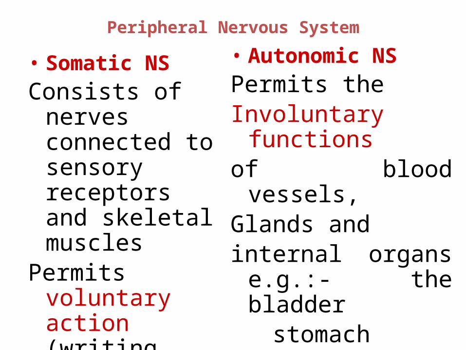
Peripheral Nervous System
• Somatic NSConsists of nerves
connected to sensory receptors and skeletal muscles
Permits voluntary action (writing your name)
• Autonomic NSPermits theInvoluntary functionsof blood vessels,Glands and internal organs e.g.:-
the bladder stomach heart
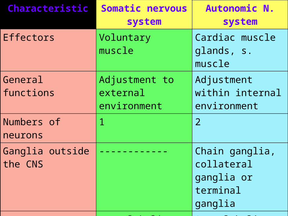
Characteristic Somatic nervous system
Autonomic N. system
Effectors Voluntary muscle Cardiac muscle glands, s. muscle
General functions Adjustment to external environment
Adjustment within internal environment
Numbers of neurons 1 2
Ganglia outside the CNS
------------ Chain ganglia, collateral ganglia or terminal ganglia
Neurotransmitter acetylcholine Acetylcholine, adrenaline, noradrenaline
Center Anterior Horn cells Lateral Horn cells
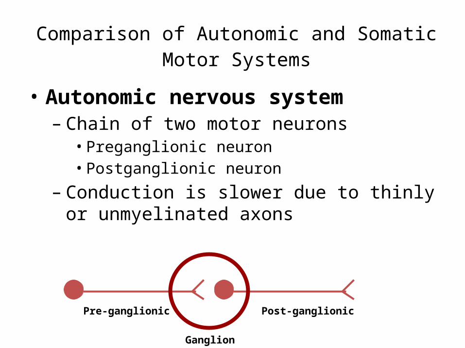
Comparison of Autonomic and Somatic Motor Systems
• Autonomic nervous system– Chain of two motor neurons
• Preganglionic neuron• Postganglionic neuron
– Conduction is slower due to thinly or unmyelinated axons
Pre-ganglionic
Ganglion
Post-ganglionic
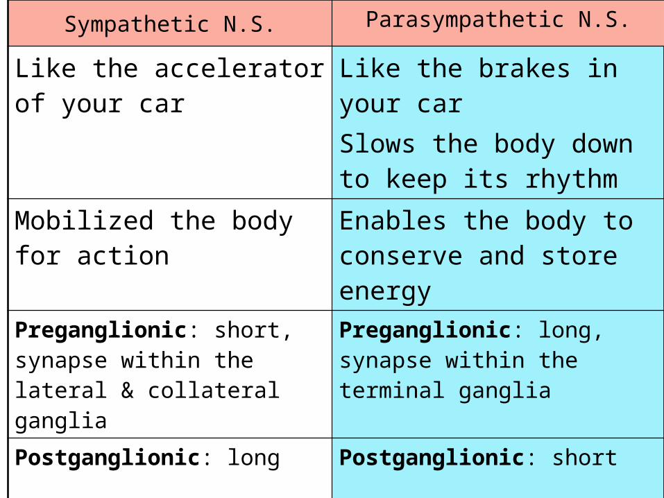
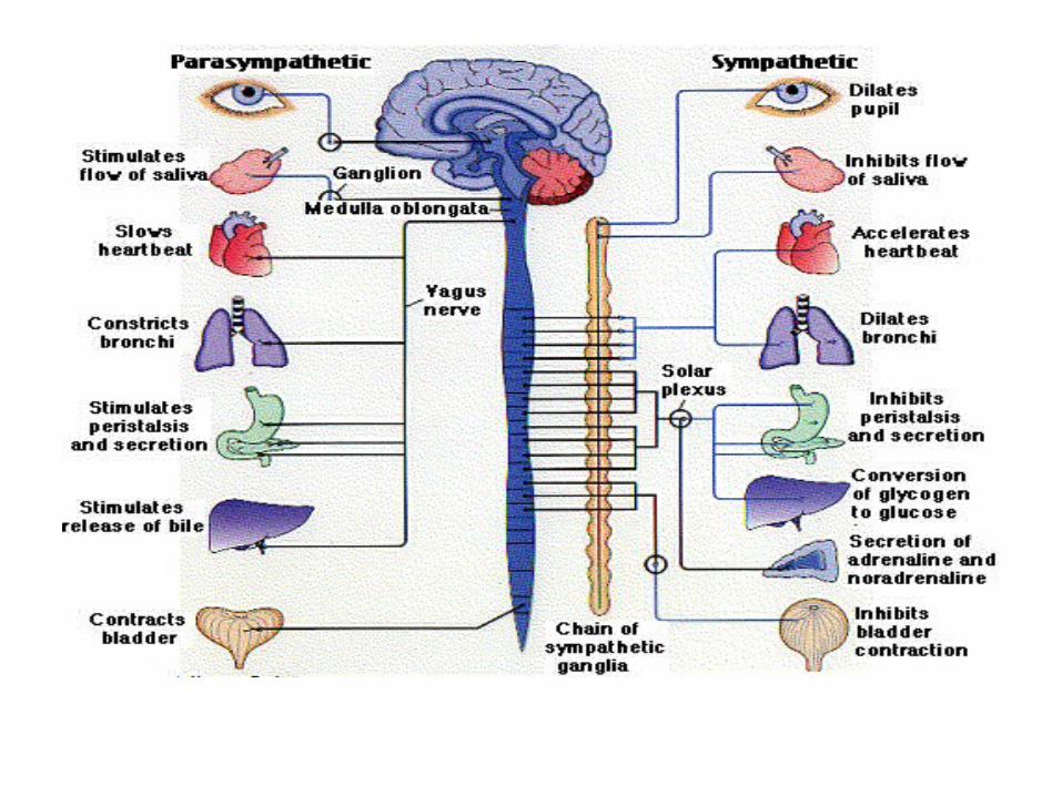
Sympathetic N.S. Parasympathetic N.S.
Like the accelerator of your car
Like the brakes in your carSlows the body down to keep its rhythm
Mobilized the body for action
Enables the body to conserve and store energy
Preganglionic: short, synapse within the lateral & collateral ganglia
Preganglionic: long, synapse within the terminal ganglia
Postganglionic: long Postganglionic: short
Has a wide distributions Has a restricted distributions
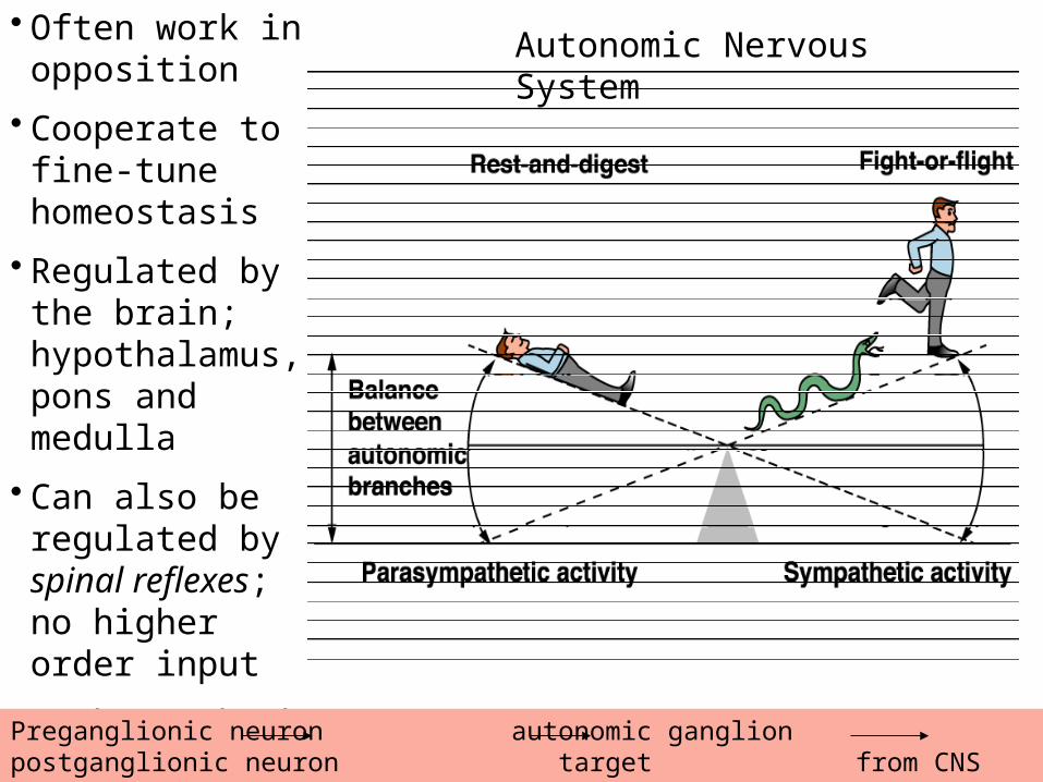
Autonomic Nervous System• Often work in
opposition• Cooperate to fine-
tune homeostasis• Regulated by the
brain; hypothalamus, pons and medulla
• Can also be regulated by spinal reflexes; no higher order input
• Pathways both consist of a two neuron system
Preganglionic neuron autonomic ganglion postganglionic neuron target from CNS outside CNS
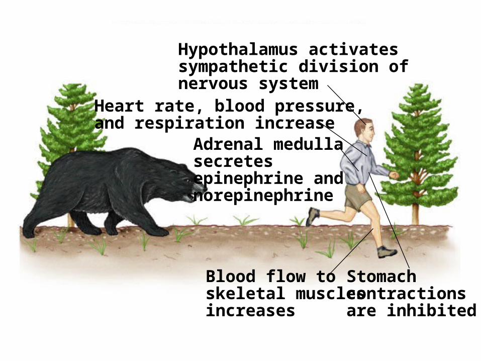
Fig. 45.34(TE Art)Hypothalamus activatessympathetic division ofnervous system
Heart rate, blood pressure,and respiration increase
Blood flow toskeletal musclesincreases
Stomachcontractions are inhibited
Adrenal medulla secretes epinephrine and norepinephrine
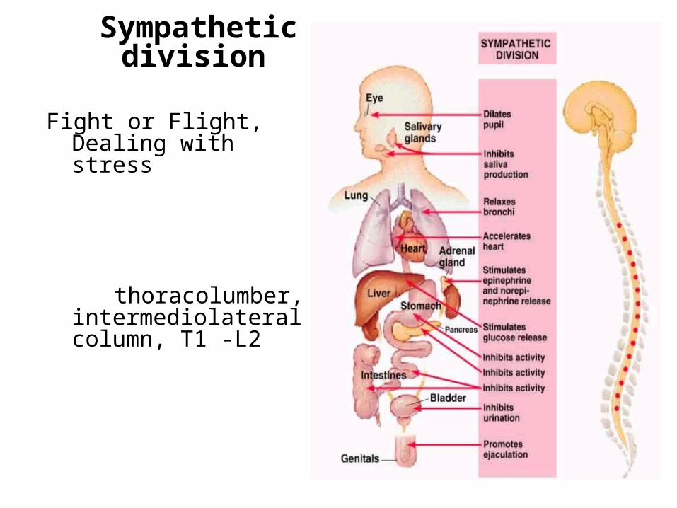
Sympathetic division
Fight or Flight, Dealing with stress
thoracolumber, intermediolateral column, T1 -L2
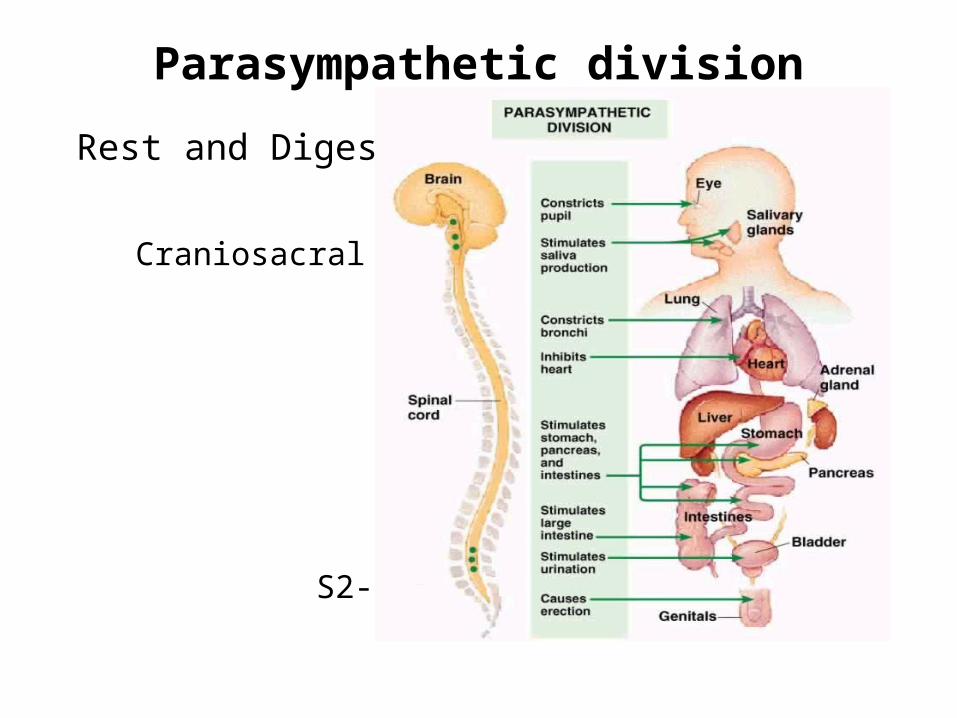
Parasympathetic division
Rest and Digest Craniosacral
S2-S4
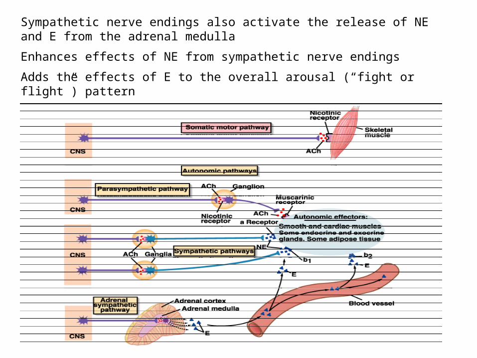
Sympathetic nerve endings also activate the release of NE and E from the adrenal medulla
Enhances effects of NE from sympathetic nerve endings
Adds the effects of E to the overall arousal (“fight or flight”) pattern
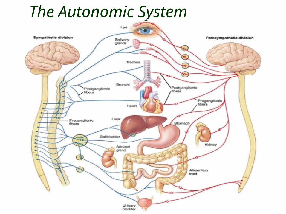
The Autonomic System
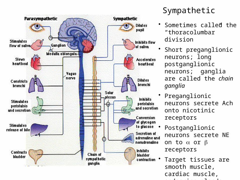
Sympathetic
• Sometimes called the “thoracolumbar” division
• Short preganglionic neurons; long postganglionic neurons; ganglia are called the chain ganglia
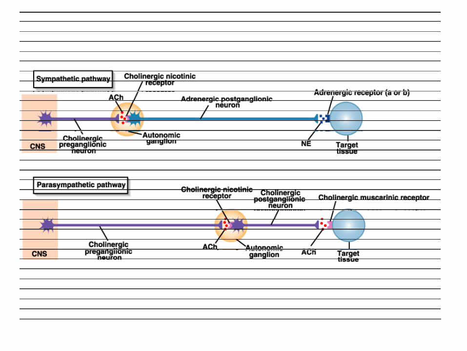
• Preganglionic neurons secrete Ach onto nicotinic receptors
• Postganglionic neurons secrete NE on to a or b receptors
• Target tissues are smooth muscle, cardiac muscle, endocrine glands, brown fat
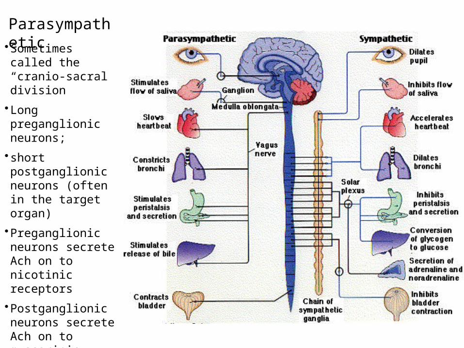
Parasympathetic•Sometimes called the “cranio-sacral division
•Long preganglionic neurons;
• short postganglionic neurons (often in the target organ)
•Preganglionic neurons secrete Ach on to nicotinic receptors
•Postganglionic neurons secrete Ach on to muscarinic receptors
•Target tissues are smooth muscle, cardiac muscle, exocrine glands, brown fat
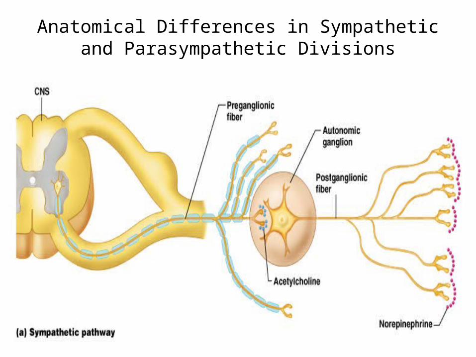
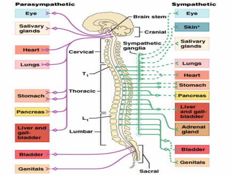
Anatomical Differences in Sympatheticand Parasympathetic Divisions
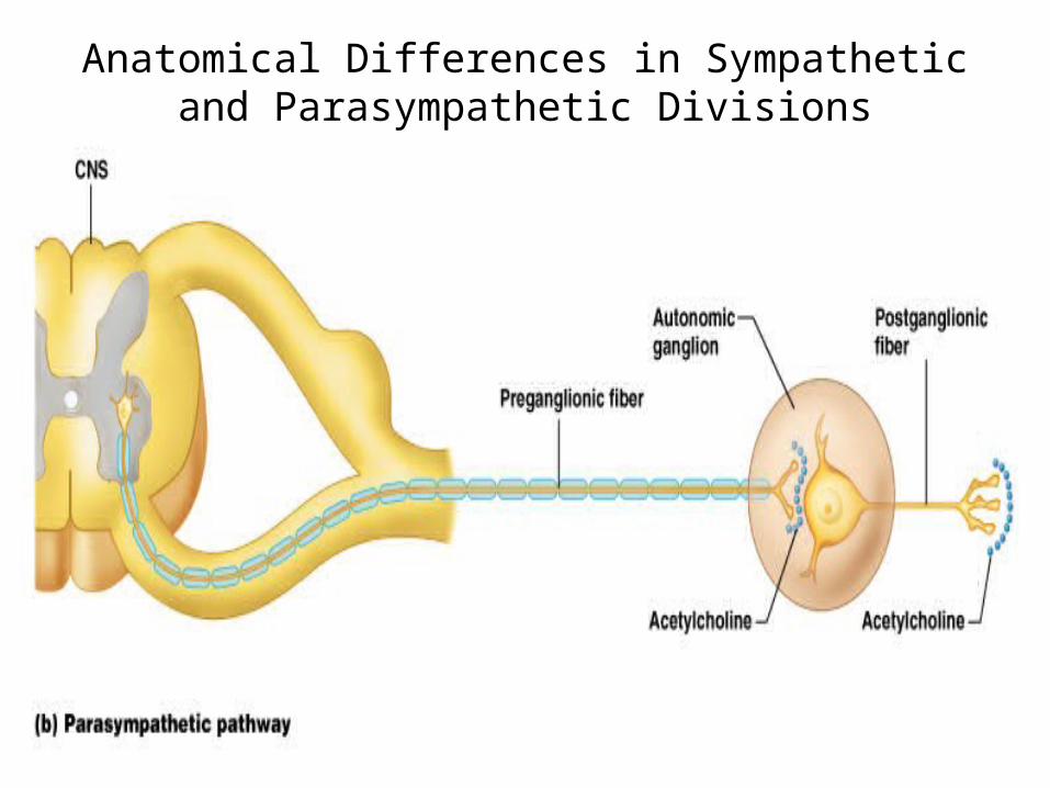
Anatomical Differences in Sympatheticand Parasympathetic Divisions
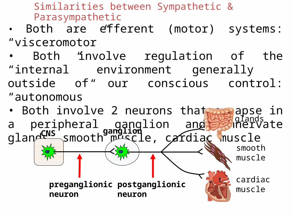
Similarities between Sympathetic & Parasympathetic
• Both are efferent (motor) systems: “visceromotor”• Both involve regulation of the “internal” environment generally outside of our conscious control: “autonomous”• Both involve 2 neurons that synapse in a peripheral ganglion and Innervate glands, smooth muscle, cardiac muscle
CNS ganglion
preganglionicneuron
postganglionicneuron
glands
smoothmuscle
cardiacmuscle
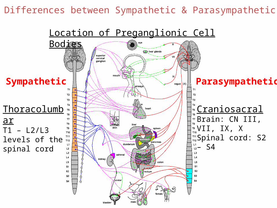
Differences between Sympathetic & Parasympathetic
Location of Preganglionic Cell Bodies
ThoracolumbarT1 – L2/L3 levels of the spinal cord
CraniosacralBrain: CN III, VII, IX, XSpinal cord: S2 – S4
Sympathetic Parasympathetic

SympatheticCNS ganglion
short preganglionicneuron
long postganglionicneuron
target
ParasympatheticCNS ganglion
long preganglionicneuron
target
short postganglionicneuron
Differences between Sympathetic & Parasympathetic
Relative Lengths of Neurons
Parasympathetic
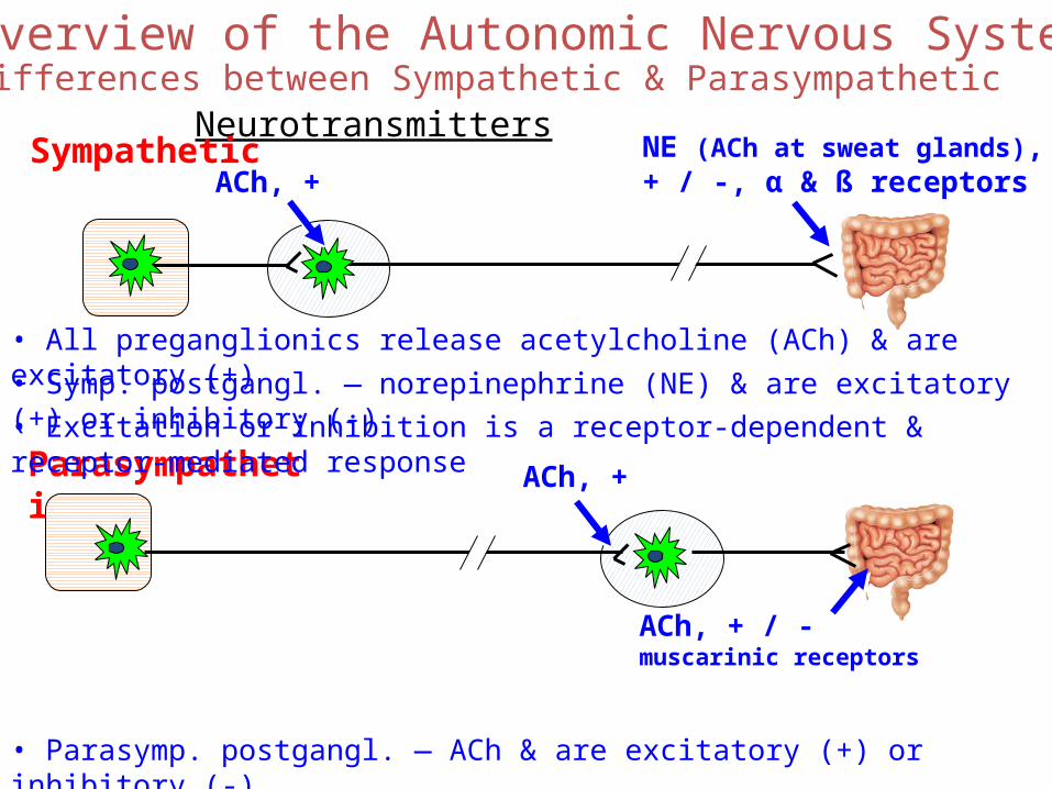
Overview of the Autonomic Nervous SystemDifferences between Sympathetic & Parasympathetic
Neurotransmitters
ACh, +
NE (ACh at sweat glands),+ / -, α & ß receptors
ACh, + / -muscarinic receptors
• All preganglionics release acetylcholine (ACh) & are excitatory (+)• Symp. postgangl. — norepinephrine (NE) & are excitatory (+) or inhibitory (-)
• Parasymp. postgangl. — ACh & are excitatory (+) or inhibitory (-)
Sympathetic
• Excitation or inhibition is a receptor-dependent & receptor-mediated response
ACh, +
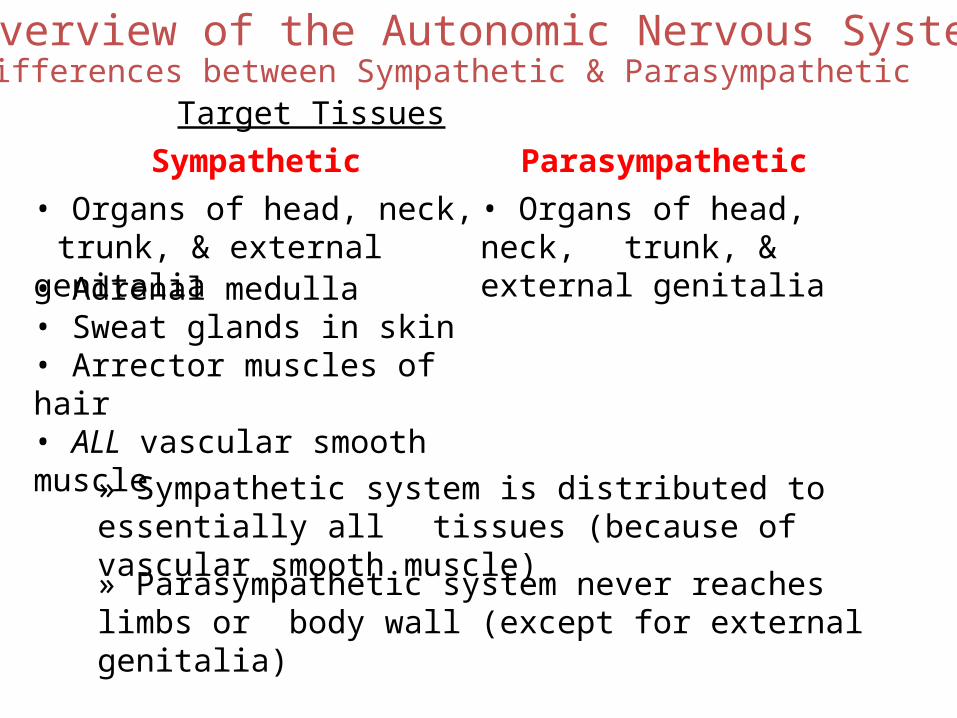
Overview of the Autonomic Nervous SystemDifferences between Sympathetic & Parasympathetic
Target TissuesParasympatheticSympathetic
• Organs of head, neck, trunk, & external genitalia
• Organs of head, neck, trunk, & external genitalia
• Adrenal medulla• Sweat glands in skin• Arrector muscles of hair• ALL vascular smooth muscle
» Sympathetic system is distributed to essentially all tissues (because of vascular smooth muscle)
» Parasympathetic system never reaches limbs or body wall (except for external genitalia)
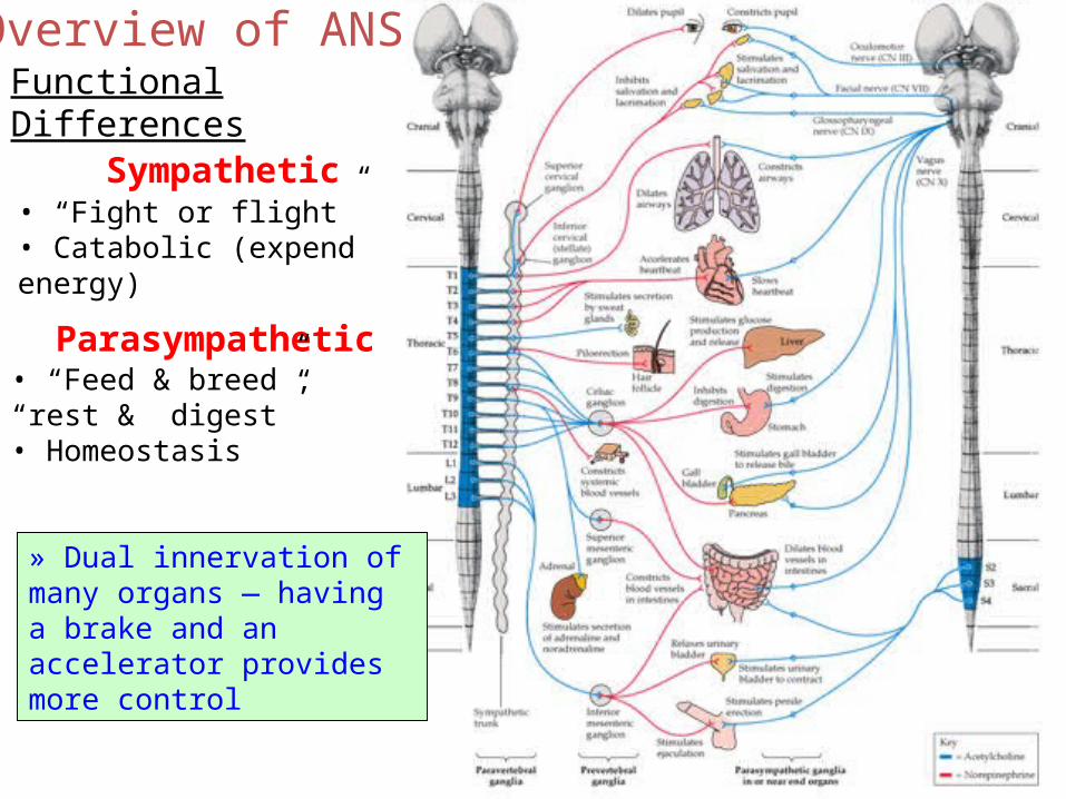
Overview of ANSFunctional Differences
Sympathetic• “Fight or flight”• Catabolic (expend energy)
Parasympathetic• “Feed & breed”, “rest & digest”• Homeostasis
» Dual innervation of many organs — having a brake and an accelerator provides more control
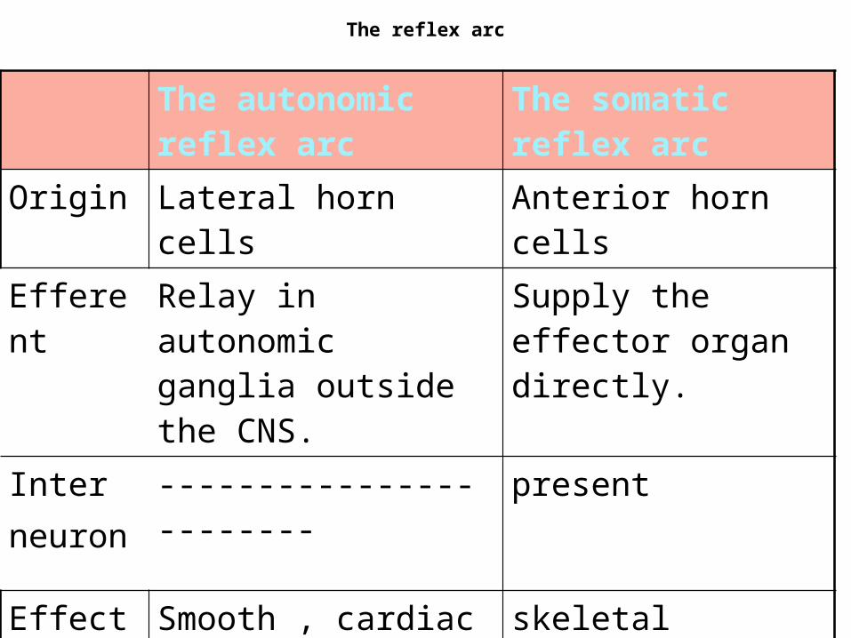
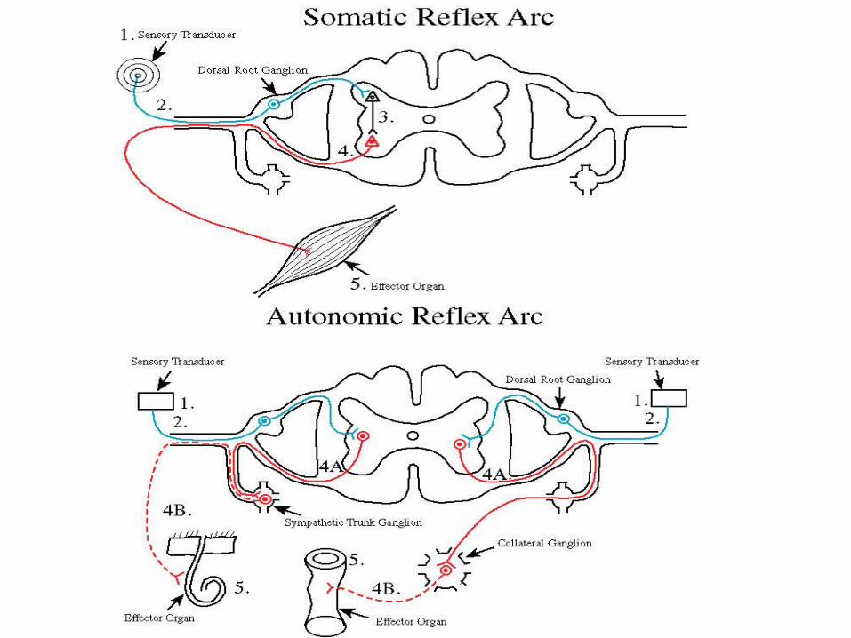
The reflex arc
The autonomic reflex arc
The somatic reflex arc
Origin Lateral horn cells Anterior horn cells
Efferent Relay in autonomic ganglia outside the CNS.
Supply the effector organ directly.
Interneuron
------------------------ present
Effector organs
Smooth , cardiac muscles,glands
skeletal
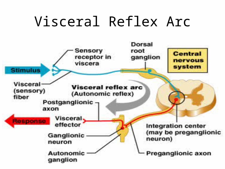
Visceral Reflex Arc
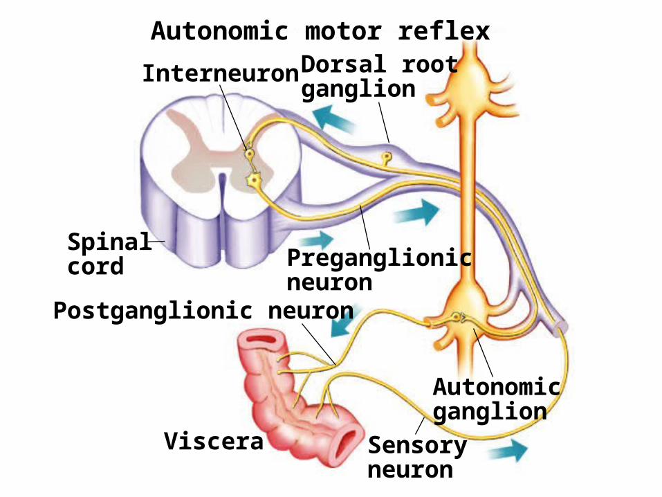
Fig. 45.32(TE Art)
Viscera
Autonomicganglion
Postganglionic neuron
Autonomic motor reflex
Interneuron Dorsal rootganglion
Preganglionicneuron
Sensoryneuron
Spinalcord
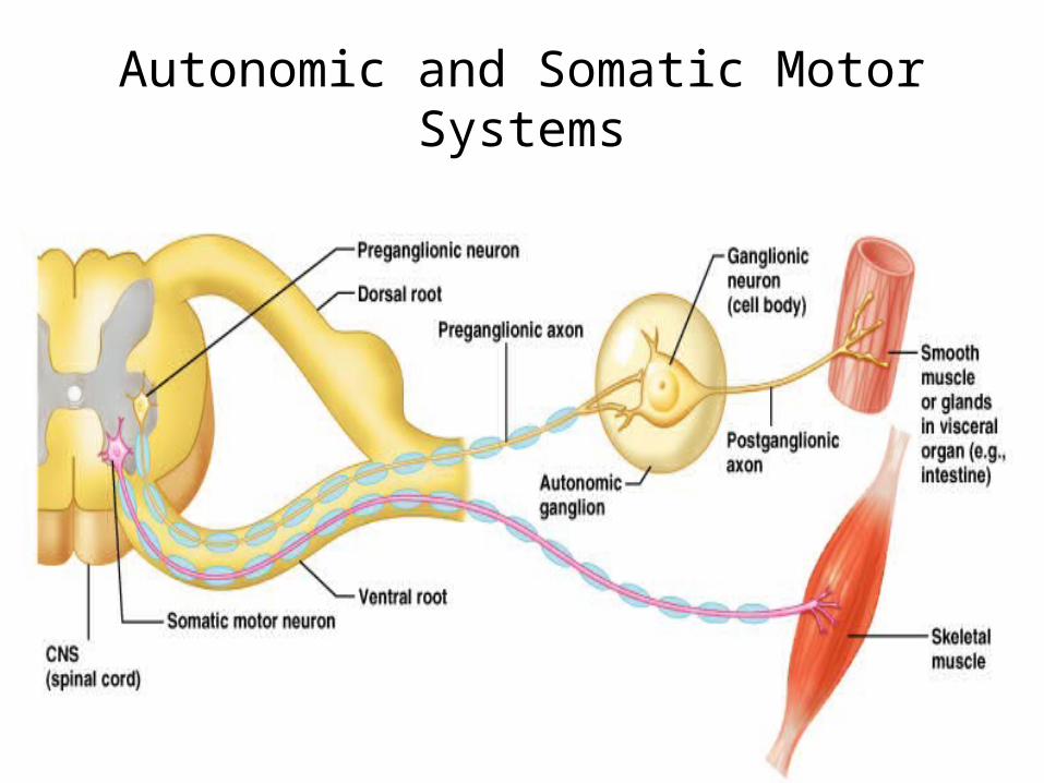
Autonomic and Somatic Motor Systems
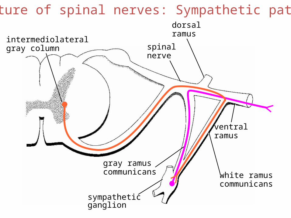
spinalnerve
dorsalramus
ventralramus
gray ramuscommunicans white ramus
communicans
sympatheticganglion
intermediolateralgray column
Structure of spinal nerves: Sympathetic pathways
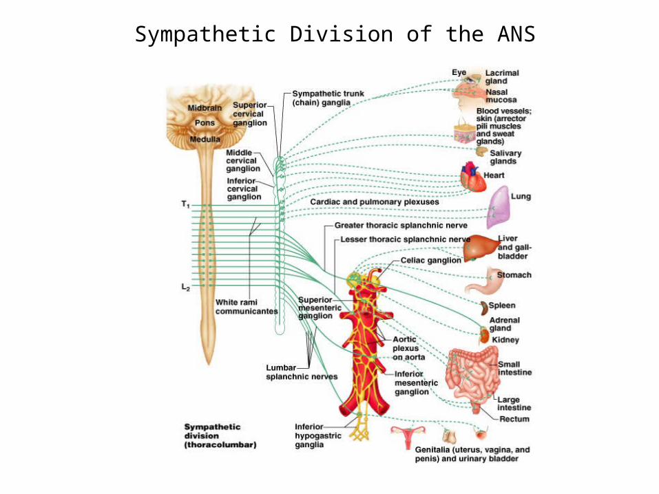
Sympathetic Division of the ANS
somatic tissues(body wall, limbs)
visceral tissues(organs)
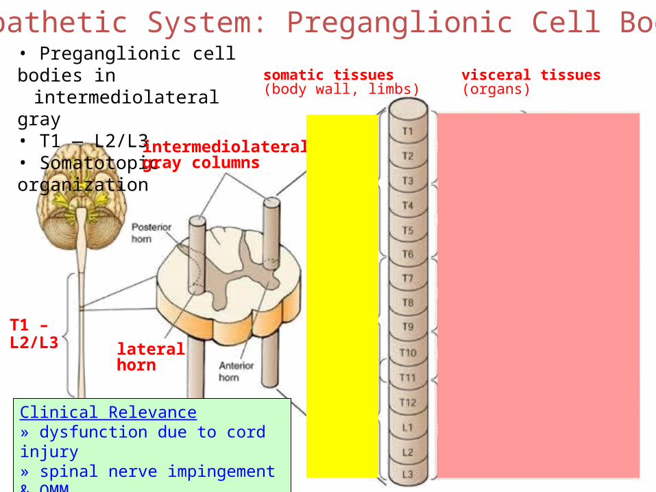
Sympathetic System: Preganglionic Cell Bodies• Preganglionic cell bodies in
intermediolateral gray• T1 — L2/L3• Somatotopic organization
intermediolateralgray columns
lateralhorn
T1 –L2/L3
Clinical Relevance» dysfunction due to cord injury» spinal nerve impingement & OMM» referred pain
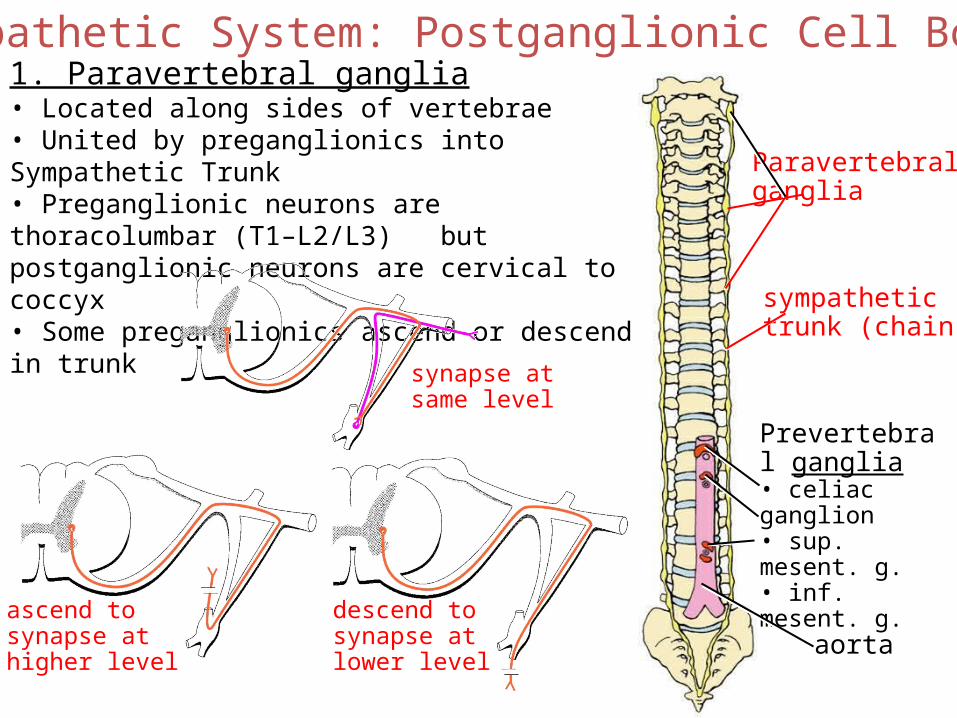
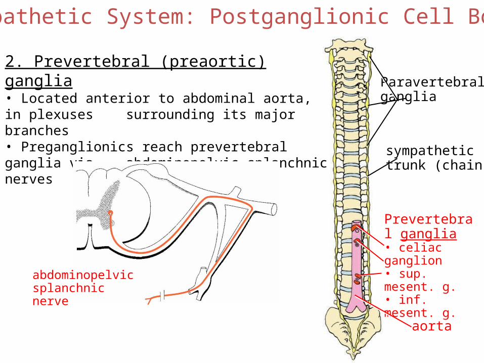
Sympathetic System: Postganglionic Cell Bodies
Paravertebralganglia
Prevertebral ganglia• celiac ganglion• sup. mesent. g.• inf. mesent. g.
aorta
sympathetictrunk (chain)
1. Paravertebral ganglia• Located along sides of vertebrae• United by preganglionics into Sympathetic Trunk• Preganglionic neurons are thoracolumbar (T1–L2/L3)
but postganglionic neurons are cervical to coccyx• Some preganglionics ascend or descend in trunk
synapse atsame level
ascend tosynapse athigher level
descend tosynapse atlower level
Sympathetic System: Postganglionic Cell Bodies
Paravertebralganglia
Prevertebral ganglia• celiac ganglion• sup. mesent. g.• inf. mesent. g.
aorta
sympathetictrunk (chain)
2. Prevertebral (preaortic) ganglia• Located anterior to abdominal aorta, in plexuses surrounding its major branches• Preganglionics reach prevertebral ganglia via abdominopelvic splanchnic nerves
abdominopelvicsplanchnicnerve
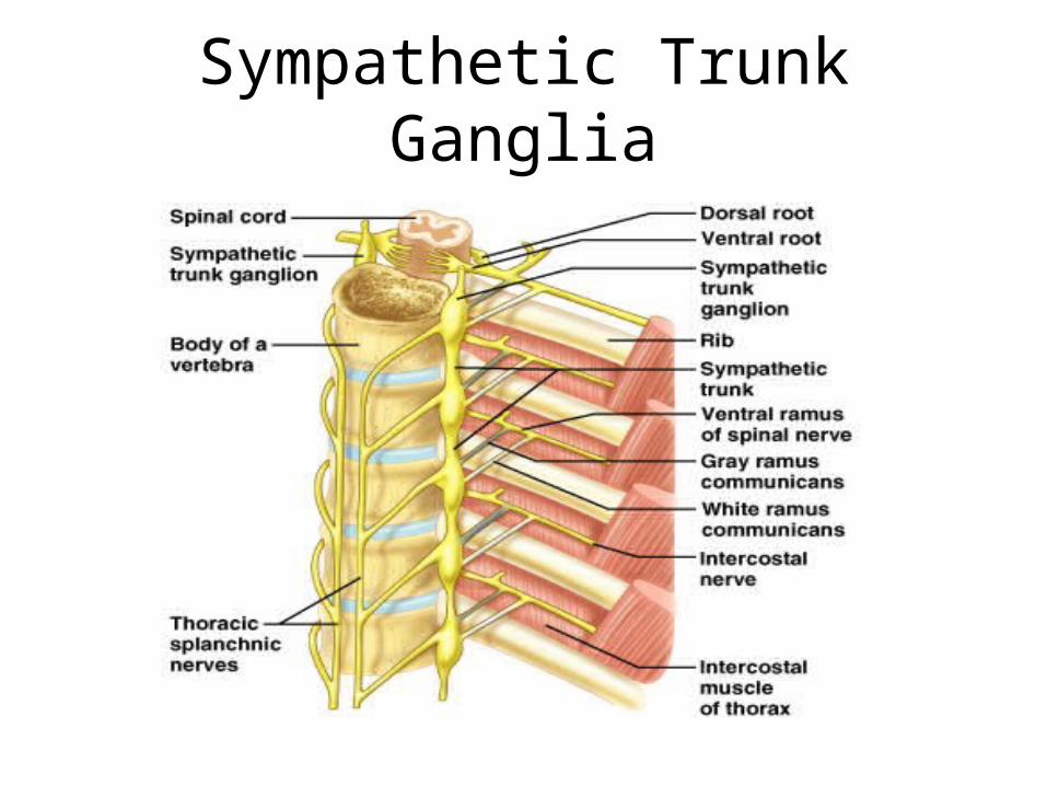
Sympathetic Trunk Ganglia
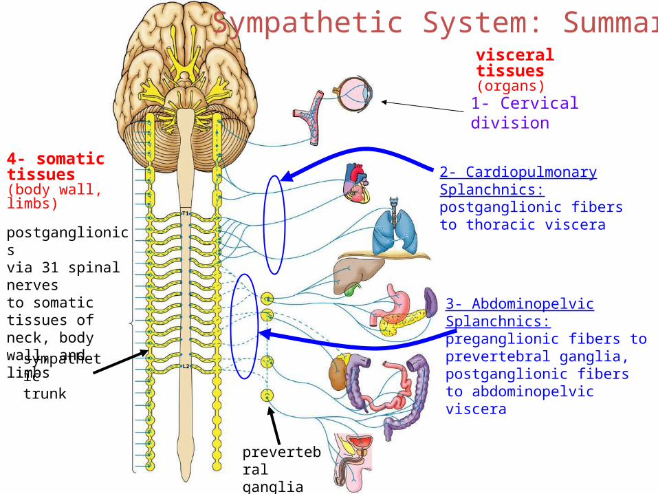
Sympathetic System: Summary
T1
L2
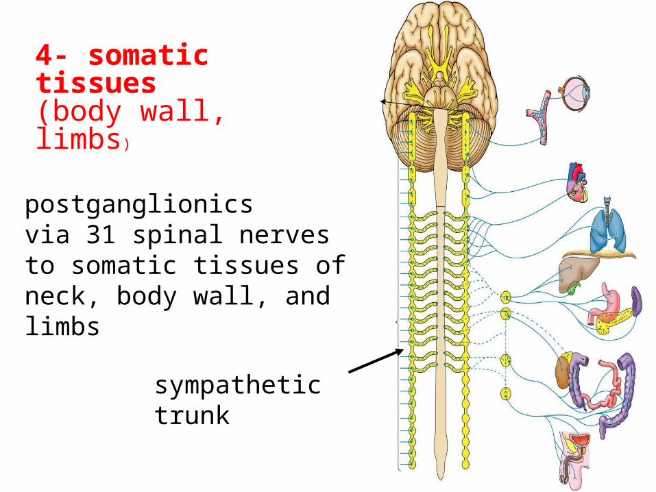
4- somatic tissues(body wall, limbs)
visceral tissues(organs)
postganglionicsvia 31 spinal nervesto somatic tissues of neck, body wall, and limbs
sympathetictrunk
prevertebralganglia
2- Cardiopulmonary Splanchnics: postganglionic fibers to thoracic viscera
3- Abdominopelvic Splanchnics: preganglionic fibers to prevertebral ganglia, postganglionic fibers to abdominopelvic viscera
1- Cervical division
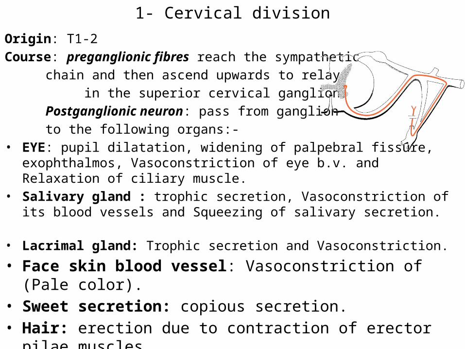
1- Cervical divisionOrigin: T1-2Course: preganglionic fibres reach the sympathetic
chain and then ascend upwards to relay in the superior cervical ganglion.
Postganglionic neuron: pass from ganglion to the following organs:-
• EYE: pupil dilatation, widening of palpebral fissure, exophthalmos, Vasoconstriction of eye b.v. and Relaxation of ciliary muscle.
• Salivary gland : trophic secretion, Vasoconstriction of its blood vessels and Squeezing of salivary secretion.
• Lacrimal gland: Trophic secretion and Vasoconstriction.• Face skin blood vessel: Vasoconstriction of (Pale color).• Sweet secretion: copious secretion.• Hair: erection due to contraction of erector pilae muscles..• Cerebral vessels: Weak vasoconstriction

Sympathetic Pathways to the Head
(2) Cardiopulmonary division
Origin: Lateral horn cells of upper 4-5 thoracic segments.Course: Preganglionic neurons reach the sympathetic chain to relay in
the three cervical ganglion and upper four thoracic ganglion. The postganglionic arise from these ganglia supply the following structures:-
• Heart: Increase all properties of cardiac muscle (contraction, rhythmicity, excitability, conductivity.
• Coronary vessels, its sympathetic supply. At first it causes vasoconstriction, and then it causes vasodilatation due to accumulation of metabolites.
• Bronchi: Broncho dilation, decrease bronchial secretions and vasoconstriction of pulmonary blood vessels.
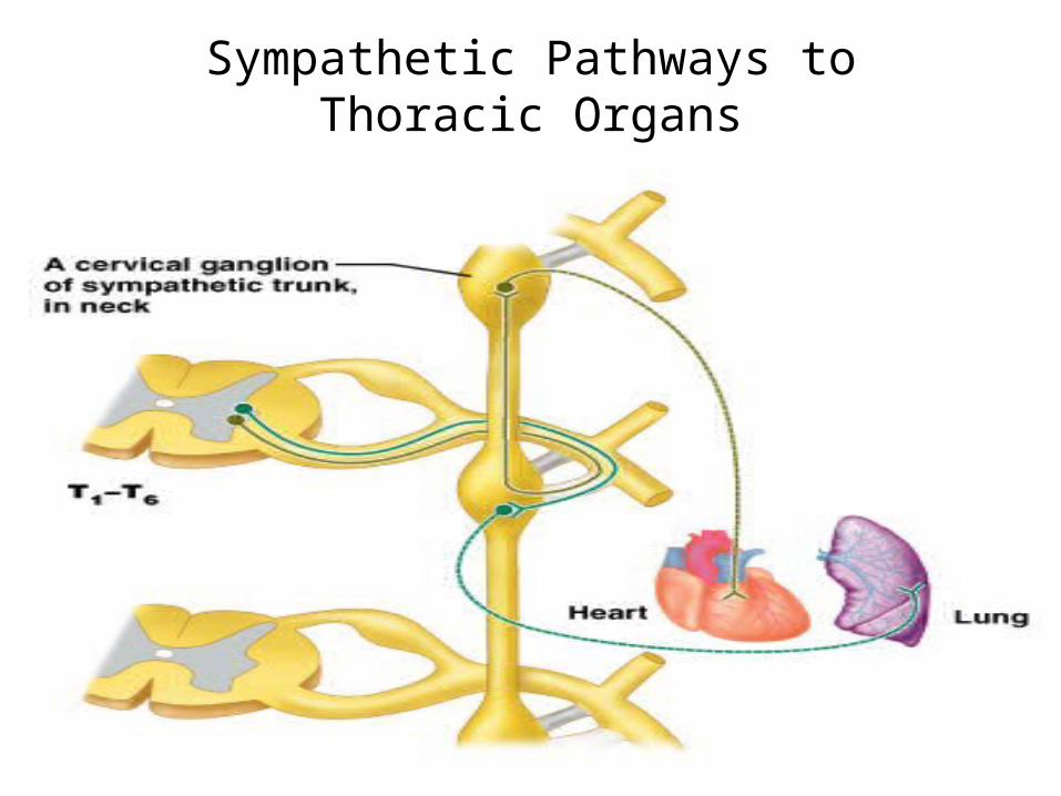
Sympathetic Pathways to Thoracic Organs
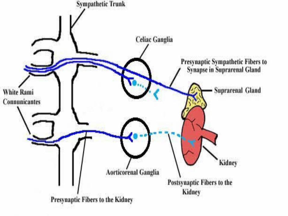
3- Splanchnic division
Origin: lateral horn cells of the lower six thoracic and upper four lumber segments.Course: Preganglionic neurons originate from these segments reach the sympathetic chain
where they pass without relay, and then they divided into two branches:(1) Greater splanchnic nerve (2) Lesser splanchnic nerve.
Greater splanchnic nerve:• Origin: Preganglionic nerves fibers emerge from lateral horn cells of lower six thoracic
segments and then relay in the collateral ganglion in the abdomen.• Course: Postganglionic nerve fibers arise from these ganglia (celiac, superior mesenteric and
inferior mesenteric ganglia) and supply the abdominal organs causing the following effects:• Vasoconstriction: of most arteries of stomach, small intestine, proximal part of large
intestine, kidney, pancreas and liver.• Relaxation of the musculature of: stomach, small intestine and proximal part of large
intestine.• Contraction of sphincters: of the stomach and intestine leading to (food retention).• Contraction of the capsule: of the spleen leading to evacuation of about 200 ml of blood. • Breakdown of the glucose in the liver: (glycogenolysis) leading to increase of blood glucose
level.• Stimulation of adrenal medulla: Secrete adrenaline and noradrenalin.
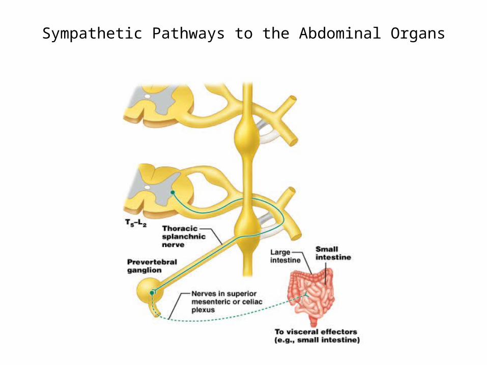
Sympathetic Pathways to the Abdominal Organs
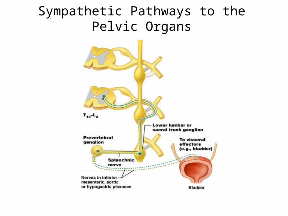
Sympathetic Pathways to the Pelvic Organs
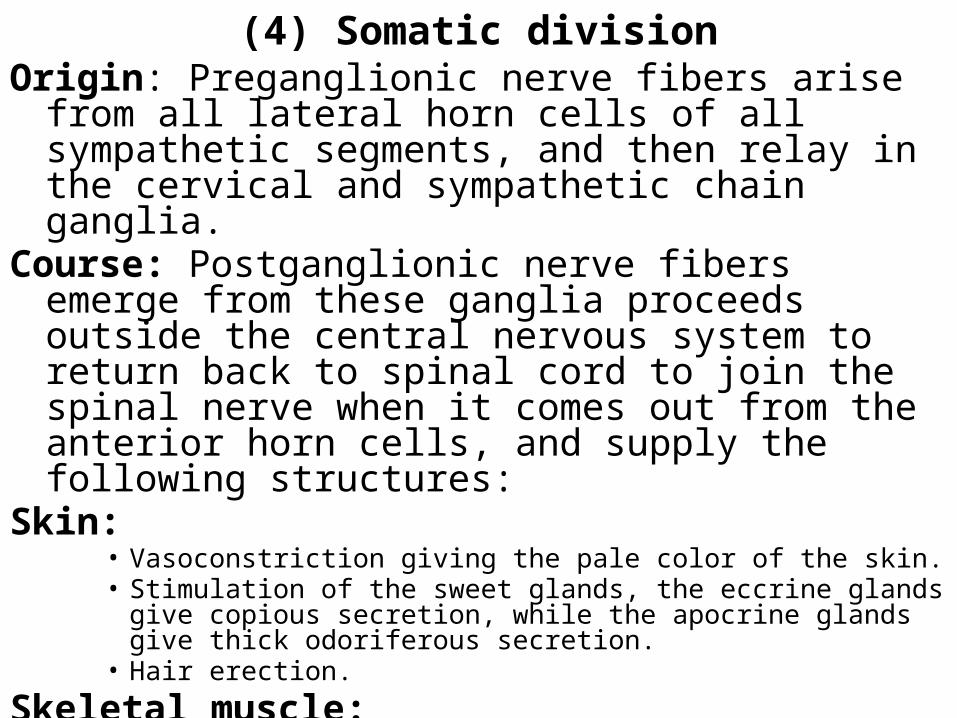
(4) Somatic divisionOrigin: Preganglionic nerve fibers arise from all lateral horn
cells of all sympathetic segments, and then relay in the cervical and sympathetic chain ganglia.
Course: Postganglionic nerve fibers emerge from these ganglia proceeds outside the central nervous system to return back to spinal cord to join the spinal nerve when it comes out from the anterior horn cells, and supply the following structures:
Skin: • Vasoconstriction giving the pale color of the skin.• Stimulation of the sweet glands, the eccrine glands give copious secretion,
while the apocrine glands give thick odoriferous secretion.• Hair erection.
Skeletal muscle: • Its blood vessels show vasodilatation (V.D.) due to cholinergic effect or
vasoconstriction (V.C.) due to a adrenergic effect. • The type of stimulation depends upon the nature of stimulation.• Muscles: its stimulation causing delayed fatigue and early recovery.
4- somatic tissues(body wall, limbs)
postganglionicsvia 31 spinal nervesto somatic tissues of neck, body wall, and limbs
sympathetictrunk
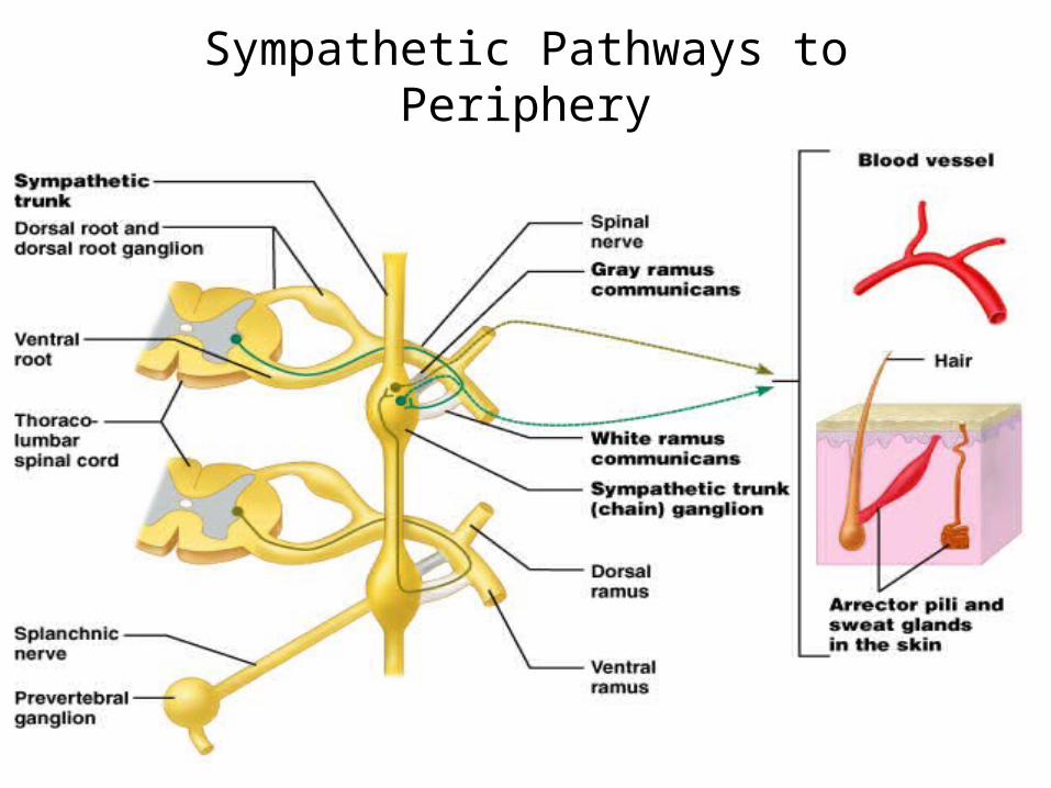
Sympathetic Pathways to Periphery
Figure 15.9
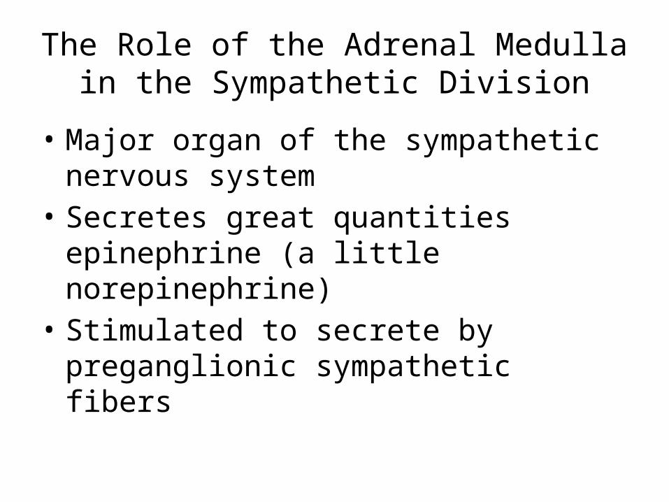
The Role of the Adrenal Medulla in the Sympathetic Division
• Major organ of the sympathetic nervous system
• Secretes great quantities epinephrine (a little norepinephrine)
• Stimulated to secrete by preganglionic sympathetic fibers
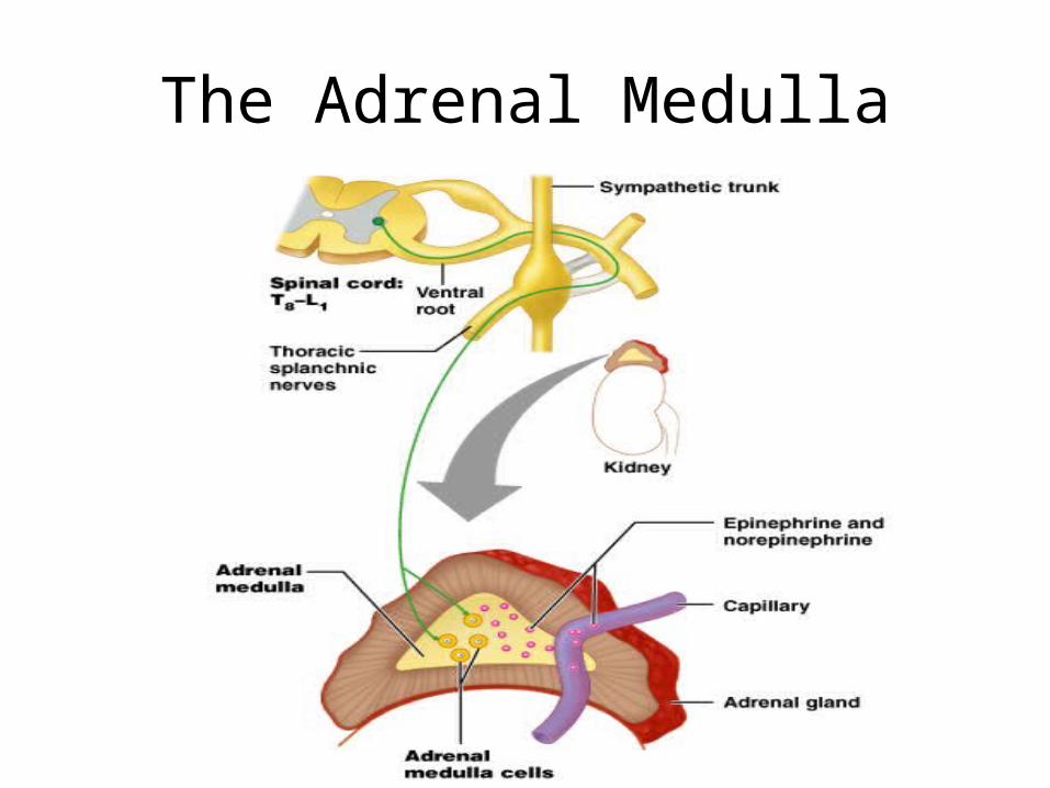
The Adrenal Medulla
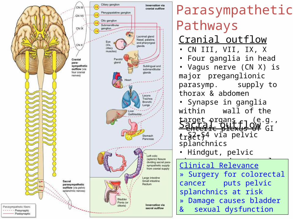
ParasympatheticPathways
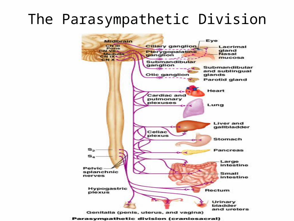
Cranial outflow• CN III, VII, IX, X• Four ganglia in head• Vagus nerve (CN X) is major
preganglionic parasymp. supply to thorax & abdomen• Synapse in ganglia within wall of the target organs (e.g., enteric plexus of GI tract)
Sacral outflow• S2–S4 via pelvic splanchnics• Hindgut, pelvic viscera, and
external genitalia
Clinical Relevance» Surgery for colorectal cancer
puts pelvic splanchnics at risk» Damage causes bladder & sexual dysfunction
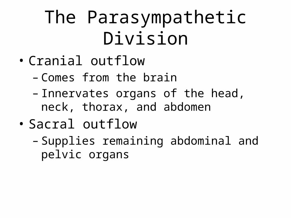
The Parasympathetic Division
• Cranial outflow – Comes from the brain– Innervates organs of the head, neck, thorax, and
abdomen• Sacral outflow
– Supplies remaining abdominal and pelvic organs
The Parasympathetic Division
Cranial Nerves
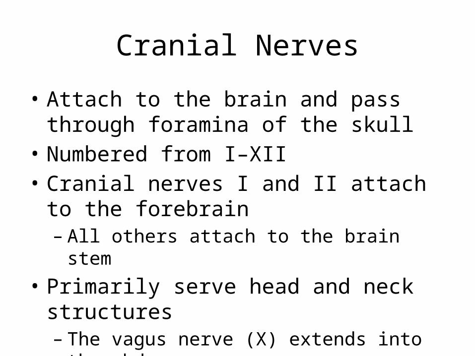
• Attach to the brain and pass through foramina of the skull
• Numbered from I–XII• Cranial nerves I and II attach to the forebrain
– All others attach to the brain stem• Primarily serve head and neck structures
– The vagus nerve (X) extends into the abdomen
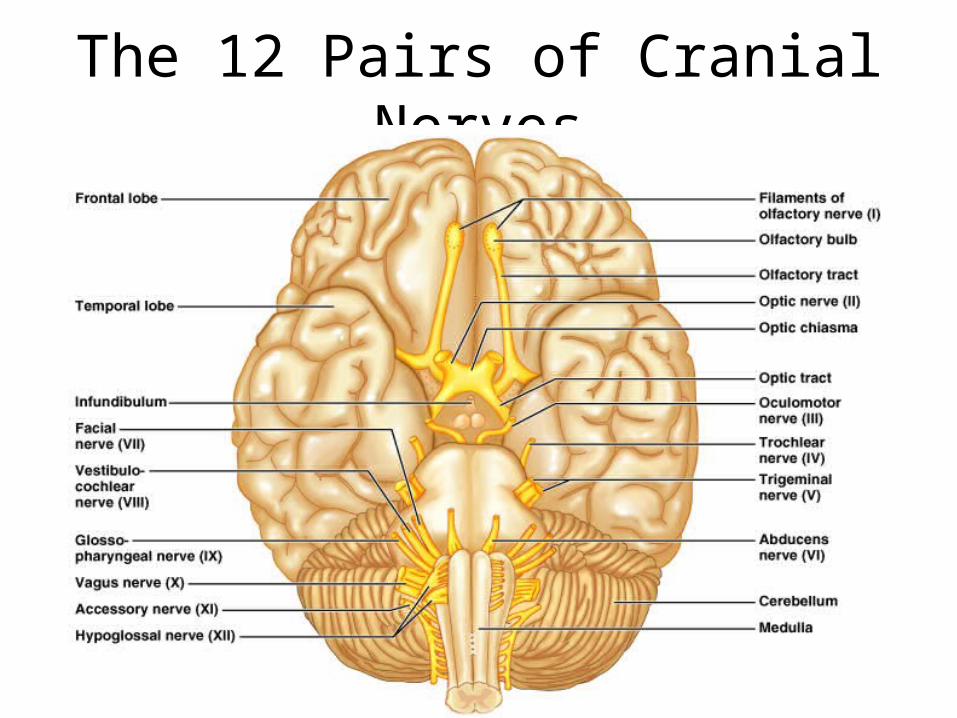
The 12 Pairs of Cranial Nerves

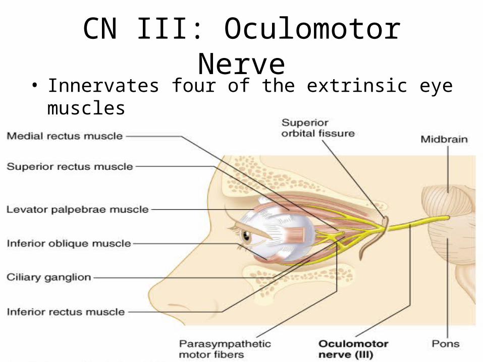
CN III: Oculomotor Nerve• Innervates four of the extrinsic eye muscles
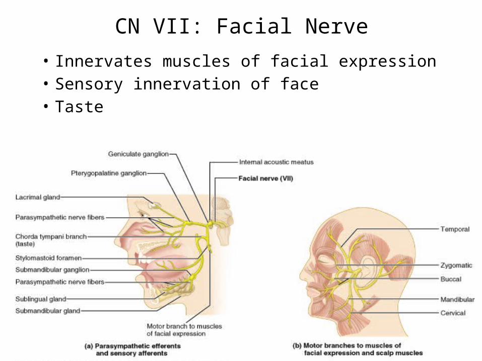
CN VII: Facial Nerve
• Innervates muscles of facial expression• Sensory innervation of face• Taste
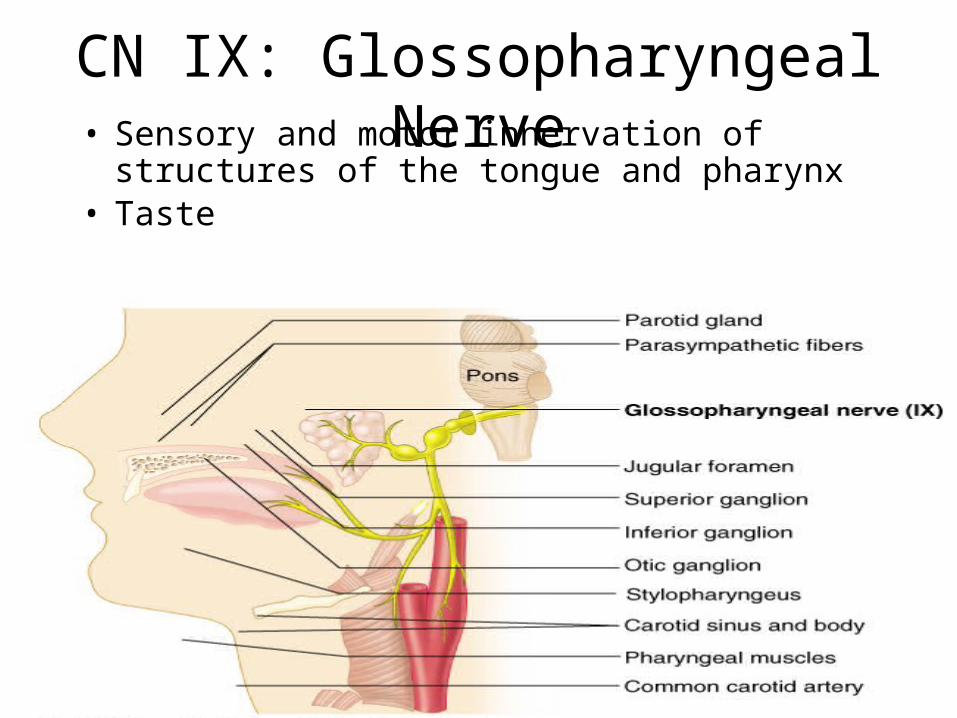
CN IX: Glossopharyngeal Nerve• Sensory and motor innervation of structures of the
tongue and pharynx• Taste
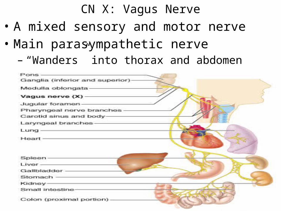
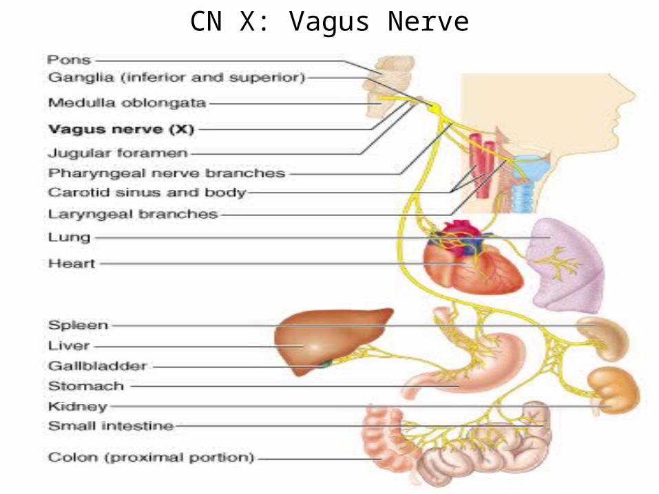
CN X: Vagus Nerve• A mixed sensory and motor nerve • Main parasympathetic nerve
– “Wanders” into thorax and abdomen
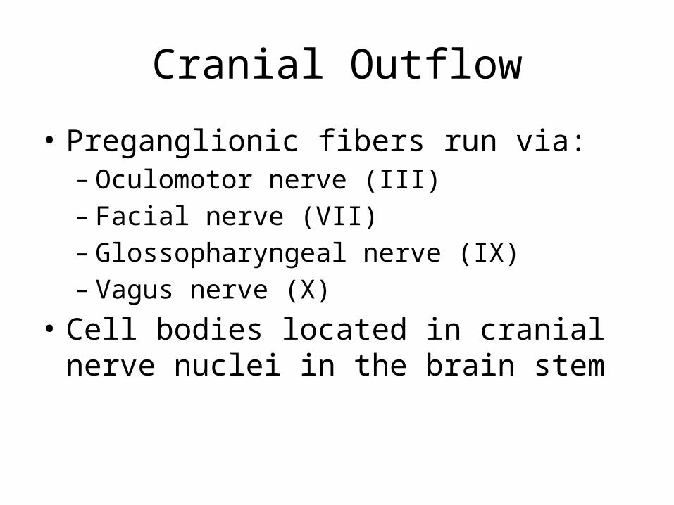
Cranial Outflow
• Preganglionic fibers run via:– Oculomotor nerve (III)– Facial nerve (VII)– Glossopharyngeal nerve (IX)– Vagus nerve (X)
• Cell bodies located in cranial nerve nuclei in the brain stem
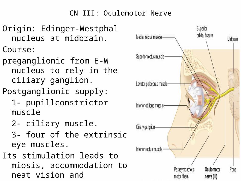
CN III: Oculomotor Nerve
Origin: Edinger-Westphal nucleus at midbrain.
Course: preganglionic from E-W nucleus to rely
in the ciliary ganglion.Postganglionic supply:
1- pupillconstrictor muscle 2- ciliary muscle.3- four of the extrinsic eye muscles.
Its stimulation leads to miosis, accommodation to neat vision and movements of the eye ball.
CN III: Oculomotor Nerve• Innervates four of the extrinsic eye muscles
CN VII: Facial Nerve
Origin: The superior salivary nucleus which is a part of facial nucleus in the lower part of pons.
Course: Preganglionic nerve fibers run in the chorda tympani nerve which is a part of facial nerve and relay in:-
- Submaxillary ganglion- Sphenopalatine ganglion.• Postganglionic nerve arises from Submaxillary ganglion
supply submandibular and sublingual salivary glands and anterior 2/3 of the tongue.
• Postganglionic nerve arises from Sphenopalatine ganglion supply the mucosa of the soft palate and nasopharynx and Lacrimal glands.
• Its stimulation causes vasodilatation and secretion at their effector organs.

CN VII: Facial Nerve
• Innervates muscles of facial expression• Sensory innervation of face• Taste
CN IX: Glossopharyngeal Nerve
Origin: Glossopharyngeal nerve nucleus in the upper part of the medulla oblongata called inferior salivary nucleus, and then relay in the otic ganglion.
Course: Postganglionic nerve fibers arise from otic ganglion supply the parotid salivary gland and posterior 1/3 of the tongue
Its stimulation causes vasodilatation and secretion at their effector organs
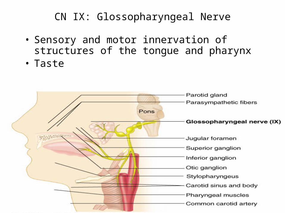
CN IX: Glossopharyngeal Nerve
• Sensory and motor innervation of structures of the tongue and pharynx
• Taste
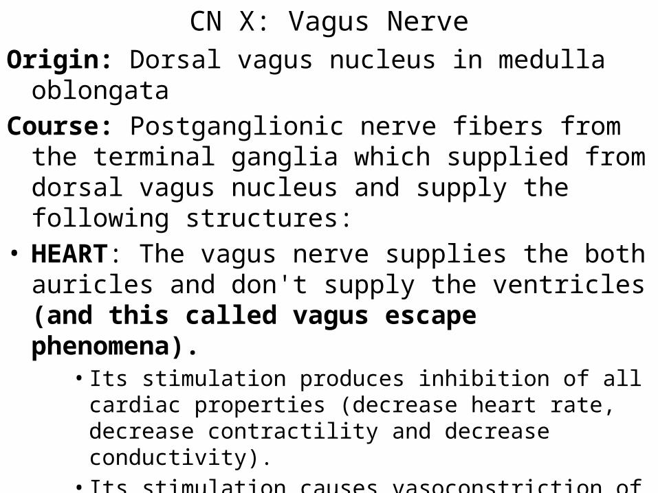
CN X: Vagus NerveOrigin: Dorsal vagus nucleus in medulla oblongata Course: Postganglionic nerve fibers from the terminal ganglia
which supplied from dorsal vagus nucleus and supply the following structures:
• HEART: The vagus nerve supplies the both auricles and don't supply the ventricles (and this called vagus escape phenomena).
• Its stimulation produces inhibition of all cardiac properties (decrease heart rate, decrease contractility and decrease conductivity).
• Its stimulation causes vasoconstriction of coronary vessels and reduction of O2 consumption by cardiac muscle.
• These responses lead to bradycardia.
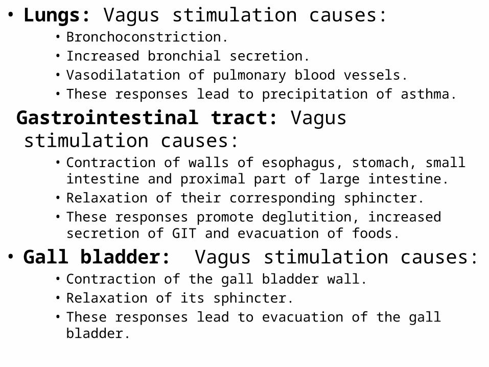
• Lungs: Vagus stimulation causes:• Bronchoconstriction.• Increased bronchial secretion.• Vasodilatation of pulmonary blood vessels.• These responses lead to precipitation of asthma.
Gastrointestinal tract: Vagus stimulation causes:• Contraction of walls of esophagus, stomach, small intestine and proximal
part of large intestine.• Relaxation of their corresponding sphincter.• These responses promote deglutition, increased secretion of GIT and
evacuation of foods.
• Gall bladder: Vagus stimulation causes:• Contraction of the gall bladder wall.• Relaxation of its sphincter.• These responses lead to evacuation of the gall bladder.
CN X: Vagus Nerve

Sacral OutflowOrigin: Preganglionic nerve fibers arise from the lateral
horn cells of the 2nd, 3rd and 4th sacral segments.Course: These preganglionic passes without relay, then the
right and left branches unit together to form the pelvic nerve, the pelvic nerve relay in the terminal ganglia, where the postganglionic nerve fibers emerge and supply the following structures:- Urinary bladder: parasympathetic stimulation causes:- Contraction of the bladder wall - Relaxation of its sphincter.- These responses lead to micturition.
Rectum and descending colon: parasympathetic stimulation causes:
- Contraction of its wall.- Relaxation of internal anal sphincter.- These responses lead to defecation.
Seminal vesicles and prostate: parasympathetic stimulation -causes:
- Secretion of these glands.Erectile tissue: parasympathetic stimulation causes:
- Vasodilatation which lead to erection.
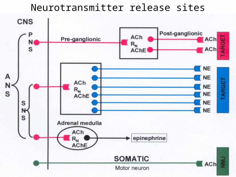
Neurotransmitter release sites
Acetylcholine receptors
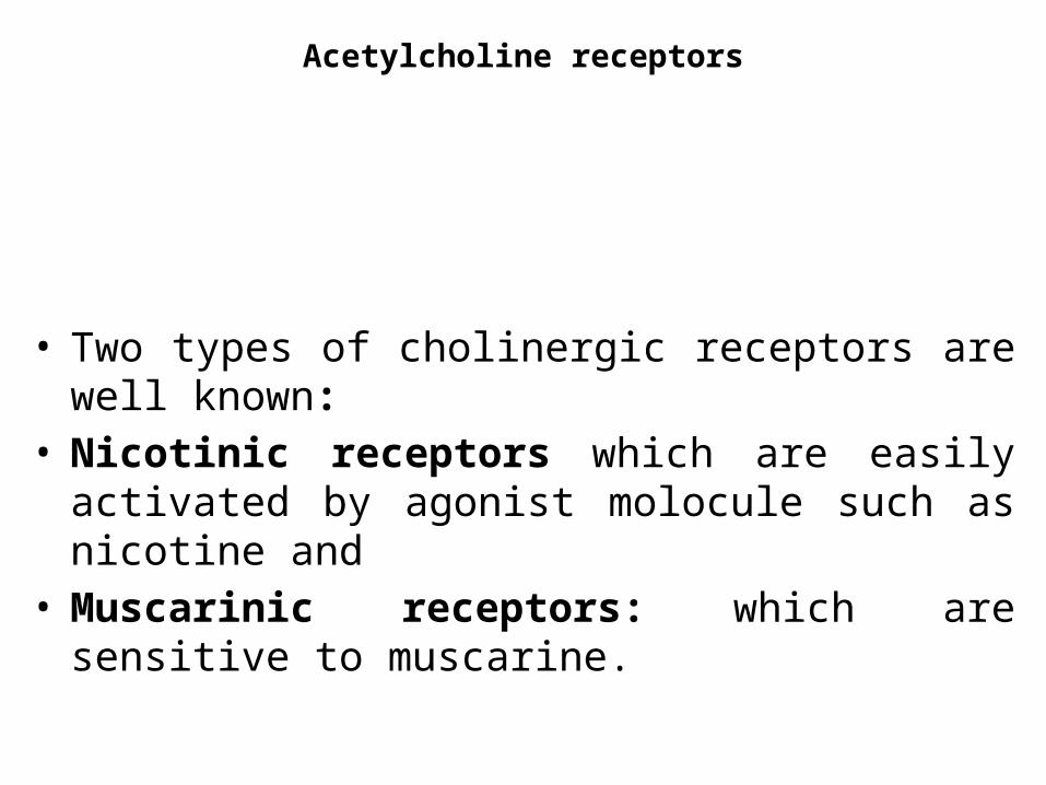
• Two types of cholinergic receptors are well known: • Nicotinic receptors which are easily activated by agonist
molocule such as nicotine and • Muscarinic receptors: which are sensitive to muscarine.
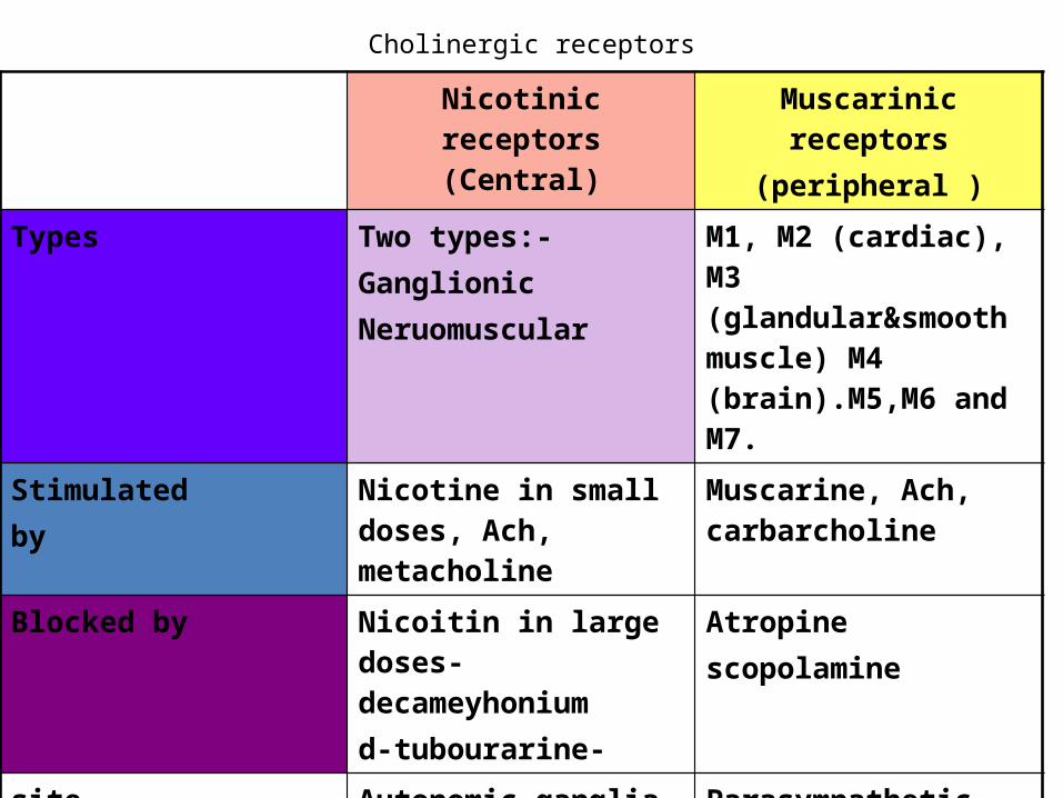
Cholinergic receptors
Nicotinic receptors(Central)
Muscarinic receptors(peripheral )
Types Two types:- GanglionicNeruomuscular
M1, M2 (cardiac), M3 (glandular&smooth muscle) M4 (brain).M5,M6 and M7.
Stimulatedby
Nicotine in small doses, Ach, metacholine
Muscarine, Ach, carbarcholine
Blocked by Nicoitin in large doses- decameyhoniumd-tubourarine-
Atropinescopolamine
site Autonomic ganglia M.E.PAdrenal medullaPreganglionic neuron.
Parasympathetic(pre-postganglionic) Sympathetic postganglionic nerve endings (sweat glands & skeletal muscle).
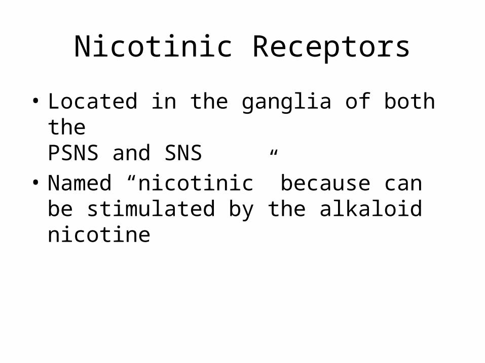
Nicotinic Receptors
• Located in the ganglia of both the PSNS and SNS
• Named “nicotinic” because can be stimulated by the alkaloid nicotine
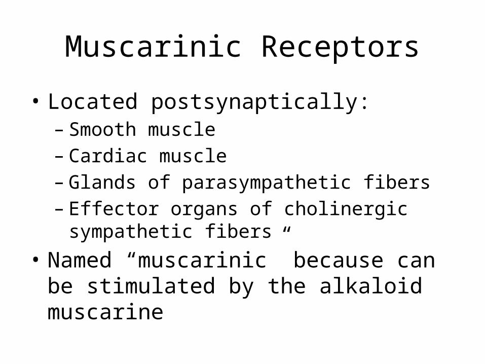
Muscarinic Receptors
• Located postsynaptically:– Smooth muscle– Cardiac muscle– Glands of parasympathetic fibers– Effector organs of cholinergic sympathetic fibers
• Named “muscarinic” because can be stimulated by the alkaloid muscarine
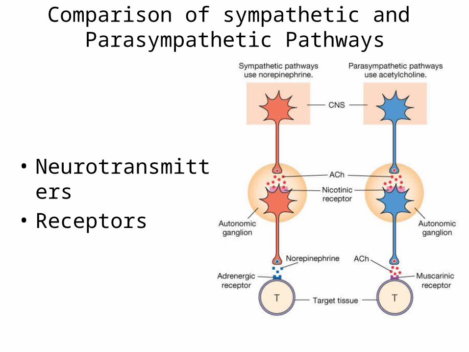
• Neurotransmitters• Receptors
Comparison of sympathetic and Parasympathetic Pathways