Embed Size (px)
Citation preview
LA rECHNIQUE DES ANTICORPS FLUORESCENTS
ET SES APPLICATIONS A LA MICROBIOLOGIE
par
M. MOURARET
OFFICE DE LA RECHERCHE SCIENTIFIQUE ET TECHNIQUE OUTRE -MER
Services Scientifiques Centraux - Bondy
1965
-1-
- INTRODUCTION
La technique des anticorps fluorescents a été élaborée en
1941 et 1942 par CaONS et al. dans le but d'obtenir le moyen de localiser les antigènes dans les tissus de mammifères. C'est seule
ment depuis une dizaine d'années qu'elle a commencé a être employée
dans l'étude des maladies infectieuses; elle a été par la suite de
plus en plus largement utilisée pour reconnaître les anticorps pro
duits par l'organisme et pour identifier et localiser les microorganismes pathogènes. Actuellement un très grand nombre d'études
concernent cette technique ou font nention de son application, si
bien qu'il est difficile de les signaler toutes. Un relevé aussicomplet que possible en a été tenté ici, à l'exclusion des études
intéressant les protozoaires, les nématodes et la localisation dans
les tissus des antigènes qui ne sont pas d'origine microbienne. Cerelevé bibliographique fait suite à un exposé de la technique, des
différentes méthodes d'utilisation et de quelques exemples d'appli-
cation.
Le principe de la technique consiste à fixer une substance
fluorescente sur des molécules d'anticorps sans altérer notablement
leur activité immunologique. Lorsque l'anticorps ainsi marqué est
mis en contact sur frottis ou sur coupe de tissus avec l'antigène
homologue, un8 réaction i~unologique a lieu et les molécules d'an
ticorps sont retenues par l'antigène, in situ. Vexposition ulté
rieure de la préparation aux radiations ultraviolettes sous le mi
croscope, provoque une emission lumineuse par lR substance fluorescente fixée sur l'anticorps, révélant ainsi le lieu où l'antigèneet l'anticorps sont combinés.
-2-
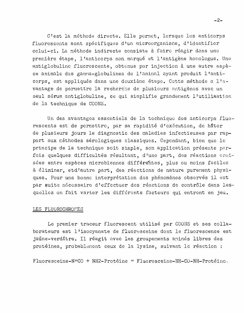
C'est la méthode directe. Elle permet, lorsque les anticorps
fluorescents sont spécifiques d'un microorganisme, d'identifier
celui-ci. La méthode indirecte consiste à faire réagir dans une
première étape, l'anticorps non marqué et l'antigène hO[1010gue. Une
antiglobuline fluorescente, obtEnue par injection à une autre espè
ce animale des gawna-glo bulines de l'animal ayant produit l' anti-·
corps, est appliquée dans une deuxième étape. Cette méthode a l'a
vantage de permettre la recherche de plusieurs antigènes avec un
seul sérum antiglobuline, ce qui simplifie grandement l'utiliaation
de la technique de COONS.
Un des avantages essentiels de la technique des anticorps fluo
rescents est de permettre, par sa rapidité d'exécution, de hâterde plusieurs jours le diagnostic des maladies infectieuses par rap
port aux méthodes sérologiques classiques. Cependant, bien que le
principe de la technique soit simple, son application présente pc?crfois quelques difficultés résultant, d'une part, des réactions croi
sées entre espèces microbiennes différifntes, plus ou moins faciles
à éliminer, etd'autre part, des réactions de nature purement physi
ques. Pour une bonne interprétation des phénomènes observés il est
par suite n6cessaire d'effectuer des réactions de contr61e dans les
quelles on fait varier les différents facteurs qui entrent en jeu.
LES FLUOROCHROIŒS
Le premier traceur fluorescent utilisé par COONS et ses collaborateurs est l'isocyanate de fluoresceine dont le fluorescence est
jaûne-verdâtre. Il réagit avec les groupements aminés libres des
protéines, probablement ceux de la lysine, suivant le réaction :
Fluoresceine-N=CO + NH2-Protéine = Fluoresceine-NH-CO-NH-Protéine,
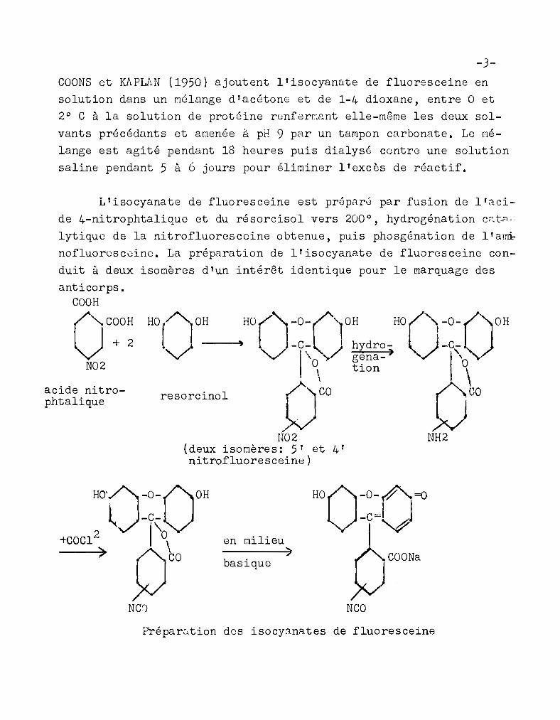
-3COONS et KAPLAN (1950) ajoutent l'isocyannte de fluoresceine en
solution dans un Mélange d'acétone et de 1-4 dioxane, entre 0 et
2° C à la solution de protéine renferClant elle-même les deux sol
vants précédants et amenée à pH 9 par un tampon carbonate. Le nélange est agité pendant 18 heures puis dialysG contre une solution
saline pendant 5 à 6 jours pour éliminer l'excès de réactif.
resorcinol
HO[loH HO:)-0-('10H HO('1-0-00H.... .Jo --~, l -c- / hydro- ) -c-'V \"'0\ ~i~~-) 1\\
/yo f1coW »fJ02 NH2
(deux isomères: 5' et 4'nitrofluoresceine)
L'isocyanate de fluoresceine est préparé par fusion de l'aci
de 4-nitrophtalique et du résorcisol vers 200°, hydrogénation c~t~·
lytique de la nitrofluoresceine 0 btenue, puis phosgénation de l' at;D,;.
nofluorusc0ine. La préparation de l'isocyanate de fluoresceine con
duit à deux isomères d'un intérêt identique pour le marquage des
anticorps.COOH
oc~o:N02
acide nitrophtalique
NG')
en r:lilieu)
basique
HoO-o~A=o-c-vCOONa
NCO
Préparution des isocyanates de fluoresceine
-4-
COONS et KAPLAN (1950) préconisent cependant la séparation de cesisomères dans le but d'élininer les inpuretés. La séparation a lieuau stade de la préparation où est obtenue la nitrofluoresceine.Celle-ci est transform6e en diacétate puis soumise à une cristallisation fractionnée. REPENTIGNY et Jid::ES (1954) procèdent à la purification au stade aminofluoresceine, par séparation sur colonne deKieselguhr, méthode qui permet de limiter les pertes de produit.
L'isocyanate de fluor~sceine étant instable doit être préparé avant chaque utilisation. Cependant caONS (1956) rapporte qued'après les observations de I~RSW.LL (1951), l'isocyanate de fluores ceine en so lut ion dans l'acétone conserve son activit(, pendant
au moins un an lorsqu'il est protégé de la lumière de la chaleuret des moisissures. Une importante ar:lélioration a par la suite été
apportée par GOLDHAN et Cf.RVER (1957) à la technique de narquagedes anticorps par l'isocyanate de fluoresceine. Ces auteurs procèdent à l'imprégnation d'un papier pour chromatographie par une solution d'isocyanate de fluoresceine dans un mélange de dioxane et
d'acétone, puis sèchent le papier sous ventilateur. La préparationainsi obtenue, dont on connait exactement la teneur en isocyanate,peut être conservée dans un dessicateur au-delà de sept mois. Aumoment de l'emploi on fait réagir dans un bain glacé la solution deprotéine à pH 9,0 et une certaine quantité de papier imprégné. La
présence de solvant organique n'est plus ici nécessaire ce qui limite la dénaturation des protéines.
L'instabilité de l'isocyanate de fluoresceine a amené l'abandon de ce produit au bénéfice de l'isothiocyanate proposé par RIGGSet al. (1958). La préparation des deux composés diffère seulementdans sa phase finale. L'isothiocyanate est obtenu par action duthiophosgèn~ sur le diacétate d'ar1inofluoresceine. Le thiophosgène
liquide est d'un emploi plus pratique que le phosgène gazeux. L'isothiocyanate a l'avantage d'être stable a l'état solide; en outre,
acide 1-diméthylaminonaphtalène-5-sulfonique
-5-
COl!lme l'ont montré IU.RSHALL et al. (1958), il pernet la fixation de
fluoresceine en l'absence de solvant organique. Le r~rquage est ob
tenu par addition progressive d'isothiocyanate en poudre à la solu
tion de protéine.
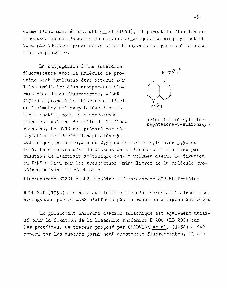
La conjugaison d'une substance
fluorescente avec la molécule de protéine peut égale~ent être obtenue par
l'interr!lédiaire d'un groupement chlo
rure d'acide du fluorochromc. HEBER
(1952) a proposé le chlorure de l'aci
de 1-diméthylaminonnphtalène-5-sulfo
nique (D;~NS), doht la fluorescence
jaune est voisine de celle de la fluo
resseine. Le DANS est préparé par C"16
thylation de l'acide 1-naphtalène-5-
sulfonique, puis broyage de 2,5g du dérivé r.1éthylô avec 3,5g de
rC15. Le chlorure d'acide dissous dans l'acétone cristallise pardilution de l'extrait 2.c6tonique dans 6 volumes d'eau. La fixationdu DANS a lieu par les groupeuents amine libres de la r.101écule pro
téique suivant la réaction :
Fluoro8hrome-S02Cl + NH2-Protéine = Fluorochrorne-S02-NH-Protéine
REDETZKI (1958) a ~ontré que le marquage d'un sérum anti-alcool-des
hydrogénase par le DANS n'affecte pas la réaction antigène-anticorps
Le groupement chlorure d'acide sulfonique est également utili
sé pour :a fixation de la lissamine rhodamine B 200 (RB 200) sur
les protéines. Ce traceur proposé par CHAD\lICK et al. (1958) a étéretenu par les auteurs parmi neuf substances fluorescentes. Il Grlet
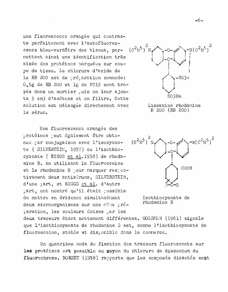
une fluorescence orangée qui contraste parfaitement avec l'autofluorescence bleu-verdâtre des tissus, permettant ainsi une identification trèsaisée des protéines marquées sur cou
pe de tissu. Le chlorure d'acide dela RB 200 est de ~ré~aration comDode:
0,5g de RB 200 et 19 de PC15 sont bro
yés dans un mortier ~uis on leur ajoute 5 cm) d'acétone et on filtre. Cettesolution est mélang8e directement avec
le sérum.
-6-
Lissamine rhodamineB 200 (RB 200)
Isothiocyanate derhodamine B
Une fluorescence orangée des
~rotéines ~ eut également être obtenue rar conjugaison avec l'isocyana
te ( SILVERTEIN, 1957) ou l'isothiocyanate ( RIGGS et al.1958) de rhodamine B. En utilisant la fluoresceine
et la rhodamine B :,our rilarquer res:-octivement deux antis8rums, SILVERSTEIN,d'une ~art, et RIGGS et al. d'autre:.art, ont Dontré qu'il étai t ~ossible
de r.J.ettre en évidence simultanG[1entdeux microorganismes sur une r:Cl1e lr6
l~aration, les couleurs émises l~,ar lesdeux traceurs étant nettement différentes. GOLDrfuN (1961) signale
que l'isothiocyanate de rhodamine B est, cOr.J.me l'isothiocyanate de
fluoresceine, stable et disionible dans le commerce.
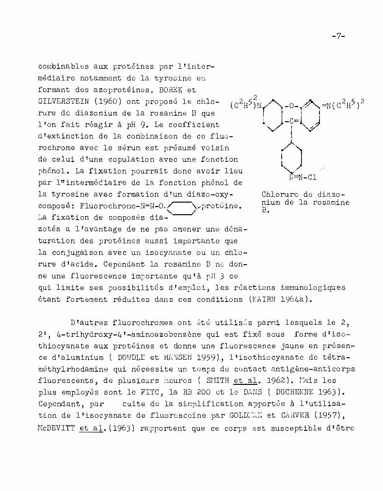
Un quatrième mode de fixation des traceurs fluorescents surles protéines est possible au woyen du chlorure de diazonium du
fluorochrome. BORDET (1958) rapporte que les composés diazotés so~t
-7-
Chlorure de diazonium de la rosamineB.
immunologiques
1964a) •
rochrome avec le sérum est présumé voisin
de celui d'une copulation avec une fonction
phénol. La fixation pourrait donc avoir lieupar lit interméè.iaire de la fonction phénol de
la tyrosine avec formation d'un diazo-oxy
composé: Fluorochrorne-N=N-0".r-'\ ....prot6ine.. " /La fixation de composés dia-
zotés a l'avantage de ne pas amener une déna
turation des protéines aussi importante que
la conjugaison avec un isocyanate ou un chlo
rure d'acide. Cependant la rosamine B n0 don
ne une fluorescence imrortante qu'à pH 3 ce
qui limite ses possibilités d'emploi, les réactions
étant fortement réduites dans ces conditions (NAIRN
combinables aux protéines par l'inter
médiaire notamment de la tyrosine en
formant des azoprotéines. BOFŒK et
SILVERSTEIN (1960) ont proposé le chlo
rure de diazonium de la rosamine B quel'on fait réagir à pH 9. Le coefficientd'extinction de la combinaison de ce fluo-
D'autres fluorochromes ont été utilises parmi lesquels le 2,
2', 4-trihydroxy-4'-aminoazobenzène qui est fixé sous forme d'isothiocyanate aux protéines et donne une fluorescence jaune en présence d'aluminium ( DOWDLE et Hf.NSEN 1959), l'isothiocyanate de tétra
mèthylrhodamine qui nécessite un t(~mps de cuntact antigène-anticorpsfluorescents, de plusieurs heures ( SHITH et al. 1962). r~ais les
plus employés sont le FITC, la RB 200 et le DAnS ( DUC HENNE 1963).
Cependant, par cuite de la simplification apport8e à l'utilisa
tion de l' isocyanate de fluorosccine par GOLE '.:.:J et CARVER (1957),
r-TcDEVITT et al. (1963) rapportent que ce corps est susceptible d'être
-8-
encore employé, l'intensité de fluorescence des molécules de flua··resceine fixée par l'isocyanate Gtant supérieure à celle des molé
cules fixées par l'isothiocyanate.
LE rS\RQŒ.GE DES ANTICORPS
Toutes les techniques employées ont en commun avec celle de
COONS, l'addition progressive de fluorochrome à la solution de
globulines amenée à pH 9 et refroidie entre 0° et 2° C. Le mélangeest maintenu en agitation à cette température pendant plusieurs
heures. ;\.ux modifications concernant le fluorochrome et son mode de
fixation s ' ajoutent des pe rfectionnerJents apportés à la purifica-tion effectuée après la conjugaison et destinée à suppri~er les
réactions non spécifiques.
Le sérum utilisé doit avoir une teneur élevée en anticorps.
L'immunisation est effe ctuée par .Ane rréparation ct' antigènes aussipure que possible, au moins pour la première injection. Un adjuvant
( alun ou émulsion de Iijcobactéries àans l'huile) est habituelle
ment utilisé avec les protéines solubbs (COOIJS 1958). Le sérum est
préalablement débarrassé des albumines pour éviter la dilution des
anticorps fluorescents à2ns une grande quantité de protéines inac
tives. COONS (195$) préconise une floculation des globulines à 4°C,
par le sulfate d'amnonium à de~i-saturation ajouté progressivement
au séru.m. Le précipité est centrifubé, lavé par du sulfate d'awnanium à demi-saturation puis dissous dans une solution saline tampon
née. L'ion arrl.l:lonium est ensuite éliminé par dialyse contre une sol,,
tion il 0, 85~; de CINa car il interfère avec la réaction, pro bable
ment en entrant en compétition avec les protéines pour l'isothio
cyanate de fluoresceine. KAUFAr~J et CHERRY (1961) rapportent en effet que le titre de color[~tion d'un sérum anti-Brucella conjugué en
-9-
présence d'une concentration 0,04 r: en sulfate d' amr.10nium, estquatre fois plus faible que celle du sérum marqué en l'absence de
de sel. RIGGS et al.(195b) procèdent à trois précipitations par le
sulfate d'ammoniurl à demi-saturation pour le Marquage des anticorpspar l'isothiocyanate de fluoresceine. Lorsque le marquage est effec
tué par le chlorure d'acide de la RB 200, il est préférable de pro
céder à la floculation des globulines après la conjugaison, la dé
termination du titre d'anticorps ne pouvant être effectuée que sur
le sérum entier ( CHAmnCK et al. 1058). La perte de colorant qui
en résulte n'est pas un inconvénient car celui-ci est bon marché.
Des techniques de conjugaison pour les trois rrincipaux fluo
rochromes ont été proposés par NAIRN (1964a): Le sérum est amené
à une concentration de 50 à 60 mg de protéine par cm); il est tam
ponné à pH 9,0 par addition de deux volumes de tampon carbonatebicarbonate de concentration 0,51'1 et maintenu entre 0 0 et 2 0 C. Le
FITC est ajouté à raison de ) mg par cm) de sérum sous forme de
poudre ou en solution dans le tampon ou dans un peu d'acétone,danslequel il est beaucoup plus soluble que l'isocyanate (RIGGS et al.
1958). L1agitation est poursuivie pendant une nuit. Le chlorure
d'acide de la RB 200 préparé à partir de 0,5 g de fluorochrome et
19 de PC15, en solution dans 5 cm) d'acétone est ajouté à raison
de 0,1 cm) par cm) de sérum. L'agitation est poursuivie pendant30 minutes. Le DAnS est employé dissous dans le minimum d'acétone
dans la proportion de 3 mg par cm3 de sérum. L'agitation est pour
suivie pendant 6 heures.
D'après t~RSHALL et al.(195a), en l'absence de solvant orga
nique la dénaturation des protéines est peu importante. Ces auteurs
ont montré que l'addition d'isothiocyanate de fluoresceine en pou
dre ou bien d'isocyanate ou d'isothiocyanate de fluoresceine fixés
sur papier suivant la technique de GOLmJ1.N et CARVER (1957), ne
provoquait pas de modification notable du titre d'agglutination de
-10-
six antisérums différents par rapport aux fractions non marquées.
Par contre le marquage par l'isothiocyanate et surtout l'isocyanate
en présence de solvants organiques amenait une diminution parfois
très im1iortante du titre d'agglutination. L'addition de fluorochro~e en l'absence de solvant organique est effectuée par RINDERKNECHT
(1960,1962) suivant une technique qui rappelle celle de GOLDI::J\N et
Cl\.RVER : le FITC et les chlorures d'acide du Df',NS et de la RB 200
sont absorbés sur terre de Diato~écs ( Célite ). Le fluorochrome
étant sous forme très divisée, sa fixation sur les protéines est
très rapide; 30 minut83 suffisent.
GOLDSTEIN et al.(1961) rapportent qu'une purificationdes globulines sur colonne de DE~E-cellulose, préalablement à laconjugaison avec l'isothiocyanate de fluoresceine n'amène pas une
diminution de la fluorescence non spécifique observée sur impressi')ns de ra te de cobaye colorées par la [;1(~thode indirecte. La fl1l0
res~ence non spécifique était en relation avec la teneur des pro
téines en fluoresceino; elle augmentait avec celle-ci. Il est maintenan~ êtabli que la fixation de fluorochrome est hétérogène et
que ce sont les protéines excessivement marquées qui sont responsa
bles ~es colorations non spécifiques. CURTAIN (1958) a séparé parélectrcphorèse différentes fractions de~2-g1obulines ayant fixé
des quantités croissantes de fluoresceine et signale que les colo
ratione non spGcifiques sont d'autant plus intenses que la teneur
des gloJulines en fluoresceine est plus grande.
La disparition du groupement amine libre ayant réagi avec le
fluorochrome et habituellement chargé positivement au-dessous de
pH 10 et l'introduction du groupement carboxylique libre de la fluoresceine chargé négativement au-dessus de pH 5, entraînent une
augmentation de deux unités de charge négative de la protéine par
molécule de fluoresceine fixée entre pH 5 et pH 10 (NAIRN 1964a).
-11-
Alors que les globulines de chèvre anti-lapin migrent vers la catho
de à pH 8:6, elles se déplacent vers l'anode après fixation de fluorescei ne : GOLDSTEIN .§l.:L a~. 1961). D'après NAIlli! (1964a) à pH 7 lesprotéines du sérum ont une charge négative qui se trouve accrue par
la fixavion de i'luoi.'ochrome si bien que les protéines se comportentcomme des colorants acides qui réagissent avec les tissus positivement chargés en donnant dos colorations non spécifiques. L'auteur
rapporto une étude de I~YERSBACH et SCHUBERT (1960) d'après laquelle par élévation du pH les tissus deviennent négativement chargés
et les colorations non spécifiques sont réduites.
GRIFFIN et ~1.(1961) sont parvenus à réduire les fluorescences non spécifiques en déterminant la durée de la réaction de conjugaison et la quantité minimum de FITC nécessaire pour obtenir untitre de coloration spécifique maxiQum et un titre de coloration
non spécifique mimirnum. Les conditions retenues pour le sérum antiStreptocoque A sont: un rapport FITC sur sérum de 1/40 et une durée
de réaction de une heure ou plus. Ce résultat a pu être obtenu avec
du FITC chromatographiquement pur. La teneur en isothiocyanate de
fluoresceine des préparations non purifiées de ce corps varient de
20 à 60% ce qui rend difficile le calcul de la quantité de colorantqui doi t ê~re employé pour la conjugaison ( FELTON et rULLON, 1961).
LTaddi~ion progressive de fluorochrome au sérum a pour butd'amener une fixation assez homogène et par suite de limiter lescolorations non spécifiques par les fractions excessivement marquées. Pour la recherche des antigènes viraux dans les tissus,CLARK et SHEFILR[ (1963) préconisent le marquage des anticorps par
diffusion de l'isothiocyanate de fluoresceine à travers une membrane à dialyse., La solution de ~"-globulines purifiée sur colonne deDEAE-cellulose, ajustée à 1% dans un tampon carbonate-bicarbonate
de concentration 0 )025H est placée en tube à dialyse dans 10 volu",mes de solution à 0)01% de FITC dans le même tampon. La réaction
-12-
est poursuivie pendant 24 heures à 4° C sur agitateur magnétique.
La spécificité des anticorps ainsi obtenus est bien supérieure à
celle des anticorps marqués par addition de FITC en poudre à la
même solution de~'-globulines pures suivio ct JunG 8.bsorp'tion 'stir
poudr.~ de tissu.
L'excès de fluoresceine fixée par les anticorps pourrait nepas être la seule cause de fluorescence non spécifique: la fraction
" 19 S " des globulines est également susceptible d'intervenir (Kc
DEVITT et al. 1963). HOLF et al. (1963) préconisent un fractionnement des globulines sur colonne de diéthylaminoéthanol-Sephadex A
50 préalablement à la conjugaison, pour le marquage des antiglobu
lines utilisées dans la Méthode indirecte. La fraction" 7 S " éluÉe
en premier lieu est conjuguée avec l'isothiocyanate de fluoresceine.
On évite ainsi, lors de la localisation des antigènes dans les tis
sus, les fluorescences non spécifiques qui sont dues à la fraction
" 19 S "
PURIFICATION DES ANTICORPS FLUORESCENTS
A l'issue de la conjugaison du fluorochrome avec les anticorps
des substances fluorescentes apportées par la matière colorante em
ployée ou provenant de l'hydrolyse du fluorochrome ou formées au
cours de la réaction ( composé YlBn de CHAmnCK et NAIRN 1960), sont
présentes dans la préparation. Ces substances désignées par U.F.E.
(unreacted fluorescent material) sont adsorbées sur les protéines
du sérum. Elles sont responsables de colorations non spécifiques et
doivent être éliminées. Les anticorps marqués par le FITC pur ren
ferment moins d' U•F. T,l. et sont plus faciles à purifier que les an
ticorps fluorescents obtenus avec certains échantillons commerciaux
de FITC impur ( NAIRN, 1964a). Des fluorescences non spécifiques
résultent égalen;ent de la fixation d'une quantité excessive de fluorochrome sur les anticorps. La localisation sur coupe de tissu ou
-13-
impressions de tissu sur lame, de certaines particules virales etd'antigènes solubles qui sont présents à concentration faible et
variable est rendue difficile par lGS fluorescences non spécifiques.
Par contre les bactéries et d'autres types de part':"culGs virales
peuvent être localisées assez facilewent par les anticorps fluores
cents ( GOLDSTEIN et al. 1961). La ~éthode de purification employéepar COONS et 10\.PL1·;JJ (1950) comporte une dialyse suivie d'une agita
tion du sérum avec du foie broyé, séché à l'acétone ou lyophilisé.
L'absorption sur poudre de tissu entraîne une dir:1inution de la teneur eH protéine et fluorcsceine et du rapport fluoresceine sur
protéine. Au-delà de deux absorptions il n'y a plus de réduction
importante de la teneur en protéine et fluoresceine ( GOLDWASSER et
SHEPARD 1958). DI~JEEN et lill!. (1957) ont proposé une extraction par
l'acétate d'éthyle suivie d'une absorption sur tissu.
CHAmaCK et al. (1959) rapportent qu'une purification par la
méthode de CaONS et KicPLAN enlève seuler,1ent 65 5~ de l 'U.F .11. de
sérums marqués par l'isocyanate de fluorcsceine. Une absorption ultérieure par le charbon activé en éliminait encore 33%. FOTHERGILL
et Ni~IRN (1961) procèdent à l'extraction de l'U.F.n. par la poudrede charbon activé, agité pendant 1 heure avec le sérum fluorescent.
Le rapport fluorochro~e sur protéine diminue puis tend vers unevaleur constante lorsque la quantité de charbon crôit. La quantité
minimum d'absorbant nécessaire est respectiver:J.ent de 15 mg et 75 mg
/~r ç~ • .~e serum marque par la RB 200 et l'isothiocyanate de fluorescelne.
Une diminution de la teneur en protéines en résulte; elle est de 5à 101~ dans le preoier cas et de 30 à 405; dans le deuxième cas. La
perte importante de protéines ayant lieu avec les sérums marqués
par le FITC conduit à adopter de préférence, pour ces sérums, la
purification sur Sephadex.
-14-
GOLDSTEIN et al. ( 1960) puis ZUAfll'J et DfdI (1961) ont proposé
l'utilisation dTune colonne de gel de Sephadex ( polydextrane )
pour la purification des anticorps fluorescents. Ce matériel présen··
te des pores dans lesquels ne peuvent diffuse~ que les petites mo10'
cules. Il en résulte une vitesse de deplacement plus réduite pour
celles-ci que pour les grandes molécules de protéine ce qui permetleur séparation. La colonne est lavée par une solution saline tampon
née à pH 7 puis imprégnée de sérum fluorescent. La même solution
déplace d'abord les anticorps fluorescents q;li sont ramenés à pH 7et dilués, puis l'U.F .It. Cette opération der:1ande seulement 10 à 15
minutes alors que par dialyse 5 à 6 jours étaient nécessaires; maisles colorations non spécifiques ne sont pas moins inportantes qu'en
procédant par dialyse ( GOLDSTEIN et al.1961). KILLANDER et al. (1961)ont montré que le pouvoir colorant des antisérums R.insi purifiésétait analogue à celui des antisérums dialysés. La perte de pro~C;np.
est négligeable. La purification des anticorps narqués par le DANS
est conduite en lumière UV, à lTobscurité, leur faible coloration
ne permettant pas de suivre leur passage. GORDON et al. (1962) uti
lisent une colonne de Sephadex conjointemont à la purification du
sérum par floculation au moyen dTEthodine (6,9-diamino-2-ethoxyacri
dine) suivie d'une précipitation des globulines par lTacétone. L'Etho
dine rfsiduelle est retenue sur la colonne.
Après élimination de ITU.F.H. puis absorption éventuelle sur
poudre de tissu, les anticorps fluorescents peuvent être concentrésau moyen dTune précipitation par le sulfate dTammonium à 40% suivie
dTune dissolution dans une solution s~line tamponnée. Les anticorps
sont prêts à l'emploi après dialyse d'une müt (N1I.IRN 1964a).
Des sérums fluorescents ne donnant que de faibles colorations
non spécifiques et qui, par suite, ne nécessitent pas d'absorption
sur poudre de tissu, ont été préparés par purification sur colonne
-15-
de diéthyla~ino-éthyl DEAE-cellulose (RIGGS et al. 1960). 1e sérum
entier est conjugué avec le FITC, dialysé pendant une nuit contre
un tampon phosphate de pH 6,3 et de concentration 0,0175T1 puis introduit dans la colonne équilibrée avec le ~êP1e tampon. L' élution
est effectuée par le tampon contenant des quantités croissantes de
CINa. Dans le cas de séruns antiviraux de singe et furet seules les
fractions éluées par le tampon en l'absence de CINa avaient un ti
tre de coloration élevé. Par contre pour des sérums antibactériens
de lapin, les fractions correspondant aux concentrations 0,075N et0,125M de CINa avaient le titre le plus élevé. Les fractions éluées
au-delà de la concentration 0, 15H donnent d'importantes colorationsnon spécifiques. CURTI,n~ (1961) a séparé sur colonne de DElŒ-cellu
lose des gamma-globulines de lapin anti-albumine humaine dont le
titre d'anticorps est d'autant plus élevé, et qui donnent des colorations non spécifiques d'autant plus réduites, qu'elles ont été
éluées plus rapidement.
GOLDSTEIN et al. (1961) ont montré que l'addition d'une quan
tité trop élevée de FITC au sérum entraîne une fixation excessive
de fluorochrome qui interdit la séparation sur colonne de DEAE-cel
lulose de fractions ne donnant pas de coloration non spécifique.
Lorsque 6 à 8 mg de FITC cristallisé étaient ajoutés par g de pro
téine, il était possible d'isoler des globulines fluorescentes dont
le rapport fluoresceine sur protéine se situait entre 2,0 et 3,5 X
10-3 • Ces fractions ne donnaient pas de fluorescence non spécifique
lorsqu'elles étaient utilisées à une concentration de 1mg de proté
ine par cm3; au-delà de 2 r'1g une fluorescence non spécifique était
OCSGrT c.
NcDEV=TT et al.(1963) rapportent égalerllent que la fixationd'une quanti~é excessive de fluorochrome sur la fraction globuline
du sérum ccnjuguée avec le FITC chromatographiquement pur entraîne
-16-
une perte très importante d'anticorps lors du fractionnement ulté
rieur sur colonne de DEAE-cellulose. Une proportion importante des
anticorps se trouve alors associée à des colorations non spécifi
ques dans la fraction éluée par une solution normale de CHJa.
Par passage des globulines fluorescentes sur colonne de DEAEcellulose il est possible d'éliminer non seulenent les fractions
ayant fixé une trop grande quantité de fluorochromes, responsables
de fluorescences non spécifiques, mais également les fractions in
suffisamment marquées qui inhibent la fluorescence spécifique
( GOLDSTEIN et al. 1962 ). Ces dernières renfermaient 35~ de la to
talité des globulines lorsque le marquage était effectué avec 8 mg
de FITC par g de protéines employées â la concentration 2~. Il
atteignait 80~o lorsque la concentration des protéines était ramenée
â l~o avant la conjugaison.
La purification sur colonne de DE~E-cellulose a également été
employée pour supprimer les réactions croisées entre plusieurs sou
ches de virus de la grippe. La conjugaison devait être effectuée
ici sur la fraction globuline du sérum ( FROH;'11AGEN et r1ARTINS 1<;(3)
Toutefois en procédant au marquage de la totalité du sérum les ré
actions croisées pouvaient être évitées par élimination des proté
ines indésirables au moyen d'une floculation par le Rivanol ( lactate de 2-ethoxy-6,9-diaminoacridine) suivie d'une floaulation des
globulines par le sulfate dl ammonium â 40;~ de la saturation et en
fin passage sur colonne de Sephadex.
-17-
PROPRIETES DES PROTEINES COIJJUGUEES AVEC UN FLUOROCHROIffi.
L'intensité de fluorescence des anticorps conjugués avec un
fluorochrome est très inférieure à celle du fluorochrome libre.
La diminution d'intensité de fluorescence serait due à une inté
raction entre le fluorochrome et la protéine. Elle s'accentue avec
le degré de conjugaison et au-delà d'une valeur appelée degré optimum de conjugaison, l'augmentation du nombre de molécules de
fluorochrome fixée n'augmente plus la fluorescence de la protéine
(NfI.IRN 1964a).
r:cDEVITT et al. (1963) signalent d'autre part que le mode de
fixation du fluorochrome influe sur la fluorescence. Le pourcenta
ge d'''efficacité d'emission ti d'antitoxines diphtériques de cheval
marquées par la fluoresceine, déterniné comparativement à des so
lutions de fluoresceine, était respectivement de 14% et 36% lorsque la conjugaison était effectuée avec l'isothiocyanate et avec
l'isocyanate de fluoresceine. Ces résultats étaient obtenus pour
des rapports fluoresceine sur protéine compris entre des limites
assez importantes qui n'influaient pas sur le pourcentage d'''effi
cacité d'emission". Pour les protéines conjuguées avec la RB 200
la fluorescence est égale à 30~ de l'intensité observée à l'état
libre (NAIlliJ 1964a).
La quantité de fluorochrome fixé est déterminée par mesure
de la densité optique, effectuée à la longueur d'onde du maximum
d'absorption du colorant: 575 mfPour la RB 200, 495 mf pour leFITC et 340 m~ pour le DANS. (NAIRN 1964a). La concentration enprotéine est mesurée par micro-Kjeldahl ou par détermination de
la différence de densité optique entre le biuret seul et le mé
lange biuret-protéine ( GOLD\JASSER et SHEPARD 1958) ou bien encore
-18-
par la densité optique à 280 m (FRorTI:HhGEN et T:ARTINS 1963). Les
valeurs obtenues pernettent de calculer le rapport fluorochrome
sur p:otéine du sérum fluorescent. La conr~aissance de ce rapport
est inportante car il conditionne les propriétés de la préparation:fluorescences non spécifiques s'il e~~ ~rop élevé, fluorescence
non décelable s:il est-trop faible.
îJAIF..N (1964a) rapporte que la fixàtion de la RB 200 sur les
protéines fait apparaitre une fluorescence dans le rouge qui s'a
joute à la fluorescence jaune orangée du fluorochrome libre. Unchangement de couleur par disparition des plus grandes longueurs
d'onde de fluorescence a lieu au cours de fortes expositions aux
U.V., des 2nticorps conjugués avec la RB 200 et le DANS. Il tra
duirait une décomposition photochimique des anticorps fluorescents
avec libération du fluorochrome que l'auteur a mis en évidence
par filtration sur gel. Aucun changement de couleur n'est observé
avec les anticorps marqués par le FITC dont la couleur de fluores
cence est id8ntique à celle du colorant libre. La lumière émisepar des préparations de microorganismes colorés par des anticorps
conjugués avec le DANS blanchit au cours de l'exposition aux U.V.,
ce qui permet de les différencier des colorations non spécifiques,
lesquelles n'ont pas cette propriété ( \JOLOCHO\'J, 1959). Il se pro
duit en outre une diminution d'intensité de la fluorescence des
préparations 'iWccours dj..}'exposit'!on prol~ée aux U.V.(~lit~,
:'-1964a ) •
REPEN'l'ImrY et SONE!" \1961) ont "'montré qu tau cours de trois
minutes d'exposition aux U.V. la variation d'intensité de fluores
cence des microorganismes ayant fixé un anticorps fluorescent,
n'est que partiellement attribuable auf'luorochrome. Elle résulte,
d'une part, d'un changement d'intensité de la fluorescence primai
re du nicroorganismes, laquelle s'accroit pour Corynebacterium
-19-
diphteriae et diminue pour Staphylococcus aureus et, d'autre part,
d'une augmentation d'intensité de la fluorescence secondaire ap
portée par l'anticorps conjugué avec l'isothiocyanate de fluores
ceine. Contrairement à ce dernier, l'isothiocyanate de rhodamine
B n'entraîne pas de variation de fluorescence secondaire au cours
d'une exposition de trois Qin~tes aux U.V.
Les anticorps fluorescents sont très généralement employés en
milieu neutre. Cependant WlIRN (1964a) rapporte que l'intensitéde fluorescence des anticorps narqués par la HE 200 est deux fois
plus intense à pH 4,0 et pH 10,5 qu'au voisinage de la neutralité,
bien que la fluorescence du colorant libre soit peu affectée par
le pH. Quant aux anticorps conjugués avec la fluoresceine, l'in
tensité de fluorescence est la plus élevée à pH 8,0. VJHJTER et
ITIODY (1959b) n'observent pas d'influence du pH, entre les valeurs
5,5 et 7,5, sur la fluorescence de préparations de Pasteurella
pestis colorées par des globulines conjuguées avec l'isocyanate de
fluoresceine.
Les anticorps marqués par un fluorochrome peuvent être con
servés pendant de nombreux mois sans r:loè.ification de leurs propri
étés. Un sérum anti-Pasteurella pestis conjugué avec l'isocyanate
de fluoresceine, conservé pendant un an entre 0 et 50 C ou bien
à l'état congelé ou lyophilisé ne présentait pas d'altérntion du
titre d'agglutination. Seul le titre de coloration était plus
réduit dnns le deuxième mode de conservation (\lINTER et HOODY,
1959b). Un stocknge de 14 mois entre 0 et 4° C ou à -20 0 C nediminuait pas le pouvoir colorant d'un sérum anti-Histoplasma
capsulatum conjugué avec l'isocyanate de fluorosceine (GORDON,1959)
-20-
Des séruQs conjugués avec la III 200 avaient conservé leurs proprié
tés après b mois de stockage entre -15 et _20 0 C ( CHAmJICK et al.
1958). Il suffisait de procéder préalablement à leur utilisation
à une absorption sur charbon ou poudre de tissu pour éliminer le
fluorochrome libéré.
La conjugaison des sérums avec un fluorochrome introduit quel
ques modifications de leurs propriétés. Une altération consécutive
à la fixation de fluoresceine a été mise en évidence par SONEA et
REPENTIGNY (1961) au moyen de la technique de diffusion sur gélose:
des lignes secondaires n'étdient plus observées après le couplage.
D'autre part J·I00DY et al. (1961) rapportent que le titre d'aggluti
nation de sérums anti-Brucella est deux à quatre fois plus faible
après fixation d'isothiocyanate de fluor8sceine. C~tte diminution
est plus ou moins i~portante suivant la technique utilisée (IARSHALL et al. 1958). Cos nodifications de propriété résultant de la
conjugaison n'ont cependant qu'une importance relative, les réac
tions par imnunofluorescence présentant quelqu8 différence avec
les réactions sérologiques babituelles. En effet, THOI~SON et al.(1957) rapportent que les réactions sérologiques croisées sont
plus fréquentes par la technique des anticorps fluorescents que
par les méthodes sérologiques classiques. Ce desaccord résulte de
ce que la simple combinaison de l'anticorps fluorescent avec une
cellule microbienne constitue un test positif alors que les tests
d'agglutination et précipitation nécessitent une seconde étapedans laquelle les particules élémentaires de complexe antigène
anticorps se réunissent en agrégat. La fréquence des désaccords
serait liée à celle du phénomène de "prozone" régissant l'aggluti
nation.
-21-
LES lIETHODES DI UTILISi,TIO~~ DES f.lITICOHPS FLUOHESCEl !TS_..--..~-.----.------ ---
Les préparations devant recevoir les anticorps fluorescentssont préalablement fixées. Pour les étalements de microorganismes
sur lar.le) on pro cede le plus généralement par chauffage. CHf.mnCK
et SLADE (1960) disposent les préparations pendant 30 secondes sur
plaque chauffé entre 60 et 65 0 C. 1'100DY et al. (1956) n'ont pasobs~~vé de différence significative de fluorescence entre des pré
parations de I~lleomyces Bseudonallei fixées par la chaleur, le
méthanol ou le formol. Des préparations de Pasteure~la pestis colorées par 11antisérum homologue de l'antigène d'enveloppe présen
taient une intensité de fluorescence satisfaisante après fix8ticn
par la chaleur, le dioxane ou le fOrr.l01 à 0,5;;, faible a près fixation par l f éthanol ou le r.lethanol et nulle après action du formol
à 10);; ( vHHTER et IIOODY 1959b). LaI3l1EC et aL(1959) ont retenu
pour des étalenents de Shigell~ flex_ner~, le traitement par le ITl2
thanol absolu de préference à la fixation par la chaleur, laquelle
amène une fluorescence du fond résultant probablement de l'extrac
tion de matériel antigénique soluble. DOrJ!,LDSON et vJHIT1~KER (1960)
fixent par l'acétone des frottis de Bordetella~rtussis obtenus
de prélèvements effectués dans les voies respiratoires.
La fL:ation des virus sur tissu est généralement effectuée
par 1 1acétone 0 HIlJŒIA. et HDrJ=ELEH (1961) fixent le virus de lapoliomyélite developpé sur culture de tissu, par traitement de
10 minutes dans l'acétone à la tenpérature ambiante. GOLDIJASSER et
KISSLING (1958) procèdent par irlf'lersion de 18 à 24 heures dansl'acétone entre -15 et _20 0 C, d'impressions de tissu infecté par
le virus rabique. Un traitement de 15 minutes dans l'alcool absolu
est employé par NAGARA.J (1965) pour des coupes de tige et feuille
infectées par le virus de la nosaïque du tabac.
-22-
I~thode dirüctu.
C'est la plus simple. Le sérum fluorescent est appliqué pen
dant 30 minutes en atmosphère hunide sur la préparation diantigene.
Celle-ci est ensuite lavée par une solution saline tamponnée â pH
7,0. 3i le sérum conjugué contient un anticorps spécifique d'un
antigène de la préparation, une combinaison a lieu et le complexe
antig~ne-anticorps formé est observable en lumière U.V. 3inonl'anticorps est éliminé par le lavage et il n'y a pas de fluores-
cence.
Les contrôles suivants doivent être effectués: l'utilisa
tion de globulines normales fluorescentes ne doit pas entraîner
de fluorescence de la préparation. Le sérum spécifique non marqué)
appliqué préalablement au sérum fluorescent doit inhiber la fluo
rescence en empêchant la formation du complexe antigène-anticorps
fluorescent.
Pour une raison inconnue, ce dernier test proposé par COONS
et KAPLAN (1950) n'est pas toujours efficace. tUODY et al. (1956)lui ont substitué l'inhibition en une étape effectuée par addition
simultanée d'antisérum non marqué et du même antisérum fluorescent.
Ce test a par la suite été developpé par GOLDI~N (1957) pour la
recherche d'anticorps dans un sérun inconnu non marqué. Ce dernier
est ajouté â une égale quantité de sérum fluorescent connu et le
mélange est appliqué pendant 1 heure â 37° C sur une préparationde l'antigène homologue. L'absence ou la réduction de fluorescencepar rapport â une préparation de l'antigène ayant reçu seulement
l'anticorps fluorescent traduit la présence, dans le sérum inconnu,
d'anticorps spécifique de l'antigène.
-23-
L'identification des nicroorganismes par la technique des
anticorps fluorescents est fréquemnent rendue nalaisée par lesréactions croisées ayant lieu avec des microorganismes hétérolo
gues. THOIlh.SOI~ et al. (1959) ne sont pas parvenus à identifier
Salmonella typhosa dans des échantillons de selles à cause de
l'absence de spécificité de l'antisérum. Il est souvent possible
d'éliminer les réactions croisées soit par une dilution du sérum,soit par son absorption sur les microorganismes hétérologues.
GORD0N (1959) a supprimé la coloration de Blastomyces dermatitidis
par un sérum fluorescent anti-Histoplasma capsulatum en diluant
4 fois le sérum dans une solution saline de pH 7. r:OODY et al.
(1958) ont préparé un antisérum fluorescent spécifiques des sou
ches de Streptococcus de groupe A en naintenant pendant 1 heure
à 37° C le mélange d'antisérum et de cellules du groupe C. Ulté
rieurement REDYS et al. (1960) ont observé que ce traitement n'é
tait pas pleinement efficace et lui ont substitué une inhibitionde la réaction croisée par app+ication de sérun non marqué anti
groupe C, préalablement à celle du sérum anti-groupe A fluores
cent. Sept sérums fluorescents spécifiques d'un seul facteur antigénique de Staphylococcus aureus ont été obtenus par COHEN et
OEDING (1962) en employant des souches appropriées pour l'immunisation et en effectuant une ou deux absorptions du sérum sur différentes souches de la m~ne espèce.
Par une préparation particulière des cellules employées pour
l'immunisation il est également possible de supprimer des réac
tions croisées. \JINTER et HOODY (1954a et b) ont Montré que pour
être spécifique de Pasteurella pestis, l'antisérum doit renferner
uniquement l'anticorps homologue de l'antigène d'enveloppe. Ceciimplique pour l'immunisation l'utilisation de cellules cultivées
à 37° C et tuées par le formol. En effet, l'antigène d'enveloppe
-24-
n'est pas produit en quantité d~celable à 20° C et la stérilisa
tion par la chaleur le détruit. L'immunisation effectuée par des
cellules ainsi traitCes provoque la formation d'anticorps homolo~
gue de l'antigène somatique, non spécifique de P. pestis.
PITTI1,N et r·mODY (1960) rapportent que des globulines fluores
centes de sérum normal de lapin et de sérum anti-Strep~coccus de
groupe A colorent Staphylococcus aureus. Cependant la réaction
croisée est intervenue 4 fois seulement au cours de 1800 examens
cliniques de recherche de Streptocoque. L'absorption préalable
des sérums sur Staphylocoque n'est donc pas indispensable. COHEN
et al. (1961) ont montré que cette réaction croisée avait lieu avec
les types de Staphylococcus l et III de Cowan mais non avec le
type II.
Quant aux colorations non spécifiques résultant de réactions
purement physico-chimiques entre le sérum fluorescent et les tis
sus, elles peuvent être éliminées par une purification appropriéedu sérum. Cependant une autre méthode a été proposée par S::ITH et
al.(1959). Elle consiste à pratiquer une double coloration: dusérum normal ou de l'albumine de sérum de boeuf conjugués avec la
RB 200 sont ajoutées à l'antisérum marqué par la fluorcsceine.
L'application de ce mélange sur des impressions de tissu renfer
mant l'antigène homologue permet d'obtenir une fluorescence spéci
fique plus intense se détachant sur un fond rouge orangé.
l~thode indirecte.
Elle a été employée pour la prenlere fois par \ffiLLBR et COONS
(1954) pour la détection de virus sur cultures de tissus. La pré
paration est d'abord recouverte pendant )0 minutes par l'antisérum
", ....
-25-
non marqué. Elle fixe l'anticorps. Après lavage par une solution
saline tamponnée à pH 7 on applique une deuxième couche constituée
par un sérum fluorescent dirigé contre les gamma-globulines de
l'espèce animale ayant produit l'anticorps, puis on lave à nouveau.
D'une part, cette méthode permet de rechercher dans plusie~
sérums les anticorps produits lors de maladies infectieuses, au
moyen d'un même sérum anti-globulines humaines. D'autre part,aplJ]_i«
quée à la mise mise en évidence des microorganismes elle présente
sur la méthode directe l'avantage de n'exiger qu'un seul sérum
fluorescent anti-globulines pour l'identification de plusieurs mi
croorganismes, les anticorps étant produits par une même espèce
apimale. Cependant d'après GOLDEAN (1961) cet avantage a perdu de
son intérêt par suite des simplifications apportées à la technique
de fixation du fluorochrone. Elle conserve toutefois une supério
rité qui réside dans sa grande sensibilité, laquelle serait 10 f~is
plus élevée que celle de la méthode directe (COONS 1956). Cette
augmentation de la sensibilité résulte de ce que chaque mOlécule
d'anticorps fixée sur l'antigène peut recevoir à son tour plusj~11r~
molécules d'anti-globulines, ce qui multiplie d'autant le nombre
de molécules fluorescentes fixées dans le complexe. r~NNUCCI (1963)rapporte ses observations et celles de JANKOVIC concernant la pos-,
sibilité d'une nouvelle multiplication de la sensibilité par uti
lisation de trois couches: la premlere est un antisérum humain,
la deuxième un sérum de lapin anti-globulines humaines et la troi
sième un sérum fluorescent de chèvre anti-globulines de lapin.
Héthode de la coloration du complément.
Ce sont les observations de LIU et HEYL (1957) relatives à l'in
tensification de la coloration par une addition de sérum frais,
-26-
lors de l'identification d'un virus par la Méthode indirecte qui
ont incité GOLDlJJ.SSER et SHEP!.FW (1956) a rechercher une méthode
de co+oration du complément. Du sérum frais de cobaye est ajouté
en m@me temps que l'anticorps à la préparation d'antigène. Celleci reçoit ensuite un sérum fluorescent de lapin anti-globulines
de cobaye. Le sérum renfermant l'anticorps est préalablement chauf
fé pendant )0 minutes à 56° C pour inactiver le complément afin
qu'il n'entre pas en compétition avec celui du cobaye.
Par rapport à la méthode indirecte qui nécessite un sérum
anti-globulines par espèce animale ayant fourni l'anticorps,cette
méthode apporte une plus grande simplification. Un seul sérum
anti-globulines suffit pour obtenir la coloration de plusieurs
préparations ayant reçu des anticorps obtenus dans diverses es
pèces animales.
HINUI'J\. et al. (1961) ont montré que cette néthode était
encore plus sensible que la précédente pour la nise en évidence
du virus de la poliomyelite.L'augmentation de la sensibilité per
met l'utilisation de sérums fluorescents plus dilués ce qui amène
une diminution de la fluorescence non spécifique. Ces avantagesont été confirrrlés lors de l'étude de myxovirus (HIlmI.Th. et al.
1962) •
- - - - - - - - - ---------~------------
(o )
'.R E FER ~ NC"E S: B l'B L lOURA P HI QUE S
TECHNIQUESET ETUDES GENE~iLES
.ALBRECHT (P.), SOKOL (F. ). Folia Microb., 1961, Q., 49-54.·FLuorescent antibody method. Optimal conditions for conjugation of 1-dimethylaminonaphtalene-5-sulphonyl chIoride with~-globulin.
,
J.LEXJ·>.l'JDER (U.R.H.), POTTER (.J~L.). Immunology, 1963,9.,450-452 ..Rhodamine-conjugated papain as a counterstain in fluorescence microscopy.
i\LLÈN (J.C.). Fed. Proc., 1962,~, 15. Identification of ·antigensin solution by the fluorescent antibody technique. ~
ALLEN (J •.C.) •. J •. Lab. clin. Med., 1963,.Q.G., 517-524.·Immunofluores:cence appliedto protein solutions.
L.NTONil. (D. d! ). J;1fI.NNUCCI (E.). A. Sclavo, 1961, ,1, 37-48. Gli anticorpi fluorescenti nella diagnostica dilaboratorio ( Fondamentadella 'metoéiiche ). . ~ ..
BilLS (IIi.). Stud. Cerc. Inframicrob., ~~M~, 1964~ .li, 205-208.0~.=8:i%5~:r~:r~~:ST=~':R:@l:f:r:i~~@~:4:&)J'~ S'tudiul cornparativ al Un"r tenni-ci pentru o6tinerGa de anticorpi fluorescenti. .
BEDj·I.RIDA (G.). R. Emoterap. Immunoemat., 1961, La metodicad~gli
anticorpi fluorescenti nelle diagnosi delle malattie infettive edelle emopatie autoimmuni.
BEUTNER (E.B.·}·.· New York StateJ-. Med., 1961, Q1, 444-454. Ultraviolet light microSCQPY and t~e f'luorescent antibodymethod.
BEUTNER (E.H.)'. Bacter~ R., 1961,'~, 4"9;:;.76. Imm1Înofluorescentstaining. The fluorescent antibody method.
BLUNDELL (G. P. ). l\mer. J. clin. Path., .1961, 32, 25.5 •. Qeneral remarks on the status of fluorescentantibody technîcs as a diagnostic serologic test.
BOREK (F.), SILVERSTEIN(:i..B.).{>.rcn. Biochem .. Bioph., 1960, al,293-297. A new fluorescent label for antibody proteine
! .
-2BOREK (F.). B. orge monde Santé, 1961, 2.:k, 249-256. The fluorescentantibody method in medical and biological research.
CARTER (C.H.), LEISE (J.l1.). Bacter. Proc., 1957, 147. Specifiestaining of various bacteria with a single fluorescont anti-rabbitglobulin.CARTER (C.H.), LEISl: (J.E.). J. Bacter., 1958, 1.9-, 152-154. Spécifio staining of various bacteria with a single fluorescent antiglobuline
EHAmnCK (C.S.), McENTEGl\.RT (IvI.G.), NAIRN (R.C.). Immunology, 1958,--J" 315-3.27. Fluores cent .protein tracers.o: A trial ot: new fluoro chro-
.mes and the development of an alternative tofluorescein.
CHADWICK (C. S. ), McENTEGART (M. G.), NAIRN (R. C. ). Lancet, 1958 , l,412-414. F:luorescent protein t~aGers. A simple alt,ernative to fluo-res~ein. ..
EHADWICK (C.S.), NAIRN (R.C.), I1cENTEGl\.RT (!'-1.G.). Biochem. J.,1959,13.,.41. Investigation of the unreacted t:J.,uorescent mate~ial in fluo-resGein-protein conjugates.· .
CHADWICK (C.S.), NAIRN (R •. C.). Immunology; 1960, .3,,363-:370. Fluorescent proteins the unreacted fluorescentmateria:l in ·fluoresceinconjugates and studies of conjugates with other green fluorochromes.
EHARNY (J.).1 BROUILLAUD (J.). R. Sery. biol. veter ~ Armées., 1963,~, 88-96. La technique des anticorps fluorescents.
l ,
'GHERCHENKO (1. 1. ). Zh.: Mikrob. Epidem. Immunob., 1963,1&,2, 93-10l.Les anticorps fluorescents. en microbiologie (Russe) ~..:':_:':_::'._-
CHERRY (W.B.). Amer. J. clin. Path., 1961, li, 256-257. The useand limitations of the fluorescent antibody technic in the identification of bacteria in body fluids and exudates, and from cultures.
CHERRY (v/.13.), GOLmrû\N (11.), CidiSKI (T.R.), ... Fluores-cent antibody techniques in t0e dia nosis of communicable diseases.1960,\iashington,. D.. C. ,U.S ..Govt. Printing ..Office. Cite par l\.IRN(R. C.) Fluorescent protein tracin~, 1964). " .. ' .CHEVLJAGUINE (V.Ja.). Ouspekhi sovrem. Biol., 1958, 45, 218-233.MIse en évidence des antigènes au moyen des anticorps fluorescents(Russe) •
. CrOFFI (L.A.). Diagn. Lab. Clin.,·195B.,-.l2, 93.,.118 .. Gli anticorpifluorescenti.
CLARK (H.F.), SHEPARD (C.C.). Virology, 1963, âQ, 642-~44. A dialysis technique for .preparing-l'.luorescent anti~'9.<:iY. ( .'.
," .....
CLAYTON (R.M.). Nature, 1954, ~, 1059-1060. Localization of embryonic antigens by antisera labelled with fluorescent dyes.
(1 )
( 2 )
-3-
COCHlliiNE (C.G.). A. Inst. Pasteur, 1960, 22, 329-349. La techniquedes anticorps fluorescents. Jlpplications à la microbiologie et auphénomène d'Arthus.
CO LOBERT' (L.), DE110N'T (G. Y, DOI'/[;\NSKI (B. f. C. R. Soc. Biol., 1959,liJ, 1029-1031. De la préparation de l'isocyanate de fluoresceinedestiné à la micrQÇopio de fluorescence.
COONS (A.H.);' CREECH (H.J.), J'ONES '(R.N. ). Proc. 'Soc. exptl BioLMed., 1941, k1, 200-202. Immunological properties of an antibodycontaining fluo,res,cent gr,oup.
COONS (A.H.), GREECH (H.J.), JONES (R.N.), BERLINER (E.). J. Immun.,1942, ki, 159-170. The demonstration of pneumococcal antigens intissues by' the use of fluorescent antibody.
COONS (A.H.), KllPLAN (NI.H. ).J. exp'tl IJIed., 1950,91, 1-13.'Localization of antigen in tissue cells. II. Improvements in a methodfor the detection of antigen by means of fluorescent antibody.
COONS (A.H.). Ann. R. IJ!icrob., 1954, 8,' 333-35.2., L,abelled antigensand antibodies. .
COONS (A.H.), LEDUC (E.H.), CONOLLY (J .lf.). J. exptl Med., 1955,;1.02., 49-60. Studies on antibody ,pro d.:u ct ion • 1. !l method for thehi'st'o·chemical d~'monstration ol speci,fic antibody and i ts appl.ica-tions to a study. of the hyperimmunerabbit. . .
COONS (il.H.). Interna~,:j.._Q.!l~l.~evi~wof cyt0:L.2..&Y, ,.caderlic Pre9~ New-Yprk. 1956, 5., 1~23. . Listochemistry. with label,ed a.ntibody.
COONS (h.• H.). Generalcytochemical m~thods, "·,co.è.e:-iic Pre'ss New-Yo~k1958, l, 399-422. ,Fluorescent antibody methods •.
COONS (Il.H.). Schweiz. Z. allg. Path. Bakter., 1959, 22" 693-699.Antibodies and antigenè labeled'Wieh fluore~cein.
COONS (A.H.). Schweiz. Z. allg. Path. Bakter., 1959, 22" 700-723.The diagnostic applicat.i.on of fluorescent antibodies.
• • ~.. .•• • • • 1
COONS (A.H.). Publ: Hlth Rep., 1960, li, 937-943. Immunofluores cence.
COONS (A.H.). J. Immun., 1961, al, 499-503. The beginnings of immunofluores cence.
COREY (H.S.Jr.), McKINN-'EY (R.I"J:.). flnalyt. Biochem., 1962, b:, 57-68.Chromatography of nitrofluoresceins aminofluoresceins and fluorescein isothiocyanates.
COTRAN (R.S.)' 'VIVALDI (E.), ZANGWILL (D.P.), KASS (E.H.). Amer.J.Path., 1963, ll, 1-31. Retrograde Proteus pyel?nephritis.in rats.Bacteriologic, pathologic, and fluorescent-antlbody studles.
-4-
.'
CRAJ;ffiR (H.), LANGER (E.). Histochimie, 1961, 2" 176-185. Zur mikroskopi,chen Darstellung komplementbindender il.ntigen-Antikorper-komplexe mit fluoreszeinmarkiertem Antikomplement.
CURTAIN (C.C.). Nature, 1958, 18~, 1305-1306. Electrophoresis of"fh.lOr~$cent antibody.
CURTAIN (C.C.). J. Histochem. Cytochem., 1961, ~, 484-486. The chromatographic purification of fluorescent antibody.
CURT;'.IN (c. C. ). il. Post raduate Course in Cell culture, : Cell Culture Society of Victoria, Eelbourne, 19 3, 139-14. Cité par NAIRN(R.C.). Fluorescent protein traci~g, 1964). Fluorescent antibody.
DASHKEVICH (1.0.), IUKHAILOV (I.F.). J. MicTOh.,~Epidem.· Immunob.,1957, ~, 838-843. The production and use of fluorescent immunesera.
l ,,
, ~ .~
DASHKEVICH (1.0., DYAKOV (S.I.), NIKITIN (V.I,l.), OSIPOViJ.- (I.V.).Zh. Mikrob. Epidem. Immunob., 1962, 33., n07, 101-107.Cont~ibutionsà la méthode de pré:parations bactériologiques avec les anticorpsfluorescents (Russe).
DEDrJION (R.E.), HOLMES(A.H.), DEINHARDT (F.). J. Bacter., 19651 112,734-739. Preparation of fluorescein isothiocyanate labeledy-g Ç>bulin by dialysis gel filtration and Ion-Exchange chromatographyincombination. '
DINEEN (JitK•.),'ADA,.(G.L.). Nature, 1957, J-ED, 1284. Rapid extraction with ethylacetate of free fluorescein derivates fromfluorescein isocyanate-globulin conjugates.
DOWDLE (W:.R.)','Hl\.NSEN (P.A.). J. Bacter., 1959, Il, 669-671. Labeling of antibodies with fluorescent azo dyes.
, 1 •
DUCHENNE (J.). pnarm. biol., 1963, l, 389-400. Lfirnmunofluorescenceet ses applicaticns.
E~~~RT (E.W.). Arch. Biochem. Bioph., 1958 , 1l, 1-8. Observationson the absorption tpectra of fluorescein, fluorescein derivativesand :conjugates'.
Er~RT (E.W.), COLE 'R.M.), ifAY (E.L.), LONGLEY (J.B.). J. Histochem. Cytochem., 1958, ~, 161-173. Studies on streptococcal hyalu
":ronidase and anti-hya:"uronidase. II. The localization of sites ofabsorption 'of streptoc")ccal hyaluronidase (Group C)with fluorescent antibody.
-5-
EVli.NS (E.E.,), KENT (S.P •. ). J. Histochem. Cytochem., 1962, lQ, 8-13.The use of basic polysaccharides in.histochemistry and cytochemistry: I. The demonstration of acidic polysaccharides in tissue sections with fluorescein labelled f~spergillus polysaccharide.
FEDINEC (A.A.). hnat. Rec.,1962, ~, 304. Localization of tetanustoxin with fluorescent antibody technique.
FELTON. (L.C.), MdIILLION (C.R.)~_Analyt. Biochem., 1961, ,?, 178-180.Chromatographically pure fluorescein and tetramethylrhodamine isothiocyanate.
l , ,
FLECK (J.), MINCK (R •.), KIRN (A.). f.... Inst.Pasteur, 1962, lQ2" 243246. Etude de l'action inhibitrice spécifique des antisérums su.:'les cultures L des bactéries.III. Localisation de l'antigène H parla technique de lrimmuno-fluorescence.
FOTHERGILL (J.E.), NAIRN (R.C.). Nature, 1961, m" 1073-1074.Purificatio~ of fluorescent protein conjugates: comparison of char-coal and "Sephadex". .
FOX (E.N.). Proc. Soc. exptl BioL I.1ed., 1962, lQ2, 577-579. Heasurement of streptQcoccal antigen synthesis with fluorescent antibody.
. ..FRm®UiliGEN (L. H. ), SPENDLOVE (R. S. ). J. Immun., 1962, a2, 124-131The staining properties of human SGrum proteins conjugated with purified fluorescein isothiocyanate.
FRü1JIrH-L\GEN (L.H.), l'li\RTINS (r,".J.). J. Immun., 1963, 9Q, 116-120.A comparison of fluorescein-labeled gamma globulins purified byrivanol and DEAE·chromatography. . .
GEORGE (W.), VJALTON (K. VI. ). Nature, 1961, 192., 1188-1189. Purification and concentration of dye-protein conjugates by gel filtration.
GERJEC (r~.). Vo,jnosani~ :eregl., 1961, J-ll, 574-579. (Biol. I.bstr.àl. Ref. 19651). Quick laboratory diagnosis by means of fluorescentantibC?~y~ (Yougoslave).
GILL (F.A.), COLE (R.M.). Fed. Proc., 1964,,?1; 509. THe behaviorof immunofluorescent complexes on the surface of group A Streptoco cci after phago cytosis .by macrophages.' ,
GINOD~~N (L.M.). Problems Virol., 1957, ~, 202-208. The use offluorescent antibodies its present status.
-6-
GKUCK (E.). Cancer Res., 1962, ~, 895-897. Fluorescent antibodies... in' cancer research :-A ·review.
GLUBOKINA (A.L.), Ki·1.Bl\NOV:1. (YEJ... ),'LEVINA (N.), PISHCHURINJ\. (M.H.)J. Microb. Epidem. Immunob., 1960,11, 3g5-392. Technique of obtaining and applying in microbiology sera labelled with fluoresceinisol:lyanate.
GOLDMl\.N (M.), CARVER (R. K. ). Science 1957, 12p" 839-840. Preser·ving· fluoresceiniaocyanàt.efor simpiifield preparation of fluores-Gent antibody. .
(2bis)GOLDI~N (M.). Int. R. trop. Med., 1961, l, 215-245. Immunochemicalstaining withfluorescent antibody.
1 •
GOLDMt'\.N (M.:), CARVER (R. K. ). Exptl' Cell Res., 1961, n, 265-280.r~icrofluorirnetry of cells· stained with fluorescent antibody.
GOLDSTEIN (G.), SLI2YS {r.s.), CHASE (M.W.). Bacter. Proc., 1960,139. Nonspecific' fluorescence of tissues treated with fluorescentglobulins.
(j01DSTEIN (G.), SLI2YS :(I.S.), CHI\.SE (M.V/lI). J. exptl Med., 1961,ill, 89-110. Studiès on fluorescent antibody staining. 1. nonspecific fluorescence with fluorescein-coupled sheep anti-rabbit globulins.
GOLDSTEIN (G')l SPALDING (B.H')l HUNT (\J.B.Jr.). Proc. Soc. exptlBiol.Med., 196~, 111, 416-421. ~tudies on fluorescent antibody staining.II. Inhibition by sub-optirnally conjugated antibody globulins.
GOLDWASSER (R.A.),·SHEP;\.RD (C.C.). J. ImmuXJ.., 1958,80,122-131Staining of complement and modifications of fluorescent antibodypro cedures.
GORDON (M.A. ), EDVvl\.RDS (M.R.), TOEPKINS (V.N.). Proc. Soc. eXptlBiol. Med., 1962, 1Q2, 96-99. Refinement of fluorescent antibodyby gel filtration.
t .. ,. , .
'(jRI'FFIN(C.W.), Cf1.RSKI (T.R.), \VhRNER (G.S.). J. Bacter., 1961,~, 534-537. Labeling procedures employing crystalline fluoresceinisothiocyanate •
. GUSTAFSON (A.), HUNDLEY (J. B. ). Pro c. North Dakota Acad. Sei., 1962,12, 75-78. Fluorescence microscopy: A new diagnosis tool in NorthDakota.(Biol. Abstr.~. Ref.7351).
HAHN (J.J.), COLE (R.IJi.). J. exptllJled., 1963, ill, 659-666. Streptococcal M.antigen location and synthesis, studied by immunofluorescence.
HSU (K.C.), RIFKIND (R.l\.), ZABRJSKIE (J.B.). Science, 1963, Jk.?,1471-1473. Fluorescent,electron microscçpic, and immunoelectrophoretic studies of labelled antibodies.
-7-
HALL(~~T.), HANSEN (P.A.). Zbl. Bakter. Parasite Infekt. Hyg. Abt.I1962 , lli, 548-555.Chelatedazo dyes used as counterstains in thefluorescent antibody technic.
HEAD (VJ.F,.Jr.). J. pharm. SeL, 1962, il, 662-665. Immunochemicalstudy of the diphteria toxin- fluorescent ~r~~itoxin system.
HESS (.R.), ,PEARSE (A.G.E.). Nature, 1959, lli, 260-261. Labelling.ofproteins with cellulose-reactive dyes.
HILL .(A.G.S.), DE.·~rJE (H.\I.), COONS .(il.H.). J. exptl Il[ed., 1950, ~,35-44. Localization of antigen in tissue cells.V. Capsular polysaccharide of Friedl~nder bacillus, type B, in the mouse.
HIRI'l.MOTO (R.) J BERNECKY (J.) ~ JURAND (J.), J-IJèI1LIN (M.). J. Histo chemCytochem., 19b3, 12, 14-15. 'l'he use of paired fluorescent labeledantibodies •
.HlRAgOTO (R.)1 ,BERNECKY (J.), JUfu\ND (J.), HAI1LIN (M.). J. Histochem. Cytochem.
i19b4, J2., 271-274. The effect of hydrogen ion concentra
tion on f uorescent labelled antibodies.
HIRSÇHBERG' (N. ). J. Bacter., 1962, ~, 1126. Simplified method forstaining srnears for fluorescent antibody.HOLBOROVJ'(E.J.}. Imnunological methods, Dlackwell, ·Scientiiïc Pu"Ellications ,Oxfor9-o 1964, 155':174•. Fluorescent antibody techniques.~OVNANIAN (H.P.), BRENNb.N (T.A.), BOTI\.N (E.A.). J. Bacter., 1964,~, 473-476. Quantitative rapid.im~unoflvorescencemicroscopy. 1.In$trurnentation. '
(J)"JEANES, (A.L.). G'.lY's Hosp. Rep~ G.B., 1964, ID, 136-142. The application of irnmunofluorescence techniques to the diagnosis of'infection.
JENTZSCH (K.P.). l'lonatsh.veter-Med., 1963,:1Q, 123-128. Die Bedeutung der fluoreszierenden ~ntik6rper fQr Diagnistik und Forschung.
jENTZSCH··("k~"tJ.)." Z"ol. Baktèr. Parasite Infekt. Brg. Abt.I, 1964,.J,92., 262-26 4,. F;Luoreszenz-Coombs-Test zum Nachweis inkompletter Brucella-Antikorper.
JOHNSON (G.D.). Nature, 1961, 191, 70-71. Simplified procedure forremoving non~sp~cifi~ st~in~ng co~ponents"from fluorescein-labelledconjugates.
KAPL/I.N (M.H.), COONS (li..H.), DEANE (H.\I.). J. exptl r'1ed., 1950,21,15-30. Localization of antigen in tissue cells. III. Cellular distribution of pneumococcal polysaccharides types II and III in thernouse.
-8-
KAPLAN (M •.H.) •. J. exptl,.Med.... , 1958, lQ1, 341-352. Localization ofstreptococcal antigeris in tissues.I.Histologic distribution andpersistence of M proteins, types 1, 5, 12, and 19 in the tissues ofthe mouse. . ',KAUFMAN (L:.), CHERRY (VJ.B.)": J. Immun., :1961, fi1, 72-79. Technicalfactors affecting the preparation of fluorescent antibody reagents.
KILLANDER (J."), PONTEN (J.), RODER-CL.). Nature, 1961,ill, 182-183.Rapid preparation of fluorescent antibodies using gel-filtration.
KLAINER (.A~S~), H,r\DOFF· (IJJ..ù.), COOPER (L.Z.), vJEISTEIN (1.). Scien~, 1964, ID, 714-715~ Staphylococca],. alpha-hemolysin: detectionon the erythrocyte membrane by immunofluorescence.
:K1EIN (P.), BURKHOLDER~(P.). Schweiz Z. allg. Path. Bakter., 1959,~, 729-731. Die Darstellung von fixièrtem Komplément mit markiertem Antikomplement.
KUNZ (Ch.), GABLER (F.),·HERZOG (F.~.Mikroscopie, 1961,.J.Q, 1-7.Kontrastfluoreszenz eine neue Methode der Fluoreszenz-Mikroscopie.
KUZMIN (N.A.). Zh. Mikrob. Epidem. Immunob., 1962, Jl, 23-27. L'adsorption des serums pour l'analyse parf-luorescence (Russe).
LlPKIN (M.E.), VESELOV (V .f•• ), PU~HKOVl\ (K.T.). J. Microb. Epidem.Immunch, 1961, ]Z, 1994-1998. Experience with use ,of fluorescentantisera inpratical work.
. • r. ' ,
.LItJ (C. ). Clin. Pediat., 1963, 2" 490-497. Immunofluores cent technicApplication in the study and diagnosis of infectious diseases.
:LONG~ (R.de. ).·Naturê~·1961, l2Q, 1126~1127.·Use ,of agàr diffusionand fluorescent antibody. ,.,'.' .
Ml\COTELA.:.RUIZ (E.). Prensa med. mex., 1962, 22, 163-172. (Biol.,Abstr. Ml. Ref. 23925). La têchnièa, de'-anticuerpos' fluorescentes.Sue aplicaciones en dermatologia experimental. ' ~
r~~ESTRorΠ(G.). Nature, 1963, 121, 409-410. Demonstration of leptospiral and viral antigens, in'f-ormalin-fixed tissues.
IvIt'.. IBOROfll\ (G.M.), Di,SHKEVICH (I.O. ). Zh. Mikrob. Epidem. Immunob.,1963, bQ; 55-59. Purification~santicorpsfluorescents par séparation du fluorochrome libre au moyen de résines. I. Purificàtîondes anticorps fluorescents antimicrobiens au moyen de AB-17-anio-nique (Russe). . . _ " _ ..' '.
, ,
t.'fÙ.NNUCCI :(E.). Trasfus. Sangue, 1963, fi, 486-507. LTimmunofluor.es. cenza; studio dei fenomeni che intervengono nella reazione in baseaIle attuali conoscenze.
(5)
-9-
ItlARSHALL (J.D.,), SHITH (C ..vJ.,), EVELAND (w.c.). Bacter. Proc., 1958,136':'13"7. lm evaluation of various antisera conjugated with fluorescein by four methods.
N'JI.R3Hf1.LL (J. D~ ), .EVELiI.1W (VI. C. ), Sr~ITH (C. vI. ). Pro c. ~.o c.! exptl Biol.Med., 1958, .9..a, 898-900. Superiority of fluorescein isothiocyanate(RIGGS) for fluorescent antibodytechnic with a modification of itsapplication.
Ilj.Y (J.H.). Exptl Cell Res., 1962, 22, 170-172. Sites of cell-wallextension demonstrated by the use of fluorescent antibody.
,
L1h YERSBI'I.CH (H.). il.cta Histo chem., 1958,,2,, 351-368. Immunohistologische Methoden.II.Ein weiterer Markierungsfarbstoff Dimethyl-1Naphylaminsulfons~ure-5.
rr.wDEVITT (H.O.), PETERS (J .H.), . POLL!l.RD (J .vJ.), HARTER (J .G. ),CO ONS (A. H. ). J. Immun., 1963, 9.Q, 634.,.642. Purification and analysis of fluoresc~in-labeledantisera by column chromatography.
McKINNEY (R.Il.), SPIL1i\NE (J.T.), PEiiRCE (G.W.). i\.nalyt. Biochem.,1964, 1, 74-86. Determination of purity of fluorescein isothiocyanates.
MEYERS (H.). Fed. Pr()..h, 1964,. al, 505. The effect ·of complement onthe permeability of cell membranes in immune reactions using theimmuno-fluorescent technique.
. . . ".
MEYSEL'lJI (N.), KABANOVA (Ye.N.), PISHCHURINl, (N.t:.). Izv. Akad.Nauk Sere biol., 1957, 4, 718-732. Les anticorps fluorescents etleur emploi en cytologie et microbiologie (Russe).. . .
l/lIKHAILOV (I.F.). J. Hicrob. Epidem. Immunob., 1958, ~, 1312-1319.Possible uses of the method of fluorescent antisera.
l/lIKHAILOV (I.F.), LI-LI. J. 1'1icrob. Epidem. Immunob., 1960,ü,392-398. On the. variants of the fluorescent sera method.
. .
NIIKHAILOV (1. F • ). J. I:icrob.-- Epidem. Immunob., 1961,]l, 424-432.Study of the properties of theantigen-antibody complex by meansof fluorescent antibodies.
Î::IKHAI10V' (f.F~), DliSHKEVICH (1.0.). J. Microb. Epidem. Immunob.,1961, ]l, 1284-1289. Detection of fixed complement by the fluorescent antibody technique.
-10-
HIKWlILOV.(LF.),STANISLf,VSKY (E.S.). Zh. Mikrob. Epidem. Immunob1963, àQ, 6, 74-79. Coloration des structures de bactéries isoléesau moyen des anticorps fluorescents (Russe).
EIKHAILOV (LF.) •. Zh •. r1ikrob. Epidem.lmmunob.,1963, Ml, 7,94-97.Critères de fluorescence spécifique des bactéries colorées par lesanticorps fluorescents (Russe).
BILLER (J.N.), BOAK (R.f..), C;.RPENTER (C.B.), FI.ZZAN (F.). Amer.J.Eed. Techn., 1963, ~, 25-32. Immuno-fluorescent methods in thediagnosis of infectiousdiseases.
MOODY (H. D. ) , .THOnt.SON (13. E. ), VJINTER (C. C. ) ,. HhLL (A. D. ). Bacter.Proc., 1959, 89. Sensitivity of bacterial agglutination and fluorescent antibody reactions.
~mLLER· (F.), GIESE (G.), RICKEN (D.). Z. Hyg. Infekt.- Krank.,1961, là1, 434-446. Untersunhungen über die Spezifit~t des Nachweises von gebundenem Komplement mit fluorescein-markiertem Antikomplement.
ri • \ 1. . . .
BULLER (F.). Zbl. Bakter. Parasite .Infekt. Hyg. !.bt I., 1962iili,
361-362. Fluoreszenzserologische Beobachtungen über die Komp ementbindung an I.ntigen i.ntikorper-Komplexe.
. . . ..' ,
NAIRN (R. C. ). ·Endeavour, 1961, 2.Q, 78-84. I:arquage des proteinespar fluorescence et méthode des anticorps fluorescents.
NAIRN (R.C.). Fluorescent protein tracing, 1962, Livingstone,London, Edinburgh.
NAIRN (R. C. ). Fluorescent.. protein tracing, 1964, Livingstone,London, Eginburgh.
NAIRN (R.C.). RecentAdv. cli~. Path., Se~. IV,ChllrCh.ill, LOIldon.1964 398-413. General uses of fluorescent antibody. . ..NELS6!,! (J. D~ ), WHITAKER. (J.il! ) • .t'cmer. J •. Dis. Childr., 1961, .lQZ,684-685. Experiences with a fluorescentantibody clinical laboratory.
. .O~I~VEZKAYA-PISCHURINA, (H. r1.) ,. InKHAILO~ (G. 1. )} KOSTNIKOVA· (N. r~. )KABANOVA (E.A.). Izv. Hkad. Nauk Sere b~ol., 19b3, J,441-444.Contribution à l'emploi de lTisothiocyanatedefluoresceine pourle ffiRrquage des anticorps (Russe).
l , •
PARONETTO (F.). Proc. Soc. exptl Biol.Hed., 1963, ID, 394-397.,The fluorescent antibbdy. technique applied to titration and identification of antigensfri solution of antisera.
-11-
PATON (A.Ill.). J. appl. Bacter., 1964~ 2:1,237-243. The adaptationof the immunofluorescence technique ror use in bacteriological investigations of plant tissue.
~1r.i:I~Ü (A.), BURTIN (P.). R. franç. Etudes clin. biol., 1961, 9.,610-612. Préparation et utilisation des anticorps fluorescents.
PETERS (J.H.), IJ.[cDEVITT (H.O.), POLLf~RD (L.\J.), Hi.RTER (J.B.),COONS(i~.H.). F,ed. Proc., 1961,~, 17. Purification of fluorescentconjugates by column chromatography.
PETERS (J. H. ). Stain Techn., 1963, J11, 260-262. Construction anduse of a ~mall Sephadex column for the separation of fluorescentantibodies.
PETZOLDT (D.). Ned. Welt, 1964, ~, 282-290. Der FTA-Test ( Prinzip,MEthodik, Resultate ). .
PITAL (A.), J~NOWITZ (S.L.). J. Bacter., 1963, Q2, 888-889. Enhancement of staining intensity in thè fluoresc€nt-antibody reaction.
POETSCHKE (G.); 'UEHLEKE (H.), KILLISCH (L.). Z. Immun.-Forsch.exptlTherap. ,1957, lli, 393-405. Untersuchungen mit fluorescein-markierten ltntikorpern.1. i.llgemeines und l'lethodisches.
POETSCHKE (G. ); 'UEHLEKE (H.), KILLISCH (L.). Schweiz. Z. allg. Path.Bakter., 1959, ~, 758-765. Untersuchungen mit fluoreszenz-I·'[arkiertmAntikorpern. V. Gleichzeitiger Naehweis mehrerer Antigene durch V·~·l"
sehieden gef~rbte fluoreszierende Antikorper am Beispiel von Protas. morganii und Baeillus cereus.
PORRO (T.J.), Di.DIK (S.P.), GREEN (H.), r.l0RSE (H.T.). Stain Techn.,1963, la, 37. Fluorescence and absorption spectra of biolog~ealdyes. . ..
PRICE (G.R.), CHRISTENSON (J.). Illikroscopie, 1957, 12., 147-151Combined phase and fluorescence microscopy.
REDETZKI (H.M.). Proc. Soc. exptl Biol.lied., 1958,.9.a,120-122.Labelling of antibodies by 5-dimethylamino-1-naphtalene sulfonylchloride. Its effect on antigen-antibody reaction.
REPENTIGNY (J.DE.), JAl/lES (A.T.). Nature, 1954, lli, 927-928. Achromatographie separation of the aminofluorescein isomers. .REPENTIGNY (J. DE. ), FRAPPIER (1'>.). Canad. J. IJIierob., 1956, 2.,677-683. Studies on Hemophilus pertussis liquid cultures. III. Localization of surface antigens by means of fluorescent antibody.
REPENTIGNY (J.DE.). R. Canad. Biol., 1958,11,492-502. Some limitations in the use of the fluorescent antibody technique.
-12-
REPENTIGNY (J.DE. ),$ONEü (S.). l';. Inst •. Pasteur, 1962, m, 182191. ,Etude microfluoror:J.étrique des inicroDrganismes. III. La fluorescence ~econdaire ajout~e par des a~ticorps fluorescents.
RIGGS (J. L. ). Synthes.is of îluores ce.nt cornpaunds and their use, forlabeling antiOo,s!Y. T1aster"Thesis, Unbr?,rsity Kansas, 1957. "
RIGGS (J .L.), SEH'!l'l1D (R.J.), BURCKH;,LTER (J.H.), DOVINS (c.n.),EETCl",LF (T.G.). l,mer. J,.Path., 195~,,' 3k, 1081-1097. ;Isothiocyan?-te campounds as fluorescent labeling agents for immune serum.
RIGGS(J.L.);'LOH (,p.C.), EVELHNP (IIl.C.). Proc. Soc.__exptl Biol.Hed., 1960, .lQ2, 655-658. ;. Simplè fractionatTàil method for preparation of fluarescein-labeled gauma globuline
RINDERKNLCHT (H.). Experientia, 1960, lQ., 430-431. i" new techniquefor the fluares cent labelling of proteins. ' "', ,.
RINDERKNECHT (H.). Nature,. 1962, 12.1, 167-.168. Ultra-rapid fluorescent labelling of proteins:' '.
RUBENSTEIN (H.S.), FINE ,(J.), COONS (J',. fi. ). Fed. Proc., 1962, .G-l,275. Thë- dï,stri bution of endotoxin in t~e ,<;log in lethal endotoxemia as determined by fluorescent antibody.
SI,CC}U (R.), COST;d~ZI (G.), I-LIJCINI U... l1I.). B. Soc. ital. Biol.sperim.',. 1962, :l§.,928-932. Sull' attivita co.mplementare del, sieradi cavta conjugato a isqtiocianato di fluoresceina.
Si\LIDO-RENGELL (F.). Salud. publ. I;It.x,., 1962, lx, 403':"407. La tecnica de anticuerpos fluorescentes come pruebasero10gica diagnostio~.
SCm':IDT (\CC.\. J. exptl r:re~d 0 ,1952, ,25,' 105-118. Cr JUp. Ji Streptoca c.:.~ polysaccharide: studies on its preparation chemical composi~
tian, and cellular localization after intravenous injection intomice.
. . . .
SCHHIDT (E.L.), Bl.. NKOLE (R.O.). Bacter. Proc., 1961,53. Detectionof soil, micraorganisI:1es by ,fluorescent .ant~Qody techniques •..
scmaDT (E.L.), Bl.. NKOLE (H.O.). SGils organisms, Proc. colloq.Soi1fauna, micrafl., Ossterbeck, 19620 North Holland Publishing Cie,Amsterdam, 1963, 197-204., The use,cl.f fluoresc,ent an~ibody with theburied cS'lid~,technique. '
SCill:ID'J;' (E.L.), B/,NKOLE (R.O.). Sçience, 1962, .JJQ,776-777. Detec~·tion of, i\.spergillus flavusin so~l. 1:?Y, iumunofluorescent staining.
-13-
SEID1ER' (E.)) 1UNZENAUER (K •.), HENZE (K.). Dtsche Gesundh.-Wes.,1964, 12, 2003-2066. l'Iodifikationen bei der Herstellung fluoreszenzmarkierter Antikorper für die Immunohistologie nach der gethodevon Goldstein et al.
SEIWi\1D (R.J.), RIGGS (J.1.), BURCKHt'.1TER (J.H. )', DmmS (T'1. )',METCt1F (T.G.). Bacter. Proc., 1958, 136. Isothiocyanate compoundsas fluorescent labelling agents for immune serum.
SHjd~TJ.HENKO (1. V. ). J. I1icrob. Epidem. Immunob •. , 1960, .il, 2033-20;6~ study of para-agglutination by the fluorescent antibody technique •.'
SI1VERSTEIN (;•• H. ). J. Histo chem. Cytochem., 1957, .5., 94-95. Contrasting fluorescent-labels for two antibodies.
SI1VERSTEIN (!l.r,'!.), EVE1:.ND (VJ.C.), }/[[.RSH:.11 (J .D.Jt.). Bacter.·Proc., 1957, 147. Rapid identification of organisms with fluorescent antibodies of contrasting colors.
S!lITH (C. W. ), MJ\RSHI.11 (J. D. Jr. ), EVE1j"dm (vv. C. ). Proc. Soc. exptlBiol. Med., 1959, JD2, 179-181. Use of contrasting fluorescent dyeas counterstain in- fixed tissue preparatidn•.
SHITH (C~W.)) 1-ŒTZGER (J.F.), HOGGl.N (M.D.). Amer. J. clin. Path.,1962, la, 2b~42. Immunofluorescence as applied to pathology.
SMITH (;JI.1.), C:>.RSKI (T.R.), GRIFFIN (C.W.). J. Bacter., 1962,§3.,1358-1359. Ifudification of fluorescent antibody procedures employing crystalline: tetTamethylrhodamine isofuio cyana te.
SONEA 'S.), REPENTIGNY (,J.DE.). Canad. J. Hicrob., 1961,1,835-838 •Technicues for the standardization of fluorescent antibodies usedin dla~nostic microbiology. .~-_.
SPEND10VE (R.S.), 1ENNETTE (E.H.). J. Immun., 1962, a2, 106-112.A simpli:'ied immunofluorescent plaque method~ ... ' .'
STI..DTSBIŒDER ',S.). i.cta .Clin. belg., 1964, 12, 212-216. 1'immuno-'fluorescence,:en b,ioJ:.ogie clinique.. . -
STEINER (R.F.), EDE1HOCH (H.). Chem. R., 1962~ 92.;457-483. Fluor8scent protein conj~gates.
ST01BIKOV..(E.P. ).'Yeterinari:ya, 1961, ~.1,. 16-79. l' emploi du rivanolpour la productionè.é gamma globulins fluorescents de serum de Bru-cella (Russe). .
(6)
-14-
STULBERQ'(C.S.).i.mer. J. Dis. Childr., 1961, lQ1, 1J7-:-139. ImmunofltibreSCence~sa diagnostic tool.
TAB1\KOV (P.K.), CHIBRIKOVi'. (E.V.), SHURKn~i\. (1.1.), VELNER (E.I.).Zh. IJIikrob. Epidem. Im'1lunob., 1962, 31, 10, 26-30. Une méthode rapide d'obtention d'anticorps marqués par, des colorants fluorescents(Russe).. .
TALIfJ\.GE (D.II.), Bl'.KER (H.R.), fdŒSON (\J.). J. infect. Dis~, 1954,2à, 199-212. The separation and analysis of labell~d antibodies.
TAS~HINI (P.), Rf.PPiI.PORT (B.Z.). Proc. Soc. _~xpyl Biol. lied. ,1960,lQi, 73-76. Use of optical density of fluorescent conjugates foranalysis of co-precipitating antibody.
TELLEI1 (E.), I~iER;I.NZE (D. R. ), PLOT KIN (H~). J. Albert Einstein Hed.Cent., 1963, li, 73-76. (Biol. i.bstr. bJx. Ref. 11855 ). Application of fluorescent antibody t~c~nic to pathology.
THIVOLET (J.), KR;. TCHKO Ui..), SE?ETDJIAN O',i.). Presse med., 1963 ,XL2740-2742. Utilisation de la méthode d'immunofluorescence pour lediagnostic immunologique. "'lpplications pratiques.
,
TOBIE (J.E.). J. Histochem. Cytochem., 1958, Q, 271-277. Certaintechnical aspects of fluorescent microscopy and the Coons fluorescent antibody technique.
TOKur,'[;'I.RU (T.). J. Immun., 1962, §.2, 195-203. A kinetic study on thelabeling of serum globulin with fluorescein isothiocyanate by meansof the gel filtration technique.
TONOIfIUR.:. (K.), '1'. lNi\.BE (O. )'. J. Bacter., 1964, §1, 226-227•. ,Localization of cell-bound a-amylase in Aspergillus oryzae demonstratedby fluore,scent antibody technique.
UEHLEKE (E.). Naturwissenschaften, 1958, la, 87. I1arkierung vonProteinen mit fluoreszierenden Farbstoffen.
UEHLEKE (ri.::. z. Naturfbrsch., 1958, .JJ:Q., 722-724. Neue T;1ê5g1ichkeiten zur Hertellung fluoreszenz-markierter Proteine.
UEHLEKE (H.). Schwelz. Z. allg. P~th. Bakter~, 1959, ~, 724-729.Untersuchungen mit fluoreszenz-markierten Antikorpern.IV. Die Iilarkierung von Antikorpern mit Sulfochloriden fluoreszierender Farbs~
fe.
VAISr'u'I.N (.\..), H;.:J1E.LIN (:t.,), ,GUTHE ,(T.). B. Orge monde Santé, 1963,2..2, ,1-6. La technique des anticorps flùorescents pratiquée sur sangdesséché et élué. Comparaison avec le FT~, le TPI et les épreuvesde l'antigène lipoidique pratiqués sur le sérum. .
(7 )
-15-
VEZNIK (Z.), VEZNIKOV,'. (D.), Z;.K (K.). Folia TJIicrob., 1963, ~, 189190. Use of the fluorescent antibody method for the detection ofpathogens in gynaecology.
WAGNER (M.). Zbl. Bakter. Parasite Infekt. Hyg. 1\bt.I, 1962, l.?.i,124-128. Die Verwendung von Sephadex zur schnellen Reinigung fluoresZenz-marlüerter i.ntikê5rperlê5sungen.
WAGNER'OIl.), UNGER (H.). Zbl. Bakter. Parasite Infekt. Hyg. f.l.bt.I,196J, W, 482-487. l.. nflirbung von Bakterienkolonien mit fluoreszierenden l~ntikê5rpern. Eine Methode zur makroskopischen Betrachtungund quantitativen i.uswertung der Immunofluoreszenz.
WALKER (R.V.). J. Immun.,1962, ~, 153-163. Studies on the immunerespo'nse of guinea pig8 to the envelope substance of pasteurellapestis.I.Immunogenicity and persistence of large doses of fractionl in guinea pigs observed with fluorescent antibody.
~iALKER (R. V. j. J. Immun., 1962, ml, 164-173. Studies on the immuneresponse of guinea pigs to the envelope substance of Paô.teurellapestis.II.Fluorescentantibody studies of cellular and tissue responae in mice and guinea pig~,~o large doses of fraction i.
WHITE (R. G. ). Nature,' 1958, 1.$.~, 1383-1384. Antibody production bysinglecells.
WH~TE ~R.G.). Tools of biolo ical research, 1960, 89-104. BlackwellOxford. (Cité par N;.IRI~ R. C. • F1uor'escent protein trD.cin~L 1964).Fluorescent antibody techniques .i . ..
WOLF (P.L.,)" PE11.RSON (B~), ROSENBLf.TT (M.), J;.KSCHITZ (Eol). Amer.J. clin.Path. ,1963, Ml, 430. L, new highly 'specifie method forimmunofluorescence. ' .
\VOLOCHOH (H.). J. Bacter., 1959, Xl, 164-166. Fluorescent la tElsfor antibody proteins. fl.pplication. to bacterial ~dentifi{;ation.
ZUBZHICKIJ (JU.N.). Dokl. l.kad. 'Nauk, '1964, ill-,'·927-9·29.0riginedu phénomène du "bord lumineux" au cours de l'utilisation de serumslumine.scents (Russe).
: ,. ~
-16-
Addendum
..(0) BORDET (P.). Bactériologie médicale, Flammarion, Paris, 1951
(oise à jour 1958) 1037-1130. Iumunité.! ' "
(1) COFFIN (D.L.) .L?_mer. Veter. ==ed..Ass., 1957, llQ, 438-440.Fluorescent antibody technique inv~terinary research.
(2) COTRAN (R.), KASS (E.H.). Fed. Proc., 1962,~, 24. (BioLAbstr. 12. Ref. 15615). Localizationof bacteria and bacterialantigens in experimental pyelonephritis. A fluürescent-antibody study.
(2bis)GOLDnU~ (E.). J. exp._.. .J1e~ 1957, 1Q~, 557-573. Staining Toxoplasma gondii with fluorescein-labelled .antib0dy.• IL li. newserologic test for antibodies to Tox'oplasma based' upon inhibition of specific staining.
(3) HUNTER'(D.K.), ZIFF (E.), HESS (E.V.). Texas Rep. Biol. T'led.,1963, 61, 466-467. Gonococcal antibody respons'e -in' gonoccalarthritis and Reiter's syndrome by an immunofluorescent method.
. .(4) LEWIS .·(V.J. ),JONES (\J.L.), BROOKS (J.B.), CHERRY ('V!.B.). ~.
llicrob., 1964, 12., 343-348. Technical considerations in thepreparation of fluorescent-antibody conjugates.
l '
I:ARSHALL (J.D.); HANSEN' (P.J•• ). Vir~inia J. Sci~,' 1960, 11,175-176. (Biol. Î,bstr. 32. Ref. 4465 . HistobactBriology.Ehestudy and identification of microorganisme in fixed tis~ti~
by fluorescent antibody.
TAYLOR (C.E.D.), LEA (D.J.),'HEIIlER (C.V.), TOI'ILINSON (A:·J.)Proc •. roy. Soc. Iled .. 1963, ;;.Q, 478. Fluorescent antibody tech-
. niques in diagnostic bacteriology~
IJHITE (R.G.). Proc. roy. Soc. Cede ,1963, 512, 474-478. Theapplication of fluorescent antibody technique in bacteriologyand virology.
-17-
"
CHA M P l G NON S
AL-DOORY (Y.), Gœ-mON (H.A.). Bacter. Proc., 1962, 84. Fluorescentarrtibody studyof pathogenicdematiaceous fungi.
AL-DOORY (Y.), GORDON (l,I.Il.). J. Bacter., 1963, aQ., 332-338. Application of fluorescent-antibody procedures to the study of pathogenic dematiaceous fungi •. J. Differentiation of Cladosporium carrionii and Cladosporium bantianum.
AL-DOORY (Y.), GORDON (IC.A.).13acter. Proc., 1963, 90. Furtherfluol?escent antibodystudies of pathogeni c dematiaceous fuogi.
CARSKI'(T.R.), COZAD (G.C.), LilRSH (H.VJ.). flmer. J~clin. Path.;1962,,32, 465-4690 Detection of Histoplasma capsulatum in sputumby means,of fluorescent antihody staining.,
EVELAND (W.C.), I-'If.RSHALL (J.Do), SILVERSTEIN (A.n.), 'JOHNSON (F.B'.),IVERSON (L.), vHNSLO\J (D.J.)" il.mer. J. Path., 1957) 3..l, 616-617.Specific immunological·staining.üf CryptGCOCCUS neofor~ and itspolysaccharide in tissue. .
.GORDON (rJ.L.A.). Proc. Soc. exptl Biol.Med., 1958,.9.1,694-698.- Dif';':férentiation of·.yeasts bymeans of fluorescent antibody.
. .GORDON (M.A.). J. Bacter., 1959,11, 678-681. Fluorescent stalningof Histoplasma capsulatum.
GORDON (l,l. f.\.. ). 'Fune;i and, fungous diseases ,Charles C.' Thomas, Springfield, 1962, ill, 207..;.219. Differentiation and classificatibrt 'ofyeasts by the Coons fluorescent antibody technic.(Biol. Abstr.~.Ref.
19354).' ,', '.. 'GORDON (M. A. ), AL .... DOORY (Y.). J. Bacter., 1965, .a2, 551"556 .1l.pplication of fluorèscent antibody procedures to the study .ofPathogenic dematiaceous ·fungi. II. Serological relationships of the GenusFonsecaea.
'KAPLAN (VJ.), IVENS (l,l.S.). J. invest. Dermat., 1960, Jj;,151-159.Fluorescent antibody staining of Sporotrichum schenckii in culturesand clinical materials.
KAPLAN (\V.), KilUFI'If.N (L.). Sabouraudia, 1961, 1, 137~144. The a,pplication of fluorescent antibodytechniques to medical mycology.
-18-
KAPLii.N (vI.), Ki.UFHAN (L.). Proc. U. S. live Stock sanit. t.ss., 1962,417-425. (Cité par N;,IRN (R.C.). Fluorescent protein tracing, 1964,
_~ r. The fluorescent antibody techniqueand fungal diseases.
KAPLAN (W.), KAUFT1i\.N (L.), ;.JELLO (L.). Bacter. Proc., 1962,84.Preparation of a fluorescent antibody specifie for the yeast phaseof Blastomyces dermatitidis.
KAPLAN (W.), OCHOA (i,. G.). J. labo clin. Eed., 1963, gz, 835-841.Application of the fluorescent antibody technique to the rapid diagnosis of sporotrichosis.
~\.PLAN (\Il.), K"UFr!U\N (L.). TIycopa~h. Bycol. appl., 1963,12, 173180. Specifie fluorescent antiglobulins for the detection and identification of Blastomyces dermatitidis yeast- phase cells.
Kh.SE (A.), r·'JI.RSHiŒL (J .D.Jr. ). l.mer. J. clin. Path., 1960, 3.Jx., 5256. h study of Cryptococcus neoformans by means of the fluorescentantibody technic.
KAUFKAN (L.), }G~PLllN (\J.). Bacter. Proc., 1961, 123. Fluoresceinlabeled specifie Histoplasna caps~latum antiglobulin for fluorescent antibody procedures.
KJ~UF:r.h.N (L.), KAPLlI.N (H.). J~ Bacte·r., 1961, ~, 729-735. Praparation of a fluorescent antibody specifie for the yeast phase of Histoplasma capsulatum.
KAUFI'IùN' (L.), SCHUBERT (J.H.), KI,PLAl-J (vv.). J. labo clin. To1ed.,1962, i2, 1033-1038. FLuorescent antibody inhibition test for histoplasmosis.
IŒUFI,'[;.N (L.), KI.PLlI.N (W.). Bacter. Proc., 1962, 84. Serological relationships of fluorescent antibodiesproduced against yeast andmycelial phase antigens of Histoplasma capsulatum.
Ki\UFr'1AN (L.), KAPLAN (vJ.). J. Bacter., 1963, ai, 986-991. Serological characterization of pathogenic fungi by means of fluorescentantibodies. 1. j,ntigenic relationships between yeast and mycelialforms of Histoplasma 6apsulatum and Blastomydes dermatitidis.
Ki"\.UFMAN (L.), BR;,NDT (B.). J. Bacter., 1964, fü, 120-126. Fluorescent-antibody studies -of the mycelial ·form of Histoplasma capsulatumand morphologically simila:r fungi.
Ki\UF~1AN (L.), BR/.NDT (B.), IkLi',UGHLIN (D.). Amer. J. Hyg., 1964i:El, 181-185. Evaluation of 'thE;; fluorescent antibody and agar geprecipitin tests for detecting Histoplasmaantibody in antieomplementary sera.
-19-
KEr:~p (G.), SOLOTOROVSKY (L.). Bacter. Proc., 1960, 136. Fluorescentantibody-studies of Candida a1bicans infections in mice.
KEl:P (G.), SOLOTOROVSKY (E.). J. Immun., 1962, M, 777-781. F1uores'cent antibody studies of pathogenesis in experimenta1 Candida a1bicans infections of mice.
., .'
KUNZ (Ch.). Zb1. Bakter. Parasite Infekt. Hyg. I,bt.I. 1958 , ill,446-448. Fluoreszenz-serologische Untersuchungen zum NachweisvonCandida albicans .!',ntigen bei FIÜggeburtenpneumonien.
KUNZ (Ch.). Schweiz. Z. allg. Path. Bakter., 1958, ~, 892-899. Untersuchungen mit fluorescein-markierten jcntikërpern an Hefen.
KUNZ (Ch.). Schweiz. Z. allg. Path. Bakter., 1959, ~, 742-746. VJeitere fluoreszenz-serologische Untersuchungen an Pilzen.
KUNZ (Ch.). ,·,rch. Klin. exper. Dermat., 1959,2Q2, 200-205. Fluoreszenz-serologische Untersuchungen an einem pathogenen Pil.zstamm (Sporotrichum schenckii ).
KUNZ (Ch.), KLhUSHOFFER (H.). ;,pp1. l''Iicrob., 1961,9.,469-471. Puritycontrol of the production of baker's yeast employing· a fluorescentantiserum.
~YNCH (H.J.Jr.), PLEXICO (K.L.). New England J. l'led.i 1962, 2M,811-:-814. j, rapid methbd for screening sputums for Histoplasma cap-sulatum employing the fluorescent-antibody technic.- .. . .. - .
lru·,RSH1.LL (J .D~), IVERSON (L.), EVEL,d'JD (vJ.C.), Ki,SE (:.• ). i.mer.' J.:Path., 1959, Ji, 684-685. Comparison of fluorescent antibody staining and spe~ial histologie stains for the identification ~f Cryptn-Coccus neoformans. <
r,:;I.RSHiŒL (J.D~), IVERSOIJ (L.), EVEL,dJD (VI.C.), K1\SE (A.). Lab. Invest., 1961, lQ, 719-:-.728. ;·.pplications and limitations of .the fluores-cent antibody staining the specifie diagnosis of-cryptococcosis.
1,~TZGER (J. F. ), KiiSE (II..), SrHTH (C. w. ). l!lycopathologia, 1962, 11,335-344. Identification of pathogenic fungi in surgical and autopsyspecimens by irrxüunofluorescence.
MI URt":" ('1'.), KlI.Si\I ('1'.). Tohoku J. exptl Ned., 1964,~, 72-80. Difference in result of the fluorescent antibody staining of dermatophytes due to the difference of fixing procedure.
NONAIlI (E.), KAGAV,:A (S.). Jap. J. Dermat., 1962,12.,634-635. (Biol.Abstr. ~. Ref. 16115). Fluorescent antibody staining of Sporotrichum schenckii and Candida albicans.
-20-
PORTER (B.I1 .. ), cmÏIFoRT (B.K.), T'IENGl=S (R.H.), Hi,BERn;l.NN (R.T.),SHITH (C. D. ). J. Bacter., 1965, .a2, 748-75 L Co rrelation of fluorescent antibody, histopathology and culture on tissues fro~ 372 animals examined for histoplasmosis and blasto~ycosis.
PROCKNOW (J.J.), CONNELLY (P.Jr.). J. Lab._ clin. J'1ed. ,1960, 1Q,937. Pathogenesis of experimcntal histoplaSDosis emloying the fluo-rescent antibody technique. .......
~:'Y (C.G.)PROCKNOVI (J.J.), CONNELLY (l·L.P.Jr.). l,rch. Path., 1962, Il, 313-324.Fluorescent antibody technique in histoplasmosis. Ils applied to thepathogenesis of experimental infection in the mouse.
SCHhNSCHUL.;, (R.G.). I,ustral. J. exptl Biol. med. Sci. , 1964, h:2,173-180. The appli~ation of the fluorescent antibody technique tothe detection of Candida albicans in oral pathological material •
. STERNBERG (T.H.), KEDDIE (F.H.). l,rch. Dermat., 1961, ~, 999-1003.Immunofluorescence studies in Tinéa versicolore
THQr1.,\S (B.), LYNCH (H.J.Jr.), VARGA (D.T.), PLEXICO (K.L.). Bacter.Proc., 1962,83. Rapid diagnosis of histhplasmosis and blastomycosis by fluorescent antibody (Fl\) technique.THONIl.S (B.K.), PORTEH (D.H.), VhHGA (D.T.?, FURCOLOVI (J'1.L.), Dill'l.rmSBERG (J •. ). Bacter. ?ro c. ,1963, 90. (@::i?t:éo=~~=W:j~:I=RW:=~Rr&:r:h::=~~!!:~~~&a~=~~a~-~Gbasr=lgQ~~. Correlation of fluorescent antibody( F.l\.. 7 and culture on 1489 sputa examined for Histoplasma capsula,turne
VOGEL (R.A.), Pil.DULA (J.F.). Proc. Soc. exptl Biol. Ned., 1958 , .9.a,135-139. Indirect staining reactions with fluorescent antibody fordetection of antibodies ta pathogenic fungi.
VOGEL (R~f\.·), SELLERS (T.F.), \VOODVI)i.RD (P.). J. amer. med. Ass.,1961, .l.:W" 921-923. Fluorescent antibody techniques applied tO.thestudy of human cryptococcosis.
-21-
B ~ C TER lES
llLLÈN (J.C.), CLUFF (L.E.). Proc. Soc. exptl Biol. Hed., 1963, ill,-194':'199. Identification of toxinogenic C. diphteriae with fluorescent antitoxin: demonstration of its nonspecificity.
i\.R:lI (T.), KU ROD;l. (S.), ITO (III.). J. BD.cter., 1962, lU, 20-26. Possible utility of a fhwrescent antibody technique in the serologi-cal identification ofnntngonistic Streptomyces. -
J,RU (L.), SCfi.RP;'. (B.). Il.. Sclavo, 1962, lx. , 471-476. L'F.T.il., nella~iagposi della lue.
BALS(i:.), ZILISTElI.NU (C.), Hum. I1ed. R..., .. 1962, §.,·15~i7. Il. methodof orientation by means of fluorescent antibodies in the coprological diagnosis of typhoid fever.
BI.RILE (N.F.), 111 i.LI211\ (W.F.), RIGGS (D.B. ). Bacter. Proc., 1961,83. Immunofluorescence of PPLO in tissue cultures.
BARILE (H.F.). J. Bacter., 1962, §..1, 430-432. Cultivation of pleuropneumonia-like organisms on membrane discs·for microscopic examination.
BARI1E (l'l.F.), :'1.LI2Ii',. (\J.F.), RIGGS (D.B.).J. I~acter., 1962, §k,130~136. Incidence and detection-of pleuropneumonia-like organisms in cell cultures by fluorescent antibody and cultural procedures •.
Bi.TTY (1.), WALKER (P.D.). J. Path. Bacter., 1963,~, 517-521Differen~iation of Clostridiumsepticum and Clostrtlium chauvoei bythe use 'of ,fluorescent labelled·antibodies.
BELLI (C.), TESSf.RI (L.). Arch. Sci. med., 1964, ill, 165-172.l·l.pplicazionedella teahnica immunofluorof?copica allo studio dellaosteopatologia.
BELLONE (A.G.), BONELLI (n.). G. ital. Dermat., 1961, ~, 293-308.Il test .di immunofluorescenza (FTi., 200-) nella sierodiagnostica della sifilidein confronte con il T.P.I. test e la sierologica classica.
-22
BIEGELEISEN (J.Z.Jr.) EOODY (I;;:.D.). Bacter. Proc., 1960, 140-14l.Genus specificity of kluorescein-labelled anti-Brucella melitensisglobuline
BIEGELEISEN (J.Z.Jr.), IIIOODY (ffi.D.). Bacter. Proc., 1961, 124.Demonstration of Brucella antigen in tissue impression smears bythe fluorescent antibody technic.
BIEGELEISEN (J .Z.Jr.); , CHERRY (VJ.B.), SK,'.LIY (P.), I,100DY (H.D.).i~mer. J. Hyg., 1962, Xi, 230-239. The demonstration of Bacillusanthracisin envir6rimAnthl specimens·by cnnventionnal and fluores-Clint antibody ,techriiques. '
BIEGELE~SEN (J.Z.Jr.)" BR1\DSHf.\iJ LB.,R.), I100DY (r1I.D.). J. Immun.,196Z,~, 109-112. Demonstration of Brucella antibodies in humanserum. ;, comparison of the fluorescent antibodies and agglutination techniques.
BIEGELEISEN (J.Z.Jr.), MOODY' (E.D.), ]'IL.\RCUS '(B.B.), FLYNT (J.vJ.).Amer. J. veter. Res., 1962, ~, 592-595. The use of fluoresceinlabeled anti-Brucella suis globulin for demonstrating Brucellaantigen ,in anÏl:la'l t'issues. .
BIEGELEISEN (J.Z.Jr.), J"~,RCUS (B.B.), NICHOLSON (L.R.), CHERRY (vi.B.). Bacter. Proc., 1962, 82. Fluorescent globulins for the identification of Herellea vaginicola strains in dried smears.
BIEGELEISEN (J. Z. ,Ir. ). 1'~. L. Gray, Edito r. Second Symposium on Listeric infection. Veterinary research laboratory, J'Iontana State Collège, Bozeman, Eontana, 1963, 183-185. (DioL ;~bstr.ki.Ref.78988).··Fluorescent antibody studies on ListerJJllTIonocytogenes.. . .
BIEGELEISEN (J.Z.Jr. ).J. Bacter., 1964, §.1, 1257-1256. Immunofluo'resc·ence techniques in retrospec\ive diagnosis of human listeriosis
BIEGELEISEN (J.Z.Jr.). J. Bacter., 1964, aa, 260-261. Immunofluorescent staining of Bacillus anthracis in dried beef.
BLil.GOVESCHENSKY (V .A. ) ," KUL:sERG (j\. Yll.. ) ,- BtJ1I,TOV;. (T. 1.), KO RN (N.YA. )~Zh~ Mikrob. Epidem. Immuriob.; 1962,.11,3, 18-23. Obtentiond'un serum fluorescent spécifique du bacille de l'anthrax (Russe).
BLOBEL (H.), BEm/~;\N (D.T.). J. ImtmÙi., 1960; '~, 244-249.Serologicstudies purified staphylococcal coagulase with special referenceto fluorescence microscopy.
BOREL (J. L.) , DUREL (P. ).- Path.' BibL, 1959, 1, 2317-2324. L' immunofluorescencè :appliquée au diagnostic de la syphilis •
.5LULi'.NGER (P.), ROBERTSON (H.). Cann.d. J. camp. Hed. veter. Sci.,1961, ~, 299-306. Fluorescein-labelled antibody technique for thedemonstration of Leptospira pomona.
-23-
B&\ND (p.). Publ. Hlth Lp.b., 1961, .l2, 18-22. (Bipl. J" bstr. .:lli.Ref.6812 ) •.. Public pea1:th streptocpc,cal control programs by use Ç)f thef1uoresce!1t. antibody technic.
BROvVN~1. ), COPELOFF '(r·;;.B.), PEi.COCK (Vi.L.). 1.mer. J. Obst. Gxnec.,1962, §à, 753-757. Study of gonorrhea in treated and untreated asymptomatic women as determined by fluorescent antibody and culturemethods.
BUCK.(J,.J'l.), SPRUYT (D.J.). i.mer. J. Hyg., 1964,~, 91-102. Seroreactivity iri the venerea] di;;ease research laboratory slide test andthe fluorescent treponemal antibody test. i~ study of patterns ins~lected disease and control groups in Ethiopia.
Ci~RON(G.), 1.C;.RTIlJEAU· CR. )" REPENTIGNY (J. DE. ), SONEi.(S.). CanadaJ. publ. Hlth, 1964, ,U., 38. IrnmunofluoreScence quantitative de quelques souches de Staphylocoques fraîchement isolées d'infections humaines différentes.
CJ'.RSKI (T.R.), SHEPldW (C.C.'). J. Bacter., 1961, lU, 626-635. Pleuropneumonia-like (11ycoplasma) infections of tissue culture.
Cl\RSKI (T.R.), HOSHI\JJ'~R"l lI.), Y•. RBROUQH (VI.B.), ROBINSQN (R.Q.).i ..mer. J. Hyg., 1962, Xi,' 28-32. H survery of respir.atoryillnessesin a population. II. Fluorescent antibody aspects.
CÙ.RTER (C.B.). J. Bactcr. , 1959, 'Il,670-677. Staining of, coagulasepositive Staphylococ,ci with fluorescent antisera.
CENSUJ\LES (S.), GJ'lROFil.10 (V.). R. Ist. sieroter. ital., 1959,31.,161-167. ~nt~qorpi antitreponernici,nella sifilide ~mana svelati conla reazione difluorëscënza.
Clli,mnCK (P.), SU.DE (J. H. R. ). J. Hyg., 1960, ia, 147-:-1~6. Identification of bacteria by specifie antibody conjugatedwit~ flu9rescein
,isothiocyanate.
CHiimnCK (P.), jJ.BBOT (L.). Canada J. lflicrob., 1964, lQ, 853-859.Specificity and sensitivity of a microcolony technique for fluorescent antibody identifîcation of pathogenic Escherichiéi coli serotypes.
CHANOCK' (R.rr;:.) ,H..\YFLICK -(L.), Bi,RILE (H.F.). Proc. nat. Il.cad. Sei.1962, ~, 41-49. Growth on artificial medium of on agent associatedwith atypical pneumonia and its identification as a PPLO.
-24-
CHANOCK n~. H. ), DIENES (1.,), El. TON 01. ï-1. ), EDVII.FLDS (D. G. ), FREUNDT(E,A.), HAYFLICK (lt), HERS (J.F.), JENSEN (K.E.), LIU (C.), IfARMION (B.P.), MORTON (H.E.), BUFSON (l':.;.')' SEITN (P.F.), SmlJERSON(N.L.), ThYLOR-ROBIIJSOH (D.). Science, 1963, l1&, 662. I1ycoplasma
, ,'pneurnoniae::pro-posed nomenclature for atypical pneumo nia organism, (Eaton agent).
CHERRY (VJ.B.)~'FREEI:lhN (E.E.). ZbL Bakter. Parasita Infekt. Hyg.hbt.I. 1959, ~, 582-604. Staining bacterial smears with fluorescent antibody. V. The rapid identification of Bacillus anthracis inculture and in human and murine tissues.
CHERRY (\v.B.), THer1J\SON (B.N~), POI:ALES-LEBRON (A.), EWING (W.H.).B. Org. monda Santé, 1961, ~, 159-171. Rapid presumptive identification of enteropathogenic Escherichia coli in faecal smears bymeans of fluorescent antibody. 3, Field evaluation.
CHIBRIKOVI. (P.V.), SCHURKINA (LL),'Tf~Bf-l.KOV (P.K.), r~OSOLOVA (O.N.)Zh. r:'ikrob. Epidem. Imrnunob., 1962, J1, 3, 9-14. La possibilitéd'emploi des anticorps fluorescents pour la détection rapide des vibrions dans l'eau (Russe).
CIOGLI1\ (L.), SCII.RP:~ (B.). ,1.. Sclavo, 1962, Ji, 300-317. La reazionedi immunofluorescenza nella diagnostica dèlla difterite. Nota 1specificita deI test e parentele antigeni fra i CorynBbatteri.
CLARK (B.vJ.), Bl.ILEY (J.S.), FmJLER (R.C.), BROVVN (T.rr..). J. Bacter.,1963, ~, 111-118. Identification of Hycoplasmataceae by the fluo-rescent antibody method. '
CLYDE (W.A.Jr~), DENNY (F.W.Jr.), DINGLE (J.H.). J. clin. Invest.,1961, kQ, 1638-1647. Fluorescent-stainable antibodies to the Eatonagent in human primary atypical pneumo nia transmission studies.
COFFIlJ (D.L.) ,rtIAE8TRONE (G.). ;.mer. J. veter. Res., 1962, 2.lf 159-164. Detection of leptospires by fluorescent antibody. ' ,
COHEN (F~), Pl~GE (R.H.L STULBEHG (C.S.). l~mer. J.Dis. Child~1961, lQ2., 82-90. Immunofluores cence in diagnosti c bacteriology.IILThe identification of enteropathogenic E. coli serotypes in fecalsmears.COHEN (J.O.),'OEDING (P.), Bacter. Proc., 196:1,124. Differentiation of strains of Staphylococci by means cf fluorescentantiboiEs.
COHEN (J.O.), COVJf.RT (G.S.), CHERRY (Vl.B.).J. Bacter., 1961, ~,110-114. ~ntibodies against Staphylococcus aureus in nonimmunizedrabbits. . . '
-25-
COHEN (J.O.), OEDING (P.). J. Bacter., 1962, M, 735-741. Serological typing of 'Staphylococci by meansof fluorescent antibodies. I.Development of specifie reagents for seven serological factors.
COLE (R.H.), Hf,HN (J.J.). Science, 1962, ill, 722-724. Cellwallreplication in Streptococcus pyogenes. Immunofluoréscent method applied during growth to show that new wall isformed aquatorially.
COLE (R.I~.). Dacter. l'roc., 1963,26. Cell wall replication in Sal-monella typhosa, followed by irununofluoresc~nce. .
COMES- (R. ), FRi.Df, (1.). G. Microb., 1961, 9., 159-169. Flagelli eantigeni flagellari in S. typhi coltivata in terrèno contenentecloruro di litio.
COTRAN (R.S.), THRUPP (L.D.), HAJJ (S.N.), Zil.NmnLL (D.P.), VIVALDI(E.), Kl.SS (E.H.). J. Lab.clin. Hed., 1963, Qj" 987-1004. Retrograd~ E. coli pyelonephritis in the rat: a bacteriologic, patnologic,am fluorescent antibody study.
COVERT (S.V.}; 'KENT (J.F.), STEVENS (R.W.). Proc. Soc. exptl Biol.Med., 1961, 1Q.Q., 729-731. Fluorescent treponemal anti body test usirgthe Reiter treponeme.
CSEillt,ELY (E.) ,T:J;.RSCHESELLI (W:), j'i.LLEGRA (F.), SANTINI (R.). lh.Ist. sieroter. ital" 1962, 3.2, 503-507. Rilievi comparativi trareazione di imtnunofluorescenza e reazioni di deviazione delcomplemento nella sifilide.
1 •
DACRES (H.G.), GROTH (i\.~H.Jr. '.J. Bactèr., 1959, 1JJ., '298-299. Identification of Erysip~lothrix insidiosa with fluorescent antibody.
DACRES (W.G.). Âmer.J. veter. Res., 1961, ~, 570-572. Fluoresceinlabeled antibody technique for identification of leptospiral sero-types. ' '
DACRES (\J.G.). J.mer.J. veter. Res., 1963, Zk, 1321-1323. Fluorescein-lahied antibody technique for~idehtificationof leptospiralserotypes: Refixation of formalin-fixed organisms-with osmic'acidvapor.
DAGUET (G.L.), PILLOT (J.). ;•• Inst.' Pas~eur, 1962, ~~, 364-366.Sérologie de la syphilis. i.nticbrps responsables, de l'immunoi'luorescence et antiproteine de groupe dans les serums donnant des réactions cardiolipidiques faussement positives.
. .
DAGUET (G.L.). h.J2.oc. franç. Derma~.• Syphil., 1964, 11, 359~363.Applications de l'immunofluorescence en dermato-vénéréologie.
-26-
Dl~NIELSSo.N (D.), LAURELL (Ge). J,.cta Paediat., 1961, .2Q, 339-345.Identification of enteropathogenic E. coli 0.111: B4 by means offluorescent antibodies. '
Dl'dHELSSON(D.). :i.cta D~rmato-Vener., 1963, à.l, 451-464. The demonstration of N. gonorrhoeae with the aid of fluorescent antibodies.I. Immunological studies of antigonococcal sera and theirf1uorescein-lab.~11edglobulins, vlith particular regard to specificity.
DANILOVA (T.A.), KORN (T.YA.). Zh. r:rikrob. Epidem. Imr1unob., 1964,àl, Il, 16-23. la possibilité d'êlimination des réactions croiséesentre les Streptocoques et divers groupes de Staphylocoques en appliquant la méthode des anticorps fluorescents (Russe).
DâSHKEVICH (1.0.), D'IùKOV (S.I.), ERH!\.KOV (N.V.), l Vi'. NOV;, (B.T.),H!i.IBORODI, '(G.M.). J. Hicrob. Epidem.. Immunob., 1959, lQ, 126-133.The staining of Salmonella'typhosa witll fluorescent antibodies.
. .DASHKEVICH (1.0.), DIAKOV (S.I.). J. r,Ticroo.Epidem. Immunob. ,1959,lQ, 53-58. Type-specific staining of Shigella ftexneri using fluo-rescent antibodies. .
Dfi.SHKEVICH (1.0.), D'Yl',KOV (S.L), FERH,\KOV (N.V.),'IV,\NOVi', (Iil.T.)OSIPOVA (LV.). J. Hicrop. EpiqeQ. Immunob., 1960, ll, 2037-2043.The use of the indirect fluorescent antibody technique for species-specific and type-specific staining ofcertairi pathogenicbacteria.
DAVIS (B.R.), E\JING (W.H.). Bacter. Proc., 1962,82. Serologicrelationships that may lead to erroneous diagnoses'of Escherichiacoli infections by fluorescent antibody technics.
DE1,CON (VJ.E.)j'Fl\LCONE (V.H.),Hi\RRIS (1\.• ). Proc. Soc. ' exptl BioLr1ed., 1957, ~, 477-480. A fluorescent test for treponemal antibo-dies. '
DEhCON (vv.E.)'; PE1'.COCK \II/.L.) ,'FREErJIi\N" (E.M. '),,-HARRIS C'•• ). Proc.Soc. exptl Biol.ged., 1959, lQl, 322,-325. Identification of Neisseria gonorrheae by means of flUorescent antib6dies.
DEi,CON (\'J.E.), PE1·i.COCK (W.L.), FREEMhN (E~I:.), H,',RRIS (AD.),,BUNCH (W.L.Jr'.). PubL Hlth Rep., 1960, Xi, 125-129. Fluorescentantibody tests for detection of the Gonococcus in women.
DEi~CON (vJ.E.), FREEl!Ii,N (E.B.). J. invest. Dermat., 1960, l6t,249253.~ Fluorescent treponemal antibody studies.
-27-
DEHCON (\ir. E.); ·FREEr::.N CE. I~. ), H,'.RRIS (;i.). Proc. So c. exptl Biol.T'Ied., 1960,' lQ1, 827-829. Fluore.scenttreponemal antibody test •.Modification based on quantitation (FT1'.-200).
DEl\COH(VJ.E.).,B, drg. mo.nd.· Santé, 1961,2Jx" 349-355. Fluorescentantïbody methods for Hèisserta gonorrhoeae identification.
DEi.CON (vI.E.), HUNTER (E.F.). Froc. 'Soc. eX12...tl BioL r:e~ 1962,llQ, 352-356. Treponemal antigens as related to identification andsyphilisserology~
DEL1'lCRET:.lZ (J .J,f~NK (E.). Dermatologica, 1961, ill, 206-233.Sensibilité et spécificité des tests tréponémiques en particulierdes tests d f immobilisation des Treponemes et d fimmuno-fluoresc ence.
DONiL1DSON (P. )',VJHITi.KER (J.f•• ).,.mer. J~ Dis. Childr., 1960,93"423-427. Diagnosis of pertussis by fluorescent antibody stainingof na;opharyngeal SMears. .'
DOVJDLE . (W. R.), GREEN (H.), ENGELBRECHT (r·I.l>.• ). Virginia J. Sei., .1957, IL 288-289. (BioL :cbstr.l2.. Rèf.,85260')~-'IdentificationôfE.,cherichia coli by"""'t11ë fluo rescent antiboq.y· t~,chrlique •.
DOWDLE (W.R.), lli.LL (C;T.), H~NSEN (P.~~).'viigI~ia J. Sei., 1958,9., 387. (Biol. :.bstr. JQ. Ref. 85261). Rapid detection of Salmonella ~yphosa-oy fluorescent.antibody technique.
, ,
DROPSY (G.), BOCHET (J.). Feuillets BioL, 1962,1,9-11. Recherche,identification et typage des ColibacilleS pathogènes des gastro~~n~térites infantiles par la technique des immuns anticorps fluorescents.
, . .
E,.TON (n.D.), LIU (C.). J. Bacter., 1957, ]k, 784-787. Studies onsensitivity to streptomycin of the atypical pneumonia agent.
ED\J",\RDS (E.i.~). Publ. Hlth Rep.; 1962, 12, 427-430. Detecting Treponema pallidum in primary lesi6,ns by the fluorescent antibody technique.
EFTHYIITOU(C.). Dissert •. I.bstr., 1961, ~, 1794. [.n antigenic analysis of Lactobacillus acidophilus by the agglutination and fluorescent antibody techniques.
EGOROV (V.L.). Zh. Mikrob. Epidem. Immunob., 196J, i:xQ, 11, 146-147.Expériences utilisant les serums fluorescents pour le diagnosticde la dysenterie (Russe). . .
ELDERING- (G. ) , .EVEL,;"ND (H. C. ). Bacter. Proc., 1961~ 124 •. Fluores ...cent antibodystaining and agglutination reactions in a study ofBordetella pertussis cultures.
-28-
ELDERING (G.), EVELdm C\i.C.), KErmRICK CP. L. ). J. Ba ct er., 1962,1l, 745-749. Fluorescent antibody staining and agglutination reactions in Bordetella pertussis cultures.
ENG (J.), NIELSEN (H.A.), ,\;EREIDE (K. ).B.uOrg. monde Santé, 1963,~, 533-535. l'.. comparative study of fluorescent treJ?oner.1alantibody (FT,,) and Treponema pallidum. ir.1no bilization (TPI) testing in 50untreated syphilitic patients.
ESTELii (L.h.), SHUEY (H.E.). "mer. J. clin. Path., 1963, Ml, 591597. Comparison of fluorescent antibody, precipitin and bacitracindisk methods in the identification of group A Streptococcie
EVEL:.. ND (liJ.C.), SIUTH (C.VJ.), IG.RSH;,LL (J.D.), BROvJN (E.R.). Bacter.Proc., 1959, 91. Studies of o~~anisms of the tribe Mimae in cultureand tissue by thef:J-uoreseent antibody :technique.
EVELAND (W.C.). r:.L.Gray, Editor. Second Symposium on Listeric infection. Veterinary research laboratory, I,1ontana State Collège,Bozeman, r~ontana,1963, 186-189 . Fluorescent antibody studies onListeria monocytogenes. (Diol. "bstr .,h2. Re~. 7(994).EVELllND (1i;.C.). J. Bacter.; 1963, §2,1448-1450. Demonstration o~Listeria monocytogenes in direct examination of spinal fluid byfluorescent-antibody tecbnique. .
FIFE (E.H.Jr.), BRY1'.N (B.n.), S;.NDERS (R.H.), IlliSCHEL (L.H.). ;.mer.J. clin. Path., 1961, lQ 105-113. Evaluation of the flu~rescent
treponemal antibody (FT, .. ~test for syphilis. Comparison with Trepo~pallidum immobilization (TPI) test.
FINKELSTEIN (R.S.), L}.BREC (E.H.). J.J'cter. ,1959, 1fJ., 886-889.Rapid identification of cholera vibrios with fluorescent antibody.
FR:I.NEK (J.), PROCH"'.. ZKll (O.). Folia Microb., 1965, lQ, 77-84. Fluorescent antibody demonstration of Pasteurella tularensis.
ilo.' i
FR, .. NEK (J.), VJOLFOV;~(J'.). Folia Hicrob., 1965,J,Q, 85-92. Use ofthe immunofluorescence method in an epidemic focus of tularaemia.
FRIBOURG-BL~'.rJC (;••.) 1 NIEL (G.). B. Soc. françe m~D,e~mat. Syphil.,1960, 92, 946-951.' ;,pplication de l,'immunofluorescenG~ au diagnostic sérologique de la syphili••
FRIBOURG-BU.NC (.'•• ), NIEL (G.). Presse med.,. 1962, 1Q, 41, 18751878. Le diagnostic sérologique de la syphilis par la techniquedTimmunofluorescence.
FRIEDr,ILN (M.E.) ~ vJHIT~,,_'(<I.,D_.) .J. Bacter., 1965, a2, 1155-1157.ImmunoflUorescent derÏ1onstration of Cell associated staphylococcal'enterotoxin.
-29-
FRY (C.S.)., vJILKINSON.( ..:\..E.). Brit. J. vener_. Dis., 1963,.12,190191. ~ note on the use of Evans blue as a background stain in thefluorescent treponemal antibody test.
GECK (P.), OSV;>.TH (P.), VOLT~',y (B.), Bl',CKH,\USZ (R.), LOSONCZY' (G.),VIGH (G.), BOGH.. \.R (S.). ;,cta Thcrob. ;\.cad. Sci. Hung., 1963, lQ,1-6. Imr:,unofluorescence and passive haemagglutination in infantileonterocolitis.
GEORG;,LI\. (D.L.), BOOTHROYD (1.:.). Nature, 1965, ~, 521-522. Prépara~ion of fluorescent polyvalent Salmonella antisera.
GILLISSEN (G.). Zbl. Bakter. Parllsit. Infekt. Hyg. übt.I, 1963, l,8E,81-91. Die fluorescenzserologische Darstellung von r1ykobakterien.
GOODBURN (G.M.), 1;1t\RMION (B. P.), KEND1'.LL (E.J .C.). Brit. med. J.,1963, 5336, 1266-1270. Infection with Eaton's primary atypical pneumonia agent in England.
GRÙBOVSKII (p.r-1.). J. Bicrob. Epidem. Immunob. , 1961, 32" 193-197.The serological diagnosis of acute dysentery by the fluorescent ântibody technique.
H;I.1PEREN (S.), DONùLDSON (P~),'SULKIN (S.E.). J. Bacter., 1958, 1Q,223-224. Identification of Streptococci in ba~rial mixtures andclinical specimens with fluorescent antibody.
ft\RRIS U,D.), BOSSiŒ' (H.r;l.), DEùCON (VJ.E.), BUNCH (H.L.). Brit. J.vener. Dis., 1960, iQ, 178-180. Comparison of the fluorescent treponemal antibody test with other tests for syphilis on cerebrospinal fluids.
Hl>.RRIS (..:,.), DE;,CON(Vi. E. ), TIEIimt':NN JJ. ), PK\COCK (V. L. Jr. ). PuhLHlth Rep., 1961, 1Q, 93-96. Fluorescent antibody meth6d of detectinggonorrhea in asymptom~tic females.
HOBSON (P.N.), r,:".CK.\Y (E.S.r1.), IJ1'.NN (S.O.). Res. Corresp., 1955,a, 30. The use of fluorescent antibody in the identification ofrumen bacteria in situ.
HOBSON (P.N.), Mi.NN (S.O.). J. gen. Microb., 1957, lQ., 463-471Some studies on the identification of rumen bacteria with flŒÔrescent antibodies.
HOBSON (P.N.), IJLNN (S.O.), OXFORD (l,.E.). J. gen. rÜcrob., 1958,12, 462-472. Some studies on the occurence an~ properties of a large gram-negative coccus from the rumen.
HOUJERD;,:(J.), ELDERING (G.). J. Bacter., 1963, aQ, 449-451. Culture and fluqrescent antibody methods in diagnosis pf whoQping co~gh.
-30-
HUDSON (B·.U.), QU.·,N (8.F.), K;·,R~rlI.\N (L.) •. J. Hyg., 1962, 9Q,443450. Efficacy of fluorescent antibody methods for detection of Pasteurella pestis in carcasses of albino laboratory mice stored forvarious periods.
IV1',NOVü (S.P·.). J. I:licrob. :Cpidem.lmmunob., 1960, ll, 2016-2022.Identification of bacteria of the typhoid-paratyphoid group bymeans of the fluorescent antibody technique.
JJ';,BLON (J.H.), l'''1\ZZ:,RELL:J. (J •. l.), S,'lSL;JJJ (H.S.). Circulation, 1961,6JJ..., 964. ;ln approach to the prevention of initial attacks of rheumatic fever: I. Use of fluoreacent antibody technic in identification of group ~ beta hemolytic Streptococcie
J;I.BLON (J .I:1. ), BRUST (B.). Bacter. Proc., 1963, 89. Suppression ofcross reactions in fluorescent antibody identification of group il
Streptococcie
j;,EGER (R.F.), SPERTZEL (R.O.), KUEHNE (R.W.). ;lPPl. 1.1icrob., 1961,~, 585-587~' Detection oY air~borne Pasteurella tularensis usingthe fluorescent antibody technique.
J;J.NNEY (G.C.), BER?:'\N (D.T.). ,·.r:ler. J. veter. Res., 1962,.0.,596598. Staining intra·cellular Brucella organisms by means of fluores-cent antibodies. '.
JEUTZSCH (K.D.) .•,rch. Exper.Veterintirme:Q, 1963, 11, 921-935. Nachweis von Bruc ellen mit fluores zierenden ilntikorpern. 3. Iii tteilung:Der :'ndirekte Hachweis von Brucella-.,ntikorpern in Rinderseren mitFluoresceinkonjugaten.
JONES I.U.L.), r,10DDY (E.D.). Bacter. Proc., 1960, 141. Staining toxigeni~ Corynebacterium diphteriae with fluorescent antitoxine
K;·.B..NOV" (:C.li.:, GLUBOKIrVc (:J..1.). J. l1icrob. Epidem. Immunob.,1958, l, 5-8. ~he use of antibodies labeled with fluorescein forthe detection cf dysentery bacilli~
fuI.Bl~NOV~". (E.;•. " I10RDVINOV~', (N.B.), KUZNETSOV;\. (N.S.), IlINDLIN;~ (R.S.), BOTVINl~~KüYA' (M.E. ), rHKH~·J.ILOV:, (Yu.N.). J. 1;1icrob. Epidem.Imm.unQb., 1960, :.1, 2022-2027. The use of fluorescent antisera forthe diagnosis of tysentery and coli-enteritis.
IÜI.RldGI.VJI, (\J.VI.), I-1~F;J.RLüND (C.R.), BORI0d~ (E.K.). Bacter. Proc.,1963, 58. Typing of group 1l Streptocoèci by the F;1. method.
-31
K1\.R/.KAHi\ (H.\J.), BORJ'1:.N (E.K.), HcF,',RL~.Im (C.R.). J. Bacter., 1964,'§..1, "1377-1382. Typing of group :\. Streptococs:i by immunof1uores cence~. Preparation and~properti~s of type1~fluorescein1abe1edantibo-
. dy.,
fuI.RTr~.N (L.). Zoonoses Res., 1960, l, 1-27. The ro1e of rabbits insy1vatic p1ague epidemmio1ogy, with special attention to human cases in New rr;:exico and use of the fluorescent antibody technique fordetection of Pasteu~e11a pastis in field specimens.
Ic:.UFlT:l.N (L.), CHERRY (\J.B.). Bacter. Proc., 1960,139. Influenceof residual ammoR~um sulfate on the preparation of Bruce1la abortusfluorescent antibody.
KELLOGG (D.S.Jr.), DE.iI.CmJ (I·LE.). Proc. Soc. exptl Biol. lied. ,1964,lli, 963-9650 ~-~1Œ=iœr~).i. new. rapïdimmunofluorescent staining technique for identification of Treponema palli-dum and Neisseria gonnrrhoeae. '
KENDRICK (P.L.), ,ELDERINÇ} (Go), EVEL,>lIJD (H.C.). Bacter... Proc., 1960,140. l.pplication of fluorescent antibody techniques tothe diagnosis of pertussis.
KENDRICK (P~L~), ELDERING (G.), EVEL;d~D (\J.C.). ,',mer .. J. Dis. Childr., 1961, 1Ql, 149-154. Fluorescent'antibody techniques. l':ethodsf.r i~entifica~ion of Bordetella pertussis.
KENT (J.F.), CCV12RT (S.V~), REILLY (H.H.), L;.USON (~i.B.). Proc. Soc.exptl Biol. I1eé.., 1962, .lQ2, 584-589. 'Evaluation 01' fluoresc.e-nt\reponemal antibody (RFL, and FT.:.· II) and other tests (RPCF andTPI) for syphil~s.
KIRSH (D.), SHEP"RD (C.C.). Fad. Proc., 1961, ~)16. Serologierelationships among mycobacteria.
KOHNIIJOS (G.N.)i'IOr1PKINS (V.N.). ,.mer.J. clin. Path., 1963, k:Q,319-324. il. simp e ~nethod of eliminating 'th@cross-reaction of Sta-phylococcus in the fluorescent antibody technic. .'
'K~RN {lv'I.Ya.)~ MAYOROWi. (G.'F.). Zh.I1ikrob. Epidem. Immunob., 1963,k:Q, 11, 51-5b. Quelques causes de ~loration de Staphylococcus pardes serums fluorescents hétérologues (Russel. .' . .
KREIER (J. P.), RISTIe (I1~). ~ .. mer. J, veter. Res., 1961, 22., 790-794.Studies in anaplasmosis IV. Development of the causative agent indeer erythrocytes transfused into calves.
-32-
KULIKOV, •.·(E~N.)~ V"n!b~. (B.L.)f, ~UZ'lnHJ. (YuftT.), BLINOV;i. (L.L.),SUVORKOV,c (".D. J. Zh. "ùkrob. l~pldeG1. Immunob., 1963, JxQ, 6, 131.Emploi de méthodes rapides de di2gnostic de1nborntoires de la dysenterie ( augMentation du titre de phage et Méthode des anticorpsfluorescents) (Russe).
L,\.BREC (E.H.), FORHi,L (S.B.), SCHNEIDER (H.). Bncter. Proc. ,1958,135. Serologicnl an81ysis of Shigelln flexneri by the fluorescentantibody technique.
Li~BREC (E.H." FORI:1iL (S.B.), SCHNEIDER (H.). J. Bncter., 1959, 1Ji,384-391. Serologicnl identification of Shigelln flexneri by mennsof fluorescent nntibody.
L'.BREC (E.H.), FORH"L (S.B.). Bacter. Proc., 1960, 133. Studies ofexperimental bacillClry dysentery by the fluorescent antibodytechnique.
L,\BREC (E.H.), FOREi.L (S.B.). J. Immun., 1961, ~, 562-572. Experimental Shigella infections. IV. Fluorescent antibody studies Gf aninfection in guinea pigs.
L,·I.RIONOV (:i..P.), KUZ'EIN (J',.). Veterinariya, 1959, l, 68-73. La détection de l'agent des toxicoses infectieuses paratyphiques à l'ai-de des anticorps fluorescents (Russe). . . '.
'C ~
LI,VERGNE (DE.E.), BURDIN (J~C.), PERCEBOIS (G.), SCHLITT (J. ) •..h...Inst. Pasteur Lille, 1962, 11, 99-103. Intérêt de l'imm.unofluorescence pour la recherche des E. coli pnthogènes dans les selles.
LEIBOVITZ (i•• ), OBERHOFER (T.R.), l'~LCHJlM (J .T.), DIESTELHORST (T.N.). l.mer. J. clin. PClth., 1963, .Ml, 480-486. Enhancement of specifi city of the fluorescent antibody treponemal test as compared withthe TPI test.
LEVIN:. (E. N~ ). J. Micro b. Epidem.· Immunob., 1958, 25]., 6-11. Fluo rescein-labelled nntibody for the detection of the anthrax Bacillus-I.
LEVINi. (E.N.), NEIEi.RK (F.I1.). J. Microb. Epidem. Immunob., 1960,11, 779-784. Detection of Haemophilus pertussis by the fluorescentantibody technique.
H:.LIZL'I. (\J.F.), BARILE (II.F.) RIGGS (D.B.). Nature, 1961, m,190-191. Irnmunofluorescence· of.pleuropneumonia-like organisms isolated from tissue cell cultures.
-33-
Mfl.NCINI (L.), ROSSI (H.), BENI (G.). Hed. clin. sper., 1962,16.,577582. Primi-:risultati dell'impiego del Treponema di Reiter come antigen riella prova diimmunofluorescenza,.
r1'..NCINI (L.), SEDJl.TI (P.), ROSSI (H.). Progr. med.• , 1962,.JJl, 791798 • Sulla tecnica della reazlone d 'lmmunof.luorescenza nella diagnostica della lues.
Mi'f'JNVCCI (E.), SP1'.GNOLI (U.). :l. Sclavo, 1961, l,· 49-62. La reaziorie di immunofluorescenza ~er la ricerca degli anticorpi della lue.(Precisazioni di technica). -., .MARIE. (J.),.HERZOG (F.). B. Jlcad. nat. r1ed •• 1962, ill, 417-421.Diagnostic immédiat de la cO'~lueluche par la technique des anticorpsfluorescents. r1ise en éyidenc.e de Bci:rdètella pertussis de.·ns le 'mucus nasal. Premiers résultats.
rJIi\HIJ1ION (B"~P.),GObDBUHN (G~rJ..). Nature 1961, l.a2,' 247...248.Effect of an organic gold salt on Eaton ~ s primary atypica1 pneumonia agent and other observations.
i,it.RSHl\LL (J.D.),.EVELi,.I~D (W.C.),SI'IITH (C.H.), HARR (J.R.). Bacter.Proc. l 1959, 90. Fluorescent antibody studies' of Erysipe10thrixinsidl.osa.
I/[i,RSHi\.LL (J. D. ), EVEL;l.ND (VI. C. ), SHITH (e. \; 0 ). Amer. J. veter. Res~1959, ~, 1077-1080. The identification of viable and nonviableErysipe10thrix insidios~ with fluorescent antibody•.
IJll'..RSHALL (J .D.Jr. ). Dissert. :,bstr., 1964, 6k, 492i. The use "ofimmunofluorescence for the identification of members of the genusPasteurella in chemically fixed tissues.
MÙRTIN(L.J~ ) ,O-'BRtEN (1:I~) .J. B~cter., 1965, a2, 570-573. Detection of enteropathogenic Escherichia coli in feca1 cultures by useof a modified fluorescent antibody technique.
It..'.RTIWlU (B ~ ). Ganàd~ med. 1\.SS. J., 1962,.al, 947-953. Les anticorps fluQrescents dans le diagnostic bacteriologique des Escherj.·chia coli entero-pathogènes. Comparaison avec l'isolement de cesger~"~~_J?é1r culture classique •
Mf'l.YOROVA (G.F.), KOHN (l·i.Ya.). Zh. Hikrob. Epidem. Immunob., 1963,kQ, 9, 42-48. Une étude de la spécificité du sérum fluorescent anti-pertussis (Russe). .. _. - . .-
., . ....
r;IcGIlVRAN (H. H. ), vmiTE' (J.):):. )., EIGELSB:,CH (H. T. ), KERPSACK( R. vJ. ) •l,mer. J. Path., 1962,' -lû,;- "2'59-271. JVT..orphologic and immunohistochemical studies of the pathogenesis of infection and antibody formation subsequent to vaccination of Tillcaca irus with an attenuatedstrain of Pasteurella tularensis. I. Intracutaneous vaccination.
-34-
- . . '- , ..
NIETZGEH.,(J.F.),· SrUTH(C.VJ.). U. S. armed Forces med.J., 1960, 1111185-1189. Hapid identification of Neisseria meningitidis by fluotrescent antibody technic. -
MEYSSEL (M.I'I.), K,\BjjK)V1\ (E.l~. ). Dokl. flkad. Nauk, 1959, lli, 205207. Reconnaissance des colonies de bactéries.du groupe intestinalpar sérologie luminescente (Husse) •
.H=çKHftILqV (I.F.), LI-LI, J.Nicrob~ Epidem. Immunob., 1958 , Z,2,1882-1888. The possibilities of using fluorescent antisera in thebac~eriologica1 diagnosis of orgnniàms o~h~ntestinal group.
ràKHAILOV (I.F~), LI-LI. J. T1icrob. Ep1.d~m. Immunob., 1959, lQ,33-40. Detection of Vibrio comma on objects of the external environmen~ by means of fluoresGent antisera.
IUKHAILOV (I.F.), KOVlŒEVl\' (V. V.). Zh. Hikrob. Epidem. Immunob' t1964 àl,3, 33-39. Caractères spécifiques de fluorescence des rormes S et R des bactéries colorées au moyen d'anticorps fluorescents(Russe) •
rilILLER(J.H.), IIDRiiSCHI (T.). H.L.Gray, Editor. Second SympQsiumon L.steric infection. Veterinary research laboratory, Montana State Collège, Bo~eman, Montana, 1963, 325-331. (Biol. ~bstr. ~.Ref.78930). RelatioQ.of Listeria monocytogenes in vaginal flora as detecced by immunofluorescence and the interuption of pregnancy.
rULLER (J.N.), HHANG (:LJ.), DOAK (H.A.), CflRPENTER Të.r~.).·Amer.
J. clin. Path. J 1964 li 337~339. Complexities of the fluorescenttreponemal antibody {FT:l ~ test,' and i ts preliminary evaluation inthe serologie diagnosis of syphilis.
MI ROLYUBOVI.. (L. V. ). Zh. Hlkrob. Epidem.lmmunob., 1962, .11, 3, 14l7.Une possibilité de détection des bacilles typhiques et paratyphiques dans le sang par les serums fluorescents (Russe).
MONTGOMERY. (C.H. ){ SUHRLAND (S~}"KNOX .. (J.H.) •. Publ.,Hlth Lab.,. 1960, 1l1, 44-48. Biol. i.bstr •.lQ. Ref. 42036). Behavior of the.fluorescent treponemal antibody test in early syphilis. .
r~ONTGOrft.ERY (C.H.), SUHRLAND (S.), KNOX (J.). J. invest. Dermat •.1960, .ll,·95-10~.Observ<,?-tibns..concerning fluorescent treponemaiantibody test for syphilis.". .
MOODY (M.D.), GOLDrJ1\N (Ivl.), THor,:rASON (13.T:.). J. Bacter., 1956, 12.,357-361. Stainingbacterial smearswith fluorescent antibody. T.General methods for' NIalleomyces pseudomallei.
-35-
HOODY.(H.D.), ELLIS (E.C.), UPDYKE (B.L.). Bacter. Proc., 1958,135.Grouping Streptococci indried smears with fluorescent ahtibody.
MOODY (M.D.), ELLIS (E.C.), UPDYKE (E.L.). J. B2cter. 1958, li,553-560. Staining bacterial smears with fluorescent antibody. IV.Grouping Streptococci in dried smears with fluorescent antibody.
f;100DY (H.D.), \lINTER (C.C.). J. infect. Dis., 1959, .J..Qk:, 288-294.Rapid identification of Pasteurella pestis with fluorescent antibody.III. Staining Pasteurella pestis in tissue impression,smears.
MQODY (IJ:. D.• ),JONES(\J~ L. ). Bacter. Proc., 1960, 145-146. Differentiation of diphtheria and diphtheroid bacilli with fluorescent antibody.
IJIOODY (l'JI.D.), BIEGELEISEN (J .Z.Jr.), '}:':c,YLOR (G.C.J. J. Bacter.,1961, §.l, 990-995. Detection of Brucellae and thelr antibodies'byfluorescent antibody and agglutination tests.
f-100DY (H.D •.), B.oKEH (C.N.), ?ITI-l\N (B.). Becter. Proc., 1962, 83.Identification of group Il Streptococci by a simplified fluorescenceinhibition test.
MOODY (IiI.D.), SIl:.:GEL (ii..C.), PITTf..LN (B •. ),.VHNTER. (C.C.). l\mer. J.pub!. Hlth, 1963, .il, 1083-1092. Fluorescent-antibody' identification of group il Streptococci from throat swabs.
l'100DY (r.1.D~ ),'JONES (tl.L.). J. Bacter., 1963, .§Q, 285-293. Identification of Corynebacterium diphthriea with fluorEscent antibacterial reagent.
JvlOORE (N.B.Jr.), VidmER5TOEP (E~B.)1 UENDE (R.D.), KNOX (J.Fi. ).Publ. Hlth Rep., 1963, la, 90-92. F uorescent gonococcal antibody
.~echnique in gonorrhea in the male.
HO RRIS (J. il.), HlJ'LISIO( C. G. ), BOZEI~\.N (F. H. ), QUINTO (R. S.)" ~,"Bacter. Proc., 1961, 123. Fluorescent antibody study of the humanleprosy bacillus.
r,101LLTON.J.J.E.), HOVJXRTI.( (j .;:.).Cornell Veter. ,1.957, b:1, 524-532.The demonstration of Leptospira canicola in hamster kidneys.bymeans of fluorescent antibody.
NELSON (J.D.;, vlHITAKER (J.h.). J. Pedüitr., 1960; 5.2, 6$4-688.Diagnosis of enteropathogenic E. coli diarrhea by fluorescein-,labeleci anti'b')dies. . . .
NELSON (J.D.). J. amer.med. Ass., 1961, .l1Q, 26-30. Epidemiological application of the fluorescent antibody technique. Study of adiarrhea outbreak in a premature nursery.
-36-
NELSON·{J.D.); SHELTOIJ (s.). J. Lab. clin. Hed., 1963,.Q2., 935-942.Immunofluorescent studies of Ljsteria monocytogenes and Erysipelothrix insidiosa. ~pplication t6 clinical diagnosis.
NIEL(G. ), FRIBOURG-BL;,NC (.ie.). ;e. Inst.Pasteur, 1962, lQ2., 616-628.Technique actuelle du test dl imclUno-fluorescence appliqué au diagnostic de la syphilis.
NIEL (G.), FRIBOURG-BL,\.NC.(,\.). B. Orge monde Santé, 1963, 6..2J 429442. Immuno-fluorescence et sérologie de la syphilis.
• 1
NIELSEN (H .... )" IDSOE (O.)." Acta Path. I,1icrob. scand., 1963, 52,331..347. Evaluation of the fluoresoent treponemal antibody test(FT]', ) •
No'EL (J. K. ). Dissert. lebstr~,. 1964, ~, 4922. Tube agglutinationand fluorescent antibody techniqu~for identification of avianpleuropneumonia-like organisms.
OL,\NSKY {S.), McCORIIICK (G.E.). ,'crch. Dermat., 1960,·lti, 59-65.Experiences with the fluores&ent treponemal antibody test for syphilis (FTH-test).
OVCINNIKOV (N.E.). B.Org. monde Santé, 1963,251,781-788. ]en ap'praisal of the fluorescent antibodymethod in gonnorrhoea.
P;,GE (R.H.), Ci'eLDRONEY (G.), STULBERG(C.S.). I3acter. Proc.l
1960,139-1'40. Use of contrast-ing fluorescent conjugates. for the l.dentification of H. influenzae in cerebrospinal fluide
Pi\.GE (R.H.), STULBERG (C.S. ).Bnct. Proc., 1961, 123. Fluorescent'antibodies in the epidemiologic control of infant·ile d.iarrhae.
P;eGE (R.H.), 'C;,LDRONEY (G.L.), STULBERG (C.S.). fe[ller. J.bis. Childr., 1961, lQ.J., 155-159. Imnunofluorescence in diagnosis bacteriology.I. Direct identification of Haemophilus influenzae in smearsof cerebrospinal fluid sediments.
PieGE (R.H.), STULBERG (C.S.). ;\.mer. J. Dis.Childr., 1962, lQk, 149156. Immunofluorescence in epidemiologic control of ·E. coli diarrh~ea.
PI,TON (i;'.IiI.). J. appL Bacter., 1960, n, 526-532. The role of'Pse~domonas in plantdisease~
PEEPLES'{vJ.J.)) SPIELE/d~ (D.\J.), HOODY (H.D.). Publ. HlthRep.,1961, ~, 651-054. Field application of fluorescent antibody technique for identification of group fI. S.trepto.coccus.
-37-
PETRJ\.N' (E. l. ). J\.mer.' J • clin. Path., 1964, il, 224-226. Comparisonof the fluorescent antibody and the bacitracin disc methods for theidentification of group .~ Streptoco~ci.
. PETUELY (F.), LINDNER (G.). l\.rch. Kinderheilk., 1958, ill, 248-252.Eine einfache schnellmethode ~ur Erkennung von pathogenen Colikeimen in Stuhlausstrichen mit Hilfe von fluoreszurenden ,·,ntikorpern.(Barkeirung r.1it 1-Dimethylamino-naphtalinsulfosaüre-5 aIs Fluoreszenzfarbstoff.) •
PETUELY (F.), LINDNER (G.). Schweiz. Z. all~EathJ3akter., 1959,~, 747-751. Die l'iethodik der Darmfloraanalyse mit fluoreszenzrnarkierten ..ntikorpern (Flarkierung mit l ...Dimethylamino.;.Naphtalinsulfosaü-re-5 ) • ..
PETUELY (F.), LINDNER (G.). Zbl. Bakter. Parasite Infekt.' Hyg. t,bt.I1960, 111, 340-352. Kritische Untersuchungen ~ber die Darmflora.II.gitteilung : Die r'lethode derquêl.ntitativen i\rialy·se -mit Hilfe vonfluoreszenz~a~kierten~ntikorpern.
'PILLOT (J.), d'.\ZÙMÈUjj·, (S,.):'i'.:Tnst. Pasteur., 1963, 'lQ.1c, 137-141.L'immunofluorescence indirecte et la réaction.de fixttion du complement réalisées avec Leptospira icteroha8morrhagia'e~
PITTr.L',N (B.), IIlOODY U=.D.). Bacter.'Proc., 1960,140. Staining ofStaphylococcus aureus with fluorescein-Iabeled globulin from non-immunized and Strepto~occus-immunizedanimaIs. 4
PODKOPiŒV (V.rI.). Zh. r1ikrob. Epidem. l~fJ!unoQ.-,,_, 1964, li, 2, 80-84.Fractions 'globuliniques fluorescentes de l'antiserum précipitantdu bacille de l'anthrax et leur activité spécifique (Russe). ......
POETSCHKE (G.), 'KILLISCH (L.), UEHLEKE (H.). Z. Immun.-Fors~h exptlTherap., 1957, ill, 406-415. Unter~uchungen r1it fluor~scein-markierten i,ntikorpern. II. Das serologische Verhal ten der L-Phase von,Proteus.
P0ETSCHKE (G.), KILLISCH (L.). Schweiz. Z. allg. Path. ,Bakter.,1959,ZZ,' 765-770~Untersuchunge'rl mit fluoreszenz-markierten' f,ntikorpernVI. Fluoreszenz-~erologischeUntersuchungen an Spirochâten (T.pallidum, Reiter-Spiroch~ten), Bor. recurrens und Schleimhautspiroch~ten.
PRI'1'ULIN (-r.L.), KUZTJ:rN (IJ.;,.). Veterinariya,1959, 1, 69-73. Unrapide diagnostic de l'anthrax au moyen des anticorps fluorescerit's(Russe). .
Ri,UCH (H.C.), R.,NTZ (L.;".). Bacter. Proc., 1962, 81. Immunofluorescent identification of group A Streptococci in direct throat smears.
Pui.UCH (H.C.), Rid'IJTZ (L ••',.). J. Lab. clin!_-lled., 1963,9.1,529-536.Immunofluorescent identification of group l\ Streptococci in directthroat smears.
-38-
REDTr.OND (D.L.), KOTCHER (E.). J. gen •. I~icrob., 1963, 33, 89-94.Comparison of cultural and immunofluorescent procedures in the identification of Haemophilus vaginalis .
. REDYS (J.J')l..,ROSS ,(E.R.), BORTr.;.N(E.K.),.Bacter. Proc., 1960,139.Inhibition 01" common-antigen fluorescence in grouping Streptococciby the' fluorescent antibody [1..;thod.
REDYS (J.J.), ROSS (B.R.), BOm:V.N (B.K.). J. Bacter., 1960, flQ,823829. Inhibition of common-antigen fluorescence in grouping Streptococci by the fluorescent antibody ~ethod.
REPENTIGNY (J .de. ), SONE.'. (S.), FR;.PPIER (,"')' J. Bacter., 1963, aQ1348-1349. Comparison of quantitative imrJ1unofluorescence and immunodiffusion for the evaluation of antigenic materials form §taphyloco~ aureus •.
RITZ (H.L.). Proc. Soc. exptl Biol. Eed., 1963, ID, 925-929. Localization of Nocardia in dental plaque by immunoflubrescence.
ROBERTSON (h.), BOUL"\HGER (P.). Canad. J. publ. Hlth, 1962, ,il, 3435. The fluorescein~labeled antibody technique in the demonstrationof Leptospira pomona.
ShSI.HIRA (T.). Jap. J. Bacter., 1963, .;ut, 335-343. (Biol.;.bstr •.!.ti.Ref. 43081). Serodiagnosis of syphilis by fluorescent treponemalantibody (FT") test. I. FT~ test using the Reiter Treponeme (RFTA\est) (Japonais).
SCW.FFER '(J. )', LEvaS (V.), J'LLSON (J.), UALCHER (D.·). l,mer. J. Dis.Childr., 1963, .lQ.Q, 170-173. (ffi~==i-A.-J'5"t:to::;=~=R=e4'=i=::!~'9€H.:I.ntepartum survey for enteropathogeni~cherichiacoli. Detection by cultural and fluorescent antibody Qethods.
SCmHDT (W.C.), l·L·i.RSH,·.LL (J.D.), EVEL;I.ND (VJ.C.). Bacter. Proc.;1959,91. Investigations of Listeria monocytogenes by fluorescent antibody.
SEDATI (P.), T'lùNCINI (L"), ROSSI·(H.). ged. clin. sper., 1962, li,257-276. Osservazioni sull 'impiego della metodica di imr.1Unofluo'res-
. cenzct nella diagno$tica ~rologica della sifilide.- .
SELL (S.H.\I.), CHEI,THJi.]''1 (\l.J. ) ,YOUNG (D.), '~ŒLCH (K.). l.mer. J. Dis.Childr. ; 1963 ,.l.Qi, ·466.... 469. (Ê4.eiT=i<l36:t;~W=*:e4'7~â~m.Haemophilus influenzae in respiratory lnf'ectlons. 1. Tj'ping by immunofluorescent techniques.
-39-
SELL (S~H.î~.), SIdmEHS (R.S.), CHI~;',TH;,J: (\J.J.). l~mer. J. Dis. Childr1963 ~ lQ5., 470-474~ (=ffi.=e=3:.==M.fs:t:rp::.~=;:ne:F.=~).Haer:lophilus influenzae in respiratory lnf'ectl0ns. II. Specific serological antibodiesidentified by agglutination and immunofluorescent techniques.
SHAPIRO (L.H.), LENTZ (J.W.). Obstet. Gynec., 1963, ~, 435-437.Fluorescent antibody technic in diagnosis of gonorrnea in females.
SHAUGHIJESSY (J.H.9, LESKO (M."), DORIGAN (F.), FORSTER (e-;.~F.),HORRISSEY (R.f•• 9, KESSNER (D.g.). l'.. mer. J. Hyg., 1962,]2,44-51.An extensive community outbreak of diarrhea due to enteropathogenic Eacherichia coli 0111:B4. II.·H comparative study of fluorescent antibody identification and standard bacteriologic methods •
.3HELDON (\J,H.). Proc. Soc. exptl Biol. I1ed., 1953, fUt, 165-167.Leptospiral antigen demonstrated by the fluorescent antibodyteo~
nique in human muscle lesions of Leptospirosis icterohemorrhagiae.
SHEPI.RD (C.C.), KIRSH (D.). Fed. Proc., .1961, ~, 16. Disruption ofTJ1ycobacteria and their stainability by fluOrescent antibody.
SHEP1~RD. (C.C.), KIRSH (D.). ,Proc. Soc. expt,l Biol. 11ed., 1961, .1QQ,685-691. Fluorescent antibody stainability and other consequencesof the disruption of 11ycobacteria •
.3ILVERSTEIN (l~.IJI.), KENT (J.F.). 8th ;,nn. Symp. Recent :,dv. studyVe~.e:r;. Dis.~. 1957).. 5. U.~. Govt Printing ?ff.ice,.\'!ashington, D.C.(Clte par N.. IRN (li.C. ).Fluorescent~~~,lntraclng, 1964), St:ü...
ning of intact Tre'pommél pallidum " with fluorescent-labelled antibo-dies. 1 1
,.3INI.TSKI,I (;... I.~). J. I:Iicrob. E idem., Imnunob. 1960,.11, 2028-2032.(Biol. "..bstr. 3.2, Ref. 234 .• Th.e use of the indirect method ofstaining Pasteurella pestis with fluorescent antibody.I. The specifi city of the staining and the morphological features characteri~ing ~he fluorescence of Pasteurell~ pestis in vaccines.
SLl'~CK (J.:'I. J/ g60RE' (D.\J.Jr.). Bacter. Proc., 1960, 142. Fluorescent antibody studies with f.ctinomyces bovis.
SLl~CK (J.H.), \JINGER (I•. ), MOORE (D.VJ.Jr.). J. Bacter., 1961,~,54-65. Serological grou,ping of ;~ctinomyces by mea,ns o.f fluorescentantibodi.es.
-.
Sm:TH (C~W.), 11..RSH:.. LL (J.D.Jr.), EVELidm (v'J.C. ).Bacter. Proc.,1959, 91. Investigation of Listeria ,monocytogenes by the fluorescent antibody technique.
SMITH (C.W.), r-:',RSPVI.LL (J.D.), EVELf.. ND (H.C.). Proc. Soc. exptlBiol. lled., 1960, l.Ql, 842-845. Identification of Listeria monocytogenes by the fluorescent antibody technic.
-40-
SEITH(C.vJ.), I:E'J:'ZGER (J.F.). r.:.L.Gray, Editor. Second Symposiumon Li~steric i I1fection. Veterinary research laboratory, r:ontana"State Collège, BozeG1an, r:ontana, 1963, 179-182. (Biol. Abstr. k:i.Ref. ·79011). Identification of Listeria monocytogenes in experimentally infected animal tissue by immunofluorescence.
SI=ITH (P.B·.) •.Dissert. Abstr., 1959, Z,Q, 1969. The detection ofStaphylococcus aureus in nonfat dry milk by the fluorescent antibody. te c!mi que •
Sr=ITH (P~B.·) Detection of Staphylococcus aureus in non-fat dry.milk by the fluorescent antibody technique. Thesis, University of~Jisconsin, Eadison, 1959. .
S~CITH (P.B.), r:cCOY (E.), vJILSON (J.B.). J. dairy Sci., 1962, k:i,7(f9~734. Identification of Staphylococci·. in nonfat drymilk by the fluorescent antibody technique.
S~TTH (P.B.). J. Bacter., 1965, 29" 198-204. Clinical applicationof immunofluores:c~nc·e. 1. Grouping(1-hem'olytic Str'eptococci.
SPAGNOLI (U.), r·:ANNUCCI (E.). Rass. Dermat. Sifil., 1960 U,323336. Prirrii::r8sultati con~la reazione di fluorescenza (FTA) applicata alla sier6diagnosi della sifilide.
STREM'iER (C.lJ.), VJILLIAr:S (P.B.), HAHG (H.L.L.), JOHNSON (R.S.),r:cGUIRE ('E.D.), ABZLOVJ -Hd, GLASER (R.J.). Amer. J. Dis. Childr.,1960, lQ,Q, 552-553. Evaluation of bacitracin disk and fluorescentantibody techriiques in identifyinggroup A;1-hemolytic Streptococci.
STREAEER (C.vJ.), HILLIAr1S (P.E.), IJANG nI.L.), JOHNSON (R.S.) L
EcGUIRE(C.D.), hBELOVJ (IJ.), GLASER (R.J.). hmer. ~. Dis. Childr.,1962, lQà, 157-160. Identification of group A Streptococci~ Bacitracin dise and fluorescent antibody techniques compared with theLaricefield~recipitinmethod. .-.
-41-
STULBERG'(C.S:), COHEN (F.l, P.:.GE (R.H.). Bacter. Proc., 1960, 14l.Use of fluorescent antibodies to id€ntify avariety of enteropathogenic E. coli serotypes in fecal smears from infants with diarrhea.
·T.\YLOR (C.E.D.). Recent fldv.clin. ?ath-:.., Series IV. Churchill, London. (çjtP pap N.'lBN (Jl"..c. ). E11olQ;r __ c::c p nt prQt~il4. t~~:i~g, ~).
Routine application in diagnostic bacteriology.
TAYLOR (C.E.D.), HEIBER (G.V.), LEil (D.J.), Tor'ILINSON (A.J.H.). ~clin. Path., 19b4, 11, 225-230. J\ comparison of a fluorescent anti-
'bodytechnique with a cultural method in the detoètion of infectionswith Shigella sonnei.
~HIVOLET (J.), GROSPIRON (D.), TlUR.:.T (g.). R. Hyg. Med. soc., 1960,~, 501-509. Utilisation do l'immunofluorescence pour le diagnosticde la syphilis. (Note préliminaire ).
THIVOLET (J.), GROSPIRON (D.), MURAT (N.). J'c. Inst. Pastèur, ~1960,,22, 920-924. Ilise en évidence, par les techniques d' immunofluorescence, d'anticorps au cours de l'infection syphilitique.
~HIV01ET (J.), CHERBY-GROSPIRON (D.) ~ ;\.. Inst. Pasteur," 1961, lQl,869-875. Etude des facteurs de non-spécificité des réactions··d'~mmuno-fluorescence. Intérêt dans la recherche des anticorps contrele.Trep6nème de la syphilis. . '
THIVOLET (J.), SOHIER (R')7 PIC;~RD (M.), BLaNC (G.). A. Inst. Pasteur, 1963, .lQ5., 749-756. 1l pplication de la méthode d' imounofluorescence au ~iagnostic do la pnoumopathie due au mycoplasma de Eaton.
THüT'1',SON (B.rJI.), MOODY (Til.D.), GOLm'L'iN (1-1.). J. Bacter., 1956,12.,362-367. Staining bacterial smears with fluorescent antibody. II.Rapid detection of varying numb9rs of T1alleomyces pseudomallei incontamihated materials and in infected animaIs.
THŒiLi.SON (B.M.), CHERRY (W.B.), BOODY (I1.D.). J. Bacter., 1957, 1./x,525-532. Staining bacterial smears with fluorescent antibody.III.Jin~igeniG analysis of Salmonella typhosa by means of fluorescentanti body' and agglutination reactions". .
. _.'.
THOIfu.SON (B.11.), CHERRY ni.B.), ED\JJ\RDS (P.R.). Bacter. Proc., 1958,134-135. Fluores~ent antibody examination of fecal smears for ente-ric pathOgéns·..·~· -'--... . .
THOI'u',SON (B.H.), CHERRY (H.B.), EliJIHG (Il.H.). Bacter. Proc., 1959,90. Rapid identification of serotypes of Escherichia èoli with polyvalent antibody globulins labeled with fluoroscein.
THOIli.-'SON (B.M.)',· CHERRY· (\i.B.); -EmJaRDS{P.R.). J. Bacter.... ) 1959,12, 478-486. Staining bacterial smears with fluorescent antibody.VI. Identification of Salmonellao in faecal specimens.
-42-
THm'1\SON (B.r1~), CHERHY (\J.B.). Bacter. Proc., 1960,142. Detectionof enteropathogenic Eschèrichia coli in dacron and cotton swabs byfluorescent antibody and cultural techniques.
THOI,'1LSON. (B.H. ), CHERRY (\i.B~), D,\.'v'IS (B. R. ), POI':.'.LES-LEBRON (lo•• ).B. Orge monde Santé, 1961, 2,l, 137-152. Rapid presumptive identificati'on ofenteropat-ha-genic Escherichia co'li in faecal smears bymeans of fluorescent antibody. 1 Preparation and testing of reagents.
THOI'I"SON (B.rI~), CHERRY (IJ.B.), POI;.LES-LEBRON (iL.). B. Orge mondeSanté, 1961, 2i,153-158. Rapid'preumptive identification of enteropathogenic Escherichia coli in faecal smears by means of fluorescent antibody. 2.Use of various types of swabs for collection andpreservation of faecal specimens.
THOIIü\SOl~ (B.IJ'i.), HONTi,GUE (rl.B.T.S.), CHERRY (hT.B.). Bacter. Proc.,1962, 98. The incidence of enteropathogenic Escherichia coli among
'childrenhospitalized with diarrheaas determined by fluorescentantibody and isolation procedures. .
TRU~NT (J.P.). Bacter. Proc., 1959, 90. Staining smears of Salmonella with fluorescent antibàdy.
TUCKER (C.B.) ~ Cl\?'iERON (G.T:I.), BUCH.;\N:LN· (R.L. ~, DILLON, U,. t,'GR"YSON (J .H. ). Publ. Hlth Rep., 1962, 12, 1089-1094. an evaluation of the Kolmer R8iter protein and fluorescent treponemal antibody tests.· ..
-Vl·~ISn,\.N (;~.), H,,~lELIN (i,.). Presse med., 1961, ftl, 1157-1159. fLPplication au diagnostic sérologique de la syphilis de la méthodedfimmunofluorescence. ..
VanDRHTIELEN .(G. C. ), BOTES (J •.vJ •. ) , CL;·,i.SSEN (N.), ROSS (VI. F. ) ,VILJOEN Ut). S. afric. med. J.,1963, 32,216. Fluorescent antibody for the diagnosis of Brucella ovigenitalium infection in sheepsemen sm~~rs. '. . .
VILLEL1\ (R.L.~, HlLLLING (L.W.)-,BIEGELEISEN (J.Z.). Amer. J.,clin.Path., 1963, 1&, 151-156. J', case of listeriosis of the newbornwith fluorescent antibody histologie studies.
VOINO-Yj1.SENETSKY (1':. V• ), KILVKIN (T. N. ). Zh. Hikro b. Epidem.Immunob., 1964, àl, 4, 98-100. Une étude de la localisation intraepitheliale des agents de la dysenterie au moyen des anticorps fluo-rescents (l1.usse]. . . . .....
~JJ\.LKER (P.D.), B1i.TTY (1.). J. appL Bacter., 1963,.6.2, V•• Facetsof Clostridium sporogenes and Cl. bQtuIimum as shown by fluorescentlabelled antlbodie's"
-43-
VlhLKER (P.D.), Bl\.TTY(1..). J. appl. Bacter., 1964,22,137-139.Fluorescent studies in the Genus Clostridium.I. The location of antigens on the surfnce of Clostridiun sporogenes during sporulationand germination.
VV.. LKER (P. D. ), Bil.TTY (1. ).. J. appl.Bacter." 1964, 22, 140-142.Fluorescent studies in the Genus Clostridium.II. il. rapid method fordifferentiating Clostridium botulinum types ;" Band F, types C andD, -an~ type E. SUFRITZ (O.).WARFIELD' (H.), ZUELZEH (VI.H.), STCV3EnG(C.S.). il.mer.J. Dis. Childr.1960, JQQ, 642-643. (B~~r7~5S~FT=~T=~eFT!i'~~+.Rapid identification of groupe i\ ~ Lcr:1olytic Streptococci-by the fluorescent antibody technique.vJARFIELD (!v1.;~.), PAGE (R~H.), ZUELZEH (VI.vJ.), STULDEHG (C.S.). Lmer.J. Dis. Childr., 1961, lQl, 16o-163.(~~~~~=ee5~~~~~=ae~~~~~~).Immunofluorescence in diagnostic bacteriology.II.Identification ofgroup ~ Streptococci in thrQat smears.
WEIGEL • Z. ges. Inn. ged. Gren~g., 1963, .JJ1, 233. Die Bedeutungfluoreszenz-mikroskopisch,er Untersuchungen zum Erregernachweis.
1
vJHIT1ŒER (J.), PAGE (R.H~), STULBERG (C.S.), ZUELZER (VI.V.). Amer.J. Dis. Childr., 1958, .25., 1-8. Hapid identification of enteropathogenic Escherichia coli 0127:B8 by the fluorescent antibody tech-nique. ----'
WHITÙKER'(J.,'\..), DONi'l.LDSON (P.), NELSON (J,.D.). New Englanél J. Med.,1960, ~, 850-851. Diagnosis of pertussis by the fluorescent antibody methoçl. . "
WHITilKER (J.), NELSON (J.D.), FINK (C.VV.). Pediatrics, 1961,22,214-218. The fluorescent antitoxin test for the imnediate diagnosisof diphtperia.
VJHITE (J.D.), BLUNDELL (G.P.). Bacte.r. Proc., 1958,,1.36. The useof fluorescent antibody technic for demonstration of Pasteurellatularensis in formalin-fixed tissue.
WHITE (F. H.), RISTIC( M.) ~ J • infect. Dis., 1959, lQk~ ;118-123.Detection of Leptospira pomona in guinea pig and bovine urine withfluorescein-labeled antibody.
WHITE (F~H.), STOLIKER (H.E.), GALTOn (g.I~.). li.mer. J. veter. Res.,1961, ~, 650-654. Detection of Leptospires in naturally infecteddogs using fluorescein-labeled antibody.
-44-
HHITE (J.D.). ~',mer. J_~lin.. Path., 1961, 32, 257-2,60. The use andlimitations of the f1ubrescent antibody technic in the identification and localizatiori of bacteria in specimens of tissue.
vVHITE (J.D.), I~cGi,VH"IJ (:~.H.), 2RICIŒTT (2.,\. ),TULIS (J.J.),EIGELSB,.CH (H. T. ). ,'.rner. J. clin. Path., 1962, il, 405-413. I:orphologic and immunohistocheraical studies of the pathogenesis of infection and antibody formation subsequent to vaccination of r:acacairus with 'an attenuated strain of -Pasteurella tularensis. II. i,ero-genic vaccination.- - -- ---'-
HILKINSON (il.• E.). Brit. J. vener. Dis., 1961, U, 59-63. Fluorescent treponemal antibody test. ~ preliminary report.
\JILKIJSON C,.E.). Proc. roy. Soc. r:ed., 1963, iQ, 478-481. Thefluoresc,ent treponemal antibody test in the serologi.ca1y.of, syphilis.
. . _- alagnosls\HNTER (C.C.), I100DY (I:.D.). Bacter. Proc., 1957, 146-147. Rapididentification of pasterurella pestis with fluorescent antibody.
WINTER (C.C.), V:OODY O=.D.). J. infect. Dis., 1959~J.Qk, 274-280.Rapid identification of Pasteurella Qestis with fluorescent antibody.I. Production of specifie antiserum with whole cell Pasteurellapestis ,antigen.
\lINTER (C.C.), :':OODY (I~.D.). J. infect. Dis., 1959~ l.QJx, 281-287.Rapid identification of Pasteurella pestis withfluorescent antibody. II. Specifie identification of Pasteurel1a pestis in driedsmears.
\JOOD (C.), IJHITE (R.G~ • Brit. J. expt1 Path., 1956, 11, 49-58.Experimental glomerulo-nephritis produced in mice by subcutaneous injections of heat-killed Proteus mirabilis.
Yl'.GER (R.H.), SPERTZEL (R.O. );JAEGER (R.F.), TIGERTT (W.D.). Proc.Soc. exptl Biol. Led., 1960, 1..Qi, 651-654. Domestic fowl-source ofhigh titer Pasteurella tularensis serum in the fluorescent antibody-technic.
Y1l.R.i.SHUS (D.J·•• ), SIEGEL (i.• C~). Circulation, 1960, ~, 835.Evaluation of rapid d~agnosis of group ~ Streptococci by fluorescent antibody technics.
Zll.CCHEO (D.), GROSSI (C.E.). 11.. Sclavo, 1962, à, 601-608. Osservazioni immunois.tologiche sulla propagazione della tossina tetanicaper' :via nervosa ~- Riéerche preliminari.'
ZINOVIEV:. (1. S. ), SHPJ.GINi, (1:. K. ). Zh. Hikrob. Epidem. ImmunQ.L1962, 11, 12, 112-115. L'emploi de serum fluorescent pour accélé-rer le diagnostic de la dysenterie (Russe). , .
ZW;.;~N (J.), VanDi.E ([•• F.). ;,cta Histocherl., 1961, il, 306-308. 11apid separation of fluorescent antisera and unconjugated dye.
-45-
VIRUS ET RICKETTSIES
AlKEN (~.r;.), HOOPES (K.B.), STAIR (E.L.), RHODES (I:.B. ).J. amer.Veter. Led. L1.SS~, 1964, ill, 1395-1397.' Rapid diagnosis of hogcholera: A tissue-ir.lpression fluorescent-antibody technique.
ALBRECHT (L), KRI-ZANOVA (O.), SZANTO (J.). Acta Virol., 1961,i,232-235. Location of influenza virus haemagglutination inhibitordemonstrated in chick embryo cells by fluorescent antibodies.
ALBRECfIT' (P. ), BLASKOV-IC· (D. ) ~. STYK (B.), KOLLER(H.). Acta Virol.,1963, 1, 405-413. Course of influenza in intransally infectedmice examined by the fluorescent antibody technique.
ATANASIU (P.), LEPINE (P.), DRhGONAS (P.). A. Inst. Pasteur, 1963,lQi, 813~824.Etude cinétique du virus rabique en culture de tissusà l'aide des anticorps' fluorescents .ètdes coupes' ultra-fines.
!.VAKIAN (A.A.'), 1I.LTCHTEITJ (It.D.), KIRILLOVA (Ph.E~), VYKOVSKI (A.)'Vopr. Virus~'1961; §" 196. Perfectionnement du diagnostic delaboratoire dusmallpox (Russe).
BALAEVA'(N.rl.), NIKOL3KAY.·.(V.N.). Zh. r,iik.rob. Epidein.·Immunob~,1962, J.1, 11, 137-140. L '·-emploi des anticorps fluores cents pout'la détection de Rickettsia dans le sang d'animaux d'expérience(Russe) •
BALiŒVA (N:r:.), KORN U:.YA.), KULBERG (h.YA.). Zh. Eikrob. Epidem.Imounob., 1963, kQ, 1, 52-57. Détection d'anticorps de Rickettsiaprowazeki par la méthode sérologique fluorescente (Russe).
BALS (IT.) NAFT}'" (1.),' ZILISTEANU (E.), G6mH CU (I~.). T'li erob. Pàrasite Epidem.,'1961, Q, 409-411. Decelarea aparitiei virusului gripal in membrana corioalantoidiann a oului de gaina embrinnat, prinmetodaanticorpilo-r' fluorescenti.· . ,'.
BALS (E.),· NAF':'li. (1.), ZILISTEiI.:m '(E. ), GROBNICU(I".). Rum. T1ed ...!b., 1962, Q, 3-S. (Biol. Iibstr. M. Ref. 11(62). Det~ction of theappearance of irQluenza virus in the chorio-allantoic membraneof the chicken emb!'yo by the fluorescent antibody methÇld.
~ ..~." . ~ .;
BALS' (I~.G. ), NAFTI. '1.), ZILISTEANU (E.), NICÙLESCU (J.) ,GROBNICU01.). IIicrob. Paras:'t. Epiden.-1. 1962, 7, 541-544. Contributii ladiagnosticul serolog~c al gripei cu ajutorul anticorpilor fluorescenti.
-46-
BALS (M.), ROI:A.N (li.). Rum. I:e_~_.R., 1962, ,R, 14-15. Intrace11ularvisualization of the Zoster virus (by the indirect fluorescent antibody technique with varicella convalescent serum and fluorescentantihuman serum).
BARi~TjünDJf.JA (R.K.), HEHSON (A.), LA.BZOFFSKY (N.l,.). Canad. J.Eicrob'~, 1963, 2,.563-565. J.pplication of immunofluorescence in thediagnosis of influenza from throat washings.
BIEGELEISEN (J.Z.), SCOTT (L.V.), LEliJIS (V.). Science, 1959, lZ.2,640~641. Rapid diagnosis of herpes simplex virus infections withfluorescent antibody.
BINDRICH (H.). Zbl. Bakter. Parasite Infekt. Hyg. Abt.I., 1963,lQ2,1}S·-146. Untersuchungen über die Virusarten der Rhinotracheitis unddes Exanthema coitale vesiculosum bovis mittels der Fluoreszenzmikroscopie.
'BLltSKOVIC (D')f A~BRECH~ (P.), Ll'lCKOVIC (V.), L~SSO. (J • ) , . RATHOYA(V.), STYK (B. J, hcta Vlrol., 1963,1, 192. Rapld dlagnosls of lnfluènza by the fluorescent antibody method.
BOAND (A.V.Jr~), KEI:PF (J.E.), HiI.NSON (R.J.). J. Immun., 1957, Xi,416-421. Phagocytosis of influenza virus. I~ In vitro observations.
BOGGS (J.D.), leAIS (R.F.), l'IcLEAN (I.vJ.), RIGHTSEL (vJ.). J. Dis.Childr., 1957, %, 534-535. Fluorescent antibody studiès with agentsisolated in tissue culture from acute infectious hepâtitis •.
BOSS (J.H.), JONES (VLA.). Arch. Path.! 1963, 1.Q, 4-8. Hepatic localization of infectious agent in murine viral hepatitis. A fluores-
. cent mièroscopièal study. .
BOYER (G.S.), DENNY (F.~J.), GINSBERG (H.S.). J. exptl TIed., 1959,1Q.2, 85-96.•. Intracellular localization of type 4 adenovirus. II. Cy
. tologi·cal.and fluorescein-labelled antJbody studies.
'B'OYER (G.S·.), DENNY (F.li.),'CaNSDERG (H.S.). J. exptl Hed.,1959,1lQ, 827-844. (Biol. j,bstr. Ji. Ref. (033). Sequential cellular
changes produced by types 5 and 7 adenoviruses in HeLa cells and inhûman armiotic cells. Cytological studies aided byfluorescein label-led antibody. '
BOZEI:i\N {F.r:.)? ELISBERG (B.L~). Proc. Soc. exptl Biol. I~ed., 1963,ill, 568-573. ~Biol. h.bstr. g. Ref. 7343). Serological diagnosisof scrub typhus by i~direct i~munofluorescence.
-47-
BRL.HDT (C.D.) •. Virology, 1961, li, 1-10. Cytopathic action of myxoviruses on cu1tivated marlma1ian ce11s.
Blt\UNE (Tt O. ). Dissert. :.bstr., 1964, ~, 4914-4915. (BioL J"lbstr.ki. Ref.105603). Standardization and application of the fluorescentantibody technique in the diagnosis of avian viral respiratory diseases.
..BREITENFELD (P.li.), SCH1\FER (17. )., Virology, 1957, ik, 328-345. Theformation of fowl plague virus antigens in infected cells, as studied with fluorescent antibodies.
BROUN (E.R.), BITTNER (J.J.). Froc. Soc. exptl Biol. Eed., 1961,l.QQ, 303-306. Fluorescent antibody reactions against the mouse mam-mary tumol7hgent.. . .... .
BROWN (E. R. ), DITTl'JER (J. J • ). Froc. So c. exptl Biol. Hed., 1961,l.Q.Q, 844-847. Hdaptation of fluorescent-microscopy to determinationof genetic variat.ions in mouse mammary tumour agent.
BROWN (G.C.). àrch. ges. Virusforsch., 1963,11, 30-34. Fluorescentantibody techniques for the diagnosis .of enteric infections.
BRmJH (G.C.), l'Ii'...\SShB (H.F.), VERONELLI (J .1\. ), FR;lNCIS (T.J.).Science, 1964, Jk2, 943~945. Rubella antibodies in human serum:Detection by the.;fh,lorescent-antibodytechnique.
lndlrectBUCKLEY· (S.E.), \JHITlJEY (E.), Ri.PP (F.). Proc. Soc. exptl BioL I·:ed1955, .2Q, 226-230. Identification by fluorescent antibody of developmental forms of psittacosis virus in tissue culture.
BUCKLEY (S.!:.). i·>.rch. ges. Virusforsch. ,1955, Q, 388-400. Visualization of poliorlyelitis virus by fluorE3scent antibody. ,
BUCKLEY (S.E.). Arner. J .• Path., 1957,.11,691-707. Cytopathologyof poliornyelitis. virus in tissue culture. Fluorescent antibody andtinctorial studies.
BURGDORFER (vv. ), L;.. CKl,r;J1.N (D.). J. Bacter., 1960 ,.{1Q~ 131-136,. Identification of the virus of Colorado tick fever i'n mouse tis:sUes'bymeans of fluorescent nntibodies.
BURGDORFER (VJ.), LilCKI~;lN (D.B.). J. infect. Dis._, 1960, lQl, 241244. (BioL l"..bstr. lQ. Ref. 17875). Identificf.ltion of Riskett~iarickettsii in the wood tick, Dermacentor andersoni, by means offluorescent antibody.
BURGDOHFER(H.). Path. r.:icrob., 1961" ~, 27-:39. Evaluation of thefluorescent antibody technique for the detection pf R.ocky HO).lnt'ainspotted fever Rickettsiae in various tissues.
-48-
BURNSTEIN (T.), R,NG (F. B. ). B. Johns Hopkins Hosp., 1958, ~,135-157. Infection of the upper respiratory tract of the chick witha mild (vaccine) strain of Newcastle disease virus.II. Studies onthe pathogenesis of the inf~ction.
Ci,IHNS (J.). Virology, 1960,11, 603-623. The initiation of vaccinainfection.
Cl',RSKI (T.R.). Fed. Froc., 1959, 19., 561. Fluorescent antibody stu-dy of simian foamy agent. ~
Ci\RSKI (T.R.). J. Immun., 1960, ilix, 426-433. J, fluorescent antibody study of the simian foamy agent.
Ci,RSKI (T.R.). l.mer. J. clin. Path., 1961, .:u., 260-262. The useand limitations of the fluorescent antibody technic in the identification and loculization of virus es.
Ci\.RSKI' (T.R.), vlILSNHCK (R.E.), SIKE3 (R.K.). l.mer. J. veter. Res.,1962, 2]., 104~-1052. (Biol. ;.bstr. li. nef.7546). Pathogenesis ofrabies in wildlife. II. Fluorescent anti body studies.
CHEEVER (F.S.). Bacter. R., 1964,~, 400-401. Labeling techniquesin diagnosis of enterovirus infection.
CHU (T.H.), CHEEVER (F.S~', COONS (,'•. H. ),D!',NIELS (J.B.). Proc.Soc.exptl BioL I1ed., 1951, 1Q, 571-574. Distribution of mumps virusin the experimentally infected IJ[onkey.
CqFFIN(D.L.), COONS (:•. H.), Cl.B!\SSO (V.J.). J. exptlr!:ed., 1953,~, 13-20. ~ histological study of infectious canine hepatitis bymeans of fluorescent antibody.
COFFIN (D.L.), LIU (C.). Virology, 1957, l, 132-145. Studies oncanine distemper infection by means of fluorescein labeled antibody.II. The pathology and diagnosis of the naturally occuring disease in dogs and the antigenic nature of the iri~ltision body.
COHEN (S.E.), GORDOlJ (1.), R,'.PP (F~', I:l,C!',ULi,Y (J.C.), BUCKLEY (S.::.Proc. Soc. exptl. BioL Iled., 1955, .2Q, 118-122. Fluorescent antibodyand complement fixation tests of agents isolated in tissue culturefrom measiles patients. ,
COOK (r.i.K.), CHidmCK (R.E.), FOX (H.H.), HUEBNER (R.J.), BUESCHER(E.L.),JOHNSON (R.T.). Brit. med. J.J1960~ 5172,905-911. Roleof Eaton agent in disease of lower respiratory tract. Evidence forinfection in adults. .
COONS (~'.. H.)~·SIJYDER (J.C.), CHEEVER ..~(F.S.), J,1URKtY (E.S.). J.exptlIlad., 1950, 2l, 31-38. Localization of antigen in tissue cells.IV.Antigens of Hickettsia and mumps virus.
-49-
COONS U•. H.). Bacter. R., 1964, ~, 397-399. Labeling techniquesin the diagnosis of viral diseases.. . .
DEIBEL (R.), HOTCHIN (J.E.). Virology, 1959, .9,,367-380. Quantitativè applications of fluorescent antibody technique to influenzavirus infected cell cultures.. , , ,
DIDERHOlI~'l (H.). l',cta Path.I,:icrob. scand., 1963, j2, 348-352. (Biol.i\.b~tr. k#. Ref. 2940). ;. fluorescent antibody study on the formationof simian virus-4ü in monkey kidney cells.
DINTER (Z.), HERNODSSON (S.), HERl:ODSSON (L.). Virology, 1964, 22,297-304. Studies on myxovirus yucaipa: its classification as a member of the paramyxovirus group.
DONhLD (H.B.), LIU (C.). Virology, 1959,2,20-29. Cytological studies of chick embryo cells infected with the virus of primary atypical pneumonia.
DONiŒDSON (P.), SULKIN (S.E.). Texas Hep. Biol. Eed., 1957,15.',421.Identification of feline pneumonitis virus in tissue cultures byfluores,::ein-labeled antibody.•
DON;,LDSON (P.), Dl\VIS (D.E.), Vh.TKINS (J.li.), SUL KIN (S.E.). Bacter.Proc., 1958, 65. Tissues culture and fluorescent antibody techniquesfor rapid isolation and i~entificat~on of agents of the LGV-psitta-cosis group. '.. -
DONALDSON (P.,), Dli.V,IS (D.E.), Hi'.T.KINS(J.R.), SULKIN (S.E.). J.mer.J. veter. Res., 195'8';--'.l.2;-~9.50-954. The isolation and identificationof ornithosis infection in turkeys by tissue culture and immunocytochemical staining.
- DOUDLE (~J.R.), rtlj~SEN (P.il.). J. infect. Dis., i96l,~;-125~'1)5.
A phîge-fluorescent antiphage staining system for Bacillus anthracis.
DROUHET (V.). il.. I:lst.' Pasteur, :1960, 9Jl, 61[):.tï21. 'Lésions cellulaires provoquées par les réovirus '(virus ECHO 10). ~nticorps fluorescents ~t étude cytochimique.
DZAGUROV (S.G.), CHUJ;1\KOV (I1i.P.), Ki,m.:YSHEV;. (~. Ya.), KOLYl.SKIN;. (G.r.), GRi.EVSKiI.YÙ (H.;I..), KUROCHKIN1', (N.2~), Pl,NOSY;,N (L.B.). Vopr-!..Virus., 1963, §., 471-474. (3iol. "bstr. k:i. :~ef. 4751~2). Tho usebfthe fl~orescent antibody methoŒito-IOcate SV40 in the cell and toreveal it quickly in the vaccine.
El'.STERBROOK (K.B.), Di',VERN (C.I.). Virology, 1963, 12, 509-520. Theeffec~ of 5-Bromodeoxyuridine on the multiplication of vaccinia virus.
-50-
EASTERDAY (B.C~ J, JAEGER(R.F.). J. infect. Dis., 1963, ill, 1-6.(Biol. Abstr. ~. Ref. 1(929). The detection of Rift Valley fevervirus by a tissue culture fluoresriein-labeled antibody method.
ENDERS (J.F. ,. Amer. J. m~d. Sci., 1956, ~, 622-637. Observationson certain virus es causing exanthematous diseases in man.
ETCHEBARNE (1:.), BERNAL (P.G.), LEYTOIJ (G.R.). J. Immun., 1960,.9k,6-10. (BioL Abstr. Jj,. ReL 386[)2). Purification of rabies antibodies in horse serum and diagnostic importance of the fluorescent
. antibody technique.
EVJY (G.A.), LIU (C.). Jo Bacter., 1962, .QJ.,20S-209. Fluorescentantibody staining of PR8 influenza virus adsorbed on chicken erythrocytes.
FERNAiJDES (Ii.V.), IJIKTOR (Y.J.), KOPROIJSKI (H.). Virology, 1963,21, 128-131. r:echanism of the cytophatic effect of rabies virus int is sue- culture.
FHJK (Ii.A.), I:ALIIGREN' (R.A.). J. nat. Cancer Inst., 1963,11, 11111121. (Biol. Abstr. ~. Ref. 288(6). Fluorescent antibody studiesof the viral antigen in a murine leukemia (Rouscher).
FRANKLIN (R.). Virology, 1958, ~, 81-95. The growth of fowl plaguevirus in tissue cultures of chicken macrophages and giant cells.
FRAlJ~LIH (R.r.:.), BREITENFELD (p.r:.)., Virology, 1959, 8, 293-307.The abortive infection of Earlesfs' L-Cells by fowl prague virus.
FRASER (K.B.), lJiI.IRN (R.C.), EcEIJTEGART (r
-51-
FURUSii.\JA (:C.), CUTTING (I·J.). Virology, 1960, lli 632-634. Detectionof viral DNK in the inclusiori bodies of ectrome ia-infected tumorcells with fluorescence microscopy. ..
, .GJ:DEKE (R.). Schweiz. __2. allg. Path. Bakte~ 1959, ll, 751-758.Lokalïsation eines ECHO-9-Virus in Geweben infizierter S~uglings
," m~use mittels fluoreszierender jintikorper.
GILFILLf.N (R.F.). Bacter•. ?:t.:'Q~-!..., 1961, 166. (Biol. ,'.bstr. 32. Hef.81175). Fluorescent antibody detection of influenza virus in tryssin-elastase dispersed chick embryo lung cultures.
GOLIDIN (R.B.). Vopr. Virus., 1961, Q, 37-44. Etude des rickettsioses expérimentales au moyen des anticorps fluorescents.I. Emploides gamrna-globulins immunes fluorescents pour détecter Rickettsiaburneti. (Russe).
GOLIDIN (R.B.), I~T10SENKOV,i. (N.L). Vopr. Virus., 1961, §" 591-598.Etude de rickettsioses expérimentales au moyen des anticorps fluorescents (Russe).
GOLD\JASSER (R.;~.), KISSLING (R.E.). Froc. Soc. exptl Biol. Hed.,1958, ~, 219-223. FluorGscen~ antibody staining of street and fixed rabies virus antigens.
GOLDV1.l\SSER (R.A.), KISSLING' (ILE.), Cf.HSKI (T.R.), HOSTY (T.S.).B. Orge monde Santé, 1959, 2Q, 579-588. Fluorescent antibody staining of rabies virus antigens in the salivary glands of rabid animals.
GOLmri,SSER (R.Ii.), SHEPüHD(C.C.). J.Imr.mn., 1959,a?, 373-,380.Fluorescent antibody nethods in the differentiation of murine andepidemic typhus sera: specificity changes resulting from previousimmunization.
GROOT (C.J.de.), 'l'!;ETZG:CR (J.F.), SIaTH (C.U.), HOGG1\N (E.D.).Virology, 1960, lZ, 317-320. Demonstration of yellow feyer virusin human cell culture by ic1L1Unofluorcscence.
I-L\.NSON (R.J.), KEEPF (J.E.), BOid'JD (l•• V.). J. Im,1Un._, 1957, J5l,422-427. Phagocytosis of influenza virus. II. Its occurrence innormal and imraune, mice. . . '.
HlI.TCH (Il.H.)~ 'KüLTER (S.S.), j',-JELLO (G.~J.). Froc. Soc. exptl Biol.r.1ed.! 1961, .l.Q1, 1-4. (Biol. i.bstr. lQ. Ref. 65213). Identification of poliovirusisolates with fluorescent antibody.
HiiTCH (l,l.H.). Bacter. rroc.é
1962, 137. (BioL i.bstr. Jxil. Ref.20045 ). Identification of oxsackie and ECHO virus isolates withfluorescent antibody.
-52-
I-L\TCH (I~.FI.). ,[-'roc. S'oc. exptl Biol._Ecd., 1963, lli, 161-165.(Biol. "bstr. b:!i. Ref. 20843). Identifi cation of Coxsackie andECHO virus isolates with fluorescent antibody.
HENLE (G.• )', DE'IJ'JH,',RDT(F.), RODRIGUEZ (J.). Virology, 1959,~, 388391. The development of polyoma virus in mouse embryo cells as revealed by fluorescent nntibody staining.
HENRY (C~), FRüHKLIiJ (R.I:.). Viro'logy, 1959, 9., 84-95. Growth ofmouse encephalo?yelitis virus in Earlels L cells.
HERI"IODSSON (S.). Virology...t. 1963, .G.Q, 333 - 343 • Inhibition of interferon by an infection with parainfluenza virus type 3 (PIV-3).
HE RS (J ~ F . F. ). "me r • H. r espi r • Dis., 1963, .aa, 316- 3 33 • (Bio l •l-l.bstr. 0.. Hef. 43092). Fluorescent antibody technique in respiratory viral diseases •
. HILLIS (H.D.), l'~OFFlI.T· (l':.l •• J.), HOLTERHfdm (O.A.). l\cta Path. Hicrob. scand. ~ 1960, 5,Q, 419-429. The development of soluble ( S),and viral (V) antigens of influenza J'. virus in tissue culture asstudied- by the fluOrescent· antibody' technique. J. Studies on theabortive cycle of replication in HeLa cells.
HINUl:Lil. (Y.), HUIIT:ELER (K.). J. Imc1Un., 1961, g], )67-375. Studieson the complement-fixing antigens of poliomyelitis.III. Intracel-lular development of antigen.' ,.
HINUILI. (Y.), T'IIYi~;~üTO (T.), TtIt:JRi,I (Y.), ISHID;\. (N.). Lancet, 1962,7248, 179-1GO. (BioL ~',bstr. li. l,,;f, 11307). Det e ct ion of CoxsackieviruG antigen in urinary cellsby inmunofluorcscence.
HINUI~\ (Y.), OHT1\ (H.), IIIY;l.~=OTO (T.), ISHIDA (N.). J. Immun., 1962a2, 19-26. Evaluation of the complenent nethod of fluorescent antibody technique with r.1yxoviruses.
HINU~I,\ (Y.), raYI.I,IOTO (T.), OHT:I.· (R. ), ISHIDA (N.). Virology, 1963,&Q, 405-412. Counting of Sendai virus particles by an immunofluorescent technique.
HOGG;lN (l'I.D.), ROIZI';;',N (B.), RO"',NE (LR.). Bacter. Froc._, 1960,124.Immunologic and histochemical studies of a strain of hJrpes simplexcausing sy~cytia and wa~roplaques in cell culture.
HOGGi\.N (I:I.~.), I:ETZGER (J.F.),'Sr:ITH (C.U.). Fed" Proc., 1961, 2.Q,426. Immunofluorescent and histochenical studies of equine abortion virus hepatitis in hamsters.
(1 )
-53-
HOLTE.RMAHN (O.jl,~), .IULLIS (l'J.D.), r,10FFllT (1:.1•• ). "'.cta Path. Hicrob.scand., 1960, iQ, 398-408. Thedevelopment of soluble (S), and viral(V) antigens of influenza l'l virus in tissue culture as studied bythe fluorescent antibody technique. 1. Studies employing a low multiplicity of infection in becf embryo kidney cells.
HOOK (E.VJ.), LUTTIŒLL (C.N.), SL1.TEN (K.~; vJilGNER (R.R.). Amer. J.Path., 1962, li, 593-602. (Biol. llbstr. li. Ref. 23364). Hembrrhagic encephalopathy in chicken embryos infected with influenza virus
IV. Endothelial localization of viral antigens determined by immuno-fluorescence.' .
HOPKINS (S.R.), J"d'JNEY (G.C.).li.mer. J. veter. Res., 1962,21,603607. (Biol. llbstr. 12. Ref. 6828). The fluorescent antibody technique applied to vesicular stomatitis virus using serum fra'ctionatedwith ethodin. . .
ISHIZAKI (R.), SHIEIZU (T. ),·11{.TUHOTO (M.). Arch. ges .. Virusforsch.1962, .12., 346-363. Sequential development of antigens of e'quinerhinopneumonitis virus in cultured horse kidney cells as studiedwith fluorescent antibodies.
ITO (N.), NISHIOKù (K.). Jap. J. Eicrob., 1959,3" 71-S3. Two reag\.;nts for fluorescent antibody technique and the visualization ofthe viral· an'ti:gen synthssized in Ehrlich ascites tumor c'ells infec-ted wi,th ED virus ( Studies on viraloncolysis 4). . .
JERU SHj~lI·lY (Z.), ~\EINSKI (E.) À KOHN (ll..), DE VRIES (!I..). Proc. So c.exptl Biol. Med., 1963, llà, 667-690. Interaction of Newcastle disease vïrus with IT'G.;akaryocytes incell c'ultures of 'guinea pig bonemarrow. . ,
JOHNSON (R.T.). J. exptl Med., 1964, lli, 343-356. (Biol.f.bstr. JJj..Ref. 52166). The pathogenesis of Herpes virus .. encephâTuïs •1. Viruspathways to the nervous system of suckling mice demonstrated by .'.fluorescent antibody staining.
JOKLIK (W.K.), RODRICK (J.M.). Virology, 1959,2, 396-416. Bi6chemical studies on vaccinia virus in cultured cells .1. Incorporationof adenine-S-C
14into normal and infected cells.
KABII.NOVi\. (E.1\.), Mf.STYUKOVA (Yu.N.), PISHCURINlI. O:~J:.) .. 11. ctÇl Virol.1958, ~, 250-252. Studies on vaccinia virus multiplication in rabbit cornea expithelial cells using fluorescent antibodies.
KALTER (S.S.), H;~TCH (B.H.), ;.JELLO (G.~v. )-. Bacter. Proc~ ~ 1959,89-90. The laboratory diagnosis of poliomyelitiswith fluorescentantibodies. . .
KA lIlEYA IIi\. (S.), Tld0.HlJ.SHI· (H. ), rrOYOSHIl:jf. (K.), Kl\ TO (S.), Kli.IiIAHORA(J.). Biken's J., 1959, ~, 341-344. Studies on the inclusion bodiesof ectromelia virus using the fluorescent antibody technique.
-54-
KAPLAN (E.H.J, FORSEK (Z.), KOPRo\:SKI (H.). B. Orge monde Santé,1960, 22" 434-435. Derlonstration of rabies virus in tissue culturewith fluorescent antibody technique.
Kil.TO (5.), TfŒJ,KcSHI (N.), KJi.IŒYi\Il1, (S.), Kl',I:icHORf, (J.). Biken's J.1959, 2., 353-363. le study on the C1orphological and cyto-immunological ralationship between the inclusions of variola, cow-pox, rabbitpox vaccinia ( variola origin ) andvaccinia IHD and a consideration of the therm Il Guarniari body fI.
KIRSH (D.), KISSLING (R.). B. Orge monde Santé, 1963,2.2, 126-128.The use of immunofluorescence in the rapid presumptive diagnosisof variola.
KISCH (;,.L.), JOHNSON (K.H.), Cf-L\NOCK (R.T,;.). Virology, 1962, .lQ,177-189. Iwnunofluorescence with respiratory syncytial virus.
KOLLER (T'1.), GONCZOL (E.). fl.cta Hicrob. J,cad. Sci. Hung., 1963, lQ,183-188. Study of the multiplication of varicella-zost'er virus byfluorescent antibody test. .
KONN (M.). Tohoku J. exptl Me~ 1961, !il, 253-260. Fluoocarbon trea~ment for purification of antigens of Ehrlich ascites tumor cells.
KOVi,CS (E.), BA Ri,THVVIDJi\Ji, (R.K.), Lf,BZOFF}tSKY (N.i\.). Nature, 1963,2QQ, 497-498. Visunlization of poliovirus type III in paraffin sections of monkey Spinal cord by indirect immuno-fluorescence.
KOV,\CS (E.) l Bi,Ri\Tl~\tJIDJ!'l.Ji, (K.), HiHflVAS (J. J. ), TY.:ORRISSEY (L. i"'. ),LABZOFFSKY (N.A.). Canad. J. publ. Hlth., 1964, ~, 39-40. (Biol.Abstr. ~. Ref. 78961). Direct visualizRtion of fluorescent Iabelledpoliovirls in cells ( mico, monkeys).
KRASNIK ':F. L·). l\cta Virol., 1963, 1, 190. Demonstration of Rickettsia prowazek~ in cell cultures by the fluorescent antibody method.
KRi\TCH~O (il.), NETTER (R.), THIVOLET (J.). 1\. Inst. Pasteur,1964,1Q1, 184-191. ~éthode rapide de recherche du virus vaccinal par mise en culture de ce~lules et identification par immunofluorescencedirecte.
KRY1.1IENCZYK (J.). J. Insect. Path., 1963, .i, 309-317. Demonstrationof-nuclear polyhedro~is in Bombyx: f.lori (Linnaeus) by fluorescent antibody technique.
KUDO (H.). Tohoku J. e::r..ptl ged., 1962, Xl, 28(,-297 • .l"pplication oftheimmunofluorescent tehnique in testing the purity of Sendai virus.
-55-
KUNDII~ (\iJ.D.), 'LIU (C.), LOU (T.Y.). Fed. t'roc., ,1960,12, 403.(:Biol. ;·,bstr • .15.. Hef. 67432). Fluorescent antibody studies on thep5TFi'Ogenesls of TiJest Hile virus.
KUNDI1J (\J.D.), LIU (C.), HYSELL (1.). 5th Int. Congo Hicrob. ;',bstr.I:ontreal uebec Canada.NO.D.30.10 •• 97. (Diol. I,bstr.ll. Ref.19620 • Fluorescent antibody and i~fectivity studies on the pathogeneGis of reovirus type 1 in suckling nice.
KUNDIlJ (\J.D.), LIU (C.)~ 'Proc. Soc. exptl Biol. I~ed~, 1963, ill,359-360. (BioL "bstr. ll. Ref. 29563). ~ffects of acetone, ul traviolet irradiation and formalin on Hest Nile virus infectivity andimuunofluorescent antigenicity.
KUNDIN (Iii ~ D. ), LIU (~~), HYSELL (P.), HfiJ:'\CHIGE (S.) • Arch,.ges ..Virusforsch., 1963, ~, 514-528. Studies on \Jest Nile virus infection by means of fluoresc~nt antibodies. 1. Pathogenesis of WestHile virus infection inexperimentally inoculated suckling nice.
KUNDIN (\J.D.). i',rch. ges. Virusforsch., 1963, ~, 529-536. Studieson \Jest Nile virus infection by means of fluorescent antibodies. II.Pathogenesis of vJest Nile virus infection in experimentally inoculated chicks.
KUNZ (Ch.). Zbl. Bakter. Parasit. Infekt. Hyg.Ilbt.I. 1962, ~,362-365. Fluoreszenz-serologische Untersuchungen an virusinfiziertan Zellen.
L1,RSON (V.g.). Virology, 1962, M, 497-500. Preparation of fluorescein-labeled Coxsackie 1,14 antibodies from immune mouse asciticfluide
LE'BRUN (J.). B. Eicrosc. appl., 1956, fi. , 94-99. Etude de la localisation cellulaire du virus de l'herpes simplex par la méthode desanticorps fluorescents.
LEBRUN (J.). Virology, 1956, 2" 4'96-510. Cellular localization ofherpes simplex virus by P1eans of fluorescent antibody.
LEBRUN (J.). ,\.• Inst. ?asteur, 1957, 91, 225 -229. L'antigène poliomyelitique au cours du développement intracellulrüre du vîrus •.
LESSO (J •. ), SZIlNTO (J.), ,',LBRECHT (P.). fi.cta Virol., 1963, 1, 3741. 11umps virus infection of HeLa cells studied by the fluorescentantibody r18thod. .
LEVINli. (E.N.), B;I.UŒVn (I~.I~.) .J. Eicrob. Epidem. In['Juno·b., 1960,11, 2013-2016. Detection of rickettsias by means of fluorescent antibodies.
-56-
LEVINTHJ\L (J;D.), ,TJ'dG~CS,(B.), EicTOH (I,:.D.). Proc. amer. [css. CancerRes ., 1962, .l, 339.' ?olyoma infection of newborn mice studied withfluorescent antibody.
LEVINTHAL (J. D.) ,Jld\OBOVITS 0:.) & Eh TOH (Il. D. ). Viro logy, 1962~;&·314-319. Polyo01a disease and tumors in mice: the distribution 01' 'viral antigen detected by imnunoflUorescence.
LEVINTH:\L (J.D.), Tl'cKfLCS (B.), Ei,TOH (II.D.). J1.cta Unio int. ContraCancrum, 1963, 12, 318-321. The distribution of polyoma viral antigen asdatected by immunofluorescence in the tissues of infectedrriice.
LEVINTHiΠ(J.D.), ;Lm'l\D-Z;~DEH (C.), VanHOOSIER (G.), TREHTIN (J.T.)Proc. amer. lCSS. Cancer Res., 1964, i, 40. (Biol. i\.bstr. Jx:i. Ref.10h898). Iml.lunofluorescence of human adeno 12 virus ( hamster tumors)
Lt;VY (H.B.), BRODSKY (1.). ;:1.. N.Y. ;·.cad. ScL, 1959, ll, 51-61.Sorne biocher,'lical affects of infection with Frïend's leukemia virus.
LEVY (H. B. ).- VirGlogy, 1961; li, 173-184. Intracellular sites ofpoliovirus reproduction. ' .
LIU (C.), El1.TON (M.D.). Bacter. l'roc., 1952, 61. Study and isolationof primary atypical pneumonia virus in chick embryos by means offluorescein-labelled antibody. .
LIU (C.). J. exptl I;Ied., 1955, lQl, 665-676. Studies on influenzainfection ferrets by means of fluorescein-labelled antibody. 1. Thepathogenesis and diagnosis of the disease.
LIU (C.). J. exptl Hed., 1955, ill, 677-686. Studies on influenzainfection in ferrets by means of fluorescein-labelled antibody.II.The role of soluble antigen in nuclear fluorescence and cross-reactions.
LIU (C.). Froc. Soc. exptl Biol. I:~ 1956, .26., 883-887. Rapiddiagnosis of human influenza infection from nasal smears by meansof fluorescein-labeled antibody.
LIU (C.,); COiFIN (D.L.). Fed.· Proc.~,1956, li, 600. iL study of canine distemper in ferrets and dogs by means of fluorescent antibody.
LIU (C.'),. El,TON (rI.D.),.HEYL (.J.T.). B. N.Y. J1.cad. r:ed.,·1956,.12.,170-171. Studies on primary atypical pneumonia.
LIU (C.), HEYL (J.T.). Fed. Proc., 1957, 1Q, 423. Serological studyof primaryatypical pneumonia.
-57-
LIU (C.). J. exptl l:ed., 1957, .1QQ, 455-466. Studies on primaryatypical pneumonia. 1. Localization, isolation, andcultivation ofa virus in chick embryos. .
LIU (C.), COFFIN (D.L.). Virology, 1957, 1, 115-131. Studies oncanine d~emper infection by means of fluorescein-labelled antibody.I. The pathogenesis, pathology ~nd diagnosis of the digease inexperimentally infected ferrets.
LIU (C.), E,",TON (};l.D.), HEYL (J.T.). J. exptlEed., 1959, 545-556.Studies on primary atypical pneumonia. II. Observations concerningthe development and irnDunological characteristics of antibody inpatients.
LIU (C.). lC. N.Y. iccad. Sci., 1959, §.1, 193-196., Relationships ofviral antigens to inclusion bodies.
LIU (C.). Ergebn. Hikrob. Immunofor.sch. exptl Ther., 1960, .11,242258. The use of fluorescent antibody in the diagnosis and studyof viral and rickettsial infections.
LIU (C.). J. exptl Eed., 1961, ID, 111-123. Stùdies on prirnaryatypical pneumonia. III. il factor in normal serum' which enhancesthe reaction between PflP virus and convalescent serum.
LIU (C.). l..mer. R. respire Dis., 1961, al, 130-138. (Biol. i ..bstr.lQo Ref. 38204). Diagnosis of influenzal infection by means offluorescent antibody staining.
LIU (C.) CHU (LS.);SHi"..RP (E.), DETERT (t,.). Fed. Proc., 1961,2Q, 446. tDiol. l",bstr~lQ, Ref. 57197). Fluorescent antibody studieson parainfluenza viruses.
LIU (C.), CHU (I.S.). ~'cmer. J. Dis. Childr., 1961, lQ2" 653-654.(Biol. nbstr. JxQ. Hef. 24228). Studies on the pathogenesis of para~nlluenza'~lrus infection .by means of fluorescent anti~ody.
LOH (P.C.), RIGGS (J.L.). Fed__ Proc:...!...., 1961, 2.Q, 16. i~n imrnuno-cytochemical study of vaccinia virus infected HeLa cells.
,
LOH (P.C.), RIGGS (J.L.)o J. exptl Hedo, 1961, 11.4, 149-160. Demonstration of the sequential development of vaccinial antigensand virus in infected cells: observations with cytochemical anddifferential ~luoresc~nt procedures.
LOUIS (C.J.). Brit. ,J. Cancer, 1960, ,J,k, 216-223. Fluoresceinglobulin affinities of the shope papilloma.
LVOVii. (A.I.). l'>-cta Virol., 1962, §., 283. FluOrescent antibody method used in thick-borne enceDhalitis virus isolated experimentsfrom patients. • .
LVOVi\ (i,.L) 8th Int. Con. Eicrob. i,bstr.NO.D.25.9.p.8~( Biol. Âbstr. kl. Ref. 19dy for study of multiplication of Japanesetissue culture.
-58-
Cc.nada
I~\i~SSi",B (H.F.'). 'Proc.So~. exptl Biol. Hed., 1962, lQ9., 897-900.( Biol.: /I.bstr. li. Ref. 11175). Fluorescent antibody st'udîes intissue culture of parainfluenza 3 infection.
r1hCOTELi,,-HUIZ (E.), 9TERNBEHG CT.H.), rime ORR. B. Soc. franç. Dermat.SyphiL, 1961, Q1i, 374-378. icpplication de la technique desanticorps fluorescents à l'étude du pityriasis rosé de Gibert.
liIr\DDEN (P.i,.). iLmer. J. veter. Res., 1962, 21, 921-924. Structuresof il.naplasma marginale observed by us.ing fluorescent antibody technique.
IiIil.LLUCCI (L. ).• ' Virology, .. 1965 ,z.,.2" 30-37. Observations. on the growth of mouse hepatitis virus (IillV-3) in mouse macrophages.
MicLMGhEN (R.i,.), R},BOTTI (G.), Ri,BSON (ù.S.). J. nat. Cancer Inst.,1960, 2JJ:" 581-587. (Biol.r ;,b,str. li. Ref. 4826~ Intrac.ellularlocalization of polyoma virus antigen demonstrated with fluoresceinlabeled antiserums.
I1iiLHGREN (H.f.... ), FINK (H.:,.), IIILLS' (H.). J. nat. Cancer Inst.,1960, 2JJ:" 995-1001. (Biol. f~bstr. .:li. Hef. 48267). 'Demonstrationof the intracellular location of Rous sarcoma virus antigen by fluorescein-labeled antiserums.
I·1icRTIN (C. l':. ), KUNIN (C. H. ), GOTTLIEB' (L. S. i, B:~RNES (11.1'1.), LIU (C)FINL;1.ND (H.), ;crch. int. Med., 1959, lQl, 515-5-31. 1>.sian influenzaA in Boston, 1957-1958. 1. Observations in thirty-two influenza-associated fatal cases.
l'IJi.TTERN (C.F.T.), LI CHI (L.). Vir~logy, 1962, la, 257-265~-StŒdieson the sites and kinetics of Coxsackie J:c9 virus multiplication inthe monkey kidneycell.
I1i\YOR (H.D.). Texas Rep . Biol. Hed., 1961, .12, 106-122. Cytochemical and fluQr~scent an-t;,ibody studi~s on the growth of poliovirusin tissue culture. - ,. . . ,. ,..
gcQUEEN'(J.L.), LEWIS (Ii,.L.), SCHHErI)EH (N.J.). j,mer. J. pubL Hlth1960, 2Q, 1743-1752. Rabies diagnosis by fluorescent antibody :1.Its evaluation in a public health laboratory.
l(ELLORS (R.C.). Cancer Res., 1960, ~, 744-746. Tumor cell localizatibn of the antigens of the Shope papilloma virus and,the Roussarcoma virus.
-59-
r,::ELLORS (R.C.),;'rWNROE(S.). J. exptlI,1ed., 1960, ill, 963-974.(Liol. ~bstr. iQ. Ref. 14566). Cellular localization of Rous sarcoma virus as studied with fluorescent antibody.
I,lENGEL,ING (W.L. )'r: GUTE~HST (D~E.), FEaNE1IU~ (I,.L. )1n;IRTL~ (E.C.)~anad. J. comp. Led. veter. SCL, 1963, 2:2, 162-164. ~.:J.LoL übstr.k:i. Ref. 25510). Demonstration of an antigenic relationship betweenhog cholera and bovine viral diarrhea' viruses by irnmunofluorescence
I1ENGELING (W.L.), PIRTLE (E.c.), TOmmy (J.P.)~ 'Canad. J. comp. T'led.veter. Sci., 1963, 2:2, 248-252. (BioL i,bstr. ki. Ref. 43044). Iden-·tification of hog cholera viral antigen by immunofluorescence. j, p._.plication as a diagnostic and assay Method~
r,ŒTTLER (N.);'BUCKLEY (S.M.), Cf,S;\LS (J.). Proc. Soc. exptlBioLl~ed., 1961, lQ.1, 684-688. Propagation of Junin virus the ethiologi-·cal agent of argentinian hemorrhagic fever, in H~La ciell cultures.
r·IETZGER (J.F.), BANKS (I.S.), 'SMITH (C.W.), HOGGAN (lI.D.). Proc.Soc. exptl Biol. Hed., 1961, .lQQ, 212-214. Demonst ration of Venezuelan equine encephalomyelitis in tissue culturebyimmunofluorescence.
IGIfJ:S (C.;,.). Brit. J. exptl ?ath., 1959, lill.., 533-542. T~lC responseof mice to large intravenous injections of ectromelia virus. 1. The
, fate of itijected virus.
rUM:S (C.A.). Brit. J. exptl Path., ,1959, lxQ, 543-550. The responseof mice to large .intravenous in je ctions of ectror~elia virus. II.The growth of virus in the liver,. .
IHI,IS (C.A.). Brit. J. exptl Path., 1960, il, 52-59. Intracerebralinjections and the growth of viruses in the mouse brain.
IvIIHS (C.;,.). Bacter. H., 1964, ~, 30-71. ;,spects of the pathogenesis of virus diseases.
~IINUIi.l (y.), OHTl, (R.), I:IIYldIOTO (T.), I3HIDi, (N.). J. Ir.:tr;1Un., 1962~, 19-26. Evaluati~n of the complement method oT fluorescent antibody technique with myxoviruses.
IvIOJ;i'Fi\T (l'I.f,.J.), BOLTE'RI~,NN (0.1,.), HILLIS (vl.D. ).' ;,cta Path. Eicrobscand., 1960, iQ, 409-418. The developDent of soluble (S) and viral(V) antigens of influenza A virus in tissue culture as studied bythe fluorescent antibody technique. 2. Studies employing a highmultiplicity of infection in beef ombryo kidney cells.
MOULTON (J.E.), BROWN (C.H.)o Proc. Soc. exptl Biol. Med" 1954,.ê.Q., 99-102. ",nt igenicity of canine" distemper inclusion bodies asdemonstrated by fluorescent antibody technic.
-60-
MOULTON (J.E.). Proc. Soc. exptl Biol. Iled., 1956,.9.l, 460-464.Fluorescent anti'body studies of demyelination in canine distemper.
" . - . ~ ...
MOULTON (J.E.), P,'.U1ER (:•• C.). Cornell Veter., 1959, lx9., 349-359.üttempts to demonstrate the transmissible agent of scrapie in experimentally infected goats by mean~:rfluorescent,antibody.
MOULTON (J .E. ), F&,ZIER (L.r:.). Virology, 1961, li, 91-101. Deoxyribonucleic acid and protein changes in dog kidney cells infectedwith infectious canine Hepatitis virus.
llIULLER (R.), KLEIN (P.). Dtsch.med. Hschr, 1959, §k, 2195-2198.Fluorezenz-serologische Darstellung der Komple~entbindung an VirusAntikorper-Komplexe in der Gewebekultur.
MULLER (F.). Klin. Hachr., 1961, ].2, 380-381. Der fluorescenz-serologis che Nachweis der Komplementbindung als virologisch-diagnostische IlIethode bei Enterovirus-Infektionen.
BULLER (F.). l.rch. ges. Virusforsch.,· 1961, il~' 400-418. Quantitative Untersuchungen über den direkten unindirekten fluorescenzserologischen Nachweis der Komple~entbindung an Enterovirus-l.ntikorper-komplexe.
r,ruR~\Y (H.G.8.). Lancet, 1963, l, 847-848. The diagnosis of smallpox by immunofluorescence.
MUSSGAY (H.). Zbl. Bakter. Parasite Infekt. Hyg. Abt.l. 1957/58,11l, 413-423. Untersuchungen mit fluoreszierenden Ântikorpern überdie Bildung von Spezifischon i ... ntigen des Virus der I-laul-und Klauenseuche in Gewebekulturzellen.
NiUSSGi~y (M.).' Zbl. Bakter. Parasi t. Infekt. Hyg. Je bt. 1. 1958 , ill,231-~+7. Der Nachweis von Teschenvirus-Antigen un Gewebekulturzellen mit Hilfevon fluoreszi~renden~ntikorper~.·
N;\.GARi',.J (j,.N.), BL:,CK (L.IiI.). Virology, 1961, J-j" 289-294. Localizatio.n ..of wounp..:...\umor virus antigen in plant tumors by the. use fluorescent antibodies.
Nii.Gi.RAJ (A.N.), SINHil (R.C.), BL1.CK (L.M.). Virol?B.L.. 1961, li, 205208 •. JI. smear technique for detecting virus antigen in individualvectors by the use of fluorescent antibodies. .
NAGARAJ (il.N.). Virology, 1962, M, 329-332. Fluorescent antibodystahing of viral antigen in dissociated plant cells.
NAGfd~\J (i,.N •.). Virology, 1965, Q; 133-142. lmmunofluorescenc~ studies on synthesis and distribution of tobacco mosaic virus antigenin tobacco. .
-61-
NAYAK (d~P.), KE1LEY (G.W.), YQUNG (G.A.)J UNDERD;iHL (N.R.). Proc.Soc. exptl Biol. Hed., 1964, ili, 200-200. ProgrGssive descendinginfluenza infection in Mice determined by i~llioofluorescence.
NI COLI (J.), JOLIBOIS (C.), BOHD,',S (J. ),DET:r,nCHI (J. L J\. Inst.Past eur, 1964, .lQ1, 453-457. l\ntigènes solubles des Il Pox Virtl s Il
II. Diagnostic rapide de la variole étude des réactions de précipitation en gélose et d'immunofluorescence.
NOYES (VJ.F.),H,\'TSON,·(B.K.). J. exptlBed., 1955,~, 237-242.Studi0S on the increase of vaccine virus in cultured human cellsby means of the fluorescent antibody technique.
NOYES (\iV.F.), IJIELLORS (H.C.). J. exptll'1ed., 1957,,1..QQ, 555-562.Fluorescent antibodyœtection of the antigens of the papilloma virus in papillomas of the wild and domestic rabbit.
. .NOYES (W.F.). J. exptl Hed., 1959, lQ.2, 423-427. Studies on theShope rabbit papilloma virus. II. The location of infective virusin papillomas of the cottontail rabbit.
NOYES (V/.F.). Virology, 1960, ~, 488-492. Development of Rous sarcoma virus antigen in' cultured chick elTlbryo Cells.
NOYES (H.F.). Virology, 1965, 6..5" 358-363. Studies o.n the human'. war\ virus. II. Changes in primary human cell cultures.
ODDO (F.G.). G. I-hcrob., 1957, l;.;, 153-165. (a1.Ql.. i~bstr. Jk. Ref.10(47). L'évoluzione dell.' infezione di cel,lule HeLa con virus poliomielitico studiata attraverso le alterazioni norphologièhe e ladimonstrazione di antigene virale a mezzo di anticorpi fluorescenti.
o 'DÊA (J.F. ), DINEEN (J. K. ). II. gen. )hcrob., 195.7, ,;Ll, 19-24.Fluorescent antibody studies with herpes simplex virUs in unfixedpreparation of trypsinized tissue cultures.
OSATO (T.), ISHIDfl. (N.). Tohoku J. exptl Hed., 1961, Il, ,201-214.The development of Sendai virus in Earle's L cells es revealed byfluorescent antibody staining. Growth characteristics of myxoviruses in tissue culture, fourth report.
OSATO (T.), IUR;d~D (E.I,.), GRi.CE (J.T.Jr.) •. Nature;1964,2Ql, 52-. 54. Propagation and irnmunofluorescent investigation of Friend virusin tissue culture.
-62-
P;\GE (R.H.,), STULBERG (C.S.). Bacter. Proc. 1962, 137. (I;3iol. j,bstr.li. Ref. 11353). Rapid identification of enterovirusinfes:t,ion ~yimmunoflu6rescence.
PAUCKER (K .. ); SKURSKi .. (Z.), t,VERNER (H.). Virolo,ay, 1962, 11, 301311. Quantitative studies on viral interference in suspended L cells.1. Growth characteristics and interfering activities of vesiculars~omatitis, Newcastle disease and influenza 1" viruses.
Pi\ULUZZl (S.), FEL ICI Ul.). Rend. Ist. super. Sanita, 1961, 2)x, 5156. L'infezione cellulare da virus ECHO 9 studiata con la tecnicadegli anticorpi fluorescenti.
PERE l Ri:' (H.G.), j,LLISON (1\.C.), Bi,LFOUR (B.). Virology, 1959,1,300-314. lfultiplication of adenovirus type 5 studied by infectivity~itrations and by the fluorescent antibody technique.
PHILIPSON (L.). Virology, 196],., li, 263-268. Adenovirus assay bythe fluorescent cell counting procedure.
POETSCHKE (G.). Progr. med. Virol., 1961, l, 79-157. Der Nachweisvon Viren und Virusantigenen mit Hilfe fluoreszenz-markierter ~nti-korper. ..
PRINCE (J.• H.), GINSBERG (H.S.). J. exptlI1ed., 1957,l..Q2., 177-188.lmmunohistochemical studicson the interaction betwe~n Ehrlich ascites tumor cells and Newcastle disease virus.
RABIN (E.R.:, HùSS"',N (S.;\'.), JENSON(B.), llJELNICK (J.L.).. l'mer. J.Path., 1964 , àà, 775-797. Coxsackie virus B3 myocarditis in mice.An. elec~ron microscopic)~ immunofluorescent and virus-assay study.,(B~o1. j-,bstr.~•. Ref. 76965).. . .RAPP (F.), SEL:GIi~,N (S.J.), GORDON (1.). Bacter. Proc., 1959,.79.l. A method fo~ estimating infectious virus units by counts of immunofluorescent :oci.
RAPP (F.), SEL:GI,~AN (S.J~', Jf\.HOSS (L.B.), GORDON (1.). Proc. Soc.exptl Biol. I:eè.., 1959, 101,289-294. Quantitative deterrdnationof infectious units of meé1.s1es virus by counts of immunofluorescentfocL
,Fù\PP (F.), GORDON ':1.), R',KER. (R.f.}. J .biophys. bioch~m. Cytol.,1960, 1, 43-48. Observations of measles virus infection of culturedhuman cells. 1. i:, study of development and spread of virus antigenby means of immunofluorescence.
,RAPP (F.), FRIEND :"C.).Virology, 1962, .11, 497-499. Detection ofcytoplasmic-deoxyribonucleic acid and nucleoproteins with antinuclear serum.

-6.3-
RliPP (F.), fU~SNUSSEN (~.E.);'BENYESH-ImLNlCK (IL). J. Immun., 196.3,.91, 709-719. (Biol. "bstr. k:i~ Ref. 2911-29). The immunofluorescentfocus technique-In studYlng the replication of cytomegalovirus.
RA?P (F.), Vi,NDEHSL:LCE (D.). Viro'logy, 1964, ll, .321-.3.30. Spreadof zoster virus in human embryonic lung cells and the inhibitoryeffect of lododeoxyuridine.
RAPP (F.), HSU (T.C.). Virology, 1965, Q" /+01-411. Vir4ses andmammalian chromosomes. IV. Replication of herpes simplex virus indiploid chinese hamster cells.
REDA (1. !1. ) ~ ROTT (R.), SCHÏ:FER (~J. ). Viroiogy, 1964, 22" 422-425.Fluorescent antibody stud:i:es with NDV-infected cell systems.
REI/[EZOV (P.I.), TOPLENINOVil. (lC.Jc.). Veterinariya, 1961, .2, 84-86.Empl,oi de la ,mét~ode iqqirecte qes an~icorps fluorescQnts ,pour lediagnostic de lachoric:imèningit p lyn'phocytique (Russe).
RHIM (J.S.), JORD1\N (L.E.), lfu\YOR (H.D.). Virology, 1962, 11, .342.355. qytochemical fluor~scent-a,ntibody ,and electron microscopicstudies on the growth of réovirus(ECHO 10) in tissue culture •.
RIGGS (J.L.), BROWN (G.C.).Fed. 1-'roc., 1960, i9.,402. (Bi;l. ;,bstrli. Ref. 67474). Demonstration of virus antigen in human post mortem tissue by fluorescentantibody' technique~ .'
RIGGS (J.L.), BHOHlJ (G.e.). Froc. Soc • .exptl Biol. lied., 1962, llQ,8.3.3-8.37. ll.pplication of d:i,rect a,nd indirect immunofluoresQence foridentifiqation of enteroviruses arid ti trating their antibodies.
RISTIC' (M')1 WHITE (F.H.), S~NDEHS (D.A. )~Amer. J. veter. Res.,1957, la, 9~4-92b. Detection of Anaplasoa marginale by mea~s offluorescein-labeled antibody.
,RISTIC (M.), WHITE (F.H.). Science, 1960, l]l, 987-988. Detectionof an Anaplasma marginale antibody o:>mplex formed in vivo.
RIVENSON (S.);'BANCHERO (E.F.de.), ZAKIN(T~.I,:.). 11. :r:nvest. Ganaderas, 1962, li, 275-284. (Biol. i,bstr. b::i. Hef. 11935), lldentlf'lcation del virus aftoso rlèdlante la tecnica de antic1..)..erpos,fluorescentes.
ROBERTS (l1..N.), DOWNS (C.E.). J. Bacter., 1959,12,194-204. Studyon the growth of Coxiella burnetii in the 1 strain mouse fibroblastand the chick fibroblaste
ROBERTS (J.i,. ).Brit. J. exptl Path., 1962, ka, 462-468. Histopathogenesis of mousepox.II." Cùtaneous infection.
-64-
ROBERTS (J .it. ). Brit. J. exptl Path. , 1962, b:1, 451-4610 Histopathogenesis of mousepox.I. Respiratory infection•.
RODRIGUEZ (J.), DEII~;~RDT (F.). Virology, 1960, 12., 316-317. Préparation of a semipermanent mounting medium for fluorescent antibodystudues.
RO~ZH;.N (B. ), SCHLUEDEHBERG (I•• E.). ?roc. Soc. exptl Biol. Med.,19b1, ~, 320-323. Virus infection of cells in'mitosis. II, Heas-les virus infection of mitotic HEp-2 cells.' .
ROIZlf;..\.N (B.). Virology, 1961, ll, 387-401. Virus infeotion of cells.in mitosis.I. Observations on the recruitment of cells in karyokinesis into giant cells induced by herpes simplex virus and bearing onthe site of virus antigen formation.. .ROSS (R.W.), ORLhNS (E.). J~ Path. Bacter., 1958, lQ, 393-402. Theredistribution of nucleicacid and the appearance od specific antigen in HeLa cells infected with herpes virus.
ROSS (E.R.), Bom::;\.I~ (E.K.). Bacter. Proc., 1962, 147. (BioL .i\bstr.Ml • .Ref. 19~')8 ). Direct and indirect fluorescent antibody techniques for the psittacosis group agents.
ROSS (M,R.), BOH1l1U~ (E.K.). J.I3acter., 1963, .a.2, 851-858. Directand indirect fluorescent-antibody ~echniques for the psittacosislymphogranuloma venereum- trRchoma group of agents.
RUBIN (H.), VOGT (P.K.). Virology, 1962,11, 184-194. ;~n avian leukosis virus associated with stocks of Rous sarcoma virus.
SACHS (L.), FOGEL (r~.) •. Virology, 1960, li, 722-736. Polyoma virussynthe~is in tumor cells as measured by the fluorescent antibodytechnique.
Sf\.XEN (L.), Vi\INIO (T.),. TOIVONEN (3. J • ) .J .Dat. Cancer Inst., 1962,2,2, 597-631. Effect of polyorna virus on mOUSe kidney rudiment in vitro.
SCHAAp· (G.J.F.T; VELTHOEN (: .• Ji.). j~ntonievan Leeuwenhoek J. l1i.crob.Serol., 1963,2,2, 216. Technical remarks on the application of thefluorescent antibody test(F~ test) in the diagnosis of rabies.
SCt1ŒFFER (H.), OR3r (E.V.), vJIDELOCK (D.L Bacter. R., 1964,211,402-408. Applications of immunofluorescence in public health virology.
SCHRJU®1 (G.), ROTTGEil (B.). Z. NaturfQrsch., 1959, ·llB , 510-515.(BioL lcbstr • .15.. Ref. 63545). Untersuchungen über das Tabakmosaikvirus mit fluoreszierenden icntikorpern.
-65-
SHAvl (E~D.), NEWTON U'.), POWELL (A.W.), FRIDfl.Y (C.J.). Virol~,1961, l.5., 208-210. Fluorescent antigen-antibody reactions in Coxsackie and ECHO enteroviruses.
SH.EIN .(H.}J[.), .LEVINT,It\L (J .D. ).• Viroloey, 1962, 17, 595-597. Fluorescent antibody and complement fixation tests for det~ction of SV40virus in cell cultures.
SHEININ (R.). Virology, 1964, ~, 368-376. Studies on the effect of5-fluoro-2-deoXyuridine on polyoma T formation in mouse embryo celm
SHERMA.N (F.E.), Dl'l.VIS (R.L~ )., }1'cYI:il.KEH (\1.). il.cta Neuropath., 1961,l, 27~-28~. (Biol. ~,bstr. lil. 11ef. 20052). Subacute inclusion encephalitis.RepoTt of a case, withob.. ervations on the fluorescentanti-herpes simplex antibody reaction.
, \
SIBOULET (A.), Gi\.LISTIN (P.), HURlEZ (C.). B. Soc. fran9. Dermat.Syphil., 1962, Q2, 898-901. Note préliminaire sur l'interêt de latechnique d'immuno-fluorescence dans le diagnostic de la blenorrhéeà inclusions.
SINHi\. (R.C.;, BLil.CK (L.r~.). Virology, 1962, .11, 532-587. Studieson the smea~s technique for detecting virus antigens in an insectvector by use of fluorescent antibodies.
SLOTNICK. (V. B. ), ROSAI10FF (E.1.). Virology, 1963, .12, 589-592.Localization 0: varicella virus in tissue culture.
mIITHIES (L. K•. , OLSON {C.). Cancer Res., 1961, ~, 1557-1559.Antigen of bov~ne cutaneous papilloma detected by fluorescent antibodies.
SrlITHIES (L.K.), ',:L:30N (C.). Cancer Hes., 1964, 2J;.;, 674-681. Correlation ef immunofluorescence and infectivity inthe developing bovine cutaneous papil~oma.
SOKOLOV (N.N.), Fl·,EFfI.NOVICH (r.i.I.', lTEKLER (L.B.). }\.cta Virol.,1963, 1, 209-216. o~ the nature of tick-bàrne encephalitis virus.1. h comparative stu1y of nucleic acids and specific antigen insheep embryo kidney cell cultures infected with tick-borne encephalitis virus by fluorescence r:licros co py.
SOKOLOV (N.N.), P;'l.RFiUmVICH (r.1.I.), MEKLER (L.B.). ;~cta Virol.,1963, 1, 217-224. On ~he nature of tick-borne encephalitis virus.II. ft comparative stuèy of nucleic acids and specific antigen ineells from brains of wiite miee infeeted with tiek-borne encephalitis virus by fluoreseer.ee mieroseopy.
-66-
SONT:ERVILLE (IL.G.), n"lCFI,RL~\NE (P.S.). Lancet, 1964,1, 911-912.The rapid diagnosis of virus infections by immunofluorescent techniques applied to blood leucocytes.
SOUTHilN (C.), NOYES (\J.F.), IŒLLORS (R.). Virologv, 1958 , i, 395398. Virus in human Célncer cells in vivo.
STil.IR (E.L.), RHODES (E.B.), "lIIŒH (J.E.), UNDERDi,HL (N.R~). Proc.Soc. exptl Biol. lied., 1963, ru" 656-660. (Biol. ;',bstr. Jxi. Ref.25528). h hog cholera v~rus-fl'Uorescent anti"5"'ë3CIY system: Its pot ential use in study of e~bryonic infection.
SUGIURJ" Ul.), ENOMOTO (C.), l"YL\,L (11.). Jap. J. exptl Hed., 1962, 32,415-432. (Biol. übstr. lxZ. Ref. 23020). Development of influenzavirus antigens in tissue culture cells followed with the aid of fluorescent antibody technique.
T~~Ki\H1\.SHI O~.), Kid/ŒYAMi. (S.), 10'"TO (S.), YJd1J\HORA (J.). Biken's J.1958, 1, 198-200. i, study of myxoma virus inclusions by fluoresceinlabeled antibody.
T:,KI"WLSHI (H."
I0,Tü (S.), KAEJ,HORi, (J.). Yv',~1EYU}IA (S.). Biken l 3 J.1959,2,333-340. (Biol. Hbstr. 32. lief. 48189). i, study on the multiplication of rabbit myxoma virus with the fluorescent antibodytechnique.
TATENO (1.), SUZUKI (S.), K;,~J;d1UW,,(i,.Jr.), Ki,\V.SHU1fl. (H.), KUSANO(N. ), ;\,GY;\MA (Y.), SUGIU&\ (1'".), i,Ki,O (Y.), ol Ki,\J"\ (K.), HOIv1r1f~ (N.)NiiTTO (11.). Jap. J. exptJ.c Eed., 1962, 32., 531-559. Diagnosis ofinfluenza by meansof fluorescent antibody technique.
TATENO (1.), SUZtJKI (S.), NiLK1\I1JRA (S.), KIT"\I'::OTO (O.). Jap. J.exptl Hed., 1962, 32., 561-573. Sensitivity of conventional serologicalmethods in the diagnosis of influenza virus infection in the lightof fluorescent antibody technique.
TAYLOR (J.). Virology, 1965, .Q" 340-349. Studies on the mechanisr.J.of action of interferon. 1. Interferon action and RNA synthesis inchick embryo fibroblasts infected with semliki forest virus •.
THOM/cS (J.B.), SIKES (R~K.), RICKER (A.S.). J. Ionun., 1963, 9.l,721-723. (Biol. l\bstr. lti. Ref. 43065). Evaluation of indirect fluorescent antibody technique for detection of rabies antibody in human sera.
-67-
TOI0\REVICH, (K.N.), KR,~SNIK (F.I.), GOLDIN (R.B.). l\cta Virol. ,1963,1, 478. The use of fluorescent antibody technique in serologicaldiagnosis of ornithosis.
TOLLE (1..).' Dtsch tierarztl. Vl~3chr, 1961, Ra, 193-202. (Cité parNAIRN (R.C.). Fluorescent protein tracing, 1964). Die fluoreszenzserologische Diagnose der Rinderleukose.
TOPLENINOVA (K.A.). Vopr. Virus., 1961, g, 174-177. L'applicationde la methode indirecte des anticorps fluorescents pour la détection du virus de la rage (Russe).
TRÙ.VER 0;;:.1.), NORTHROP (R.L.), W;I.LKER (D.L.). :)roc. Soc. exptlBiol. Med., 1960, lQà, 268-273. SIte of intracellular antigen production by myxoviruses.
VAINIO (T.), SAXEN (L.), TOIVONEN (S.). Virology, 1963, ~, 380-385The acquisition of cellular resistance to polyoma virus during embryonic differentiation.
VAINIO'(T.), S1\.XEN (L.1, TOIVONEN(S.). Acta Path. I1icrob. s'("and.,1963, 211, 205-211. (Biol. fl.bstr. b:i. Ref. 47174). Viral susceptibilitY and embryonic dif~entiation. 2 Immunofluorescence studies ofviral infection in the developing mouse kidney in_ vitro.
VOGT (P.K.), RUBIN (H.). Virology, 1961, ll, 528-544. Localizationof infectious virus and viral antigen in chick fibroblasts during
,suocessive stages of infectionwith Rous sarcoma virus.
VOGT (P.K.), RUBIN (H.). Virology, 1963, 12, 92-104. Studies on theassay and multiplication of avian myuloblnstosis virus.
VORONINA (F. V. ), PILLE (E. R.). Vopr. Virus., 1963, ti, 594-600. Le'.multiplication de quelques virus simiens en culture de tissu, étudiée au moyen des anticorps f1uorêscents (Russe).
H;I.TSON (B.K.). Proc. Suc. exptl Biol. lIed., 1952, 12,,222-224. Distribution of mumps virus in tissue cultures as doterminedby fluorescein-labeled antiserum.
ViJATSON (B.K,.'). J. exptl I,1ed., 19:52, 92, 653-664., Fate of rnumps virus in the embryonated egg as determined by specific staining withfluores~ein-labelled immune serum.
ViJATSON (B.K.). Fed. Proc., 1953, .l.&,464-465. Inmunohistochemicalstudiesof growth of influenzél and mumps virus in chick embryo.
yJATSON (B.K.), COONS (A.H.). J. exptl Bed., 1954,.9.2, 419-428.Studies of influenza virus infGcl.icm in the chick er1bryo using fluorescent antibody.
-68-
W;'l'SON (B. K. ). J. èxptl Hed., 1961, ill, 13-23. Inmunocytologica1effects of v<"..ried inoculn of influenzn virus.
VŒLLEH (T.H.), COONS (Jl,.H.). Froc. Soc. exptlBiol. I~8d., 1954, aQ,. 789-794. Fluor8scent nntibody studieswith agents of vnricelln and
herpes zoster propngated in vitro.
VJHEELOCK (E.F.), T,J'IT1 (1.). Virology, 1959, ~, 532-536. r:itosisand division in HeLa cells infected with influenza of Newcastle disease virus.
\VHEELOCK (E. F. ). Fed. Froc., 1960, 12, 409. (Biol. i,bstr. 32. Ref.67504). Interaction of ~yxoviruses with HçL~ ceIls as revealed byfluoresGent antibody•.
VJHEELOCK (E.F.), T;,n~ (1.). J.exptl Med.,1961, ID, 301-316. Enumeration of cell-infecting particles of NewcRstle disease virus bythe fluorescent antib0dy technique.
WHEELOCK (E.F.), TlIJ1I1 (1.). J. exptl Bed., 1961, lli, 317-338. Effect of multiplicity of infection on Newcastle disease virus HeLacell interaction.
vJILLIAl'IS (r:I.G.), SHEIJ'HN (R.). Virology, 1961, JJ., 368-370. Cytological studies of r10use er.1bryo cells infected wi th polyoma virus,using acridine orange and fluorescent nntibody.
\JILSNi,CK (R.F.)~ J. a~er. Veter. Eed. J~ss., 1960, ill, 319-320.(Biol. ;,bstr. jQ. Ref. 21233). The fluorescent antibody diagnosisof rabies.
WINOCOUll (E.), Sl\CHS (L.). Virology, 1960, il, 699-721. Cell-virusinteractions wi th thepQlyoma virus. 1. StHdi ,3 on the lytic interaction in the mouse embryo system.
VJORLEY (J.F.), SCHIŒIDER (I.H.). jJhytopathology, 1963, il, 12551257. (Biol. Abstr. Jx:i.H~f, $4255). Progre~;:;Jye distribution ofsouthern bean mosaic virus antigen in beanleaves deterlilined·witha fluorescent antibody stain.
ZHD;.NOV (V.lt), BUKRINSK.;.Yj:. (t,.G.), I.ZII.DOVi.JN.D.). J. Immun. ,1961,~, 647-653. Fluorescent microscopic study of Sendai incomplete vi-rus production in tissue culture cells. ..'
ZII\iIT'J1ERIvII\.NN (T.), SCH,.FER (W.). Virology, 1960, li, 676-698. Effectof 'p-fluorophenylalanine on fowl plague virus multiplication.
11.ddendum'.
(1) ISHIDA (N.), HINUT;;\. (Y.), SEKINO" (K. ), SHIIt\TORI (T~), KUDO (H.),IKEDI, (T.), Sl:,TO (S.). Tohoku J. exptl r:ed., 1963, 1J1, 390-397.Experimental human influenzn infections studied by means of imrmnofluorescence staining of nasal s~ears.

![Maladies de système en réanimation.ppt [Lecture seule]jrur.org/documents/2006/2006-Gayet.pdf · SGS anticorps anti-SSA syndrome des antisynthétases anticorps anti-JO1](https://img.pdfslide.fr/doc/110x75/5b9d099809d3f253238b639a/maladies-de-systeme-en-re-lecture-seulejrurorgdocuments20062006-gayetpdf.jpg)
![Sclérodermie et syndrome des anticorps … · la sclérodermie systémique [7,8]. Parmi ces travaux scientifiques qui ont étudié la fréquence des anticorps antiphospholipides,](https://img.pdfslide.fr/doc/110x75/5b9a3e0f09d3f207308d6082/sclerodermie-et-syndrome-des-anticorps-la-sclerodermie-systemique-78.jpg)