Embed Size (px)
Citation preview
411Vet. Res. 36 (2005) 411–436© INRA, EDP Sciences, 2005DOI: 10.1051/vetres:2005001
Review article
Zoonotic aspects of Mycobacterium bovis and Mycobacterium avium-intracellulare complex (MAC)
Franck BIETa*, Maria Laura BOSCHIROLIb, Marie Françoise THORELb, Laurence A. GUILLOTEAUa
a UR918 Pathologie Infectieuse et Immunologie, INRA Centre de Tours, 37380 Nouzilly, Franceb Unité Zoonoses Bactériennes, Agence française de sécurité sanitaire des aliments,
23 avenue du Général de Gaulle, 94706 Maisons-Alfort Cedex, France
(Received 4 May 2004; accepted 17 August 2004)
Abstract – Pathogens that are transmitted between the environment, wildlife, livestock and humansrepresent major challenges for the protection of human and domestic animal health, the economicsustainability of agriculture, and the conservation of wildlife. Among such pathogens, the genusMycobacterium is well represented by M. bovis, the etiological agent of bovine tuberculosis,M. avium ssp. paratuberculosis (Map) the etiological agent of Johne disease, M. avium ssp. avium(Maa) and in a few common cases by other emergent environmental mycobacteria. Epidemiologicsurveys performed in Europe, North America and New Zealand have demonstrated the existenceand importance of environmental and wildlife reservoirs of mycobacterial infections that limit theattempts of disease control programmes. The aim of this review is to examine the zoonotic aspectsof mycobacteria transmitted from the environment and wildlife. This work is focused on the speciesof two main groups of mycobacteria classified as important pathogens for humans and animals: first,M. bovis, the causative agent of bovine tuberculosis, which belongs to the M. tuberculosis complexand has a broad host range including wildlife, captive wildlife, domestic livestock, non-humanprimates and humans; the second group examined, is the M. avium-intracellulare complex (MAC)which includes M. avium ssp. avium causing major health problems in AIDS patients and M. aviumssp. paratuberculosis the etiological agent of Johne disease in cattle and identified in patients withCrohn disease. MAC agents, in addition to a broad host range, are environmental mycobacteriafound in numerous biotopes including the soil, water, aerosols, protozoa, deep litter and freshtropical vegetation. This review examines the possible reservoirs of these pathogens in theenvironment and in wildlife, their role as sources of infection in humans and animals and their healthimpact on humans. The possibilities of control and management programmes for thesemycobacterial infections are examined with regards to the importance of their natural reservoirs.
Mycobacterium / zoonosis / wildlife / environment
Table of contents
1. Introduction...................................................................................................................................... 4122. Environmental and animal reservoir of M. bovis............................................................................. 414
2.1. Environmental reservoir.......................................................................................................... 4142.1.1. Locations...................................................................................................................... 4142.1.2. Physiological characteristics for environmental survival ............................................ 414
* Corresponding author: [email protected]
412 F. Biet et al.
2.2. Animal reservoir ......................................................................................................................4142.2.1. Wildlife as a source of M. bovis ..................................................................................4142.2.2. Physiological characteristics for host adaptation .........................................................4162.2.3. Spread in domestic livestock ........................................................................................416
3. Health impacts of M. bovis ...............................................................................................................4163.1. Transmission and route of infection ........................................................................................4163.2. Human pathology.....................................................................................................................4183.3. Risk factors ..............................................................................................................................418
4. Environmental and animal reservoir of MAC agents.......................................................................4194.1. Environmental reservoir ..........................................................................................................419
4.1.1. Locations ......................................................................................................................4194.1.2. Physiological characteristics for environmental survival.............................................4194.1.3. Interactions with protozoa and insects .........................................................................420
4.2. Animal reservoir ......................................................................................................................4204.2.1. Wildlife as a source of the MAC agents.......................................................................4204.2.2. Physiological characteristics for host adaptation .........................................................4234.2.3. Spread in domestic livestock ........................................................................................424
5. Health impacts of the MAC..............................................................................................................4245.1. Transmission and route of infection ........................................................................................4245.2. Human pathology.....................................................................................................................4255.3. Risk factors ..............................................................................................................................426
6. Control .............................................................................................................................................4267. Conclusion ........................................................................................................................................427
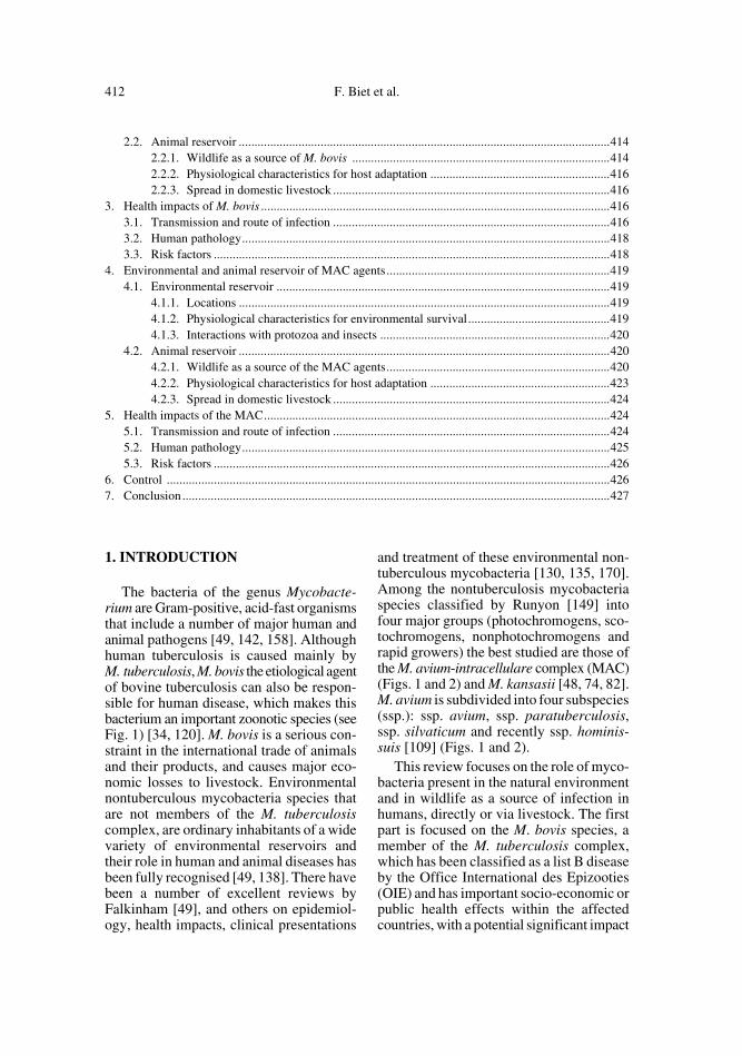
1. INTRODUCTION
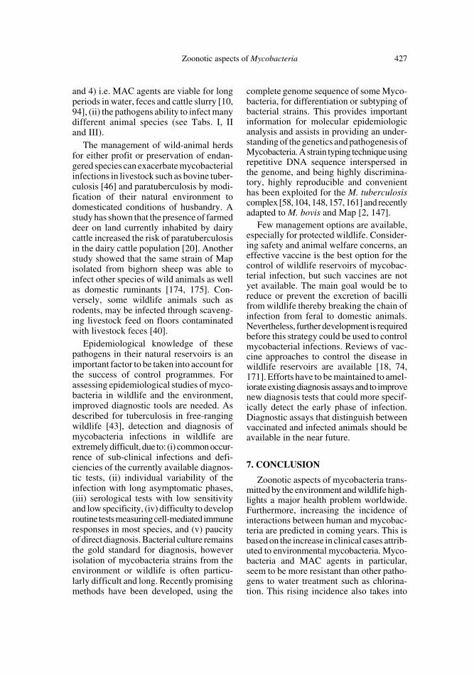
The bacteria of the genus Mycobacte-rium are Gram-positive, acid-fast organismsthat include a number of major human andanimal pathogens [49, 142, 158]. Althoughhuman tuberculosis is caused mainly byM. tuberculosis, M. bovis the etiological agentof bovine tuberculosis can also be respon-sible for human disease, which makes thisbacterium an important zoonotic species (seeFig. 1) [34, 120]. M. bovis is a serious con-straint in the international trade of animalsand their products, and causes major eco-nomic losses to livestock. Environmentalnontuberculous mycobacteria species thatare not members of the M. tuberculosiscomplex, are ordinary inhabitants of a widevariety of environmental reservoirs andtheir role in human and animal diseases hasbeen fully recognised [49, 138]. There havebeen a number of excellent reviews byFalkinham [49], and others on epidemiol-ogy, health impacts, clinical presentations
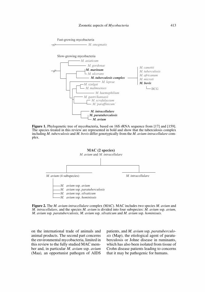
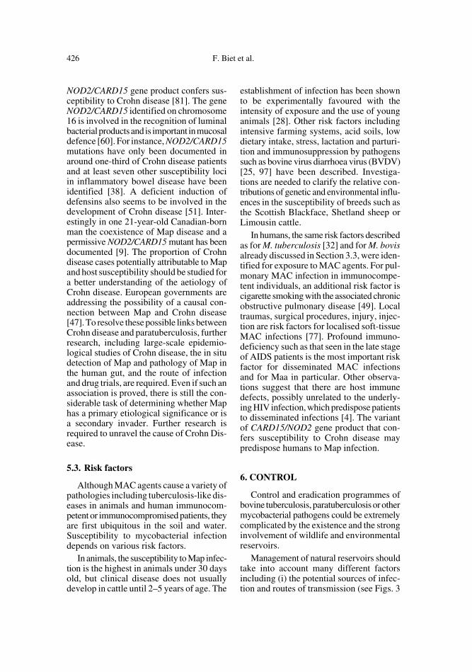
and treatment of these environmental non-tuberculous mycobacteria [130, 135, 170].Among the nontuberculosis mycobacteriaspecies classified by Runyon [149] intofour major groups (photochromogens, sco-tochromogens, nonphotochromogens andrapid growers) the best studied are those ofthe M. avium-intracellulare complex (MAC)(Figs. 1 and 2) and M. kansasii [48, 74, 82].M. avium is subdivided into four subspecies(ssp.): ssp. avium, ssp. paratuberculosis,ssp. silvaticum and recently ssp. hominis-suis [109] (Figs. 1 and 2).
This review focuses on the role of myco-bacteria present in the natural environmentand in wildlife as a source of infection inhumans, directly or via livestock. The firstpart is focused on the M. bovis species, amember of the M. tuberculosis complex,which has been classified as a list B diseaseby the Office International des Epizooties(OIE) and has important socio-economic orpublic health effects within the affectedcountries, with a potential significant impact
Zoonotic aspects of Mycobacteria 413
on the international trade of animals andanimal products. The second part concernsthe environmental mycobacteria, limited inthis review to the fully studied MAC mem-ber and, in particular M. avium ssp. avium(Maa), an opportunist pathogen of AIDS
patients, and M. avium ssp. paratuberculo-sis (Map), the etiological agent of paratu-berculosis or Johne disease in ruminants,which has also been isolated from tissue ofCrohn disease patients leading to concernsthat it may be pathogenic for humans.
Figure 2. The M. avium-intracellulare complex (MAC). MAC includes two species M. avium andM. intracellulare, and the species M. avium is divided into four subspecies: M. avium ssp. avium,M. avium ssp. paratuberculosis, M. avium ssp. silvaticum and M. avium ssp. hominisuis.
Figure 1. Phylogenetic tree of mycobacteria, based on 16S rRNA sequence from [17] and [159].The species treated in this review are represented in bold and show that the tuberculosis complexincluding M. tuberculosis and M. bovis differ genotypically from the M. avium-intracellulare com-plex.
414 F. Biet et al.
2. ENVIRONMENTAL AND ANIMAL RESERVOIR OF M. BOVIS
2.1. Environmental reservoir
2.1.1. Locations
M. bovis is considered to be an obligateintracellular pathogen whose most efficientway of infection is direct animal contact[136]. However, experimental evidence hasshown that M. bovis can survive for longperiods outside an animal host in an envi-ronment directly or indirectly contaminatedby discharges of infected animals, suggest-ing other possible ways of transmission.Yet in cattle, the natural host of M. bovis andthe main source of human spread, transmis-sion via the oral route or even the respira-tory route by inhalation of dust particles infields where no wildlife reservoir are impli-cated in transmission to livestock, wouldplay a less important role since the excre-tion of the organisms in faeces even fromheavily infected cattle occurs irregularlyand at a low frequency [107]. There are norecords of human infection by M. boviscoming from a direct environmental source,revealing that this way of transmission isnot the most important one for this patho-gen.
2.1.2. Physiological characteristicsfor environmental survival
The success of tubercle bacilli as patho-gens comes mainly from its ability to persistin the host for long periods and cause dis-ease by overcoming host immune responses[57] (see Sect. 2.2.2). Nevertheless, the pos-sibility of surviving for long periods in theenvironment is explained by the mycobac-terial impermeable cell wall [14] and slowgrowth [62]. In contrast, other featuresrender these species more sensitive to envi-ronmental survival, like a more enhancedpH sensitivity of the tuberculosis complexcompared to MAC species [21, 35]. Genomiccomparisons between MAC and M. tuber-
culosis complex members will not onlyallow to elucidate differences in virulencedeterminants between these two mycobac-terial complexes but also the disparities inenvironmental survival factors.
2.2. Animal reservoir
2.2.1. Wildlife as a source of M. bovis
Domestic and non-domestic animalsmay be considered either as maintenance(or reservoir) hosts or non-maintenance (orspill-over) hosts for bovine tuberculosis(see Tab. I). In reservoir host species, infec-tion can persist through horizontal transferin the absence of any other source ofM. bovis and may as well be transmitted toother susceptible hosts. In contrast, spillo-ver hosts become infected with M. bovis butthe infection only occurs sporadically orpersists within these populations if a truemaintenance host is present in the ecosys-tem. If the source of infection is removed,the prevalence for this disease is reducedand it can only be maintained in the longterm by re-infection from another source[76].
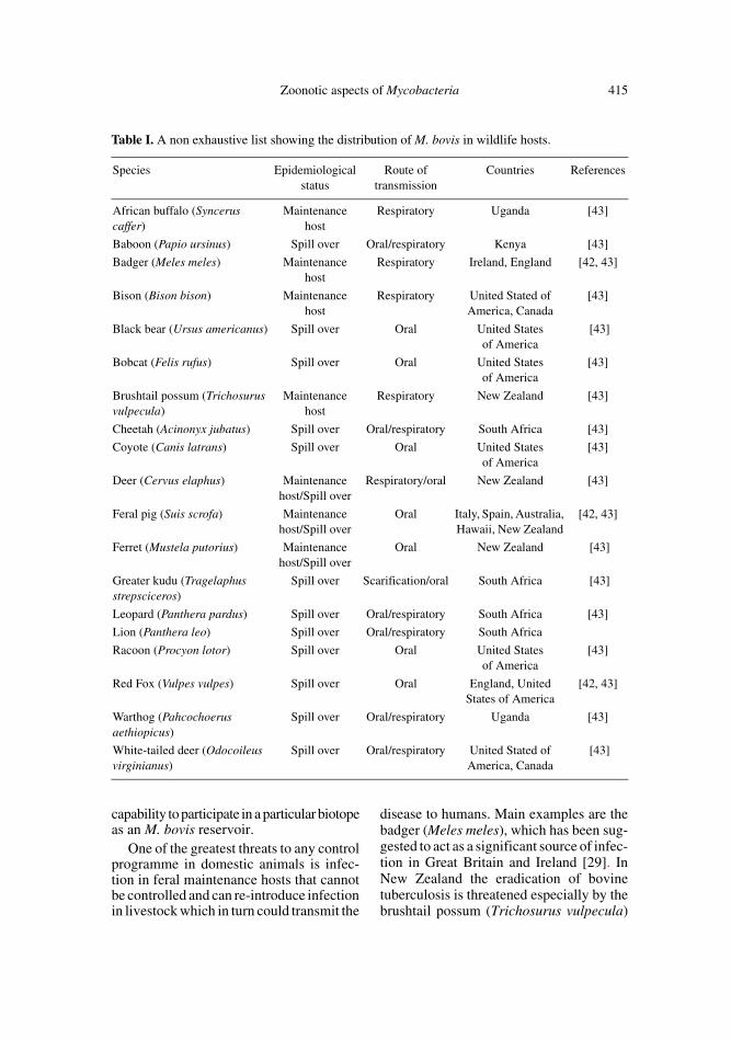
A main trait of M. bovis is its broad hostrange, actually the largest of any member ofthe M. tuberculosis complex. M. bovis causesdisease in a wide range of domestic but alsofree-ranging and farmed wildlife animals aswell as in humans. Only a small proportionof these animal species that become infectedcan act as maintenance hosts of this organ-ism. Table I is a non-exhaustive list sum-marising two excellent reviews by G.W. deLisle [42, 43], which describe M. bovis inreservoirs and spill-over wildlife species aswell as their distribution.
It is worth examining which factors rendera species as a maintenance host. Physio-pathogenesis, i.e. the capacity of excretion,ethology (for example gregarious or not gre-garious behaviour) and ecology (alimentarybehaviour, population density and interac-tions with other species) determine their
Zoonotic aspects of Mycobacteria 415
capability to participate in a particular biotopeas an M. bovis reservoir.
One of the greatest threats to any controlprogramme in domestic animals is infec-tion in feral maintenance hosts that cannotbe controlled and can re-introduce infectionin livestock which in turn could transmit the
disease to humans. Main examples are thebadger (Meles meles), which has been sug-gested to act as a significant source of infec-tion in Great Britain and Ireland [29]. InNew Zealand the eradication of bovinetuberculosis is threatened especially by thebrushtail possum (Trichosurus vulpecula)
Table I. A non exhaustive list showing the distribution of M. bovis in wildlife hosts.
Species Epidemiological status
Route of transmission
Countries References
African buffalo (Syncerus caffer)
Maintenance host
Respiratory Uganda [43]
Baboon (Papio ursinus) Spill over Oral/respiratory Kenya [43]
Badger (Meles meles) Maintenance host
Respiratory Ireland, England [42, 43]
Bison (Bison bison) Maintenance host
Respiratory United Stated of America, Canada
[43]
Black bear (Ursus americanus) Spill over Oral United States of America
[43]
Bobcat (Felis rufus) Spill over Oral United States of America
[43]
Brushtail possum (Trichosurus vulpecula)
Maintenance host
Respiratory New Zealand [43]
Cheetah (Acinonyx jubatus) Spill over Oral/respiratory South Africa [43]
Coyote (Canis latrans) Spill over Oral United States of America
[43]
Deer (Cervus elaphus) Maintenance host/Spill over
Respiratory/oral New Zealand [43]
Feral pig (Suis scrofa) Maintenance host/Spill over
Oral Italy, Spain, Australia, Hawaii, New Zealand
[42, 43]
Ferret (Mustela putorius) Maintenance host/Spill over
Oral New Zealand [43]
Greater kudu (Tragelaphus strepsciceros)
Spill over Scarification/oral South Africa [43]
Leopard (Panthera pardus) Spill over Oral/respiratory South Africa [43]
Lion (Panthera leo) Spill over Oral/respiratory South Africa
Racoon (Procyon lotor) Spill over Oral United States of America
[43]
Red Fox (Vulpes vulpes) Spill over Oral England, United States of America
[42, 43]
Warthog (Pahcochoerus aethiopicus)
Spill over Oral/respiratory Uganda [43]
White-tailed deer (Odocoileus virginianus)
Spill over Oral/respiratory United Stated of America, Canada
[43]
416 F. Biet et al.
[172]. The presence of M. bovis infection inwhite tailed deer (Odocoileus virginianus)in Michigan poses a serious menace to thecontrol and eradication programmes forbovine tuberculosis in the United States[131]. Infection with M. bovis has also beendescribed across a range of animals such asbuffalo, kudu, lion, baboon and antelope inthe Kruger National Park in South Africa,having severe consequences on the biodi-versity of this region [172]. In France, ahigh non-negligible proportion of M. bovisinfected wild deer (Cervus elaphus) werefound in regions where cattle outbreakswere reported, opening-up the suspicion oftransmission from wildlife (Boschiroli et al.,unpublished results).
2.2.2. Physiological characteristics for host adaptation
Only some of these characteristics willbe discussed in this section. For furtherinformation, see reference [160]. Althoughthe course of infection, clinical signs anddevelopment of disease can vary within dif-ferent host species, it can be presumed thatcertain essential physiological characteris-tics are common for successful infection inany susceptible host. The analysis of thecomplete genome sequence of M. bovis [59]provides a means to dissect these character-istics. To begin with, the cell wall protectsthe bacteria from harsh environments butalso promotes intracellular persistence [14].The ability to infect and persist in the mac-rophage by inhibiting phagosome-lysosomefusion, creating a privileged compartmentand remaining sequestered away from theterminal endocytic organelles, is central tothe success of the pathogen [45]. The pres-ence of acidic, glycine-rich proteins (PEand PPE families) also found in M. leprae[30] and M. marinum [140] whose genes areinvolved in virulence are worth mentioning.Another important genetic factor implicatedin the attenuation of the M. bovis BCG strainis the lack of the RD1 locus [140], which isinvolved in a novel described secretion sys-tem [140].
Latency is another important aspect oftubercle bacilli pathogenesis. The molecu-lar basis for the persistence phenotype andthe pertinent host immune mechanisms thatcontribute to the maintenance of tuberculo-sis latency are just beginning to be under-stood. The bacillus releases peripheral cell-wall lipids into their host cells, which inducethe granulatomous response. This representsactive manipulation of the host’s responseto ensure the maintenance of the infection.The granuloma appears as a balance struc-ture that walls off the infection and limitsits metastasis. However, the very prisonthat limits spread could well restrict thecapacity of the host to activate the macro-phages required to kill the bacteria [150].
2.2.3. Spread in domestic livestock
Within domesticated animals, cattle,farmed buffalo and goats are consideredreservoir hosts of M. bovis, while pigs, cats,dogs, horses and sheep are considered spill-over hosts. For further reading, see [36].The realisation that wildlife is infected withM. bovis may result in apparent failure pro-grammes to eradicate the infection fromcattle [44]. Knowledge of wildlife tubercu-losis through appropriate surveillance pro-grammes in feral animal populations maybe important in the research strategies forthe total elimination of livestock tuberculo-sis.
3. HEALTH IMPACTS OF M. BOVIS
3.1. Transmission and route of infection
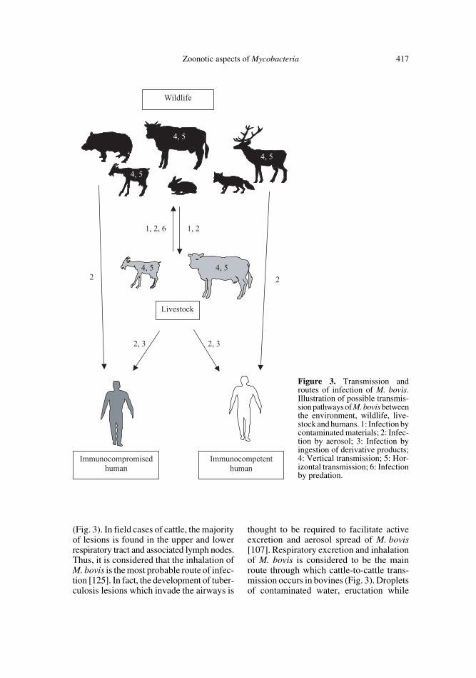
In cattle as well as in other animal hosts,the route of transmission of M. bovis can bededuced by the pattern of lesions observedin slaughtered animals. Animals with lesionsrestricted to the thoracic cavity are pre-sumed to have been infected by the inhala-tion of aerosols, while those with lesions inmesenteric lymph nodes are thought to haveacquired the infection by ingestion [136]
Zoonotic aspects of Mycobacteria 417
(Fig. 3). In field cases of cattle, the majorityof lesions is found in the upper and lowerrespiratory tract and associated lymph nodes.Thus, it is considered that the inhalation ofM. bovis is the most probable route of infec-tion [125]. In fact, the development of tuber-culosis lesions which invade the airways is
thought to be required to facilitate activeexcretion and aerosol spread of M. bovis[107]. Respiratory excretion and inhalationof M. bovis is considered to be the mainroute through which cattle-to-cattle trans-mission occurs in bovines (Fig. 3). Dropletsof contaminated water, eructation while
Figure 3. Transmission androutes of infection of M. bovis.Illustration of possible transmis-sion pathways of M. bovis betweenthe environment, wildlife, live-stock and humans. 1: Infection bycontaminated materials; 2: Infec-tion by aerosol; 3: Infection byingestion of derivative products;4: Vertical transmission; 5: Hor-izontal transmission; 6: Infectionby predation.
418 F. Biet et al.
ruminating infected pastures or inhalatingcontaminated dust particles can also be analternative way of aerogenous infection.This is, in fact, suspected to be the mostlikely way cattle could get infected in a con-taminated environment by badger excretions[134]. Ingestion of M. bovis directly frominfected animals or from contaminated pas-tures, water or fomites is considered second-ary to respiratory spread, as deduced fromthe minor presence of mesenteric lesions incattle cases [107]. Congenital infections andvertical transmission to calves as well asgenital transmission are uncommon in regionswhere intensive eradication programmesoperate. Within wildlife, routes of trans-mission are listed in Table I and illustratedin Figure 3.
Infection of humans may occur by theinhalation of aerosols or through the con-sumption of contaminated milk (see Fig. 3).The aerosols are the result of animal excre-tion but can also be produced by handlinglesioned carcasses [124]. This route ofinfection leads to respiratory tuberculosis.Human to human transmission is possible ifan immunodeficient status of the potentialhost is encountered [64].
3.2. Human pathology
Tuberculosis in humans caused byM. tuberculosis or M. bovis is indistinguish-able clinically, radiologically and patholog-ically [171]. In countries where bovine tuber-culosis is uncontrolled, or in developedcountries before strict control campaignsand milk pasteurisation, most human casesoccur in young persons and result fromdrinking contaminated milk. This alimen-tary route of infection leads to extra-pulmo-nary forms of tuberculosis, where infectioncan become established in the cervix andless frequently in the axillary lymph nodesleading to chronic skin tuberculosis [113].Adult humans at professional risk, espe-cially farmers or abattoir workers as well asveterinarians, are generally infected withM. bovis by the respiratory route through
aerosols from infected cattle and developtypical pulmonary tuberculosis.
The implementation of bovine eradica-tion schemes together with the pasteurisa-tion of milk has had a major impact on thedisease with the result that human tubercu-losis due to M. bovis is now rare in devel-oped countries. However, a small numberof cases still occur in elderly people as aresult of reactivation of dormant infections[171].
3.3. Risk factors
In animals, age, behaviour, environmentand prevailing farm practices can have asignificant influence [107]. Nutritional defi-ciencies are associated to reduced resistanceto bovine tuberculosis [71]. Immunologicaldysfunction in cattle may enhance bovinetuberculosis infection, although this hasnever been assessed.
In humans, risk factors for mycobaterialinfections, being especially well describedfor M. tuberculosis [32] include the inten-sity of exposure, age, immune system, HIVcoinfection, genetic factor, vaccination sta-tus and also socio-economic factors. Pro-fessional exposure and life style, as discussedin Section 3.2, can also be considered as riskfactors when M. bovis is the etiologicalagent in human tuberculosis. Reactivationoccurs under stress or in old age, sincemycobacteria in a latent state may becomesubject to less stringent control by host sys-tems [113]. The endemic nature of the dis-ease in domestic stock or wildlife and thelikely contact with humans, particularlythose infected with HIV, poses a serioushealth problem, since humans could beginto actively transmit the infection withinpopulations. Another risk could come fromthe increasing contact of humans withinfected wildlife animal species, and there-fore bovine tuberculosis could become a“leisure” zoonoses (unpublished data). Thisis a possibility for hunters that handle heav-ily contaminated animal carcasses capableof producing infective droplets.
Zoonotic aspects of Mycobacteria 419
4. ENVIRONMENTAL AND ANIMAL RESERVOIR OF MAC AGENTS
4.1. Environmental reservoir
4.1.1. Locations
Environmental mycobacteria such as themembers of MAC constitute a very inter-esting group in terms of ecology. They pos-sess properties that enable them to grow innatural biotopes without losing their path-ogenicity for certain living beings. Somestrains induce infections via natural biotopes,which can be regarded as reservoirs in thechain of transmission. In spite of their path-ogenicity, they possess a number of prop-erties resembling in many respects those ofthe saprophytes: growth over a wide tem-perature range, sometimes better at 20 °Cthan 37 °C, rapid adaptation to new sub-strates and the capacity to increase theirgrowth rate on synthetic media. MACagents grow well between pH 4.0 and 7.5[21] with an optimal pH between 5.4 and6.5 [137]. In contrast, M. tuberculosis has
a comparatively narrow range for optimalgrowth between pH 6.0 and 6.5. Outside ofliving beings, mycobacteria species of MAChave been found in many biotopes includ-ing the soil, wastewater, water tank, munic-ipal water, aerosols, protozoa, deep litter,fresh tropical vegetation, animals and humans(Tab. II). Among the several opportunisticpathogens affecting patients infected withhuman immunodeficiency virus (HIV),members of MAC, mainly Maa, are thecause of significant problems for the clini-cal management of this immunosuppres-sive disease. Potable water is considered asthe primary source of MAC infection inhumans [146] and has been shown to be asource of Maa infection in virus-inoculatedSimian immunodeficiency macaques [101].Food has also been shown to be a possiblesource and route of transmission of Maa,isolated in patients and food [178].
4.1.2. Physiological characteristics for environmental survival
Mycobacteria of MAC have the capacityto survive and multiply under a wide range
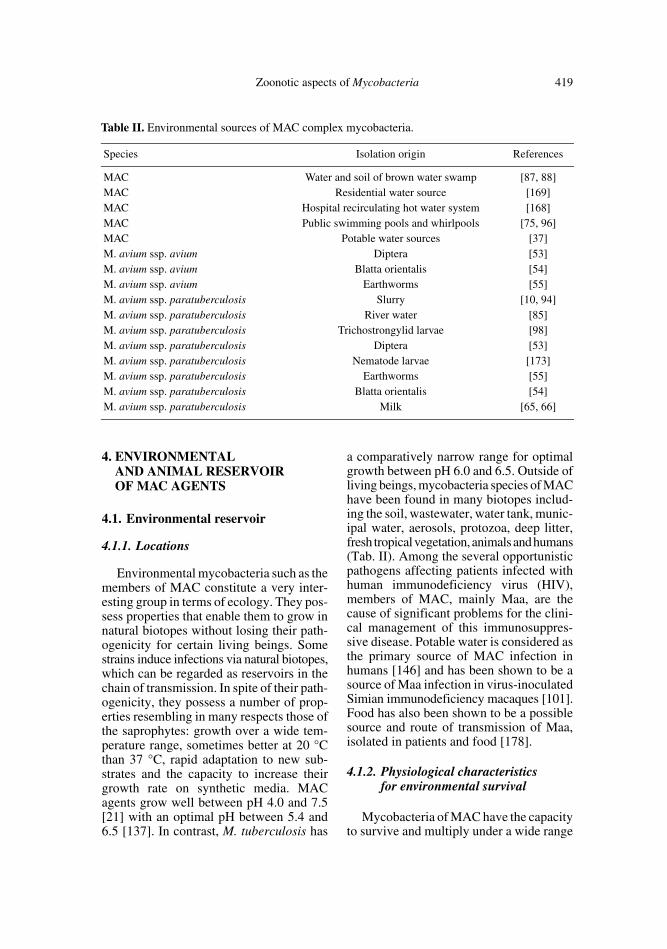
Table II. Environmental sources of MAC complex mycobacteria.
Species Isolation origin References
MAC Water and soil of brown water swamp [87, 88]MAC Residential water source [169]MAC Hospital recirculating hot water system [168]MAC Public swimming pools and whirlpools [75, 96]MAC Potable water sources [37]M. avium ssp. avium Diptera [53]M. avium ssp. avium Blatta orientalis [54]M. avium ssp. avium Earthworms [55]M. avium ssp. paratuberculosis Slurry [10, 94]M. avium ssp. paratuberculosis River water [85]M. avium ssp. paratuberculosis Trichostrongylid larvae [98]M. avium ssp. paratuberculosis Diptera [53]M. avium ssp. paratuberculosis Nematode larvae [173]M. avium ssp. paratuberculosis Earthworms [55]M. avium ssp. paratuberculosis Blatta orientalis [54]M. avium ssp. paratuberculosis Milk [65, 66]
420 F. Biet et al.
of environmental conditions [49, 138], includ-ing low pH [21], extreme temperature [152],starvation, chlorine or ozone treatment [128]and low oxygen level [16]. Thus, their abil-ity to utilise many substances as nutrientsenables them to grow successfully in manybiotopes. Their adaptation for life in theenvironment is linked to physiological char-acteristics of the mycobacteria such as theimpermeable cell wall [14, 141] and slowgrowth [62] (see also Sect. 4.2.2).
4.1.3. Interactions with protozoa and insects
In contrast to M. bovis, protozoa andinsects play an important role in the dissem-ination of some of the other species of myco-bacteria and interactions with animals orhumans [55, 138]. As described in Table II,Maa and Map have been isolated from manydifferent insects and protozoa. The interac-tions between mycobacteria and insects arevery important to the evolution of myco-bacterial pathogenesis. Many protozoa har-bor bacteria and their ability to survive phago-cytosis is a considerable advantage to water-borne bacilli. Maa inhibits lysosomal fusionand possibly kills infected amoebae. Maacan also invade and replicate in Dictyostel-ium discoideum [156]. Compared to bacilligrown in medium, amoeba-grown Maa aremore invasive towards amoebae, HT-29human epithelial cells and macrophages[27] and more virulent in beige mice [27].Other advantages for these mycobacteria touse protozoa as hosts reside in the fact thatthey are protected from antimicrobial effects[111] and that they can survive during encyst-ment. Mycobacteria can indeed use the pro-tozoan cysts as carriers to survive starvationand toxic stress and can be released uponexcystment [138, 160].
Protozoa may play a central role in theevolution of mycobacteria pathogenesis. Theselection of mycobacteria that can infect andreplicate within protozoa has likely resultedin mycobacteria also becoming intracellularpathogens in animals as exemplified by the
adaptation of Legionella pneumophilia tothe infection of human macrophages [153].
Insects that have been in contact withmaterial contaminated with these environ-mental pathogens may spread mycobacte-ria. Members of MAC were isolated fromDiptera (see Tab. II) collected from bothcattle herds infected by Map and M. intra-cellulare and cattle without mycobacterialinfections [53]. Earthworms constitute asignificant component of soil organisms.Most ingested microorganisms pass throughthe digestive tract and are excreted in thefaeces. However, some species can propa-gate in the digestive tract and survive inegg cocoons of the earthworm [39, 151].Whittington et al. have shown that the nem-atode parasite of sheep might be able to helpin the transmission of Map [98, 173]. Therole of earthworms as vectors of mycobac-terial infection in cattle and goat farms hasbeen identified for Maa and Map [55].Other omnivorous insects such as cock-roaches, which frequently infect hospitals,laboratories and other contaminated habi-tats, may also be an environmental reservoirof pathogen mycobacteria. Allen et al. havedemonstrated the survival of mycobacteriain Blatta orientalis that had ingested infectedhuman sputum [1]. Recently Fisher et al.have proposed the cockroach (Blatta orien-talis) as a passive vector of causal agents ofavian tuberculosis and paratuberculosis [54].
4.2. Animal reservoir
4.2.1. Wildlife as a source of the MAC agents
Mycobacterium members of MAC causeinfections and diseases in a wide range ofdifferent animal species. Concerning ssp.Maa and Map, the known host range includesruminant and non-ruminant wildlife (Tab. III)[8, 40, 166, 167]. This section is divided intothree parts. The first part describes the dis-ease in wild ruminants, the second describesthe disease in wild non-ruminants and thelast part describes the disease in birds.
Zoonotic aspects of Mycobacteria 421
(i) In wild ruminants, the Maa and Mapinfections have been documented world-wide, including in the USA [2, 121, 162],Europe [90, 155, 167, 177], Australia [50],New Zealand [31], Japan [117, 118] or Africa[126]. The data are essentially described aslesions and clinical signs in Map infectedwild ruminants, which are similar to thosein infected domestic ruminants, and wherethe disease may be fatal [19, 174]. In deer,the disease is characterized by loss of con-dition, diarrhoea, faecal staining of the peri-neum, low serum albumin and total protein
concentration. In other cases, the infectionis clinically unapparent [28, 41]. In bisons,histologic results are similar to paratuber-culosis described in cattle [19].
(ii) In the wild boar (Sus scrofa), the prev-alence and the pathogenesis caused by MACagents Maa and Map have been recentlyreviewed [99]. The lesions, rarely observed,are localised in the head lymph nodes. Thenumber of wild boars with tuberculouslesions increases with age. However myco-bacteria are more frequently isolated from
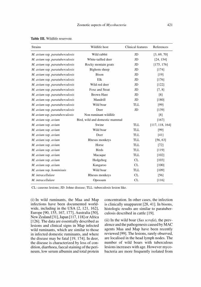
Table III. Wildlife reservoir.
Strains Wildlife host Clinical features References
M. avium ssp. paratuberculosis Wild rabbit JD [3, 69, 70]
M. avium ssp. paratuberculosis White-tailled deer JD [24, 154]
M. avium ssp. paratuberculosis Rocky montain goats JD [175, 176]
M. avium ssp. paratuberculosis Bighorn sheep JD [174]
M. avium ssp. paratuberculosis Bison JD [19]
M. avium ssp. paratuberculosis Elk JD [176]
M. avium ssp. paratuberculosis Wild red deer JD [122]
M. avium ssp. paratuberculosis Foxe and Stoat JD [7, 8]
M. avium ssp. paratuberculosis Brown Hare JD [8]
M. avium ssp. paratuberculosis Mandrill JD [180]
M. avium ssp. paratuberculosis Wild boar TLL [99]
M. avium ssp. paratuberculosis Deer JD [139]
M. avium ssp. paratuberculosis Non ruminant wildlife [8]
M. avium ssp. avium Bird, wild and domestic mammal [167]
M. avium ssp. avium Swine TLL [117, 118, 164]
M. avium ssp. avium Wild boar TLL [99]
M. avium ssp. avium Deer TLL [41]
M. avium ssp. avium Rhesus monkeys TLL [56, 63]
M. avium ssp. avium Horse TLL [72]
M. avium ssp. avium Birds TLL [119]
M. avium ssp. avium Macaque TLL [102]
M. avium ssp. avium Hedgehog CL [103]
M. avium ssp. avium Kangaroo CL [100]
M. avium ssp. hominisuis Wild boar TLL [109]
M. intracellulare Rhesus monkeys CL [56]
M. intracellulare Opossum CL [116]
CL: caseous lesions; JD: Johne disease; TLL: tuberculosis lesion like.
422 F. Biet et al.
the wild boar without clinical signs of tuber-culosis [33]. Ray et al. [143] have alsodescribed a more frequent isolation of myco-bacteria, including M. bovis, Maa and Map,from the tissue of wild boar without tuber-culous lesions. In non-ruminant wildlife,the occurrence of Map infections have beenrecently documented in Scotland [8, 40].Following the isolation of Map in rabbits,the studies were extended to other wildlifespecies in farms with a history of paratuber-culosis in livestock. Map was isolated fromfoxes, stoats, weasels, badgers, wood mice,rats, brown hares, jackdaws, rooks andcrows [8]. The clinical signs of paratuber-culosis in non-ruminant wildlife are largelyunknown. Lesions seem to be similar toearly, subclinical infections described forruminants and clinical signs are not system-atically observed on positive animals [8,133].
Naturally acquired infections with Maaand M. intracellulare have been reported innon-domestic mammal species and non-human primates, as well as in exotic hoofedanimals (Tab. II) [166, 167]. MAC agentshave also been isolated from kangaroos,macaques and mandrills [79, 105, 115, 163,180].(iii) In many countries, the disease causedprincipally by Maa serotype 1,2,3, occurs indomestic and wild birds as well as in a vari-ety of fowl, game birds and water-fowl (seeThorel et al. for a review [167]). The diseasecaused by Maa is characterized by itschronic nature, its persistence in a flock oraviary once established, and its tendency toinduce wasting and finally death. However,a few clinical signs of the disease are com-monly observed in chickens and birds, onlyduring the advanced stages of the disease.Macroscopic lesions are disseminated throughthe organism most often observed in theliver, spleen, intestine and bone marrow.
These data clearly show the existenceand the importance of a wildlife reservoir ofmycobacteria of MAC that is still mainlyundetermined. The interspecies transmis-sion may occur between livestock and wild-
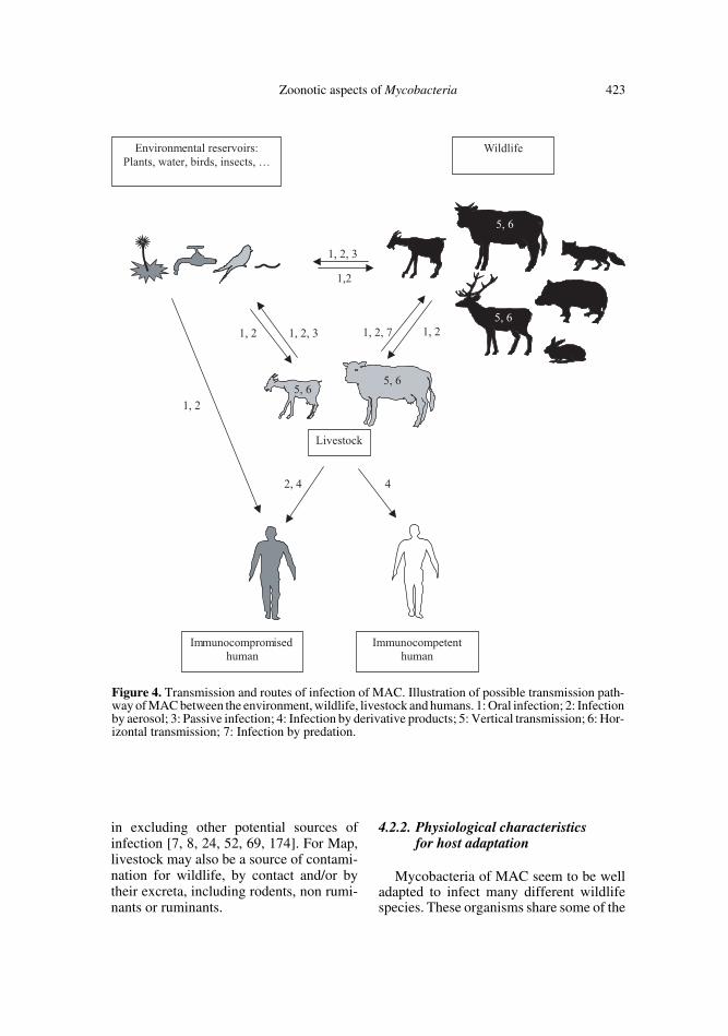
life and vice-versa. Interspecies transmis-sion has been demonstrated experimentallybetween non-ruminant wildlife and live-stock [114]. These possible transmissionshave important implications with respect tothe attempted control or eradication of thisdisease in both wild and domestic animals.The routes and mode of transmission areillustrated in Figure 4. Studies of the trans-mission of Map in livestock could be trans-posed to Map transmission in wildlife. Thefaecal-oral route, i.e. through ingestion offaecal contaminants, milk or colostrum, isthe principal pathway of infection in thehost. Ingestion of mycobacteria has beenproposed as the primary route of infectionin paratuberculosis and experimental oralinoculation of organisms has produced entericdisease experimentally in sheep and cattle.Other experimental Map infections havebeen reported by using different routesof inoculation such as intravenous [89],intramammary [93], intrauterine [108]. Otherpossible alternative transmission pathwayscould help to understand the epidemiologyof Johne disease. One such pathway couldbe the aerosol transmission via the respira-tory tract which is generally agreed as beingthe principal route of infection of M. bovisin cattle [123]. In cattle, vertical transmis-sion during pregnancy has also been pro-posed since Map has been isolated from theuterus [91, 132], fetal tissues [95] andsemen [92, 173].
Predation is also a possible form of trans-mission of MAC to carnivores (Fig. 4). ForMap, the prevalence in predators, includingfox stoats and weasels, is 62% and is higherthan in the prey species, including rabbits,rats and wood mice, whose prevalence is10% [69, 70]. The high prevalence of Mapin some non-ruminant wildlife species andtheir interaction with susceptible ruminantlivestock raises the possibility that theyplay a role in the epidemiology of the dis-ease in the latter. The risk of transmissionfrom wildlife to livestock has frequentlybeen suggested, but it is hard to be provenin the field mainly due to the long incuba-tion period of the disease and the difficulty
Zoonotic aspects of Mycobacteria 423
in excluding other potential sources ofinfection [7, 8, 24, 52, 69, 174]. For Map,livestock may also be a source of contami-nation for wildlife, by contact and/or bytheir excreta, including rodents, non rumi-nants or ruminants.
4.2.2. Physiological characteristics for host adaptation
Mycobacteria of MAC seem to be welladapted to infect many different wildlifespecies. These organisms share some of the
Figure 4. Transmission and routes of infection of MAC. Illustration of possible transmission path-way of MAC between the environment, wildlife, livestock and humans. 1: Oral infection; 2: Infectionby aerosol; 3: Passive infection; 4: Infection by derivative products; 5: Vertical transmission; 6: Hor-izontal transmission; 7: Infection by predation.
424 F. Biet et al.
same physiological characteristics and molec-ular determinants of virulence describedfor other slowly growing mycobacteria asM. tuberculosis or M. leprae, see [49, 82,158] for a review. These organisms are sur-rounded by a cell wall and an envelope char-acteristic of mycobacteria which conferstheir distinctive feature of acid fastness [82].However, MAC-specific C-mycoside glyc-opeptidolipids (GPL) [13, 15] seem to berelated to the resistance of MAC to antimi-crobial agents. Members of MAC share ahigh percentage of DNA and rRNA homol-ogy. Map shares over 98% DNA homologywith Maa and they have homologous majorantigens [6, 74]. However, phenotypic dif-ferences between these two subspecies, dif-ferentiated by the dependence of Map onmycobactin [165] and genetically by thepresence of multiple copies of the Map spe-cific insertion element IS900 [67], are impor-tant. These organisms induce, in a wide-rang-ing animal species, different pathologiesassociated to distinct clinical signs [28, 82]and host adaptation [49, 74, 82]. With theavailability of published microbial genomes,the genomic approach will help in identify-ing novel genes involved in the physiolog-ical adaptation of MAC members to thesedifferent wild animal species. This approachwill also help to identify specific genes thatcause distinct pathologies, avian tuberculo-sis and Johne disease, with different clinicalsigns and in different target hosts, despitesharing more than 98% DNA identity.
4.2.3. Spread in domestic livestock
Livestock also represent an importantreservoir, not developed in this review andwell described in others [74, 166, 167]. Asillustrated in Figure 4, livestock could eitherbe infected by mycobacteria present inwildlife and in the environment or be asource of contamination for wildlife andhumans and a particularly susceptible pop-ulation [12, 90, 129]. Livestock could alsobe a vector of infection in humans directlyor by their derivative products (see Fig. 4).
MAC agents are described to be respon-sible for the infection in a very large rangeof agricultural and domestic animal species.The range of domestic animals infected byMAC members includes domestic birds,chickens, cattle, swine, farmed deer, sheepand goats, and horses but also cats and dogswhich has been reviewed by Harris andBarletta and Thorel et al. [74, 167]. Themajority of MAC infections in livestock aredetected at slaughter and the diagnosis isconfirmed by bacteriological procedures. Itis most probable that a common environ-mental reservoir of infection exists withinwildlife.
5. HEALTH IMPACTS OF THE MAC
5.1. Transmission and route of infection
For humans, exposure to MAC organ-isms, present in wildlife and in naturalbiotopes including protozoa and insects,can occur by a variety of routes. Birds aremajor agents of Maa spread as they excretebacilli in large amounts in their faeces,where bacteria can persist in the soil or inwater for long periods afterwards. Knowl-edge of the route of infection, pathogenesis,and levels of excretion will assist in deter-mining the potential of each species to actas a reservoir of infection (Fig. 4). As forother environmental mycobacteria, munic-ipal and natural water are important ways ofMAC infection. While environmental myco-bacteria are opportunistic pathogens in avariety of immunocompromised patients, awide prevalence results in all humans beingcommonly and continuously exposed at alow level (50 to 500 bacilli per day). Onlya very small percentage of human-mycobac-teria interaction progress to outright myco-bacterial infection but such progression ismuch more common in immunocompro-mised patients, especially those with AIDS[4]. Genomic restriction fragment patternsof Maa from hospital water isolates are sim-ilar to those from AIDS patient isolates [5].Numerous studies have attempted to deter-mine the routes, oral or aerosol, leading to
Zoonotic aspects of Mycobacteria 425
Maa infection in AIDS patients but no evi-dence was related to one and a combinationof both routes is likely [22, 83, 144].
For Map, studies need to be done todetermine the possible ways of direct trans-mission between wildlife and humans.
It is necessary to develop a better under-standing of the epidemiology of MAC andtheir diseases, especially the transmissionpathways, in animals, domestic ruminantsand humans with important implicationswith respect to the attempt to control thesediseases.
5.2. Human pathology
While humans are highly susceptible toM. tuberculosis and M. leprae infection,most people who are exposed to these bac-teria never develop clinical disease, indicat-ing that the normal immune system can con-trol these organisms [86]. This observationis even more applicable for MAC organ-isms because, despite evidence of exposurerates, the incidence of clinical disease isremarkably low (10 cases per 100 000 pop-ulation). For a review see [49, 82, 138].
In immunocompetent patients, the infec-tions caused by MAC agents are principallypulmonary [4]. In children a recent studyhas shown that the most predominant spe-cies in cervical lymphadenitis caused bynontuberculous mycobacteria was M. scrof-ulaceum (60%) followed by the MAC agents(40%) [84].
The number of MAC infection cases inimmunocompetent patients has been over-whelmed by the high frequency (e.g. 25 to50%) of MAC infections in AIDS patients[80, 127]. Among the members of MAC,Maa predominate (87 to 98%) in AIDSpatients and induce disseminated mycobac-teremia rather than bacteria restricted to thelungs as for immunocompetent patients.Maa appears to have a particular predilec-tion for infecting and disseminating in HIV-infected patients. It has been suggested thatMaa isolates that cause disease in AIDS
patients are not simply gratuitous opportun-ists but possess specific genetic determi-nants that confer an ability to penetrate andmultiply within macrophages and host cellsand contribute to the existing immunodefi-ciency [73]. One of the most interestingaspects of MAC infection in AIDS patientsis the discovery of polyclonal infection, onepossible explanation for the inability to cor-relate the outcome of antibiotic treatmentwith susceptibility patterns [179]. Recently,human infection with Map in a patient withHIV was reported [145]. This report raisesthe question of systematic Map detection,which is not yet possible by routine tech-niques. It raises other questions as to whyMap has not been detected before andwhether this lack of detection was becauseof its slow and difficult growth, or becauseit has been misidentified with Maa, orbecause its occurrence in infections is low.
The isolation of Map from tissues of Crohnpatients [23, 26, 106] has led to concernsthat Map may be pathogenic for humans[112]. Physical and causal association ofMap in Crohn disease is still controversial.Since cell-wall deficient Map usually can-not be identified by Zeihl-Neelsen staining,identification of Map in humans eitherrequires fastidious culture or detection ofMap DNA or RNA, which is not alwaysreproducible [74]. However, the Koch pos-tulates may be met for Map [68]. Map hasbeen isolated, with technical difficulties ofMap culture, from patients with Crohn dis-ease [26, 61]. Milk and water are potentialsources for acquiring Map [78, 110]. How-ever, only a few samples of milk, positiveby PCR for the presence of Map, have beenshown to be positive for culture, suggestingthat either Map remains undetectable becauseof a too low number of viable Map in a sam-ple or due to the absence of live Map in thesample [11]. Serological response to Mapdoes not conclusively prove that the subjecthas had an active infection [11]. The devel-opment of Crohn disease depends upon aninteraction between the host and environ-mental factors but also genetic factors. Inhumans, it has been suggested that the
426 F. Biet et al.
NOD2/CARD15 gene product confers sus-ceptibility to Crohn disease [81]. The geneNOD2/CARD15 identified on chromosome16 is involved in the recognition of luminalbacterial products and is important in mucosaldefence [60]. For instance, NOD2/CARD15mutations have only been documented inaround one-third of Crohn disease patientsand at least seven other susceptibility lociin inflammatory bowel disease have beenidentified [38]. A deficient induction ofdefensins also seems to be involved in thedevelopment of Crohn disease [51]. Inter-estingly in one 21-year-old Canadian-bornman the coexistence of Map disease and apermissive NOD2/CARD15 mutant has beendocumented [9]. The proportion of Crohndisease cases potentially attributable to Mapand host susceptibility should be studied fora better understanding of the aetiology ofCrohn disease. European governments areaddressing the possibility of a causal con-nection between Map and Crohn disease[47]. To resolve these possible links betweenCrohn disease and paratuberculosis, furtherresearch, including large-scale epidemio-logical studies of Crohn disease, the in situdetection of Map and pathology of Map inthe human gut, and the route of infectionand drug trials, are required. Even if such anassociation is proved, there is still the con-siderable task of determining whether Maphas a primary etiological significance or isa secondary invader. Further research isrequired to unravel the cause of Crohn Dis-ease.
5.3. Risk factors
Although MAC agents cause a variety ofpathologies including tuberculosis-like dis-eases in animals and human immunocom-petent or immunocompromised patients, theyare first ubiquitous in the soil and water.Susceptibility to mycobacterial infectiondepends on various risk factors.
In animals, the susceptibility to Map infec-tion is the highest in animals under 30 daysold, but clinical disease does not usuallydevelop in cattle until 2–5 years of age. The
establishment of infection has been shownto be experimentally favoured with theintensity of exposure and the use of younganimals [28]. Other risk factors includingintensive farming systems, acid soils, lowdietary intake, stress, lactation and parturi-tion and immunosuppression by pathogenssuch as bovine virus diarrhoea virus (BVDV)[25, 97] have been described. Investiga-tions are needed to clarify the relative con-tributions of genetic and environmental influ-ences in the susceptibility of breeds such asthe Scottish Blackface, Shetland sheep orLimousin cattle.
In humans, the same risk factors describedas for M. tuberculosis [32] and for M. bovisalready discussed in Section 3.3, were iden-tified for exposure to MAC agents. For pul-monary MAC infection in immunocompe-tent individuals, an additional risk factor iscigarette smoking with the associated chronicobstructive pulmonary disease [49]. Localtraumas, surgical procedures, injury, injec-tion are risk factors for localised soft-tissueMAC infections [77]. Profound immuno-deficiency such as that seen in the late stageof AIDS patients is the most important riskfactor for disseminated MAC infectionsand for Maa in particular. Other observa-tions suggest that there are host immunedefects, possibly unrelated to the underly-ing HIV infection, which predispose patientsto disseminated infections [4]. The variantof CARD15/NOD2 gene product that con-fers susceptibility to Crohn disease maypredispose humans to Map infection.
6. CONTROL
Control and eradication programmes ofbovine tuberculosis, paratuberculosis or othermycobacterial pathogens could be extremelycomplicated by the existence and the stronginvolvement of wildlife and environmentalreservoirs.
Management of natural reservoirs shouldtake into account many different factorsincluding (i) the potential sources of infec-tion and routes of transmission (see Figs. 3
Zoonotic aspects of Mycobacteria 427
and 4) i.e. MAC agents are viable for longperiods in water, feces and cattle slurry [10,94], (ii) the pathogens ability to infect manydifferent animal species (see Tabs. I, IIand III).
The management of wild-animal herdsfor either profit or preservation of endan-gered species can exacerbate mycobacterialinfections in livestock such as bovine tuber-culosis [46] and paratuberculosis by modi-fication of their natural environment todomesticated conditions of husbandry. Astudy has shown that the presence of farmeddeer on land currently inhabited by dairycattle increased the risk of paratuberculosisin the dairy cattle population [20]. Anotherstudy showed that the same strain of Mapisolated from bighorn sheep was able toinfect other species of wild animals as wellas domestic ruminants [174, 175]. Con-versely, some wildlife animals such asrodents, may be infected through scaveng-ing livestock feed on floors contaminatedwith livestock feces [40].
Epidemiological knowledge of thesepathogens in their natural reservoirs is animportant factor to be taken into account forthe success of control programmes. Forassessing epidemiological studies of myco-bacteria in wildlife and the environment,improved diagnostic tools are needed. Asdescribed for tuberculosis in free-rangingwildlife [43], detection and diagnosis ofmycobacteria infections in wildlife areextremely difficult, due to: (i) common occur-rence of sub-clinical infections and defi-ciencies of the currently available diagnos-tic tests, (ii) individual variability of theinfection with long asymptomatic phases,(iii) serological tests with low sensitivityand low specificity, (iv) difficulty to developroutine tests measuring cell-mediated immuneresponses in most species, and (v) paucityof direct diagnosis. Bacterial culture remainsthe gold standard for diagnosis, howeverisolation of mycobacteria strains from theenvironment or wildlife is often particu-larly difficult and long. Recently promisingmethods have been developed, using the
complete genome sequence of some Myco-bacteria, for differentiation or subtyping ofbacterial strains. This provides importantinformation for molecular epidemiologicanalysis and assists in providing an under-standing of the genetics and pathogenesis ofMycobacteria. A strain typing technique usingrepetitive DNA sequence interspersed inthe genome, and being highly discrimina-tory, highly reproducible and convenienthas been exploited for the M. tuberculosiscomplex [58, 104, 148, 157, 161] and recentlyadapted to M. bovis and Map [2, 147].
Few management options are available,especially for protected wildlife. Consider-ing safety and animal welfare concerns, aneffective vaccine is the best option for thecontrol of wildlife reservoirs of mycobac-terial infection, but such vaccines are notyet available. The main goal would be toreduce or prevent the excretion of bacillifrom wildlife thereby breaking the chain ofinfection from feral to domestic animals.Nevertheless, further development is requiredbefore this strategy could be used to controlmycobacterial infections. Reviews of vac-cine approaches to control the disease inwildlife reservoirs are available [18, 74,171]. Efforts have to be maintained to amel-iorate existing diagnosis assays and to improvenew diagnosis tests that could more specif-ically detect the early phase of infection.Diagnostic assays that distinguish betweenvaccinated and infected animals should beavailable in the near future.
7. CONCLUSION
Zoonotic aspects of mycobacteria trans-mitted by the environment and wildlife high-lights a major health problem worldwide.Furthermore, increasing the incidence ofinteractions between human and mycobac-teria are predicted in coming years. This isbased on the increase in clinical cases attrib-uted to environmental mycobacteria. Myco-bacteria and MAC agents in particular,seem to be more resistant than other patho-gens to water treatment such as chlorina-tion. This rising incidence also takes into
428 F. Biet et al.
account the increasing percentage of myco-bacterial infections in the population withpredisposing conditions, AIDS, age, immu-nosuppressive regiments after transplanta-tion for example, and socioeconomic fac-tors. It is also a reflection of better researchon these novel opportunistic mycobacterialspecies that are and will be better identifiedby more rapid and sophisticated methods.
As countries engage in programmes tocontrol bovine tuberculosis and paratuber-culosis in domestic animals, the determina-tion of the role of wildlife and the environ-ment as sylvatic reservoirs of mycobacteriapathogens such as Maa, Map or M. boviswill become increasingly necessary. Thiswill require the use of the appropriate diag-nostic procedures to perform robust epide-miological investigations on different wild-life species. Research in order to understandthe physiological ecology of mycobacteriain wildlife and the environment is needed tofully discover the effects that mycobacteriahave on human health and to allow newapproaches for management and control oftheir environmental and wildlife reservoirs.
ACKNOWLEDGMENTS
We are grateful to Prof. Joseph Falkinhamand Mrs Victoria Boschiroli for advice and help-ful comments. We thank Dr Roland Brosch foradaptation of Figure 1.
REFERENCES
[1] Allen B.W., Excretion of viable tuberclebacilli by Blatta orientalis (the Oriental cock-roach) following ingestion of heat-fixed spu-tum smears: a laboratory investigation, Trans.R. Soc. Trop. Med. Hyg. 81 (1987) 98–99.
[2] Amonsin A., Li L.L., Zhang Q., BannantineJ.P., Motiwala A.S., Sreevatsan S., Kapur V.,Multilocus short sequence repeat sequencingapproach for differentiating among Mycobac-terium avium subsp. paratuberculosis strains,J. Clin. Microbiol. 42 (2004) 1694–1702.
[3] Angus K.W., Intestinal lesions resemblingparatuberculosis in a wild rabbit (Oryctolagus
cuniculus), J. Comp. Pathol. 103 (1990) 101–105.
[4] Arasteh K.N., Cordes C., Ewers M., Simon V.,Dietz E., Futh U.M., Brockmeyer N.H., L’ageM.P., HIV-related nontuberculous mycobac-terial infection: incidence, survival analysisand associated risk factors, Eur. J. Med. Res.5 (2000) 424–430.
[5] Aronson T., Holtzman A., Glover N., BoianM., Froman S., Berlin O.G., Hill H., StelmaG., Comparison of large restriction fragmentsof Mycobacterium avium isolates recoveredfrom AIDS and non-AIDS patients with thoseof isolates from potable water, J. Clin. Micro-biol. 37 (1999) 1008–1012.
[6] Bannantine J.P., Baechler E., Zhang Q., Li L.,Kapur V., Genome scale comparison of Myco-bacterium avium subsp. paratuberculosis withMycobacterium avium subsp. avium revealspotential diagnostic sequences, J. Clin. Micro-biol. 40 (2002) 1303–1310.
[7] Beard P.M., Henderson D., Daniels M., PirieA., Buxton D., Greig A., Hutchings M.R.,McKendrick I., Rhind S., Stevenson K., SharpJ.M., Evidence of paratuberculosis in fox(Vulpes vulpes) and stoat (Mustela erminea),Vet. Rec. 145 (1999) 612–613.
[8] Beard P.M., Daniels M.J., Henderson D., PirieA., Rudge K., Buxton D., Rhind S., Greig A.,Hutchings M.R., McKendrick I., StevensonK., Sharp J.M., Paratuberculosis infection ofnonruminant wildlife in Scotland, J. Clin.Microbiol. 39 (2001) 1517–1521.
[9] Behr M.A., Semret M., Poon A., Schurr E.,Crohn’s disease, mycobacteria, and NOD2,Lancet Infect. Dis. 4 (2004) 136–137.
[10] Berg-Jorgensen J., Survival of Mycobacte-rium paratuberculosis in slurry, Nord. Vet.Med. 29 (1977) 267–270.
[11] Bernstein C.N., Blanchard J.F., RawsthorneP., Collins M.T., Population-based case con-trol study of seroprevalence of Mycobacte-rium paratuberculosis in patients with Crohn’sdisease and ulcerative colitis, J. Clin. Micro-biol. 42 (2004) 1129–1135.
[12] Bono M., Jemmi T., Bernasconi C., Burki D.,Telenti A., Bodmer T., Genotypic characteri-zation of Mycobacterium avium strains recov-ered from animals and their comparison tohuman strains, Appl. Environ. Microbiol. 61(1995) 371–373.
[13] Brennan P.J., Structures of the typing antigensof atypical mycobacteria: a brief review ofpresent knowledge, Rev. Infect. Dis. 3 (1981)905–913.
Zoonotic aspects of Mycobacteria 429
[14] Brennan P.J., Nikaido H., The envelope ofMycobacteria, Annu. Rev. Biochem. 64 (1995)29–63.
[15] Brennan P.J., Aspinall G.O., Shin J.E., Struc-ture of the specific oligosaccharides from theglycopeptidolipid antigens of serovars in theMycobacterium avium-Mycobacterium intra-cellulare-Mycobacterium scrofulaceum com-plex, J. Biol. Chem. 256 (1981) 6817–6822.
[16] Brooks R.W., George K.L., Parker B.C.,Falkinham J.O., Gruff H., Recovery and sur-vival of nontuberculous mycobacteria undervarious growth and decontamination condi-tions, Can. J. Microbiol. 30 (1984) 1112–1117.
[17] Brosch R., Pym A.S., Gordon S.V., Cole S.T.,The evolution of mycobacterial pathogenic-ity: clues from comparative genomics, TrendsMicrobiol. 9 (2001) 452–458.
[18] Buddle B.M., Skinner M.A., Chambers M.A.,Immunological approaches to the control oftuberculosis in wildlife reservoirs, Vet. Immu-nol. Immunopathol. 74 (2000) 1–16.
[19] Buergelt C.D., Layton A.W., Ginn P.E., TaylorM., King J.M., Habecker P.L., Mauldin E.,Whitlock R., Rossiter C., Collins M.T., Thepathology of spontaneous paratuberculosis inthe North American bison (Bison bison), Vet.Pathol. 37 (2000) 428–438.
[20] Cetinkaya B., Erdogan H.M., Morgan K.L.,Relationships between the presence of Johne’sdisease and farm and management factors indairy cattle in England, Prev. Vet. Med. 32(1997) 253–266.
[21] Chapman J.S., Bernard J.S., Tolerances ofunclassified mycobacteria. I. Limits of pH tol-erance, Am. Rev. Respir. Dis. 86 (1962) 582–583.
[22] Chin D.P., Hopewell P.C., Yajko D.M.,Vittinghoff E., Horsburgh C.R.J., HadleyW.K., Stone E.N., Nassos P.S., Ostroff S.M.,Jacobson M.A., Mycobacterium avium com-plex in the respiratory or gastrointestinal tractand the risk of M. avium complex bacteremiain patients with human immunodeficiencyvirus infection, J. Infect. Dis. 169 (1994) 289–295.
[23] Chiodini R.J., Crohn’s disease and the myco-bacterioses: a review and comparison of twodisease entities, Clin. Microbiol. Rev. 2(1989) 90–117.
[24] Chiodini R.J., Van Kruiningen H.J., Easternwhite-tailed deer as a reservoir of ruminantparatuberculosis, J. Am. Vet. Med. Assoc. 182(1983) 168–169.
[25] Chiodini R.J., Van Kruiningen H.J., MerkalR.S., Ruminant paratuberculosis (Johne’s dis-ease): the current status and future prospects,Cornell Vet. 74 (1984) 218–262.
[26] Chiodini R.J., Van Kruiningen H.J., MerkalR.S., Thayer W.R., Coutu J.A., Characteris-tics of an unclassified Mycobacterium speciesisolated from patients with Crohn’s disease, J.Clin. Microbiol. 20 (1984) 966–971.
[27] Cirillo J.D., Falkow S., Tompkins L.S.,Bermudez L.E., Interaction of Mycobacte-rium avium with environmental amoebaeenhances virulence, Infect. Immun. 65 (1997)3759–3767.
[28] Clarke C.J., The pathology and pathogenesisof paratuberculosis in ruminants and otherspecies, J. Comp. Pathol. 116 (1997) 217–261.
[29] Clifton-Hadley R.S., Wilesmith J.W., RichardsM.S., Upton P., Johnston S., The occurrenceof Mycobacterium bovis infection in cattle inand around an area subject to extensive badger(Meles meles) control, Epidemiol. Infect. 114(1995) 179–193.
[30] Cole S.T., Eiglmeier K., Parkhill J., JamesK.D., Thomson N.R., Wheeler P.R., HonoreN., Garnier T., Churcher C., Harris D., MungallK., Basham D., Brown D., Chillingworth T.,Connor R., Davies R.M., Devlin K., DuthoyS., Feltwell T., Fraser A., Hamlin N., HolroydS., Hornsby T., Jagels K., Lacroix C., MacleanJ., Moule S., Murphy L., Oliver K., QuailM.A., Rajandream M.A., Rutherford K.M.,Rutter S., Seeger K., Simon S., Simmonds M.,Skelton J., Squares R., Squares S., Stevens K.,Taylor K., Whitehead S., Woodward J.R.,Barrell B.G., Massive gene decay in the lep-rosy bacillus, Nature 409 (2001) 1007–1011.
[31] Collins D.M., Cavaignac S., de Lisle G.W.,Use of four DNA insertion sequences to char-acterize strains of the Mycobacterium aviumcomplex isolated from animals, Mol. Cell.Probes 11 (1997) 373–380.
[32] Collins H.L., Kaufmann S.H., Prospects forbetter tuberculosis vaccines, Lancet Infect.Dis. 1 (2001) 21–28.
[33] Corner L.A., Barrett R.H., Lepper A.W.,Lewis V., Pearson C.W., A survey of myco-bacteriosis of feral pigs in the Northern Terri-tory, Aust. Vet. J. 57 (1981) 537–542.
[34] Cosivi O., Grange J.M., Daborn C.J., RaviglioneM.C., Fujikura T., Cousins D., RobinsonR.A., Huchzermeyer H.F., de Kantor I., Mes-lin F.X., Zoonotic tuberculosis due to Myco-bacterium bovis in developing countries,Emerg. Infect. Dis. 4 (1998) 59–70.
430 F. Biet et al.
[35] Cotter P.D., Hill C., Surviving the acid test:responses of gram-positive bacteria to lowpH, Microbiol. Mol. Biol. Rev. 67 (2003)429–453.
[36] Cousins D.V., Mycobacterium bovis infectionand control in domestic livestock, Rev. Sci.Tech. 20 (2001) 71–85.
[37] Covert T.C., Rodgers M.R., Reyes A.L.,Stelma G.N. Jr., Occurrence of nontubercu-lous mycobacteria in environmental samples,Appl. Environ. Microbiol. 65 (1999) 2492–2496.
[38] Cuthbert A.P., Fisher S.A., Mirza M.M., KingK., Hampe J., Croucher P.J., Mascheretti S.,Sanderson J., Forbes A., Mansfield J., SchreiberS., Lewis C.M., Mathew C.G., The contribu-tion of NOD2 gene mutations to the risk andsite of disease in inflammatory bowel disease,Gastroenterology 122 (2002) 867–874.
[39] Daane L.L., Häggblom M.M., Earthworm eggcapsules as vectors for the environmentalintroduction of biodegradative bacteria, Appl.Environ. Microbiol. 65 (1999) 2376–2381.
[40] Daniels M.J., Hutchings M.R., Beard P.M.,Henderson D., Greig A., Stevenson K., SharpJ.M., Do non-ruminant wildlife pose a risk ofparatuberculosis to domestic livestock andvice versa in Scotland? J. Wildl. Dis. 39(2003) 10–15.
[41] De Lisle G.W., Havill P.F., Mycobacteria iso-lated from deer in New Zealand from 1970–1983, N. Z. Vet. J. 33 (1985) 138–140.
[42] De Lisle G.W., Mackintosh C.G., BengisR.G., Mycobacterium bovis in free-living andcaptive wildlife, including farmed deer, Rev.Sci. Tech. Off. Int. Epizoot. 20 (2001) 86–111.
[43] De Lisle G.W., Bengis R.G., Schmitt S.M.,O’Brien D.J., Tuberculosis in free-rangingwildlife: detection, diagnosis and manage-ment, Rev. Sci. Tech. Off. Int. Epizoot. 21(2002) 317–334.
[44] Delahay R.J., De Leeuw A.N., Barlow A.M.,Clifton-hadley R.S., Cheeseman C.L., Thestatus of Mycobacterium bovis infection in UKwild mammals: a review, Vet. J. 164 (2002)90–105.
[45] Deretic V., Fratti R.A., Mycobacterium tuber-culosis phagosome, Mol. Microbiol. 31 (1999)1603–1609.
[46] Donnelly C.A., Woodroffe R., Cox D.R.,Bourne J., Gettinby G., Le Fevre A.M., McIn-erney J.P., Morrison W.I., Impact of localizedbadger culling on tuberculosis incidence inBritish cattle, Nature 426 (2003) 834–837.
[47] European Union Scientific Committee onAnimal Health, Possible links between Crohn’s
disease and paratuberculosis, [on line]http://europa.eu.int/comm/food/fs/sc/scah/out38_en.pdf [consulted 23 Febuary 2004].
[48] Evans S.A., Colville A., Evans A.J., CrispA.J., Johnston I.D., Pulmonary Mycobacte-rium kansasii infection: comparison of theclinical features, treatment and outcome withpulmonary tuberculosis, Thorax 51 (1996)1248–1252.
[49] Falkinham J.O., Epidemiology of infection bynontuberculous mycobacteria, Clin. Micro-biol. Rev. 9 (1996) 177–215.
[50] Feizabadi M.M., Robertson I.D., Cousins D.V.,Dawson D., Chew W., Gilbert G.L., HampsonD.J., Genetic characterization of Mycobacte-rium avium isolates recovered from humansand animals in Australia, Epidemiol. Infect.116 (1996) 41–49.
[51] Fellermann K., Wehkamp J., Herrlinger K.R.,Stange E.F., Crohn’s disease: a defensin defi-ciency syndrome? Eur. J. Gastroenterol. Hepa-tol. 15 (2003) 627–634.
[52] Ferroglio E., Nebbia P., Robino P., Rossi L.,Rosati S., Mycobacterium paratuberculosisinfection in two free-ranging Alpine ibex,Rev. Sci. Tech. 19 (2000) 859–862.
[53] Fischer O., Matlova L., Dvorska L., SvastovaP., Bartl J., Melicharek I., Weston R.T., PavlikI., Diptera as vectors of mycobacterial infec-tions in cattle and pigs, Med. Vet. Entomol. 15(2001) 208–211.
[54] Fischer O.A., Matlova L., Dvorska L., SvastovaP., Pavlik I., Nymphs of the Oriental cock-roach (Blatta orientalis) as passive vectors ofcausal agents of avian tuberculosis and paratu-berculosis, Med. Vet. Entomol. 17 (2003)145–150.
[55] Fischer O.A., Matlova L., Bartl J., Dvorska L.,Svastova P., du Maine R., Melicharek I., BartosM., Pavlik I., Earthworms (Oligochaeta, Lum-bricidae) and mycobacteria, Vet. Microbiol.25 (2003) 325–338.
[56] Fleischman R.W., du Moulin G.C., EsberH.J., Ilievski V., Bogden A.E., Nontubercu-lous mycobacterial infection attributable toMycobacterium intracellulare serotype 10 intwo rhesus monkeys, J. Am. Vet. Med. Assoc.181 (1982) 1358–1362.
[57] Flynn J.L., Chan J., Immunology of tubercu-losis, Annu. Rev. Immunol. 19 (2001) 93–129.
[58] Frothingham R., Meeker-O’Connell W.A.,Genetic diversity in the Mycobacteriumtuberculosis complex based on variable num-bers of tandem DNA repeats, Microbiology144 (1998) 1189–1196.
Zoonotic aspects of Mycobacteria 431
[59] Garnier T., Eiglmeier K., Camus J.C., MedinaN., Mansoor H., Pryor M., Duthoy S., GrondinS., Lacroix C., Monsempe C., Simon S., HarrisB., Atkin R., Doggett J., Mayes R., Keating L.,Wheeler P.R., Parkhill J., Barrell B.G., ColeS.T., Gordon S.V., Hewinson R.G., The com-plete genome sequence of Mycobacteriumbovis, Proc. Natl. Acad. Sci. USA 100 (2003)7877–7882.
[60] Girardin S.E., Boneca I.G., Viala J., ChamaillardM., Labigne A., Thomas G., Philpott D.J.,Sansonetti P.J., Nod2 is a general sensor ofpeptidoglycan through muramyl dipeptide(MDP) detection, J. Biol. Chem. 278 (2003)8869–8872.
[61] Gitnick G., Collins J., Beaman B., Brooks D.,Arthur M., Imaeda T., Palieschesky M., Pre-liminary report on isolation of mycobacteriafrom patients with Crohn’s disease, Dig. Dis.Sci. 34 (1989) 925–932.
[62] Gonzalez-y-Merchand J.A., Garcia M.J.,Gonzalez-Rico S., Colston M.J., Cox R.A.,Strategies used by pathogenic and nonpatho-genic mycobacteria to synthesize rRNA, J.Bacteriol. 179 (1997) 6949–6958.
[63] Goodwin B.T., Jerome C.P., Bullock B.C.,Unusual lesion morphology and skin test reac-tion for Mycobacterium avium complex inmacaques, Lab. Anim. Sci. 38 (1988) 20–24.
[64] Grange J.M., Yates M.D., Zoonotic aspects ofMycobacterium bovis infection, Vet. Micro-biol. 40 (1994) 137–151.
[65] Grant I.R., Pope C.M., O’Riordan L.M., BallH.J., Rowe M.T., Improved detection ofMycobacterium avium subsp. paratuberculo-sis in milk by immunomagnetic PCR, Vet.Microbiol. 77 (2000) 369–378.
[66] Grant I.R., Hitchings E.I., McCartney A.,Ferguson F., Rowe M.T., Effect of commer-cial-scale high-temperature, short-time pas-teurization on the viability of Mycobacteriumparatuberculosis in naturally infected cows’milk, Appl. Environ. Microbiol. 68 (2002)602–607.
[67] Green E.P., Tizard M.L., Moss M.T., ThompsonJ., Winterbourne D.J., McFadden J.J., Hermon-Taylor J., Sequence and characteristics ofIS900, an insertion element identified in ahuman Crohn’s disease isolate of Mycobacte-rium paratuberculosis, Nucleic Acids Res. 17(1989) 9063–9073.
[68] Greenstein R.J., Is Crohn’s disease caused bya mycobacterium? Comparisons with leprosy,tuberculosis, and Johne’s disease, LancetInfect. Dis. 3 (2003) 507–514.
[69] Greig A., Stevenson K., Perez V., Pirie A.A.,Grant J.M., Sharp J.M., Paratuberculosis in
wild rabbits (Oryctolagus cuniculus), Vet. Rec.140 (1997) 141–143.
[70] Greig A., Stevenson K., Henderson D., Perez V.,Hughes V., Pavlik I., Hines M.E., McKendrickI., Sharp J.M., Epidemiological study ofparatuberculosis in wild rabbits in Scotland, J.Clin. Microbiol. 37 (1999) 1746–1751.
[71] Griffin J.M., Hahesy T., Lynch K., SalmanM.D., McCarthy J., Hurley T., The associa-tion of cattle husbandry practices, environ-mental factors and farmer characteristics withthe occurrence of chronic bovine tuberculosisin dairy herds in the Republic of Ireland, Prev.Vet. Med. 17 (1993) 145–160.
[72] Gunnes G., Nord K., Vatn S., Saxegaard F., Acase of generalised avian tuberculosis in ahorse, Vet. Rec. 136 (1995) 565–566.
[73] Hampson S.J., Portaels F., Thompson J.,Green E.P., Moss M.T., Hermon-Taylor J.,McFadden J.J., DNA probes demonstrate asingle highly conserved strain of Mycobacte-rium avium infecting AIDS patients, Lancet 1(1989) 65–68.
[74] Harris N.B., Barletta R.G., Mycobacteriumavium subsp. paratuberculosis in VeterinaryMedicine, Clin. Microbiol. Rev. 14 (2001)489–512.
[75] Havelaar A.H., Berwald L.G., GroothuisD.G., Baas J.G., Mycobacteria in semi-publicswimming-pools and whirlpools, Zentralbl.Bakteriol. Mikrobiol. Hyg. 180 (1985) 505–514.
[76] Haydon D.T., Cleaveland S., Taylor L.H.,Laurenson M.K., Identifying reservoirs of infec-tion: a conceptual and practical challenge,Emerg. Infect. Dis. 8 (2002) 1468–1473.
[77] Hellinger W.C., Smilack J.D., Greider J.L.,Alvarez S., Trigg S.D., Brewer N.S., EdsonR.S., Localized soft-tissue infections withMycobacterium avium/Mycobacterium intra-cellulare complex in immunocompetent patients:granulomatous tenosynovitis of the hand orwrist, Clin. Infect. Dis. 21 (1995) 65–69.
[78] Hermon-Taylor J., Bull T.J., Sheridan J.M.,Cheng J., Stellakis M.L., Sumar N., Causationof Crohn’s disease by Mycobacterium aviumsubspecies paratuberculosis, Can. J. Gastro-enterol. 14 (2000) 521–539.
[79] Holmberg C.A., Henrickson R., Lenninger R.,Anderson J., Hayashi L., Ellingsworth L.,Immunologic abnormality in a group of Macacaarctoides with high mortality due to atypicalmycobacterial and other disease processes,Am. J. Vet. Res. 46 (1985) 1192–1196.
432 F. Biet et al.
[80] Horsburgh C.R., Mycobacterium aviumcomplex infection in the acquired immuno-deficiency syndrome, N. Engl. J. Med. 324(1991) 1332–1338.
[81] Hugot J.P., Chamaillard M., Zouali H.,Lesage S., Cezard J.P., Belaiche J., Almer S.,Tysk C., O’Morain C.A., Gassull M., BinderV., Finkel Y., Cortot A., Modigliani R.,Laurent-Puig P., Gower-Rousseau C., MacryJ., Colombel J.F., Sahbatou M., Thomas G.,Association of NOD2 leucine-rich repeatvariants with susceptibility to Crohn’s dis-ease, Nature 411 (2001) 599–603.
[82] Inderlied C.B., Kemper C.A., BermudezL.E., The Mycobacterium avium complex,Clin. Microbiol. Rev. 6 (1993) 266–310.
[83] Jacobson M.A., Hopewell P.C., Yajko D.M.,Hadley W.K., Lazarus E., Mohanty P.K.,Modin G.W., Feigal D.W., Cusick P.S.,Sande M.A., Natural history of disseminatedMycobacterium avium complex infection inAIDS, J. Infect. Dis. 164 (1991) 994–998.
[84] Jindal N., Devi B., Aggarwal A., Mycobac-terial cervical lymphadenitis in childhood,Indian J. Med. Sci. 57 (2003) 12–15.
[85] Jorgensen J.B., Survival of M. paratubercu-losis in slurry, Nord. Vet. Med. 29 (1977)267–270.
[86] Kaufmann S.H., How can immunology con-tribute to the control of tuberculosis? Nat.Rev. Immunol. 1 (2001) 20–30.
[87] Kirschner R.A. Jr., Parker B.C., FalkinhamJ.O. 3rd, Epidemiology of infection by non-tuberculous mycobacteria. Mycobacteriumavium, Mycobacterium intracellulare, andMycobacterium scrofulaceum in acid, brown-water swamps of the southeastern UnitedStates and their association with environ-mental variables, Am. Rev. Respir. Dis. 145(1992) 271–275.
[88] Kirschner R.A. Jr., Parker B.C., FalkinhamJ.O. 3rd, Humic and fulvic acids stimulatethe growth of Mycobacterium avium, FEMSMicrobiol. Ecol. 30 (1999) 327–332.
[89] Kluge J.P., Merkal R.S., Monlux W.S.,Larsen A.B., Kopecky K.E., Ramsey F.K.,Lehmann R.P., Experimental paratuberculo-sis in sheep after oral, intratracheal, or intra-venous inoculation lesions and demonstra-tion of etiologic agent, Am. J. Vet. Res. 29(1968) 953–962.
[90] Komijn R.E., de Haas P.E., Schneider M.M.,Eger T., Nieuwenhuijs J.H., van den HoekR.J., Bakker D., van Zijd Erveld F.G., vanSoolingen D., Prevalence of Mycobacteriumavium in slaughter pigs in the Netherlandsand comparison of IS1245 restriction frag-
ment length polymorphism patterns of por-cine and human isolates, J. Clin. Microbiol.37 (1999) 1254–1259.
[91] Kopecky K.E., Larsen A.B., Merkal R.S.,Uterine infection in bovine paratuberculosis,Am. J. Vet. Res. 28 (1967) 1043–1045.
[92] Larsen A.B., Kopecky K.E., Mycobacteriumparatuberculosis in reproductive organs andsemen of bulls, Am. J. Vet. Res. 31 (1970)255–258.
[93] Larsen A.B., Miller J.M., Mammary glandexposure of cows to Mycobacterium paratu-berculosis, Am. J. Vet. Res. 39 (1978) 1972–1974.
[94] Larsen A.B., Merkal R.S., Vardaman T.H.,Survival time of Mycobacterium paratuber-culosis, Am. J. Vet. Res. 17 (1956) 549–551.
[95] Lawrence W.E., Congenital infection withMycobacterium johnei in cattle, Vet. Rec. 68(1956) 312.
[96] Leoni E., Legnani P., Mucci M.T., Pirani R.,Prevalence of mycobacteria in a swimmingpool environment, J. Appl. Microbiol. 87(1999) 683–688.
[97] Lepper A.W., Wilks C.R., Kotiw M.,Whitehead J.T., Swart K.S., Sequential bac-teriological observations in relation to cell-mediated and humoral antibody responses ofcattle infected with Mycobacterium paratu-berculosis and maintained on normal or highiron intake, Aust. Vet. J. 66 (1989) 50–55.
[98] Lloyd J.B., Whittington R.J., Fitzbibbon C.,Dobson R., Mycobacterium paratuberculo-sis is present in ovine trichostrongylid larvalsuspension produced in faecal cultures arti-ficially contaminated with the bacteria, Vet.Rec. 148 (2001) 261–263.
[99] Machackova M., Matlova L., Lamka J., SmolikJ., Melicharek I., Hanzlikova M., Docekal J.,Cvetnic Z., Nagy G., Lipiec M., Ocepek M.,Pavlik I., Wild boar (Sus scrofa) as a possiblevector of mycobacterial infections: review ofliterature and critical analysis of data fromcentral Europe between 1983 to 2001, Vet.Med. Czech. 48 (2003) 51–65.
[100] Mann P.C., Montali R.J., Bush M., Myco-bacterial osteomyelitis in captive marsupials,J. Am. Vet. Med. Assoc. 181 (1982) 1331–1333.
[101] Mansfield K.G., Lackner A.A., Simianimmunodeficiency virus-inoculated macaquesacquire Mycobacterium avium from potablewater during AIDS, J. Infect. Dis. 175 (1997)184–187.
[102] Maslow J.N., Brar I., Smith G., NewmanG.W., Mehta R., Thornton C., Didier P.,
Zoonotic aspects of Mycobacteria 433
Latent infection as a source of disseminateddisease caused by organisms of the Myco-bacterium avium complex in simian immu-nodeficiency virus-infected rhesus macaques,J. Infect. Dis. 187 ( 2003) 1748–1755.
[103] Matthews P.R., McDiarmid A., Mycobacte-rium avium infection in free living hedge-hogs (Erinaceus europaeus L.), Res. Vet.Sci. 22 (1977) 388.
[104] Mazars E., Lesjean S., Banuls A.L., GilbertM., Vincent V., Gicquel B., Tibayrenc M.,Locht C., Supply P., High-resolution minis-atellite-based typing as a portable approachto global analysis of Mycobacterium tuber-culosis molecular epidemiology, Proc. Natl.Acad. Sci. USA 98 (2001) 1901–1906.
[105] McClure H.M., Chiodini R.J., AndersonD.C., Swenson R.B., Thayer W.R., CoutuJ.A., Mycobacterium paratuberculosis infec-tion in a colony of stumptail macaques(Macaca arctoides), J. Infect. Dis. 155(1987) 1011–1019.
[106] McFadden J.J., Butcher P.D., Chiodini R.,Hermon-Taylor J., Crohn’s disease-isolatedmycobacteria are identical to Mycobacte-rium paratuberculosis, as determined byDNA probes that distinguish between myco-bacterial species, J. Clin. Microbiol. 25(1987) 796–801.
[107] Menzies F.D., Neill S.D., Cattle-to-cattletransmission of bovine tuberculosis, Vet. J.160 (2000) 92–106.
[108] Merkal R.S., Miller J.M., Hintz A.M.,Bryner J.H., Intrauterine inoculation ofMycobacterium paratuberculosis into guineapigs and cattle, Am. J. Vet. Res. 43 (1982)676–678.
[109] Mijs W., de Haas P., Rossau R., Van derLaan T., Rigouts L., Portaels F., van Soolin-gen D., Molecular evidence to support a pro-posal to reserve the designation Mycobacte-rium avium subsp. avium for bird-typeisolates and “M. avium subsp. hominissuis”for the human/porcine type of M. avium, Int.J. Syst. Evol. Microbiol. 52 (2002) 1505–1518.
[110] Millar D., Ford J., Sanderson J., Withey S.,Tizard M., Doran T., Hermon-Taylor J.,IS900 PCR to detect Mycobacterium paratu-berculosis in retail supplies of whole pas-teurized cows’ milk in England and Wales,Appl. Environ. Microbiol. 62 (1996) 3446–3452.
[111] Miltner E.C., Bermudez L.E., Mycobacte-rium avium grown in Acanthamoeba castel-lanii is protected from the effects of antimi-crobials, Antimicrob. Agents Chemother. 44(2000) 1990–1994.
[112] Mishina D., Katsel P., Brown S.T., GilbertsE.C., Greenstein R.J., On the etiology ofCrohn disease, Proc. Natl. Acad. Sci. USA93 (1996) 9816–9820.
[113] Moda G., Daborn C.J., Grange J.M., CosiviO., The zoonotic importance of Mycobacte-rium bovis, Tuber. Lung Dis. 77 (1996) 103–108.
[114] Mokresh A.H., Butler D.G., Granulomatousenteritis following oral inoculation of new-born rabbits with Mycobacterium paratuber-culosis of bovine origin, Can. J. Vet. Res. 54(1990) 313–319.
[115] Montali R.J., Bush M., Cromie R., HollandS.M., Maslow J.N., Worley M., WitebskyF.G., Phillips T.M., Primary Mycobacteriumavium complex infections correlate withlowered cellular immune reactivity in Mat-schie’s tree kangaroos (Dendrolagus mat-schiei), J. Infect. Dis. 178 (1998) 1719–1725.
[116] Moore T.D., Allen A.M., Ganaway J.R.,Sevy C.E., A fatal infection in the opossumdue to Mycobacterium intracellulare, J.Infect. Dis. 123 (1971) 569–578.
[117] Morita Y., Maruyama S., Katsube Y., Path-ogenicity of Mycobacterium avium Serovar 1isolated from swine in Japan for the firsttime, J. Vet. Med. Sci. 56 (1994) 77–81.
[118] Morita Y., Maruyama S., Katsube Y., Prev-alence of atypical mycobacteriosis in slaugh-tered swine in Gunma Prefecture and theserovars of the isolates, J. Vet. Med. Sci. 56(1994) 475–479.
[119] Morita Y., Arai M., Nomura O., MaruyamaS., Katsube Y., Avian tuberculosis whichoccurred in an imported pigeon and patho-genicity of the isolates, J. Vet. Med. Sci. 56(1994) 585–587.
[120] Morris R.S., Pfeiffer D.U., Jackson R., Theepidemiology of Mycobacterium bovisinfections, Vet. Microbiol. 40 (1994) 153–177.
[121] Motiwala A.S., Amonsin A., Strother M.,Manning E.J., Kapur V., Sreevatsan S.,Molecular epidemiology of Mycobacteriumavium subsp. paratuberculosis isolates recov-ered from wild animal species, J. Clin.Microbiol. 42 (2004) 1703–1712.
[122] Nebbia P., Robino P., Ferroglio E., Rossi L.,Meneguz G., Rosati S., Paratuberculosis inred deer (Cervus elaphus hippelaphus) in thewestern Alps, Vet. Res. Commun. 24 (2000)435–443.
434 F. Biet et al.
[123] Neill S.D., O’Brien J.J., Hanna J., A mathe-matical model for Mycobacterium bovis excre-tion from tuberculous cattle, Vet. Microbiol.28 (1991) 103–109.
[124] Neill S.D., Hanna J., O’Brien J.J.,McCracken R.M., Transmission of tubercu-losis from experimentally infected cattle toin-contact calves, Vet. Rec. 124 (1989) 269–271.
[125] Neill S.D., Pollock J.M., Bryson D.B.,Hanna J., Pathogenesis of Mycobacteriumbovis infection in cattle, Vet. Microbiol. 40(1994) 41–52.
[126] Nel E.E., Mycobacterium avium-intracellu-lare complex serovars isolated in SouthAfrica from humans, swine, and the environ-ment, Rev. Infect. Dis. 3 (1981) 1013–1020.
[127] Nightingale S.D., Byrd L.T., Southern P.M.,Jockusch J.D., Cal S.X., Wynne B.A., Inci-dence of Mycobacterium avium-intracellu-lare complex bacteremia in human immun-odeficiency virus-positive patients, J. Infect.Dis. 165 (1992) 1082–1085.
[128] Norton C.D., LeChevallier M.W., A pilotstudy of bacteriological population changesthrough potable water treatment and distri-bution, Appl. Environ. Microbiol. 66 (2000)268–276.
[129] O’Grady D., Flynn O., Costello E., QuigleyF., Gogarty A., McGuirk J., O’Rourke J.,Gibbons N., Restriction fragment length pol-ymorphism analysis of Mycobacterium aviumisolates from animal and human sources, Int.J. Tuberc. Lung Dis. 4 (2000) 278–281.
[130] Olivier K.N., Nontuberculous mycobacterialpulmonary disease, Curr. Opin. Pulm. Med.4 (1998) 148–153.
[131] Payeur J.B., Church S., Mosher L., Robinson-Dunn B., Schmitt S., Whipple D., Bovinetuberculosis in Michigan wildlife, Ann. NYAcad. Sci. 969 (2002) 259–261.
[132] Pearson J.K.L., McClelland T.G., Uterineinfection and congenital Johne’s disease incattle, Vet. Rec. 67 (1955) 615–616.
[133] Perez V., Garcia Marin J.F., Badiola J.J.,Description and classification of differenttypes of lesion associated with natural paratu-berculosis infection in sheep, J. Comp. Pathol.114 (1996) 107–122.
[134] Phillips C.J., Foster C.R., Morris P.A.,Teverson R., The transmission of Mycobac-terium bovis infection to cattle, Res. Vet. Sci.74 (2003) 1–15.
[135] Phillips M.S., von Reyn C.F., Nosocomialinfections due to nontuberculous mycobac-teria, Clin. Infect. Dis. 33 (2001) 1363–1374.
[136] Pollock J.M., Neill S.D., Mycobacteriumbovis infection and tuberculosis in cattle,Vet. J. 163 (2002) 115–127.
[137] Portaels F., Pattyn S.R., Growth of mycobac-teria in relation to the pH of the medium,Ann. Microbiol. (Paris) 133 (1982) 213–221.
[138] Primm T., Lucero P., Christie A., FalkinhamJ.O., Health impacts of environmental myco-bacteria, Clin. Microbiol. Rev. 17 (2004) 98–106.
[139] Quist C.F., Nettles V.F., Manning E.J., HallD.G., Gaydos J.K., Wilmers T.J., LopezR.R., Paratuberculosis in key deer (Odocoi-leus virginianus clavium), J. Wildl. Dis. 38(2002) 729–737.
[140] Ramakrishnan L., Federspiel N.A., FalkowS., Granuloma-specific expression of Myco-bacterium virulence proteins from the gly-cine-rich PE-PGRS family, Science 288(2000) 1436–1439.
[141] Rastogi N., Barrow W.W., Cell envelopeconstituents and the multifaceted nature ofMycobacterium avium pathogenicity anddrug resistance, Res. Microbiol. 145 (1994)243–252.
[142] Raviglione M.C., Snider D.E.J., Kochi A.,Global epidemiology of tuberculosis. Mor-bidity and mortality of a worldwide epi-demic, JAMA 273 (1995) 220–226.
[143] Ray J.A., Mallmann V.H., Mallmann W.L.,Morrill C.C., Pathologic and bacteriologicfeatures and hypersensitivity of pigs givenMycobacterium bovis, Mycobacterium avium,or group 3 Mycobacteria, Am. J. Vet. Res. 33(1972) 1333–1345.
[144] Reddy V.M., Mechanism of Mycobacteriumavium complex pathogenesis, Front. Biosci.3 (1998) 525–531.
[145] Richter E., Wessling J., Lugering N.,Domschke W., Rusch-Gerdes S., Mycobac-terium avium subsp. paratuberculosis infec-tion in a patient with HIV, Germany, Emerg.Infect. Dis. 8 (2002) 729–731.
[146] Ristola M.A., von Reyn C.F., Arbeit R.D.S.,Soini H., Lumio J., Ranki A., Buhler S.,Waddell R., Tosteson A.N., Falkinham J.O.,Sox C.H., High rates of disseminated infec-tion due to non-tuberculous mycobacteriaamong AIDS patients in Finland, J. Infect. 39(1999) 61–67.
[147] Roring S., Scott A.N., Glyn Hewinson R.,Neill S.D., Skuce R.A., Evaluation of varia-ble number tandem repeat (VNTR) loci inmolecular typing of Mycobacterium bovisisolates from Ireland, Vet. Microbiol. 101(2004) 65–73.
Zoonotic aspects of Mycobacteria 435
[148] Roring S., Scott A., Brittain D., Walker I.,Hewinson G., Neill S., Skuce R., Develop-ment of variable-number tandem repeat typ-ing of Mycobacterium bovis: comparison ofresults with those obtained by using existingexact tandem repeats and spoligotyping, J.Clin. Microbiol. 40 (2002) 2126–2133.
[149] Runyon E.H., Anonymous mycobacteria inpulmonary disease, Med. Clin. North Am. 43(1959) 273–290.
[150] Russell D.G., Highlighting the parallelsbetween human and bovine tuberculosis, J.Vet. Med. Educ. 30 (2003) 140–142.
[151] Schönholzer F., Hahn D., Zeyer J., Originsand fate of fungi and bacteria in the gut ofLumbricus terrestris L. studied by imageanalysis, FEMS Microbiol. Ecol. 28 (1999)235–248.
[152] Schulze-Röbbecke R., Buchholtz K., Heatsusceptibility of aquatic mycobacteria, Appl.Environ. Microbiol. 58 (1992) 1869–1873.
[153] Segal G., Shuman H.A., Legionella pneu-mophila utilizes the same genes to multiplywithin Acanthamoeba castellanii and humanmacrophages, Infect. Immun. 67 (1999)2117–2124.
[154] Shulaw W.P., Gordon J.C., Bech-Nielsen S.,Pretzman C.I., Hoffsis G.F., Evidence ofparatuberculosis in Ohio’s white-tailed deer,as determined by an enzyme-linked immu-nosorbent assay, Am. J. Vet. Res. 47 (1986)2539–2542.
[155] Sigurdardottir O.G., Nordstoga K., BaustadB., Saxegaard F., Granulomatous enteritis ina pig caused by Mycobacterium avium, Vet.Pathol. 31 (1994) 274–276.
[156] Skriwan C., Fajardo M., Hagele S., Horn M.,Wagner M., Michel R., Krohne G., SchleicherM., Hacker J., Steinert M., Various bacterialpathogens and symbionts infect the amoebaDictyostelium discoideum, Int. J. Med.Microbiol. 291 (2002) 615–624.
[157] Skuce R.A., Brittain D., Hughes M.S., NeillS.D., Differentiation of Mycobacteriumbovis isolates from animals by DNA typing,J. Clin. Microbiol. 34 (1996) 2469–2474.
[158] Smith I., Mycobacterium tuberculosis patho-genesis and molecular determinants of viru-lence, Clin. Microbiol. Rev. 16 (2003) 463–496.
[159] Springer B., Wu W.K., Bodmer T., Haase G.,Pfyffer G.E., Kroppenstedt R.M., SchroderK.H., Emler S., Kilburn J.O., Kirschner P.,Telenti A., Coyle M.B., Bottger E.C., Isola-tion and characterization of a unique groupof slowly growing mycobacteria: description
of Mycobacterium lentiflavum sp. nov, J.Clin. Microbiol. 34 (1996) 1100–1107.
[160] Steinert M., Birkness K., White E., Fields B.,Quinn F., Mycobacterium avium bacilligrow saprozoically in coculture with Acan-thamoeba polyphaga and survive within cystwalls, Appl. Environ. Microbiol. 64 (1998)2256–2261.
[161] Supply P., Magdalena J., Himpens S., LochtC., Identification of novel intergenic repeti-tive units in a mycobacterial two-componentsystem operon, Mol. Microbiol. 26 (1997)991–1003.
[162] Thoen C.O., Mycobacterium avium infec-tions in animals, Res. Microbiol. 145 (1994)173–177.
[163] Thoen C.O., Richards W.D., Jarnagin J.L.,Mycobacteria isolated from exotic animals,J. Am. Vet. Med. Assoc. 170 (1977) 987–990.
[164] Thorel M.F., Comparative study of serotypesof “Mycobacterium avium” isolated fromhuman and animal, Ann. Microbiol. (Paris)131 (1980) 71–76 (in French).
[165] Thorel M.F., Krichevsky M., Levy-FrebaultV.V., Numerical taxonomy of mycobactin-dependent mycobacteria, emended descrip-tion of Mycobacterium avium, and descrip-tion of Mycobacterium avium subsp. aviumsubsp. nov., Mycobacterium avium subsp.paratuberculosis subsp. nov., and Mycobac-terium avium subsp. silvaticum subsp. nov,Int. J. Syst. Bacteriol. 40 (1990) 254–260.
[166] Thorel M.F., Huchzermeyer H.F., MichelA.L., Mycobacterium avium and Mycobac-terium intracellulare infection in mammals,Rev. Sci. Tech. Off. Int. Epizoot. 20 (2001)204–218.
[167] Thorel M.F., Huchzermeyer H., Weiss R.,Fontaine J.J., Mycobacterium avium infec-tions in animals. Literature review, Vet. Res.28 (1997) 439–447.
[168] Von Reyn C.F., Maslow J.N., Barber T.W.,Falkinham J.O., Arbeit R.D., Persistent col-onisation of potable water as a source ofMycobacterium avium infection in AIDS,Lancet 343 (1994) 1137–1141.
[169] Von Reyn C.F., Arbeit R.D., HorsburghC.R., Ristola M.A., Waddell R.D., TvarohaS.M., Samore M., Hirschhorn L.R., LumioJ., Lein A.D., Grove M.R., Tosteson A.N.,Sources of disseminated Mycobacteriumavium infection in AIDS, J. Infect. 44 (2002)166–170.
[170] Wallace R.J.J., Brown B.A., Griffith D.E.,Nosocomial outbreaks/pseudo-outbreaks
436 F. Biet et al.
To access this journal online: www.edpsciences.org
caused by nontuberculous mycobacteria,Annu. Rev. Microbiol. 52 (1998) 453–490.
[171] Wedlock D.N., Skinner M.A., de Lisle G.W.,Buddle B.M., Control of Mycobacteriumbovis infections and the risk to human pop-ulations, Microbes Infect. 4 (2002) 471–480.
[172] Weyer K., Fourie P.B., Durrheim D., LancasterJ., Haslov K., Bryden H., Mycobacteriumbovis as a zoonosis in the Kruger NationalPark, South Africa, Int. J. Tuberc. Lung Dis.3 (1999) 1113–1119.
[173] Whittington R.J., Lloyd J.B., Reddacliff L.A.,Recovery of Mycobacterium avium subsp.paratuberculosis from nematode larvae cul-tured from the faeces of sheep with Johne’sdisease, Vet. Microbiol. 81 (2001) 273–279.
[174] Williams E.S., Spraker T.R., SchoonveldG.G., Paratuberculosis (Johne’s disease) inbighorn sheep and a Rocky Mountain goat inColorado, J. Wildl. Dis. 15 (1979) 221–227.
[175] Williams E.S., Snyder S.P., Martin K.L.,Pathology of spontaneous and experimentalinfection of North American wild ruminants
with Mycobacterium paratuberculosis, Vet.Pathol. 20 (1983) 274–290.
[176] Williams E.S., Snyder S.P., Martin K.L.,Experimental infection of some North Amer-ican wild ruminants and domestic sheep withMycobacterium paratuberculosis: clinicaland bacteriological findings, J. Wildl. Dis.19 (1983) 185–191.
[177] Windsor R.S., Durrant D.S., Burn K.J.,Avian tuberculosis in pigs: Mycobacteriumintracellulare infection in a breeding herd,Vet. Rec. 114 (1984) 497–500.
[178] Yoder S., Argueta C., Holtzman A., AronsonT., Berlin O.G., Tomasek P., Glover N.,Froman S., Stelma G., PCR comparison ofMycobacterium avium isolates obtained frompatients and foods, Appl. Environ. Micro-biol. 65 (1999) 2650–2653.
[179] Young L.S., Mycobacterium avium complexinfection, J. Infect. Dis. 157 (1988) 863–867.
[180] Zwick L.S., Walsh T.F., Barbiers R., CollinsM.T., Kinsel M.J., Murnane R.D., Paratuber-culosis in a mandrill (Papio sphinx), J. Vet.Diagn. Invest. 14 (2002) 326–328.
![Pneumopathies bactériennes et leurs complications...chez l’enfant [12] et ne doit pas être pris en compte en première intention. Mycobacterium tuberculosis est une cause rare](https://img.pdfslide.fr/doc/110x75/60baca262d51ad50ac49fad3/pneumopathies-bactriennes-et-leurs-complications-chez-laenfant-12-et-ne.jpg)