Embed Size (px)
Citation preview
Improved In Vitro Culture of Plasmodium falciparumPermits Establishment of Clinical Isolates with PreservedMultiplication, Invasion and Rosetting PhenotypesUlf Ribacke1.¤a, Kirsten Moll1*., Letusa Albrecht1¤b, Hodan Ahmed Ismail1, Johan Normark1¤c,
Emilie Flaberg1, Laszlo Szekely1, Kjell Hultenby2, Kristina E. M. Persson1, Thomas G. Egwang3,
Mats Wahlgren1*
1 Department of Microbiology, Tumor and Cell Biology, Karolinska Institutet, Stockholm, Sweden, 2 Department of Laboratory Medicine, Karolinska Institutet, Huddinge,
Sweden, 3 Med Biotech Laboratories, Kampala, Uganda
Abstract
To be able to robustly propagate P. falciparum at optimal conditions in vitro is of fundamental importance for genotypicand phenotypic studies of both established and fresh clinical isolates. Cryo-preserved P. falciparum isolates from Ugandanchildren with severe or uncomplicated malaria were investigated for parasite phenotypes under different in vitro growthconditions or studied directly from the peripheral blood. The parasite cultures showed a minimal loss of parasite-mass andpreserved percentage of multiple infected pRBCs to that in peripheral blood, maintained adhesive phenotypes and goodoutgrowth and multiplication rates when grown in suspension and supplemented with gas. In contrast, abnormal andgreatly fluctuating levels of multiple infections were observed during static growth conditions and outgrowth andmultiplication rates were inferior. Serum, as compared to Albumax, was found necessary for optimal presentation of PfEMP1at the pRBC surface and/or for binding of serum proteins (immunoglobulins). Optimal in vitro growth conditions of P.falciparum therefore include orbital shaking (50 rev/min), human serum (10%) and a fixed gas composition (5% O2, 5% CO2,90% N2). We subsequently established 100% of 76 frozen patient isolates and found rosetting with schizont pRBCs in everyisolate (.26% schizont rosetting rate). Rosetting during schizogony was often followed by invasion of the bound RBC asseen by regular and time-lapse microscopy as well as transmission electron microscopy. The peripheral parasitemia, thelevel of rosetting and the rate of multiplication correlated positively to one another for individual isolates. Rosetting wasalso more frequent with trophozoite and schizont pRBCs of children with severe versus uncomplicated malaria (p,0.002;p,0.004). The associations suggest that rosetting enhances the ability of the parasite to multiply within the human host.
Citation: Ribacke U, Moll K, Albrecht L, Ahmed Ismail H, Normark J, et al. (2013) Improved In Vitro Culture of Plasmodium falciparum Permits Establishment ofClinical Isolates with Preserved Multiplication, Invasion and Rosetting Phenotypes. PLoS ONE 8(7): e69781. doi:10.1371/journal.pone.0069781
Editor: Tobias Spielmann, Bernhard Nocht Institute for Tropical Medicine, Germany
Received November 7, 2012; Accepted June 12, 2013; Published July 22, 2013
Copyright: � 2013 Ribacke et al. This is an open-access article distributed under the terms of the Creative Commons Attribution License, which permitsunrestricted use, distribution, and reproduction in any medium, provided the original author and source are credited.
Funding: The work was supported by grants from the European Union (EvimalaR), the Swedish Research Council, Karolinska Institutet, and the SwedishInternational Development Cooperation Agency (Sida/SAREC). The funders had no role in study design, data collection and analysis, decision to publish, orpreparation of the manuscript.
Competing Interests: Thomas G. Egwang is an employee of Med Biotech Laboratories. There are no patents, products in development or marketed products todeclare. This does not alter the authors’ adherence to all the PLOS ONE policies on sharing data and materials.
* E-mail: [email protected] (MW); [email protected] (KM)
¤a Current address: Department of Immunology and Infectious Diseases, Harvard School of Public Health, Boston, Massachusetts, United States of America¤b Current address: de Genetica, Evolucao e Bioagentes, UNICAMP, Instituto de Biologia Cidade, Universitaria Zeferino Vaz, Campinas, Sao Paulo, Brazil¤c Current address: Department of Molecular Biology, Umea University, Umea, Sweden
. These authors contributed equally to this work.
Introduction
Cytoadherence and rosetting in Plasmodium falciparum malaria,
the binding of parasitized erythrocytes (pRBC) to endothelial
cells and uninfected erythrocytes (RBC), may cause excessive
micro-vascular sequestration, obstruction of blood flow and
severe disease [1,2,3,4,5]. Malaria is a selective force on human
populations and erythrocyte polymorphisms have evolved that
provide resistance to severe malaria through different mecha-
nisms [6,7,8,9,10,11,12]. Rosettes are smaller and weaker in
blood group O erythrocytes compared with groups A, B, and
AB [13], and blood group O has been found to confer
resistance to severe malaria due to reduced rosetting [14,15].
Similarly, CR1 deficient, thalassemic and hemoglobin S- or C-
containing RBCs possess an impaired ability to adhere to
pRBCs and are thought to account for a protective role in
malaria [16]. The decreased rosetting is associated with the lack
of CR1, the small size of the thalassemic RBCs and with
distortion of the mechanical properties and altered PfEMP1
display of HbS-containing RBCs [12,17]. However the biolog-
ical advantage of the rosetting phenomenon for the parasite has
yet not been established.
Both the peripheral parasitemia and the sequestered biomass
of P. falciparum are higher in patients with severe malaria than
in patients with uncomplicated disease [18]. This could be
accounted for by differences in host immune responses or in the
expression of erythrocyte receptors, but underlying variations in
PLOS ONE | www.plosone.org 1 July 2013 | Volume 8 | Issue 7 | e69781
the ability of the parasite to multiply could equally well lead to
differences in parasite-mass. A correlation between parasite
multiplication rates and severe malaria has previously been
reported [19] and a relationship between rosette formation and
parasite density has been observed in Saimiri monkeys [20] but
the underlying mechanisms are not known. Discrepancies in
growth may, amongst others, rely on the rates of replication, the
number of formed merozoites and the invasion ligand repertoire
presented by the merozoites. Rosettes formed by schizont-
containing pRBCs have previously been found with the primate
malaria parasite P. fragile [21] and P. falciparum [22]. In children
with severe disease the parasites have been found to form
rosettes and giant rosettes at a high frequency and bind to
multiple receptors [1,23,24], even though contradictory results
have been reported [25,26], making this biological phenomenon
attractive to investigate for its potential role in generating high
levels of parasitemia. Indicative of a role of rosetting in
merozoite invasion is also the occasional observation of several
newly infected RBCs surrounding a bursting schizont and the
presence of ring-infected RBCs in the microvasculature of
children who succumb in cerebral malaria [5].
Measures of rosetting, multiplication and RBC invasion have
traditionally been hampered by difficulties adapting fresh and
cryo-preserved clinical isolates to in vitro culture. Loss of parasite
mass due to poor outgrowth and low multiplication rates can
have devastating effects on both genotype and phenotype
representations within heterogeneous samples. Similarly, pheno-
types such as rosetting and cytoadhesion may be lost due to lack
of specific host factors or limitations in mimicking the
rheological and micro-aerophilic environment of the vasculature.
P. falciparum propagated in vitro under static conditions employ-
ing the classical candle jar method looses its capacity to form
knobs at the surface of the pRBC, adhere to host cells and also
to switch PfEMP1 expression [27,28,29,30]. In previous work it
has been suggested that serum-lipid components are required
for the final relocation of the PfEMP1 molecule from the
Maurer’s clefts to the pRBC surface [31,32]. Other factors such
as complement factor D, albumin and naturally occurring
antibodies to the anion transport protein band 3 have been
attributed a role in rosetting [33]. In addition, the necessity of
the presence of human serum for the formation of rosettes has
been shown for both laboratory strains as well as patient
isolates, where rosettes frequently contain non-immune immu-
noglobulins [1,4,34,35,36,37,38]. To be able to robustly
investigate P. falciparum it is therefore needed to propagate the
parasite at conditions in vitro that mimic the environment of the
human microvasculature as much as possible.
Optimized growth conditions as established herein allowed us to
achieve an outgrowth of 100% of 76 patient isolates and ensured
correct display of the parasite derived molecules on the pRBC
surface. In order to analyze a possible correlation between
rosetting and multiplication rate, parameters such as multiplica-
tion, outgrowth, multiple infection and rosetting rates with
trophozoite as well as schizont stages for isolates derived from
severe malaria patients were compared to those of uncomplicated
cases. We demonstrate that rosetting during schizogony, with a
concordant invasion of uninfected RBCs upon rupture, is a
phenomenon, which is observed in clinical isolates but is less
common in laboratory clones. Our data imply, that the ability of
the parasite to multiply might be enhanced by rosetting within the
malaria patient, suggesting the close proximity to uninfected RBCs
in rosettes might facilitate merozoite invasion in P. falciparum
malaria.
Results
Advantageous in vitro Growth Conditions for Clinical P.falciparum Isolates
Optimal growth conditions are of importance when comparing
the multiplication of parasites obtained from patients. A systematic
evaluation was therefore undertaken to ensure that 100%, all of 76
cryopreserved clinical isolates (Table S1, Figure S1), endured
during in vitro growth with minimal loss of parasite mass and with
the preservation of the original parasite phenotype. A comparison
of four different in vitro growth conditions was carried out based on
10 of the 76 isolates. Cultivation involving growth in suspension on
an orbital shaker (50 rev/min) with the use of a fixed gas
composition (5% O2, 5% CO2 and 90% N2) to maintain a stable
micro-aerophilic environment was found preferred as compared to
static cultivation with or without the addition of the gas mixture
(candle-jar method). All isolates could thereby be established with
a minimal loss of parasites during outgrowth (,20% loss,
Figure 1A), with a reduction of multiple invaded erythrocytes to
the levels observed in the peripheral blood of the patients, from
,20% when grown statically to ,3% when grown in suspension
(Figure 1B), with high rates of parasite multiplication (Figure 1C)
and a preserved capacity to form rosettes over time (Figure 1D, E)
as compared to static conditions in the absence (two isolates failed
to be adapted to in vitro growth under this condition) or presence of
gas.
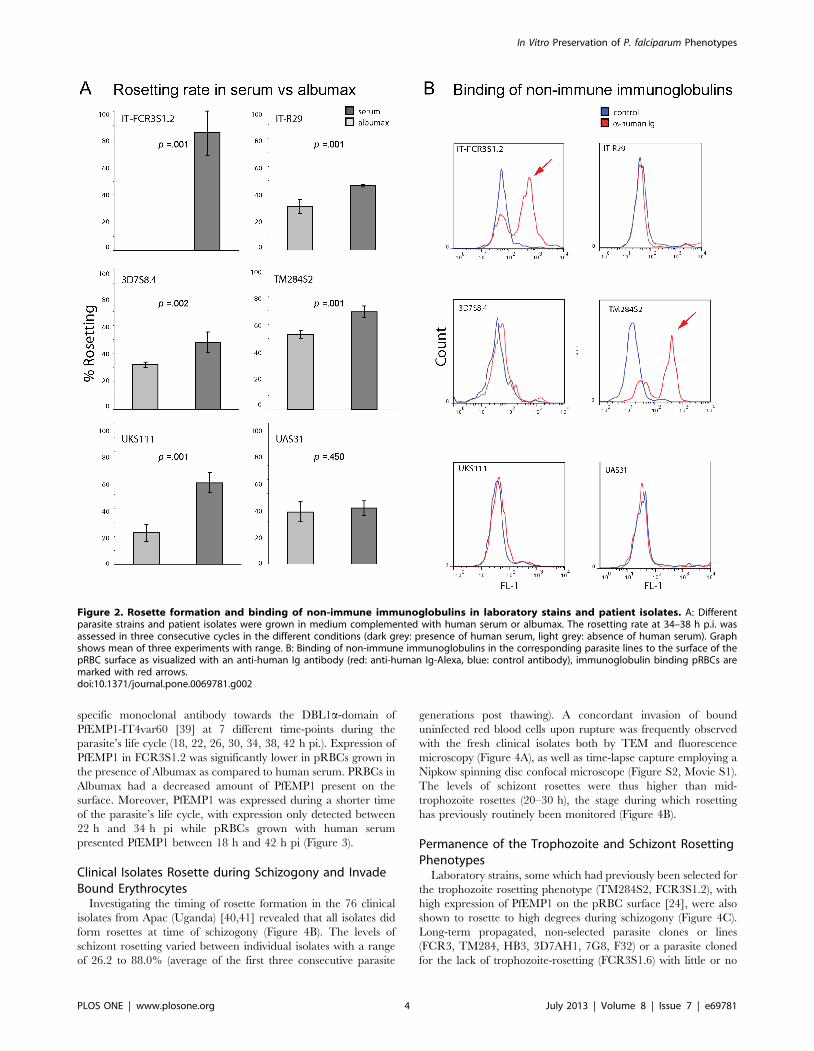
Advantageous in vitro Growth Conditions for Formationof Rosettes in P. falciparum Strains
Growth conditions in vitro that resemble conditions met by the
parasite in vivo are important for the correct development of the
adhesive phenotype of the parasite. Different laboratory parasite
strains (FCR3S1.2, R29, 3D7S8.4, TM284S2) and two patient
isolates with a rosetting phenotype were propagated and analyzed
in presence or absence of human serum. Parasites were adapted
for three generations to medium with or without serum and
afterwards monitored for their capacity to form rosettes during
three consecutive generations. The dependence of rosetting on the
presence or absence of human serum varied in different parasite
strains (Figure 2A). FCR3S1.2 pRBCs did not show any rosettes
when grown in absence of human serum, while the rosetting rate
was around 80% in the presence of human serum. Switching of
culture conditions rapidly re-established the rosetting phenotype in
the presence of human serum, while it was lost by a change to
conditions without human serum (data not shown). Rosetting
disappeared partly in the parasite strain R29, 3D7S8.4, TM284S2
and the patient isolate UKS111 when no human serum was
present. In contrast the patient isolate UAS31 did not show any
loss in the ability to form rosettes when depleted of human serum.
No correlation between the capacity to bind non-immune Ig to the
pRBC surface and dependence of rosetting on presence of human
serum was observed, except for the parasite strain FCR3S1.2 with
a high capacity to bind non-immune human serum which
completely lost the rosetting phenotype when human serum was
removed from the cultures (Figure 2B).
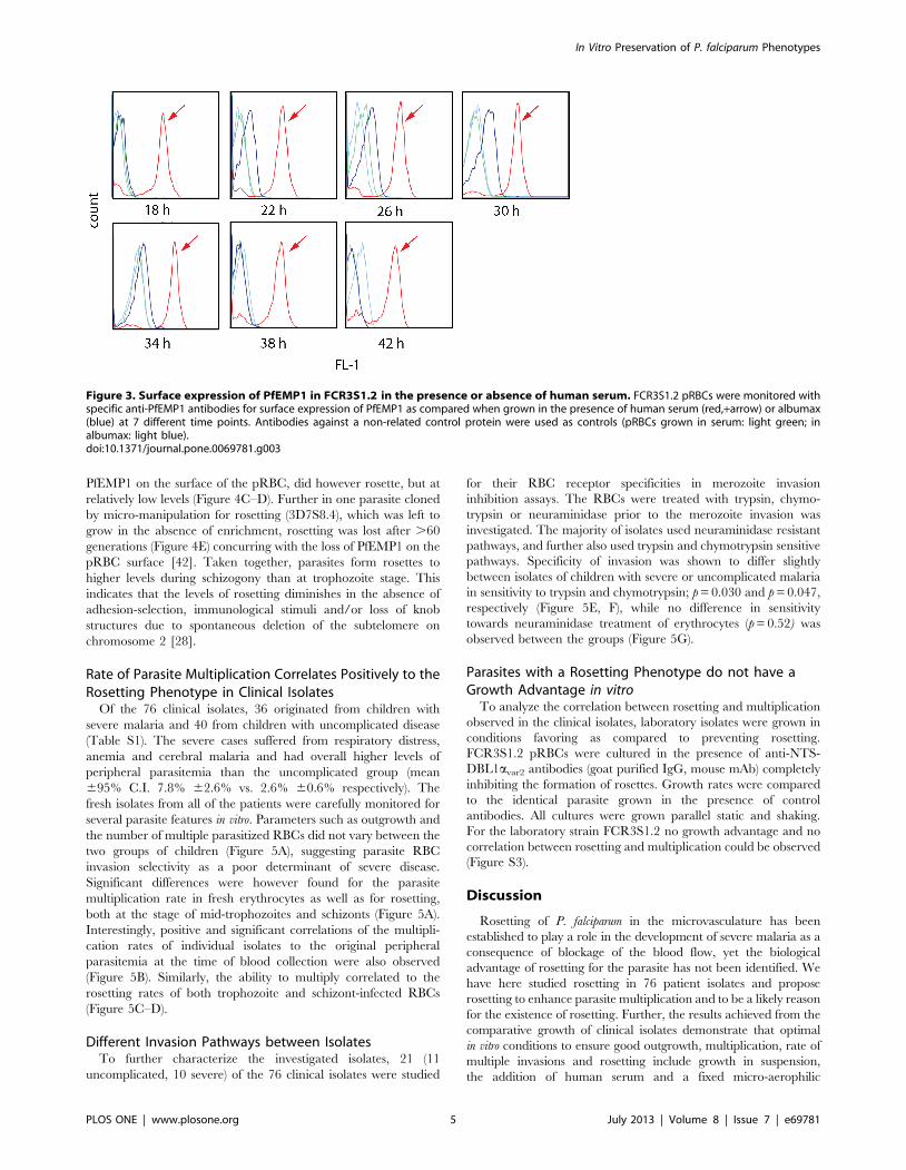
Importance of in vitro Growth Conditions for PfEMP1Surface Expression by P. falciparum Strains
Expression of the parasite derived surface protein PfEMP1 was
analyzed in the different growing conditions: The parasite strain
FCR3S1.2 expressing the PfEMP1 encoded by the IT4var60 gene
was grown in presence or absence of human serum and the
exposure of PfEMP1 on the pRBC surface was monitored with a
In Vitro Preservation of P. falciparum Phenotypes
PLOS ONE | www.plosone.org 2 July 2013 | Volume 8 | Issue 7 | e69781
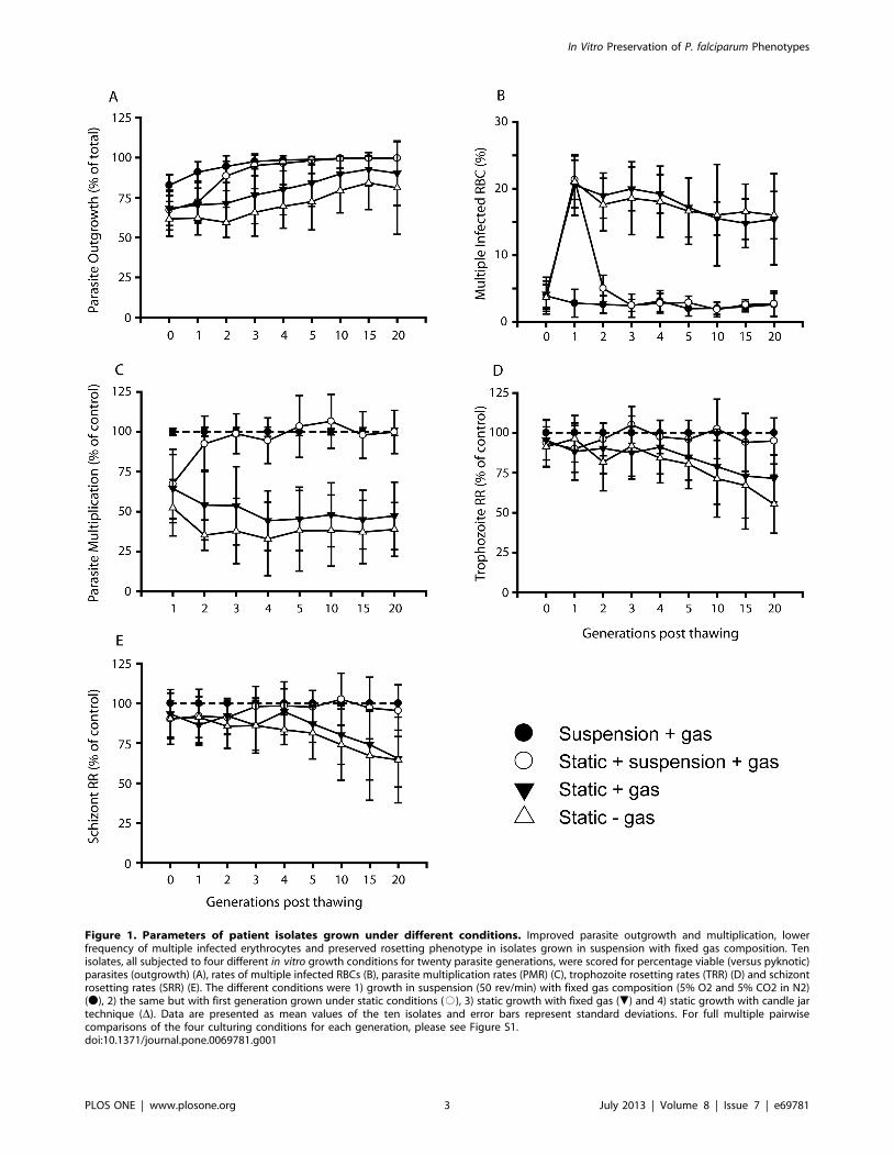
Figure 1. Parameters of patient isolates grown under different conditions. Improved parasite outgrowth and multiplication, lowerfrequency of multiple infected erythrocytes and preserved rosetting phenotype in isolates grown in suspension with fixed gas composition. Tenisolates, all subjected to four different in vitro growth conditions for twenty parasite generations, were scored for percentage viable (versus pyknotic)parasites (outgrowth) (A), rates of multiple infected RBCs (B), parasite multiplication rates (PMR) (C), trophozoite rosetting rates (TRR) (D) and schizontrosetting rates (SRR) (E). The different conditions were 1) growth in suspension (50 rev/min) with fixed gas composition (5% O2 and 5% CO2 in N2)(N), 2) the same but with first generation grown under static conditions (#), 3) static growth with fixed gas (.) and 4) static growth with candle jartechnique (D). Data are presented as mean values of the ten isolates and error bars represent standard deviations. For full multiple pairwisecomparisons of the four culturing conditions for each generation, please see Figure S1.doi:10.1371/journal.pone.0069781.g001
In Vitro Preservation of P. falciparum Phenotypes
PLOS ONE | www.plosone.org 3 July 2013 | Volume 8 | Issue 7 | e69781
specific monoclonal antibody towards the DBL1a-domain of
PfEMP1-IT4var60 [39] at 7 different time-points during the
parasite’s life cycle (18, 22, 26, 30, 34, 38, 42 h pi.). Expression of
PfEMP1 in FCR3S1.2 was significantly lower in pRBCs grown in
the presence of Albumax as compared to human serum. PRBCs in
Albumax had a decreased amount of PfEMP1 present on the
surface. Moreover, PfEMP1 was expressed during a shorter time
of the parasite’s life cycle, with expression only detected between
22 h and 34 h pi while pRBCs grown with human serum
presented PfEMP1 between 18 h and 42 h pi (Figure 3).
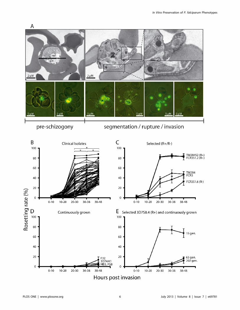
Clinical Isolates Rosette during Schizogony and InvadeBound Erythrocytes
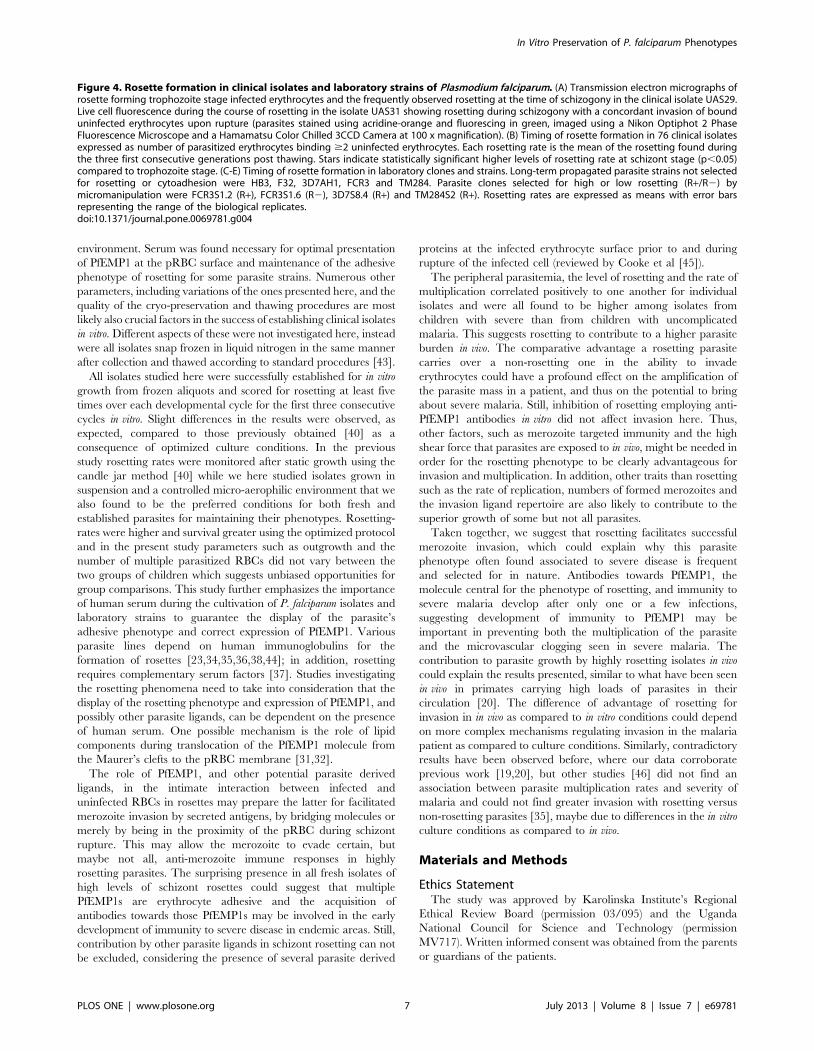
Investigating the timing of rosette formation in the 76 clinical
isolates from Apac (Uganda) [40,41] revealed that all isolates did
form rosettes at time of schizogony (Figure 4B). The levels of
schizont rosetting varied between individual isolates with a range
of 26.2 to 88.0% (average of the first three consecutive parasite
generations post thawing). A concordant invasion of bound
uninfected red blood cells upon rupture was frequently observed
with the fresh clinical isolates both by TEM and fluorescence
microscopy (Figure 4A), as well as time-lapse capture employing a
Nipkow spinning disc confocal microscope (Figure S2, Movie S1).
The levels of schizont rosettes were thus higher than mid-
trophozoite rosettes (20–30 h), the stage during which rosetting
has previously routinely been monitored (Figure 4B).
Permanence of the Trophozoite and Schizont RosettingPhenotypes
Laboratory strains, some which had previously been selected for
the trophozoite rosetting phenotype (TM284S2, FCR3S1.2), with
high expression of PfEMP1 on the pRBC surface [24], were also
shown to rosette to high degrees during schizogony (Figure 4C).
Long-term propagated, non-selected parasite clones or lines
(FCR3, TM284, HB3, 3D7AH1, 7G8, F32) or a parasite cloned
for the lack of trophozoite-rosetting (FCR3S1.6) with little or no
Figure 2. Rosette formation and binding of non-immune immunoglobulins in laboratory stains and patient isolates. A: Differentparasite strains and patient isolates were grown in medium complemented with human serum or albumax. The rosetting rate at 34–38 h p.i. wasassessed in three consecutive cycles in the different conditions (dark grey: presence of human serum, light grey: absence of human serum). Graphshows mean of three experiments with range. B: Binding of non-immune immunoglobulins in the corresponding parasite lines to the surface of thepRBC surface as visualized with an anti-human Ig antibody (red: anti-human Ig-Alexa, blue: control antibody), immunoglobulin binding pRBCs aremarked with red arrows.doi:10.1371/journal.pone.0069781.g002
In Vitro Preservation of P. falciparum Phenotypes
PLOS ONE | www.plosone.org 4 July 2013 | Volume 8 | Issue 7 | e69781
PfEMP1 on the surface of the pRBC, did however rosette, but at
relatively low levels (Figure 4C–D). Further in one parasite cloned
by micro-manipulation for rosetting (3D7S8.4), which was left to
grow in the absence of enrichment, rosetting was lost after .60
generations (Figure 4E) concurring with the loss of PfEMP1 on the
pRBC surface [42]. Taken together, parasites form rosettes to
higher levels during schizogony than at trophozoite stage. This
indicates that the levels of rosetting diminishes in the absence of
adhesion-selection, immunological stimuli and/or loss of knob
structures due to spontaneous deletion of the subtelomere on
chromosome 2 [28].
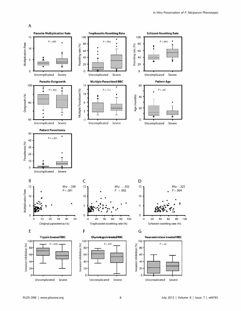
Rate of Parasite Multiplication Correlates Positively to theRosetting Phenotype in Clinical Isolates
Of the 76 clinical isolates, 36 originated from children with
severe malaria and 40 from children with uncomplicated disease
(Table S1). The severe cases suffered from respiratory distress,
anemia and cerebral malaria and had overall higher levels of
peripheral parasitemia than the uncomplicated group (mean
695% C.I. 7.8% 62.6% vs. 2.6% 60.6% respectively). The
fresh isolates from all of the patients were carefully monitored for
several parasite features in vitro. Parameters such as outgrowth and
the number of multiple parasitized RBCs did not vary between the
two groups of children (Figure 5A), suggesting parasite RBC
invasion selectivity as a poor determinant of severe disease.
Significant differences were however found for the parasite
multiplication rate in fresh erythrocytes as well as for rosetting,
both at the stage of mid-trophozoites and schizonts (Figure 5A).
Interestingly, positive and significant correlations of the multipli-
cation rates of individual isolates to the original peripheral
parasitemia at the time of blood collection were also observed
(Figure 5B). Similarly, the ability to multiply correlated to the
rosetting rates of both trophozoite and schizont-infected RBCs
(Figure 5C–D).
Different Invasion Pathways between IsolatesTo further characterize the investigated isolates, 21 (11
uncomplicated, 10 severe) of the 76 clinical isolates were studied
for their RBC receptor specificities in merozoite invasion
inhibition assays. The RBCs were treated with trypsin, chymo-
trypsin or neuraminidase prior to the merozoite invasion was
investigated. The majority of isolates used neuraminidase resistant
pathways, and further also used trypsin and chymotrypsin sensitive
pathways. Specificity of invasion was shown to differ slightly
between isolates of children with severe or uncomplicated malaria
in sensitivity to trypsin and chymotrypsin; p = 0.030 and p = 0.047,
respectively (Figure 5E, F), while no difference in sensitivity
towards neuraminidase treatment of erythrocytes (p = 0.52) was
observed between the groups (Figure 5G).
Parasites with a Rosetting Phenotype do not have aGrowth Advantage in vitro
To analyze the correlation between rosetting and multiplication
observed in the clinical isolates, laboratory isolates were grown in
conditions favoring as compared to preventing rosetting.
FCR3S1.2 pRBCs were cultured in the presence of anti-NTS-
DBL1avar2 antibodies (goat purified IgG, mouse mAb) completely
inhibiting the formation of rosettes. Growth rates were compared
to the identical parasite grown in the presence of control
antibodies. All cultures were grown parallel static and shaking.
For the laboratory strain FCR3S1.2 no growth advantage and no
correlation between rosetting and multiplication could be observed
(Figure S3).
Discussion
Rosetting of P. falciparum in the microvasculature has been
established to play a role in the development of severe malaria as a
consequence of blockage of the blood flow, yet the biological
advantage of rosetting for the parasite has not been identified. We
have here studied rosetting in 76 patient isolates and propose
rosetting to enhance parasite multiplication and to be a likely reason
for the existence of rosetting. Further, the results achieved from the
comparative growth of clinical isolates demonstrate that optimal
in vitro conditions to ensure good outgrowth, multiplication, rate of
multiple invasions and rosetting include growth in suspension,
the addition of human serum and a fixed micro-aerophilic
Figure 3. Surface expression of PfEMP1 in FCR3S1.2 in the presence or absence of human serum. FCR3S1.2 pRBCs were monitored withspecific anti-PfEMP1 antibodies for surface expression of PfEMP1 as compared when grown in the presence of human serum (red,+arrow) or albumax(blue) at 7 different time points. Antibodies against a non-related control protein were used as controls (pRBCs grown in serum: light green; inalbumax: light blue).doi:10.1371/journal.pone.0069781.g003
In Vitro Preservation of P. falciparum Phenotypes
PLOS ONE | www.plosone.org 5 July 2013 | Volume 8 | Issue 7 | e69781
In Vitro Preservation of P. falciparum Phenotypes
PLOS ONE | www.plosone.org 6 July 2013 | Volume 8 | Issue 7 | e69781
environment. Serum was found necessary for optimal presentation
of PfEMP1 at the pRBC surface and maintenance of the adhesive
phenotype of rosetting for some parasite strains. Numerous other
parameters, including variations of the ones presented here, and the
quality of the cryo-preservation and thawing procedures are most
likely also crucial factors in the success of establishing clinical isolates
in vitro. Different aspects of these were not investigated here, instead
were all isolates snap frozen in liquid nitrogen in the same manner
after collection and thawed according to standard procedures [43].
All isolates studied here were successfully established for in vitro
growth from frozen aliquots and scored for rosetting at least five
times over each developmental cycle for the first three consecutive
cycles in vitro. Slight differences in the results were observed, as
expected, compared to those previously obtained [40] as a
consequence of optimized culture conditions. In the previous
study rosetting rates were monitored after static growth using the
candle jar method [40] while we here studied isolates grown in
suspension and a controlled micro-aerophilic environment that we
also found to be the preferred conditions for both fresh and
established parasites for maintaining their phenotypes. Rosetting-
rates were higher and survival greater using the optimized protocol
and in the present study parameters such as outgrowth and the
number of multiple parasitized RBCs did not vary between the
two groups of children which suggests unbiased opportunities for
group comparisons. This study further emphasizes the importance
of human serum during the cultivation of P. falciparum isolates and
laboratory strains to guarantee the display of the parasite’s
adhesive phenotype and correct expression of PfEMP1. Various
parasite lines depend on human immunoglobulins for the
formation of rosettes [23,34,35,36,38,44]; in addition, rosetting
requires complementary serum factors [37]. Studies investigating
the rosetting phenomena need to take into consideration that the
display of the rosetting phenotype and expression of PfEMP1, and
possibly other parasite ligands, can be dependent on the presence
of human serum. One possible mechanism is the role of lipid
components during translocation of the PfEMP1 molecule from
the Maurer’s clefts to the pRBC membrane [31,32].
The role of PfEMP1, and other potential parasite derived
ligands, in the intimate interaction between infected and
uninfected RBCs in rosettes may prepare the latter for facilitated
merozoite invasion by secreted antigens, by bridging molecules or
merely by being in the proximity of the pRBC during schizont
rupture. This may allow the merozoite to evade certain, but
maybe not all, anti-merozoite immune responses in highly
rosetting parasites. The surprising presence in all fresh isolates of
high levels of schizont rosettes could suggest that multiple
PfEMP1s are erythrocyte adhesive and the acquisition of
antibodies towards those PfEMP1s may be involved in the early
development of immunity to severe disease in endemic areas. Still,
contribution by other parasite ligands in schizont rosetting can not
be excluded, considering the presence of several parasite derived
proteins at the infected erythrocyte surface prior to and during
rupture of the infected cell (reviewed by Cooke et al [45]).
The peripheral parasitemia, the level of rosetting and the rate of
multiplication correlated positively to one another for individual
isolates and were all found to be higher among isolates from
children with severe than from children with uncomplicated
malaria. This suggests rosetting to contribute to a higher parasite
burden in vivo. The comparative advantage a rosetting parasite
carries over a non-rosetting one in the ability to invade
erythrocytes could have a profound effect on the amplification of
the parasite mass in a patient, and thus on the potential to bring
about severe malaria. Still, inhibition of rosetting employing anti-
PfEMP1 antibodies in vitro did not affect invasion here. Thus,
other factors, such as merozoite targeted immunity and the high
shear force that parasites are exposed to in vivo, might be needed in
order for the rosetting phenotype to be clearly advantageous for
invasion and multiplication. In addition, other traits than rosetting
such as the rate of replication, numbers of formed merozoites and
the invasion ligand repertoire are also likely to contribute to the
superior growth of some but not all parasites.
Taken together, we suggest that rosetting facilitates successful
merozoite invasion, which could explain why this parasite
phenotype often found associated to severe disease is frequent
and selected for in nature. Antibodies towards PfEMP1, the
molecule central for the phenotype of rosetting, and immunity to
severe malaria develop after only one or a few infections,
suggesting development of immunity to PfEMP1 may be
important in preventing both the multiplication of the parasite
and the microvascular clogging seen in severe malaria. The
contribution to parasite growth by highly rosetting isolates in vivo
could explain the results presented, similar to what have been seen
in vivo in primates carrying high loads of parasites in their
circulation [20]. The difference of advantage of rosetting for
invasion in in vivo as compared to in vitro conditions could depend
on more complex mechanisms regulating invasion in the malaria
patient as compared to culture conditions. Similarly, contradictory
results have been observed before, where our data corroborate
previous work [19,20], but other studies [46] did not find an
association between parasite multiplication rates and severity of
malaria and could not find greater invasion with rosetting versus
non-rosetting parasites [35], maybe due to differences in the in vitro
culture conditions as compared to in vivo.
Materials and Methods
Ethics StatementThe study was approved by Karolinska Institute’s Regional
Ethical Review Board (permission 03/095) and the Uganda
National Council for Science and Technology (permission
MV717). Written informed consent was obtained from the parents
or guardians of the patients.
Figure 4. Rosette formation in clinical isolates and laboratory strains of Plasmodium falciparum. (A) Transmission electron micrographs ofrosette forming trophozoite stage infected erythrocytes and the frequently observed rosetting at the time of schizogony in the clinical isolate UAS29.Live cell fluorescence during the course of rosetting in the isolate UAS31 showing rosetting during schizogony with a concordant invasion of bounduninfected erythrocytes upon rupture (parasites stained using acridine-orange and fluorescing in green, imaged using a Nikon Optiphot 2 PhaseFluorescence Microscope and a Hamamatsu Color Chilled 3CCD Camera at 100 x magnification). (B) Timing of rosette formation in 76 clinical isolatesexpressed as number of parasitized erythrocytes binding $2 uninfected erythrocytes. Each rosetting rate is the mean of the rosetting found duringthe three first consecutive generations post thawing. Stars indicate statistically significant higher levels of rosetting rate at schizont stage (p,0.05)compared to trophozoite stage. (C-E) Timing of rosette formation in laboratory clones and strains. Long-term propagated parasite strains not selectedfor rosetting or cytoadhesion were HB3, F32, 3D7AH1, FCR3 and TM284. Parasite clones selected for high or low rosetting (R+/R2) bymicromanipulation were FCR3S1.2 (R+), FCR3S1.6 (R2), 3D7S8.4 (R+) and TM284S2 (R+). Rosetting rates are expressed as means with error barsrepresenting the range of the biological replicates.doi:10.1371/journal.pone.0069781.g004
In Vitro Preservation of P. falciparum Phenotypes
PLOS ONE | www.plosone.org 7 July 2013 | Volume 8 | Issue 7 | e69781
In Vitro Preservation of P. falciparum Phenotypes
PLOS ONE | www.plosone.org 8 July 2013 | Volume 8 | Issue 7 | e69781
ParasitesParasites used were long-term propagated parasite strains not
selected for rosetting or cytoadhesion (HB3, F32, 3D7AH1, FCR3,
R29 and TM284), parasite clones selected for high or low rosetting
by micro-manipulation (FCR3S1.2, FCR3S1.6, TM284S2 and
3D7S8.4) and 76 clinical isolates collected in Apac, Uganda 2002
[40], see table S1. The isolates were all randomly selected from
unique patients and were shown to be genetically distinct by msp1,
msp2, glurp and csp genotyping (data not shown). Patients were
under the age of five and subdivided into two groups, severe
(n = 36) or uncomplicated (n = 40) falciparum malaria, using WHO
guidelines [47] and the modified Blantyre score [48]. The
parasitemia and the number of multiple infected RBCs were
estimated from Giemsa-stained thin smears obtained directly from
the peripheral blood of the patient [40].
Evaluation of in vitro Growth conditions for ThawedClinical Isolates
Ten clinical isolates were thawed according to standard
procedures and transferred to 5 ml malaria culture medium
[43,49] (tissue culture flasks 25cm2, Falcon, nr. 353082) containing
12.5% non-immune human AB+ serum and sub-cultivated in fresh
O+ RBCs during the first cycle in vitro (see below for details). Each
sub-cultivated isolate were subjected to four different culture
conditions (all at 37uC) involving the use of a gas mixture (5% O2
and 5% CO2 in N2), classical candle jar [49], growth in static
manner, growth in suspension on an orbital shaker (50 rpm)
[50,51] or combinations thereof. The differently treated cultures
were evaluated for outgrowth, the rate of multiple parasitized
RBCs, multiplication rates and rosetting rates (see below for
details).
Evaluation of Parasite Outgrowth, Multiple Infected RBCs,Parasite Multiplication Rates and Rosette Formation
All 76 isolates were upon thawing successfully grown until
mature trophozoite stages before sub-cultivated at least 1:10 to a
parasitemia in the range 0.1–0.3% in fresh O+ RBCs. The same
serum batch was used as supplement for all cultures, and an equal
number of uncomplicated and severe isolates were cultured in the
same batch of O+ RBCs (both serum and RBCs were pooled from
at least three individual donors to counteract oddities of individual
donors and to remove RBC polymorphisms as potential
confounders). Rosetting rates were assessed as previously described
[43] by staining 10 ml drops of resuspended parasite culture (0.5 -
2.5% parasitemia) with 2ml acridine orange (10mg/ml in PBS).
Rosetting rates were counted from at least 100 pRBCs in duplicate
in a Nikon Optiphot 2 Phase Fluorescence microscope (Nikon) and
pictures were taken using a Hamamatsu Color Chilled 3CCD
Camera (Hamamatsu Photonics). A trophozoite rosette was
defined as two or more uninfected RBCs bound to one or two
trophozoite-infected RBCs and a schizont rosette as two or more
uninfected RBCs bound to one or two schizont-infected RBCs.
The parasites were analyzed five times during intra-RBC
progression (0–10, 10–20, 20–30, 30–38 and 38–48 hours post
invasion) for the three first parasite generations. The rosetting-
rates presented for each time-point are the averages of the first
three consecutive parasite generations post thawing. Thin smears
stained with Giemsa were prepared when cultures were in ring
stages. Parasitemia was determined in at least 5,000 RBCs for the
original culture. The same was performed on both cultures after
sub-cultivation and after invasion in the next consecutive parasite
generation in order to compute multiplication rates. Further, the
number of viable versus pyknotic parasites and the number of
multiple infected RBCs was assessed. We have previously reported
the trophozoite rosetting rates for the samples studied here after
cultivation under static conditions using the candle-jar method in
Kampala (Uganda) [40]. The parasites studied herein are from
cryo-preserved stocks of the same samples propagated on an
orbital shaker (50 rev/min) with the use of a fixed gas composition
(5% O2, 5% CO2 and 90% N2) in Stockholm (Sweden).
For the evaluation of the importance of human serum for
rosetting and correct PfEMP1 surface expression, parasites were
grown in parallel in malaria complete medium containing 10%
human serum, or 0.5% Albumax II respectively.
Analysis of Surface Expression of PfEMP1 by FlowCytometry
Trophozoite-infected RBCs selected at seven different time-
points (18, 22, 26, 30, 34, 38 and 42 h p.i.) were incubated in a
final concentration of 50 ug/ml with a strain-specific mAb (a-
NTS-DBL1avar2 [39]) diluted in PBS/FCS (2%) for 30 min at RT.
After incubation with the primary antibody, three washes with
PBS/FCS was performed followed by 30 min incubation with
anti-mouse IgG second antibody ALEXA488 (Molecular Probes,
dilution 1:100) at RT. For nuclear staining ethidium bromide was
added at final concentration of 2.5mg/ml for 5 min at RT. The
pRBCs were washed three times and resuspended in PBS/FCS.
The cell acquisition was done using flow cytometry (FACSCalibur,
BD Bioscience, http://www.bd.com) where 5000 pRBCs were
counted. The analysis was performed using the software FlowJo.
Binding of non-immune human Ig was visualized as above using
an anti-human Ig FITC-coupled antibody (DAKO, F0200,
dilution 1:100). As a negative control a monoclonal antibody
generated in mouse against a non-related bacterial protein has
been used in all experiments.
Invasion AssaysPRBCs of the parasite strain FCR3S1.2 at trophozoite stage
were adjusted to a parasitemia of 0.5% and a hematocrit of 2.5%,
mixed with the purified IgG fraction of an a-NTS-DBL1avar2 goat
serum, a a-NTS-DBL1avar2 mAb (1, 0.5 and 0.25 mg/ml, in
duplicates) and cultivated in 96-well plates until reinvasion of
merozoites was completed. The applied antibodies were a-NTS-
DBL1avar2 mouse mAb or purified polyclonal goat IgG generated
against and specific for the DBL1a-domain of ITvar60 expressed
by FCR3S1.2 pRBCs [39].
Parasitemia was measured at ring stage (around 15 h p.i.) and
assays were performed under shaking versus non shaking
conditions under a fixed gas composition. In addition, goat IgG
was pre-absorbed by incubation with uninfected RBCs for 30 min
at RB prior to the invasion experiments. As a negative control,
Figure 5. Phenotypic characteristics of clinical isolates and age of children with severe or uncomplicated malaria. (A) Boxed plotsshowing group comparisons of isolates originating from patients with severe (n = 36) and uncomplicated (n = 40) malaria in respect to patient age,parasitemia, growth and rosetting during the first generation post thawing. (B-D) Scatter plots showing Spearman rank correlations between parasitemultiplication rates and original peripheral parasitemia of the patient or rosetting rates with mid-trophozoite or schizont infected RBCs for individualisolates. (E-G) Inhibition of invasion into trypsin-, chymotrypsin or neuraminidase treated erythrocytes by 11 P. falciparum isolates from children withuncomplicated malaria and 10 isolates of children with severe malaria.doi:10.1371/journal.pone.0069781.g005
In Vitro Preservation of P. falciparum Phenotypes
PLOS ONE | www.plosone.org 9 July 2013 | Volume 8 | Issue 7 | e69781
normal goat IgG or mouse IgG was used in the same
concentrations.
PRBCs were stained with Acridine Orange and counted by flow
cytometry (50,000 events). Experiments were repeated three times
and the effect was determined by relating the observed parasitemia
in the presence of PfEMP1var2-specific antibodies to invasion in
the presence of corresponding non-immune immunoglobulins.
Invasion into Enzyme Treated ErythrocytesHuman O+ erythrocytes were washed with RPMI 1640 (Gibco,
Invitrogen), and subsequently incubated with neuraminidase
(62.5 uM/ml; Sigma), trypsin (1mg/ml; Sigma) and chymotrypsin
(1 mg/ml; Sigma) for 45 minutes at 37 0C with periodic shaking.
Controls were only treated with RPMI 1640. After incubation,
enzyme treated cells were washed once with RPMI 1640
containing 20% human serum and twice with RPMI 1640
containing 10% human serum to inhibit enzyme activity [52].
Late-pigmented trophozoites to schizont stage were enriched
using magnetic bead column separation (Miltenyi Biotec, Ger-
many). Invasion assay was performed in 96 U-bottom culture
plates with total of 50 ml of parasite suspension at 0.5–1.0%
parasitemia and enzyme treated erythrocytes. All samples were
tested in triplicate. Plates were incubated in a gassed box and
incubated for 48 hours at 37uC. Parasitemia was estimated using
hydroethidine (10 ug/ml; Sigma) in flow cytometry (FACS Scan;
BD) after 48 hours. Inhibition by enzyme treatment was
determined as [1 2 (proportion of enzyme-treated cells invad-
ed/proportion of untreated cells invaded)] 6 100. Results
presented are in comparison with control treated cells [52].
Statistical AnalysisSigmaStat 3.1 (Systat Software, Erkrath, Germany) was used for
statistical analyses. Group comparisons (severe vs. uncomplicated)
for parasite multiplication rate and rosetting at mid-trophozoite
stage were analyzed using the non-parametric Mann-Whitney
rank sum test due to non-normality of the data, whereas all other
group comparisons were conducted using Student’s t-test. Corre-
lations between multiplication rates and rosetting rates as well as
multiplication rates and original peripheral parasitemia were
assessed using Spearman rank correlations. The four culture
conditions were evaluated and compared using one way ANOVA
with pairwise multiple comparisons using the Tukey for all
generations. The temporal differences in rosetting (trophozoites vs
schizonts) among the clinical isolates were analyzed by Kruskal-
Wallis ANOVA on ranks with pairwise multiple comparison
procedures using the Tukey test. A priori assumption of statistical
significance was in all cases determined to p,0.05.
Transmission Electron MicroscopyTransmission electron microscopy (TEM) was used to visualize
rosettes at both trophozoite and schizont stages. RBCs and pRBCs
of the clinical isolate UAS29 were fixed in 2% glutaraldehyde and
0.5% paraformaldehyde in 0.1 M sodiumcacodylate buffer
containing 0.1 M sucrose (pH 7.4) at room temperature for
30 min. Fixed cells were rinsed in 0.15 M sodiumcacodylate buffer
(pH 7.4), pelleted by centrifugation, post fixed in 2% osmium
tetroxide buffer (pH 7.4) at 4uC for 2 hours, dehydrated in ethanol
and acetone before embedded in LX-112 (Ladd Research
Industries, Burlington, VT, USA). Sections were contrasted with
uranyl acetate followed by lead citrate and examined in Leo 906
transmission electron microscope at 80 kV. Digital images were
taken using a Morada digital camera (Olympus Soft Imaging
System, Munster, Germany) [53].
Time-lapse Capture of Rupture and Invasion fromRosettes
The time-lapse movie was made using an automated confocal
microscope, a custom modified LCI system (Perkin-Elmer, Upp-
lands Vasby, Sweden). The system includes a motorized Axiovert
fluorescence microscope 200 M (Zeiss, Gottingen, Germany), a
CSU10 Nipkow spinning disc (Yokogawa, Japan), a motorized
XY-table (Marzhauser), an ORCA ER cold CCD camera;
detector array 1344 x 1024 px (Hamamatsu, Hamamatsu City,
Japan), a 3-line Argon-Krypton Laser; 488 nm, 568 nm, 647 nm
(Melles Griot, Stockholm, Sweden) and halogen illumination for
bright field and phase contrast mode. A 63X/NA 1.25 Oil Ph3
Plan – Neofluor ‘/0.17, Zeiss objective was used. The images
were captured using the movie automation FiveColorMovie with
autofocus function, developed by authors (EF+LS) in the visual
programming language environment of Openlab automator. For
each time-point, 4 phase-contrast images (350 ms exposure/
image) were captured through a 1mm thick z-depth (0.3 mm/step).
The interval between time-points was 30 seconds.
Supporting Information
Figure S1 Pairwise multiple comparisons of parasitegrowth and rosetting for the four evaluated cultureconditions. Statistical analyses of the data presented in Figure 1,
describing the differences in parasite outgrowth, multiple invaded
RBCs, parasite multiplication and rosetting for all parasite
generations (G0–G20) analyzed. The different conditions were 1)
growth in suspension (50 rev/min) with fixed gas composition (5%
O2 and 5% CO2 in N2) (N), 2) the same but with first generation
grown under static conditions (#), 3) static growth with fixed gas
(.) and 4) static growth with candle jar technique (D). All
statistically significant pairwise multiple comparisons are shaded in
light green (p,0.05), and non-significant data in orange.
Quadrants lacking numbers indicate too small differences in mean
values among the groups and therefore failed test due to low
power.
(PDF)
Figure S2 Time-lapse capture of schizont rupture froma rosette under static growth. Timing of schizont rupture in
rosetting FCR3S1.2. Four phase-contrast images were captured
through a 1 mm thick z-depth using a Nipkow spinning disc
confocal microscope with 30 second intervals between time-points,
visualizing the invasion of bound erythrocytes from schizont
rosettes upon rupture. In total 5.5 minutes of real-time capture is
represented, with timing indicated in the bottom-right of each
panel. The time-laps capture can also be viewed as continuous in
Movie S1. Panel 1 (0–1 min): A schizont pRBC (white arrow)
ruptures after 1 min while still attached to five RBCs, with a
concomitant egress of merozoites. A few free merozoites are visual
(red arrows). Panel 2 (1.5–2.5 min): Four of the bound RBCs are
immediately invaded by merozoites (green arrows), while free
merozoites can still be seen (red arrows). Panel 3 (3–4 min): The
cells from the original rosette still attach to the remnants of the
ruptured pRBC and each other and the fifth RBC gets invaded by
merozoite (green arrow). Panel 4 (4.5–5.5 min): All of the RBCs
from the original rosette have been turned into pRBCs (white
arrows) and still surround and attach to the remnant pRBC ghost
and each other, albeit the interactions are weakening.
(TIF)
Figure S3 Correlation between rosetting and invasionin the laboratory strain FCR3S1.2. Invasion inhibition using
rosette-disruptive antibodies against the PfEMP1 variant displayed
In Vitro Preservation of P. falciparum Phenotypes
PLOS ONE | www.plosone.org 10 July 2013 | Volume 8 | Issue 7 | e69781
by FCR3S1.2. Goat IgG aNTS-DBL1a, non-absorbed or pre-
absorbed on RBCs or monoclonal aNTS-DBL1a antibodies were
added to early trophozoite stage pRBCs at concentrations of 1, 0.5
and 0.25 mg/ml, parasites were grown with gas under shaking
conditions, allowed to invade and parasitemia was measured
thereafter. Non-related antibodies from the same species were
used as controls. The level of invasion was comparable in the
presence or absence of antibodies blocking invasion and no
correlation between the level of rosetting and invasion could be
observed for this long term propagated laboratory strain. Graph
shows median of three experiments with range.
(PDF)
Table S1 Summary of patient and P. falciparum isolatedata.(DOC)
Movie S1 Time-lapse capture of schizont rupture andmerozoite invasion from rosettes. Timing of schizont
rupture and merozoite invasion in rosetting FCR3S1.2. Four
phase-contrast images were captured through a 1 mm thick z-
depth using a Nipkow spinning disc confocal microscope with 30
second intervals between time-points, visualizing the invasion of
bound erythrocytes from schizont rosettes upon rupture. In total
7.5 min of real-time capture is represented, with timing indicated
in the bottom-right of the screen. Events shown in this movie are
described in detail in figure legend for Figure S2.
(MOV)
Acknowledgments
We thank Ingrid Lindell for technical support with the transmission
electron microscopy.
Author Contributions
Conceived and designed the experiments: UR KM KEMP TGE LS MW.
Performed the experiments: UR KM LA HAI JN EF KH. Analyzed the
data: UR KM LS MW. Contributed reagents/materials/analysis tools: JN
KH LS TGE. Wrote the paper: UR KM KEMP MW.
References
1. Carlson J, Helmby H, Hill AV, Brewster D, Greenwood BM, et al. (1990)Human cerebral malaria: association with erythrocyte rosetting and lack of anti-
rosetting antibodies. Lancet 336: 1457–1460.
2. Kaul DK, Roth EF Jr, Nagel RL, Howard RJ, Handunnetti SM (1991)
Rosetting of Plasmodium falciparum-infected red blood cells with uninfected redblood cells enhances microvascular obstruction under flow conditions. Blood 78:
812–819.
3. Miller LH, Good MF, Milon G (1994) Malaria pathogenesis. Science 264: 1878–1883.
4. Rowe A, Obeiro J, Newbold CI, Marsh K (1995) Plasmodium falciparumrosetting is associated with malaria severity in Kenya. Infect Immun 63: 2323–
2326.
5. Silamut K, Phu NH, Whitty C, Turner GD, Louwrier K, et al. (1999) Aquantitative analysis of the microvascular sequestration of malaria parasites in
the human brain. Am J Pathol 155: 395–410.
6. Pasvol G, Weatherall DJ, Wilson RJ (1978) Cellular mechanism for the
protective effect of haemoglobin S against P. falciparum malaria. Nature 274:701–703.
7. Friedman MJ (1978) Erythrocytic mechanism of sickle cell resistance to malaria.
Proc Natl Acad Sci U S A 75: 1994–1997.
8. Luzzi GA, Merry AH, Newbold CI, Marsh K, Pasvol G (1991) Protection by
alpha-thalassaemia against Plasmodium falciparum malaria: modified surfaceantigen expression rather than impaired growth or cytoadherence. Immunol
Lett 30: 233–240.
9. Carlson J, Nash GB, Gabutti V, al-Yaman F, Wahlgren M (1994) Natural
protection against severe Plasmodium falciparum malaria due to impaired
rosette formation. Blood 84: 3909–3914.
10. Luzzi GA, Merry AH, Newbold CI, Marsh K, Pasvol G, et al. (1991) Surface
antigen expression on Plasmodium falciparum-infected erythrocytes is modifiedin alpha- and beta-thalassemia. J Exp Med 173: 785–791.
11. Rowe JA, Moulds JM, Newbold CI, Miller LH (1997) P. falciparum rosetting
mediated by a parasite-variant erythrocyte membrane protein and complement-receptor 1. Nature 388: 292–295.
12. Fairhurst RM, Baruch DI, Brittain NJ, Ostera GR, Wallach JS, et al. (2005)Abnormal display of PfEMP-1 on erythrocytes carrying haemoglobin C may
protect against malaria. Nature 435: 1117–1121.
13. Carlson J, Wahlgren M (1992) Plasmodium falciparum erythrocyte rosetting is
mediated by promiscuous lectin-like interactions. J Exp Med 176: 1311–1317.
14. Rowe JA, Handel IG, Thera MA, Deans AM, Lyke KE, et al. (2007) Bloodgroup O protects against severe Plasmodium falciparum malaria through the
mechanism of reduced rosetting. Proc Natl Acad Sci U S A 104: 17471–17476.
15. Fry AE, Griffiths MJ, Auburn S, Diakite M, Forton JT, et al. (2008) Common
variation in the ABO glycosyltransferase is associated with susceptibility to severe
Plasmodium falciparum malaria. Hum Mol Genet 17: 567–576.
16. Cyrklaff M, Sanchez CP, Kilian N, Bisseye C, Simpore J, et al. (2007)
Hemoglobins S and C interfere with actin remodeling in Plasmodiumfalciparum-infected erythrocytes. Science 334: 1283–1286.
17. Cholera R, Brittain NJ, Gillrie MR, Lopera-Mesa TM, Diakite SA, et al. (2008)Impaired cytoadherence of Plasmodium falciparum-infected erythrocytes
containing sickle hemoglobin. Proc Natl Acad Sci U S A 105: 991–996.
18. Dondorp AM, Desakorn V, Pongtavornpinyo W, Sahassananda D, Silamut K,et al. (2005) Estimation of the total parasite biomass in acute falciparum malaria
from plasma PfHRP2. PLoS Med 2: e204.
19. Chotivanich K, Udomsangpetch R, Simpson JA, Newton P, Pukrittayakamee S,
et al. (2000) Parasite multiplication potential and the severity of Falciparum
malaria. J Infect Dis 181: 1206–1209.
20. Le Scanf C, Vigan-Womas I, Contamin H, Guillotte M, Bischoff E, et al. (2008)Rosetting is associated with increased Plasmodium falciparum in vivo multipli-
cation rate in the Saimiri sciureus monkey. Microbes Infect 10: 447–451.
21. David PH, Handunnetti SM, Leech JH, Gamage P, Mendis KN (1988)
Rosetting: a new cytoadherence property of malaria-infected erythrocytes.
Am J Trop Med Hyg 38: 289–297.
22. Wahlgren M, Carlson J, Udomsangpetch R, Perlmann P (1989) Why do
Plasmodium falciparumm-infected erythrocytes form spontaneous erythrocyterosettes? Parasitol Today 5: 183–185.
23. Heddini A, Pettersson F, Kai O, Shafi J, Obiero J, et al. (2001) Fresh isolates
from children with severe Plasmodium falciparum malaria bind to multiplereceptors. Infect Immun 69: 5849–5856.
24. Fernandez V, Treutiger CJ, Nash GB, Wahlgren M (1998) Multiple adhesive
phenotypes linked to rosetting binding of erythrocytes in Plasmodium falciparummalaria. Infect Immun 66: 2969–2975.
25. al-Yaman F, Genton B, Mokela D, Raiko A, Kati S, et al. (1995) Humancerebral malaria: lack of significant association between erythrocyte rosetting
and disease severity. Trans R Soc Trop Med Hyg 89: 55–58.
26. Horata N, Kalambaheti T, Craig A, Khusmith S (2009) Sequence variation ofPfEMP1-DBLalpha in association with rosette formation in Plasmodium
falciparum isolates causing severe and uncomplicated malaria. Malar J 8: 184.
27. Udeinya IJ, Graves PM, Carter R, Aikawa M, Miller LH (1983) Plasmodiumfalciparum: effect of time in continuous culture on binding to human endothelial
cells and amelanotic melanoma cells. Exp Parasitol 56: 207–214.
28. Biggs BA, Kemp DJ, Brown GV (1989) Subtelomeric chromosome deletions in
field isolates of Plasmodium falciparum and their relationship to loss of
cytoadherence in vitro. Proc Natl Acad Sci U S A 86: 2428–2432.
29. Gysin J, Pouvelle B, Fievet N, Scherf A, Lepolard C (1999) Ex vivo
desequestration of Plasmodium falciparum-infected erythrocytes from humanplacenta by chondroitin sulfate A. Infect Immun 67: 6596–6602.
30. Beeson JG, Brown GV, Molyneux ME, Mhango C, Dzinjalamala F, et al. (1999)
Plasmodium falciparum isolates from infected pregnant women and children areassociated with distinct adhesive and antigenic properties. J Infect Dis 180: 464–
472.
31. Frankland S, Adisa A, Horrocks P, Taraschi TF, Schneider T, et al. (2006)Delivery of the malaria virulence protein PfEMP1 to the erythrocyte surface
requires cholesterol-rich domains. Eukaryot Cell 5: 849–860.
32. Frankland S, Elliott SR, Yosaatmadja F, Beeson JG, Rogerson SJ, et al. (2007)
Serum lipoproteins promote efficient presentation of the malaria virulence
protein PfEMP1 at the erythrocyte surface. Eukaryot Cell 6: 1584–1594.
33. Luginbuhl A, Nikolic M, Beck HP, Wahlgren M, Lutz HU (2007) Complement
factor D, albumin, and immunoglobulin G anti-band 3 protein antibodies mimicserum in promoting rosetting of malaria-infected red blood cells. Infect Immun
75: 1771–1777.
34. Scholander C, Treutiger CJ, Hultenby K, Wahlgren M (1996) Novel fibrillarstructure confers adhesive property to malaria-infected erythrocytes. Nat Med 2:
204–208.
35. Clough B, Atilola FA, Black J, Pasvol G (1998) Plasmodium falciparum: theimportance of IgM in the rosetting of parasite-infected erythrocytes. Exp
Parasitol 89: 129–132.
36. Treutiger CJ, Scholander C, Carlson J, McAdam KP, Raynes JG, et al. (1999)
Rouleaux-forming serum proteins are involved in the rosetting of Plasmodium
falciparum-infected erythrocytes. Exp Parasitol 93: 215–224.
37. Somner EA, Black J, Pasvol G (2000) Multiple human serum components act as
bridging molecules in rosette formation by Plasmodium falciparum-infectederythrocytes. Blood 95: 674–682.
In Vitro Preservation of P. falciparum Phenotypes
PLOS ONE | www.plosone.org 11 July 2013 | Volume 8 | Issue 7 | e69781
38. Rowe JA, Shafi J, Kai OK, Marsh K, Raza A (2002) Nonimmune IgM, but not
IgG binds to the surface of Plasmodium falciparum-infected erythrocytes andcorrelates with rosetting and severe malaria. Am J Trop Med Hyg 66: 692–699.
39. Angeletti D, Albrecht L, Blomqvist K, Quintana Mdel P, Akhter T, et al. (2012)
Plasmodium falciparum Rosetting Epitopes Converge in the SD3-Loop ofPfEMP1-DBL1alpha. PLoS One 7: e50758.
40. Normark J, Nilsson D, Ribacke U, Winter G, Moll K, et al. (2007) PfEMP1-DBL1alpha amino acid motifs in severe disease states of Plasmodium falciparum
malaria. Proc Natl Acad Sci U S A 104: 15835–15840.
41. Yeka A, Banek K, Bakyaita N, Staedke SG, Kamya MR, et al. (2005)Artemisinin versus nonartemisinin combination therapy for uncomplicated
malaria: randomized clinical trials from four sites in Uganda. PLoS Med 2: e190.42. Mok BW, Ribacke U, Rasti N, Kironde F, Chen Q, et al. (2008) Default
Pathway of var2csa switching and translational repression in Plasmodiumfalciparum. PLoS One 3: e1982.
43. Moll K, Ljungstrom I, Perlmann H, Scherf A (2008) Methods in Malaria
Research (ed 5th). Manassas, VA, USA and Paris, France 5th edition.44. Treutiger CJ, Carlson J, Scholander C, Wahlgren M (1998) The time course of
cytoadhesion, immunoglobulin binding, rosette formation, and serum-inducedagglutination of Plasmodium falciparum-infected erythrocytes. Am J Trop Med
Hyg 59: 202–207.
45. Cooke BM, Lingelbach K, Bannister LH, Tilley L (2004) Protein trafficking inPlasmodium falciparum-infected red blood cells. Trends Parasitol 20: 581–589.
46. Deans AM, Lyke KE, Thera MA, Plowe CV, Kone A, et al. (2006) Low
multiplication rates of African Plasmodium falciparum isolates and lack of
association of multiplication rate and red blood cell selectivity with malaria
virulence. Am J Trop Med Hyg 74: 554–563.
47. WHO O (2009) Severe falciparum malaria. Trans R Soc Trop Med Hyg 94: 1–
90.
48. Molyneux ME, Taylor TE, Wirima JJ, Borgstein A (1989) Clinical features and
prognostic indicators in paediatric cerebral malaria: a study of 131 comatose
Malawian children. Q J Med 71: 441–459.
49. Trager W, Jensen JB (2005) Human malaria parasites in continuous culture.
1976. J Parasitol 91: 484–486.
50. Butcher GA (1981) A comparison of static thin layer and suspension cultures for
the maintenance in vitro of Plasmodium falciparum. Ann Trop Med Parasitol
75: 7–17.
51. Allen RJ, Kirk K Plasmodium falciparum culture: the benefits of shaking. Mol
Biochem Parasitol 169: 63–65.
52. Persson KE, McCallum FJ, Reiling L, Lister NA, Stubbs J, et al. (2008)
Variation in use of erythrocyte invasion pathways by Plasmodium falciparum
mediates evasion of human inhibitory antibodies. J Clin Invest 118: 342–351.
53. Muschiol S, Bailey L, Gylfe A, Sundin C, Hultenby K, et al. (2006) A small-
molecule inhibitor of type III secretion inhibits different stages of the infectious
cycle of Chlamydia trachomatis. Proc Natl Acad Sci U S A 103: 14566–14571.
In Vitro Preservation of P. falciparum Phenotypes
PLOS ONE | www.plosone.org 12 July 2013 | Volume 8 | Issue 7 | e69781