Embed Size (px)
Citation preview
International Journal of
Molecular Sciences
Review
Modifications in Glass Ionomer Cements: Nano-SizedFillers and Bioactive NanoceramicsShariq Najeeb 1, Zohaib Khurshid 2, Muhammad Sohail Zafar 3, Abdul Samad Khan 4,Sana Zohaib 5, Juan Manuel Nuñez Martí 6, Salvatore Sauro 7, Jukka Pekka Matinlinna 8 andIhtesham Ur Rehman 9,*
1 Department of Restorative Dental Sciences, Al-Farabi Colleges, P.O Box 361724, Riyadh 11313, Saudi Arabia;[email protected]
2 Department of Dental Biomaterials, College of Dentistry, King Faisal University, P.O. Box 400,Al-Hofuf 31982, Saudi Arabia; [email protected]
3 Department of Restorative Dentistry, College of Dentistry, Taibah University,Madina Munawwarrah 41311, Saudi Arabia; [email protected]
4 Interdisciplinary Research Centre in Biomedical Materials, COMSATS Institute of Information Technology,Defence Road, off Raiwind Road, Lahore 54000, Pakistan; [email protected]
5 Department of Biomedical Engineering, College of Engineering, King Faisal University,Al-Hofuf 31982, Saudia Arabia; [email protected]
6 Preventive and Minimally Invasive Dentistry (Spanish Course), Departamento de Odontología,Facultad de Ciencias de la Salud, Universidad CEU-Cardenal Herrera, Valencia 46115, Spain;[email protected]
7 Dental Biomaterials, Preventive and Minimally Invasive Dentistry (Bilingual course),Departamento de Odontología, Facultad de Ciencias de la Salud, Universidad CEU-Cardenal Herrera,Valencia 46115, Spain; [email protected]
8 The University of Hong Kong, Faculty of Dentistry, Dental Materials Science, Hong Kong, China;[email protected]
9 Department of Materials Science and Engineering, The Kroto Research Institute, The University of Sheffield,North Campus, Broad Lane, Sheffield S3 7HQ, UK
* Correspondence: [email protected]; Tel.: +44-(0)-114-222-5946; Fax: +44-(0)-114-222-5945
Academic Editor: Mohamed N. RahamanReceived: 28 April 2016; Accepted: 9 July 2016; Published: 14 July 2016
Abstract: Glass ionomer cements (GICs) are being used for a wide range of applications in dentistry.In order to overcome the poor mechanical properties of glass ionomers, several modifications havebeen introduced to the conventional GICs. Nanotechnology involves the use of systems, modificationsor materials the size of which is in the range of 1–100 nm. Nano-modification of conventional GICsand resin modified GICs (RMGICs) can be achieved by incorporation of nano-sized fillers to RMGICs,reducing the size of the glass particles, and introducing nano-sized bioceramics to the glass powder.Studies suggest that the commercially available nano-filled RMGIC does not hold any significantadvantage over conventional RMGICs as far as the mechanical and bonding properties are concerned.Conversely, incorporation of nano-sized apatite crystals not only increases the mechanical propertiesof conventional GICs, but also can enhance fluoride release and bioactivity. By increasing thecrystallinity of the set matrix, apatites can make the set cement chemically more stable, insoluble, andimprove the bond strength with tooth structure. Increased fluoride release can also reduce and arrestsecondary caries. However, due to a lack of long-term clinical studies, the use of nano-modifiedglass ionomers is still limited in daily clinical dentistry. In addition to the in vitro and in vivo studies,more randomized clinical trials are required to justify the use of these promising materials. Theaim of this paper is to review the modification performed in GIC-based materials to improve theirphysicochemical properties.
Keywords: glass ionomer cement; restorative dentistry; nanotechnology; adhesive dentistry
Int. J. Mol. Sci. 2016, 17, 1134; doi:10.3390/ijms17071134 www.mdpi.com/journal/ijms
Int. J. Mol. Sci. 2016, 17, 1134 2 of 14
1. Introduction
The concept of using synthetic biomaterials to replace lost or damaged tissue is not new [1,2].For instance, plaster of Paris was pioneered as bone substitution around at the end of the 19th century.Dental silver amalgams are restorative materials that are still being used after more than 150 years [2,3].A good example of modern dental materials is glass ionomer cement (GIC) that has revolutionizedthe restorative approaches, particularly in minimally invasive dentistry [4]. Bioactivity implies theinduction of cellular growth, proliferation and tissue formation by a biomaterial. Additionally,bioactivity also signifies an anti-bacterial effect of a material to prevent or cure infection in thetissues. GICs contain alumino-fluorosilicate glasses which have inherent bioactive properties due tothe presence of silicates and fluorides [3]. Each modification with any significant outcome has beenconsidered in relation to effects on the final properties of GICs. In addition, the current status andfuture perspectives of nano-modified glass ionomers have been assessed.
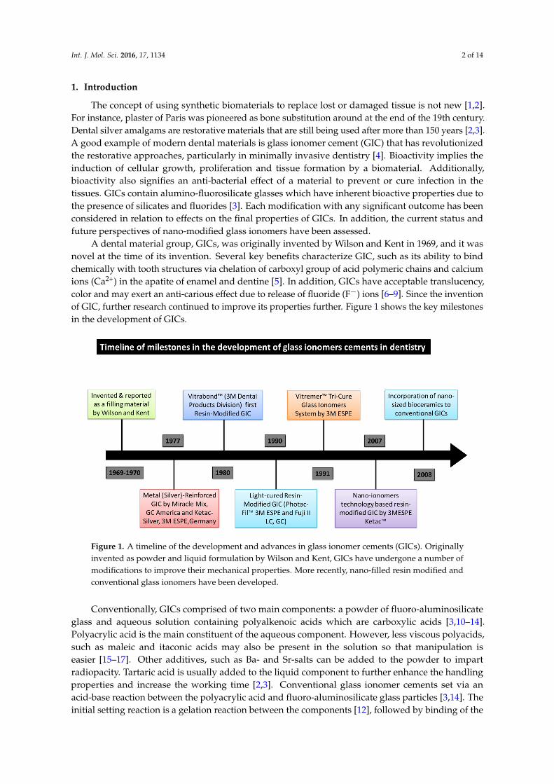
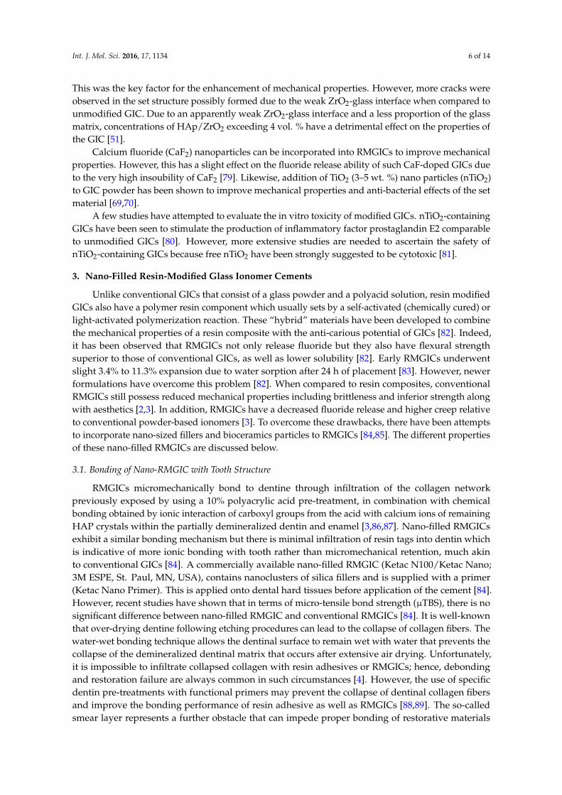
A dental material group, GICs, was originally invented by Wilson and Kent in 1969, and it wasnovel at the time of its invention. Several key benefits characterize GIC, such as its ability to bindchemically with tooth structures via chelation of carboxyl group of acid polymeric chains and calciumions (Ca2+) in the apatite of enamel and dentine [5]. In addition, GICs have acceptable translucency,color and may exert an anti-carious effect due to release of fluoride (F´) ions [6–9]. Since the inventionof GIC, further research continued to improve its properties further. Figure 1 shows the key milestonesin the development of GICs.
Int. J. Mol. Sci. 2016, 17, 1134 2 of 13
Keywords: glass ionomer cement; restorative dentistry; nanotechnology; adhesive dentistry
1. Introduction
The concept of using synthetic biomaterials to replace lost or damaged tissue is not new [1,2]. For instance, plaster of Paris was pioneered as bone substitution around at the end of the 19th century. Dental silver amalgams are restorative materials that are still being used after more than 150 years [2,3]. A good example of modern dental materials is glass ionomer cement (GIC) that has revolutionized the restorative approaches, particularly in minimally invasive dentistry [4]. Bioactivity implies the induction of cellular growth, proliferation and tissue formation by a biomaterial. Additionally, bioactivity also signifies an anti-bacterial effect of a material to prevent or cure infection in the tissues. GICs contain alumino-fluorosilicate glasses which have inherent bioactive properties due to the presence of silicates and fluorides [3]. Each modification with any significant outcome has been considered in relation to effects on the final properties of GICs. In addition, the current status and future perspectives of nano-modified glass ionomers have been assessed.
A dental material group, GICs, was originally invented by Wilson and Kent in 1969, and it was novel at the time of its invention. Several key benefits characterize GIC, such as its ability to bind chemically with tooth structures via chelation of carboxyl group of acid polymeric chains and calcium ions (Ca2+) in the apatite of enamel and dentine [5]. In addition, GICs have acceptable translucency, color and may exert an anti-carious effect due to release of fluoride (F−) ions [6–9]. Since the invention of GIC, further research continued to improve its properties further. Figure 1 shows the key milestones in the development of GICs.
Figure 1. A timeline of the development and advances in glass ionomer cements (GICs). Originally invented as powder and liquid formulation by Wilson and Kent, GICs have undergone a number of modifications to improve their mechanical properties. More recently, nano-filled resin modified and conventional glass ionomers have been developed.
Conventionally, GICs comprised of two main components: a powder of fluoro-aluminosilicate glass and aqueous solution containing polyalkenoic acids which are carboxylic acids [3,10–14]. Polyacrylic acid is the main constituent of the aqueous component. However, less viscous polyacids, such as maleic and itaconic acids may also be present in the solution so that manipulation is easier [15–17]. Other additives, such as Ba- and Sr-salts can be added to the powder to impart radiopacity. Tartaric acid is usually added to the liquid component to further enhance the handling properties and increase the working time [2,3]. Conventional glass ionomer cements set via an
Figure 1. A timeline of the development and advances in glass ionomer cements (GICs). Originallyinvented as powder and liquid formulation by Wilson and Kent, GICs have undergone a number ofmodifications to improve their mechanical properties. More recently, nano-filled resin modified andconventional glass ionomers have been developed.
Conventionally, GICs comprised of two main components: a powder of fluoro-aluminosilicateglass and aqueous solution containing polyalkenoic acids which are carboxylic acids [3,10–14].Polyacrylic acid is the main constituent of the aqueous component. However, less viscous polyacids,such as maleic and itaconic acids may also be present in the solution so that manipulation iseasier [15–17]. Other additives, such as Ba- and Sr-salts can be added to the powder to impartradiopacity. Tartaric acid is usually added to the liquid component to further enhance the handlingproperties and increase the working time [2,3]. Conventional glass ionomer cements set via anacid-base reaction between the polyacrylic acid and fluoro-aluminosilicate glass particles [3,14]. Theinitial setting reaction is a gelation reaction between the components [12], followed by binding of the
Int. J. Mol. Sci. 2016, 17, 1134 3 of 14
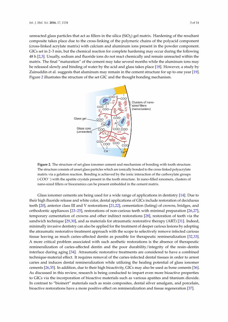
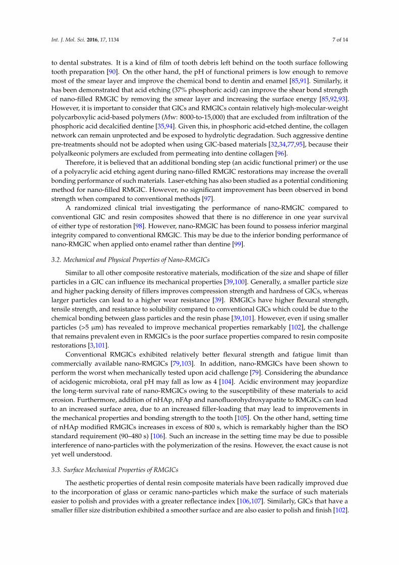
unreacted glass particles that act as fillers in the silica (SiO2) gel matrix. Hardening of the resultantcomposite takes place due to the cross-linking of the polymeric chains of the polyacid component(cross-linked acrylate matrix) with calcium and aluminum ions present in the powder component.GICs set in 2–3 min, but the chemical reaction for complete hardening may occur during the following48 h [2,3]. Usually, sodium and fluoride ions do not react chemically and remain unreacted within thematrix. The final “maturation” of the cement may take several months while the aluminum ions maybe released slowly and binding of water by the acid and glass takes place [18]. However, a study byZainuddin et al. suggests that aluminum may remain in the cement structure for up to one year [19].Figure 2 illustrates the structure of the set GIC and the thought bonding mechanism.
Int. J. Mol. Sci. 2016, 17, 1134 3 of 13
acid-base reaction between the polyacrylic acid and fluoro-aluminosilicate glass particles [3,14]. The initial setting reaction is a gelation reaction between the components [12], followed by binding of the unreacted glass particles that act as fillers in the silica (SiO2) gel matrix. Hardening of the resultant composite takes place due to the cross-linking of the polymeric chains of the polyacid component (cross-linked acrylate matrix) with calcium and aluminum ions present in the powder component. GICs set in 2–3 min, but the chemical reaction for complete hardening may occur during the following 48 h [2,3]. Usually, sodium and fluoride ions do not react chemically and remain unreacted within the matrix. The final “maturation” of the cement may take several months while the aluminum ions may be released slowly and binding of water by the acid and glass takes place [18]. However, a study by Zainuddin et al. suggests that aluminum may remain in the cement structure for up to one year [19]. Figure 2 illustrates the structure of the set GIC and the thought bonding mechanism.
Figure 2. The structure of set glass ionomer cement and mechanism of bonding with tooth structure. The structure consists of unset glass particles which are ionically bonded to the cross-linked polyacrylate matrix via a gelation reaction. Bonding is achieved by the ionic interaction of the carboxylate groups (-COO−) with the apatite crystals present in the tooth structure. In nano-filled ionomers, clusters of nano-sized fillers or bioceramics can be present embedded in the cement matrix.
Glass ionomer cements are being used for a wide range of applications in dentistry [14]. Due to their high fluoride release and white color, dental applications of GICs include restoration of deciduous teeth [20], anterior class III and V restorations [21,22], cementation (luting) of crowns, bridges, and orthodontic appliances [23–25], restorations of non-carious teeth with minimal preparation [26,27], temporary cementation of crowns and other indirect restorations [28], restoration of teeth via the sandwich technique [29,30], and as materials for atraumatic restorative therapy (ART) [31]. Indeed, minimally invasive dentistry can also be applied for the treatment of deeper carious lesions by adopting the atraumatic restorative treatment approach with the scope to selectively remove infected carious tissue leaving as much caries-affected dentin as possible for therapeutic remineralization [32,33]. A more critical problem associated with such aesthetic restorations is the absence of therapeutic remineralization of caries-affected dentin and the poor durability/integrity of the resin–dentin interface during aging [34]. Atraumatic restorative treatments are considered to have a combined technique-material effect. It requires removal of the caries-infected dental tissues in order to arrest caries and induces dental remineralization while utilizing the healing potential of glass ionomer cements [26,35]. In addition, due to their high bioactivity, GICs may also be used as bone cements [36]. As discussed in this review, research is being conducted to impart even more bioactive properties to GICs via the incorporation of bioactive materials such as various apatites and titanium dioxide. In contrast to “bioinert” materials such as
Figure 2. The structure of set glass ionomer cement and mechanism of bonding with tooth structure.The structure consists of unset glass particles which are ionically bonded to the cross-linked polyacrylatematrix via a gelation reaction. Bonding is achieved by the ionic interaction of the carboxylate groups(-COO´) with the apatite crystals present in the tooth structure. In nano-filled ionomers, clusters ofnano-sized fillers or bioceramics can be present embedded in the cement matrix.
Glass ionomer cements are being used for a wide range of applications in dentistry [14]. Due totheir high fluoride release and white color, dental applications of GICs include restoration of deciduousteeth [20], anterior class III and V restorations [21,22], cementation (luting) of crowns, bridges, andorthodontic appliances [23–25], restorations of non-carious teeth with minimal preparation [26,27],temporary cementation of crowns and other indirect restorations [28], restoration of teeth via thesandwich technique [29,30], and as materials for atraumatic restorative therapy (ART) [31]. Indeed,minimally invasive dentistry can also be applied for the treatment of deeper carious lesions by adoptingthe atraumatic restorative treatment approach with the scope to selectively remove infected carioustissue leaving as much caries-affected dentin as possible for therapeutic remineralization [32,33].A more critical problem associated with such aesthetic restorations is the absence of therapeuticremineralization of caries-affected dentin and the poor durability/integrity of the resin–dentininterface during aging [34]. Atraumatic restorative treatments are considered to have a combinedtechnique-material effect. It requires removal of the caries-infected dental tissues in order to arrestcaries and induces dental remineralization while utilizing the healing potential of glass ionomercements [26,35]. In addition, due to their high bioactivity, GICs may also be used as bone cements [36].As discussed in this review, research is being conducted to impart even more bioactive propertiesto GICs via the incorporation of bioactive materials such as various apatites and titanium dioxide.In contrast to “bioinert” materials such as resin composites, dental silver amalgam, and porcelain,bioactive restorations have a more positive effect on remineralization and tissue regeneration [37].
Int. J. Mol. Sci. 2016, 17, 1134 4 of 14
No dental material today has ideal properties for any dental application [1–3,38]. This said,GICs also carry a number of drawbacks such as brittleness, subsequently prone to fracture [39], poorwear resistance, and inadequate surface properties [40,41], as well as they are sensitive to moisturein the oral cavity when newly placed [42]. These said aspects restrict the use of GICs for manyclinical situations. In order to overcome the poor mechanical properties of glass ionomers, severalmodifications have been introduced to the conventional GICs [43–46]. The key modifications includethe combination of glass ionomer cements with auto-cured or photo-cured resin systems to produceresin-modified glass ionomer cements (RMGICs) [46,47]. Additionally, modification of GICs byincorporation polyvinyl phosphonic acid [48,49], fiber-reinforcement [50], bioactive apatite without [43]or with zirconia [51,52], zinc [53,54], strontium oxide [55], stainless steel [56], silica particles [57],amino acids [58], and N-vinylpyrrolidone [45] have all aimed at improving the mechanical andphysical properties.
Nanotechnology involves the use of systems, modifications or materials which have the size in therange of 1–100 nm [59–61]. Key applications of nanotechnology in dentistry include implant surfacemodifications [62], production of reinforced polymeric composites by incorporation of nano-sizedparticles [60], and caries prevention [63]. Recent studies have suggested that incorporation ofnano-sized particles or “nanoclusters” can improve the mechanical properties of dental restorativematerial such as resin composites [64–66]. Similar approaches have been attempted to improve thephysical and mechanical characteristics of GIC using nanotechnology [43,44].
There are two approaches for the manufacture of nano-size particles: top-down andbottom-up [61]. The so-called top-down nanofabrication involves the production of nano-size particlesby removing the bulk material. Some examples of top-down fabrication include are milling, machining,and lithography. On the other hand, the so-called bottom-up nanofabrication involves productionof nano-sized particles atom by atom. Some examples of bottom-up nanofabrication are: tissueregeneration, protein synthesis, and biomimetic dental implant coatings. Production of nano-sizedparticles for incorporation to GICs, is manly carried out through top-down nanofabrication of bulkmaterials such as apatites, silicate glasses, and some metal oxides.
2. Powder-Modified Nano Glass Ionomers
It is well-documented that incorporation of nano-sized particles may improve the mechanicalproperties of polymeric dental materials [60,67]. De Caluwé et al. showed that doping conventionalGICs with nano-sized glass particles can decrease the setting time and enhance the compressionstrength and elastic modulus [68]. The main advantages of decreasing setting times of direct restorativematerials are e.g., enhanced ease of handling and manipulation. These decrease the treatment time,benefitting both the clinician as well as the patient. Enhancing the mechanical properties adds to theserviceability and self-life of restorative materials as they are able to withstand the masticatory andocclusal forces more efficiently. The process of mastication is quite complex as it involves forces inmultiple directions. Therefore, owing to the quantitative nature of in vitro research, it is difficult totranslate the results obtained in the laboratory to clinical practice.
Certain procedures, such as thermo-cycling are aimed at artificially aging restorative materialsin the laboratory so the effect of oral temperature and moisture may be assessed. Given this,thermo-cycling has more deleterious effects on the mechanical properties of nano-filled GICs comparedto conventional GICs. This may also compromise the long-term survival rate of such materials.However, in most studies, the in vitro testing of modified GICs against various cell-lines has beencarried out, and none of them has reported the effect of such cements on animals. The chemicalstructure of glass ionomers can be mainly assessed by spectroscopic methods. Fourier transforminfrared (FTIR) spectroscopy has been used to observe the effect of nano-modification of GICs viaincorporation of apatite nano-crystals [44,45]. However, the effect of modification by using some otherparticles, TiO2 and ZrO2, has not been evaluated or reported.
Int. J. Mol. Sci. 2016, 17, 1134 5 of 14
As described above, in order to improve the mechanical, biological, and physical properties ofpowder-liquid formulations of GICs, various types of nano-size powders have been incorporated tothe glass powder component. The mechanical properties have been summarized in Table 1.
Table 1. Some powder modifications of glass ionomer cements and their reported properties. HA,hydroxyapatite; FA, fluorohydroxyapatite; FAS, fluoroaluminosilicate; TiO2, titanium oxide; ZrO2,zirconium oxide.
GIC Formulation Mechanical Properties
Liquid Powder Nano FillerPercentage and Size
CompressiveStrength (MPa)
TensileStrength (MPa)
FlexuralStrength (MPa)
Reference(s)
Polyacrylic acidcopolymer Unmodified FAS glass No nano fillers, glass
size: 3.34–9.6 µm 161 11.8 14.8 [45]
Polyacrylic acidcopolymer FAS Glass + HA 5 wt. %, 100–200nm 178 19 31 [44]
Polyacrylic acidcopolymer FAS Glass + FA 5 wt. %, 100–200 179 23 33 [44]
Polymer of AA, NVP,IA (8:1:1) FAS Glass + HA 5 wt. %, 100–200 nm 183.8 23.5 36 [45]
Polyacrylic acidcopolymer FAS Glass + TiO2 3%, size variable 176.27 - 23.17 [69,70]
Polyacrylic acidcopolymer FAS Glass + HA/ZrO2
4 vol. %, particledimension: 20 ˆ 200 nm 176.30 12.67 - [52]
2.1. Modification Using Nano-Apatite
Due to their chemistry being similar to that of mineralized bone and dental tissues, hydroxypatiteand fluorohydroxyapatite have been used in many fields of dentistry such as implant dentistry [71],and caries prevention [59,72]. For instance, nano-hydroxyapatite (nHAp) crystals can favorremineralization of enamel [73,74]. Recently, resin composites modified by the addition of nHAphave been observed to have superior mechanical properties than unmodified resin composites [75,76].Similarly, addition of nHAp or nano-fluoroapatite (nFAp) to the powder component of conventionalGIC has a positive impact on the compressive, tensile, and flexural strengths of the set cement afterbeing stored in distilled water for 7 days [44].
FTIR spectroscopy has revealed that addition of apatite to GIC powder increases the crystallinityof the set GIC, hence improving the chemical stability and water insolubility [44,45]. Such modificationsresult in better survival rates than commercially available GICs [44]. Since nFAp has lower solubilitythan nHAp or nFAp-containing GICs have better mechanical properties and bond strength comparedto nHAp-containing GICs [44,45]. It has been suggested that the enhanced mechanical propertiesof apatite-modified GICs are the result of ionic interaction between the polyacrylic acid and theapatite crystals [45]. This enhancement is more pronounced when the nHAp-containing powder isadded to polyacrylic acid, itatonic acid, and N-vinylpyrrolidone copolymers, instead of the standardpolyacrylic copolymer. This has been attributed to the additional physiochemical interaction betweenthe N-vinylpyrrolidone and the apatite crystals [45]. Moreover, nano-apatite containing glass ionomersare expected to have superior bonding to the tooth surface due to the possibility of the formation ofthe strong ionic linkages between the apatite crystals/particles in the cement and Ca-ions in the toothstructure [77]. Additionally, decreasing the particle size of apatites from micrometer scale to nanometerscale increases the surface area remarkably, and infiltration of the crystals into demineralised dentineas well as enamel pores; this may enhance bonding at the tooth-ionomer interface [78].
2.2. Modification with Nano-Sized HAp/Zr, CaF2 and TiO2 Particles
It has been recently reported by Gu et al. that the combined incorporation of HAp and zirconia(HAp/ZrO2) at concentrations of 4 vol. % to the GIC powder can improve the mechanical properties ofthe set GIC [52]. When specimens of set GIC were analyzed using scanning electron microscopy (SEM)a dense, uniform dispersion of the glass and HAp/ZrO2 particles within the matrix were revealed.
Int. J. Mol. Sci. 2016, 17, 1134 6 of 14
This was the key factor for the enhancement of mechanical properties. However, more cracks wereobserved in the set structure possibly formed due to the weak ZrO2-glass interface when compared tounmodified GIC. Due to an apparently weak ZrO2-glass interface and a less proportion of the glassmatrix, concentrations of HAp/ZrO2 exceeding 4 vol. % have a detrimental effect on the properties ofthe GIC [51].
Calcium fluoride (CaF2) nanoparticles can be incorporated into RMGICs to improve mechanicalproperties. However, this has a slight effect on the fluoride release ability of such CaF-doped GICs dueto the very high insoubility of CaF2 [79]. Likewise, addition of TiO2 (3–5 wt. %) nano particles (nTiO2)to GIC powder has been shown to improve mechanical properties and anti-bacterial effects of the setmaterial [69,70].
A few studies have attempted to evaluate the in vitro toxicity of modified GICs. nTiO2-containingGICs have been seen to stimulate the production of inflammatory factor prostaglandin E2 comparableto unmodified GICs [80]. However, more extensive studies are needed to ascertain the safety ofnTiO2-containing GICs because free nTiO2 have been strongly suggested to be cytotoxic [81].
3. Nano-Filled Resin-Modified Glass Ionomer Cements
Unlike conventional GICs that consist of a glass powder and a polyacid solution, resin modifiedGICs also have a polymer resin component which usually sets by a self-activated (chemically cured) orlight-activated polymerization reaction. These “hybrid” materials have been developed to combinethe mechanical properties of a resin composite with the anti-carious potential of GICs [82]. Indeed,it has been observed that RMGICs not only release fluoride but they also have flexural strengthsuperior to those of conventional GICs, as well as lower solubility [82]. Early RMGICs underwentslight 3.4% to 11.3% expansion due to water sorption after 24 h of placement [83]. However, newerformulations have overcome this problem [82]. When compared to resin composites, conventionalRMGICs still possess reduced mechanical properties including brittleness and inferior strength alongwith aesthetics [2,3]. In addition, RMGICs have a decreased fluoride release and higher creep relativeto conventional powder-based ionomers [3]. To overcome these drawbacks, there have been attemptsto incorporate nano-sized fillers and bioceramics particles to RMGICs [84,85]. The different propertiesof these nano-filled RMGICs are discussed below.
3.1. Bonding of Nano-RMGIC with Tooth Structure
RMGICs micromechanically bond to dentine through infiltration of the collagen networkpreviously exposed by using a 10% polyacrylic acid pre-treatment, in combination with chemicalbonding obtained by ionic interaction of carboxyl groups from the acid with calcium ions of remainingHAP crystals within the partially demineralized dentin and enamel [3,86,87]. Nano-filled RMGICsexhibit a similar bonding mechanism but there is minimal infiltration of resin tags into dentin whichis indicative of more ionic bonding with tooth rather than micromechanical retention, much akinto conventional GICs [84]. A commercially available nano-filled RMGIC (Ketac N100/Ketac Nano;3M ESPE, St. Paul, MN, USA), contains nanoclusters of silica fillers and is supplied with a primer(Ketac Nano Primer). This is applied onto dental hard tissues before application of the cement [84].However, recent studies have shown that in terms of micro-tensile bond strength (µTBS), there is nosignificant difference between nano-filled RMGIC and conventional RMGICs [84]. It is well-knownthat over-drying dentine following etching procedures can lead to the collapse of collagen fibers. Thewater-wet bonding technique allows the dentinal surface to remain wet with water that prevents thecollapse of the demineralized dentinal matrix that occurs after extensive air drying. Unfortunately,it is impossible to infiltrate collapsed collagen with resin adhesives or RMGICs; hence, debondingand restoration failure are always common in such circumstances [4]. However, the use of specificdentin pre-treatments with functional primers may prevent the collapse of dentinal collagen fibersand improve the bonding performance of resin adhesive as well as RMGICs [88,89]. The so-calledsmear layer represents a further obstacle that can impede proper bonding of restorative materials
Int. J. Mol. Sci. 2016, 17, 1134 7 of 14
to dental substrates. It is a kind of film of tooth debris left behind on the tooth surface followingtooth preparation [90]. On the other hand, the pH of functional primers is low enough to removemost of the smear layer and improve the chemical bond to dentin and enamel [85,91]. Similarly, ithas been demonstrated that acid etching (37% phosphoric acid) can improve the shear bond strengthof nano-filled RMGIC by removing the smear layer and increasing the surface energy [85,92,93].However, it is important to consider that GICs and RMGICs contain relatively high-molecular-weightpolycarboxylic acid-based polymers (Mw: 8000-to-15,000) that are excluded from infiltration of thephosphoric acid decalcified dentine [35,94]. Given this, in phosphoric acid-etched dentine, the collagennetwork can remain unprotected and be exposed to hydrolytic degradation. Such aggressive dentinepre-treatments should not be adopted when using GIC-based materials [32,34,77,95], because theirpolyalkeonic polymers are excluded from permeating into dentine collagen [96].
Therefore, it is believed that an additional bonding step (an acidic functional primer) or the useof a polyacrylic acid etching agent during nano-filled RMGIC restorations may increase the overallbonding performance of such materials. Laser-etching has also been studied as a potential conditioningmethod for nano-filled RMGIC. However, no significant improvement has been observed in bondstrength when compared to conventional methods [97].
A randomized clinical trial investigating the performance of nano-RMGIC compared toconventional GIC and resin composites showed that there is no difference in one year survivalof either type of restoration [98]. However, nano-RMGIC has been found to possess inferior marginalintegrity compared to conventional RMGIC. This may be due to the inferior bonding performance ofnano-RMGIC when applied onto enamel rather than dentine [99].
3.2. Mechanical and Physical Properties of Nano-RMGICs
Similar to all other composite restorative materials, modification of the size and shape of fillerparticles in a GIC can influence its mechanical properties [39,100]. Generally, a smaller particle sizeand higher packing density of fillers improves compression strength and hardness of GICs, whereaslarger particles can lead to a higher wear resistance [39]. RMGICs have higher flexural strength,tensile strength, and resistance to solubility compared to conventional GICs which could be due to thechemical bonding between glass particles and the resin phase [39,101]. However, even if using smallerparticles (>5 µm) has revealed to improve mechanical properties remarkably [102], the challengethat remains prevalent even in RMGICs is the poor surface properties compared to resin compositerestorations [3,101].
Conventional RMGICs exhibited relatively better flexural strength and fatigue limit thancommercially available nano-RMGICs [79,103]. In addition, nano-RMGICs have been shown toperform the worst when mechanically tested upon acid challenge [79]. Considering the abundanceof acidogenic microbiota, oral pH may fall as low as 4 [104]. Acidic environment may jeopardizethe long-term survival rate of nano-RMGICs owing to the susceptibility of these materials to aciderosion. Furthermore, addition of nHAp, nFAp and nanofluorohydroxyapatite to RMGICs can leadto an increased surface area, due to an increased filler-loading that may lead to improvements inthe mechanical properties and bonding strength to the tooth [105]. On the other hand, setting timeof nHAp modified RMGICs increases in excess of 800 s, which is remarkably higher than the ISOstandard requirement (90–480 s) [106]. Such an increase in the setting time may be due to possibleinterference of nano-particles with the polymerization of the resins. However, the exact cause is notyet well understood.
3.3. Surface Mechanical Properties of RMGICs
The aesthetic properties of dental resin composite materials have been radically improved dueto the incorporation of glass or ceramic nano-particles which make the surface of such materialseasier to polish and provides with a greater reflectance index [106,107]. Similarly, GICs that have asmaller filler size distribution exhibited a smoother surface and are also easier to polish and finish [102].
Int. J. Mol. Sci. 2016, 17, 1134 8 of 14
Although purely mechanical degradation (abrasion) produced by tooth brushing simulates surfacewear of nano-RMGCs to a significantly lesser degree compared to conventional RMGICs, in vitrostudies suggest that there is no statistical difference between the surface roughness and hardness ofnano-RMGICs and conventional RMGICs after bacterial and chemical degradation [108]. Nevertheless,to date, studies have established that no matter what size of filler is present in the RMGICs, theirsurface roughness and hardness remain significantly lower than those of resin composites followingbacterial, mechanical, and chemical degradation due to lower wear resistance and higher solubilityof the former [108–110]. Therefore, it can be concluded that commercially available nano-RMGICsdo not possess any substantial advantage or disadvantage, in terms of surface mechanical properties,compared to conventional restorative materials.
3.4. Fluoride Release from Nano-Ionomers
It has been well established that fluoride is released from glass ionomers [8,111–116]. At highconcentrations, fluoride has been thought to reduce the rate of demineralization, enhance theremineralization process, inhibit the growth, and attachment of bacteria on tooth surfaces and impedethe formation of a complex bacteria biofilm [117]. Because the fluoride ions in the set ionomer structuredo not take part in the setting reaction, they are released into the surrounding environment via an ionexchange process. Furthermore, the glass ionomers can also absorb salivary fluoride and act as fluoridereservoirs capable of releasing the ions which may have the potential to inhibit caries formation [118].However, to date, it has not been established whether the amounts of fluoride released from glassionomer cements is sufficient to impede dental caries [115,116]. Indeed, several studies have suggestedthat the cumulative fluoride release from nano-RMGICs and conventional RMGICs are comparablewith each other but still significantly lesser than conventional GICs [79,119,120]. Nevertheless, theexact amount of fluoride released by nano-RMGICs compared to other RMGICs and GICs is stilldebatable. It has been observed that, although there is a slightly increased fluoride release fromnano-RMGICs at a pH of 4, cumulative fluoride released after 84 days and per specimen surface in aday remains comparable to those of conventional RMGICs [79]. So far, no long-term clinical studiesassessing the secondary caries in teeth restored by nano-ionomers cements are available in literature.This said, it has still not been ascertained whether or not the anti-carious activity of these cements isany better than conventional GICs in the clinical scenario. So far, with the exception of CaF2-modifiedionomers, no studies have focused on investigating the effect of nano-modification of the powdercomponent of conventional ionomers.
4. Conclusions
Nano-modification of conventional GICs and RMGICs can be achieved by the incorporation ofnano-sized fillers to RMGICs, reducing the size of the glass particles and introducing nano-sizedbioceramics to the glass powder. Commercially available nano-filled RMGIC (Ketac Nano) does nothold any significant advantage over micro-filled RMGICs in terms of flexural strength and tensilestrength. Bonding properties of nano-filled RMGIC are still a matter of concern. Conversely, recentadvancements, like the introduction of nano-sized apatites, have not only improved the mechanicalproperties of conventional GICs, but have also enhanced fluoride release in vitro. By increasing thecrystallinity of the set matrix, apatite crystals can make the set cement more stable and improve thebond strength with tooth structure. An increased fluoride release can help in reducing secondarycaries around restorations. However, a problematic issue is the possibility of the failure of theglass-bioceramic interface which may affect the mechanical properties of the set cement. Moreover,very few studies focusing on the nano-modification of GIC have concentrated on effects they mighthave on the pulpal cells. Hence, more mechanical, biological studies, and eventually, clinical trialsare needed and essential to ascertain the status of nano-modified GICs in clinical practice. On top ofthat, there is more to be learned about the effect of nano-modification of the powder component in
Int. J. Mol. Sci. 2016, 17, 1134 9 of 14
glass ionomers and on their fluoride release. Perhaps surprisingly, none of the studies have focused oninvestigating this aspect to date.
Author Contributions: Shariq Najeeb conducted the primary literature search, constructed the tables andwrote the majority of this manuscript. Zohaib Khurshid, Sana Zohaib and Abdul Samad Khan constructedthe illustrations and contributed to the writing of the manuscript. Salvatore Sauro provided expertopinion on minimally invasive dentistry and applications of glass ionomers. Juan Manuel Nuñez Martí,Muhammad Sohail Zafar, Ihtesham Ur Rehman and Jukka Pekka Matinlinna contributed to the writing andorganizing the text.
Conflicts of Interest: The authors declare no conflict of interest.
References
1. Nicholson, J.W. The Chemistry of Medical and Dental Materials; Royal Society of Chemistry: Milton, Cambridge,2002; Volume 3.
2. McCabe, J.F.; Walls, A. Applied Dental Materials; John Wiley & Sons: Hoboken, NJ, USA, 2013.3. Anusavice, K.J.; Shen, C.; Rawls, H.R. Phillips’ Science of Dental Materials; Elsevier Health Sciences: St. Louis,
MO, USA, 2012.4. Sauro, S.; Pashley, D. Stabilize to stabilize dentine-bonded interfaces through reminerlizing operative
procedures—State of art. Int. J. Adhes. Adhes. 2016. in press. [CrossRef]5. Tyas, M.J.; Burrow, M.F. Adhesive restorative materials: A review. Aust. Dent. J. 2004, 49, 112–121. [CrossRef]
[PubMed]6. Preston, A.J.; Higham, S.M.; Agalamanyi, E.A.; Mair, L.H. Fluoride recharge of aesthetic dental materials.
J. Oral Rehabil. 1999, 26, 936–940. [CrossRef] [PubMed]7. Forsten, L. Resin-modified glass ionomer cements: Fluoride release and uptake. Acta Odontol. Scand. 1995,
53, 222–225. [CrossRef] [PubMed]8. Forss, H.; Jokinen, J.; Spets-Happonen, S.; Seppä, L.; Luoma, H. Fluoride and mutans streptococci in plaque
grown on glass ionomer and composite. Caries Res. 1991, 25, 454–458. [CrossRef] [PubMed]9. Preston, A.J.; Mair, L.H.; Agalamanyi, E.A.; Higham, S.M. Fluoride release from aesthetic dental materials.
J. Oral Rehabil. 1999, 26, 123–129. [CrossRef] [PubMed]10. Wilson, A.D.; Kent, B.E. The glass-ionomer cement, a new translucent dental filling material. J. Appl.
Chem. Biotechnol. 1971, 21, 313. [CrossRef]11. McLean, J.W.; Gasser, O. Glass-cermet cements. Quintessence Int. 1985, 16, 333–343. [PubMed]12. Wilson, A.D. The chemistry of dental cements. Chem. Soc. Rev. 1978, 7, 265–296. [CrossRef]13. Davidson, C.L. Advances in glass-ionomer cements. J. Appl. Oral Sci. 2006, 14, 3–9. [CrossRef] [PubMed]14. Wilson, A.D.; Nicholson, J.W. Acid-Base Cements: Their Biomedical and Industrial Applications; Cambridge
University Press: Cambridge, UK, 2005; Volume 3.15. Prosser, H.J.; Powis, D.R.; Wilson, A.D. Glass-ionomer cements of improved flexural strength. J. Dent. Res.
1986, 65, 146–148. [CrossRef] [PubMed]16. Smith, D.C. Development of glass-ionomer cement systems. Biomaterials 1998, 19, 467–478. [CrossRef]17. Guggenberger, R.; May, R.; Stefan, K.P. New trends in glass-ionomer chemistry. Biomaterials 1998, 19, 479–483.
[CrossRef]18. Nicholson, J.W. Chemistry of glass-ionomer cements: A review. Biomaterials 1998, 19, 485–494. [CrossRef]19. Zainuddin, N.; Karpukhina, N.; Hill, R.G.; Law, R.V. A long-term study on the setting reaction of glass
ionomer cements by 27 Al MAS-NMR spectroscopy. Dent. Mater. 2009, 25, 290–295.20. De Amorim, R.G.; Leal, S.C.; Frencken, J.E. Survival of atraumatic restorative treatment (ART) sealants and
restorations: A meta-analysis. Clin. Oral Investig. 2012, 16, 429–441. [CrossRef] [PubMed]21. Van Dijken, J.W. 3-Year clinical evaluation of a compomer, a resin-modified glass ionomer and a resin
composite in class III restorations. Am. J. Dent. 1996, 9, 195–198. [PubMed]22. Abdalla, A.I.; Alhadainy, H.A.; Garcia-Godoy, F. Clinical evaluation of glass ionomers and compomers in
class V carious lesions. Am. J. Dent. 1997, 10, 18–20. [PubMed]23. Leevailoj, C.; Platt, J.A.; Cochran, M.A.; Moore, B.K. In vitro study of fracture incidence and compressive
fracture load of all-ceramic crowns cemented with resin-modified glass ionomer and other luting agents.J. Prosthet. Dent. 1998, 80, 699–707. [CrossRef]
Int. J. Mol. Sci. 2016, 17, 1134 10 of 14
24. Pascotto, R.C.; de Lima Navarro, M.F.; Capelozza Filho, L.; Cury, J.A. In vivo effect of a resin-modified glassionomer cement on enamel demineralization around orthodontic brackets. Am. J. Orthod. Dentofac. Orthop.2004, 125, 36–41. [CrossRef]
25. Jokstad, A.; Mjör, I.A. Ten years’ clinical evaluation of three luting cements. J. Dent. 1996, 24, 309–315.[CrossRef]
26. Murdoch-Kinch, C.A.; McLean, M.E. Minimally invasive dentistry. J. Am. Dent. Assoc. 2003, 134, 87–95.[CrossRef] [PubMed]
27. Peumans, M.; de Munck, J.; Mine, A.; van Meerbeek, B. Clinical effectiveness of contemporary adhesivesfor the restoration of non-carious cervical lesions. A systematic review. Dent. Mater. 2014, 30, 1089–1103.[CrossRef] [PubMed]
28. Terata, R.; Nakashima, K.; Kubota, M. Effect of temporary materials on bond strength of resin-modifiedglass-ionomer luting cements to teeth. Am. J. Dent. 2000, 13, 209–211. [PubMed]
29. McLean, J.W.; Powis, D.R.; Prosser, H.J.; Wilson, A.D. The use of glass-ionomer cements in bonding compositeresins to dentine. Br. Dent. J. 1985, 158, 410–414. [CrossRef] [PubMed]
30. Andersson-Wenckert, I.E.; van Dijken, J.W.; Kieri, C. Durability of extensive class II open-sandwichrestorations with a resin-modified glass ionomer cement after 6 years. Am. J. Dent. 2004, 17, 43–50.[PubMed]
31. Frencken, J.E.; Makoni, F.; Sithole, W.D. Art restorations and glass ionomer sealants in Zimbabwe: Survivalafter 3 years. Community Dent. Oral Epidemiol. 1998, 26, 372–381. [CrossRef] [PubMed]
32. Yamaga, R.; Nishino, M.; Yoshida, S.; Yokomizo, I. Diammine silver fluoride and its clinical application.J. Osaka Univ. Dent. Sch. 1972, 12, 1–20. [PubMed]
33. Sauro, S.; Osorio, R.; Watson, T.F.; Toledano, M. Influence of phosphoproteins’ biomimetic analogs onremineralization of mineral-depleted resin–dentin interfaces created with ion-releasing resin-based systems.Dent. Mater. 2015, 31, 759–777. [CrossRef] [PubMed]
34. De Munck, J.; van Landuyt, K.; Peumans, M.; Poitevin, A.; Lambrechts, P.; Braem, M.; van Meerbeek, B.A critical review of the durability of adhesion to tooth tissue: Methods and results. J. Dent. Res. 2005, 84,118–132. [CrossRef] [PubMed]
35. Sauro, S.; Watson, T.F.; Thompson, I.; Toledano, M.; Nucci, C.; Banerjee, A. Influence of air-abrasion executedwith polyacrylic acid-bioglass 45S5 on the bonding performance of a resin-modified glass ionomer cement.Eur. J. Oral Sci. 2012, 120, 168–177. [CrossRef] [PubMed]
36. Jonck, L.M.; Grobbelaar, C.J.; Strating, H. The biocompatibility of glass-ionomer cement in joint replacement:Bulk testing. Clin. Mater. 1989, 4, 85–107. [CrossRef]
37. Najeeb, S.; Zafar, M.S.; Khurshid, Z.; Siddiqui, F. Applications of polyetheretherketone (PEEK) in oralimplantology and prosthodontics. J. Prosthodont. Res. 2016, 60, 12–19. [CrossRef] [PubMed]
38. Powers, J.M.; Wataha, J.C. Dental Materials: Properties and Manipulation; Elsevier Health Sciences: St. Louis,MO, USA, 2014.
39. Xie, D.; Brantley, W.A.; Culbertson, B.M.; Wang, G. Mechanical properties and microstructures ofglass-ionomer cements. Dent. Mater. 2000, 16, 129–138. [CrossRef]
40. Peutzfeldt, A.; Garcia-Godoy, F.; Asmussen, E. Surface hardness and wear of glass ionomers and compomers.Am. J. Dent. 1997, 10, 15–17. [PubMed]
41. Ahmed, N.; Zafar, M.S. . Effects of wear on hardness and stiffness of restorative dental materials. Life Sci. J.2014, 11, 11–18.
42. Um, C.M.; Øilo, G. The effect of early water contact on glass-ionomer cements. Quintessence Int. 1992, 1, 23.43. Moshaverinia, A.; Roohpour, N.; Chee, W.W.L.; Schricker, S.R. A review of powder modifications in
conventional glass-ionomer dental cements. J. Mater. Chem. 2011, 21, 1319–1328. [CrossRef]44. Moshaverinia, A.; Ansari, S.; Moshaverinia, M.; Roohpour, N.; Darr, J.A.; Rehman, I. Effects of incorporation
of hydroxyapatite and fluoroapatite nanobioceramics into conventional glass ionomer cements (GIC).Acta Biomater. 2008, 4, 432–440. [CrossRef] [PubMed]
45. Moshaverinia, A.; Ansari, S.; Movasaghi, Z.; Billington, R.W.; Darr, J.A.; Rehman, I.U. Modification ofconventional glass-ionomer cements with N-vinylpyrrolidone containing polyacids, nano-hydroxy andfluoroapatite to improve mechanical properties. Dent. Mater. 2008, 24, 1381–1390. [CrossRef] [PubMed]
46. Wilson, A.D. Resin-modified glass-ionomer cements. Int. J. Prosthodont. 1989, 3, 425–429.
Int. J. Mol. Sci. 2016, 17, 1134 11 of 14
47. Soncini, J.A.; Maserejian, N.N.; Trachtenberg, F.; Tavares, M.; Hayes, C. The Longevity of amalgam versuscompomer/composite restorations in posterior primary and permanent teeth: Findings from the newengland children’s amalgam trial. J. Am. Dent. Assoc. 2007, 138, 763–772. [CrossRef] [PubMed]
48. Khouw-Liu, V.H.W.; Anstice, H.M.; Pearson, G.J. An in vitro investigation of a poly (vinyl phosphonic acid)based cement with four conventional glass-ionomer cements. Part 1: Flexural strength and fluoride release.J. Dent. 1999, 27, 351–357. [CrossRef]
49. Moshaverinia, A.; Roohpour, N.; Chee, W.W.L.; Schricker, S.R. A review of polyelectrolyte modifications inconventional glass-ionomer dental cements. J. Mater. Chem. 2012, 22, 2824–2833. [CrossRef]
50. Lohbauer, U.; Walker, J.; Nikolaenko, S.; Werner, J.; Clare, A.; Petschelt, A.; Greil, P. Reactive fibre reinforcedglass ionomer cements. Biomaterials 2003, 24, 2901–2907. [CrossRef]
51. Gu, Y.W.; Yap, A.U.J.; Cheang, P.; Khor, K.A. Zirconia–glass ionomer cement—A potential substitute formiracle mix. Scr. Mater. 2005, 52, 113–116. [CrossRef]
52. Gu, Y.W.; Yap, A.U.; Cheang, P.; Khor, K.A. Effects of incorporation of HA/ZrO2 into glass ionomer cement(GIC). Biomaterials 2005, 26, 713–720. [CrossRef] [PubMed]
53. Zoergiebel, J.; Ilie, N. Evaluation of a conventional glass ionomer cement with new zinc formulation: Effectof coating, aging and storage agents. Clin. Oral Investig. 2013, 17, 619–626. [CrossRef] [PubMed]
54. Boyd, D.; Towler, M.R. The processing, mechanical properties and bioactivity of zinc based glass ionomercements. J. Mater. Sci. Mater. Med. 2005, 16, 843–850. [CrossRef] [PubMed]
55. Deb, S.; Nicholson, J.W. The effect of strontium oxide in glass–ionomer cements. J. Mater. Sci. Mater. Med.1999, 10, 471–474. [CrossRef] [PubMed]
56. Kerby, R.E.; Bleiholder, R.F. Physical properties of stainless-steel and silver-reinforced glass-ionomer cements.J. Dent. Res. 1991, 70, 1358–1361. [CrossRef] [PubMed]
57. Tjandrawinata, R.; Irie, M.; Yoshida, Y.; Suzuki, K. Effect of adding spherical silica filler onphysico-mechanical properties of resin modified glass-ionomer cement. Dent. Mater. J. 2004, 23, 146–154.[CrossRef] [PubMed]
58. Kao, E.C.; Culbertson, B.M.; Xie, D. Preparation of glass ionomer cement using N-acryloyl-substituted aminoacid monomers—Evaluation of physical properties. Dent. Mater. 1996, 12, 44–51. [CrossRef]
59. Hannig, M.; Hannig, C. Nanomaterials in preventive dentistry. Nat. Nanotechnol. 2010, 5, 565–569. [CrossRef][PubMed]
60. Najeeb, S.; Khurshid, Z.; Matinlinna, J.P.; Siddiqui, F.; Nassani, M.Z.; Baroudi, K. Nanomodified peekdental implants: Bioactive composites and surface modification—A review. Int. J. Dent. 2015, 2015, 381759.[CrossRef] [PubMed]
61. Khurshid, Z.; Zafar, M.; Qasim, S.; Shahab, S.; Naseem, M.; AbuReqaiba, A. Advances in nanotechnology forrestorative dentistry. Materials 2015, 8, 717–731. [CrossRef]
62. Le Guéhennec, L.; Soueidan, A.; Layrolle, P.; Amouriq, Y. Surface treatments of titanium dental implants forrapid osseointegration. Dent. Mater. 2007, 23, 844–854. [CrossRef] [PubMed]
63. Hannig, M.; Hannig, C. Nanotechnology and its role in caries therapy. Adv. Dent. Res. 2012, 24, 53–57.[CrossRef] [PubMed]
64. Curtis, A.R.; Palin, W.M.; Fleming, G.J.P.; Shortall, A.C.C.; Marquis, P.M. The mechanical properties ofnanofilled resin-based composites: The impact of dry and wet cyclic pre-loading on bi-axial flexure strength.Dent. Mater. 2009, 25, 188–197. [CrossRef] [PubMed]
65. Terry, D.A. Direct applications of a nanocomposite resin system: Part 1-the evolution of contemporarycomposite materials. Pract. Proced. Aesthet. Dent. 2004, 16, 417–432. [PubMed]
66. Chen, M.-H. Update on dental nanocomposites. J. Dent. Res. 2010, 89, 549–560. [CrossRef] [PubMed]67. Xia, Y.; Zhang, F.; Xie, H.; Gu, N. Nanoparticle-reinforced resin-based dental composites. J. Dent. 2008, 36,
450–455. [CrossRef] [PubMed]68. De Caluwe, T.; Vercruysse, C.W.; Fraeyman, S.; Verbeeck, R.M. The influence of particle size and fluorine
content of aluminosilicate glass on the glass ionomer cement properties. Dent. Mater. 2014, 30, 1029–1038.[CrossRef] [PubMed]
69. Elsaka, S.E.; Hamouda, I.M.; Swain, M.V. Titanium dioxide nanoparticles addition to a conventionalglass-ionomer restorative: Influence on physical and antibacterial properties. J. Dent. 2011, 39, 589–598.[CrossRef] [PubMed]
Int. J. Mol. Sci. 2016, 17, 1134 12 of 14
70. Garcia-Contreras, R.; Scougall-Vilchis, R.J.; Contreras-Bulnes, R.; Sakagami, H.; Morales-Luckie, R.A.;Nakajima, H. Mechanical, antibacterial and bond strength properties of nano-titanium-enriched glassionomer cement. J. Appl. Oral Sci. 2015, 23, 321–328. [CrossRef] [PubMed]
71. Javed, F.; Vohra, F.; Zafar, S.; Almas, K. Significance of osteogenic surface coatings on implants to enhanceosseointegration under osteoporotic-like conditions. Implant Dent. 2014, 23, 679–686. [CrossRef] [PubMed]
72. Ong, J.L.; Chan, D.C.N. Hydroxyapatite and their use as coatings in dental implants: A review. Crit. Rev.Biomed. Eng. 2000, 28. [CrossRef]
73. Huang, S.B.; Gao, S.S.; Yu, H.Y. Effect of nano-hydroxyapatite concentration on remineralization of initialenamel lesion in vitro. Biomed. Mater. 2009, 4, 034104. [CrossRef] [PubMed]
74. Huang, S.; Gao, S.; Cheng, L.; Yu, H. Remineralization potential of nano-hydroxyapatite on initial enamellesions: An in vitro study. Caries Res. 2011, 45, 460–468. [CrossRef] [PubMed]
75. Zakir, M.; Al Kheraif, A.A.A.; Asif, M.; Wong, F.S.L.; Rehman, I.U. A comparison of the mechanical propertiesof a modified silorane based dental composite with those of commercially available composite material.Dent. Mater. 2013, 29, e53–e59. [CrossRef] [PubMed]
76. Yap, A.U.J.; Pek, Y.S.; Kumar, R.A.; Cheang, P.; Khor, K.A. Experimental studies on a new bioactive material:Haionomer cements. Biomaterials 2002, 23, 955–962. [CrossRef]
77. Lucas, M.E.; Arita, K.; Nishino, M. Toughness, bonding and fluoride-release properties ofhydroxyapatite-added glass ionomer cement. Biomaterials 2003, 24, 3787–3794. [CrossRef]
78. Lee, J.J.; Lee, Y.K.; Choi, B.J.; Lee, J.H.; Choi, H.J.; Son, H.K.; Hwang, J.W.; Kim, S.O. Physical properties ofresin-reinforced glass ionomer cement modified with micro and nano-hydroxyapatite. J. Nanosci. Nanotechnol.2010, 10, 5270–5276. [CrossRef] [PubMed]
79. Moreau, J.L.; Xu, H.H. Fluoride releasing restorative materials: Effects of pH on mechanical properties andion release. Dent. Mater. 2010, 26, e227–e235. [CrossRef] [PubMed]
80. Garcia-Contreras, R.; Scougall-Vilchis, R.J.; Contreras-Bulnes, R.; Kanda, Y.; Nakajima, H.; Sakagami, H.Effects of tio2 nano glass ionomer cements against normal and cancer oral cells. In Vivo 2014, 28, 895–907.[PubMed]
81. Hall, S.; Bradley, T.; Moore, J.T.; Kuykindall, T.; Minella, L. Acute and chronic toxicity of nano-scale TiO2
particles to freshwater fish, cladocerans, and green algae, and effects of organic and inorganic substrate ontio2 toxicity. Nanotoxicology 2009, 3, 91–97. [CrossRef]
82. McCabe, J.F. Resin-modified glass-ionomers. Biomaterials 1998, 19, 521–527. [CrossRef]83. Cattani-Lorente, M.A.; Dupuis, V.; Payan, J.; Moya, F.; Meyer, J.M. Effect of water on the physical properties
of resin-modified glass ionomer cements. Dent. Mater. 1999, 15, 71–78. [CrossRef]84. Coutinho, E.; Cardoso, M.V.; de Munck, J.; Neves, A.A.; van Landuyt, K.L.; Poitevin, A.; Peumans, M.;
Lambrechts, P.; van Meerbeek, B. Bonding effectiveness and interfacial characterization of a nano-filledresin-modified glass-ionomer. Dent. Mater. 2009, 25, 1347–1357. [CrossRef] [PubMed]
85. El-Askary, F.; Nassif, M. Bonding nano-filled resin-modified glass ionomer to dentin using different self-etchadhesives. Oper. Dent. 2011, 36, 413–421. [CrossRef] [PubMed]
86. Mitra, S.B. Adhesion to dentin and physical properties of a light-cured glass-ionomer liner/base. J. Dent. Res.1991, 70, 72–74. [CrossRef] [PubMed]
87. Lin, A.; McIntyre, N.S.; Davidson, R.D. Studies on the adhesion of glass-ionomer cements to dentin.J. Dent. Res. 1992, 71, 1836–1841. [CrossRef] [PubMed]
88. Nakaoki, Y.; Nikaido, T.; Pereira, P.N.R.; Inokoshi, S.; Tagami, J. Dimensional changes of demineralizeddentin treated with hema primers. Dent. Mater. 2000, 16, 441–446. [CrossRef]
89. Kugel, G.; Ferrari, M. The science of bonding: From first to sixth generation. J. Am. Dent. Assoc. 2000, 131,20S–25S. [CrossRef] [PubMed]
90. Wilson, A.D.; Prosser, H.J.; Powis, D.M. Mechanism of adhesion of polyelectrolyte cements to hydroxyapatite.J. Dent. Res. 1983, 62, 590–592. [CrossRef] [PubMed]
91. Korkmaz, Y.; Gurgan, S.; Firat, E.; Nathanson, D. Shear bond strength of three different nano-restorativematerials to dentin. Oper. Dent. 2010, 35, 50–57. [CrossRef] [PubMed]
92. Imbery, T.A.; Namboodiri, A.; Duncan, A.; Amos, R.; Best, A.M.; Moon, P.C. Evaluating dentin surfacetreatments for resin-modified glass ionomer restorative materials. Oper. Dent. 2013, 38, 429–438. [CrossRef][PubMed]
Int. J. Mol. Sci. 2016, 17, 1134 13 of 14
93. Hamama, H.H.; Burrow, M.F.; Yiu, C. Effect of dentine conditioning on adhesion of resin-modified glassionomer adhesives. Aust. Dent. J. 2014, 59, 193–200. [CrossRef] [PubMed]
94. Sidhu, S.K.; Schmalz, G. The biocompatibility of glass-ionomer cement materials. A status report for theamerican journal of dentistry. Am. J. Dent. 2001, 14, 387–396. [PubMed]
95. Hoshika, S.; Munck, J.D.; Sano, H.; Sidhu, S.K.; van Meerbeek, B. Effect of conditioning and aging on thebond strength and interfacial morphology of glass-ionomer cement bonded to dentin. J. Adhes. Dent. 2015,17, 141–146. [PubMed]
96. Takahashi, M.; Nakajima, M.; Tagami, J.; Scheffel, D.L.S.; Carvalho, R.M.; Mazzoni, A.; Cadenaro, M.;Tezvergil-Mutluay, A.; Breschi, L.; Tjäderhane, L. The importance of size-exclusion characteristics of type icollagen in bonding to dentin matrices. Acta Biomater. 2013, 9, 9522–9528. [CrossRef] [PubMed]
97. Korkmaz, Y.; Ozel, E.; Attar, N.; Ozge Bicer, C. Influence of different conditioning methods on the shearbond strength of novel light-curing nano-ionomer restorative to enamel and dentin. Lasers Med. Sci. 2010, 25,861–866. [CrossRef] [PubMed]
98. Perdigao, J.; Dutra-Correa, M.; Saraceni, S.H.; Ciaramicoli, M.T.; Kiyan, V.H. Randomized clinical trial of tworesin-modified glass ionomer materials: 1-Year results. Oper. Dent. 2012, 37, 591–601. [CrossRef] [PubMed]
99. El Wakeel, A.M.; Elkassas, D.W.; Yousry, M.M. Bonding of contemporary glass ionomer cements to differenttooth substrates; microshear bond strength and scanning electron microscope study. Eur. J. Dent. 2015, 9,176–182. [PubMed]
100. Gu, Y.W.; Yap, A.U.; Cheang, P.; Kumar, R. Spheroidization of glass powders for glass ionomer cements.Biomaterials 2004, 25, 4029–4035. [CrossRef] [PubMed]
101. Mathis, R.S.; Ferracane, J.L. Properties of a glass-ionomer/resin-composite hybrid material. Dent. Mater.1989, 5, 355–358. [CrossRef]
102. Mitsuhashi, A.; Hanaoka, K.; Teranaka, T. Fracture toughness of resin-modified glass ionomer restorativematerials: Effect of powder/liquid ratio and powder particle size reduction on fracture toughness.Dent. Mater. 2003, 19, 747–757. [CrossRef]
103. Pameijer, C.H.; Garcia-Godoy, F.; Morrow, B.R.; Jefferies, S.R. Flexural strength and flexural fatigue propertiesof resin-modified glass ionomers. J. Clin. Dent. 2015, 26, 23–27. [PubMed]
104. Hefferren, J.J.; Koehler, H.M. Foods, Nutrition & Dental Health; Pathotox Publishers: Park Forest South, IL,USA, 1981.
105. Lin, J.; Zhu, J.; Gu, X.; Wen, W.; Li, Q.; Fischer-Brandies, H.; Wang, H.; Mehl, C. Effects of incorporation ofnano-fluorapatite or nano-fluorohydroxyapatite on a resin-modified glass ionomer cement. Acta Biomater.2011, 7, 1346–1353. [CrossRef] [PubMed]
106. Moraes, R.R.; Gonçalves, L.d.S.; Lancellotti, A.C.; Consani, S.; Correr-Sobrinho, L.; Sinhoreti, M.A.Nanohybrid resin composites: Nanofiller loaded materials or traditional microhybrid resins? Oper. Dent.2009, 34, 551–557. [CrossRef] [PubMed]
107. St-Georges, A.J.; Bolla, M.; Fortin, D.; Muller-Bolla, M.; Thompson, J.Y.; Stamatiades, P.J. Surface finishproduced on three resin composites by new polishing systems. Oper. Dent. 2005, 30, 593. [PubMed]
108. De Paula, A.B.; Fucio, S.B.; Ambrosano, G.M.; Alonso, R.C.; Sardi, J.C.; Puppin-Rontani, R.M. Biodegradationand abrasive wear of nano restorative materials. Oper. Dent. 2011, 36, 670–677. [CrossRef] [PubMed]
109. De Fúcio, S.B.; de Paula, A.B.; de Carvalho, F.G.; Feitosa, V.P.; Ambrosano, G.M.; Puppin-Rontani, R.M.Biomechanical degradation of the nano-filled resin-modified glass-ionomer surface. Am. J. Dent. 2012, 25,315–320. [PubMed]
110. De Paula, A.B.; de Fucio, S.B.; Alonso, R.C.; Ambrosano, G.M.; Puppin-Rontani, R.M. Influence of chemicaldegradation on the surface properties of nano restorative materials. Oper. Dent. 2014, 39, E109–E117.[CrossRef] [PubMed]
111. Forsten, L. Fluoride release and uptake by glass-ionomers and related materials and its clinical effect.Biomaterials 1998, 19, 503–508. [CrossRef]
112. Skrtic, D.; Antonucci, J.M.; Eanes, E.D.; Eichmiller, F.C.; Schumacher, G.E. Physicochemical evaluation ofbioactive polymeric composites based on hybrid amorphous calcium phosphates. J. Biomed. Mater. Res. 2000,53, 381–391. [CrossRef]
113. Glasspoole, E.A.; Erickson, R.L.; Davidson, C.L. A fluoride-releasing composite for dental applications.Dent. Mater. 2001, 17, 127–133. [CrossRef]
Int. J. Mol. Sci. 2016, 17, 1134 14 of 14
114. Anusavice, K.J.; Zhang, N.Z.; Shen, C. Effect of CaF2 content on rate of fluoride release from filled resins.J. Dent. Res. 2005, 84, 440–444. [CrossRef] [PubMed]
115. Ullah, R.; Zafar, M.S.; Al-Munawwarah, A.-M.; Arabia, S. Oral and dental delivery of fluoride: A review.Fluoride 2015, 48, 195–204.
116. Zafar, M.S.; Ahmed, N. Therapeutic roles of fluoride released from restorative dental materials. Fluoride2015, 48, 184–194.
117. Wiegand, A.; Buchalla, W.; Attin, T. Review on fluoride-releasing restorative materials—Fluoride releaseand uptake characteristics, antibacterial activity and influence on caries formation. Dent. Mater. 2007, 23,343–362. [CrossRef] [PubMed]
118. Powers, J.M.; Sakaguchi, R.L. Craig’s Restorative Dental Materials, 13/e; Elsevier: St. Louis, MO, USA, 2006.119. Neelakantan, P.; John, S.; Anand, S.; Sureshbabu, N.; Subbarao, C. Fluoride release from a new glass-ionomer
cement. Oper. Dent. 2011, 36, 80–85. [CrossRef] [PubMed]120. Paschoal, M.A.B.; Gurgel, C.V.; Rios, D.; Magalhães, A.C.; Buzalaf, M.A.R.; Machado, M.A.d.A.M. Fluoride
release profile of a nanofilled resin-modified glass ionomer cement. Braz. Dent. J. 2011, 22, 275–279.[CrossRef] [PubMed]
© 2016 by the authors; licensee MDPI, Basel, Switzerland. This article is an open accessarticle distributed under the terms and conditions of the Creative Commons Attribution(CC-BY) license (http://creativecommons.org/licenses/by/4.0/).