Embed Size (px)
Citation preview
LE JOURNAL CANADIEN DES SCIENCES NEUROLOGIQUES
The Encephalopathy of Sepsis Alan C. Jackson, Joseph J. Gilbert, G. Bryan Young and Charles F. Bolton
ABSTRACT: Twelve fatal cases of encephalopathy associated with sepsis were examined in a ten-year retrospective study. The sources of infection and organisms isolated were variable. Six of the patients had focal neurologic signs; five had seizures. The level of consciousness varied from drowsiness to deep coma, and electroencephalograms revealed diffuse or multifocal abnormalities. Computed tomographic head scans and cerebrospinal fluid examinations were usually unremarkable. Eight patients had disseminated microabscesses in the brain at autopsy. Four patients had proliferation of astrocytes and microglia in the cerebral cortex, a feature associated with metabolic encephalopathies. Additional findings included cerebral infarcts, brain purpura, multiple small white matter hemorrhages, and central pontine myelinolysis. Although sepsis may cause encephalopathy by producing disturbances in cerebral synaptic transmission and cerebral energy production through a toxic mechanism, bacterial invasion of the brain with the formation of disseminated microabscesses is also an important cause.
RESUME: L'encephalopathie de la septicemic Au cours d'une etude retrospective de 10 ans, nous avons 6tudie 12 cas mortels d'encephalopathie associee a une septicemic Les sources d'infection et les organismes isoles sont variables. Six patients avaient des signes neurologiques focalises; 5 avaient des crises epileptiques. L'etat de conscience variait d'un assoupissement au coma profond alors que les electroenc6phalogrammes r^velaient des anomalies diffuses et multifocals. La tomodensitometrie et ['analyse du liquide cephalorachidien etaient normales. A I'autopsie on trouva chez 8 patients des micro-abces diffus au cerveau. Chez 4 patients on trouva une proliferation des astrocytes et de la microglie dans le cortex cerebral, une caracteYistique souvent associee aux encephalopathies metaboliques. On nota aussi des infarctus ceYdbraux, du purpura, des petites hemorragies de la substance blanche et la myelinolyse centrale de la protuberance. Une septicemic peut causer une encephalopathie en produisant des changements dans les mecanismes de la transmission synaptique ou la production d'energie par toxines, mais I'invasion bacterienne elle-meme du cerveau en produisant des micro-abces diffus peut egalement etre une cause.
Can. J. Neurol. Sci. 1985; 12:303-307
Sepsis causes malfunction of many organs, including the brain (Eiseman et al 1977; Finley 1976; Pine et al 1983; Polk and Shields 1977). The mortality rate is high (Pine et al 1983). In sepsis there is bloodstream invasion by microorganisms or absorption of their toxins with overt, usually severe, clinical symptoms or signs (Sanford 1985). The brain malfunction occurring in association with sepsis has not been studied systematically, and has received little attention in the literature (Drayna et al 1981; Pendlebury et al 1983; Pine et al 1983). In a ten year retrospective study of clinical and neuropathologic records, twelve patients were identified who developed a diffuse encephalopathy without any recognized cause other than sepsis itself. The clinical and pathologic features of this encephalopathy are described, and possible mechanisms of its production are discussed.
METHODS
Consecutive neuropathologic autopsy records at Victoria Hospital between July 1, 1973 and June 30, 1983 (2,172 cases) were reviewed. Patients with encephalopathy, defined as a diffuse or multifocal disturbance of cerebral function, were initially identified from an index of neuropathologic diagnoses. All records were then reviewed, and only patients with evidence of sepsis, including a source of bacterial or fungal infection and a febrile response, were included. Most patients had positive blood cultures. Patients with other recognized causes of encephalopathy including major biochemical abnormalities, drug intoxication, major structural central nervous system (CNS) meningitis were excluded. Patients with infective endocarditis were also excluded because the clinical features and neurologic
From the Departments of Clinical Neurological Sciences and Pathology, Victoria Hospital and The University of Western Ontario, London, Ontario
Received February 28, 1985. Accepted in revised form July 16, 1985 Presented in Part at the American Neurological Association 108th Annual Meeting in New Orleans, Louisiana, October 1983. and at the 19th Canadian Congress of Neurological Sciences in Edmonton, Alberta, June, 1984 Reprint requests to: Dr. C.F. Bolton, Department of Clinical Neurological Sciences. Victoria Hospital. London. Ontario. Canada N6A 4G5
303 https://www.cambridge.org/core/terms. https://doi.org/10.1017/S0317167100035381Downloaded from https://www.cambridge.org/core. IP address: 54.39.106.173, on 03 Mar 2020 at 02:33:11, subject to the Cambridge Core terms of use, available at
THE CANADIAN JOURNAL OF NEUROLOGICAL SCIENCES
complications of this disorder are well-known (Pruitt et al 1978). The hospital charts, electroencephalograms (EEGs), and computed tomographic (CT) head scans (performed on an EMI-ScannerCTlOlO) were reviewed. The neuropathologic records and slides, including special stains, were reviewed by a single neuropathologist (J.J.G.).
RESULTS
Twelve patients met the above criteria (Table 1). These criteria were rigid, and sepsis was likely to have contributed to encephalopathy in many additional cases. The mean age of the patients was 62 years (range: 34-83). The initial sites of infection were variable. The encephalopathy was present on admission to hospital in five patients. In seven it developed while in hospital, with a mean onset of 17 days (range: 2-52 days) following admission. The mean survival in hospital after the development of the encephalopathy was 26 days (range: 5-131 days). Four patients had surgical procedures, including abdominal surgery in three. The mean interval between surgery and development of encephalopathy was 18 days. The patients' level of consciousness ranged from drowsiness to deep coma. Ten patients were comatose at some point during hospitalization, and two had waxing and waning levels of consciousness. Focal neurologic signs were present in six patients. These included hemiparesis (two patients), partial abducens palsies, conjugate gaze paresis, intemuclear ophthalmoplegia, and upper motor neuron facial paresis. Five patients had focal or multifocal seizures, and one had generalized convulsions. Disorders that predisposed to infection were present in 75% of the patients.
The infections were polymicrobial in six cases (Table 2), and included Candida albicans in three. Bacteremia was documented with positive blood cultures in ten of the twelve cases. All patients had received multiple antibiotics, including drugs that penetrate the blood-brain barrier. Eight patients had cerebrospinal fluid (CSF) examinations, which were performed a mean of 6.0 days either following admission to hospital or after the onset of encephalopathy. Only two patients showed a mild pleocytosis, one predominantly lymphocytic with 7 white cells per mm3
(case 1) and the other predominantly neutrophilic with 35 white cells per mm3 (case 7). All CSF cultures were sterile. The CSF protein ranged from 0.17-0.90 g/L, and glucose from 4.9-11.0 mmol/L. EEGs revealed diffuse or multifocal abnormalities. One patient had a questionable single small hypodense lesion in the parieto-occipital region on a CT head scan. No corresponding lesion was found subsequently on neuropathologic examination (18 days later). Seven patients had normal CT scans (six received contrast enhancement). Four patients did not have CT scans performed.
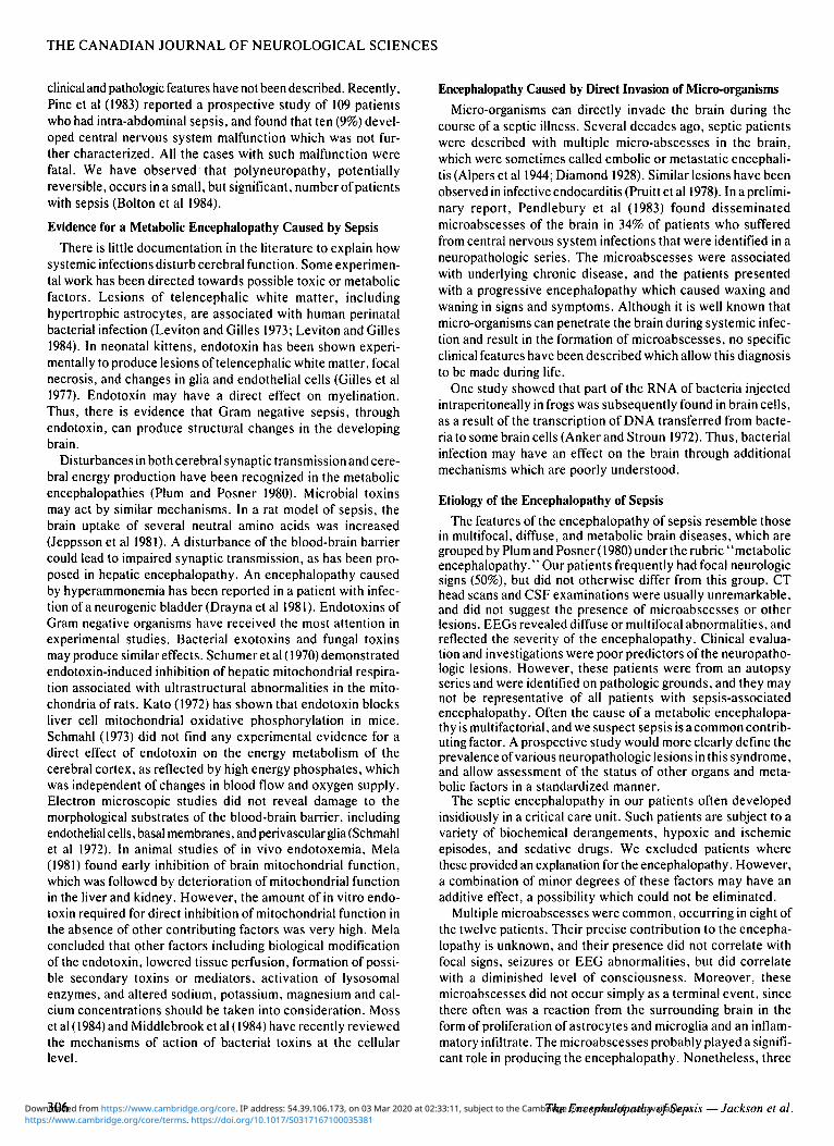
The most common neuropathologic finding present in 8 of the 12 patients, was multiple microabscesses disseminated throughout the brain. The microabscesses consisted of a collection of polymorphonuclear leukocytes, macrophages, microglia, and necrotic debris (Figure 1). Reactive astrocytosis was often present around the microabscesses. Occasionally the offending organisms were demonstrated with special stains. The microabscesses were not diagnosed during life, and microbial isolation studies were not performed on fresh brain tissue. Although the level of consciousness was more impaired in patients with microabscesses, there was no correlation with the presence of
Table 1
Case
1
2
3
4
5
6
7
8
9
10
11
12
: Clinical
Age (yr.)
60
68
54
65
82
69
58
83
34
61
54
59
findings in
Sex
F
M
F
M
F
M
F
F
M
F
F
M
12 patients with encephalopathy associated with sepsis
Initial Site of Infection
Lungs
Gall Bladder
Kidney
Vascular Graft
Joint
Uncertain
Lungs
Intra-abdominal Perforation
Kidney
Intra-abdominal Perforation
Intra-abdominal Perforation
Uncertain
' Focal Signs
Absent
Present
Terminal
Absent
Absent
Present
Present
Present
Absent
Absent
Present
Present
Seizures
Multifocal
None
None
Multifocal
None
None
Multifocal, Generalized
Multifocal
None
Focal
None
None
Predisposing Factors
Diabetes Mellitus
Ethanol Abuse
None
Ethanol Abuse
Rheumatoid Arthritis
Ethanol Abuse
None
Diabetes Mellitus
Gastric Adenocarcinoma
None
Diabetes Mellitus
Ethanol Abuse
304 The Encephalopathy of Sepsis — Jackson et al. https://www.cambridge.org/core/terms. https://doi.org/10.1017/S0317167100035381Downloaded from https://www.cambridge.org/core. IP address: 54.39.106.173, on 03 Mar 2020 at 02:33:11, subject to the Cambridge Core terms of use, available at
LE JOURNAL CANADIEN DES SCIENCES NEUROLOGIQUES
Table 2: Laboratory and pathologic features
Case Organisms Blood Cultures EEG Findings Pathology
1
2
3
4
5
6
7
8
9
Polymicrobial
Klebsiella pneumoniae
Staphylococcus aureus
Pseudomonas aeruginosa
Staphylococcus aureus
Polymicrobial
Pseudomonas aeruginosa
Polymicrobial
Escherichia coli
Positive
Positive
Positive
Positive
Positive
Positive
Negative
Positive
Positive
Diffuse suppression
Not done
Diffuse suppression
Multifocal epileptiform activity
Multifocal epileptiform activity
Diffuse suppression
Multifocal epileptiform activity
Not done
Not done
Multiple microabscesses, central pontine myelinolysis
Multiple microabscesses
Multiple microabscesses, cerebral infarcts (terminal)
Proliferation of astrocytes and microglia
Multiple microabscesses, cerebral infarcts
Proliferation of astrocytes and microglia
Brain purpura
Proliferation of astrocytes and microglia
Multiple microabscesses,
10
12
Polymicrobial
Polymicrobial
Polymicrobial
Negative
Positive
Positive
Burst suppression
Technically unsatisfactory
Burst supression
metabolic encephalopathy, central pontine myelinolysis, hemorrhages
Multiple microabscesses, central pontine myelinolysis
Multiple microabscesses
Multiple microabscesses, cerebral infarcts
microabscesses and focal signs, seizures, or EEG abnormalities. Of the four patients with an infection caused by a single Gram negative organism, only two had microabscesses. Microabscesses or abscesses were commonly found in other organs at autopsy. Four patients had mild proliferation of astrocytes and microglia in the cerebral cortex, changes commonly associated with metabolic encephalopathies (Adams and Sidman 1968). Three of these four patients had no other structural brain lesions. The patient with brain purpura (case 7) had thrombotic thrombocytopenic purpura which developed in association with a bacterial infection. The patient with multiple small white matter hemorrhages had laboratory features and pathologic lesions of disseminated intravascular coagulation (case 9). Only one of the three patients with microscopic evidence of central pontine myelinolysis had documented hyponatremia with rapid correction of the serum sodium.
DISCUSSION
Numerous metabolic derangements are associated with sepsis, including a hyperdynamic circulation, hypercatabolic state, and altered hormonal patterns with elevated catecholamines (Finley 1976). When sepsis is associated with failure of multiple organ systems, a state of critical illness exists in which there is a
mortality rate of about 45% (Eiseman et al 1977). An encephalopathy has been noted previously in critically ill, septic patients (Finley 1976; Jeppsson et al 1981; Pine et al 1983), but the
*. \* • * * » • • • • • '
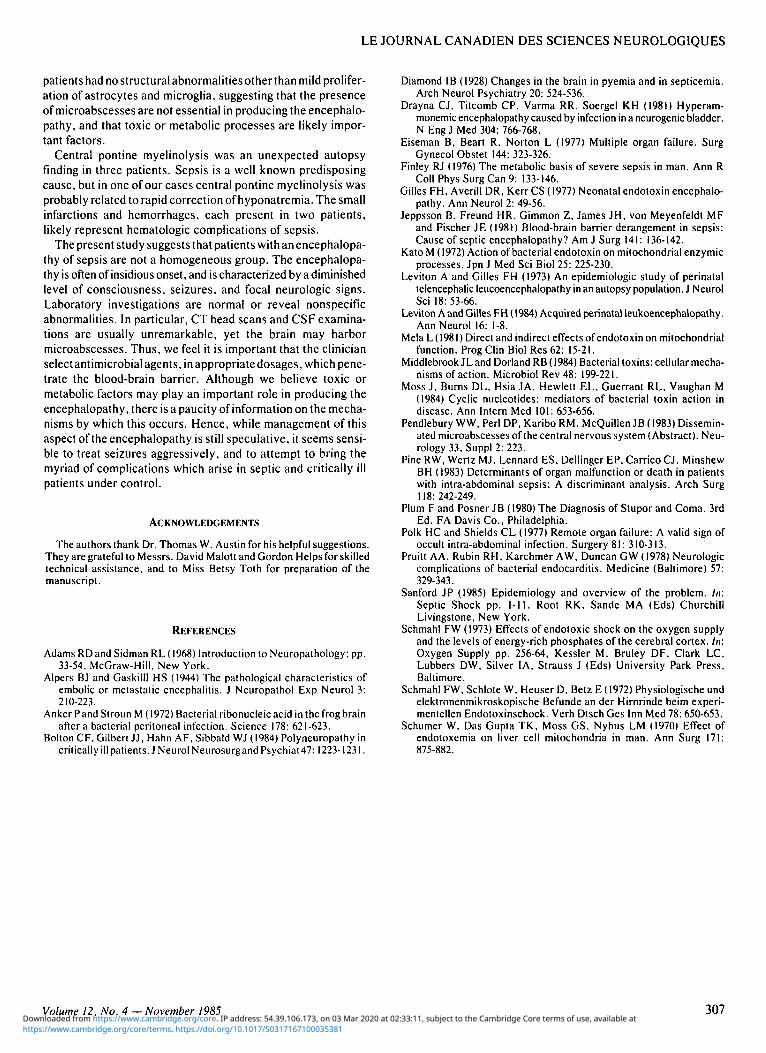
Figure 1 — Microabscess in cerebral cortex. Inflammatory infiltrate of polymorphonuclear leukocytes, macrophages, lymphocytes. The surrounding brain shows microglial proliferation. (Mag. X 627)
Volume 12, No. 4 — November 1985 305 https://www.cambridge.org/core/terms. https://doi.org/10.1017/S0317167100035381Downloaded from https://www.cambridge.org/core. IP address: 54.39.106.173, on 03 Mar 2020 at 02:33:11, subject to the Cambridge Core terms of use, available at
THE CANADIAN JOURNAL OF NEUROLOGICAL SCIENCES
clinical and pathologic features have not been described. Recently, Pine et al (1983) reported a prospective study of 109 patients who had intra-abdominal sepsis, and found that ten (9%) developed central nervous system malfunction which was not further characterized. All the cases with such malfunction were fatal. We have observed that polyneuropathy, potentially reversible, occurs in a small, but significant, number of patients with sepsis (Bolton et al 1984).
Evidence for a Metabolic Encephalopathy Caused by Sepsis
There is little documentation in the literature to explain how systemic infections disturb cerebral function. Some experimental work has been directed towards possible toxic or metabolic factors. Lesions of telencephalic white matter, including hypertrophic astrocytes, are associated with human perinatal bacterial infection (Leviton and Gilles 1973; Leviton and Gilles 1984). In neonatal kittens, endotoxin has been shown experimentally to produce lesions of telencephalic white matter, focal necrosis, and changes in glia and endothelial cells (Gilles et al 1977). Endotoxin may have a direct effect on myelination. Thus, there is evidence that Gram negative sepsis, through endotoxin, can produce structural changes in the developing brain.
Disturbances in both cerebral synaptic transmission and cerebral energy production have been recognized in the metabolic encephalopathies (Plum and Posner 1980). Microbial toxins may act by similar mechanisms. In a rat model of sepsis, the brain uptake of several neutral amino acids was increased (Jeppsson et al 1981). A disturbance of the blood-brain barrier could lead to impaired synaptic transmission, as has been proposed in hepatic encephalopathy. An encephalopathy caused by hyperammonemia has been reported in a patient with infection of a neurogenic bladder (Dray naetal 1981). Endotoxins of Gram negative organisms have received the most attention in experimental studies. Bacterial exotoxins and fungal toxins may produce similar effects. Schumer et al (1970) demonstrated endotoxin-induced inhibition of hepatic mitochondrial respiration associated with ultrastructural abnormalities in the mitochondria of rats. Kato (1972) has shown that endotoxin blocks liver cell mitochondrial oxidative phosphorylation in mice. Schmahl (1973) did not find any experimental evidence for a direct effect of endotoxin on the energy metabolism of the cerebral cortex, as reflected by high energy phosphates, which was independent of changes in blood flow and oxygen supply. Electron microscopic studies did not reveal damage to the morphological substrates of the blood-brain barrier, including endothelial cells, basal membranes, and perivascular glia (Schmahl et al 1972). In animal studies of in vivo endotoxemia, Mela (1981) found early inhibition of brain mitochondrial function, which was followed by deterioration of mitochondrial function in the liver and kidney. However, the amount of in vitro endotoxin required for direct inhibition of mitochondrial function in the absence of other contributing factors was very high. Mela concluded that other factors including biological modification of the endotoxin, lowered tissue perfusion, formation of possible secondary toxins or mediators, activation of lysosomal enzymes, and altered sodium, potassium, magnesium and calcium concentrations should be taken into consideration. Moss et al (1984) and Middlebrook et al (1984) have recently reviewed the mechanisms of action of bacterial toxins at the cellular level.
Encephalopathy Caused by Direct Invasion of Micro-organisms Micro-organisms can directly invade the brain during the
course of a septic illness. Several decades ago, septic patients were described with multiple micro-abscesses in the brain, which were sometimes called embolic or metastatic encephalitis (Alpersetal 1944; Diamond 1928). Similar lesions have been observed in infective endocarditis (Pruitt et al 1978). In a preliminary report, Pendlebury et al (1983) found disseminated microabscesses of the brain in 34% of patients who suffered from central nervous system infections that were identified in a neuropathologic series. The microabscesses were associated with underlying chronic disease, and the patients presented with a progressive encephalopathy which caused waxing and waning in signs and symptoms. Although it is well known that micro-organisms can penetrate the brain during systemic infection and result in the formation of microabscesses, no specific clinical features have been described which allow this diagnosis to be made during life.
One study showed that part of the RNA of bacteria injected intraperitoneally in frogs was subsequently found in brain cells, as a result of the transcription of DNA transferred from bacteria to some brain cells (Anker and Stroun 1972). Thus, bacterial infection may have an effect on the brain through additional mechanisms which are poorly understood.
Etiology of the Encephalopathy of Sepsis The features of the encephalopathy of sepsis resemble those
in multifocal, diffuse, and metabolic brain diseases, which are grouped by Plum and Posner (1980) under the rubric' 'metabolic encephalopathy." Our patients frequently had focal neurologic signs (50%), but did not otherwise differ from this group. CT head scans and CSF examinations were usually unremarkable, and did not suggest the presence of microabscesses or other lesions. EEGs revealed diffuse or multifocal abnormalities, and reflected the severity of the encephalopathy. Clinical evaluation and investigations were poor predictors of the neuropathologic lesions. However, these patients were from an autopsy series and were identified on pathologic grounds, and they may not be representative of all patients with sepsis-associated encephalopathy. Often the cause of a metabolic encephalopathy is multifactorial, and we suspect sepsis is a common contributing factor. A prospective study would more clearly define the prevalence of various neuropathologic lesions in this syndrome, and allow assessment of the status of other organs and metabolic factors in a standardized manner.
The septic encephalopathy in our patients often developed insidiously in a critical care unit. Such patients are subject to a variety of biochemical derangements, hypoxic and ischemic episodes, and sedative drugs. We excluded patients where these provided an explanation for the encephalopathy. However, a combination of minor degrees of these factors may have an additive effect, a possibility which could not be eliminated.
Multiple microabscesses were common, occurring in eight of the twelve patients. Their precise contribution to the encephalopathy is unknown, and their presence did not correlate with focal signs, seizures or EEG abnormalities, but did correlate with a diminished level of consciousness. Moreover, these microabscesses did not occur simply as a terminal event, since there often was a reaction from the surrounding brain in the form of proliferation of astrocytes and microglia and an inflammatory infiltrate. The microabscesses probably played a significant role in producing the encephalopathy. Nonetheless, three
306 The Encephalopathy of Sepsis —Jackson et al. https://www.cambridge.org/core/terms. https://doi.org/10.1017/S0317167100035381Downloaded from https://www.cambridge.org/core. IP address: 54.39.106.173, on 03 Mar 2020 at 02:33:11, subject to the Cambridge Core terms of use, available at
LE JOURNAL CANADIEN DES SCIENCES NEUROLOGIQUES
patients had no structural abnormalities other than mild proliferation of astrocytes and microglia, suggesting that the presence of microabscesses are not essential in producing the encephalopathy, and that toxic or metabolic processes are likely important factors.
Central pontine myelinolysis was an unexpected autopsy finding in three patients. Sepsis is a well known predisposing cause, but in one of our cases central pontine myelinolysis was probably related to rapid correction of hyponatremia. The small infarctions and hemorrhages, each present in two patients, likely represent hematologic complications of sepsis.
The present study suggests that patients with an encephalopathy of sepsis are not a homogeneous group. The encephalopathy is often of insidious onset, and is characterized by a diminished level of consciousness, seizures, and focal neurologic signs. Laboratory investigations are normal or reveal nonspecific abnormalities. In particular, CT head scans and CSF examinations are usually unremarkable, yet the brain may harbor microabscesses. Thus, we feel it is important that the clinician select antimicrobial agents, in appropriate dosages, which penetrate the blood-brain barrier. Although we believe toxic or metabolic factors may play an important role in producing the encephalopathy, there is a paucity of information on the mechanisms by which this occurs. Hence, while management of this aspect of the encephalopathy is still speculative, it seems sensible to treat seizures aggressively, and to attempt to bring the myriad of complications which arise in septic and critically ill patients under control.
ACKNOWLEDGEMENTS
The authors thank Dr. Thomas W. Austin for his helpful suggestions. They are grateful to Messrs. David Malott and Gordon Helps for skilled technical assistance, and to Miss Betsy Toth for preparation of the manuscript.
REFERENCES
Adams RD and Sidman RL (1968) Introduction to Neuropathology; pp. 33-54, McGraw-Hill, New York.
Alpers BJ and Gaskilll HS (1944) The pathological characteristics of embolic or metastatic encephalitis. J Neuropathol Exp Neurol 3: 210-223.
Anker P and Stroun M (1972) Bacterial ribonucleic acid in the frog brain after a bacterial peritoneal infection. Science 178: 621-623.
Bolton CF, Gilbert JJ, Hahn AF, Sibbald WJ (1984) Polyneuropathy in critically ill patients. J Neurol NeurosurgandPsychiat47:1223-1231.
Diamond IB (1928) Changes in the brain in pyemia and in septicemia. Arch Neurol Psychiatry 20: 524-536.
Drayna CJ, Titcomb CP, Varma RR, Soergel KH (1981) Hyperam-monemic encephalopathy caused by infection in a neurogenic bladder. N Eng J Med 304: 766-768.
Eiseman B, Beart R. Norton L (1977) Multiple organ failure. Surg Gynecol Obstet 144: 323-326.
Finley RJ (1976) The metabolic basis of severe sepsis in man. Ann R Coll Phys Surg Can 9: 133-146.
Gilles FH, Averill DR, Kerr CS (1977) Neonatal endotoxin encephalopathy. Ann Neurol 2: 49-56.
Jeppsson B, Freund HR, Gimmon Z, James JH, von Meyenfeldt MF and Fischer JE (198!) Blood-brain barrier derangement in sepsis: Cause of septic encephalopathy? Am J Surg 141: 136-142.
Kato M (1972) Action of bacterial endotoxin on mitochondrial enzymic processes. Jpn J Med Sci Biol 25: 225-230.
Leviton A and Gilles FH (1973) An epidemiologic study of perinatal telencephalic leucoencephalopathy in an autopsy population. J Neurol Sci 18: 53-66.
Leviton A and Gilles FH (1984) Acquired perinatal leukoencephalopathy. Ann Neurol 16: 1-8.
Mela L (1981) Direct and indirect effects of endotoxin on mitochondrial function. Prog Clin Biol Res 62: 15-21.
Middlebrook JL and Dorland RB (1984) Bacterial toxins: cellular mechanisms of action. Microbiol Rev 48: 199-221.
Moss J, Burns DL, Hsia JA, Hewlett EL. Guerrant RL, Vaughan M (1984) Cyclic nucleotides: mediators of bacterial toxin action in disease. Ann Intern Med 101: 653-656.
Pendlebury WW, Perl DP, Karibo RM, McQuillen JB (1983) Disseminated microabscesses of the central nervous system (Abstract). Neurology 33, Suppl 2: 223.
Pine RW, Wertz MJ, Lennard ES, Dellinger EP. Carrico CJ, Minshew BH (1983) Determinants of organ malfunction or death in patients with intra-abdominal sepsis; A discriminant analysis. Arch Surg 118:242-249.
Plum F and Posner JB (1980) The Diagnosis of Stupor and Coma. 3rd Ed. FA Davis Co., Philadelphia.
Polk HC and Shields CL (1977) Remote organ failure: A valid sign of occult intra-abdominal infection. Surgery 81: 310-313.
Pruitt AA, Rubin RH, Karchmer AW, Duncan GW (1978) Neurologic complications of bacterial endocarditis. Medicine (Baltimore) 57: 329-343.
Sanford JP (1985) Epidemiology and overview of the problem. In: Septic Shock pp. 1-11, Root RK, Sande MA (Eds) Churchill Livingstone, New York.
Schmahl FW (1973) Effects of endotoxic shock on the oxygen supply and the levels of energy-rich phosphates of the cerebral cortex. In: Oxygen Supply pp. 256-64, Kessler M, Bruley DF, Clark LC, Lubbers DW, Silver IA, Strauss J (Eds) University Park Press, Baltimore.
Schmahl FW, Schlote W, Heuser D, Betz E (1972) Physiologische und elektronenmikroskopische Befunde an der Hirnrinde beim experi-mentellen Endotoxinschock. Verh Dtsch Ges Inn Med 78: 650-653.
Schumer W, Das Gupta TK, Moss GS, Nyhus LM (1970) Effect of endotoxemia on liver cell mitochondria in man. Ann Surg 171: 875-882.
Volume 12, No. 4 —November 1985 307 https://www.cambridge.org/core/terms. https://doi.org/10.1017/S0317167100035381Downloaded from https://www.cambridge.org/core. IP address: 54.39.106.173, on 03 Mar 2020 at 02:33:11, subject to the Cambridge Core terms of use, available at